Note : Les descriptions sont présentées dans la langue officielle dans laquelle elles ont été soumises.
CA 02497988 2005-03-07
WO 2004/022721 PCT/US2003/028081
Docket No. 701586-053022-PCT
Express Mail Label No. EL948181598US
QUANTIFICATION OF GENE EXPRESSION
BACKGROUND OF THE INVENTION
[001] Detection and quantification of differentially expressed genes in a
number
of pathological conditions such as different benign and malignant tumors,
neurological disorders, heart disease and autoimmune disorders, would be
useful in
the diagnosis, prognosis and treatment of these pathological conditions.
Quantification of gene expression would also be useful in diagnosis of
infectious
diseases and following up effects of pharmaceuticals or toxins on molecular
level.
For example, gene expression data could be used to determine the
pharmacological
mechanism of a drug or a toxin (Libutti et al., Microarray technology and gene
expression analysis for the study of angiogenesis. Expert Opin Biol Ther. 2002
Jun;2(5):545-56).
[002] The methods for transcript detection and quantification have
traditionally
included Northern-blot hybridization, ribonuclease protection assay, and
reverse
transcriptase polymerase chain reaction (RT-PCR) based methods. However, in
addition to suffering from lack of sensitivity (except RT-PCR), these methods
are
only useful for roughly estimating the relative expression changes of each
transcript
among samples from different sources. The different RT-PCR based techniques
are
the most suitable quantification method for diagnostic purposes, because they
are very
sensitive and thus require only a small sample size which is desirable for a
diagnostic
test.
[003] Absolute quantification of transcript copy numbers in a sample is a
requirement if one wishes to compare gene expression between samples and even
within the same sample. However, quantification of nucleic acid copy numbers
is
difficult using PCR based methods because of the inherent non-linear nature of
the
PCR reaction. PCR amplification will change from an exponential phase to a
plateau
phase with the consumption of reagents or enzyme inactivation. Often, the
exponential phase of the PCR must be determined separately which may involve
1
CA 02497988 2005-03-07
WO 2004/022721 PCT/US2003/028081
sampling of the PCR reactions at different time points or performing the PCR
using
different dilutions of the template. Further, because of differences in
amplification
efficiency between templates, the starting quantities of different PCR
products cannot
be compared directly even in the linear range. Detection of PCR products has
traditionally been performed after amplification is completed. Typically, an
aliquot of
the PCR reaction product is size separated by agarose gel electrophoresis,
stained with
ethidium bromide, and visualized with ultraviolet light. Alternatively, the
primers
may be labeled with a fluorescent dye or a radioactive molecule. Comparison of
band
intensities between samples allows one to qualitatively estimate the relative
starting
concentrations of templates amplified, but this method is not quantitative and
does not
result in determination of the absolute copy number.
[004] A number of quantitative RT-PCR based methods have been described
including RNA quantification using PCR and complementary DNA (cDNA) arrays
(Shalon et al., Genoine Research 6(7):639-45, 1996; Bernard et al., Nucleic
Acids
Research 24(8):1435-42, 1996), solid-phase mini-sequencing technique, which is
based upon a primer extension reaction (U.S. Patent No. 6,013,431, Suomalainen
et
al. Mol. Biotechnol. Jun;15(2):123-31, 2000), ion-pair high-performance liquid
chromatography (Doris et al. J. Chromatogr. A May 8;806(1):47-60, 1998), and
5'
nuclease assay or real-time RT-PCR (Holland et al. Proc Natl Acad Sci USA 88:
7276-7280, 1991).
[005] It would be useful to develop a method which allows a sensitive and
accurate mRNA transcript quantification, can be easily automated and scaled up
to
accommodate testing of large numbers of sample and overcomes the problems
associated with PCR amplification. Such a method would enable diagnosing
different
pathological conditions, including viruses, bacteria and parasites, as well as
different
benign and malignant tumors, neurological disorders, heart disease and
autoimmune
disorders. Such a method would also allow quantifying the transcripts of
interest for
diagnostic, prognostic and therapeutic purposes, and would ultimately
facilitate
pharmacogenomic applications. Such a method would also allow screening a large
number of agents for effects on gene expression.
2
CA 02497988 2005-03-07
WO 2004/022721 PCT/US2003/028081
SUMMARY OF THE INVENTION
[006] The present invention relates to a method for measuring the amount of a
target nucleic acid in a sample using a standard which is designed to have one
base
difference compared with the gene of interest or a "target nucleic acid
sequence."
Use of such standard in combination with a method of "enhancing" the
difference in
the standard and the test nucleic acid sample using, for example, a base
extension
reaction carried right at the mutation site allowing amplification of the
standard and
target nucleic acids with the same efficiency and facilitating quantification
of the
target nucleic acid. Thereafter a means of quantifying the "enhanced" standard
and
target nucleic acid samples is used to determine the amount of the target
nucleic acid.
In the preferred embodiment, the quantification means is Mass Spectrometry.
[007] The method of the present invention is sensitive, accurate and highly
reproducible and it is also independent of PCR cycle number, which greatly
simplifies
the analysis. The method of the present invention is unique because different
alleles
of the same gene can be measured simultaneously, absolute quantification of
gene
expression can be achieved so that the data can be directly compared from
different
experiments, and it can be applied in high-throughput analysis and virtually
no
optimization is needed for PCR. Additionally, the method allows for accurate
determination of copy number of infectious agents such as viruses, bacteria
and
parasites in a biological specimen such as human fluids (serum, plasma, etc).
[008] The invention provides a method of quantifying the amount of a target
gene/nucleic acid or a plurality of target genes/nucleic acids in a biological
specimen
comprising adding a known concentration of a nucleic acid standard to the
biological
specimen, wherein the standard is designed to have one base difference with
the target
nucleic acid sequence; amplifying a sample with the target and standard
nucleic acids,
for example, using a polymerase chain reaction, removing the excessive dNTPs,
for
example by treating the amplified sample with a phosphatase (e.g. Shrimp
alkaline
phosphatase), and consequently enhancing the nucleic acid difference between
the
standard and the test nucleic acid, for example, by extending the differing
base in the
target and the standard nucleic acid samples. The standard and the target
nucleic acid
3
CA 02497988 2005-03-07
WO 2004/022721 PCT/US2003/028081
produce two different products, typically having one to two bases difference,
and are
subsequently quantified. The concentration of a transcript can be calculated
based
upon the amount of standard present in the amplified sample.
[009] Fore example, this invention enables detection, and more importantly,
quantification of infectious agents. It can easily be used in a high
throughput way
where around 100 infectious agents can be quantified on a 384-format silicon
chip.
[0010] In one preferred embodiment, the quantification is performed based upon
the different mass of the "enhanced" target and standard nucleic acid products
using
MALDI-TOF mass spectrometry (e.g., Using Sequenom's MassArrayTM system),
wherein the ratio of the peaks in the mass spectrum is used to calculate the
ratio of the
standard and the target nucleic acid. The concentration of a transcript can be
calculated based upon the initial amount of standard used/added in the sample
before
amplification.
[0011] In one preferred embodiment, the enhancement of the nucleic acid
difference between the standard and the target nucleic acid is performed using
primer
extension methods.
[0012] In another embodiment, the enhancement of the nucleic acid difference
in
the target and the standard after the PCR is performed using fluorescence
tagged
dNTP/ddNTP for base extension.
[0013] In yet another embodiment, the enhancement of the nucleic acid
difference
in the target and the standard after the PCR is performed using different dye-
labeled
ddNTPs which are differentially incorporated into the target and standard
nucleic
acids in a primer extension reaction.
[0014] In one embodiment, the enhancement of the nucleic acid difference in
the
target and the standard after the PCR, is performed using real time PCR.
[0015] In another embodiment, the enhancement of the nucleic acid difference
in
the target and the standard after the PCR is performed using hybridization
based
4
CA 02497988 2005-03-07
WO 2004/022721 PCT/US2003/028081
techniques wherein two oligonucleotides specific for either the target or the
standard
are designed for hybridization.
[0016] In another embodiment, the enhancement of the nucleic acid difference
in
the target and the standard after the PCR is performed using pyrosequencing
technology.
[0017] In another embodiment, the enhancement of the nucleic acid difference
in
the target and the standard after the PCR is performed using a third wave
invader
assay using an artificial single nucleotide polymorphism (SNP) as an internal
reference. In an alternative embodiment, when using pyrosequencing, no pre-
amplification is needed.
[0018] In one' embodiment, the target nucleic acid is a nucleic acid from at
least
one infectious agent.
[0019] In yet another embodiment, the invention provides a kit comprising at
least
one preferably several different primers designed to differ by one nucleic
acid from at
least one, preferably several target nucleic acids, in different vials or
preferably, all
standard nucleic acids in one vial having a known predetermined concentration
in a
buffer suitable for a PCR or direct enhancement reactions to enhance the
difference
between the standard and a corresponding target nucleic acid as described
above. The
kit also comprises a manual explaining the reaction conditions and the
measurement
of the amount of target nucleic acid(s) using the standard nucleic acid(s).
Kits
contemplated by the invention include, but are not limited to kits for
determining the
amount of infectious agents in a biological sample and kits determining the
amount of
one or more transcripts that is expected to be increased or decreased after
administration of a medicament or a drug, or as a result of a disease
condition such as
cancer.
BRIEF DESCRIPTION OF THE FIGURES
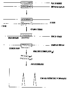
[0020] FIG 1 shows a flow chart of the real competitive PCR and Mass
Spectrometry approach for measuring gene expression. For simplicity, only one
DNA
CA 02497988 2005-03-07
WO 2004/022721 PCT/US2003/028081
strand is shown. Also extension oligos are generally around 20 bases, instead
of 7
bases shown in the flow chart.
[0021] FIG 2 shows a peak area distribution for the same oligo at the same
concentration. Oligo 47954 (5'-ATGGCCACAGTTGTATCA-3') were used at 0.3
gM and 15 nL is used for spotting onto a silicon chip prespotted with a matrix
of 3-
hydroxypicolinic acid (HPA). The absolute peals areas for oligos with the same
concentration spotted at different positions of the same chip show modest
variability
with average peak area of 12395 (arbitrary number) and standard deviation of
3737.
[0022] FIGS 3A AND 3B show peak area ratios in the mass spectrum correlate
accurately with oligo concentration ratios. Courtesy of Kai Tang (Sequenom).
4.5 nL
of solutions of two oligo mixtures at different ratios (1:1, 1:2, 1:5, 1:10,
1:20) were
analyzed using the MassArrayTM (Sequenom). FIG 3A shows the mass spectrums,
and
FIG 3B shows the plot of actual concentration ratio versus the ratio of signal
intensity
(peak area) in the mass Spectrum.
[0023] FIGS 4A-4E show Mass Spectrum for two DNA templates differs only by
one base, mixed at different ratios. In FIG 4A the ratio is 1:1; in FIG 4B the
ratio is
3:1; in FIG 4C the ratio is 10:1; in FIG 4D the ratio is 1:3; and in FIG 4E
the ratio is
1:10, but at fixed total concentration (2 * 10-7 tg/ L). The templates were
amplified
by PCR (30 cycles), base extension (40 cycles), then spotted onto a silicon
chip pre-
spotted with a matrix of 3-hydroxypicolinic acid (HPA), and analyzed with
MALDI-
TOF.
[0024] FIG 5 shows correlations between putative DNA concentration ratios and
measured DNA concentration ratios (represented by peak area ratios). PCR
amplifications are 20, 30 and 40 cycles respectively and the results are PCR-
cycle
independent. Each data point is repeated 4 times (n=4) and error bars are
shown.
[0025] FIGS 6A-6H show gene expression (GAPDH, HMBS and CXCR4)
analysis using real competitive PCR and mass spectrometry.
DETAILED DESCRIPTION OF THE INVENTION
6
CA 02497988 2005-03-07
WO 2004/022721 PCT/US2003/028081
[0026] The present invention relates to a novel approach in measuring gene
expression or amount of nucleic acid in a sample. This approach combines
competitive PCR (polymerase chain reaction), base extension and thereafter
measured. The method can be used for directly measuring copy numbers of
specific
genes, or comparing relative up or down regulations of specific genes from
different
samples.
[0027] A standard nucleic acid (either DNA or RNA) with known concentration
is added to the RNA sample (for RNA standard) or the reverse transcription
product
(for DNA standard). The reverse transcription product including the standard
is then
amplified by PCR. The standard is designed to have one base mutation
difference
compared with the gene of interest, i.e. the target nucleic acid. Thus, the
standard and
the target nucleic acid are amplified with same efficiency in PCR. And these
two can
be identified, using, for example a base extension reaction carried right at
the
mutation site.
[0028] The amount of the PCR products is consequently measured by any of a
variety of means, preferably by Mass Spectrometry (MALDI-TOF, or Matrix
Assisted
Laser Desorption Ionization - Time of Flight). The peak area ratio between the
products from the standard and the gene of interest represents the ratio of
the standard
and the gene of interest. Since the concentration of the standard is known,
the
concentration of the gene of interest can be calculated.
[0029] The method of the present invention is unique in at least the following
aspects. First of all, the natural mutations of genes can be selected to
construct
standards. Therefore, not only the expression level of the genes can be
measured, but
also the genotype of the genes expressed can be determined. Second, the usage
of a
single point mutation in PCR guarantees virtually identical amplification.
This
eliminates the problems arising from differential amplifications in other
competitive
PCR approaches where the standards generally are of different lengths with the
genes.
[0030] In the preferred embodiment, the combination of base extension and
MALDI-TOF MS detection also eliminates the problems from heteroduplex
formation encountered by traditional detection method such as gel
electrophoresis.
7
CA 02497988 2005-03-07
WO 2004/022721 PCT/US2003/028081
Also, the extension product from the standard serves as an internal standard
in
MALDI-TOF MS. Thus, the amount of the nucleic acids can be quantitatively
measured when the amount of the standard added to the reaction is known.
[0031] This approach has at least the following advantages. First, this method
requires little optimization in PCR. Second, this method is not dependent on
PCR
cycle numbers. Third, the method is highly accurate, sensitive, and
reproducible.
Fourth, the method can be used to for high throughput gene expression analysis
where
the expression of at least 50-100, or even up to at least 1000 genes can be
measured
on one 384-silicon chip.
[0032] As shown in the following examples, the analysis of GAPDH, HMBS and
CXCR4 expression in human cultured cells by this method produced results
consistent with other methods.
[0033] As used herein, the term "biological sample" refers to any biological
material obtained from any source (e.g. human, animal, plant, bacteria, fungi,
protist,
virus). For use in the invention, the biological sample should contain a
nucleic acid
molecule. Examples of appropriate biological samples for use in the instant
invention
include: solid materials (e.g tissue, cell pellets, biopsies) and biological
fluids (e.g.
urine, blood, saliva, amniotic fluid, mouth wash).
Nucleic acid molecules can be isolated from a particular biological sample
using any
of a number of procedures, which are well-known in the art, the particular
isolation
procedure chosen being appropriate for the particular biological sample.
[0034] Viruses, bacteria, fungi and other infectious organisms contain
distinct
nucleic acid sequences, which are different from the sequences contained in
the host
cell. Detecting or quantifying nucleic acid sequences that are specific to the
infectious
organism is important for diagnosing or monitoring infection. Examples of
disease
causing viruses that infect humans and animals and which may be detected by
the
disclosed processes include: Retroviridae (e.g., human immunodeficiency
viruses,
such as HIV-1 (also referred to as HTLV-III, LAV or HTLV-III/LAV, See Ratner,
L.
et al., Nature, Vol. 313, Pp. 227-284 (1985); Wain Hobson, S. et al, Cell,
Vol. 40: Pp.
9-17 (1985)); HIV-2 (See Guyader et al., Nature, Vol. 328, Pp. 662-669 (1987);
8
CA 02497988 2005-03-07
WO 2004/022721 PCT/US2003/028081
European Patent Publication No. 0 269 520; Chakraborti et al., Nature, Vol.
328, Pp.
543-547 (1987); and European Patent Application No. 0 655 501); and other
isolates,
such as HIV-LP (International Publication No. WO 94/00562 entitled "A Novel
Human Immunodeficiency Virus"; Picornaviridae (e.g., polio viruses, hepatitis
A
virus, (Gust, I. D., et al., Intervirology, Vol. 20, Pp. 1-7 (1983); entero
viruses, human
coxsackie viruses, rhinoviruses, echoviruses); Calciviridae (e.g., strains
that cause
gastroenteritis); Togaviridae (e.g., equine encephalitis viruses, rubella
viruses);
Flaviridae (e.g., dengue viruses, encephalitis viruses, yellow fever viruses);
Coronaviridae (e.g., coronaviruses); Rhabdoviridae (e.g., vesicular stomatitis
viruses,
rabies viruses); Filoviridae (e.g., ebola viruses); Paramyxoviridae (e.g.,
parainfluenza
viruses, mumps virus, measles virus, respiratory syncytial virus);
Orthomyxoviridae
(e.g., influenza viruses); Bungaviridae (e.g., Hantaan viruses, bunga viruses,
phleboviruses and Nairo viruses); Arena viridae (hemorrhagic fever viruses);
Reoviridae (e.g., reoviruses, orbiviurses and rotaviruses); Birnaviridae,
Hepadnaviridae (Hepatitis B virus); Parvoviridae (parvoviruses); Papovaviridae
(papilloma viruses, polyoma viruses); Adenoviridae (most adenoviruses);
Herpesviridae (herpes simplex virus (HSV) 1 and 2, varicella zoster virus,
cytomegalovirus (CMV), herpes viruses); Poxviridae (variola viruses, vaccinia
viruses, pox viruses); and Iridoviridae (e.g., African swine fever virus); and
unclassified viruses (e.g., the etiological agents of Spongiform
encephalopathies, the
agent of delta hepatities (thought to be a defective satellite of hepatitis B
virus), the
agents of non-A, non-B hepatitis (class 1=internally transmitted; class
2=parenterally
transmitted (i.e., Hepatitis C); Norwalk and related viruses, and
astroviruses).
Examples of infectious bacteria include: Helicobacter pyloris, Borelia
burgdorferi,
Legionella pneumophilia, Mycobacteria sps (e.g. M. tuberculosis, M. avium, M.
intracellulare, M. kansaii, M. gordonae), Staphylococcus aureus, Neisseria
gonorrhoeae, Neisseria meningitidis, Listeria monocytogenes, Streptococcus
pyogenes (Group A Streptococcus), Streptococcus agalactiae (Group B
Streptococcus), Streptococcus (viridans group), Streptococcus faecalis,
Streptococcus
bovis, Streptococcus (anaerobic sps.), Streptococcus pneumoniae, pathogenic
Campylobacter sp., Enterococcus sp., Haemophilus influenzae, Bacillus
antracis,
corynebacterium diphtheriae, corynebacterium sp., Erysipelothrix
rhusiopathiae,
9
CA 02497988 2005-03-07
WO 2004/022721 PCT/US2003/028081
Clostridium perfringers, Clostridium tetani, Enterobacter aerogenes,
Klebsiella
pneumoniae, Pasturella multocida, Bacteroides sp., Fusobacterium nucleatum,
Streptobacillus moniliformis, Treponema pallidium, Treponema pertenue,
Leptospira,
and Actinomyces israelli.
[0035] This technique can be directly applied in developing technologies for
high
throughput and accurate gene expression analysis. It could also be used to
develop
clinical diagnosis chips where measurement of at least about 2, 5, 10, 25, 50-
100 and
up to at least 1000 genes can be used for disease diagnosis.
[0036] The method for "enhancing" PCR products wherein the base difference
between the standard and the target nucleic acid has been enhanced according
to the
present invention include, but are not limited to PYROSEQUENCINGTM, real time
PCR, hybridization-based techniques, third wave invader assay, fluorescence-
based
PCR techniques, solid-phase minisequencing. Quantification of the "enhanced"
PCR
products can consequently be performed utilizing the mass difference of the
target and
the standard enhanced nucleic acid product using, for example, MALDI-TOF mass
spectrometry (MS).
[0037] The term "enhancing" as used in the present invention is intended to
cover different techniques whereby the target and the standard nucleic acid
are made
to include a difference in their mass. Therefore, because the standard and the
target
nucleic acid have preferably only one base difference, they can be
differentiated and
the difference amplified or enhanced using, for example a primer extension
techniques using labeled nucleic acids. Alternatively, the mass difference can
be
created using allele-specific hybridization probes or enzymatic cleavage of
the
different products like in the INVADER assay.
[0038] In one embodiment, the PCR products differing by one base pair are
enhanced by PYROSEQUENCINGTM (Uppsala, Sweden) which is essentially
sequencing by synthesis. A sequencing primer, designed directly next to the
nucleic
acid differing between the target and the standard is first hybridized to a
single
stranded, PCR amplified, DNA template comprising both the target and the
standard
CA 02497988 2009-11-12
WO 2004/022721 PCT/US2003/028081
PCT product, and incubated with the enzymes, DNA polymerase, ATP sulfurylase,
luciferase and apyrase, and the substrates, adenosine 5' phosphosulfate (APS)
and
luciferin. One of four deoxynucleotide triphosphates (dNTP), for example,
corresponding to the nucleotide present in the standard template, is then
added to the
reaction. DNA polymerase catalyzes the incorporation of the dNTP into the
standard
DNA strand. Each incorporation event is accompanied by release of
pyrophosphate
(PPi) in a quantity jquimolar to the amount of incorporated nucleotide.
Consequently, ATP sulfurylase quantitatively converts PPi to ATP in the
presence of
adenosine 5' phosphosulfate. This ATP drives the luciferase-mediated
conversion of
luciferin to oxyluciferin that generates visible light in amounts that are
proportional to
the amount of ATP. The light produced in the luciferase-catalyzed reaction is
detected
by a charge coupled device (CCD) camera and seen as a peak in a PYROGRAMTM.
Each light signal is proportional to the number of nucleotides incorporated
and allows
determination of the amount of the standard nucleic acid sequence. Thereafter,
apyrase, a nucleotide degrading enzyme, continuously degrades unincorporated
dNTPs and excess ATP. When degradation is complete, another dNTP is added
which
corresponds to the dNTP present in the target template the amount of which is
to be
determined. Finally, addition of dNTPs is performed one at a time.
Deoxyadenosine
alfa-thio triphosphate (dATPaS) is used as a substitute for the natural
deoxyadenosine
triphosphate (dATP) since it is efficiently used by the DNA polymerase, but
not
recognized by the luciferase. Because the amount of the standard added in the
PCR is
known, the amount of the target can be calculated from the ratio of the
incorporated
dNTPs. For detailed information about reaction conditions, see, e.g. U.S.
Patent No.
6,210,891.
[0039] Another example of the methods useful for enhancing the base difference
of the standard and the target nucleic acid of PCR products is real time PCR.
All real-
time PCR systems rely upon the detection and quantitation of a fluorescent
reporter,
the signal of which increases in direct proportion to the amount of PCR
product in a
reaction. Examples of real-time PCR method useful according to the present
invention include, TagMan and molecular beacons, both of which are
hybridization
probes relying on fluorescence resonance energy transfer (FRET) for
quantitation.
11
CA 02497988 2005-03-07
WO 2004/022721 PCT/US2003/028081
TaqMan Probes are oligonucleotides that contain a fluorescent dye, typically
on the 5'
base, and a quenching dye, typically located on the 3' base. When irradiated,
the
excited fluorescent dye transfers energy to the nearby quenching dye molecule
rather
than fluorescing, resulting in a nonfluorescent substrate. TagMan probes are
designed
to hybridize to an internal region of a PCR product (ABI 7700 (TagManTM),
Applied
BioSystems, Foster City, CA). Accordingly, two different primers, one
hybridizing to
the target and the other to the standard nucleic acid template, are designed.
The
primers are consequently allowed to hybridize to the corresponding nucleic
acids in
the real time PCR reaction. During PCR, when the polymerase replicates a
template
on which a TaqMan probe is bound, the 5' exonuclease activity of the
polymerase
cleaves the probe. Consequently, this separates the fluorescent and quenching
dyes
and FRET no longer occurs. Fluorescence increases in each cycle, proportional
to the
rate of probe cleavage.
[0040] Molecular beacons also contain fluorescent and quenching dyes, but
FRET only occurs when the quenching dye is directly adjacent to the
fluorescent dye.
Molecular beacons are designed to adopt a hairpin structure while free in
solution,
bringing the fluorescent dye and quencher in close proximity. Therefore, two
different molecular beacons are designed, one recognizing the target and the
other the
standard nucleic acid. When the molecular beacons hybridize to the target and
the
standard nucleic acids, the fluorescent dye and quencher are separated, FRET
does not
occur, and the fluorescent dye emits light upon irradiation. Unlike TaqMan
probes,
molecular beacons are designed to remain intact during the amplification
reaction, and
must rebind to target in every cycle for signal measurement. TaqMan probes and
molecular beacons allow multiple DNA species to be measured in the same sample
(multiplex PCR), since fluorescent dyes with different emission spectra may be
attached to the different probes, e.g. different dyes are used in making the
standard
probe and the target probe. Multiplex PCR allows internal controls to be co-
amplified
and permits allele discrimination in single-tube, homogeneous assays. (Ambion
Inc,
Austin, TX, TechNotes 8(1) - February 2001, Real-time PCR goes prime time).
[0041] Yet another method useful for enhancing the difference between the
target
and standard nucleic acid is the primer extension method as used in the solid-
phase
12
CA 02497988 2005-03-07
WO 2004/022721 PCT/US2003/028081
mini-sequencing (Hultman, et al., 1988, Nucl. Acid. Res., 17, 4937-4946;
Syvanen et
al., 1990, Genomics, 8, 684-692). In the original reports, the incorporation
of a
radiolabeled nucleotide was measured and used for analysis of the three-
allelic
polymorphism of the human apolipoprotein E gene. The method of detection of
the
variable nucleotide(s) is based on primer extension and incorporation of
detectable
nucleoside triphosphates in the detection step. By selecting the detection
step primers
from the region immediately adjacent to the variable nucleotide, this
variation can be
detected after incorporation of as few as one nucleoside triphosphate.
Labelled
nucleoside triphosphates matching the variable nucleotide are added and the
incorporation of a label into the detection step primer is measured. The
detection step
primer is annealed to the copies of the target nucleic acid and a solution
containing
one or more nucleoside triphosphates including at least one labeled or
modified
nucleoside triphosphate, is added together with a polymerizing agent in
conditions
favoring primer extension. Either labeled deoxyribonucleoside triphosphates
(dNTPs)
or chain terminating dideoxyribonucleoside triphosphates (ddNTPs) can be used,
and
labels are preferably dyes, including fluorescent dyes. The solid-phase mini-
sequencing method is described in detail, for example, in the U.S. Patent No.
6,013,431 and in Wartiovaara and Syvanen, Quantitative analysis of human DNA
sequences by PCR and solid-phase minisequencing. Mol Biotechnol 2000 Jun;
15(2):123-131.
[00421 Another method to enhance the difference in the target and standard
nucleic acids in the PCR products is by using fluorescence tagged dNTP/ddNTPs.
In
addition to use of the fluorescent label in the solid phase mini-sequencing
method, a
standard nucleic acid sequencing gel can be used to detect the amount of the
fluorescent label incorporated into the PCR amplification product. A
sequencing
primer is designed to anneal next to the base differentiating the standard
from the
template. A primer extension reaction is performed using chain terminating
dideoxyribonucleoside triphosphates (ddNTPs) labeled with a fluorescent dye,
one
label attached to the ddNTP to be added to the standard nucleic acid and
another to
the ddNTP to be added to the target nucleic acid. The primer extension
products are
thereafter separated using a denaturating gel in a fluorescence detecting
nucleic acid
sequencing machine or using capillary gel electrophoresis and the amount of
13
CA 02497988 2005-03-07
WO 2004/022721 PCT/US2003/028081
fluorescent label incorporated to the standard and target nucleic acids
results in a
fluorescence peak and the amount can be determined from the size of the peak.
Standard fluorescent sequencing protocols are known to one skilled in the art
(e.g.,
see Amersham Life Sciences, Uppsala, Sweden, and Applied Biosystems, Foster
City,
CA).
[0043] Alternatively, an INVADER assay can be used (Third Wave
Technologies, Inc (Madison, WI)). This assay is generally based upon a
structure-
specific nuclease activity of a variety of enzymes, which are used to cleave a
target-
dependent cleavage structure, thereby indicating the presence of specific
nucleic acid
sequences or specific variations thereof in a sample (see, e.g. U.S. Patent
No.
6,458,535). For example, an INVADER operating system (OS), provides a method
for detecting and quantifying DNA and RNA. The INVADER OS is based on a
"perfect match" enzyme-substrate reaction. The INVADER OS uses proprietary
CLEAVASE enzymes (Third Wave Technologies, Inc (Madison, WI)), which
recognize and cut only the specific structure formed during the INVADER
process.
The INVADER OS relies on linear amplification of the signal generated by the
INVADER process, rather than on exponential amplification of the target. This
allows quantification of target concentration.
[0044] In the INVADER process, two short DNA probes hybridize to the target
to form a structure recognized by the CLEAVASE enzyme. The enzyme then cuts
one of the probes to release a short DNA "flap." Each released flap binds to a
fluorescently-labeled probe and forms another cleavage structure. When the
CLEAVASE enzyme cuts the labeled probe, the probe emits a detectable
fluorescence signal.
[0045] The preferred method of quantification is MALDI-TOF MS. Details of
the method of quantification using MALDI-TOF MS are given below in the
Example.
[0046] The invention also envisions a kit comprising at least one preferably
several different primers designed to differ by one nucleic acid from at least
one,
preferably several target nucleic acids, in separate vials or tubes, or
preferably, a set
of combined standards comprising at least two different standards in the same
vial or
14
CA 02497988 2005-03-07
WO 2004/022721 PCT/US2003/028081
tube with known amount of dried standard nucleic acid(s) with instructions to
dilute
the sample in a suitable buffer, such as PBS, to a known concentration for use
in the
quantification reaction. Alterantively, the standard is pre-diluted at a known
concentration in a suitable buffer, such as PBS. Suitable buffer can be either
suitable
for both for storing nucleic acids and for, e.g., PCR or direct enhancement
reactions to
enhance the difference between the standard and a corresponding target nucleic
acid
as described above, or the buffer is just for storing the sample and a
separate dilution
buffer is provided which is more suitable for the consequent PCR, enhancement
and
quantification reactions. In a preferred embodiment, all the standard nucleic
acids are
combined in one tube or vial in a buffer, so that only one standard mix can be
added
to a nucleic acid sample containing the target nucleic acid.
[0047] The kit also preferably comprises a manual explaining the reaction
conditions and the measurement of the amount of target nucleic acid(s) using
the
standard nucleic acid(s) or a mixture of them and gives detailed
concentrations of all
the standards and of the type of buffer. Kits contemplated by the invention
include,
but are not limited to kits for determining the amount of infectious agents in
a
biological sample and kits determining the amount of one or more transcripts
that is
expected to be increased or decreased after administration of a medicament or
a drug,
or as a result of a disease condition such as cancer.
EXAMPLE
MALDI-TOF MS is Quantitative
[0048] The absolute signals (measured by peak area in the mass spectrum) are
relatively consistent in the MALDI-TOF MSexperiments in the MassArray system
(Fig 2). This is not good enough for an accurate quantitative analysis.
However, by
using an oligo with similar sequence as an internal control, we can measure
oligo
concentration accurately (Fig 3).
Real competitive PCR works in a two DNA mixture system, independent of PCR
cycle number.
CA 02497988 2005-03-07
WO 2004/022721 PCT/US2003/028081
[0049] In this experiment, two DNAs differ only by one nucleotide are mixed at
different ratios (10:1, 3:1, 1:1, 1:3, 1:10) with a constant total
concentration of 2 * 10-
7 g/ L. PCR amplifications with HotStart DNA polymerase were carried out,
followed by Shrimp alkaline phosphatase (SAP) treatment to remove excess
dNTPs.
Then, base extension experiments were carried out with ThermoSequenase with
appropriate ddNTP/dNTP mixtures (generally three different ddNTP and one
dNTP).
The extension products were detected by MALDI-TOF and peak areas were analyzed
with the RT (real time) software (Sequenom Inc.). Figure 4 shows the mass
spectrums from template mixtures of five different ratios. Figure 5 shows the
correlations between peak area ratios in mass spectrum and DNA template ratios
pre-
determined for analysis.
[0050] Same experiments were repeated on another pair of two DNAs and similar
results as above were obtained. These preliminary data clearly show, at least
in this
simple artificial system, the real competitive PCR coupled with Mass
Spectrometry
identification is potentially an accurate way to measure gene expression. The
measured peak area ratios correlate linearly (R2 > 0.999) with the known DNA
concentration ratios, up to a 100-fold range. Three gradients at a 100-fold
separation
of the standard DNA can easily extend the dynamic range to 106, sufficient for
most
practical applications.
Testing Real Competitive PCR for Human Gene Expression
[0051] Expressions of GAPDH, HMBS and CXCR4 in cultured cells were
analyzed by this real competitive PCR and MALDI-TOF approach. The competitors
for each gene are added individually to the cDNA sample at increasing
concentrations. The frequencies of the endogenous genes and their competitors
are
measured by real competitive PCR and MALDI-TOF MS. Since we know the
concentration of the competitors, the expression levels for the genes of
interest can be
calculated.
Scaling up for High Throughput Gene Expression Analysis
16
CA 02497988 2005-03-07
WO 2004/022721 PCT/US2003/028081
[0052] Microarray is an ideal (at least for the moment) method for screening
tens
of thousands of genes on a small population/condition scale (typically not
more than
50). And generally a few hundred genes were chosen by some statistical
standard as
significantly different between controls and samples. For example, Golub et
al.
reported using 38 bone marrow samples for microarray analysis and chose 50
genes
that collectively were able to distinguish between acute lymphoblastic
leukemia
(ALL) and acute myeloid leukemia (AML). The large statistical freedom from the
small sample size (38 samples) and the large gene number size (6817 genes),
together
with low accuracy of the microarray method, cast significant doubt on how well
this
predictor (50 genes) will perform on a larger patient sample size.
Economically, it is
not feasible to test this with microarray on a patient sample size of
hundreds. In our
method, we can easily measure about 100 genes expression on a 384 chip and
hundreds of patients sample can be tested. Microarray is high throughput gene-
number-wise, while our method is high throughput patient-number-wise, which
makes these two methods highly complementary.
[0053] We can also use this method to study gene expression stoichiometry. The
scientific assumption here is that genes (or their products, proteins) that
work closely
as a functional unit will have similar expression levels as well. Mass
Spectrometry
has been used to analyze protein complexes (Gavin et al., Ho et al.). We can
analyze
mRNA expression of these genes in the same complex and the estimate the
stoichiometry of these associations.
Computations
[0054] The first issue is PCR oligo design. For other RT-PCR methods such as
real time PCR, it will be devastating if the amplifications are non-specific
for your
gene of interest, because it will result in significant underestimate of the
expression
level. And what's even worse, non-specific amplification could be sample
dependent.
In our case, since we always have an internal standard in the same reaction
with the
gene of interest, this problem should be less severe. With that said, it is
still important
to avoid non-specific amplifications. Another issue in designing amplification
oligos
17
CA 02497988 2005-03-07
WO 2004/022721 PCT/US2003/028081
arises from multiplexing PCR. Extra care should be taken to avoid primer-
primer
interactions.
[00551 Computational and statistical techniques can also be applied to analyze
the
spectra. In MALDI-TOF experiments, five different positions of the same sample
spot are shot by the laser beam. And, if we do four repetitions of each
sample, we will
have 20 data points; sufficient to apply statistical models such as normal
distribution
to more accurately calculate the peak ratios. Another issue is normalizing.
Various
housekeeping genes (GAPDH, (3-actin, cyclophilin, 18s rRNA) have been used. It
might be better to use a combination of these genes for normalization.
REFERENCES
1. Amexis, G., et al., Quantitative mutant analysis of viral quasispecies by
chip-
based matrix- assisted laser desorption/ ionization time-of-flight mass
spectrometry. Proc Natl Acad Sci U S A, 2001. 98(21): p. 12097-102.
2. Bittner, M., et al., Molecular classification of cutaneous malignant
melanoma
by gene expression profiling. Nature, 2000. 406(6795): p. 536-40.
3. Cho, R.J., et al., A genome-wide transcriptional analysis of the mitotic
cell
cycle. Mol Cell, 1998. 2(1): p. 65-73.
4. Freeman, W.M., S.J. Walker, and K.E. Vrana, Quantitative RT-PCR: pitfalls
and potential. Biotechniques, 1999.26(1): p. 112-22, 124-5.
5. Gavin, A.C., et al., Functional organization of the yeast proteome by
systematic analysis of protein complexes. Nature, 2002. 415(6868): p. 141-7.
6. Golub, T.R., et al., Molecular classification of cancer: class discovery
and
class prediction by gene expression monitoring. Science, 1999. 286(5439): p.
531-7.
7. Hayward-Lester, A., P.J. Oefner, and P.A. Doris, Rapid quantification of
gene
expression by competitive RT-PCR and ion- pair reversed-phase HPLC.
Biotechniques, 1996. 20(2): p. 250-7.
18
CA 02497988 2005-03-07
WO 2004/022721 PCT/US2003/028081
8. Ho, Y., et al., Systematic identification of protein complexes in
Saccharomyces cerevisiae by mass spectrometry. Nature, 2002. 415(6868): p.
180-3.
9. Hughes, T.R., et al., Functional discovery via a compendium of expression
profiles. Cell, 2000. 102(1): p. 109-26.
10. Jurinke, C., et al., Automated genotyping using the DNA MassArray
technology. Methods Mol Biol, 2001. 170: p. 103-16.
11. Libutti, S.K. and N.G. Costouros, Microarray technology and gene
expression
analysis for the study of angiogenesis. Expert Opin Biol Ther, 2002. 2(5): p.
545-56.
12. Livak, K.J., et al., Oligonucleotides with fluorescent dyes at opposite
ends
provide a quenched probe system useful for detecting PCR product and
nucleic acid hybridization. PCR Methods Appl, 1995. 4(6): p. 357-62.
13. Lockhart, D.J., et al., Expression monitoring by hybridization to high-
density
oligonucleotide arrays. Nat Biotechnol, 1996. 14(13): p. 1675-80.
14. McCulloch, R.K., C.S. Choong, and D.M. Hurley, An evaluation of
competitor type and size for use in the determination of mRNA by competitive
PCR. PCR Methods Appl, 1995. 4(4): p. 219-26.
15. Prediger, E.A., Detection and quantitation of mRNAs using ribonuclease
protection assays. Methods Mol Biol, 2001. 160: p. 495-505.
16. Prediger, E.A., Quantitating mRNAs with relative and competitive RT-PCR.
Methods Mol Biol, 2001. 160: p. 49-63.
17. Zhang, J., I.N. Day, and C.D. Byrne, A novel medium throughput
quantitative
competitive PCR technology to simultaneously measure mRNA levels from
multiple genes. Nucleic Acids Res, 2002. 30(5): p. e20.
19
CA 02497988 2006-05-04
SEQUENCE LISTING
<110> The Trustees of Boston University
<120> Quantification of Gene Expression
<130> 08902652CA
<140> 2,497,988
<141> 2003-09-05
<150> 60/408,819
<151> 2002-09-06
<150> 60/422,030
<151> 2002-10-29
<160> 1
<170> Patentln version 3.1
<210> 1
<211> 18
<212> DNA
<213> Artificial Sequence
<220>
<223> Description of Artificial Sequence: Primer
<400> 1
atggccacag ttgtatca 18
20/1