Note : Les descriptions sont présentées dans la langue officielle dans laquelle elles ont été soumises.
CA 02501300 2013-09-26
TUBE FOR INSPECTING INTERNAL ORGANS OF A BODY
TECHNICAL FIELD OF THE INVENTION
The present invention relates generally to a medical means of monitoring
critically
ill and anesthetized patients including monitoring ventilated patients. More
specifically,
the invention is a device for monitoring patient's organs and cavities.
BACKGROUND OF THE INVENTION
Insertion of tubes into patient's body organs, cavities and tracts is a common
procedure in emergency and critical care medicine. An endotracheal tube may be
inserted
into the trachea of a patient who is in acute respiratory failure or is
undergoing general
anesthesia. The endotracheal tube must be placed quickly and accurately and
positioned
with its tip in the mid portion of the patient's trachea to prevent accidental
slipping and to
provide proper seal and ventilation of both lungs. Similarly, a naso - gastric
tube is
commonly inserted through the nose or mouth into the stomach of patients who
need
artificial feeding or evacuation of the content of the stomach. Another tube
that is
frequently inserted into a body cavity during emergency treatment is the
urinary catheter.
This catheter is threaded through the urethra into the urinary bladder. The
correct
placement of these tubes and catheters throughout their use is critically
important.
Many patients who are critically ill or undergoing general anesthesia require
artificial ventilation. For over 40 years the most common method of providing
artificial
ventilation has been by pumping compressed air into the patient's lungs
through an
endotracheal tube. This tube is inserted through the patient's mouth or nose
and passed
between the vocal cords into the trachea. Alternatively, a tube may be
inserted into the
trachea through a tracheotomy surgical incision.
For oral intubation the operator usually uses a laryngoscope, which consists
of a
handle and a blade. The operator inserts the blade into the patient's mouth
and advances it
until its tip lies in the pharynx beyond the root of the tongue. The handle is
then used to
manipulate the blade and push the tongue out of the way until the epiglottis
and the vocal
folds can be seen. The tip of the endotracheal tube can then be aimed and
pushed between
the vocal folds into the trachea. This method of insertion is used in the
majority of
intubations, but requires skill, training and experience and is only performed
by
specialized physicians and licensed paramedics.
CA 02501300 2005-07-04
2
An alternative method that is often used when difficult intubation is
anticipated is
over a fiber optic bronchoscope. First the bronchoscope is connected to a
light source to
provide the needed illumination of the field facing its tip. The shaft of the
bronchoscope
is then inserted through the endotracheal tube and moved in as far as
possible. The tip of
bronchoscope is then inserted into the patient's airway and advanced under
visualization
through the bronchoscope's eyepiece or a video display in between the vocal
folds into the
trachea. The endotracheal tube can now be pushed down the bronchoscope shaft
and
moved between the vocal folds into the trachea. The= endotracheal tube can now
be
secured and the bronchoscope removed to free up the lumen of the endotracheal
tube.
While the bronchoscopic method is safer than with the laryngoscope, the
equipment
needed is expensive, delicate and more cumbersome and is seldom found in the
field or
on emergency medical vehicles.
Securing the endotracheal tube and preventing its inadvertent movement during
use is critical to the prevention of dire accidents. Inflating a cuff that
surrounds the tube
near its tip occludes the space between the outer wall of the tube and the
inner wall of the
trachea to provide an airtight seal. The cuff is connected to the external end
of the
endotracheal tube through a thin channel in the tube's wall. The channel is
connected to a
one-way valve through which air can be injected to inflate the cuff to the
desired pressure
and volume. The cuff is also helpful in securing the tube in place, but
additional fasteners
are usually applied around the head to prevent the tube from slipping in or
dislodging.
Once the tube has been inserted, it is mandatory to verify its correct
position.
Accidental insertion of the tube into the esophagus or placing it too deep
inside the
airways, so that its tip is lodged in one of the main stem bronchi instead of
in the trachea
may lead to catastrophic consequences and asphyxiation. Many methods are
available to
verify the endotracheal tube placement. Auscultation of both sides of the
chest is usually
done to verify symmetric air entry into both lungs. A chest x-ray is another
well-tested
method of verifying the tube placement. The x-ray picture reveals the
relationships
between the endotracheal tube tip and the tracheal first bifurcation (carina).
X-ray
pictures may be and should be taken whenever an endotracheal tube is placed or
repositioned. Additionally, the tube placement may be verified through a fiber
optic
bronchoscope, by a suction bulb, or through sending and receiving an acoustic
signal.
These methods are used to verify the initial placement of the endotracheal
tube. There are
CA 02501300 2005-07-04
=
3
no currently available means for continuous monitoring of the actual placement
of the
tube.
The advantages of fiber optic visualization were combined with the simple
design
of the laryngoscope as disclosed by several patents and scientific papers.
Additionally, the
use of visualization stylets which include means for seeing the airways during
the
insertion of an endotracheal tube have been described. However, there are no
known
methods for incorporating the visualization means permanently into the
anterior face of
the endotracheal tube so that visualization of the airways can be accomplished
during the
insertion and continuously thereafter.
BRIEF DESCRIPTION OF THE DRAWINGS
Fig. 1 is a schematic isometric scheme of the tube of the invention
incorporating
three types of conduits;
Fig. 2 is a schematic isometric description of a portion of the anterior face
of the
tube of the invention into which a miniature video camera is incorporated;
Fig. 3 is a schematic description of the items commuting along the tube of the
invention, related to the performance of inspection tasks.
DETAILED DESCRIPTION OF THE PRESENT INVENTION
In accordance with the present invention, a multifunctional inspection tube is
provided for collecting information about internal cavities and spaces in the
body of a
patient or an animal in association with the insertion of an inspection tube
in the body.
The multifunctional inspection tube is a modified medical tube such as an
endotracheal
tube, catheter, a gastric feeding tube. In accordance with the present
invention the tube is
equipped with means to examine both the positioning of the inspection tube
with respect
to body organs and the functional aspects of the body during and after the
insertion. Thus,
the tube of the invention may be used to perform not only customary medical
treatment
tasks of conveying gasses and or liquids to and from the penetrated organs,
but also
inspection tasks that examine the reaction to such treatment and otherwise the
condition
of the penetrated organs. The multifunctional inspection tube of the invention
incorporates a means of receiving signals relating to the condition of the
penetrated
organs such as visual and audio signals by employing suitable sensors
incorporated at or
near the anterior face of a tube. The signals produced by the sensors are
transmitted via
CA 02501300 2005-07-04
4
wires or communication fibers running along the length of the tube to a
connector or a
wireless transmitter located at the posterior portion of the tube near its
standard connector
to the ventilation source, gastric tube feeder or urinary collecting device.
The signals are
received by a receiver containing a suitable signal conditioning means for
subsequent
processing, display, recording and or monitoring. The structural concept of
the invention
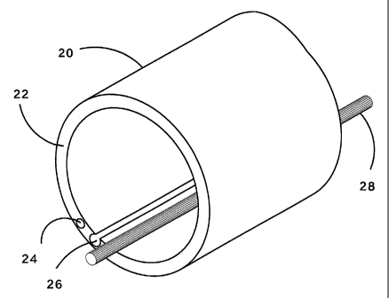
is better explained with reference to Fig. 1. A portion 20 of a
multifunctional inspection
tube of the invention is shown, including an anterior face 22. Channels and a
conductor
are associated with the wall of the tube. A totally embedded channel 24 runs
along the
length of the inspection tube within its wall, alongside an open recessed
channel 26. A
conducting element 28 runs along the length of the tube without being embedded
in the
wall of the tube, rather it is attached to the wall of the tube and occupies a
space in the
lumen of the tube. The conduit may be partially embedded in the tube or it may
be
inserted within a recess or it may be threaded within a totally embedded
channel without
being attached to the tube. Even in embodiments in which the lumen contains a
conducting element attached to the wall as described above, the lumen of the
tube is still
largely free for transferring liquids or gasses in both directions. With this
respect,
embodiment in which the channels and conducting elements embedded or wholly
inserted
in the wall may be preferable.
A typical feature of the multifunctional inspection tube is the acquisition of
internal images of the body. For acquiring the images, an image sensor may be
employed,
such as a miniature electronic camera employing a CCD or a CMOS chip
incorporated in
the anterior face of the inspection tube. The image sensor may be incorporated
removably
or permanently in the tube. Removable mounting enables to reuse, after
sterilizing, of the
imager in combination with a new tube. In one embodiment, the camera is
incorporated in
a recess in the wall of the tube as described in Fig. 2 to which reference is
now made. A
portion of an inspection tube is shown, within the inner side 40 of which, a
camera 42 is
inserted in a recessed channel, protruding from the anterior face 44 of the
tube. The signal
of the camera is transmitted by a conducting element 46, typically a copper
wire or an
optical fiber. The camera's lens is facing away from the tube. The signals
arriving from
the camera are subsequently fed to a receiver and may be subsequently
displayed on a
screen, which may be a stand-alone mini screen, an ordinary video screen, or a
portion of
the display screen ordinarily used to monitor the physiological parameters and
well -
being of the patient. In some embodiments of the invention, a fiber optical
element
CA 02501300 2005-07-04
running along the length of the tube is used to convey light to illuminate the
field of view
ahead of the anterior face of the inspection tube. Alternatively, a light
source may be
associated with the proximal face of the tube. Examples for light sources are
miniature
halogen lamps, light emitting diodes (LED), lasers, or any other kind of light-
emitting
5 source of suitable size. An alternative method of illumination is by
constructing the
inspection tube made of light - conducting material. In some embodiments of
the
invention, means for keeping the lens of the camera and /or other sensors
clean and clear
are employed. The airways, stomach or urinary bladder of an ill patient are
often filled
with secretions that may be thick and viscous. Thus, it is quite possible that
the secretions
may lodge on the lens and obscure its field of view, or on other sensors
thereby
modifying their responsiveness. To overcome such an obstacle, a constant or
intermittent
flow of air or physiological fluid is pumped through a channel in the tube's
wall, whereby
the outlet of the channel is aimed directly over and around the lens or the
sensor's active
surface. This flow may be generated by a simple flow source or by a device
that is
triggered to emit flow upon command from a human care giver, a timer or a
software
program that monitors the signal and determines when clearing action is
required.
In general, the inspection tube is used as bi - directional conveying platform
for
various elements required for the fulfillment of its inspection tasks. This is
described
schematically in Fig. 3 to which reference is now made. Tube 52 receives
activation
energy 54 of one or several types on its rear end, and downloads information
56, raw or
processed at the same end. At the anterior end 58, the tube receives signals
60 of one or
several types, and spends energy 62 as will be elaborated later on.
In some embodiments of the invention, a microphone is employed in the tube.
Such a microphone can be incorporated in the wall of the tube. Such a
microphone
receives acoustic signals from at least the vicinity of the tubes anterior,
and transfers the
signals, raw or processed to the rear of the tube for further downloading and
processing.
A plurality of sensors can be effectively employed in the anterior face of the
tube
of the invention, the non exhaustive list includes cameras, video cameras,
microphones,
pressure transducers and thermal sensors. Gas sensors, for example sensors for
particular
gasses such as oxygen and carbon dioxide may also be employed. The energy
required to
activate such sensors is supplied by conduits of energy such as electric wires
incorporated
in the tube. In addition, auxiliary energy can be supplied to the vicinity of
the anterior
face of the tube for the purpose of cleaning and clearing the sensors active
facets by
CA 02501300 2005-07-04
6
flushing them with cleaning media such as gases, humidified air or oxygen, or
liquids,
typically a physiological solution, through channels in the wall of the tube.
Liquids and or
gases for flushing are energized and conducted typically via a totally
embedded channel.
The inspection tube of the invention may be used alone or in combination with
other
catheters and tubes that are ordinarily inserted into a body organ, tract or
cavity such as
the esophagus, the stomach, the intestine, the colon, the urinary bladder, the
pleural space,
lung airways and/or the peritoneal cavity. The present technology may be
applied in
various medical practices and treatments such as: artificial ventilation of
the lung, feeding
or removing the content of the stomach, draining urine from the bladder,
draining the gas
and feces from the colon, and draining or injecting into a surgically accessed
cavity such
as the pleural space, or the peritoneal cavity.
The sensors of the tube transmit one or more signal types, which are either
preprocessed in the sensor for example on the CCD chip, or may be sent raw, to
be
further processed by analog or digital circuits to yield information relating
to the status of
the organ or body cavity inspected. The receiving and or processing devices
such as,
monitors, displays, storage means, analyzers, DSP processors, computers and
generators
of alarm signals are typically connected by one or a plurality of connectors
to the tube.
The tube of the invention may be used for insertion through orifices such as
the nose,
mouth, urethral meatus, rectum, or a surgical incision.
The transmission of raw or preprocessed signals is affected through conductors
along the tube such as wires or optical fibers, which connect to a connector
at the rear of
the tube. A wireless transmitter or transceiver may be applied anywhere
suitable on the
tube, typically at the rear, for communicating with a console containing a
receiver and
processor and or a control module.
The inspection tube of the invention may also be used to detect changes in
indications of vital functions of a patient. Accordingly, image and acoustic
signal are
being detected, processed and compared to a reference base picture or sound
structure. An
alarm is set as soon as certain changes in the indication pass a predetermined
threshold.
For example, the accumulation of secretions, or development of excessive or
diminished
lung noises are abnormal. Images as such may provide vital information
regarding body
or organ condition. Constant monitoring of images can be used to provide
dynamic
information regarding changes in blood flow, organ color or secretions level.