Note : Les descriptions sont présentées dans la langue officielle dans laquelle elles ont été soumises.
CA 02510324 2011-03-28
73612-59
INTRAOCULAR IMPLANTS
FIELD OF THE INVENTION
The present invention relates to ocular implants generally and more
particularly to intraocular implants and techniques for implanting thereof.
BACKGROUND OF THE INVENTION
The following U.S. Patents of the present inventor are believed to
represent the current state of the art:
5,814,103; 5,876,442; 5,928,283; 6,007,579 and 6,066,171.
1
CA 02510324 2005-06-16
WO 2004/054469 PCT/IL2003/001084
SUMMARY OF THE INVENTION
The present invention seeks to provide an artificial vision system.
There is thus provided in accordance with a preferred embodiment of the
present invention an artificial vision system including a sealed capsule
adapted for
intraocular placement upstream of a retina, an electronic display located
within the
sealed capsule and focusing optics located within the sealed capsule and
arranged for
focusing an image on the electronic display onto the retina.
In accordance with another preferred embodiment of the present
invention the electronic display includes an LCD display.
In accordance with yet another preferred embodiment of the present
invention the artificial vision system also includes electronic circuitry
located within the
sealed capsule for operating the electronic display, the electronic circuitry
being located
outside an optical path defined between the electronic display and the
focusing optics.
Additionally, the electronic circuitry includes a wireless data receiver
operative to
receive image data for display on the electronic display. Alternatively, the
electronic
circuitry includes a wireless energy receiver for wirelessly receiving
electrical energy
for operating the electronic display.
In accordance with still another preferred embodiment the artificial
vision system also includes wireless image transmission functionality
operative to
transmit the image data to the wireless data receiver. Preferably, the
wireless image
transmission functionality includes at least one of RF and IR image
transmission
functionality.
In accordance with yet a farther preferred embodiment of the present
invention the electronic circuitry includes an electrical power source for
providing
electrical energy for operating the electronic display. Additionally, the
power source for
providing electrical energy for operating the electronic display is a
rechargeable power
source. Alternatively, the power source for providing electrical energy for
operating the
electronic display is a wirelessly rechargeable power source. Additionally or
alternatively, the power source for providing electrical energy for operating
the
electronic display is rechargeable using at least one of ultrasonic,
electromagnetic and
photovoltaic power source.
2
CA 02510324 2005-06-16
WO 2004/054469 PCT/IL2003/001084
In accordance with still another preferred embodiment of the present
invention the artificial vision system also includes an image acquirer for
acquiring an
image to be displayed on the electronic display. Additionally, the image
acquirer is
mounted onto eyeglasses.
Preferably, the focusing optics includes a single lens. Alternatively, the
focusing optics includes multiple lenses.
There is also provided in accordance with another preferred embodiment
of the present invention a method for providing artificial vision including
implanting a
sealed capsule in a user's eye upstream of a retina, the sealed capsule
incorporating an
electronic display and focusing optics for focusing an image on the electronic
display
onto the retina, acquiring image data and transmitting the image data to the
electronic
display for display thereon.
Preferably, the transmitting includes wireless transmission to electronic
circuitry located within the sealed capsule for operating the electronic
display.
Additionally, the method for providing artificial vision also includes
wirelessly transmitting electrical energy for operating the electronic display
to a
location inside the capsule.
There is further provided in accordance with yet another preferred
embodiment of the present invention an intraocular implant, for placement
upstream of
a retina, including a telescope body defining an optical path for light to
pass
-therethrough, at least one first lens and at least one second lens enclosed
in the telescope
body, positioning means, for positioning at least one of the lenses along its
optical axis
relative to another at least one of the lenses, operative to focus objects
located at
multiple distances onto the retina and mounting structure connected to the
telescope
body for mounting the implant in an eye.
In accordance with another preferred embodiment the positioning means
includes a range finder. Alternatively, the positioning means includes a focus
resolver.
In accordance with yet another preferred embodiment of the present invention
the
positioning means is responsive to a user input.
In accordance with another preferred embodiment the positioning means
includes a mounting for at least one of the lenses, at least one magnet and at
least one
electromagnetic coil, interacting with the at least one magnet.
3
CA 02510324 2005-06-16
WO 2004/054469 PCT/IL2003/001084
In accordance with yet another preferred embodiment the positioning
means is responsive to an input from an input device external to the telescope
body.
There is also provided in accordance with yet another preferred
embodiment of the present invention an intraocular implant system for use in
an
environment wherein at least one positive lens is located outside the lens
capsule of an
eye, the system including a sealed capsule including at least one negative
lens
cooperating with the at least one positive lens to define a Galilean telescope
and at least
one air bubble.
In accordance with yet another preferred embodiment of the present
invention the at least one positive lens is produced by reshaping of the
cornea.
There is further provided in accordance with still another preferred
embodiment of the present invention an intraocular implant system including a
sealed
capsule, including at least one negative lens and at least one air bubble, and
at least one
positive lens located outside the sealed capsule.
In accordance with a preferred embodiment of the present invention the
at least one positive lens includes an eyeglass lens. Alternatively or
additionally, the at
least one positive lens includes a contact lens. Additionally or
alternatively, the at least
one positive lens includes a lens implanted in an eye. Additionally, the
implanted lens
includes an air capsule.
In accordance with another preferred embodiment, an external wall of
the sealed capsule includes the at least one negative lens.
There is also provided in accordance with a preferred embodiment of the
present invention an intraocular implant system for use in an environment
wherein at
least one negative lens is located outside the lens capsule of an eye, the
system
including a sealed capsule including at least one positive lens cooperating
with the at
least one negative lens to define a Galilean telescope and at least one air
bubble.
In accordance with yet another preferred embodiment the at least one
negative lens is produced by reshaping of the cornea.
There is yet further provided in accordance with another preferred
embodiment of the present invention an intraocular implant system including a
sealed
capsule, including at least one positive lens and at least one air bubble, and
at least one
negative lens located outside the sealed capsule.
4
CA 02510324 2005-06-16
WO 2004/054469 PCT/IL2003/001084
In accordance with a preferred embodiment of the present invention the
at least one negative lens includes an eyeglass lens. Alternatively or
additionally, the at
least one negative lens includes a contact lens. Additionally or
alternatively, the at least
one negative lens includes a lens implanted in an eye. Additionally, the
implanted lens
includes an air capsule.
In accordance with another preferred embodiment, an external wall of
the sealed capsule includes the at least one positive lens.
There is also provided in accordance with another preferred embodiment
of the present invention a method of improving vision including implanting a
sealed
capsule upstream of a retina, the sealed capsule including an electronic
display and
focusing optics and employing the electronic display and focusing optics for
focusing an
image appearing on the electronic display onto the retina.
In accordance with another preferred embodiment of the present
invention the method also includes employing a wireless data receiver
operative to
receive image data and displaying the image data on the electronic display.
Additionally
or alternatively, the method also includes employing a wireless energy
receiver for
wirelessly receiving electrical energy for operating the electronic display.
In accordance
with still another preferred embodiment of the present invention the method
also
includes wirelessly transmitting the image data to the wireless data receiver.
Preferably,
the wirelessly transmitting includes at least one of RF and IR image
transmitting.
In accordance with still another preferred embodiment of the present
invention the method also includes employing an electrical power source for
providing
electrical energy for operating the electronic display.
In accordance with yet another preferred embodiment of the present
invention the method also includes acquiring an image to be displayed on the
electronic
display. Additionally, the acquiring includes mounting an image acquirer onto
eyeglasses.
There is further provided in accordance with another preferred
embodiment of the present invention a method of improving vision including
providing
an intraocular implant including a telescope body defining an optical path for
light to
pass therethrough, at least one first lens and at least one second lens
enclosed in the
telescope body, positioning means for positioning at least one of the lenses
along its
5
CA 02510324 2005-06-16
WO 2004/054469 PCT/IL2003/001084
optical axis relative to another at least one of the lenses, operative to
focus objects
located at multiple distances onto the retina and implanting the implant
upstream of a
retina in an eye.
In accordance with yet another preferred embodiment of the present
invention the method also includes providing an input to the positioning means
and
positioning the at least one of the lenses in response to the input.
Additionally, the
providing an input includes providing an input from an input device external
to the
telescope body.
There is further provided in accordance with another preferred
embodiment of the present invention a method of improving vision including
providing
at least one negative lens located outside the lens capsule of an eye and
implanting a
sealed capsule upstream of a retina in the eye, the sealed capsule including
at least one
air bubble and at least one positive lens cooperating with the at least one
negative lens
to define a Galilean telescope. In accordance with another preferred
embodiment of the
present invention the method also includes reshaping the cornea of the eye to
produce
the at least one negative lens.
There is still further provided in accordance with another preferred
embodiment of the present invention a method of improving vision including
implanting a sealed capsule upstream of a retina in an eye, the sealed capsule
including
at least one positive lens and at least one air bubble and providing at least
one negative
lens located outside the sealed capsule.
There is further provided in accordance with another preferred
embodiment of the present invention a method of improving vision including
providing
at least one positive lens located outside the lens capsule of an eye and
implanting a
sealed capsule upstream of a retina in the eye, the sealed capsule including
at least one
air bubble and at least one negative lens cooperating with the at least one
positive lens
to define a Galilean telescope. In accordance with another preferred
embodiment of the
present invention the method also includes reshaping the cornea of the eye to
produce
the at least one positive lens.
There is still further provided in accordance with another preferred
embodiment of the present invention a method of improving vision including
implanting a sealed capsule upstream of a retina in an eye, the sealed capsule
including
6
CA 02510324 2011-03-28
73612-59
at least one negative lens and at least one air bubble and providing at least
one
positive lens located outside the sealed capsule.
In accordance with another preferred embodiment of the present
invention the providing includes providing an eyeglass lens. Alternatively or
additionally, the providing includes providing a contact lens. Additionally or
alternatively, the providing includes implanting a lens in the eye. In
accordance with
another preferred embodiment of the present invention the implanting a lens
includes
implanting a lens including an air capsule.
In accordance with another preferred embodiment of the present
invention, there is provided an artificial vision system comprising: a sealed
capsule
sized and configured for intraocular placement upstream of a retina, said
sealed
capsule including therein: an internal imaging device, sized and configured
for
intraocular placement, operative to view a scene; a transmitter, sized and
configured
for intraocular placement, operative for transmitting image information
regarding said
scene from said internal imaging device to electronic circuitry within said
sealed
capsule; an electronic display operative to display an image based on signals
received from said electronic circuitry which represent said image information
received from said transmitter; and focusing optics arranged for focusing an
image on
said electronic display onto the retina.
7
CA 02510324 2005-06-16
WO 2004/054469 PCT/IL2003/001084
BRIEF DESCRIPTION OF THE DRAWINGS
The present invention will be understood and appreciated more fully
from the following detailed description, taken in conjunction with the
drawings in
which:
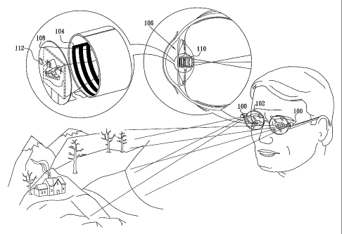
Fig. 1 is a simplified pictorial illustration of an artificial vision system
constructed and operative in accordance with a preferred embodiment of the
present
invention;
Fig. 2 is a simplified exploded view pictorial illustration of an implant
forming part of the system of Fig. 1;
Fig. 3 is a simplified partially sectional side view illustration of the
implant of Fig. 2;
Figs. 4A and 4B are simplified illustrations of the use of a variable focal
length lens arrangement in the implant system of Figs. 1 - 3;
Fig. 5 is a simplified exploded view pictorial illustration of an implant
forming part of the system of Figs. 4A & 4B;
Fig. 6 is a simplified partially sectional side view illustration of the
implant of Fig. 5;
Figs. 7A - 7G are simplified sectional illustrations showing alternative
implementations of an intraocular lens system employing a sealed capsule
arranged for
implantation in an eye and including at least one negative lens and at least
one air
bubble and at least one positive lens located outside of the sealed capsule;
Fig. 7H is a simplified sectional illustration showing another alternative
implementation of an intraocular lens system employing a sealed capsule
arranged for
implantation in an eye and including at least one negative lens and at least
one air.
bubble for use in cooperation with a positive lens formed by reshaping of the
cornea;
Fig. 8 is a simplified side view sectional illustration of an intraocular lens
system of the type shown in Figs. 7A - 7H constructed and operative in
accordance with
a further embodiment of the present invention;
Figs. 9A - 9D are simplified side view illustrations of four examples of
implanted sealed capsules of the type employed in the systems of Figs. 7A - 8;
Figs. 1 OA -10G are simplified sectional illustrations showing alternative
8
CA 02510324 2005-06-16
WO 2004/054469 PCT/IL2003/001084
implementations of an intraocular lens system employing a sealed capsule
arranged for
implantation in an eye and including at least one positive lens and at least
one air bubble
and at least one negative lens located outside of the sealed capsule;
Fig. 1 OH is a simplified sectional illustration showing another alternative
implementation of an intraocular lens system employing a sealed capsule
arranged for
implantation in an eye and including at least one positive lens and at least
one air bubble
for use in cooperation with a negative lens formed by reshaping of the cornea;
Fig. 11 is a simplified side view sectional illustration of an intraocular
lens system of the type shown in Figs. 10A - 10H constructed and operative in
accordance with a further embodiment of the present invention; and
Figs. 12A - 12D are simplified side view illustrations of four examples
of implanted sealed capsules of the type employed in the systems of Figs. 10A -
11.
9
CA 02510324 2005-06-16
WO 2004/054469 PCT/IL2003/001084
DETAILED DESCRIPTION OF PREFERRED EMBODIMENTS
Reference is now made to Fig. 1, which is a simplified pictorial
illustration of an artificial vision system constructed and operative in
accordance with a
preferred embodiment of the present invention. As seen in Fig. 1, there is
provided an
artificial vision system including a real time imaging device, such as a CCD
camera.
The illustrated embodiment includes both implanted and external imaging
devices for
the purposes for illustration, it being understood that typically either
implanted or
external imaging devices will be employed, although both could be used
together.
In the illustrated embodiment of Fig. 1, at least one and preferably plural
external imaging devices, here designated by reference numeral 100, are
typically
mounted on a pair of eyeglasses 102, as shown. The external imaging devices
100 view
a scene, preferably in stereo. The image information captured by the external
imaging
devices 100 is transmitted wirelessly, preferably by conventional IR or RF
techniques,
to electronic circuitry 104 located within a sealed capsule 106 adapted for
intraocular
placement upstream of a retina. The electronic circuitry 104 is operative to
display the
captured image as seen by the external imaging devices 100 in real time on an
electronic
display 108, such as a backlit or self-illuminated LCD display.
Focusing optics, typically in the form of a lens assembly 110, in the
sealed capsule 106, are operative to image the displayed image onto the retina
of a user.
Alternatively or additionally, an implanted imaging device, here
designated by reference numeral 112, is located on an outer surface of or
interior of
each sealed capsule 106. The internal imaging devices 112 view a scene,
preferably in
stereo. The image information captured by the internal imaging devices 100 is
transmitted in a wired or wireless manner, such as by conventional IR or RF
techniques,
to electronic circuitry 104 located within sealed capsule 106 adapted for
intraocular
placement upstream of a retina. The electronic circuitry 104 is operative to
display the
captured image as seen by the internal imaging devices 112 in real time on
electronic
display 108, such as a backlit or self-illuminated LCD display. Focusing
optics,
preferably lens assembly 110, in the sealed capsule 106, are operative to
image the
displayed image onto the retina of a user.
It is noted that the electronic circuitry 104 is located outside an optical
CA 02510324 2011-03-28
73612-59
path defined between the electronic display 108 and said focusing optics 110.
It is appreciated that, in addition to transmitting an image of a scene,
external imaging devices 100 or internal imaging devices 112 may be operative
to
transmit any other suitable digital information, such as a video image, via
electronic
circuitry 104 to electronic display 108.
Reference is now made to Figs. 2 and 3, which illustrate some details of
the implantable sealed capsule 106 which is shown implanted in a user in Fig.
1. The
sealed capsule 106 is defined by an intraocular implant housing 120 having
mounting
haptics 122 and defining a generally cylindrical capsule body 124.
Hermetically sealed
to capsule body 124 are a front sealing plate 125 and a back sealing plate
126. Back
sealing plate 126 is transparent. An internal imaging device 112 is shown
mounted on
an outside surface of front. sealing plate 125. Capsules of this type are
described in
applicants' U.S. Patent No. 6,569,199, filed October 3, 2000 and entitled
"TELESCOPIC
INTRAOCULAR LENS", and U.S. Patent No. 6,596,026, filed November 27, 2000 and
entitled "TELESCOPIC INTRAOCULAR LENS".
Preferably disposed within sealed capsule 106 is an electronic circuit and
display assembly, here designated by reference numeral 130. Assembly 130
preferably
includes electronic display 108 (Fig. 1) which is coupled to electronic
circuitry 104
(Fig. 1), preferably including a wireless receiver for image data. Display 108
is arranged
to lie generally parallel to front sealing plate 125, while electronic
circuitry 104 is
preferably embodied on a flexible circuit board 132 which is arranged to lie
in a
cylindrical configuration, peripherally of the optical path between display
108 and back
sealing plate 126, so as not to interfere with the optical pathway between the
display
108, focusing optics 110 (Fig. 1), here shown as a lens 134, and the user's
retina. It is
appreciated that even though the embodiment illustrated in Figs. 2 and 3 shows
a single
lens 134, focusing optics 110 may also comprise multiple lenses as shown in
the
embodiment of Fig. 1.
In accordance with a preferred embodiment of the present invention, the
electronic circuitry 104 also includes a wireless energy receiver such as a
resonant
11
CA 02510324 2011-03-28
73612-59
circuit (not shown) and energy storage facilities, such as a rechargeable
miniature.
battery or capacitor (not shown) for wirelessly receiving and storing
electrical energy
for operating the electrical circuitry and the electronic display.
In the embodiment of Fig. 1, an electrical power source (not shown)
-external to a user's body, such as a battery mounted in eyeglasses 102, and a
suitable
energy transmitter, such as a resonant circuit, may be used to transmit
operating power
to electronic circuit 104 inside sealed capsule 106. Any suitable electrical
power source,
such as an ultrasonic, electromagnetic and photovoltaic power source, may
alternatively
be employed interiorly or exteriorly of the capsule.
Reference is now made to Figs. 4A and 4B, which are simplified
illustrations of the use of a variable focal length lens arrangement, usable
in the implant
system of Figs. 1 - 3, as well as in other intraocular implant systems. As
seen in Figs.
4A and 4B, there is provided an intraocular implant system which includes
variable
focus optics 200 located within a sealed capsule 202 implanted within the eye
of a user.
From a consideration of Figs. 4A and 4B, it can be seen that the relative
positioning of at least two lenses 204 and 206 within variable focus optics
200 is
variable, preferably in response to an electrical control input, so as to
correctly focus
onto objects at differing distances.
The relative positioning is preferably produced by an electric displacer,
such as a piezoelectric device or a rotary electric motor in response to a
wirelessly
received viewed object distance indicating input, which may be provided by a
conventional range finder or focus resolver, such as employed in conventional
automatic focus cameras. Alternatively, a user input may be provided.
Reference is now made to Figs. 5 and 6, which illustrate some details of
the implantable sealed capsule 202 shown implanted in a user in Figs. 4A and
4B. The
sealed capsule 202 is defined by an intraocular -implant housing 220 having
mounting
haptics 222 and defining a generally cylindrical capsule body 224.
Hermetically sealed
to capsule body 224 are a front sealing plate 225 and a back sealing plate
226. Front
sealing plate 225 and back sealing plate 226 are transparent. An internal
range finding
device 212 is shown mounted on an outside surface of front sealing plate 225.
Capsules
12
CA 02510324 2011-03-28
73612-59
of this type are described in applicants' U.S. Patent No. 6,569,199, filed
October 3, 2000
and entitled "TELESCOPIC INTRAOCULAR LENS", and U.S. Patent No. 6,596,026,
filed November 27, 2000 and entitled "TELESCOPIC INTRAOCULAR LENS".
An electronic circuit and focus control assembly, here designated by
reference numeral 230, is preferably disposed within sealed capsule 202.
Assembly 230
preferably includes electronic circuitry 234, preferably including a wireless
receiver for
receiving ranging information. Electronic circuitry 234 is preferably embodied
on a
flexible circuit board 236 which is arranged to lie in a cylindrical
configuration,
peripherally of the optical path through capsule 202 via back sealing plate
226, so as not
to interfere with the optical pathway between the viewed scene, via variable
focusing
optics 240, and the user's retina.
In the illustrated embodiment, the variable focusing optics 240 comprise
a fixed lens 242 and a variable position lens 244 which is selectably
positionable along
its optical axis with respect to fixed lens 242, thus varying the focal length
of the
variable focusing optics 240.
In the illustrated embodiment, a threaded mounting 246 is provided for
lens 244, and at least one permanent magnet 250, and at least one
electromagnetic coil
252 interacting therewith, is preferably provided for selectably threading
lens 244 in
threaded mounting 246, thus varying its separation from lens 242, in response
to control
signals from electronic circuitry 234, thereby providing appropriate focusing
on a
distant viewed object.
It is appreciated that any other suitable mechanism for selectable mutual
displacement of lenses 242 and 244 may be employed.
In accordance with a preferred embodiment of the present invention, the
electronic circuitry 234 also includes a wireless energy receiver such as a
resonant
circuit (not shown) and energy storage facilities, such as a rechargeable
miniature
battery or capacitor (not shown) for wirelessly receiving and storing
electrical energy
for operating the electrical circuitry 234 and the electromagnetic coil 252.
In one embodiment of the invention, an electrical power source (not
shown) external to a user's body, such as a battery mounted in eyeglasses, and
a suitable
13
CA 02510324 2005-06-16
WO 2004/054469 PCT/IL2003/001084
range finder and energy transmitter, such as a resonant circuit, may be used
to transmit
operating power to electronic circuit 234 inside sealed capsule 202. Any
suitable
electrical power source, such as an ultrasonic, electromagnetic and
photovoltaic power
source, may alternatively be employed interiorly or exteriorly of the capsule.
It is appreciated that even though the illustrated embodiment comprises
two lenses, any suitable configuration of two or more lenses may also be
employed.
Reference is now made to Figs. 7A - 7G, which are simplified sectional
illustrations showing examples of various alternative implementations of an
intraocular
lens system employing a sealed capsule 300 implanted in the lens capsule of an
eye and
including at least one negative lens 3.02 and at least one air bubble 304 and
at least one
positive lens located outside of the sealed capsule.
Fig. 7A shows an embodiment where the positive lens is a contact lens
306. In the embodiment of Fig. 7B, the positive lens is an eyeglass lens 308.
Fig. 7C
illustrates an embodiment where the positive lens is a lens 310 implanted in
the eye.
Fig. 7C shows lens 310 implanted in the anterior chamber of the eye, it being
appreciated that alternatively lens 310 may be implanted in the posterior
chamber
between the iris and the lens capsule.
Fig. 7D shows an embodiment where two lenses are provided, a contact
lens 312 and an eyeglass less 314. In the embodiment of Fig. 7E, two lenses
are
provided, a contact lens 316 and a lens 318 implanted in the eye. Fig. 7E
shows lens 318
implanted in the anterior chamber of the eye, it being appreciated that
alternatively lens
318 may be implanted in the posterior chamber between the iris and the lens
capsule.
Fig. 7F illustrates an embodiment where the two lenses are an eyeglass lens
320 and a
lens 322 implanted in the eye. Fig. 7F shows lens 322 implanted in the
anterior chamber
of the eye, it being appreciated that alternatively lens 322 may be implanted
in the
posterior chamber between the iris and the lens capsule.
Fig. 7G shows an embodiment where three lenses are employed, contact
lens 324, eyeglass lens 326 and a lens 328.implanted in the eye. Fig. 7G shows
lens 328
implanted in the anterior chamber of the eye, it being appreciated that
alternatively lens
328 may be implanted in the posterior chamber between the iris and the lens
capsule.
Reference is now made to Fig. 7H, which is identical to Fig. 7A, wherein
refractive surgery is employed to change the curvature of the cornea 330, as
shown by
14
CA 02510324 2011-03-28
73612-59
dotted lines 332, thereby obviating the need for lens 306 (Fig. 7A).
Reference is now made to Fig. 8, which is a simplified side view
sectional illustration of an intraocular lens system of the type shown in
Figs. 7A - 7H,
constructed and operative in accordance with an. additional embodiment of the
present
invention. In this embodiment, a positive lens 350 is implanted in the eye.
Fig. 8 shows
lens 350 implanted in the anterior chamber of the eye, it being appreciated
that
alternatively lens 350 may be implanted in the posterior chamber between the
iris and
the lens capsule. In the embodiment of Fig. 8, positive lens 350 preferably
includes an
air capsule 352 to provide higher clarity focusing.
Reference is now made to Figs. 9A - 9D, which are simplified side view
illustrations of four examples of implanted sealed capsules of the type
employed in the
systems of Figs. 7A - 8. It is seen that each of the capsules includes a
sealed capsule
body 360 and associated mounting haptics 362. Capsules of this type are
described in
applicants' U.S. Patent No. 6,569,199, filed October 3, 2000 and entitled
"TELESCOPIC
INTRAOCULAR LENS", and U.S. Patent No. 6,596,026, filed November 27, 2000 and
entitled "TELESCOPIC INTRAOCULAR LENS". Disposed within the capsule is a
negative lens 364.
In the embodiment of Fig. 9A, a single air bubble 368 is disposed
rearward of negative lens 364.
In the embodiment of Fig. 9B, a single air bubble 370 is disposed
forward of negative lens 364.
In the embodiment of Fig. 9C, air bubbles 380 are disposed forward and
rearward of negative lens 364.
In the embodiment of Fig. 9D, in addition to air bubbles 390 disposed
forward and rearward of negative lens 364, a positive lens 394 is also
disposed rearward
of negative lens 364.
Reference is now made to Figs. 10A" - IOG, which are simplified
sectional illustrations showing examples of alternative implementations of an
intraocular lens system employing a sealed capsule 400 implanted in a lens
capsule of
CA 02510324 2005-06-16
WO 2004/054469 PCT/IL2003/001084
an eye and including at least one positive lens 402 and at least one air
bubble 404 and at
least one negative lens located outside of the sealed capsule. The intraocular
lens system
of Figs. 10A-10G is particularly suitable for treatment of tunnel vision.
Fig. IOA shows an embodiment where the negative lens is a contact lens
406. In the embodiment of Fig. I OB, the negative lens is an eyeglass lens
408. Fig. I OC
illustrates an embodiment where the negative lens is a lens 410 implanted in
the eye.
Fig. 1OC shows lens 410 implanted in the anterior chamber of the eye, it being
appreciated that alternatively lens 410 may be implanted in the posterior
chamber
between the iris and the lens capsule.
Fig. 1O1 shows an embodiment where two lenses are provided, a contact
lens 412 and an eyeglass less 414. In the embodiment of Fig. 10E, two lenses
are
provided, a contact lens 416 and a lens 418 implanted in the eye. Fig. IOE
shows lens
418 implanted in the anterior chamber of the eye, it being appreciated that
alternatively
lens 418 may be implanted in the posterior chamber between the iris and the
lens
capsule. Fig. 1OF illustrates an embodiment where the two lenses are an
eyeglass lens
420 and a lens 422 implanted in the eye. Fig. lOF shows lens 422 implanted in
the
anterior chamber of the eye, it being appreciated that alternatively lens 422
may be
implanted in the posterior chamber between the iris and the lens capsule.
Fig. I OG shows an embodiment where three lenses are employed, contact
lens 424, eyeglass lens 426 and a lens 428 implanted in the eye. Fig. lOG
shows lens
428 implanted in the anterior chamber of the eye, it being appreciated that
alternatively
lens 428 may be implanted in the posterior chamber between the iris and the
lens
capsule.
Reference is now made to Fig. IOH, which is identical to Fig. 10A,
wherein refractive surgery is employed to change the curvature of the cornea
430, as
shown by dotted lines 432, thereby obviating the need for negative lens 406
(Fig. 10A).
Reference is now made to Fig. 11, which is a simplified side view
sectional illustration of an intraocular lens system of the type shown in
Figs. l0A -
10H, constructed and operative in accordance with an additional embodiment of
the
present invention. In this embodiment, a negative lens 450 is implanted in the
eye. Fig.
11 shows lens 450 implanted in the anterior chamber of the eye, it being
appreciated
that alternatively lens 450 may be implanted in the posterior chamber between
the iris
16
CA 02510324 2011-03-28
73612-59
and the lens capsule. In the embodiment of Fig. 11, lens 450 includes an air
capsule 452
to provide higher clarity focusing.
Reference is now made to Figs. 12A - 12D, which are simplified side
view illustrations of four examples of implanted sealed capsules of the type
employed in
the systems of Figs. 10A - 11. It is seen that each of the capsules includes a
sealed
capsule body 460 and associated mounting haptics 462. Capsules of this type
are
described in applicants' U.S. Patent No. 6,569,199, filed October 3, 2000 and
entitled
"TELESCOPIC INTRAOCULAR LENS", and U.S. Patent No. 6,596,026, filed
November 27, 2000 and entitled "TELESCOPIC INTRAOCULAR LENS". Disposed
within the capsule is a positive lens 464.
In the embodiment of Fig. 12A, a single air bubble 468 is disposed
rearward of positive lens 464.
In the embodiment of Fig. 12B, a single air bubble 470 is disposed
forward of positive lens 464. _
In.the embodiment of Fig. 12C, air bubbles 480 are disposed forward and
rearward of positive lens 464.
In the embodiment of Fig. 12D, in addition to air bubbles 490 disposed
forward and rearward of negative lens 464, a negative lens 494 is also
disposed
rearward of positive lens 464.
It will be appreciated by persons skilled in the art that the present
invention is not limited to what has been particularly shown and described
hereinabove.
Rather the scope of the present invention includes both combinations and
subcombinations of the various features described hereinabove as well as
modifications
and variations thereof as would occur to a person of skill in the art upon
reading the
foregoing specification and which are not in the prior art.
17