Note : Les descriptions sont présentées dans la langue officielle dans laquelle elles ont été soumises.
CA 02511958 2005-06-27
WO 2004/064624 PCT/EP2003/000543
1
RETRACTOR FOR SURGICAL OPERATIONS ON THE ARTERIA HAEMORRHOIDALIS
DESCRIPTION
In order to effect ambulatory operations on the haemorrhoids, without
s anaesthesia, it is known the use of the device described in the U.S. patent
No. 5 570
692, which comprises a retractor tube closed on the end which is inserted in
the anal
cavity and opened on the external end, which is provided with a gripping
handle. The
retractor tube is provided on its lateral wall, at a short distance from its
closed end,
with an ultrasonic probe to detect the blood flow of the haemorrhoidalis
artery and is
io provided near to the probe, with a lateral window through which may be
detected and
observed the portion of the anal mucosa upon which it must be operated for the
ligature of said artery, for example by means of a curved needle or by means
of
cauterisation. The closed end of the retractor tube, may be illuminated by a
luminous
source housed in said end and connected to feeding means provided in the
handle,
is together with the feeding means of said probe. This device, for the reason
that
incorporates the ultrasound probe and that houses the luminous source in its
closed
end, presents elevated production costs, so that it is not possible to propose
the
same as a disposable product, with all the drawbacks and the limitations
deriving by
this fact.
2o Objecfi of the invention is to obviate to these and other limits of the
known prior
art, with a disposable device, for the realisation of which it has been
necessary to
resolve some technical problems connected with the removable housing in the
same
of the ultrasonic probe and other problems related to the means for the
illumination of
the lateral window for the exploration of the anal mucosa. The first of said
problems
zs has been solved providing in the retractor tube a longitudinal seat, closed
toward the
interior and opened with the end toward the outer end of the same tube, in
which
seat it is possible to removably house an ultrasonic probe which partially
projects
through a longitudinal opening of the retractor tube, to result in contact
with the anal
mucosa. The ultrasonic probe is preferably inserted and hygienically protected
in a
3o sterile, disposable and easily removable sheath, having a suitable
conformation, in
CA 02511958 2005-06-27
WO 2004/064624 PCT/EP2003/000543
2
such a manner that the same may be reutilized repeatedly in other disposable
devices of the type which is referred to. Immediately downstream of the seat
with the
ultrasonic probe, there is provided the window for the exploration of the anal
mucosa.
To solve the problem of the illumination, it has been used the technique of
the back
s illumination, known in the proctoscopies, which provides the movable
insertion of a
luminous source in the handle of the device. Instead of the use of curved
light guide
means, realised for example with optical fibre or with a bar of plastics,
connected with
one of their end to said luminous source and oriented with the other end in
the field of
view defined by the internal cavity of the retractor tube, as described for
example in
io the Italian patent No. 1 234 169, in the device according to the invention
are utilised
curved means to reflect the light inside of the retractor tube, with the
advantage of a
better luminous yield and with the advantage that such devices result distant
from the
internal surface of the same retractor and can not be soiled and blinded by
the
physiological liquid which unavoidably is produced by the anal cavity during
the
is operation which is referred to.
These and other features of the invention, and the advantages deriving
therefrom, will appear better evident from the following description of a
preferred
embodiment of the same, made by way of non-limiting example, with reference to
the
figures of the attached sheets of drawings, in which:
20 - Figure 1 is a perspective view of the device without the ultrasonic probe
and
without the illumination source;
- Figure 2 shows the device in lateral elevation, decomposed in the pieces
which compose it and with the illumination source placed between the two
portion of
the handle in which the same is inserted;
2s - Figures 3 and 4 show the device respectively in plan view from above and
in
plan view from the bottom;
- Figure 5 shows the device assembled and sectioned along the line V - V of
Figure 2;
- Figure 6 shows further details of the device sectioned along the line VI -
VI
30 of Figure 5;
CA 02511958 2005-06-27
WO 2004/064624 PCT/EP2003/000543
3
- Figures 5a and 6a show embodiments of the device respectively viewed as
in the preceding Figures 5 and 6;
- Figure 7 shows the device of the Figure 5a according to a view of the front
toward the operator.
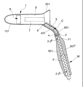
s From the Figures it is noted that the device comprises a substantially
cylindrical tube 1 having the function of retractor, closed at its terminal
end 101 which
is opportunely tapered and rounded, and on the contrary open on the initial
end 201
which has a conical shape and which is outwardly divergent. Merely by way of
example, the body 1 may have an external diameter which is comprised between
2,5
io and 3,5 centimetres, for example of about 3 centimetres, an may have a
general
length comprised between 10 and 12 centimetres, comprehensive of the divergent
end 201 which has alone the length of about 2 centimetres. However, it is to
be
understood that the device may be realised with dimensions which are different
to the
indicated dimensions, in order to comply with different use requirements. The
conical
is end 201 is outwardly projecting with a portion having a substantially
triangular plan
301, having a length of some centimetres, provided in its center line with a
longitudinal and channel-shaped rib 401, outwardly convex, which has
prevalently
the function to increase the resistance to the bending and torsion stress of
said
appendices 301 and to partially define the duct along which will be effected
the
ao reflection of the light for the illumination of the working zone. The
appendix 301,
which has for example an inclination of about 30° with respect to the
longitudinal axis
of the body 1, integrally connects to said body an elongated shell 501
realised with a
suitable ergonomic shape for the function of gripping handle, having for
example a
length of about 10 centimetres and which is forming with the axis of said body
1 an
2s internal angle of about 105°. It is to be understood that also these
last dimension
data of the device are merely indicative and that the same may be widely
modified.
The connecting zone of the shell 501 to the appendices 301, is suitably
curved. Upon
the shell 501 there is placed and fixed with the male-female fixed coupling
portions 2,
2', a complementary shell 501' which completes the formation of the gripping
handle
3o M and which is superimposed to the ribbed portion 401 of the appendix 301,
with a
CA 02511958 2005-06-27
WO 2004/064624 PCT/EP2003/000543
4
terminal portion 601 having the shape of a channel, which is connected to the
same
shell 501' with a suitable curvature, which ends in the connecting zone of the
conical
portion 201 to the cylindrical portion of the tube 1 and then in the internal
portion of
said tube which appears through the same conic mouth 201. The portion 601
realises
s with the portion 401 a tubular duct C which at least from the outside
presents a flat
shape also for the presence of lateral ribs, in such a manner to result with
high
features of resistance to the bending and to the torsion. In the conjunction
zone of
the shell 501 to the channel 601, there is provided a step 11 raised toward
said shell,
suitable for leaning the thumb of the hand which grasps the handle M, to
ensure a
to steady grasp of the same handle and to facilitate in absolute the use of
the device.
In the conjunction zone of the unit of the handle M to the appendix 301,
inside
of the two complementary shells which define the same handle, are obtained the
two
complementary portions of an annular seat 3 in which can be placed a small
disk 4
made of transparent material, which realises a division barrier between the
internal
is and absolutely sterile portion of the instrument, from the internal and
hollow portion of
the handle, in which is inserted and retained for example by means of
friction, the
end of the illumination optical waveguide F, of the known type, which may be
not
subjected to sterilisation treatments. The small disk 4 may have, if required,
optical
functions and may be made by means of a lens suitable to focalise the light on
a
2o reflection parabola 5 which covers the internal surface of the portion 601
of the duct
C and which can be, for example, realised in a very economical and reliable
manner,
with an electrochemical metal spray coating of chrome. The advantages deriving
from the backlight system described, with respect to the known systems which
use
light guides, are represented by a better luminous efficiency and especially
by the
2s fact that the same illumination means may be not blinded by the organic
liquid which
can come out from the hollow of the retractor tube, because the reflection
parabola 5
remains raised from the path of said liquid, and because also in the most
unfavourable condition shown in Figure 6, it is possible to foresee upstream
of the
small disk 3, on the ribbed zone 401, one or more drainage openings 6,
suitably
3o shaped, through which said organic liquid may freely come out. The device
is
CA 02511958 2005-06-27
WO 2004/064624 PCT/EP2003/000543
preferably realised with plastics of a changing white colour, to exalt the
effects of the
illumination inside the body 1. From the drawings, it appears that the
retractor tube 1
is laterally provided with a longitudinal and rectilinear opening 7, for
example with a
rectangular shape, which begins in the zone in which the end conic portion 201
is
s connected to the cylindrical portion of the same retractor 1 and which has a
length
which is about equal to the half length of the same retractor. In the example
which is
referred to, the ideal plane in which lies the opening 7 is parallel to the
center line
plane of the device and the same opening is placed on the right side of the
body 1 if
the device is considered with the handle M downwardly oriented, but it is to
be
io understood that said collocation may be diversified. It is not even
excluded that the
ideal plane on which the opening 7 lies, may be differently perpendicular to
the
vertical center line plane of the device, with the same window which results
placed in
the upper portion of the body 1 if the device is considered with the handle
downwardly oriented, also to cause the terminal and internal portion of the
body
is retractor, placed downstream of the zone interested by said opening, may be
better
illuminated by the beam which comes out from the reflection parabola 5. In
fact, in
said zone, the retractor body 1 presents a tapered and slightly flattened
shape, as
shown with numeral reference 701, in the initial portion of which is provided,
transversally oriented with the greatest dimension, a window 8 for example
with a
ao rectangular shape, for example having the dimensions of centimetres 1 x 2,
through
which it will appears the anal mucosa which will be efficaciously illuminated
by the
above mentioned backlight means. The window 8 is distant from the outer end of
the
body 1, which is connected to the conical portion 201, of about 4-7
centimetres, for
example of about 5-6 centimefires. In the zone which is comprised between the
rear
zs edge 208 of the window 8 and the rear side 107 of the openings 7, the body
1
presents internally and integral a flat division wall 9, which delimits inside
the same
body 1 a longitudinal chamber 10 open on the end toward the mouth 201 of the
retractor and provided with the outer and lateral opening 7 above mentioned.
In said
chamber 10 is friction inserted a ultrasound probe, not illustrated, which
will be
3o realised with such shape to opportunely project from the opening 7, to
result in
CA 02511958 2005-06-27
WO 2004/064624 PCT/EP2003/000543
6
contact with the rectal mucosa. As said in the introduction of the present
description,
the probe may be contained in a thin sterilised, disposable and easily
removable
sheath, so that the same probe may be used several times in other disposable
devices of the type which is referred to. The connection cable to the probe,
will go out
s from the mouth 201 of the retractor and it may be temporarily fixed with an
adhesive
bandage on a side of the handle M. It is to be understood that the handle M
and
other portions of the device (see further) may be laterally provided with
small loops
having the shape of pincers, integral obtained upon the shells 501, 501' and
suitable
to temporarily support the cable of the ultrasound probe above mentioned. As
to appears from Figures 3-5, the window 8 lies on a terminal portion of the
retractor
tube which is slightly flattened and in recess and the rear side 208 of said
window is
connected with an inclined plane 801 with the lateral surface of the
retractor. The
forward edge 108 of the window 8 is then characterised by the fact that it is
in relief
and to have a slightly arcuate shape, with the convexity turned toward the
outside. All
is these conditions allow to optimise the dilatation of the rectal tissues and
contextually
to avoid prolapse of the same inside the window 8, in such a manner that
through
said window the rectal mucosa presents itself in the better condition to
operate on the
same with the known and required means for the ligature of the arteria
haemorrhoidalis, which can be identified with precision by means of the said
2o ultrasonic probe.
The device shown in Figures 5a, 6a and 7 is different from the device
previously described for the several features below considered. The window 8
is, for
example, arcuate-shaped, is obtained on the retractor tube 1 substantially for
half of
its circumference, and has a length which is inferior to 1 centimetre, for
example
2s comprises between 8 and 5 millimetres. The inclined plane 801, placed
immediately
downstream of the window 8, is more wide and less inclined of that of the
Figures
from 1 to 5, and upon it there is localised the opening 7 which exposes the
sensible
portion of the ultrasonic probe S visible in Figure 7, in such a manner that
this same
portion results very close to the said window 8 and to the portion of the
arteria
3o haemorroidalis upon which the operation will be made.
CA 02511958 2005-06-27
WO 2004/064624 PCT/EP2003/000543
7
The forward side 108 of the window 8 it is not in relief as in the previous
solution, but it is lower with respect to the posterior side 208 of the same
window and
forms part of a flat portion 701' which is substantially aligned to the wall 9
for the
delimitation of the chamber 10 housing the probe S, said portion being
connected
s with a correct union to the remaining flat portion 701, in such a manner to
form in the
whole a flat portion with a sinuous profile and with a decreasing profile
toward the
rounded point 101.
Always from Figure 5a it appears that under the portions 701, 701' above
mentioned, inside the body 1 is obtained a seat 12 having for example a
rounded
io section and a conic shape, with a superior edge 112 slightly placed beyond
the
anterior edge 108 of the window 8, in such a manner to rest upon said edge and
to
insert in said seat, the terminal portion of a mandrel not shown, which holds
the
curved needle A with which will be made the ligature of the arteria
haemorroidalis
and that with the external end of the retractor tube 1 may be easily operated
by the
is operator. The axis of the seat 12 is for example parallel and suitably
displaced from
the axis of the retractor tube 1.
From Figures 5a and 7 it is noted that the initial conic portion 201 of the
retractor 1, is flattened on the side corresponding to the seat 10 for the
housing of the
probe S and on this side it carries a set of three appendices 13 upon which it
is
ao possible to firmly anchor the portion of the cable G which is near to the
same probe.
From Figure 6 it is finally noted that the reflecting portion 5 is placed only
in
the terminal and rectilinear portion of the channel C, with an inclination of
about 40-
45° with respect to the longitudinal axis of the retractor 1, for
example of about 43°.
The terminal portion of the optical waveguide F for the illumination is now
placed at a
Zs short distance from the reflecting surface 5, in such a manner to sensibly
improve the
illumination intensity of the internal cavity of the same retractor. The
longitudinal axis
of the terminal portion of the optical waveguide F inserted in the handle M,
forms with
the axis of the retractor 1 an internal angle of about 110°. Always
from Figure 6a it is
finally noted how the same terminal portion of the optical waveguide F results
raised
3o from the bottom of the channel C with the reflecting surface 5, for the
presence of the
CA 02511958 2005-06-27
WO 2004/064624 PCT/EP2003/000543
wide recessed portion 14 in the conjunction zone 401 of the handle M to the
retractor
tube 1, zone which may be provided with, if required, said drainage openingls.