Note : Les descriptions sont présentées dans la langue officielle dans laquelle elles ont été soumises.
CA 02528635 2005-12-28
Patent
File 110.02740101
Express Mail Label No. EV 201894040 US
TUMOR TREATMENT USING BETA-SHEET PEPT>DES
AND RADIOTHERAPY
GOVERNMENT FUNDING
The present invention was made with government support under Grant Nos.
CA 096090 and CA 044114, awarded by the National Institutes of Health. The
Government may have certain rights in this invention.
IS
BACKGROUND
Controlling tumor growth by targeting tumor vasculature remains the subject
of intense investigation. As angiogenesis is required in the growth of tumors,
anti-
angiogenic agents have been investigated as potential antitumor agents. The
search
for anti-angiogenic agents has focused on controlling two of the processes
that
promote angiogenesis_ the growth and adhesion of endothelial cells (EC).
Efforts
have focused on ECs primarily because ECs are more accessible than other cells
to
pharmacologic agents delivered via the blood, and are genetically stable and
thus not
easily mutated into drug resistant variants. Most anti-angiogenic agents have
been
discovered by identifying endogenous molecules that inhibit EC growth. This
traditional approach has produced a number of anti-angiogenic agents, such as
platelet factor-4 (PF4), thrombospondin, tumor necrosis factor (TNF),
interferon-y
inducible protein-l0, angiostatin, endostatin, vasostatin, and bactericidal-
permeability
increasing (BPI) protein. In toto, about forty anti-angiogenic agents,
identified using
various approaches, are currently known. While a number of anti-angiogenic
agents
have been developed, the need for better angiogenesis inhibitors and improved
methods for their use is evidenced by the absence of any major clinical
breakthroughs
(Fogarty, M., The Scientist 16, 33-35 (2002)) and the paucity of markers of
antiangiogenic therapy that can be monitored during treatment (Kerbel, R.S.,
Carcinogenesis 21, 505-I S (2000)).
CA 02528635 2005-12-28
Angiogenesis inhibitors may be used to complement other antitumor
techniques. Radiotherapy and chemotherapy are used against many new cases of
cancer reported annually in the United States. Although the primary tumor is
often
controlled with radiotherapy and/or chemotherapy, at least temporarily, there
remain
S over 500,000 cancer-related deaths annually in the United States alone (NCI
Surveillance, Epidemiology and End results program and Centers for Disease
Control
and Prevention). Results of the first successful phase III clinical trial
using an
antiangiogenic agent (bevacizumab/Avastin) in combination with a
chemotherapeutic
regimen were recently announced (Yang et al., N. Engl. J. Med. 349, 427-34
(2003);
listed in Clin. Colorectal Cancer 3, 8S-88 (2003)). This trial demonstrates
that
angiogenesis inhibitors, when used in combination with conventional
chemotherapy,
appear to be a powerful tool to combat cancer in patients.
In animal models, combining angiogenesis inhibitors with radiation therapy
(Mauceri et al., Nature 394, 287-91 ( 1998); Griffin et al., Cancer Res. 62,
1702-06
1 S (2002)), gene therapy (Wilczynska et al., Acta Biochim. Pol. 48, 1077-84
(2001 )), or
chemotherapy (Teicher et al., Cancer Res S2, 6702-4 ( 1992): Teicher et al.,
Eur. J.
Cancer 32A, 2461-6 ( 1996); Herbst et al., Cancer Chemother. Pharmacol. 41,
497-
504 ( 1998)) has been shown to be potentially beneficial. In the case of
radiation
therapy, apoptosis of endothelial cells has been recognized as a critical
component of
the radiation response (Garcia-Barros et a1_, Science 300, 1 l SS-S9 (2003);
Folkman et
al., Science 293(5528), 227-8 (2001)), independent of tumor oxygenation during
radiation (Lee et al., Cancer Res. 60, SS6S-70 (2000))_ The list of
antiangiogenic
agents demonstrated to enhance the antitumoreffects from radiation therapy
includes,
for example, thrombospondin-1 (Rofstad et al., Cancer Res. 63, 4055-61
(2003)),
angiostatin (Gorski et al., Cancer Res. S8, 5686-9 ( 1998)), various receptor
tyrosine
kinase inhibitors (Griffin et al., Cancer Res. 62, 1702-06 (2002), and anti-
VEGF and
VEGFR antibodies (Gorski et al., Cancer Res. 59, 3374-8 ( 1999)).. However,
many
of these agents are difficult and expensive to produce and/or have documented
toxicity issues. Thus, other ways or methods of treating tumors is needed.
2
CA 02528635 2005-12-28
SUMMARY OF THE INVENTION
The present invention demonstrates that infusion of ~3-sheet peptides in
combination with radiation results in tumor growth inhibition. Furthermore,
injection
of ~3-sheet peptides before, after, or during radiation treatment preferably
sensitizes
endothelial cells to radiation and significantly prolongs radiation-induced
tumor
growth delay. Changes in tumor histology and in vitro tissue culture studies
strongly
suggest that combination treatment with (3-sheet peptides and radiation is an
effective
tumor treatment strategy.
Accordingly, in one aspect, the present invention provides a method of
treating a patient with a tumor that includes delivering radiation to the
patient and
administering to the patient a (3-sheet peptide. The (3-sheet peptide is a
water soluble
peptide having at least 35% to 55% amino acids having hydrophobic side chains
in
which the ratio of amino acids having positively charged side chains amino
acids to
l5 amino acids having negatively charged side chains is at least 2:1, and in
which at least
two of the amino acids having hydrophobic side chains are positioned in the
peptide
chain with an intervening turn sequence in a manner such that the two amino
acids
having hydrophobic side chains are capable of aligning in a pairwise fashion
to form
a (3-sheet structure. Furthermore, the (3-sheet peptide is water soluble under
physiological conditions, and the peptide forms ~3-sheet structures.
In further aspects of the method of the invention, the turn sequence of the ~3-
sheet peptide is LXXGR(SEQ ID N0:33) and X is independently selected from the
group consisting of K, N, S, and D, and/or the ~-sheet peptide consists of 28
to 33
amino acids.
In a preferred aspect of the invention, the radiation and the (3-sheet peptide
treat the patient with a tumor synergistically. In one aspect, the (3-sheet
peptide of the
method is administered before delivering radiation to the patient and the (3-
sheet
peptide radiosensitizes the tumor to radiation. One type of cells that may be
radiosensitized is endothelial cells. In a further aspect, the ~3-sheet
peptide is
administered within 24 hours of delivering radiation to the patient.
3
CA 02528635 2005-12-28
The invention may include a variety of additional aspects. For instance, the
~i-
sheet peptide used in the method of the invention is administered along with a
pharmaceutically acceptable carrier. The method may also include inhibition of
angiogenesis by the (3-sheet peptide. Radiation used in the method may include
gamma ray or x-ray radiation.
The method of the invention may be used to treat a variety of tumors_ In one
aspect, the tumor is a solid tumor. In a further aspect, the solid tumor is
selected from
the group consisting of carcinomas, sarcomas, and lymphomas. For solid tumors,
the
radiation is preferably a daily dose of about 50 to 70 grays. In a further
aspect, the
solid tumor is present in the brain; breast, cervix, larynx, lung, pancreas,
prostate,
skin, spine, stomach, or uterus. The tumor treated by the method of the
invention
may also be a leukemia. For leukemia, the radiation is preferably a daily dose
of
about 20 to 40 grays.
In an additional aspect of the method of the invention, the (3-sheet peptide
is
selected from the group consisting of (3pep-1 through (3pep-30 (SEQ ID NOS:1-
30)
and their derivatives. In a further aspect, the (3-sheet peptide is (3pep-25
and its
derivatives. In yet a further aspect, the (3-sheet peptide is ~3pep-25.
The invention also provides a method of treating a patient with a tumor that
includes delivering gamma or x-ray radiation to the patient and administering
to the
patient (3pep-25 in a pharmaceutically acceptable carrier. In one aspect of
this
method, the radiation and (3pep-25 treat the patient with a tumor
synergistically. In a
further aspect, the radiation and (ipep-25 provide a synergistic effect of
200% or
more.
"Amino acid" is used herein to refer to a chemical compound with the general
formula: NHS-CRH-COOH, where R, the side chain, is H or an organic group.
Where
R is organic, R can vary and is either polar or nonpolar (i.e., hydrophobic).
The
amino acids of this invention can be naturally occurring or synthetic (often
referred to
as nonproteinogenic). As used herein, an organic group is a hydrocarbon group
that is
classified as an aliphatic group, a cyclic group or combination of aliphatic
and cyclic
groups. The term "aliphatic group" means a saturated or unsaturated linear or
branched hydrocarbon group. This term is used to encompass alkyl, alkenyl, and
4
CA 02528635 2005-12-28
..
alkynyl groups, for example. The term "cyclic group" means a closed ring
hydrocarbon group that is classified as an alicyclic group, aromatic group, or
heterocyclic group. The term "alicyclic group" means a cyclic hydrocarbon
group
having properties resembling those of aliphatic groups. The term "aromatic
group"
refers to mono- or polycyclic aromatic hydrocarbon groups. As used herein, an
organic group can be substituted or unsubstituted. One letter and three letter
symbols
are used herein to designate the naturally occurring amino acids. Such
designations
including R or Arg, for Arginine, K or Lys, for Lysine, G or Gly, for Glycine,
and X
for an undetermined amino acid, and the like, are well known to those skilled
in the
3 0 art.
The terms "polypeptide" and "peptide" as used herein, are used
interchangeably and refer to a polymer of amino acids. These terms do not
connote a
specific length of a polymer of amino acids. Thus, for example, the terms
oligopeptide, protein, and enzyme are included within the definition of
polypeptide or
1 S peptide, whether produced using recombinant techniques, chemical or
enzymatic
synthesis, or naturally occurring. This term also includes polypeptides that
have been
modified or derivatized, such as by glycosylation, acetylation,
phosphorylation, and
the like.
The term "water-soluble" is used herein to refer to compounds, molecules, and
20 the like, including the peptides of this invention, that are preferably
readily dissolved
in water. The compounds of this invention are readily dissolved in water if
about 1
mg of the compound dissolves in 1 ml of water having a temperature of about 35-
45°
C. More preferably, the peptides of this invention will have a water
solubility of at
least about 10 mg/ml and often of at least about 20 mg/ml. Even more
preferably, the
25 peptides are soluble at these concentrations under physiological
conditions, including
a pH of about 7.0-7.4 and a salt concentration of about 1 SO mM NaCI.
The term "hydrophobic amino acid side chain" or "nonpolar amino acid side
chain," is used herein to refer to amino acid side chains having properties
similar to
oil or wax in that they repel water. In water, these amino acid side chains
interact
30 with one another to generate a nonaqueous environment. fixamples of amino
acids
5
CA 02528635 2005-12-28
with hydrophobic side chains include, but are not limited to; valine, leucine,
isoleucine, phenylalanine, and tyrosine.
The term "polar amino acid side chain" is used herein to refer to groups that
attract water or are readily soluble in water or form hydrogen bonds in water.
Examples of polar amino acid side chains include hydroxyl, amine, guanidinium,
amide, and carboxylate groups. Polar amino acid side chains can be charged or
non-
charged.
The term "non-charged polar amino acid side chain" or "neutral polar amino
acid side chain" is used herein to refer to amino acid side chains that are
not ionizable
or do not carry an overall positive or negative charge. Examples of amino
acids with
non-charged polar or neutral polar side chains include serine, threonine,
glutamine,
and the like.
The term "positively charged amino acid side chain" refers to amino acid side
chains that are able to carry a full or positive charge and the term
"negatively charged
amino acid side chain" refers to amino acid side chains that are able to carry
a
negative charge. Examples of amino acids with positively charged side chains
include arginine, histadine, lysine, and the Like. Examples of amino acids
with
negatively charged side chains include aspartic acid and glutamic acid, and
the like.
The term "self-association" refers to the spontaneous association of two or
more individual peptide chains or molecules irrespective of whether or not the
peptide
chains are identical.
The term "tumor" refers a collection of cells that have developed cancer. The
collection of cells may form an aggregate, as in solid tumors, or may be
diffuse, as in
leukemias. Cancer cells contain genetic damage that has resulted in the
relatively
unrestrained growth of the cells. The genetic damage present in a cancer cell
is
maintained as a heritable trait in subsequent generations of the cancer cell
line. The
genetic damage found in cancer cells is generally found in oncogenes, and
tumor
suppressor genes, but can also occur in genes governing immunity, cell
motility, or
angiogenesis.
Unless otherwise specified, "a," "an," "the," and "at least one" are used
interchangeably and mean one or more than one.
6
CA 02528635 2005-12-28
BRIEF DESCRIPTION OF THE FIGURES
Figure 1 schematically illustrates the alignment of (3-sheet regions from the
polypeptides PF4, IL-8 and GRO polypeptides. ~3-sheet residues are blocked-in
and
lines connect the residues that are paired in the chain. The C-termini in the
sequences
were synthesized in the amide form. Numbering shown below the sequence is that
from native PF4.
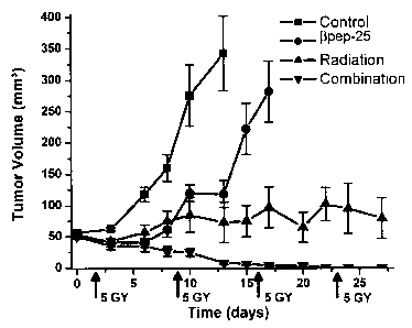
Figure 2 provides graphs showing the mean tumor growth curves for the
human ovarian carcinoma (MA 148) and the murine syngeneic mammary carcinoma
(SCK) in mice treated with (3pep-25, radiation or combination treatment. Fig.
2A
shows the mean tumor growth curve of the MA148 xenograft. Groups shown are
defined as follows. - t -, vehicle containing BSA (n = 13); - ~ -, pep-25 ( 10
mg/kg/day for 28 days, n = 10); - ~ -, radiation (n = 10); - ~~ -, combination
group (n
= 10). Fig_ 2b shows the mean tumor growth curve of SCK tumors. Groups shown
are defined as follows: -1-, vehicle containing BSA (n = 12); -1-, ~3pep-25
(20
mg/kg/day for l4 days, n = 12); -~ -, radiation (n = 13); - ~ -, a combination
group (n
= 13). Radiation was administered locally at the time points and amount
indicated by
arrows. Data shown are means of tumor volume. Error barn, SEs. In Fig. 2B, the
error bars are shown until the first animal death in each group, the means
thereafter
represent the average tumor volumes of the mice still present.
Figure 3 is a picture showing the results of histochemical analysis. Double
staining of tumor cross-sections that were stained for vessel density,
apoptosis and
proliferation. Microvessel density (MVD) is revealed by PE-labeled anti-CD31
antibody staining, apoptosis staining is revealed by using the TUNEL assay,
and
proliferation is revealed by PE-labeled anti-PCNA, all as indicated. Fig. 3A
shows
MA148 tumor section staining. Specimens of the combination treatment were not
available for histology due to tumor regression during course of treatment.
Fig. 3B
shows SCK tumor section staining. Quantifications are given in Table 2 and the
arrows indicate double staining. Original magnification 200 X.
Figure 4 provides graphs showing (3pep-25 affects tumor physiology. Fig. 4A
shows SCK tumor blood perfusion is reduced by 5 days of i_p_ injection of 10
mg/kg
7
CA 02528635 2005-12-28
(3pep-25 treatment measured by g6Rb uptake. Fig. 4B shows that the median
partial
pressure of 02 (p02) was significantly (p < 0.05) reduced in SCK tumors by 7
daily
i.p. injections of 20 mg/kg (3pep-25 treatment.
Figure 5 provides graphs showing the relative effectiveness of (3pep-25 and
angiostatin as radiosensitizers. Fig. 5A shows i.p. injections of (3pep-25 (20
mg/kg)
were given on days-I, 0, and 1 to SCK bearing mice. Radiation (10 Gy) was
applied
on days 0 and 1, 2 hours after (3pep-25 injection. Fig. 5B shows i.p.
injections of
angiostatin (25 or 50 mg/kg) were given on days -I , 0, and 1 in the SCK tumor
mouse model. Radiation (10 Gy) was applied on days 0 and 1, 2 hours after
injection
of angiostatin. Data shown are means of tumor volume. Error bars (SEs) are
shown
until the first animal death in each group, the means thereafter represent the
tumor
volumes of the mice still alive.
Figure 6 is a bar graph showing the relative changes in the volume of SCCVII
tumors after injection of 10 mg/kg of (3pep-25 or exposure to 8 Gy alone or
IS combined. Tumor-bearing animals were given i.p. injection of 10 mg/kg (3pep-
25 on
days l and 3 after irradiation. Each data point is average tumor volume ~I SE
measured in 7-10 animals per treatment group.
Figure 7 is a bar graph showing typical hematoxylin and eosin (H&E) or
pimonidazole staining of SCCV11 tumors at 5 days after first injection 10
mg/kg of
(3pep-25 (twice injections) and/or radiation exposure of 8Gy, 40x and 400x
magnification. At least three animals per group were analysed. Images shown
are
representatives of each treatment group.
Figure 8 is a graph showing the frequency distribution of measured intra-
tumor p02 in saline-treated, (3pep-25 alone, 8 Gy alone and 8 Gy and ~3pep-25
combined, constructed as function of oxygen tension, with grouping in 2 mmHg
intervals.
Figures 9A-9H provide power doppler images of tumor bearing mice. Power
doppler tumor images of vehicle (saline) treated mouse on day 3 and day 7 are
shown
in 9A and 9 B, respectively. Power doppler tumor images of a ~3pep-25 treated
mouse
on day 3 and 7 are shown in Figures 9C and 9D, respectively. Power doppler
Iumor
images of an 8 Gy treated mouse are shown on day 7 and day 14 in 9E and 9F,
8
CA 02528635 2005-12-28
respectively. Power doppler tumor images of a combination (~3pep-25 and 8 Gy)
treated mouse on day 7 and day 14 are shown in 9G and 9H, respectively. Images
shown are representative of the mean.
Figure 10 provides graphs showing ~3pep-25 specifically targets endothelial
cells and enhances the anti-proliferative activity of radiation. Fig. I OA
shows (3pep-
25 alone specifically inhibits endothelial cell proliferation and does not
affect MA 148
and SCK tumor cells. Fig. l OB shows dose response curve of 72 hours of pep-25
exposure combined with radiation exposure 4 hours after the start of (3pep-25
treatment showing an enhanced effect on proliferating HUVEC. Fig. lOC shows
l0 (3pep-25 radiosensitizes endothelial cells. Fig. lOD shows clonogenicity of
MEC is
reduced by 4 hours of ~ipep-25 exposure or 2.5 Gy and combining ~3pep-25 with
radiation caused a significant decrease (p less than 0.05) in clonogenicity
compared to
either treatment alone. (3pep-25 alone for 4 hours has little to no effect on
colony
formation of MA148 and SCK cells, as shown in Fig. IOE and IOF, respectively.
The
survival was reduced by 40-50% by exposure to 5 Gy alone, but not further
decreased
when (3pep-25 was combined with radiation.
Figure 1 I graphically illustrates ~H-Thymidine incorporation data for two
different types of endothelial cells with peptide ((3pep-1 through ~3pep-24)
concentrations of 2 x 10-~ M. Fig. 1 I A provides ~H-Thymidine incorporation
data for
FBNEC cells and FIG. I 1B provides 3H-Thymidine incorporation data for HUVEC
cells.
DETAILED DESCRIPTION OF ILLUSTRATIVE EMBODIMENTS OF THE
INVENTION
The present invention provides methods of treating tumors using a
combination of radiation and designed water-soluble (3-sheet forming peptides.
In
one aspect of the invention, the (3-sheet peptides act as radiosensitizers to
increase
tumor susceptibility to treatment by radiation. In a further aspect, the ~3-
sheet
peptides interact with radiation therapy to provide synergistic effects in
treating
tumors.
9
CA 02528635 2005-12-28
The (3-sheet peptides of the present invention were designed, in part, using
portions of chemokines_ Chemokines are small, chemotactic cytokines that
direct
migration of leukocytes, activate inflammatory responses, participate in many
other
pleiotropic functions, including regulation of tumor growth, and have been
proposed
for use in anticancer therapies (Frederick et al., Exp. Rev. in Mol. Med., 1-
17,(
2001 )). Chemokines have also been used as a starting point to design water-
soluble
~-sheet forming peptides, as described in U.S. Patent No. 6,486,125. These ~-
sheet
peptides contain an appropriate percent composition of amino acids with
hydrophobic
side chains and proper placement in the amino acid sequence to promote self-
association-induced structural collapse and stability, providing them with a
(3-sheet
structure and water solubility. Many of these peptides have been shown to
possess
pharmaceutical activity, including endotoxin neutralizing activity,
antibacterial
activity, and anti-angiogenic activity, as described in U.S. Patent
Publication No.
20030153502. One of these peptides, (3pep-25, also referred to by the trade
name
ANGINEX, is a (3-sheet forming peptide 33mer with potent anti-angiogenesis
activity
both in vitro (Griffioen et al., Biochem J. 354, 233-42 (2001)) and in vivo
(Dings et
al., Cancer Lett.;194, 55-66 (2003); van der Schaft et al., Faseb J. 16, 1991-
93
(2002)). (3pep-25 is water soluble, shelf stable, and non-toxic in animals. In
combination with a sub-optimal dose of the chemotherapeutic drug carboplatin,
(3pep-
25 treatment provided a synergistic effect and led to an improved outcome
compared
to either treatment alone. Dings et al., Cancer Res. 63, 382-85 (2003).
Structure and Preparation of Designed Water-Soluble />~Sheet Forming Peptides
Water-soluble ~3-sheet forming peptides have been designed as described in
U.S. Patent 6,486,125, issued to Mayo et al. These peptides were designed to
form
soluble folded peptides that avoided the problems of precipitation or over-
solvation
caused by forming a peptide that is either not soluble enough or with too high
an
affinity for water to form (3-sheets. ~3-sheets, also referred to as ~3-
pleated sheets, are a
periodic structural motif found in many proteins, and are categorized as a
form of
protein secondary structure. The polypeptide chain forming a (3-sheet includes
long
stretches in which the polypeptide chain is almost fully extended, and in
which
CA 02528635 2005-12-28
adjacent chains can run in the same or opposite direction, forming parallel
and
antiparallel ~-sheets, respectively. The axial distance between adjacent amino
acids
is about 3.5 A, and the sheet is stabilized primarily by hydrogen bonds that
form
between amine and carbonyl groups in adjacent polypeptide chains. The adjacent
polypeptides forming the sheet, also referred to as strands, are typically
connected by
U-turns, in which a short peptide (e.g., a tetrapeptide) forms a hairpin turn,
again
stabilized by hydrogen bonding between carbonyl and amine groups. The amino
acids making up the ~3-turn are referred to herein as a turn sequence.
The (3-sheet peptides used in the present invention were designed taking into
account a variety of parameters. These include, for example, the number or
percentage composition of amino acids with positively and negatively charged
side
chains, the number or percentage composition of amino acids with non-charged
polar
side chains, the number or percentage composition of amino acids with
hydrophobic
side chains, proper placement and pairing of amino acids in the sequence and
in
space, and specific turn character. The specific turn character refers to the
composition of side chains of the amino acids positioned in the turn sequence.
When the number of amino acids with positively and negatively charged side
chains is about equal, intermolecular electrostatic interactions shift the
solvation-
precipitation equilibrium to the precipitate state. Adjusting the overall net
charge of
the peptide to have mostly amino acids with positively charged side chains
greatly
improves solubility. Inter-peptide charge repulsion may also help to reduce
precipitation. In a preferred embodiment of the invention, the ratio of amino
acids
with positively charged side chains to amino acids with negatively charged
side
chains in a (3-sheet peptide is at least 2:1. Preferably the ratio of amino
acids with
positively charged side chains to amino acids with negatively charged side
chains is
no greater than 3:1; however, this invention also considers larger ratios of
amino
acids with positively charged side chains to amino acids with negatively
charged side
chains including, but not limited to, 4:1, 5:1, 6:1 or greater.
While the (3-sheet peptides are preferably water-soluble, they must not be too
soluble or they may become over-solvated. When the number of amino acids with
polar side chains is too high and other stabilizing forces are too low,
desired protein
CA 02528635 2005-12-28
folding may be hindered by intermolecular peptide-water associations.
Therefore, a
high content of amino acids with short chain polar side chains such as serine
and
threonine (the hydroxylated amino acids) is not preferred. The peptides of the
present
invention preferably contain less than 100%, preferably less than 50%, more
preferably, less than 20% amino acids with non-charged polar side chains.
An appropriate percent composition of amino acids with hydrophobic side
chains and proper placement in the sequence of such amino acids promotes self-
association-induced folding and stability. The trade-off is to adjust the
percent
composition of amino acids with hydrophobic side chains to avoid insolubility,
while
promoting folding and structure formation. The (3-sheet peptides thus
preferably
contain 35% to 55% amino acids with hydrophobic side chains, and in
particularly
preferred embodiments, 40% to 50% amino acids with hydrophobic side chains. In
preferred embodiments of this invention, the hydrophobic amino acids, or
combination thereof, are aliphatic, although aromatic hydrophobic amino acids
may
I S be used. Percentages are reported as the number of specified amino acids
relative to
the total number of amino acids in the peptide chain.
To generate a compact fold in a (3-sheet peptide, side-chain pairing and
packing should be optimized by encouraging desired hydrophobic interactions.
Choosing the proper placement of amino acids with hydrophobic side chains in
the
sequence and combination of hydrophobic side-chain triplets across the strands
as
well as between strands in the self-associated peptide is an important aspect
of
designing stable ~-sheet folds. Preferably, the amino acids are also
positioned in the
folded peptide to form a substantially hydrophobic surface. More preferably,
the
amino acids are positioned in the folded peptide such that one peptide
molecule is
capable of self-associating with another peptide molecule to form a multimer.
Efficient hydrophobic side-chain packing of one sheet on top of another
appears to be important for optimum folding stability and compactness.
Choosing the
proper placement of side chains, particularly hydrophobic side chains, in the
amino
acid sequence is thus important to control fold stability. Compact ~3-sheet
folding is
typically dependent on well-packed inter-strand side chain pairings.
Preferably, the
(3-sheet peptide has at least two amino acids with hydrophobic side chains,
and more
12
CA 02528635 2005-12-28
preferably, three amino acids with hydrophobic side chains that are positioned
to
align in space to form a (3-sheet structure. Between these amino acids are
turn
sequences to allow for these side chain pairings.
Specific turn character may be used in the (3-sheet peptides to promote or
stabilize a desired fold. A variety of turn sequences are known in the art. A
specific
novel folding initiation turn/loop sequence, KXXGR (Ilyina et al.,
Biochemistry 33,
13436 (1994) was used in SEQ ~ NOS:1-4 ((3pep-5, (3pep-8, ~3pep-11 and (3pep-
1).
In this sequence, each X is independently selected from the group consisting
of K, N,
S, and D. This sequence was positioned between two amino acids with
hydrophobic
side chains such that the two amino acids having hydrophobic side chains were
capable of aligning in a pairwise fashion to form a (3-sheet structure.
A (3-sheet peptide of the invention preferably has at least 20 amino acids.
Preferably the (3-sheet peptides of this invention are no greater than 50
amino acids in
length, and more preferably about 28 to about 33 amino acids in length. U.S.
Patent
6,486,152 describes how 30 particular (3-sheet peptides - (3pep-1 through
(3pep-30 -
were designed de novo. The peptides were prepared, in part, by using various
portions of a-chemokines (e.g., platelet factor 4, interleukin-8, growth-
related
polypeptide (Gro-a), and neutrophil activating peptide-2), which are
chemokines
known to attract neutrophils. Portions of these chemokines that were used to
prepare
(3-sheet peptides are shown in Fig. 1. As the (3-sheet peptides of the present
invention
include significant portions of a-chemokines, they may also be referred to as
a-
chemokine hybrid peptides. A number of (3-sheet peptides are shown in Table 1
below. All of the peptides shown in Table 1 are 33 amino acid residues long.
As can
be seen, the 30 amino acid sequences contain many similarities to one another.
All of
these (3-sheet peptides are water soluble at least up to 30 mg/mL (9 mM) at pH
values
between pH = 2 and pH = 10, and all have been shown by circular dichroism (CD)
and nuclear magnetic resonance (NMR) to form (3-sheets and significant
populations
of self-association-induced ~-sheet structure in water at near-physiological
conditions.
13
CA 02528635 2005-12-28
Table l : Amino Acid Sequence of ~-Peptides
~3pep-5 (SEQ ID NO: l )
KFIVTLRV IKAGPHSPTAQIIV ELKNGRKLSLD
~3pep-8 (SEQ ID N0:2)
ANIKLSVEMKLFKRHLKWKIIVKLNDGRELSLD
(3pep-I 1 (SEQ 1D N0:3)
ANIKLSVEMKLFCYDWKVCKIIVKLNDGRELSLD
(3pep-I (SEQ 1D N0:4)
SIQDLN V SM KLFRKQAKW KIIV KLNDGRELSLD
l5 pep-2 (SEQ ID NO:S)
ANIKLS V KW KAQKRFLKMSIN V DLSDGRELSLD
~3pep-3 (SEQ ID N0:6)
HIKELQV KWKAQKRFLKMSIIV KLNDGRELSLD
(3pep-4 (SEQ ID N0:7}
S IQDLN V SM KLFR KQAKW KIN V KLNDGRELSLD
(3pep-6 (SEQ ID N0:8)
HIKELQVRWRAQKRFLRMSIIV KLNDGRELSLD
~3pep-7 (SEQ ID N0:9)
HIKELQV KM KAQKRFLKW SII V KLNDGRELSLD
(3pep-9 (SEQ ID NO:10)
ANIKLSVKWKAQKRFLKMSIIVKLNDGRELSLD
14
CA 02528635 2005-12-28
.
pep-10 (SEQ ID NO: l l )
ANIKLSVEMKLFCRHLKCKIIVKLNDGRELSLD
(3pep-12 (SEQ ID N0:12)
ANIKLSVEMKFFKRHLKWKIIVKLNDGRELSLD
~3pep-13 (SEQ ID N0:13)
ANIKLS VEFKLFKRHLKW KIIFKLNDGREFSLD
~3pep-14 (SEQ ID N0:14)
SIQDLNVSMKLFRKQAKWKLIVKLNDGRELSLD
(3pep-15 (SEQ 1D NO:15)
SIQDLNVSMKLFRKQAKWKIILKLNDGRELSLD
(3pep-16 (SEQ 1D N0:16)
SIQDLNVSMKLFRKQAKWKIIAKLNDGRELSLD
pep-17 (SEQ ID N0:17)
SIQDLN V SM KLFRK QAKW KILV KLNDGRELSLD
(3pep-l8 (SEQ ID NO:I 8)
SIQDLKVSMKLFRKQAKWKIIVKLNDGRELSLD
(3pep-19 (SEQ ID N0:19)
SIQKLNVSMKLFRKQAKWKIIVKLNDGRELSLD
(3pep-20 (SEQ ID NO:20)
SIQDLNVSMXLFRKQAKWKIIVKLNDGRELSLD
"X" in this sequence refers to the non-common aminoacid norleucine
CA 02528635 2005-12-28
(3pep-21 (SEQ ID N0:21 )
SIQDLN V SLKLFRKQAKW KIIV KLNDGRELSLD
~3pep-22 (SEQ 1D N0:22)
SIQDLNLSM KLFRKQAKW KIIV KLNDGRELSLD
~3pep-23 (SEQ ID N0:23)
SIQDLKVSLNLFRKQAKWKIIVKLNDGRELSLD
pep-24 (SEQ 1D N0:24)
SIQFLKV SLNLDRKQAKW KIIV KLNDGRELSLD
(3pep-25 (SEQ ID N0:25)
ANIKLSVQMKLFKRHLKWKIIVKLNDGRELSLD
IS
J3pep-26 (SEQ ID N0:26)
S1QDLN V SM KLFRKQAKW KIIIKLNDGRELSLD
(3pep-27 (SEQ ID N0:27)
SIQDLNVSMKLFRKQAKWKAIVKLNDGRELSLD
(3pep-28 (SEQ ID N0:28)
SIQDLNVSMKLFRKQAKWKVIVKLNDGRELSLD
pep-29 (SEQ ID N0:29)
SIQDLN V SM KLFRKQAKW KLILKLNDGRELSLD
(3pep-30 (SEQ ID N0:30)
SIQDLNVSMKLFRKQAKWKVIIKLNDGRELSLD
16
CA 02528635 2005-12-28
The (3-sheet peptides can be further modified in a variety of ways to form
derivatives. These modifications include addition of organic groups to form
modified
polypeptides, or addition, substitution or deletion of amino acids. These
modifications preferably do not eliminate or substantially reduce the
biological
activity of the peptide. The biological activity of a polypeptide can be
determined,
for example, as described in the Examples section. Conservative amino acid
substitutions typically can be made without affecting biological activity.
Substitutes for an amino acid in the polypeptides of the invention are
preferably conservative substitutions, which are selected from other members
of the
l0 class to which the amino acid belongs. For example, it is well-known in the
art of
protein biochemistry that an amino acid belonging to a grouping of amino acids
having a particular size or characteristic (such as charge, hydrophobicity and
hydrophilicity) can generally be substituted for another amino acid without
substantially altering the structure of a polypeptide. For example, nonpolar
(hydrophobic) amino acids include alanine, leucine, isoleucine, valise,
proline,
phenylalanine, tryptophan, and tyrosine. Polar neutral amino acids include
glycine,
serine, threonine, cysteine, tyrosine, asparagine and glutamine. The
positively
charged (basic) amino acids include arginine, lysine, and histidine. The
negatively
charged (acidic) amino acids include aspartic acid and glutamic acid. Examples
of
preferred conservative substitutions include Lys for Arg and vice versa to
maintain a
positive charge; Glu for Asp and vice versa to maintain a negative charge; Ser
for Thr
so that a free -OH is maintained; and Gln for Asn to maintain a free NH2.
Other amino acids and derivatives thereof that can be used include 3-
hydroxyproline, 4-hydroxyproline, homocysteine, 2-aminoadipic acid, 2-
aminopimelic acid,'y-carboxyglutamic acid, (3-carboxyaspartic acid, ornithine,
homoarginine, N-methyl lysine, dimethyl lysine, trimethyl lysine, 2,3-
diaminopropionic acid, 2,4-diaminobutyric acid, homoarginine, sarcosine,
hydroxylysine, substituted phenylalanines, norleucine, norvaline, 2-
aminooctanoic
acid, 2-aminoheptanoic acid, statine, /3-valise, naphthylalanines, substituted
phenylalanines, tetrahydroisoquinoline-3-carboxylic acid, and halogenated
tyrosines_
17
CA 02528635 2005-12-28
Polypeptide derivatives, as that term is used herein, also include modified
polypeptides. Modifications of polypeptides of the invention include chemical
and/or
enzymatic derivatizations at one or more constituent amino acid, including
side chain
modifications, backbone modifications, and N- and C-terminal modifications
including acetylation, hydroxylation, methylation, amidation, and the
attachment of
carbohydrate or lipid moieties, cofactors, and the like.
Synthetic methods may be used to produce (3-sheet peptides, as is described in
U.S. Patent 6,486, I 25. Such methods are known and have been reported
(Merrifield,
Science, 85, 2149 ( 1963), Olson et al., Peptides, 9, 301, 307 ( 1988)). The
solid phase
peptide synthetic method is an established and widely used method which is
described, for example, in the following references: Stewari et al., Solid
Phase
Peptide Synthesis, W. H. Freeman Co., San Francisco (1969); Merrifield, J. Am.
CChem. Soc., 85 2149 ( 1963); Meienhofer in "Hormonal Proteins and Peptides,"
ed.;
C.H. Li, Vol. 2 (Academic Press, 1973), pp. 48-267; Bavaay and Merrifield,
"The
l5 Peptides," eds. E. Gross and F. Meienhofer, Vol. 2 (Academic Press, 1980)
pp. 3-285;
and Clark-Lewis et al., Meth. Enzymol., 287, 233 ( 1997). Peptides can be
readily
purified by fractionation on immunoaffinity or ion-exchange columns; ethanol
precipitation; reverse phase HPLC; chromatography on silica or on an anion-
exchange resin such as DEAE; chromatofocusing; SDS-PAGE; ammonium sulfate
precipitation; gel filtration using, for example, Sephadex G-75; ligand
affinity
chromatography, and the like. Peptides can also be readily purified through
binding
of a fusion polypeptide to separation media, followed by cleavage of the
fusion
polypeptide to release a purified polypeptide.
The (3-sheet peptides may also be prepared via recombinant techniques well
known to those skilled in the art. A polynucleotide sequence coding for a ~3-
sheet
peptide can be constructed by techniques well known in the art. It will
further be
understood by those skilled in the art that owing to the degeneracy of the
genetic
code, a sizeable yet definite number of DNA sequences can be constructed to
encode
peptides having an amino acid sequence corresponding to a particular ~3-sheet
peptide.
Once the DNA sequence has been determined, it can be readily synthesized using
commercially available DNA synthesis technology. The DNA sequence can then be
18
CA 02528635 2005-12-28
inserted into any one of many appropriate and commercially available DNA
expression vectors through the use of appropriate restriction endonucleases. A
variety of expression vectors useful for transforming prokaryotic and
eukaryotic cells
are well known in the art. The DNA sequences coding for the peptide are
inserted in
frame and operably linked to transcriptional and translational control
regions, such as
promoters, which are present in the vector and are functional in the host
cell. The
DNA sequence coding for the peptide can also be inserted into a system that
results in
the expression of a fusion protein that contains the (3-sheet peptide. For
example,
U.S. Pat. No. 5,595.887 describes methods of forming a variety of relatively
small
peptides through expression of a recombinant gene construct coding for a
fusion
protein that includes a binding protein and one or more copies of the desired
target
peptide. After expression, the fusion protein is isolated and cleaved using
chemical
and/or enzymatic methods to produce the desired target peptide.
Cancer formation and types
The (3-sheet peptides of the invention can be administered to a patient (e.g.,
a
mammal such as a human) in conjunction with radiation therapy as a method of
treating cancer. Cancer is a disease of abnormal and excessive cell
proliferation.
Cancer generally is initiated by an environmental insult or error in
replication that
allows a small fraction of cells to escape the normal controls on
proliferation and
increase their number. The damage or error generally affects the DNA encoding
cell
cycle checkpoint controls, or related aspects of cell growth control such as
tumor
suppressor genes. As this fraction of cells proliferates, additional genetic
variants
may be generated, and if they provide growth advantages, will be selected in
an
evolutionary fashion. Cancer results in an increased number of cancer cells in
a
patient. These cells may form an abnormal mass of cells called a tumor, the
cells of
which are referred to as tumor cells. The overall amount of tumor cells in the
body of
a patient is referred to as the tumor load. Tumors can be either benign or
malignant.
A benign tumor contains cells that are proliferating but remain at a specific
site. The
cells of a malignant tumor, on the other hand, can invade and destroy nearby
tissue
and spread to other pans of the body through a process referred to as
metastasis.
19
CA 02528635 2005-12-28
While cancer is defined by its nature, cancer is generally named based on its
tissue of origin. There are several main types of cancer. Carcinoma is cancer
that
begins in the skin or in tissues that line or cover internal organs. Sarcoma
is cancer
that begins in bone, cartilage, fat, muscle, blood vessels, or other
connective or
supportive tissue. Leukemia is cancer that starts in blood-forming tissue such
as the
bone marrow, and causes large numbers of abnormal blood cells to be produced
and
enter the bloodstream. Lymphoma and multiple myeloma are cancers that begin in
the
cells of the immune system. When a tumor does not contain cysts or liquid
areas, it is
generally referred to as a solid tumor. Carcinomas, sarcomas, and lymphomas
often
form a solid tumor, whereas leukemias generally do not.
Radiation therapy
Radiation therapy is used in conjunction with the (3-sheet peptides of the
invention as a method of treating cancer. While not intending to be bound by
theory,
radiation therapy works by damaging the DNA of cells. The damage is caused by
an
electromagnetic, electron or proton beam directly or indirectly ionizing the
atoms
which make up DNA chain. Indirect ionization happens as a result of the
ionization
of oxygen, forming free radicals, which then damage the DNA. In the most
common
forms of radiation therapy, most of the radiation effect is through free
radicals.
Because cells have mechanisms for repairing DNA breakage, where the DNA is
broken on both strands of the DNA are the most significant in modifying cell
characteristics. Because cancer cells generally are undifferentiated and stem
cell-like,
they reproduce more, and have a diminished ability to repair sub-lethal damage
compared to most healthy differentiated cells. The DNA damage is inherited
through
cell division, accumulating damage to the cancer cells, causing them to die or
reproduce more slowly. Radiation therapy can be used to treat almost every
type of
solid tumor, including cancers of the brain, breast, cervix, larynx, lung,
pancreas,
prostate, skin, spine, stomach, uterus, or other soft tissue sarcomas.
Radiation can
also be used to treat leukemias and lymphomas.
Radiation therapy includes the use of a variety of different types of
radiation,
as well as different methods of administering the radiation, and varying
dosages of
CA 02528635 2005-12-28
radiation. The choice of method and type of radiation best suited to a
particular
cancer can be determined by one skilled in the art. One type of radiation is
electromagnetic radiation, which includes x-rays that have energies in the
range I(?0
eV to 100 thousand electron volt (KeV), and y-rays that generally have
energies
S greater than 100 KeV. Normally, radiation is administered to subjects in the
clinical
setting using 2 million electron volt (MeV) to 10 MeV machines. Another type
of
radiation is particle beams, which are beams of fast-moving subatomic panicles
such
as electrons, protons, neutrons, heavy ions, and pions. As particle beams
often
exhibit low penetration, they are preferably used to treat cancers located on
the
surface or just below the skin. The unit used to measure radiation dosages is
the gray
(Gy), which is the equivalent of 100 rads. The dosage used varies depending on
a
variety of variables, including the type of tissue being irradiated. For
example, a
human liver can tolerate a total dose of 3,000 cGy, while human kidneys can
only
tolerate about 1,800 cGy. Radiotherapy generally involves delivering multiple
small
I S fractions of radiation over time to reduce side effects.
Radiation therapy is preferably administered daily. The dose depends
primarily on tumor type, but many other factors such as whether radiation is
given
before or after surgery, the success of surgery and its findings and many
other reasons
known by those skilled in the art. For radical (curative) cases, the typical
dose for a
solid epithelial tumor may range from about SO to 70 grays (Gy) or more, while
lymphomas (white cell) tumors might receive doses closer to about 20 to 40 Gy
given
in daily doses (a daily dose is a fraction); in adults these are typically 1.8
to 2 Gy per
fraction. These small frequent doses allow healthy cells time to grow back,
repairing
damage inflicted by the radiation. The total dose can be given in daily
fractions using
2S external beam radiation or the total dose can be given via other methods
such as
implants that deliver radiation continuously over a given timeframe. The
typical
treatment schedule is S days per week. However, there are alternative
fractionation
schedules such as the CHART (Continuous Hyperfractionated Accelerated
RadioTherapy) regimen for lung cancer, which used 2 or 3 smaller fractions per
day,
may also be used. In palliative cases, a single dose of 6-10 Gy may be given
to
painful superficial tumors (e_g_, a rib metases) to relieve pain.
21
CA 02528635 2005-12-28
Methods of delivering radiation include external radiation therapy, internal
radiation therapy, systemic radiation therapy, and prophylactic radiation
therapy.
External radiation therapy involves delivering radiation from a machine
outside the
body, and includes methods such as interoperative radiation therapy and 3-D
conformal radiation therapy. Internal radiation uses radiation delivered from
a
machine, generally within an implant, that is placed internally, very close to
or inside
the tumor. Interstitial radiation therapy and intracavitary radiation therapy
are
examples of internal radiation therapy. Preferably, external and internal
radiation are
focused to primarily effect the target tissue, which includes the tumor being
treated.
Systemic radiation therapy involves delivering radioactive materials such as
Iodine-
131 (~3~I) or Strontium-89 (g9Sr) orally or by injection. Prophylactic
radiation therapy
is conducted to prevent a tumor from obtaining a foothold in a particular
area, and
typically involves the application of external radiation. In particular,
prophylactic
radiation therapy involves delivery of radiation to the brain when the primary
cancer
I S is highly metastatic (e.g., small cell lung cancer) and has a high risk of
metastasizing
to the brain.
Angiogenesis and Tumor Growth
Angiogenesis is the generation of new blood vessels in tissue. Tumor growth
and metastasis have been shown to be angiogenesis dependent, and tumors unable
to
induce angiogenesis generally remain dormant at a microscopic in situ size.
For
example, in immunodeficient mouse models, heterotrasplanted malignant cells
sometimes fail to form grossly-identifieable tumor nodules, but nonetheless
persist as
small, non-angiogenic tumors called "no-takes." See Achilles et al., J. Nat).
Cancer
Ins. 93, 1075-1081 (2001 ). While not intending to be bound by theory, cancer
cells
that are unable to simulate angiogenesis do not appear to be able to obtain
sufficient
oxygenation and other nutrients needed for aggressive cell proliferation.
Thus, while
cancer is caused by cells that exhibit abnormal and excessive cell
proliferation, the
mere presence of tumor cells may not be sufficient in many cases to cause
cancer, and
the tumor cells may remain relatively harmless unless they are able to
stimulate
pathological angiogenesis to support their growth.
22
CA 02528635 2005-12-28
Previous research described in Griffioen et al., Blood 88, 667-673 ( 1996),
and
Griffioen et al., Cancer Res. 56, I I 1 1-1117 (1996) has shown that pro-
angiogenic
factors in tumors induce down-regulation of adhesion molecules on endothelial
cells
in the tumor vasculature and induce anergy to inflammatory signals such as
tumor
necrosis factor-a (TNFa), interleukin-1, and interferon-'y. Endothelial cells
(EC)
exposed to vascular endothelial cell growth factor (VEGF) have a severely
hampered
up-regulation of intercellular adhesion molecule-1 (ICAM-1) and induction of
vascular cell adhesion molecule-1 (VCAM-1) and E-selectin. This phenomenon,
referred to as tumor-induced EC anergy, is one way in which tumors with an
angiogenic phenotype may escape infiltration by cytotoxic leukocytes.
Because angiogenesis-mediated down-regulation of endothelial adhesion
molecules (EAM) may promote tumor outgrowth by avoiding the immune response
(Griffioen et al., Blood 88, 667-673 (1996); Kitayama et al., Cancer. Res. 54
4729-
4733 ( 1994); and Piali et al., J. exp. Med. I 81, 811-816 ( I 995)), it is
believed that
inhibition of angiogenesis would overcome the down-regulation of adhesion
molecules and the unresponsiveness to inflammatory signals. In support of this
hypothesis, a relation between E-selectin up-regulation and the angiostatic
agent
AGM-1470 has been reported (Budson et al., Biochem. Biophys. Res. Comm. 225,
141-145 (1996)). It has also been shown that inhibition of angiogenesis by PF4
up-
regulates ICAM-1 on bFGF-simulated EC. In addition, inhibition of angiogenesis
by
PF4 overcomes the angiogenesis-associated EC anergy to inflammatory signals.
Accordingly, one aspect of tumor treatment by the combination of (3-sheet
peptides and radiotherapy of the invention includes inhibiting angiogenesis
and
endothelial cell proliferation, as described further herein.
Tumor treatment b~~ /sheet peptides and radiation therapy
The ~3-sheet peptides of the invention can be administered to a patient (e.g.,
a
mammal such as a human) in conjunction with radiation therapy as a method of
treating cancer. In conjunction, as used herein, refers to administration of
the ~3-sheet
peptides either before, after, or during radiation therapy, but sufficiently
proximate in
time such that the effects of the two treatment modalities overlap. For
example, in
23
CA 02528635 2005-12-28
one embodiment, (3-sheet peptides may be administered before delivery of
radiation.
In another embodiment, ~-sheet peptides may be administered after delivery of
radiation. Examples 4 and 6, below, for instance, demonstrate the
radiosensitizing
activity of (3-sheet peptides delivered prior to administration of radiation,
while
Example 9 demonstrates the synergistic effect of (3-sheet peptides delivered
after
administration of radiation.
While the pharmacokinetics of individual (3-sheet peptides vary to some
degree, pharmacokinetic studies have demonstrated that ~3-sheet peptides can
persist
in the vasculature surrounding a tumor site for several days. For example,
administration of (3-sheet peptides 24 hours before or after administration of
radiation
is sufficiently proximate in time such that the effects of the two treatment
modalities
overlap. More preferably, the (3-sheet peptides are administered within 12
hours of
radiation therapy. The dosage of (3-sheet peptide administered will vary in
response
to a variety of factors such as the tumor size and location, the size of the
subject, and
the means of administration. Determination of an appropriate dosage can be
readily
determined based on those generally used for chemokine peptides by one skilled
in
the art. However, due to the efficacy of the (3-sheet peptides, Lower dosages
may be
suitable as well.
The cancer treated by the method of the invention may be any of the forms of
cancer known to those skilled in the art or described herein. Cancer that
manifests as
both solid tumors and cancer that instead forms non-solid tumors as typically
seen in
leukemia can be treated.
Treatment, as defined herein, is a reduction in tumor load or decrease in
tumor
growth in a patient in response to the administration of (3-sheet peptides and
radiation
therapy. The reduction in tumor load may be represent a direct decrease in
mass, or it
may be measured in terms of tumor growth delay, which is calculated by
subtracting
the average time for control tumors to grow over to a certain volume from the
time
required for treated tumors to grow to the same volume. The patient is
preferably a
mammal, such as a domesticated farm animal (e.g., cow, horse, pig) or pet
(e.g., dog,
cat). More preferably, the patient is a human.
24
CA 02528635 2005-12-28
Cancer treatment by the (3-sheet peptides used in conjunction with radiation
therapy preferably results in a synergistic therapeutic effect. A synergistic
effect, as
defined herein, occurs when treatment by a (3-sheet peptide in conjunction
with
radiation therapy results in a reduction in tumor load or growth delay that is
greater
than the reduction in tumor load or growth delay that is observed when the
effects of
separate treatment by radiation therapy and the (3-sheet peptides of the
invention are
added together, where the radiation and (3-sheet peptides dosages and
treatment
schedules are otherwise the same when used individually or in combination. The
comparison of the combined treatment with the effects of separate treatment,
added
l0 together, result in a ratio that will be greater than 1 (i.e., greater than
100%) if a
synergistic effect is present. Preferably, a synergistic effect with a ratio
of at least 2
(i.e., at least 200%) is provided by the method of the invention, and more
preferably
the synergistic effect has a ratio of at least 3 (i.e., at least 300%). For
further
discussion of the determination of a synergistic effect, see Example 3,
herein.
(3-sheet peptides used in the method of the present invention may exhibit
activity as radiosensitizing agents. A radiosensitizing agent, as defined
herein, is a
substance that increases the sensitivity of tumor cells to damage by radiation
therapy.
Radiosensitization may occur either by directly by increasing the
susceptibility of
tumor cells to damage by radiation, or by hindering the ability of tumor cells
damaged by radiation to repair the damage inflicted. An agent that functions
strictly
as a radiosensitizing agent will have no significant effect on a tumor when
used alone,
but will exhibit a substantial effect on tumor growth and/or load when used in
conjunction with radiation therapy. For instance, in Example 8, (3pep-25
demonstrated little effect on tumor growth when used alone. Likewise,
radiation
treatment alone produced an incomplete response. However, use of (3pep-25 in
combination with radiation significantly increased tumor growth delay. Example
9
demonstrates that ~3pep-25 is able, in a further aspect of the invention, to
radiosensitize the response of endothelial cells to radiation.
Cancer treatment by the ~3-sheet peptides of the present invemion may include
a variety of specific effects on tumor tissue. For instance, ~3-sheet peptides
of the
present invention may exhibit activity as angiogenesis inhibitors. An
angiogenesis
CA 02528635 2005-12-28
inhibitor is a substance that decreases angiogenesis, as described herein. For
instance, in Example 10, (3pep-14 and (3pepl6 were shown to decrease
endothelial
cell (EC) proliferation in vitro, which is a standard method in the art of
demonstrating
the efficacy of a substance as an angiogenesis inhibitor. In a further aspect
of their
activity as angiogenesis inhibitors, (3pep-14 and (3pep-16 are able to prevent
fibroblast
growth factor (bFGF) mediated downregulation of intercellular adhesion
molecule-1
(ICAM-1). In Figure 11, (3pep1-24 were all shown to have activity as
angiogenesis
inhibitors. Example 10 further provides methods for testing the angiogenesis
or
endothelial proliferation inhibiting capacity of (3-sheet peptides suitable
for testing the
efficacy of ~-sheet peptides used in the present method. In the case of tumor
angiogenesis, it may be preferable to measure the response to tumor
microvessel
density (MVD) to treatment. MVD indicates the vascularization of a tumor that
results from tumor angiogenesis, and it is expected to diminish in response to
treatment with an angiogenesis inhibitor. In Example 5, ~3pep-25 infusion was
demonstrated to decrease MVD, again supporting its activity as an angiogenesis
inhibitor. Thus, an additional aspect of the method is the ability to reduce
tumor
microvessel density.
Further aspects of the method relate to the ability of ~3-sheet peptides to
effect
tumor physiology. Aspects of tumor physiology include, for instance, tumor
oxygen
levels (p02) and tumor blood perfusion. Preferably, cancer treatment by (3-
sheet
peptides used in conjunction with radiation therapy will result in a reduction
of tumor
oxygen levels and/or tumor blood perfusion. While not intending to be bound by
theory, reduction of tumor oxygen levels and/or blood perfusion indicate a
diminished
ability of a tumor to receive the materials it needs for continued cell
proliferation,
which leads to an inhibition of tumor growth. Preferably, tumor oxygen levels
and/or
blood perfusion levels are decreased to a level at which tumor cells are not
only
prevented from proliferating, but in addition become subject to hypoxia and
necrosis.
Mechanism of Radiosensitization by / sheet peptides
While not intending to be bound by theory, the potential mechanism of action
of (3-sheet peptides warrants discussion. First, the effects of ~-sheet
peptides should
26
CA 02528635 2005-12-28
be distinguished from the effects of radiation alone. Radiation treatment
alone led to
a 50% decrease in microvessel density (MVD) in MA148 tumors. This is in
agreement with recent work of Garcia-Barros et al. who reported that the
primary
effect of radiation on the tumor is via endothelial cells (Garcia-Barros et
al., Science
300, 1155-59 (2003)). However, the faster growing SCK tumors treated solely by
radiation showed no significant change in MVD. This was surprising because the
baseline MVD in SCK control tumors was twice as high as that in MA 148 control
tumors, and it was expected that the Garcia-Barros et al. hypothesis would
hold. It
must be remembered that the kinetics of tumor cell division and tumor cell
loss in a
given tumor type remain an important factor in the response to treatment,
which may
or may not correlate to MVD. Nevertheless, the combination of ~3pep-25 and
radiation remarkably decreased MVD and increased treatment response in SCK
tumors compared to stand-alone therapy. This was clearly demonstrated in SCK
tumors where there was a synergistic decrease in MVD, whereas in the MA148
model
l5 this was implied because all tumors disappeared completely by the end of
the
treatment period.
Because MVD in tumors was reduced more by combination therapy, it was
expected that combination therapy would also cause an increase in viability
(parenchyma) and stromal cells). However, this was not the case in SCK mammary
carcinoma tumors (at least when measured after 14 days of anginex infusion and
a
single dose of 25 Gy), where only cell proliferation was highly attenuated.
Moreover,
immunohistochemical colocalization (TUNEL or PCNA with anti-CD31 antibody to
locate vessels) also revealed that both SCK and MA148 ovarian carcinoma tumors
in
control mice had a greater number of proliferating endothelial cells (EC) and
fewer
apoptotic EC compared to EC in tumors from any of the treated groups. This
provides further indication that (3-sheet peptides, as well as radiation,
disrupt the
function of EC in tumors.
While not intending to be bound by theory, the ability of ~3-sheet peptides to
specifically target EC in newly forming blood vessels in tumors appears to
make
tumor tissue more susceptible to radiation and reduces the ability of tumors
to recover
from radiation, thereby explaining the results of the tumor growth delay
assays
27
CA 02528635 2005-12-28
presented in the Examples. The Examples further indicate that ~i-sheet
peptides
sensitize EC to radiation treatment. For instance, the in vitro experiments
illustrate
the specific effects from ~ipep-25 (limited 4 hour exposure) and radiation on
EC
proliferation and on colony formation, but not on either tumor cell line (SCK
or
MA148). This is further validated by the fact that after only three daily i.p.
injections
of (3pep-25, the response of SCK tumors to radiation is significantly
improved, with
half of the tumors completely regressing. Interestingly, no tumor regressions
were
observed using the same protocol with angiostatin (Mauceri et al., Nature 394,
287-91
(1998)), another antiangiogenic agent that operates via a molecular mechanism
or
target different from (3pep-25. Pharmacologically, the half-life of ~3pep-25
in mice is
on the order of 50 to 90 minutes. As an antiangiogenic agent, the effects of
(3pep-25
on tumor growth are observed only following several days of treatment (Dings
et al.,
Cancer Lett. 194, 55-66 (2003)). In combination with radiation, however,
effects
from (3pep-25 are observed on a much shorter time scale. Previous studies with
(3pep-
25 demonstrated that this peptide functions on a shorter time scale by
inhibiting EC
adhesion to and migration on the extracellular matrix in vitro (Griffioen et
al.,
Biochem J. 354, 233-42 (2001)). This, in turn, suggests that (3-sheet peptides
function
this way in vivo as well. In all, the fact that angiostatin had little if any
effect on the
SCK tumor radiation response while (3pep-25 produced a significant response is
encouraging.
Although MVD and changes in vascular patterns are commonly used markers
of anti-angiogenic efficacy, assessing other physiological parameters can be
another
valuable way to assess anti-angiogenic efficacy. A time-dependent increase in
tumor
p0~, or blood flow upon treatment with antiangiogenic agents (Kozin et al.,
Cancer
Res. 61, 39-44 (2001 )), or inhibition of VEGF-induced protection against,
and/or
repair of, radiation damage in EC (Reinmuth et al., Faseb J. 15, 1239-41 (2001
)) have
been suggested as possible mechanisms by which anti-angiogenic agents enhance
radiation response_ Previously, it was determined that a single injection of
SU6668
transiently decreased tumor blood perfusion and permanently reduced tumor
perfusion after 1-2 weeks of daily injections of SCK-bearing animals (Griffin
et al.,
Cancer Res. 62, 1702-06 (2002)). The present Examples show that daily
injections of
28
CA 02528635 2005-12-28
(3pep-2S result in reduced blood flow and tumor oxygenation, without affecting
blood
pressure, in size-matched tumors not exceeding 1000 mm3. These data suggest
that
the time of assessment is crucial to observe the true effects from anti-
angiogenic
and/or radiation therapy. Another study (Lee et al., Cancer Res. 60, SS6S-70
(2000))
S reported a similar finding for monitoring the effects of endostatin
treatment. The fact
that large experimental tumors commonly have widespread hypoxia and necrosis,
even without any treatment, may also explain why it is difficult, if not
impossible, to
detect differences in physiology between vehicle-treated mice and anginex-
treated
mice at extended time points with late stage tumors. However, the fact that it
is
possible to detect changes in tumor physiology during the early stages of
treatment
supports the clinical use of similar markers monitored via functional MRI or
other
non-invasive imaging methods. This type of analysis has already been
demonstrated
in a phase I human colon cancer trial with bevacizumab (Avastin) (Willett et
al., Nat.
Med. 10, 14S-47 (2004)).
1 S Example 9 supplements the understanding of ~3-sheet peptides effects by
demonstration that (3-sheet peptides can inhibit re-vascularization of tumor
tissue
following radiation damage, and avoid inducing hypoxia before radiation
therapy is
given. Combination therapy did indeed cause a greater delay in SCCVII tumor
growth compared to (3pep-2S or radiation therapy alone. The delay was 10 days
post
treatment, and tumor size was maintained at the same level for approximately
one
week thereafter.
Example 9 demonstrates that low-dose administration of (3pep-2S following
irradiation delays tumor growth, indicating that using an angiogenesis
inhibitor may
be effective as an adjuvant therapy after completion of radiotherapy. It has
also been
2S suggested that radiotherapy, antitumor chemotherapy or their use in
combination
facilitates tumor disappearance macroscopically after surgery, and that
intermittent
administration of an angiogenesis inhibitor inhibits local recurrence of the
tumor.
The choice of treatment protocol depends on the intrinsic characteristics of
the tumor.
The results above demonstrate that ~3pep-2S given just prior to radiation was
more
effective than angiostatin at increasing the response to radiation of the
highly
aggressive SCK breast carcinoma in our previous study. SCK tumors are very
29
CA 02528635 2005-12-28
hypoxic, and thus addition of anginex before radiation probably did little to
influence
oxygen-mediated radiation cell killing. Hence, a complete response of the
tumor may
be achieved by using radiotherapy in combination with a (3-sheet peptide
administered at a relatively high dose before or after radiation. Example 9
highlights
the use of adjuvant therapy using an angiogenesis inhibitor to target re-
vascularization of the primary tumor and local recurrence following radiation-
induced
tumor reduction.
While not intending to be bound by theory, the results from the sonography
studies, shown in Figure 9, provided some mechanistic insight into why (3-
sheet
peptides, in combination with radiation therapy, are so effective. Sonography
is a
rapidly evolving and potentially powerful imaging method for experimental
analysis
of human cancer. The ability to longitudinally image tumor vasculature before,
during and after therapy is an extremely valuable tool in assessing effects
from
treatment (Weller et al., Cancer Res. 65(2), 533-9 (2005)). Blood flow in
vessels of
tumors from (3pep-25-treated mice was decreased one week after treatment,
compared
with those in control mice. This strongly suggests that a decrease in blood
supply to
the tumor was causal to delaying tumor growth. Griffioen et al. reported that
in the
sprout-formation assay using bovine capillary endothelial cells (ECs), ~3pep-
25 did
not affect resting ECs in confluent monolayers, but did affect actively
growing EC.
This suggests that (3pep-25 should not act on quiescent EC in normal
vasculature in
vivo. Moreover, anginex was reported to have a specific toxic effect on
endothelial
cells, and very little, if any, toxic effect MA 148 tumor cells (Dings et al.,
Cancer Lett.
194, 55-66 (2003)). This is confirmed by Example 9, which shows that SCCVII
tumor cell growth is also not affected by the presence of ~3pep-25. Therefore,
the
effect from (3pep-25 in the in vivo studies with SCCVII tumors is likely
through
endothelial cells_ Power doppler imaging supports this, and allowed in vivo
observation of the specific cytotoxic effect from ~3pep-25 on activated intra-
tumor
vessels. This effect is correlated to the observed depression of the oxygen
partial
pressure in tumors and an increase in the hypoxic fraction.
Overall, the Examples clearly demonstrate that ~3-sheet peptides in
combination with radiotherapy are effective at inhibiting tumor progression in
animal
CA 02528635 2005-12-28
models. This observation, combined with the general absence of toxicity alone
or in
combination with radiation, underscores the clinical potential of these
compounds.
Administration and Formulation of ~3-sheet peptides
The (3-sheet peptides of this invention can be administered alone in a
pharmaceutically acceptable carrier, as an antigen in association with another
protein,
such as an immunostimulatory protein or with a protein carrier such as, but
not
limited to, keyhole limpet hemocyanin (KLH), bovine serum albumin (BSA),
ovalbumin, or the like. They may be employed in a monovalent state (i.e., free
peptide or a single peptide fragment coupled to a carrier molecule). They may
also be
employed as conjugates having more than one (same or different) peptides bound
to a
single carrier molecule. The carrier may be a biological carrier molecule
(e.g., a
glycosaminoglycan, a proteoglycan, albumin or the like) or a synthetic polymer
(e.g.,
a polyalkyleneglycol or a synthetic chromatography support). Typically,
ovalbumin,
I S human serum albumin, other proteins, polyethylene glycol, or the like are
employed
as the carrier. Such modifications may increase the apparent affinity and/or
change
the stability of a peptide. The number of peptides associated with or bound to
each
carrier can vary, but from about 4 to 8 peptides per carrier molecule are
typically
obtained under standard coupling conditions.
The (3-sheet peptides can be conjugated to other polypeptides using standard
methods such as activation of the carrier molecule with a heterobifunctional
sulfosuccinimidyl 4-(n-maleimidomethyl) cyclohexane-1-carboxylate reagent.
Cross-
linking of an activated carrier to a peptide can occur by reaction of the
maleimide
group of the carrier with the sulfhydryl group of a peptide containing a
cysteine
residue. Conjugates can be separated from free peptide through the use of gel
filtration column chromatography or other methods known in the art.
For instance, peptidelcarrier molecule conjugates may be prepared by treating
a mixture of peptides and carrier molecules with a coupling agent, such as a
carbodiimide. The coupling agent may activate a carboxyl group on either the
peptide or the carrier molecule so that the carboxyl group can react with a
nucleophile
31
CA 02528635 2005-12-28
(e.g., an amino or hydroxyl group) on the other member of the peptide/carrier
molecule, resulting in the covalent linkage of the peptide and the carrier
molecule.
For example, conjugates of a peptide coupled to ovalbumin may be prepared
by dissolving equal amounts of lyophilized peptide and ovalbumin in a small
volume
of water. In a second tube, l-ethyl-3-(3-dimethylamino-propyl)-carboiimide
hydrochloride (EDC; ten times the amount of peptide) is dissolved in a small
amount
of water. The EDC solution is added to the peptide/ovalbumin mixture and
allowed to
react for a number of hours. The mixture may then be dialyzed (e.g., into
phosphate
buffered saline) to obtain a purified solution of peptide/ovalbumin conjugate.
The present invention also provides a composition that includes one or more
active agents (i.e., (3-sheet peptides) of the invention and one or more
pharmaceutically acceptable carriers. One or more (3-sheet peptides with
demonstrated biological activity can be administered to a patient in an amount
alone
or together with other active agents and with a pharmaceutically acceptable
buffer.
The ~3-sheet peptides can be combined with a variety of physiological
acceptable
carriers for delivery to a patient including a variety of diluents or
excipients known to
those of ordinary skill in the an. For example, for parenteral administration,
isotonic
saline is preferred. For topical administration, a cream, including a carrier
such as
dimethylsulfoxide (DMSO), or other agents typically found in topical creams
that do
not block or inhibit activity of the peptide, can be used. Other suitable
carriers
include, but are not limited to, alcohol, phosphate buffered saline, and other
balanced
salt solutions.
The formulations may be conveniently presented in unit dosage form and may
be prepared by any of the methods well known in the art of pharmacy.
Preferably,
such methods include the step of bringing the active agent into association
with a
carrier that constitutes one or more accessory ingredients. In general, the
formulations are prepared by uniformly and intimately bringing the active
agent into
association with a liquid carrier, a finely divided solid carrier, or both,
and then, if
necessary, shaping the product into the desired formulations. The methods of
the
invention include administering to a patient, preferably a mammal, and more
preferably a human, the composition of the invention in an amount effective to
32
CA 02528635 2005-12-28
produce the desired effect. The peptides can be administered as a single dose
or in
multiple doses. Useful dosages of the active agents can be determined by
comparing
their in vitro activity and the in vivo activity in animal models. Methods for
extrapolation of effective dosages in mice, and other animals, to humans are
known in
the art; for example, see U.S. Pat. No. 4,938,949.
The agents of the present invention are preferably formulated in
pharmaceutical compositions and then, in accordance with the methods of the
invention, administered to a patient, such as a human patient, in a variety of
forms
adapted to the chosen route of administration. The formulations include, but
are not
limited to, those suitable for oral, rectal, vaginal, topical, nasal,
ophthalmic, or
parental (including subcutaneous, intramuscular, intraperitoneal,
intratumoral, and
intravenous) administration.
Formulations suitable for parenteral administration conveniently include a
sterile aqueous preparation of the active agent, or dispersions of sterile
powders of the
l5 active agent, which are preferably isotonic with the blood of the
recipient. Parenteral
administration of (3-sheet peptides (e.g., through an LV. drip) is a preferred
form of
administration. Isotonic agents that can be included in the liquid preparation
include
sugars, buffers, and sodium chloride. Solutions of the active agent can be
prepared in
water, optionally mixed with a nontoxic surfactant. Dispersions of the active
agent
can be prepared in water, ethanol, a polyol (such as glycerol, propylene
glycol, liquid
polyethylene glycols, and the like), vegetable oils, glycerol esters, and
mixtures
thereof. The ultimate dosage form is sterile, fluid, and stable under the
conditions of
manufacture and storage. The necessary fluidity can be achieved, for example,
by
using liposomes, by employing the appropriate particle size in the case of
dispersions,
or by using surfactants. Sterilization of a liquid preparation can be achieved
by any
convenient method that preserves the bioactivity of the active agent,
preferably by
filter sterilization. Preferred methods for preparing powders include vacuum
drying
and freeze drying of the sterile injectible solutions. Subsequent microbial
contamination can be prevented using various antimicrobial agents, for
example,
antibacterial, antiviral and antifungal agents including parabens,
chlorobutanol,
phenol, sorbic acid, thimerosal, and the like. Absorption of the active agents
over a
33
CA 02528635 2005-12-28
prolonged period can be achieved by including agents for delaying, for
example,
aluminum monostearate and gelatin.
Formulations of the present invention suitable for oral administration may be
presented as discrete units such as tablets, troches, capsules, lozenges,
wafers, or
cachets, each containing a predetermined amount of the active agent as a
powder or
granules, as liposomes containing the chemopreventive agent, or as a solution
or
suspension in an aqueous liquor or non-aqueous liquid such as a syrup, an
elixir, an
emulsion, or a draught. Such compositions and preparations typically contain
at least
about 0.1 wt-% of the active agent. The amount of (3-sheet peptide (i.e.,
active agent)
is such that the dosage level will be effective to produce the desired result
in the
patient.
Nasal spray formulations include purified aqueous solutions of the active
agent with preservative agents and isotonic agents. Such formulations are
preferably
adjusted to a pH and isotonic state compatible with the nasal mucous
membranes.
Formulations for rectal or vaginal administration may be presented as a
suppository
with a suitable carrier such as cocoa butter, or hydrogenated fats or
hydrogenated
fatty carboxylic acids. Ophthalmic formulations are prepared by a similar
method to
the nasal spray, except that the pH and isotonic factors are preferably
adjusted to
match that of the eye. Topical formulations include the active agent dissolved
or
suspended in one or more media such as mineral oil, petroleum, polyhydroxy
alcohols, or other bases used for topical pharmaceutical formulations.
The tablets, troches, pills, capsules, and the like may also contain one or
more
of the following: a binder such as gum tragacanth, acacia, corn starch or
gelatin; an
excipient such as dicalcium phosphate; a disintegrating agent such as corn
starch,
potato starch, alginic acid, and the like; a lubricant such as magnesium
stearate; a
sweetening agent such as sucrose, fructose, lactose, or aspartame; and a
natural or
artificial flavoring agent. When the unit dosage form is a capsule, it may
further
contain a liquid carrier, such as a vegetable oil or a polyethylene glycol.
Various other
materials may be present as coatings or to otherwise modify the physical form
of the
solid unit dosage form. For instance, tablets, pills, or capsules may be
coated with
gelatin, wax, shellac, sugar, and the like. A syrup or elixir may contain one
or more of
34
CA 02528635 2005-12-28
a sweetening agent, a preservative such as methyl- or propylparaben, an agent
to
retard crystallization of the sugar, an agent to increase the solubility of
any other
ingredient, such as a polyhydric alcohol, for example glycerol or sorbitol, a
dye, and
flavoring agent. The material used in preparing any unit dosage form is
substantially
nontoxic in the amounts employed. The active agent may be incorporated into
sustained-release preparations and devices.
The present invention is illustrated by the following examples. It is to be
understood that the particular examples, materials, amounts, and procedures
are to be
interpreted broadly in accordance with the scope and spirit of the invention
as set
forth herein.
EXAMPLES
Example 1
Gamma and X-ray irradiation
For X-ray irradiation, mice were first anesthetized and covered with a 4
millimeter (mm) thick lead shield. The tumor-bearing legs were gently extended
into
the radiation field and exposed to X-rays at a dose rate of 1.4 Gy/minute. A
Philips
250 kV orthovoltage machine (Philips Medical Systems, Brookfield, WI) was used
for the irradiation (Griffin et al., Cancer Res 62, 1702-06 (2002)). In the in
vitro
experiments cells were irradiated with y-rays using a "QCs irradiator (Mark I
Cesium
irradiator; J.L. Shepherd and Associates, Glendale, CA) at a dose rate of 0.9
Gy/minute.
Example 2
Carcinoma Mouse Models and Radiosensitization test
To prepare the MA 148 ovarian carcinoma mouse model, female athymic nude
mice (nu/nu, 5-6 weeks old) were purchased from the National Cancer Institute
and
allowed to acclimate for one week. Exponentially growing human ovarian MA 148
epithelial carcinoma cells were cultured, harvested, suspended in serum free
RPMI
(2.0 x 10' cells/milliliter (ml)) and inoculated subcutaneously (s.c.) into
the right
CA 02528635 2005-12-28
flank of the mouse (Dings et al., Cancer Res 63, 382-85 (2003)). Treatment was
initiated after randomization of mice, when established tumors reached a size
of at
least 50 mm~. Four groups were assigned, control (n = 13), (3pep-25 (n = 10),
radiation (n = 10), and the combination of ~3pep-25 and radiation (n = 10).
(3pep-25,
dissolved in PBS was administered using osmotic mini-pumps (10 milligrams
(mg)/kilograms (kg)/day; Durect; Cupertino, CA) implanted s.c. into the left
flank.
The pumps had a treatment span of 28 days. The mice of the control group were
given PBS administered with mini-pumps. Tumors were locally irradiated with 5
Gy
once a week for 4 weeks.
l0 To prepare the SCK murine mammary carcinoma model, SCK tumor cells, 2 x
105 cells in 0.05 ml of serum-free medium, were injected s.c. into the right
hind thigh
of male A/J mice (Griffin et al., Cancer Res 62, 1702-06 (2002)). The tumors
were
allowed to grow to 125 mm3 in volume. After randomization four groups were
assigned, control (n = 12), ~3pep-25 (20 mg/kg; n = 12), radiation (n = 13)
and the
combination of ~3pep-25 and radiation (n = 13). (3pep-25 treatment (20 mg/kg)
was
initiated utilizing 14-day osmotic mini-pumps. Radiation (25 Gy) was given
once,
two days after initiation of (3pep-25 treatment.
To test for radiosensitization of the tumor endothelium, SCK tumors at a
volume of 200-225 mm~ were treated with intraperitoneal (i.p.) injections of
~3pep-25
(20 mg/kg) or angiostatin (25 and 50 mg/kg) on days -I, 0 and I . On days 0
and 1 the
injections were given 2 hours prior to tumor irradiation ( 10 Gy each day)
(Mauceri et
al., Nature 394, 287-91 ( l 998)). Human angiostatin was produced from human
plasma (Kirsch et al., Cancer Res. 58, 4654 ( 1998)). The treatment groups
each
contained 6 or 10 tumor-bearing mice in the (3pep-25 or angiostatin studies,
respectively.
Tumor volume was determined by measuring the diameters of the tumor using
metric-scale calipers (Scienceware; Pequannock, NJ) and solving the equation
to
determine the volume of a spheroid: (a' x b x 17) / 6, where 'a' is the width
and 'b' is
the length of the tumor.
36
CA 02528635 2005-12-28
Example 3
Statistical Analysis and Synergy Determination
Data sets were analyzed using a commercially available software package
(InStat 2.03, Graphpad Software, Inc.). A two-tailed Student's t test was used
to
determine the validity of the differences between control and treatment data
sets. A P
value of 0.05 or less was considered significant.
In order to determine the degree to which the combination of (3pep-25 and
radiation had synergistic effects on tumor growth delay the following formula
was
applied: observed growth delay from combination treatment l { ( tumor growth
delay
from treatment I ) + (tumor growth delay from treatment 2)}. A ratio of
greater than
I indicates a synergistic (greater than additive) effect. Growth delay was
calculated
by subtracting the average time for control tumors to grow three-fold in
volume from
I S the time required for treated tumors to increase in volume by 3-fold from
the day of
radiation.
Example 4
~3pep-25 potentiates radiation therapy
The potentiation of radiation therapy by (3pep-25 was initially demonstrated
by administering ~3pep-25 to mice bearing established human MA148 ovarian
tumor
xenografts, in combination with 5 Gy once per week for 4 weeks. Tumors were
allowed to grow to about 50 mm~ and animals were treated concurrently with
(~pep-
(systemically via implanted osmotic mini-pumps) and radiation treatment (once
25 weekly). After 2 weeks of treatment with (3pep-25 alone, tumor growth was
inhibited
up to 70% (tumor volume) compared to control, with an average delay in tumor
growth of 7 days (Fig. 2a). Radiation treatment resulted in an effective
reduction of
tumor growth leading to tumor stasis; however, when radiation was combined
with
(3pep-25, no tumor mass could be detected at the end of 4 weeks of continuous
(3pep-
25 infusion in these mice (Fig. 2a). However, regrowth of tumors was observed
two
weeks post termination of treatment.
37
CA 02528635 2005-12-28
In the study employing the SCK marine mammary carcinoma grown in
immunocompetent mice, treatment was initiated on established tumors of 125 mm3
in
volume. Anticipating the aggressiveness of this tumor model, ~3pep-25 was
given at a
dose of 20 mg/kg and a single dose of 25 Gy radiation was given on day 2.
Control
tumors grew to 3-fold of the average volume on the day of radiation exposure
for the
other groups in 4 days. Tumors of mice being treated with (3pep-25 also grew
three-
fold in volume in 4 days (Fig. 2b), whereas radiation treatment alone delayed
tumor
growth by 5 days. Combination treatment, on the other hand, resulted in a 16
days
tumor growth delay compared to control. The synergy ratio was calculated as
[tumor
growth delay caused by the combination of (ipep-25 and 25 Gy (growth delay of
16.0
days))/[growth delay by (3pep-25 (growth delay of 0 days) + growth delay by 25
Gy
(growth delay of 5 days)) = 3.2. Therefore, combination treatment had a
synergistic
effect on tumor growth delay.
I S Example 5
Histolo~ical analysis of microvessel density, cell death and proliferation
Evaluation of immunohistochemistry was carried out as follows. MA 148
ovarian xenografts were excised at the end of 28 days of ~3pep-25 infusion. In
the
SCK mouse model, tumors were taken excised after 14 days of ~ipep-25 infusion,
or
the day at which the animal was deemed too weak to survive another day and was
sacrificed. Similar size tumors without apparent widespread necrosis were
embedded
in tissue freezing medium (Miles Inc.; Elkart, IN) and snap frozen in liquid
nitrogen.
Preparation and procedures were done as described by Wild et al. (Microvasc.
Res.
59, 368 (2000)). Samples were subsequently incubated in a l :50 dilution with
phycoerythrin (PE)-conjugated monoclonal antibody to mouse CD-31 (PECAM-1 )
(Pharmingen; San Diego, CA) or a fluorescem isothiocyanate (FITC)-conjugated
PCNA (Ab-I) (Oncogene; San Diego, CA) to stain for microvessel density (MVD)
or
proliferation, respectively. At the same time the sections were also stained
for cell
death using a TUNEL (terminal deoxyribonucleotidyl transferase-mediated dUTP-
nick-end labeling) assay carried out according to the manufacturer's
instructions (in
situ cell death detection kit, fluorescein; TUNEL, Roche). The vessel density
and
38
CA 02528635 2005-12-28
architecture, as well as the proliferation and apoptosis was quantified (Dings
et a1,
Cancer Lett., 194, 55 (2001); Wild et al., Microvasc. Res. 59, 368 (2000)) and
summarized in Table 2.
In the MA148 tumors, ~3pep-25 infusion at 10 mg/kg/day for 28 days resulted
in a decrease of tumor microvessel density (MVD), as determined by CD31
staining,
indicating that the anti-tumor activity of (3pep-25 is the result of
angiogenesis
inhibition. Table 2 and Fig. 3 summarize the immunohistochemical study and the
results from digital analysis of stained tissue sections. Radiation exposure
also
reduced MA 148 MVD by a substantial margin. MA 148 tumors treated with the
combination of ~3pep-25 and radiation could not be stained because all tumors
had
regressed at the end of the 28 days of (3pep-25 infusion and 4 weekly doses of
5 Gy.
Aside from vessel density (including number, size and length), the digital
approach
discriminates vessel branch points, end points and vessel lengths. Changes in
these
architectural parameters were revealed by this digital method (Table 2). For
example,
the vessel length and the amount of end points of the vessels in tumors of
animals
treated with either ~3pep-25 or radiation were convincingly less (P less than
0.001 )
than that in control tumors. This trend was also observed with the amount of
cells
undergoing proliferation, as determined by PCNA staining. Tumors treated with
either ~3pep-25 or radiation alone showed only about half the proliferation of
control
tumors (P Less than 0.01; Table 2), with an insignificant increase in total
cell death
(stromal and parenchyma) cells), as determined by the TUNEL method.
In SCK tumors, neither (3pep-25 nor radiation treatment alone significantly
affected MVD in tumors of treated animals compared to those of controls.
However,
combination treatment did cause a decrease in MVD compared to control (P less
than
0.001 ) and ~3pep-25 treatment (P less than 0.01 ). In addition, combination
treatment
also affected vessel architecture (Table 2), for example, by significantly
reducing the
number of end points and branch points (P less than 0.001 and P less than 0_01
respectively). On the other hand, combination treatment did not enhance total
cell
death compared to either stand-alone therapy, whereas it did show a
significant
difference in total cell death compared to control tumors (P less than O.OI ,
Table 2).
39
CA 02528635 2005-12-28
Because TUNEL and PCNA staining are non-specific for cell type, double
staining was performed in conjunction with CD3l staining: This revealed that
tumors
in control mice had a greater amount of proliferating endothelial cells and
fewer
endothelial cells undergoing apoptosis compared to ~3pep-25, radiation, or
combination-treated tumors. This trend was observed in both tumor models (Fig.
3).
TABLE 2 - HISTOLOGICAL ANALYSIS OF MICROVESSEL DENSITY, CELL
DEATH AND PROLIFERATION
MA148 Vessel End Branch VesselProliferationApoptosis
Density'PoinlszPoints;Length4
Vehicle 11094 402 2.7 7.93 3561 405 1543
917
1173.4 3.5 0.62 0.66
~3PeP-25 5105 18.8 1 _5 4.22 1255 31 2407
16 840
606.95 2.65 0.82 0.665
Radiation 5540 I 8.6 I .2 4.36 1612 27962130
-~- 471
704.85 2.35 0.99 0.795
SCK Vessel End Branch VesselProliferationApoptosis
Density'Points2Points'Length
Vehicle 23452 68.1 4.1 13.7 14637 175
4323
1762 7.6 1.3 1.5 82
~peP-25 20094 66.0 5.1 I I 2707 272 2177
.0
1968 5.6 1.4 1.6 3956
Radiation 23305 49.8 9.9 14.9 2257 220 1767
2877 t4.9 2.5 3.1 7886
Combination8045 33.4 1.1 5.5 1254 42361 I 15
5865' 2.75. p.46.~ 0.75.' 3146
' Following binarization of images (magnification 200X), microvessel density
was
estimated by scoring the total number of white pixels per field. Results show
the
mean white pixel count per image ~ standard error.
' Mean number of vessel end points ~ standard error as determined after
skeletonization of the images.
3 Mean number of vessel branch points/nodes per image in pixels ~ standard
error as
determined after skeletonization of the images.
° Mean total vessel length per image in pixels ~ standard error as
determined after
skeletonization of the images.
5 P less than 0.001. Experimental group compared to vehicle, using the
Student's T
test.
b P less than 0.01. Experimental group compared to vehicle, using the
Student's T
test.
P less than 0.05. Experimental group compared to (3pep-25, using the Student's
T
test.
CA 02528635 2005-12-28
Example 6
Absence of toxicity from (3~ep-25 treatment
As an indirect measurement of general toxicity, body weights of mice were
monitored twice weekly, using a digital balance (Ohaus Florham, NJ). To
determine
hematocrit and creatinine levels, blood samples were extracted by tail vein
bleedings
on the last day of treatment and blood was collected in heparinized micro-
hematocrit
capillary tubes (Fisher; Pittsburgh, PA). For hematocrit levels, samples were
spun
down for 10 minutes in a micro-hematocrit centrifuge (Clay-Adams; NY), and the
amount of hematocrit was determined using an international microcapillary
reader
(IEC; Needham, Mass). To obtain creatinine levels, a kit was purchased from
Sigma
(Sigma Diagnostics; St Louis, MO) and used 'according to the manufacturer's
lnStIlICLI0I7S.
l5 Animals treated with (3pep-25 (alone or in combination with radiation)
showed no signs of toxicity as assessed by unaltered behavior, weight gain
during
experiments, normal hematocrit and creatinine levels, and macro- and
microscopic
morphology of internal organs on autopsy. Body weights of mice were monitored
as
an indirect measurement of general toxicity. In experiments where tumors were
irradiated, weights of mice halted initially and subsequently increased on
termination
of radiation treatment. (3pep-25 did not augment this toxicity. In the
xenograft model
on the last day of treatment, blood was drawn and hematocrit and creatinine
levels
were determined as a measure of bone marrow and kidney toxicity, respectively.
Hematocrit levels reported as a percentage of red blood cells (vehicle 49.0 ~
3.5,
(3pep-25 49.6 ~ 2.5, radiation 49.6 ~ 0.6, and combination 51.0 ~ 1.0) and
creatinine
Levels reported in ftmoles/1 (vehicle 49.2 ~ 6.1, (3pep-25 48.0 ~ 5.0,
radiation 48.5 ~
1.4, and combination 44.4 ~ 1.4) showed no significant differences among
treatment
groups. The study with SCK tumors in immune competent mice showed similar
hematocrit Levels (vehicle 49.6 -~ 0.6, (3pep-25 47.6 ~ 1.5, radiation 47.6 ~-
0.6, and
combination 47 ~ 2.6 in percentage red blood cells) and creatinine levels
(vehicle
41
CA 02528635 2005-12-28
38.4 ~ 82, (3pep-2S 40.7 ~ 5.3, radiation 39.9 ~ 12.0, and combination 39.4 ~
10.0 in
~tmoles/1).
Example 7
S (3pep-2S affects tumor Qhysiolo;~ical function
Various assays were used to determine the effects of ~3pep-2S on tumor
physiology functions. The blood perfusion in SCK tumors was measured with the
g6RbCl uptake method (Lin et al., Cancer Res. S3, 2076 ( 1993)). Anesthetized
mice
were injected with S ftCi of A6RbCl in 0.1 ml of PBS (pH 7.S) through the
lateral tail
vein, and sacrificed 60 seconds later by cervical dislocation. The tumors were
removed, weighed, and the radioactivity was counted with a well-type gamma
counter (1282 Compugamma; Pharmacia LKB Wallac, Turku, Finland). From the
radioactivity in the tissue sample and that in the reference, the percentage
of injected
86RbCl per gram of tissue was calculated.
1 S The p0~ of tumors was measured with an Eppendorf p0~ Histograph
(Eppendorf, Hamburg, Germany. A p02 electrode (300 mm diameter) was inserted l-
2 mm deep by hand into the SCK tumors through small incisions made in the skin
over the distal side of the tumor. The electrode was then advanced by a
computer-
controlled system measuring p02 along the track: the electrode was advanced by
0.7
mm forward steps, immediately withdrawn by 0.3 mm to reduce pressure artifact
and
the p0~ value recorded. A total of 5 tracks were measured in each tumor,
resulting in
approximately SO readings/tumor.
The blood pressure in awake mice was measured using a tail-cuff rodent blood
pressure unit (Gilson Medical Electronics, Middleton, Wl)_ Briefly, a Gould
P23ID
ZS pressure transducer was put on the tail of a mouse that had been warmed on
a 40°C
warming pad for 30 minutes (to enhance the signal in the tail via
vasodilatation). The
pressure transducer was connected to an ICT-2H chart recorder via an A-4023
conditioning amplifier. The cuff was inflated and released and systolic
pressure was
read by interpolating the pressure reading against a standard curve created
with a
manometer. See O'Bryan et al., J. Am. Soc. Nephrol. 1 l, 1067 (2000).
42
CA 02528635 2005-12-28
To determine the effect of (3pep-2S on physiological parameters, tumor blood
perfusion and tumor oxygenation were measured in separately treated groups of
animal s.
It was found that a limited schedule of (3pep-2S treatment (daily injection of
S 10 mg/kg i.p. for S days) of SCK-tumor bearing animals with large tumors
(about 300
mm3) resulted in reduced perfusion estimated by g6Rb uptake. In control mice,
g6Rb
uptake was 3.0% ~ 1.0% per gram of tumor tissue, and in (3pep-2S-treated, size-
matched tumors this was l .9% ~ 0.4% per gram of tumor tissue (Fig. 4, p =
0.12).
For reference, perfusion estimated in major organs (liver, spleen, muscle,
kidney and
l0 lungs) showed no significant difference between (3pep-2S-treated and non-
treated
mice (data not shown). g6Rb uptake in SCK tumors was also measured after 14
days
of (3pep-2S treatment (via osmotic pumps; see tumor growth in Fig. 3). These
tumors
were relatively large, exceeding an average of 1000 mm~, and no differences in
perfusion between (3pep-2S-treated and control groups were observed (2.85 ~
O.S%/g
1 S vs. 2.8 ~ 0.4%/g, respectively).
In addition, the median p02 was measured in other relatively large SCK
tumors from the treatment groups shown in Fig. 4. Five mice were assessed for
the
control treated group (total measurement points n = 199), and six mice in the
(3pep-2S
treated group (total measurement points n = 2SS) resulting in a mean p02 of
9.S ~ 1.4
20 mmHg and 6.0 ~ 0.7 mmHg, respectively. After 14 days of continuous,
systemic
treatment with ~3pep-2S, no difference was found between tumors from (3pep-2S-
treated and control animals. Control tumors had a median p02 of 1 _4 mmHg,
whereas
(3pep-2S-treated animals had a median p0~ of l .S mmHg. Due to the fact that
SCK-
bearing animals from the tumor response assays used to measure the p0~ were
treated
2S with 20 mg/kg (3pep-2S, the effect of daily injections of 20 mg/kg ~3pep-2S
on SCK
tumor p02 was also tested. An independent group of mice bearing SCK tumors
(initial tumor size of SO mm3) treated with 7 daily injections of (3pep-2S (20
mg/kg
i.p.) had a significantly reduced median tumor p0~ (p less than O.OS; Fig.
4b). The
average median p02 in control tumors was 6.7 ~ 0.66 mmHg, and this was reduced
to
30 4.S ~ 0.35 mmHg in (3pep-2S-treated mice with size-matched tumors of about
300
mmj. Within 2 hours after the final injection, blood pressure was taken prior
to
43
CA 02528635 2005-12-28
measuring the p02. Blood pressure, which was not affected by 7 daily
injections of
~3pep-2S, showed an average systolic pressure of 88.6 ~ 12.1 mmHg in control
(PBS
treated) mice and 85.0 ~ 1 1.8 mmHg in ~3pep-2S-treated mice.
S Example 8
2S functions as a radiosensitizer in vivo
During the course of these combination treatment studies, it was hypothesized
that (3pep-2S functions by sensitizing endothelial cells to radiation therapy.
To test
this hypothesis, (3pep-2S was administered to SCK tumor-bearing mice via i.p.
injection (20 mg/kg on days -1, 0, 1 ) two hours prior to tumor irradiation (
10 Gy
locally on days 0 and 1 ). Compared to tumors from control animals, there was
a little
effect from (3pep-2S alone on tumor growth (Fig. Sa), whereas two radiation
treatments (10 Gy each) increased tumor growth delay by approximately 4 days.
Combination treatment further delayed tumor growth and resulted in SO%
complete
1 S responses. Radiation treatment alone produced no complete responses. Based
on
time to grow 3-fold in volume from initial tumor volume, (3pep-2S in
combination
with radiation resulted in a synergy ratio of 1.6. For comparison, the well-
known
anti-angiogenic angiostatin (Gorski et al., Cancer Res S8, 5686-9 ( 1998)) was
used
with the same treatment regimen (Fig. Sb). For 3 consecutive days ( 1-, 0, 1
),
angiostatin (2S or SO mg/kg) was given i.p. to SCK tumor-bearing mice two
hours
prior to radiation therapy on days 0 and 1 ( 10 Gy each). In the fast-growing
SCK
model, combination of angiostatin and radiation did not result in any
significant delay
in tumor growth, nor did it induce any complete responses.
2S Example 9
~3pep-ZS antitumor effect with post-radiation administration
Experiments were performed using 7 to 8 week old male C3H/HeJ mice.
SCCVII tumor cells (squamous cell carcinoma) were grown in vitro using RPMI
1640 medium supplemented with 10 % fetal bovine serum and 100 Units (U)/ml
penicillin and 0.1 mg/ml streptomycin. Cells in exponential growth phase were
harvested by treatment with 0.25% trypsin solution. Approximately 1 x10⁶
viable
44
CA 02528635 2005-12-28
cells were injected subcutaneously (sc) into the right legs of the mice.
Radiation
treatment was initiated when tumors had grown to approximately 200 mm3 in size
(typically 7-10 days after inoculation). Initiation of radiation treatment is
defined as
day 0, and anginex was administered via i_p_ injection on days 1 and 3.
p02 in SCCVII tumors was measured using a polarographic electrode (POG-
203 Unique Medical Tokyo Japan). Mice were anesthetized with an i.p. injection
of
50 mg/kg (i.e., 0.2 ml for a 20 g mouse) sodium pentobarbital (Dainippon
Pharmaceutical Co. Ltd., Tokyo, Japan), and anesthetized animals were laid on
a
Plexiglas board with legs gently stretched and secured by taping the foot to
the board.
The p02 electrode, which was calibrated in 0.9% saline and alternately bubbled
with
100% nitrogen and 20.9% oxygen, was inserted by hand to about I mm deep into
the
tumor through a small incision in the skin on the distal side of the tumor.
The
electrode was then guided by a computer-controlled system through the tumor
tissue,
and the p02 was measured by advancing the electrode 0.7 mm and immediately
l5 withdrawing it 0.3 mm to reduce the compression pressure. The p0~ was
measured
and recorded by computer. Animals with tumors the size of about 150 mm~ were
used for all measurements. The p02 was measured 4 days after the first
injection of
~3pep-25 and thus 1 day after the final treatment. Five mice were used for
each time
point.
A Philips 200-kV orthovoltage machine (Philips Medical Systems, Tokyo,
Japan) was used for irradiation both for in vivo and in vitro work. The
radiation
factors are 200 kVp, 9 mA with an added filtration of 0.2 mm Cu at the final
dose rate
of 0.419 Gy/min.
For the in vitro work, a set number of cells was added to medium in tissue
culture flasks and incubated overnight to allow for cell adherence. The flasks
were
tightly capped and irradiated with 2.5 Gy or 5 Gy, with or without prior
exposure to
10 micromole (~tM) of (3pep-25 for 2.5, 4, 6, 16 hours. Immediately following
exposure to ~3pep-25, cells were gently rinsed with 4 ml of pep-25-free medium
and
cultured with fresh medium under an atmosphere of 95 % air and 5 % CO~ for 8
days
at 37 °C. Colonies were stained with crystal violet and counted. A
viable colony was
defined as one containing more than 50 cells.
CA 02528635 2005-12-28
For the in vivo work, tumor sizes averaged 200 mm3 on the day of radiation
treatment. Tumor-bearing mice were anesthetized and covered with a 4-mm-thick
lead shield. Their legs were gently extended into the X-ray field and exposed
to a
single dose of 8 Gy. Tumor size was measured using a metric scale caliper
every 2
days until the mean tumor volume of each group reached three times the volume
on
the day of treatment. The tumor volume was calculated using the formula a2b/2,
where a and b are the shorter and Longer diameters of the tumor, respectively.
An 11 Megahertz (MHz) ultrasound unit (Aplio (SSA 770A); Toshiba Co.
Ltd., Tokyo, Japan) was used to examine the effect of (3pep-25 on the internal
architecture and vascularity of implanted tumors. Power Doppler images were
taken
to assess flow activity in tumors, as well as in peripheral vessels, in a 150
x 100 x 80
mm water bath at 37 °C. Images were taken at 7 or 14 days (control and
~3pep-25
alone: 3 or 7 days) following combination therapy to ensure use of size-
matched
tumors.
Tumor-bearing mice were treated with 10 mg/kg (3pep-25 (2 injections) and/or
8 Gy in a manner identical to the in vivo tumor growth delay studies described
above.
The investigation of tumor hypoxia was initiated by i.v. injection of 60 mg/kg
pimonidazole through the lateral tail vein of tumor-bearing C3H mice.
Pimonidazole,
a substituted 2-nitroimidazole with a molecular weight of 290.7, is
preferentially
reduced in hypoxic viable cells and forms irreversible protein adducts, which
have
been optimized for detection with immunohistochemistry. The plasma half-life
of
pimonidazole is 0.5 hour in C3H mice. At 3 hour post-injection, the mouse was
sacrificed, and the tumor was dissected and immediately fixed in 10 %
formalin.
Following paraffin embedding and sectioning of fixed tumor tissue, a
monoclonal
antibody against protein adducts of pimonidazole was added. To reveal the
location
of these adducts, a secondary antibody conjugated with horseradish peroxidase
was
applied, and images of sections were acquired using a digital CCD camera under
brightfield microscopy at 40x and 400x magnification. Statistical analyses
were
performed using the Student's t-test or Turkey Kramer test. A p-value of 0.05
or less
was considered statistically significant.
46
CA 02528635 2005-12-28
The effect of ~3pep-25 on radiation-induced tumor growth delay is shown in
Figure 6, which plots the mean (n = 7 to 10) relative tumor volume as a
function of
time after initiation of treatment. The volume of control SCCVII tumors
increased
three-fold over 6 days, whereas it increased three-fold after 10 days
following
exposure to 8 Gy irradiation. In mice injected i.p. with 10 mg/kg ~ipep-25,
tumor
volume increased three-fold in 7 days, only slightly more than in control
animals.
However, the delay in tumor growth was further increased to a 10-day delay in
animals receiving both radiation and ~3pep-25 therapy. In other words, tumors
receiving combination therapy required 16 days to increase in volume by 3-fold
on
average, which was found to be significant compared to radiation therapy alone
(Student t test, p less than 0.01 ) and supra-additive.
At the end of tumor growth delay experiments, tumors were excised and
cross-sections collected and stained as described. Images from microscopic
examination of pimonidazole and H&E stained tumors are shown in Figure 7.
Images
IS from control tumors did not show any visible signal, whereas tumors from
each
treatment group did reveal hypoxic regions as indicated by the brown coloring.
Tumors from ~3pep-25 treated groups generally demonstrated the most hypoxia
and
evidence of neutrophil invasion.
The measurement of p02 in SCCVII tumors is shown in Figure 8, which
provides histograms from saline treated control groups, (3pep-25 treated
groups, 8 Gy
irradiated groups, and combination treated groups. These histograms were
constructed as a function of oxygen tension collected in 2 mmNg intervals. In
saline-
treated SCCVII tumors, the mean p02 was 19.5 ~4.9 mmHg (n = 100), which
decreased to 13.7 ~ 2.8 mmHg (n = 100) in ~3pep-25 treated mice and 12.4 ~ 2.7
mmHg (n = 100) in 8 Gy treated mice. The mean p02 was further decreased
significantly (p less than 0.01 in a Turkey Kramer test) to 9.7 ~ 1.9 mmHg (n
= 100)
in mice treated with a combination of (3pep-25 and 8 Gy radiation.
Figure 9 illustrates power Doppler detected blood flow in SCCVII tumors.
This technique allowed differentiation of infra-tumoral vessels and peripheral
vessels.
Although infra-tumoral and peripheral blood flow was observed to be
essentially the
same in control and 8 Gy irradiated mice, groups that received ~3pep-25,
either alone
47
CA 02528635 2005-12-28
or in combination, demonstrated little infra-tumoral blood flow at 14 days
after
treatment. A lack of toxicity from (3pep-2S was evidenced both in vitro and in
vivo
after exposure to pep-2S for 2.5, 4, 6 and 16 hours, followed by 2.S Gy or S
Gy
radiation.
S
Example 10
(3pep-2S selectively radiosensitizes endothelial cells in vitro
Human umbilical vein derived EC (HUVEC) and human microvascular EC
(MEC) were harvested and cultured as described by Griffioen et al. (Biochem J.
354,
233-42(2001)). MA148, a human epithelial ovarian carcinoma cell line, and SCK,
a
mammary carcinoma cell line was cultured as described by Yokoyama et al.
(Cancer
Res. 60, 2190 (2000)).
For the proliferation assay, HUVEC were seeded in a 96-well culture plate
coated with 0.2% gelatin (2 hours at 20°C). MA148 cells and SCK cells
were seeded
1 S in non-coated 96-well plates. All cell types were seeded at a
concentration of 3,000
cells per well and allowed to adhere for at least 3 hours at 37°C in S%
CO~/95% air
before treatments were initiated. The cells were then exposed to complete
medium
with 20 ng/ml basic Fibroblast Growth Factor (bFGF), with or without various
concentrations of (3pep-2S for 72 hours or as indicated otherwise_ In the
groups
receiving radiation, (3pep-2S was added to the wells 4 hours prior to
radiation
exposure and the plates were then returned to the incubator until 72 hours had
elapsed. [~H]-thymidine incorporation or the cell counting kit (CCK-8) were
used to
assess cell proliferation rates relative to untreated cells, as described by
Dings et al.
(Cancer Lett. 194, SS (2003). All measurements were done in triplicate, and
the
2S experiments were done at least three times.
An in vitro cell clonogenic assay was conducted according to the following
procedure. Cells in exponential growth phase were trypsinized, washed, counted
and
seeded into 2S cm2 tissue culture flasks in duplicate for all conditions. The
flasks
were incubated overnight at 37°C, exposed to (3pep-2S (2 pM) for 4
hours and then
irradiated, rinsed once with normal medium, and incubated with fresh medium
for 7-
10 days in a S% CO~/9S% air, 37°C incubator. The resulting colonies
were stained
48
CA 02528635 2005-12-28
with crystal violet in methanol/acetic acid ( 10: I ) and counted by hand, as
described
by Ahn et al. (Int. J. Radiat. Oncol. Biol. Phys. 57, 813 (2003)).
To further demonstrate that (3pep-25 functions as a radiosensitizer, a series
of
in vitro cell culture experiments (proliferation and colony formation assays)
were
S performed using HUVEC, MEC, MA148 and SCK cells. Both assays produced
comparable results. In the proliferation assay, ~ipep-25 alone for 72 hours
inhibited
endothelial cell proliferation dose dependently, but had no effect on MA 148
or SCK
cell growth (Fig 11 a). Treatment with (3pep-2S, 4 hours before exposure to 0-
8 Gy,
markedly decreased HUVEC growth when measured at 72 hours (Fig. 10a) compared
to radiation treatment alone. In a related study, HUVEC were exposed to (3pep-
2S for
only 24 hours before administering a single dose of radiation (4 Gy). Prior to
irradiation, (3pep-2S was washed away, and HUVEC were provided fresh cell
culture
media. EC viability was assessed 72 hours after the start of the experiment
(48 hours
after radiation) by measuring the extent of cell proliferation. In a
concentration
dependent manner, (3pep-2S again enhanced the effect of radiation on EC (Fig.
10c).
In the colony-formation assay, a 4 hour exposure to 2 p.M pep-2S (ICSo for
HUVEC proliferation) was used in combination with a sub-optimal dose 2.S or S
Gy
of radiation. (pep-2S had no effect on either tumor cell line, therefore the
ICSO of
(3pep-2S against HUVEC and MEC cells was used for tumor cells (2 p.M). pep-2S
was then removed following irradiation and cells were cultured in fresh
medium. For
this assay, MEC primary cultures were used as a model of microvascular cells
and
because HUVECs do not reliably form colonies. While 4 hours of (3pep-25 or 2.5
Gy
alone reduced MEC clonogenicity on average by 40% and 76%, respectively, 4
hours
of (3pep-25 exposure followed by 2.5 Gy reduced clonogenicity by 91 % (Fig. l
Od).
In contrast, survival of MA148 and SCK tumor cells treated with S Gy reduced
cell
survival by about 50%. (3pep-2S exposure for 4 hours alone did not
significantly
reduce tumor cell survival, and treatment with ~ipep-25 for 4 hours followed
by
irradiation with 5 Gy did not reduce survival compared to radiation alone
(Figs. 10e
and 10f)_
49
CA 02528635 2005-12-28
Example 11
AngioQenesis inhibition by (3-sheet peptides
Platelet Factor 4 (PF4), one of the most potent angiogenesis inhibitors, and
related (3pep-peptides were tested for their effects on endothelial adhesion
molecule
expression from endothelial cells (EC). Like native PF4, (3pep-14 and (3pep-16
were
found to be angiostatic as determined by measurement of EC proliferation in
vitro
(Table 3). The effect of the peptides on expression of intercellular adhesion
molecule-1 (ICAM-I) was also tested.
In a first series of experiments, PF4 and (3pep peptides were tested for their
ability to prevent bFGF (fibroblast growth factor) mediated downregulation of
ICAM-1. It was found that inhibition of angiogenesis and endothelial cell
proliferation resulted in a complete blockade of bFGF mediated ICAM-I
l5 downregulation. A 3-day preincubation of EC with 10 ng/ml bFGF resulted in
a
marked modulation of ICAM-1. Simultaneously 100 ftg/ml of each PF4, (3pep-14,
(3pep-16, or medium was added. Mean ICAM-I fluorescence intensity values were
determined. The addition of 100 ~tg/ml PF4 enhanced the expression of ICAM-1.
Simultaneous addition of bFGF and PF4 did not result in the loss of ICAM-1
expression. Also, the addition of the PF4 related peptides pep-14 and (3pep-16
resulted in a complete block of bFGF-mediated downregulation.
Since the in vivo situation of tumor-associated EC involves the low expression
or even absence of ICAM-1, the next set of experiments aimed to study the
ability to
re-induce ICAM-1 expression after bFBF preincubation. It had been demonstrated
previously that the longevity of the bFGF mediated ICAM-1 downregulation is at
least 7 days. Treatment of EC expressing downregulated ICAM-1 levels with 100
~tg/ml PF4 resulted, even in the presence of bFGF, in reinduction of ICAM-I .
(3pep-
14 and (3pep-16 showed similar results. In these experiments, HWVEC were
pretreated for 3 days with bFGF, subsequently PF4 was added for 3 days and,
where
indicated in the last 16 hours of culture 4 ng/ml TNFa was added. Human
umbilical
vein derived endothelial cells (HUVEC) were harvested from normal human
CA 02528635 2005-12-28
umbilical cords by perfusion with 0.125% trypsin/EDTA. Cells were cultured in
fibronectin (FN) coated tissue culture flasks in culture medium (RPMI-1640
with
20% human serum (NS), supplemented with 2 mM glutamine and 100 U/ml penicillin
and 0.1 mg/ml streptomycin). Immunofluorescence using indirect PE-conjugated
reagents required three separate incubations. I x 105 EC were fixed for I hour
in I %
paraformaldehyde, resuspended in 20 ~tl appropriately diluted Mab and
incubated for
1 hour on ice. Subsequently. cells were washed two times in PBSBSA (0. I %)
and
incubated for another 30 minutes with biotinylated rabbit-anti-mouse Ig (Dako,
Glostrup, Denmark). After another 2 washings, cells were incubated with
streptavidin-phycoerythrin conjugate (Dako). Stained cells were analyzed on a
FACScan flowcytometer. Data analysis was performed using PCLysys software
(Becton Dickinson, Mountain View, Calif.). Statistical significance of
observed
differences was determined using the Student's t-test.
The anergy of EC to stimulation with inflammatory cytokines was the subject
of additional experiments. For these experiments, HUVEC (human vascular
endothelial cells) were pretreated with 10 ng/ml bFGF for 3 days.
Subsequently, cells
are subcultured for 3 days with 100 ~tg/ml bFGF in the presence of PF4. For
the last
16 hours of the culture 4 ng/ml TNF-a. was added to induce upregulation of
ICAM-1.
The decreased inflammatory response of angiogenic stimulated EC was found to
be
overcome by simultaneous treatment with PF4 and similar results were found for
(3pep-14 and (3pep-16. The regulation of ICAM-I at the protein level was
confirmed
in Northern blot analysis for detection of ICAM-1 message. In these
experiments,
HUVEC were cultured for 3 days with bFGF and treated for the last 4 and 24
hours
with PF4 ( 100 pg/ml). TNF-oc was added 2 hours before isolation of RNA. RNA
from a subconfluent EC cultures (75 cm2 Petri-dishes) incubated with bFGF for
different time-points was isolated using an RNA-zol kit (Campro Scientific,
Houston,
Tex.). Total RNA ( 10 ~tg) for each sample was separated in a 0.8%
formaldehyde-
denaturing gel, transferred to nitrocellulose (Hybond N+, Amersham
International,
Amersham, UK) and hybridized to a ~zP-labelled 1.9 Kb c-DNA probe, containing
the
functional sequence of the human ICAM-1 gene (a gift from Dr. B. Seed).
Membranes were washed at a high stringency in 0.2 x SSC, 0.1% SDS at
50° C for I
51
CA 02528635 2005-12-28
hour. Filters were exposed to X-ray films (Kodak X-omat, Eastman Kodak
Company, Rochester, N.Y.) using an intensifying screen at -80° C for
not less than 12
hours. Autoradiograms were subjected to scanning using a laser densitometer
(Model
GS670, Bio-Rad, Hercules, Calif.) and data were analysed with the Molecular
Analyst PCTM software. The intensity of the major ICAM-I mRNA transcript was
normalized with respect to actin mRNA expression used as a control.
Functional impact for the observed phenomena was provided by leukocyte/EC
adhesion assays as described earlier (Griffloen et al. Cancer Res. 56, 111 I-1
I 17
( 1996)). The bFGF mediated inhibition of leukocyte adhesion to cultured HUVEC
was completely abolished in the presence of PF4 or related peptides. TNF
mediated
upregulation of adhesion to bFGF preincubated HUVEC in the presence of PF4 was
similar to the adhesion to TNF treated control cells. PHA-activated peripheral
blood
T lymphocyte were adhered for 1 hour at 37° C to TNF-a (4 ng/ml), bFGF
( 10 ng/ml)
and PF4 ( 100 ftg/ml) treated, or control (HUVEC). Non-adhering cells were
removed
and adhered cells were enumerated by an inverted microscope. Values of one
representative experiment out of three are expressed as numbers of adhered
cells per
high power field. Statistical significance is determined by the Student's t-
test.
These results indicate that the inhibition of angiogenesis and endothelial
cell
proliferation, which has been demonstrated to prevent outgrowth of solid
tumors and
metasteses, is able to overcome the downregulation of adhesion molecules and
the
anergy upon stimulation with inflammatory cytokines. In experiments to
document
the effect of other inhibitors of angiogenesis the same results were found for
thrombospondin-1 and 1P-10. However, the metalloproteinase inhibitor BB-94
(batimastat) and thalidomide, which do not affect EC growth in vitro, did not
affect
ICAM-1 expression. It can thus be concluded that the ICAM-1 regulation
coincides
with the regulatory mechanisms involving EC growth. The present results
indicate
that adhesion molecules which are necessary for the formation of an efficient
leukocyte infiltrate are not only under regulation of angiogenic factors but
are
induced under conditions of angiogenesis inhibition.
52
CA 02528635 2005-12-28
TABLE 3. INHIBITION OF EC-PROLIFERATION (3H) THYMIDINE
S INCORPORATION BY DIFERENT ANTIOGENESIS INHIBITORS
no bFGF 10 ng/ml bFGF
expt I
medium 4044 206 2881 S 1007
PF4 ( 1 pg/ml) 4656 456 28782 81
S
PF4 ( 10 pg/ml) 4066 3S 1 23868 402
PF4 ( 100 Itg/ml) I 6S 1 172 4655 421
1 expt 2
S
medium 14296 2490 29079 2506
(3pep-14 ( I pg/ml) 14184 1775 28695 1062
~ipep-14 ( I 0 pg/ml)9886 2114 29530 1608
(3pep-14 ( 100 ug/ml)3774 299 6585 132
~ipep-16 ( I ug/ml) I 5039 2020 35447 2621
pep-16 ( l 0 ug/ml) l 1881 2545 33663 2572
~ipep-16 ( 100 ug/ml)4929 749 7852 875
expt 2
2S medium 6780 713 52808 4092
PF4 ( 1 pg/m1) 6171 1227 43524 5318
PF4 ( 10 pg/m1) 3547 317 8337 704
PF4 ( 100 pg/ml) 947 170 I 654 375
(3pep-14 ( I pg/ml) 7214 1668 48443 2700
(3pep-14 (l0 ~cg/ml) 6074 899 52126 1258
(3pep-14 ( 10(? Itg/ml)1062 325 7663 71 S
(3pep-16 ( 1 pg/ml) 7450 737 47727 447
~ipep- I 6 ( l 0 ~eg/ml)6148 I 370 44919 2081
~ipep-16 ( 100 pg/ml)2669 370 27071 3277
3S expt 3
medium ND 3432 232
IP-10 ( 100 pg/ml) ND 725 9S
expt 4
medium 18904 1501 31954 1220
TSP-1 ( 10 pg/ml) 8865 639 22338 860
TSP-1 (2S ItgJml) SS6S 349 10267 797
4S
S3
CA 02528635 2005-12-28
EC proliferation was measured using a 3[H]thymidine incorporation assay.
EC were seeded in f7atbottomed 96-well tissue culture plates (5000 cells/well)
and
grown for 3 days, in culture medium. In some cultures the proliferation of EC
was
enhanced by incubation with 10 ng/ml bFGF. During the last 6 hours of the
assay,
the culture was pulsed with 0.5 ftCi [methyl-3H]thymidine/well. Results are
expressed as the arithmetic mean counts per minute (cpm) of triplicate
cultures.
(3-sheet peptides (3pep 1-24 were tested in an endothelial cell proliferation
assay using 3H-thymidine incorporation. At least half of the peptides were
somewhat
active at 2.6 micromolar at decreasing endothelial cell growth. These results
are
provided in Fig. 7. (3pep-23 and ~3pep-24 were about 30% effective at 0.26
ftM.
The peptides were also able to regulate inter-cellular adhesion molecule
(ICAM) expression. This receptor is downregulated in tumors and agents that
are
effective at upregulating ICAM are potentially useful therapeutic agents to
control
tumor growth. Those that were the most anti-angiogenic appeared to be least
effective at ICAM regulation. That the ~3-sheet peptides have the same or
similar
positive charge to mass ratios but do not share the same activities indicates
that the
peptides work specifically. For example, (3pep-8 deomonstrates little cell
proliferation activity while (3pep-24 was very good at controlling cell
proliferation.
Those skilled in the art will readily be able to use the assays provided here
and the
(3pep sequences disclosed herein to identify peptides with ICAM upregulating
activity
and peptides with endothelial cell proliferation activity without undue
experimentation.
The complete disclosure of all patents, patent applications, and publications,
and electronically available material (including, for instance, nucleotide
sequence
submissions in, e.g., GenBank and RefSeq, and amino acid sequence submissions
in,
e.g., SwissProt, PIR, PRF, PDB, and translations from annotated coding regions
in
GenBank and RefSeq) cited herein are incorporated by reference. The foregoing
detailed description and examples have been given for clarity of understanding
only.
No unnecessary limitations are to be understood therefrom. The invention is
not
54
CA 02528635 2005-12-28
limited to the exact details shown and described, for variations obvious to
one skilled
in the art will be included within the invention defined by the claims.
All headings are for the convenience of the reader and should not be used to
limit the meaning of the text that follows the heading, unless so specified.
55
CA 02528635 2006-03-21
SEQUENCE LISTING
(1) GENERAL INFORMATION:
(i) APPLICANT: REGENTS OF THE UNIVERSITY OF MINNESOTA
(ii) TITLE OF INVENTION: TUMOR TREATMENT USING BETA-SHEET
PEPTIDES AND RADIOTHERAPY
(iii) NUMBER OF SEQUENCES: 34
(iv) CORRESPONDENCE ADDRESS:
(A) ADDRESSEE: SMART & BIGGAR
(B) STREET: P.O. BOX 2999, STATION D
(C) CITY: OTTAWA
(D) STATE: ONT
(E) COUNTRY: CANADA
(F) ZIP: K1P 5Y6
(v) COMPUTER READABLE FORM:
(A) MEDIUM TYPE: Floppy disk
(B) COMPUTER: IBM PC compatible
(C) OPERATING SYSTEM: PC-DOS/MS-DOS
(D) SOFTWARE: ASCII (text)
(vi) CURRENT APPLICATION DATA:
(A) APPLICATION NUMBER: CA 2,528,635
(B) FILING DATE: 28-DEC-2005
(C) CLASSIFICATION:
(vii) PRIOR APPLICATION DATA:
(A) APPLICATION NUMBER:
(B) FILING DATE:
(viii) ATTORNEY/AGENT INFORMATION:
(A) NAME: SMART & BIGGAR
(B) REGISTRATION NUMBER:
(C) REFERENCE/DOCKET NUMBER: 76433-90
(ix) TELECOMMUNICATION INFORMATION:
(A) TELEPHONE: (613)-232-2486
(B) TELEFAX: (613)-232-8440
(2) INFORMATION FOR SEQ ID NO.: 1:
(i) SEQUENCE CHARACTERISTICS
(A) LENGTH: 33
(B) TYPE: amino acid
(C) STRANDEDNESS:
(D) TOPOLOGY:
(ii) MOLECULE TYPE: polypeptide
(vi) ORIGINAL SOURCE:
(A) ORGANISM: artificial sequence
(ix) FEATURE
(C) OTHER INFORMATION: chemically synthesized
(xi) SEQUENCE DESCRIPTION: SEQ ID NO.: 1:
Lys Phe Ile Val Thr Leu Arg Val Ile Lys Ala Gly Pro His Ser Pro
1 5 10 15
Thr Ala Gln Ile Ile Val Glu Leu Lys Asn Gly Arg Lys Leu Ser Leu
20 25 30
Asp
(2) INFORMATION FOR SEQ ID NO.: 2:
(i) SEQUENCE CHARACTERISTICS
(A) LENGTH: 33
(B) TYPE: amino acid
(C) STRANDEDNESS:
(D) TOPOLOGY:
(ii) MOLECULE TYPE: polypeptide
56
CA 02528635 2006-03-21
(vi) ORIGINAL SOURCE:
(A) ORGANISM: artificial sequence
(ix) FEATURE
(C) OTHER INFORMATION: chemically synthesized
(x1) SEQUENCE DESCRIPTION: SEQ ID NO.: 2:
Ala Asn Ile Lys Leu Ser Val Glu Met Lys Leu Phe Lys Arg His Leu
1 5 10 15
Lys Trp Lys Ile Ile Val Lys Leu Asn Asp Gly Arg Glu Leu Ser Leu
20 25 30
Asp
(2) INFORMATION FOR SEQ ID NO.: 3:
(i) SEQUENCE CHARACTERISTICS
(A) LENGTH: 33
(B) TYPE: amino acid
(C) STRANDEDNESS:
(D) TOPOLOGY:
(ii) MOLECULE TYPE: polypeptide
(vi) ORIGINAL SOURCE:
(A) ORGANISM: artificial sequence
(ix) FEATURE
(A) NAME/KEY: misc_feature
(B) LOCATION: (15) . (15)
(C) OTHER INFORMATION: Xaa can be any naturally occurring amino acid
(xi) SEQUENCE DESCRIPTION: SEQ ID NO.: 3:
Ala Asn Ile Lys Leu Ser Val Glu Met Lys Leu Phe Cys Tyr Xaa Lys
1 5 10 15
Val Cys Lys Ile Ile Val Lys Leu Asn Asp Gly Arg Glu Leu Ser Leu
20 25 30
Asp
(2) INFORMATION FOR SEQ ID NO.: 4:
(i) SEQUENCE CHARACTERISTICS
(A) LENGTH: 33
(B) TYPE: amino acid
(C) STRANDEDNESS:
(D) TOPOLOGY:
(ii) MOLECULE TYPE: polypeptide
(vi) ORIGINAL SOURCE:
(A) ORGANISM: artificial sequence
(ix) FEATURE
(C) OTHER INFORMATION: chemically synthesized
(xi) SEQUENCE DESCRIPTION: SEQ ID NO.: 4:
Ser Ile Gln Asp Leu Asn Val Ser Met Lys Leu Phe Arg Lys Gln Ala
1 5 10 15
Lys Trp Lys Ile Ile Val Lys Leu Asn Asp Gly Arg Glu Leu Ser Leu
20 25 30
Asp
(2) INFORMATION FOR SEQ ID NO.: 5:
(i) SEQUENCE CHARACTERISTICS
(A) LENGTH: 33
(B) TYPE: amino acid
(C) STRANDEDNESS:
(D) TOPOLOGY:
57
CA 02528635 2006-03-21
(ii) MOLECULE TYPE: polypeptide
(vi) ORIGINAL SOURCE:
(A) ORGANISM: artificial sequence
(ix) FEATURE
(C) OTHER INFORMATION: chemically synthesized
(xi) SEQUENCE DESCRIPTION: SEQ ID NO.: 5:
Ala Asn Ile Lys Leu Ser Val Lys Trp Lys Ala Gln Lys Arg Phe Leu
1 5 10 15
Lys Met Ser Ile Asn Val Asp Leu Ser Asp Gly Arg Glu Leu Ser Leu
25 30
Asp
(2) INFORMATION FOR SEQ ID NO.: 6:
(i) SEQUENCE CHARACTERISTICS
(A) LENGTH: 33
(B) TYPE: amino acid
20 (C) STRANDEDNESS:
(D) TOPOLOGY:
(ii) MOLECULE TYPE: polypeptide
(vi) ORIGINAL SOURCE:
(A) ORGANISM: artificial sequence
(ix) FEATURE
(C) OTHER INFORMATION: chemically synthesized
(xi) SEQUENCE DESCRIPTION: SEQ ID NO.: 6:
His Ile Lys Glu Leu Gln Val Lys Trp Lys Ala Gln Lys Arg Phe Leu
1 5 10 15
Lys Met Ser Ile Ile Val Lys Leu Asn Asp Gly Arg Glu Leu Ser Leu
20 25 30
Asp
(2) INFORMATION FOR SEQ ID NO.: 7:
(i) SEQUENCE CHARACTERISTICS
(A) LENGTH: 33
(B) TYPE: amino acid
(C) STRANDEDNESS:
(D) TOPOLOGY:
(ii) MOLECULE TYPE: polypeptide
(vi) ORIGINAL SOURCE:
(A) ORGANISM: artificial sequence
(ix) FEATURE
(C) OTHER INFORMATION: chemically synthesized
(xi) SEQUENCE DESCRIPTION: SEQ ID NO.: 7:
Ser Ile Gln Asp Leu Asn Val Ser Met Lys Leu Phe Arg Lys Gln Ala
1 5 10 15
Lys Trp Lys Ile Asn Val Lys Leu Asn Asp Gly Arg Glu Leu Ser Leu
20 25 30
Asp
(2) INFORMATION FOR SEQ ID NO.: 8:
(i) SEQUENCE CHARACTERISTICS
(A) LENGTH: 33
(B) TYPE: amino acid
(C) STRANDEDNESS:
(D) TOPOLOGY:
(ii) MOLECULE TYPE: polypeptide
58
CA 02528635 2006-03-21
(vi) ORIGINAL SOURCE:
(A) ORGANISM: artificial sequence
(ix) FEATURE
(C) OTHER INFORMATION: chemically synthesized
(xi) SEQUENCE DESCRIPTION: SEQ ID NO.: 8:
His Ile Lys Glu Leu Gln Val Arg Trp Arg Ala Gln Lys Arg Phe Leu
1 5 10 15
Arg Met Ser Ile Ile Val Lys Leu Asn Asp Gly Arg Glu Leu Ser Leu
20 25 30
Asp
(2) INFORMATION FOR SEQ ID NO.: 9:
(i) SEQUENCE CHARACTERISTICS
(A) LENGTH: 33
(B) TYPE: amino acid
(C) STRANDEDNESS:
(D) TOPOLOGY:
(ii) MOLECULE TYPE: polypeptide
(vi) ORIGINAL SOURCE:
(A) ORGANISM: artificial sequence
(ix) FEATURE
(C) OTHER INFORMATION: chemically synthesized
(xi) SEQUENCE DESCRIPTION: SEQ ID NO.: 9:
His Ile Lys Glu Leu Gln Val Lys Met Lys Ala Gln Lys Arg Phe Leu
1 5 10 15
Lys Trp Ser Ile Ile Val Lys Leu Asn Asp Gly Arg Glu Leu Ser Leu
20 25 30
Asp
(2) INFORMATION FOR SEQ ID NO.: 10:
(i) SEQUENCE CHARACTERISTICS
(A) LENGTH: 33
(B) TYPE: amino acid
(C) STRANDEDNESS:
(D) TOPOLOGY:
(ii) MOLECULE TYPE: polypeptide
(vi) ORIGINAL SOURCE:
(A) ORGANISM: artificial sequence
(ix) FEATURE
(C) OTHER INFORMATION: chemically synthesized
(xi) SEQUENCE DESCRIPTION: SEQ ID NO.: 10:
Ala Asn Ile Lys Leu Ser Val Lys Trp Lys Ala Gln Lys Arg Phe Leu
1 5 10 15
Lys Met Ser Ile Ile Val Lys Leu Asn Asp Gly Arg Glu Leu Ser Leu
20 25 30
Asp
(2) INFORMATION FOR SEQ ID NO.: 11:
(i) SEQUENCE CHARACTERISTICS
(A) LENGTH: 33
(B) TYPE: amino acid
(C) STRANDEDNESS:
(D) TOPOLOGY:
(ii) MOLECULE TYPE: polypeptide
(vi) ORIGINAL SOURCE:
59
CA 02528635 2006-03-21
(A) ORGANISM: artificial sequence
(ix) FEATURE
(C) OTHER INFORMATION: chemically synthesized
(xi) SEQUENCE DESCRIPTION: SEQ ID NO.: 11:
Ala Asn Ile Lys Leu Ser Val Glu Met Lys Leu Phe Cys Arg His Leu
1 5 10 15
Lys Cys Lys Ile Ile Val Lys Leu Asn Asp Gly Arg Glu Leu Ser Leu
25 30
Asp
(2) INFORMATION FOR SEQ ID NO.: 12:
(i) SEQUENCE CHARACTERISTICS
(A) LENGTH: 33
(B) TYPE: amino acid
(C) STRANDEDNESS:
(D) TOPOLOGY:
20 (ii) MOLECULE TYPE: polypeptide
(vi) ORIGINAL SOURCE:
(A) ORGANISM: artificial sequence
(ix) FEATURE
(C) OTHER INFORMATION: chemically synthesized
(xi) SEQUENCE DESCRIPTION: SEQ ID NO.: 12:
Ala Asn Ile Lys Leu Ser Val Glu Met Lys Phe Phe Lys Arg His Leu
1 5 10 15
Lys Trp Lys Ile Ile Val Lys Leu Asn Asp Gly Arg Glu Leu Ser Leu
20 25 30
Asp
(2) INFORMATION FOR SEQ ID NO.: 13:
(i) SEQUENCE CHARACTERISTICS
(A) LENGTH: 33
(B) TYPE: amino acid
(C) STRANDEDNESS:
(D) TOPOLOGY:
(ii) MOLECULE TYPE: polypeptide
(vi) ORIGINAL SOURCE:
(A) ORGANISM: artificial sequence
(ix) FEATURE
(C) OTHER INFORMATION: chemically synthesized
(xi) SEQUENCE DESCRIPTION: SEQ ID NO.: 13:
Ala Asn Ile Lys Leu Ser Val Glu Phe Lys Leu Phe Lys Arg His Leu
1 5 10 15
Lys Trp Lys Ile Ile Phe Lys Leu Asn Asp Gly Arg Glu Phe Ser Leu
20 25 30
Asp
(2) INFORMATION FOR SEQ ID NO.: 14:
(i) SEQUENCE CHARACTERISTICS
(A) LENGTH: 33
(B) TYPE: amino acid
(C) STRANDEDNESS:
(D) TOPOLOGY:
(ii) MOLECULE TYPE: polypeptide
(vi) ORIGINAL SOURCE:
(A) ORGANISM: artificial sequence
CA 02528635 2006-03-21
(ix) FEATURE
(C) OTHER INFORMATION: chemically synthesized
(xi) SEQUENCE DESCRIPTION: SEQ ID NO.: 14:
Ser Ile Gln Asp Leu Asn Val Ser Met Lys Leu Phe Arg Lys Gln Ala
1 5 10 15
Lys Trp Lys Leu Ile Val Lys Leu Asn Asp Gly Arg Glu Leu Ser Leu
20 25 30
Asp
(2) INFORMATION FOR SEQ ID NO.: 15:
(i) SEQUENCE CHARACTERISTICS
(A) LENGTH: 33
(B) TYPE: amino acid
(C) STRANDEDNESS:
(D) TOPOLOGY:
(ii) MOLECULE TYPE: polypeptide
(vi) ORIGINAL SOURCE:
(A) ORGANISM: artificial sequence
(ix) FEATURE
(C) OTHER INFORMATION: chemically synthesized
(xi) SEQUENCE DESCRIPTION: SEQ ID NO.: 15:
Ser Ile Gln Asp Leu Asn Val Ser Met Lys Leu Phe Arg Lys Gln Ala
1 5 10 15
Lys Trp Lys Ile Ile Leu Lys Leu Asn Asp Gly Arg Glu Leu Ser Leu
20 25 30
Asp
(2) INFORMATION FOR SEQ ID NO.: 16:
(i) SEQUENCE CHARACTERISTICS
(A) LENGTH: 33
(B) TYPE: amino acid
(C) STRANDEDNESS:
(D) TOPOLOGY:
(ii) MOLECULE TYPE: polypeptide
(vi) ORIGINAL SOURCE:
(A) ORGANISM: artificial sequence
(ix) FEATURE
(C) OTHER INFORMATION: chemically synthesized
(xi) SEQUENCE DESCRIPTION: SEQ ID NO.: 16:
Ser Ile Gln Asp Leu Asn Val Ser Met Lys Leu Phe Arg Lys Gln Ala
1 5 10 15
Lys Trp Lys Ile Ile Ala Lys Leu Asn Asp Gly Arg Glu Leu Ser Leu
20 25 30
Asp
(2) INFORMATION FOR SEQ ID NO.: 17:
(i) SEQUENCE CHARACTERISTICS
(A) LENGTH: 33
(B) TYPE: amino acid
(C) STRANDEDNESS:
(D) TOPOLOGY:
(ii) MOLECULE TYPE: polypeptide
(vi) ORIGINAL SOURCE:
(A) ORGANISM: artificial sequence
(ix) FEATURE
61
CA 02528635 2006-03-21
(C) OTHER INFORMATION: chemically synthesized
(xi) SEQUENCE DESCRIPTION: SEQ ID NO.: 17:
Ser Ile Gln Asp Leu Asn Val Ser Met Lys Leu Phe Arg Lys Gln Ala
1 5 10 15
Lys Trp Lys Ile Leu Val Lys Leu Asn Asp Gly Arg Glu Leu Ser Leu
25 30
Asp
(2) INFORMATION FOR SEQ ID NO.: 18:
(i) SEQUENCE CHARACTERISTICS
(A) LENGTH: 33
(B) TYPE: amino acid
(C) STRANDEDNESS:
(D) TOPOLOGY:
(ii) MOLECULE TYPE: polypeptide
(vi) ORIGINAL SOURCE:
20 (A) ORGANISM: artificial sequence
(ix) FEATURE
(C) OTHER INFORMATION: chemically synthesized
(xi) SEQUENCE DESCRIPTION: SEQ ID NO.: 18:
Ser Ile Gln Asp Leu Lys Val Ser Met Lys Leu Phe Arg Lys Gln Ala
1 5 10 15
Lys Trp Lys Ile Ile Val Lys Leu Asn Asp Gly Arg Glu Leu Ser Leu
20 25 30
Asp
(2) INFORMATION FOR SEQ ID NO.: 19:
(i) SEQUENCE CHARACTERISTICS
(A) LENGTH: 33
(B) TYPE: amino acid
(C) STRANDEDNESS:
(D) TOPOLOGY:
(ii) MOLECULE TYPE: polypeptide
(vi) ORIGINAL SOURCE:
(A) ORGANISM: artificial sequence
(ix) FEATURE
(C) OTHER INFORMATION: chemically synthesized
(xi) SEQUENCE DESCRIPTION: SEQ ID NO.: 19:
Ser Ile Gln Lys Leu Asn Val Ser Met Lys Leu Phe Arg Lys Gln Ala
1 5 10 15
Lys Trp Lys Ile Ile Val Lys Leu Asn Asp Gly Arg Glu Leu Ser Leu
20 25 30
Asp
(2) INFORMATION FOR SEQ ID NO.: 20:
(i) SEQUENCE CHARACTERISTICS
(A) LENGTH: 33
(B) TYPE: amino acid
(C) STRANDEDNESS:
(D) TOPOLOGY:
(ii) MOLECULE TYPE: polypeptide
(vi) ORIGINAL SOURCE:
(A) ORGANISM: artificial sequence
(ix) FEATURE
(A) NAME/KEY: mist feature
62
CA 02528635 2006-03-21
(B) LOCATION: (10)..(10)
(C) OTHER INFORMATION: Xaa can be any naturally occurring amino acid
(xi) SEQUENCE DESCRIPTION: SEQ ID NO.: 20:
Ser Ile Gln Asp Leu Asn Val Ser Met Xaa Leu Phe Arg Lys Gln Ala
1 5 10 15
Lys Trp Lys Ile Ile Val Lys Leu Asn Asp Gly Arg Glu Leu Ser Leu
20 25 30
Asp
(2) INFORMATION FOR SEQ ID NO.: 21:
(i) SEQUENCE CHARACTERISTICS
(A) LENGTH: 33
(B) TYPE: amino acid
(C) STRANDEDNESS:
(D) TOPOLOGY:
(ii) MOLECULE TYPE: polypeptide
(vi) ORIGINAL SOURCE:
(A) ORGANISM: artificial sequence
(ix) FEATURE
(C) OTHER INFORMATION: chemically synthesized
(xi) SEQUENCE DESCRIPTION: SEQ ID NO.: 21:
Ser Ile Gln Asp Leu Asn Val Ser Leu Lys Leu Phe Arg Lys Gln Ala
1 5 10 15
Lys Trp Lys Ile Ile Val Lys Leu Asn Asp Gly Arg Glu Leu Ser Leu
20 25 30
Asp
(2) INFORMATION FOR SEQ ID NO.: 22:
(i) SEQUENCE CHARACTERISTICS
(A) LENGTH: 33
(B) TYPE: amino acid
(C) STRANDEDNESS:
(D) TOPOLOGY:
(ii) MOLECULE TYPE: polypeptide
(vi) ORIGINAL SOURCE:
(A) ORGANISM: artificial sequence
(ix) FEATURE
(C) OTHER INFORMATION: chemically synthesized
(xi) SEQUENCE DESCRIPTION: SEQ ID NO.: 22:
Ser Ile Gln Asp Leu Asn Leu Ser Met Lys Leu Phe Arg Lys Gln Ala
1 5 10 15
Lys Trp Lys Ile Ile Val Lys Leu Asn Asp Gly Arg Glu Leu Ser Leu
20 25 30
Asp
(2) INFORMATION FOR SEQ ID NO.: 23:
(i) SEQUENCE CHARACTERISTICS
(A) LENGTH: 33
(B) TYPE: amino acid
(C) STRANDEDNESS:
(D) TOPOLOGY:
(ii) MOLECULE TYPE: polypeptide
(vi) ORIGINAL SOURCE:
(A) ORGANISM: artificial sequence
(ix) FEATURE
(C) OTHER INFORMATION: chemically synthesized
63
CA 02528635 2006-03-21
(xi) SEQUENCE DESCRIPTION: SEQ ID NO.: 23:
Ser Ile Gln Asp Leu Lys Val Ser Leu Asn Leu Phe Arg Lys Gln Ala
1 5 10 15
Lys Trp Lys Ile Ile Val Lys Leu Asn Asp Gly Arg Glu Leu Ser Leu
25 30
Asp
(2) INFORMATION FOR SEQ ID NO.: 24:
(i) SEQUENCE CHARACTERISTICS
(A) LENGTH: 33
(B) TYPE: amino acid
(C) STRANDEDNESS:
(D) TOPOLOGY:
(ii) MOLECULE TYPE: polypeptide
(vi) ORIGINAL SOURCE:
(A) ORGANISM: artificial sequence
20 (ix) FEATURE
(C) OTHER INFORMATION: chemically synthesized
(xi) SEQUENCE DESCRIPTION: SEQ ID NO.: 24:
Ser Ile Gln Phe Leu Lys Val Ser Leu Asn Leu Asp Arg Lys Gln Ala
1 5 10 15
Lys Trp Lys Ile Ile Val Lys Leu Asn Asp Gly Arg Glu Leu Ser Leu
20 25 30
Asp
(2) INFORMATION FOR SEQ ID NO.: 25:
(i) SEQUENCE CHARACTERISTICS
(A) LENGTH: 33
(B) TYPE: amino acid
(C) STRANDEDNESS:
(D) TOPOLOGY:
(ii) MOLECULE TYPE: polypeptide
(vi) ORIGINAL SOURCE:
(A) ORGANISM: artificial sequence
(ix) FEATURE
(C) OTHER INFORMATION: chemically synthesized
(xi) SEQUENCE DESCRIPTION: SEQ ID NO.: 25:
Ala Asn Ile Lys Leu Ser Val Gln Met Lys Leu Phe Lys Arg His Leu
1 5 10 15
Lys Trp Lys Ile Ile Val Lys Leu Asn Asp Gly Arg Glu Leu Ser Leu
20 25 30
Asp
(2) INFORMATION FOR SEQ ID NO.: 26:
(i) SEQUENCE CHARACTERISTICS
(A) LENGTH: 33
(B) TYPE: amino acid
(C) STRANDEDNESS:
(D) TOPOLOGY:
(ii) MOLECULE TYPE: polypeptide
(vi) ORIGINAL SOURCE:
(A) ORGANISM: artificial sequence
(ix) FEATURE
(C) OTHER INFORMATION: chemically synthesized
64
CA 02528635 2006-03-21
(xi) SEQUENCE DESCRIPTION: SEQ ID NO.: 26:
Ser Ile Gln Asp Leu Asn Val Ser Met Lys Leu Phe Arg Lys Gln Ala
1 5 10 15
Lys Trp Lys Ile Ile Ile Lys Leu Asn Asp Gly Arg Glu Leu Ser Leu
25 30
Asp
(2) INFORMATION FOR SEQ ID NO.: 27:
(i) SEQUENCE CHARACTERISTICS
(A) LENGTH: 33
(B) TYPE: amino acid
(C) STRANDEDNESS:
(D) TOPOLOGY:
(ii) MOLECULE TYPE: polypeptide
(vi) ORIGINAL SOURCE:
(A) ORGANISM: artificial sequence
20 (ix) FEATURE
(C) OTHER INFORMATION: chemically synthesized
(xi) SEQUENCE DESCRIPTION: SEQ ID NO.: 27:
Ser Ile Gln Asp Leu Asn Val Ser Met Lys Leu Phe Arg Lys Gln Ala
1 5 10 15
Lys Trp Lys Ala Ile Val Lys Leu Asn Asp Gly Arg Glu Leu Ser Leu
20 25 30
Asp
(2) INFORMATION FOR SEQ ID NO.: 28:
(i) SEQUENCE CHARACTERISTICS
(A) LENGTH: 33
(B) TYPE: amino acid
(C) STRANDEDNESS:
(D) TOPOLOGY:
(ii) MOLECULE TYPE: polypeptide
(vi) ORIGINAL SOURCE:
(A) ORGANISM: artificial sequence
(ix) FEATURE
(C) OTHER INFORMATION: chemically synthesized
(xi) SEQUENCE DESCRIPTION: SEQ ID NO.: 28:
Ser Ile Gln Asp Leu Asn Val Ser Met Lys Leu Phe Arg Lys Gln Ala
1 5 10 15
Lys Trp Lys Val Ile Val Lys Leu Asn Asp Gly Arg Glu Leu Ser Leu
20 25 30
Asp
(2) INFORMATION FOR SEQ ID NO.: 29:
(i) SEQUENCE CHARACTERISTICS
(A) LENGTH: 33
(B) TYPE: amino acid
(C) STRANDEDNESS:
(D) TOPOLOGY:
(ii) MOLECULE TYPE: polypeptide
(vi) ORIGINAL SOURCE:
(A) ORGANISM: artificial sequence
(ix) FEATURE
(C) OTHER INFORMATION: chemically synthesized
CA 02528635 2006-03-21
(xi) SEQUENCE DESCRIPTION: SEQ ID NO.: 29:
Ser Ile Gln Asp Leu Asn Val Ser Met Lys Leu Phe Arg Lys Gln Ala
1 5 10 15
Lys Trp Lys Leu Ile Leu Lys Leu Asn Asp Gly Arg Glu Leu Ser Leu
25 30
Asp
(2) INFORMATION FOR SEQ ID NO.: 30:
(i) SEQUENCE CHARACTERISTICS
(A) LENGTH: 33
(B) TYPE: amino acid
(C) STRANDEDNESS:
(D) TOPOLOGY:
(ii) MOLECULE TYPE: polypeptide
(vi) ORIGINAL SOURCE:
(A) ORGANISM: artificial sequence
20 (ix) FEATURE
(C) OTHER INFORMATION: chemically synthesized
(xi) SEQUENCE DESCRIPTION: SEQ ID NO.: 30:
Ser Ile Gln Asp Leu Asn Val Ser Met Lys Leu Phe Arg Lys Gln Ala
1 5 10 15
Lys Trp Lys Val Ile Ile Lys Leu Asn Asp Gly Arg Glu Leu Ser Leu
20 25 30
Asp
(2) INFORMATION FOR SEQ ID NO.: 31:
(i) SEQUENCE CHARACTERISTICS
(A) LENGTH: 33
(B) TYPE: amino acid
(C) STRANDEDNESS:
(D) TOPOLOGY:
(ii) MOLECULE TYPE: polypeptide
(vi) ORIGINAL SOURCE:
(A) ORGANISM: artificial sequence
(ix) FEATURE
(C) OTHER INFORMATION: chemokine
(xi) SEQUENCE DESCRIPTION: SEQ ID NO.: 31:
Arg His Ile Thr Ser Leu Glu Val Ile Lys Ala Gly Pro His Ser Pro
1 5 10 15
Thr Ala Gln Leu Ile Ala Thr Leu Lys Asn Gly Arg Lys Ile Ser Leu
20 25 30
Asp
(2) INFORMATION FOR SEQ ID NO.: 32:
(i) SEQUENCE CHARACTERISTICS
(A) LENGTH: 33
(B) TYPE: amino acid
(C) STRANDEDNESS:
(D) TOPOLOGY:
(ii) MOLECULE TYPE: polypeptide
(vi) ORIGINAL SOURCE:
(A) ORGANISM: artificial sequence
(ix) FEATURE
(C) OTHER INFORMATION: chemokine
66
CA 02528635 2006-03-21
(xi) SEQUENCE DESCRIPTION: SEQ ID NO.: 32:
Lys Phe Ile Lys Glu Leu Arg Val Ile Glu Ser Gly Pro His Ser Ala
1 5 10 15
Asn Thr Glu Ile Ile Val Lys Leu Ser Asp Gly Arg Glu Leu Ser Leu
20 25 30
Asp
(2) INFORMATION FOR SEQ ID NO.: 33:
(i) SEQUENCE CHARACTERISTICS
(A) LENGTH: 5
(B) TYPE: amino acid
(C) STRANDEDNESS:
(D) TOPOLOGY:
(ii) MOLECULE TYPE: polypeptide
(vi) ORIGINAL SOURCE:
(A) ORGANISM: artificial sequence
(ix) FEATURE
(A) NAME/KEY: misc_feature
(B) LOCATION: (2). (3)
(C) OTHER INFORMATION: Xaa can be any naturally occurring amino acid
(xi) SEQUENCE DESCRIPTION: SEQ ID NO.: 33:
Leu Xaa Xaa Gly Arg
1 5
(2) INFORMATION FOR SEQ ID NO.: 34:
(i) SEQUENCE CHARACTERISTICS
(A) LENGTH: 33
(B) TYPE: amino acid
(C) STRANDEDNESS:
(D) TOPOLOGY:
(ii) MOLECULE TYPE: polypeptide
(vi) ORIGINAL SOURCE:
(A) ORGANISM: artificial sequence
(ix) FEATURE
(C) OTHER INFORMATION: chemokine
(xi) SEQUENCE DESCRIPTION: SEQ ID NO.: 34:
Lys Asn Ile Gln Ser Val Asn Val Lys Ser Pro Gly Pro His Ser Ala
1 5 10 15
Gln Thr Glu Val Ile Ala Thr Leu Lys Asn Gly Arg Lys Ala Ser Leu
20 25 30
Asn
67