Note : Les descriptions sont présentées dans la langue officielle dans laquelle elles ont été soumises.
DEMANDES OU BREVETS VOLUMINEUX
LA PRESENTE PARTIE DE CETTE DEMANDE OU CE BREVETS
COMPREND PLUS D'UN TOME.
CECI EST LE TOME DE _2
NOTE: Pour les tomes additionels, veillez contacter le Bureau Canadien des
Brevets.
JUMBO APPLICATIONS / PATENTS
THIS SECTION OF THE APPLICATION / PATENT CONTAINS MORE
THAN ONE VOLUME.
THIS IS VOLUME 1 OF 2
NOTE: For additional volumes please contact the Canadian Patent Office.
CA 02544462 2006-05-02
WO 2005/042728 PCT/EP2004/052789
-1-
Immortalized Avian Cell Lines for Virus Production
The present invention relates to immortalized avian cell lines suitable for
production of biologicals or viruses for vaccination. In particular, the cell
lines
are derived from primary cells which are transformed with at least two viral
or
cellular genes, one of which causes cell cycle progression whereas the other
interferes with innate protective mechanisms of the cell induced by
dysregulated
replication. The invention moreover relates to the production of said
immortalized cell lines and their use for producing biologicals or viruses for
vaccination.
Background
Embryonated chicken eggs still are one of the main substrates for the
production
of human vaccines. They are able to support the replication of a wide range of
human and animal viruses. This spectrum includes attenuated viruses, i.e.
defective viruses that have impaired potential to replicate in human or
mammalian cells and can thus be used as vaccines. Attenuation can be
generated or maintained by continuous passage in embryonated eggs. Chicken
eggs used for human vaccine production must be certified to be free of a
defined
set of viral and bacterial contamination (specific pathogen-free or SPF). SPF
eggs are available from commercial suppliers. The broad applicability and a
long
international track record has kept this strategy alive despite clear
disadvantages:
SPF flocks of chicken and embryonated eggs are expensive and can constitute
up to 40% of the cost of vaccines. Furthermore, it is difficult to continually
maintain SPF flocks completely free of pathogens which is evidenced by
periodic
outbreaks of disease in SPF flocks. A vaccine lot cannot be released until the
SPF
supplier verifies that the parental chickens for the embryonated eggs used to
manufacture the vaccine lot were completely free of any disease. This
uncertainty adds a significant cost to the preparation of these vaccines. In
pandemic situations with sudden need for a particular vaccine (e.g. influenza)
the supply of SPF eggs may be severely limited. In addition, the large-scale
CA 02544462 2006-05-02
WO 2005/042728 PCT/EP2004/052789
-2-
processes for infecting eggs and maintaining virus growth are time consuming
and sometimes inconsistent across different vaccine batches.
With the development of cell culture techniques vaccine manufacturers have
replaced embryonated eggs with isolated chicken embryonic fibroblasts. While
the use of primary cell cultures improves the safety profile, efficiency and
reliability of the manufacturing process, it also further increases costs:
chicken
fibroblasts are prepared from SPF eggs by mincing embryos to establish and
amplify viable cells. Typical for primary animal cells the fibroblasts suffer
senescence: the doubling time increases with passaging and eventually all
cells
die. This process occurs after about 20 passages, much earlier than for rodent
or
some human cell substrates currently used in vaccine manufacture (such as
MRC-5 or WI-38). Fibroblast cultures have to be maintained in the presence of
5-10% fetal calf serum, adding additional risk factors to the manufacturing
process. They also require a solid surface for propagation and do not grow in
suspension, a preferred state for bioreactor applications. Even with the use
of
multilayer cell factories this substantially limits scale-up procedures. Due
to the
limited live span a complete set of safety tests has to be applied for each
lot of
chicken fibroblasts.
Fibroblasts are the only cell type out of the wide variety of different
tissues from
a chicken embryo that proliferates well. The predominance of fibroblasts
compared to other cell types has in some cases decreased theoretical virus
yield
because in eggs typically the chorioallantoic membrane, an epithelial cell
layer,
is the main site for virus amplification.
The discussed problems have contributed to severe influenza vaccine shortages
in the last two years (2003 and 2004). To overcome these limitations, a
permanent cell line growing in a synthetically defined medium, preferably in
suspension or at least on carriers, would be highly desired.
Some of the viruses typically grown in chicken fibroblasts have been adapted
to
certain cell lines. BHK-21 (baby hamster kidney) cells support the growth of
various vaccinia, influenza, and rabies vaccine strains (Drexler, I. et al.,
3. Gen.
Virol. 79(Pt2):347-52 (1998); Gumusderelioglu M. et al., Biotechnol. Appl.
CA 02544462 2006-05-02
WO 2005/042728 PCT/EP2004/052789
-3-
Biochem. 33:167-72 (2001); Merten, O.W. et al., Adv. Exp. Med. Biol. 397:141-
51 (1996)) and easily grow in large fermenters on carriers under serum-free
conditions (Pay, T.W. et al., Dev. Biol. Stand 60:171-4 (1985); Gallegos
Gallegos, R. M. et al., Arch. Med. Res. 26:59-63 (1995)). For vaccinia this
applies even to the highly attenuated' strain Ankara (MVA) which was developed
on chicken cells. The BHK-21 cell line is accepted for production of certain
vaccines for livestock animals (Lubiniecki, A.S., Bioprocess Technol. 10:495-
513
(1990)). However, the BHK-21 line does not meet the safety requirements for
human live vaccines. BHK cells have spontaneously formed, are highly
tumorigenic and their history is inadequately reported.
According to the FDA, CBER Discussion from May 12 , 2000 on cell substrates
the
development of "Minimally-Purified Live-Attenuated Viral Vaccines and Virus-
Vectored Vaccines" in neoplastic cells cbrived from naturally occurring tumors
from humans and other mammals or from human cells and mammalian cells that
have been transformed by unknown mechanisms is discouraged.
As an exception to the rule the VERO cell line (originating from African green
monkey) is allowed as a cell substrate for vaccine manufacture based on a
proven safety profile and the lack of transformed phenotype for a defined
number of passages. The cell line has been used extensively for the
manufacture of the polio and smallpox vaccines for clinical use. However, VERO
cells require attachment and are amenable only to carrier based processes.
Additionally MDCK cells (a spontaneous cell line from dog kidney epithelium)
with a described history have been applied to the manufacture of influenza
virus
(Tree, J. A. et al., Vaccine 19:3444-50 (2001)).
More recently, triggered by the development of vector based vaccines and gene
therapy approaches, new so-called designer cell lines of human origin are
intensely discussed and included into the spectrum of potential cell
substrates
for vaccine production (Vaccines and Related Biological Products advisory
committee, session from May 16, 2001). New permanent cell lines were created
to provide complementing genes for recombinant viruses that are replication-
deficient outside the production system. However, stable introduction of the
complementing genes requires prolonged cultivation times, which either exceed
CA 02544462 2006-05-02
WO 2005/042728 PCT/EP2004/052789
-4-
the natural limit of passage numbers available to primary cells or the
tolerated
limit of passage numbers for VERO cells before full transformation occurs.
Designer cell lines are generated in vitro with extensive documentation using
characterized genes for transformation. For example, the complementing genes
from the El region of adenoviruses by themselves exhibit transforming
properties and have allowed establishment of human cell lines, for example
PER.C6 (Fallaux, F.J. et al., Hum. Gene Ther. 9 :1909-17 (1998)). The
application of these cell lines is not limited to the viral vector they are
designed
for but may be extended to other viruses. For example, influenza virus can be
propagated on PER.C6 (Pau, M.G. et al., Vaccine 19:2716-21 (2001)). However,
this finding does not apply to all viruses relevant to vaccine development, in
particular avian viruses such as Marek's disease, infectious bursal disease,
Newcastle disease, turkey herpes, or chicken anemia viruses. While some of
these viruses replicate well on mammalian cell lines, virus growth is often
poor.
For other viruses, replication is poor and limited to particular especially
adapted
strains.
In addition, with adaptation to a primate-derived cell substrate, receptor
binding
sites on the virus are likely to change resulting in a modified antigen
pattern and
thus a general effect on im munogenicity. This genetic adaptation may reverse
attenuation for strains which have been developed via passaging in avian cells
such as MVA or chicken-adapted measles virus (Escoffier, C., Gerlier, D., J.
Virol.
73:5220-4 (1999)), or create new strains replicating more efficiently in human
cells compared to their wild type isolates. Such viruses may also obtain a
higher
pathogenic potential.
For the above reasons vaccine manufacturers are reluctant to switch to
mammalian cell lines and a need for immortal avian cell lines has developed.
The investigation of tumor induction in birds by the avian alpharetroviruses
provided first molecular insights on cell transformation in general. The
retroviral
oncogenes are derived from cellular genes with essential regulator domains
mutated or deleted. Some of the factors that have been identified in the
course
of these studies, such as v-myc or v-ras, directly affect components of both
CA 02544462 2006-05-02
WO 2005/042728 PCT/EP2004/052789
-5-
retinoblastoma (RB) and p53 pathways. Other proteins, such as v-src or v-erbB,
are constitutively activated (hence, dysregulated) signal transducers that
mimic
impinging extracellular mitogens. The problem with these factors is that they
target only one of several pathways required for efficient transformation. The
presence of v-src or v-myc predisposes the cell for transformation and
requires
additional, spontaneous and unpredictable alterations within the cell for full
transformation. The risks for the patient posed by cells transformed with one
of
the retroviral oncogenes therefore is difficult to estimate.
In other cases a single strong tumor antigen (e.g. v-jun) is able to directly
cause
tumor formation (Hard, M. et al., Curr. Cancer Drug Targets 3 :41-55 (2003)).
Many avian viral oncogenes maintain their oncogenic potential in mammalian
cells.
Cell lines created by these viruses are not suitable for vaccine
manufacturing. A
retrovirus carrying an oncogene may get activated and transferred together
with
the vaccine. Even a tumor antigen not enclosed by viral LTRs may pose a high
risk when it is able to transform mammalian cells without the help of
complementary antigens. This risk is typically estimated by consideration of
the
transforming potential, the number of vaccinees, and the amount of cellular
nucleic acid transferred with the vaccine virus. This amount is limited by the
efficiency of the purification process and currently cannot be reduced to
below
pg/dose. This criterion is especially stringent for vaccine production where a
healthy population often is inoculated at a very young age.
The same arguments apply to transforming DNA viruses such as
papillomaviruses and polyomaviruses. These viruses are equiped with aggressive
oncogenes: SV40 large T antigen is a multifunctional protein which affects
both
checkpoint control in G1 of the cell cycle and p53 activity. Therefore, large
T
readily immortalizes and transforms multiple mammalian tissues of rodent and
human origin. With the addition of small T antigen (further enhancing large T
action and additionally modulating the AKT3 pathway) it was possible to
immortalize avian cells (part of patent application US 2001-0016348). However,
even with sophisticated modern purification methods SV40 large-T antigen is
considered too aggressive for use in cell lines generated for application in
human
CA 02544462 2006-05-02
WO 2005/042728 PCT/EP2004/052789
-6-
medicine. In contrast to the above, the genes proposed in this invention
affect
checkpoint control of the cell cycle and p53 inactivation via separate
factors: a
required simultaneous transfer event of two distinct factors for
transformation
dramatically decreases any theoretical risk for the vaccinee.
US patent application 2001-0016348 describes the use of an anti-apoptotic
pathway completely unrelated to the present invention. It does not provide a
second gene that counters an internal signal for apoptosis due to forced cell
cycle progression caused by a first gene. Apoptosis can also be induced by a
variety of external simuli, for example lack of growth factors or loss of
anchorage. Transmission of this type of pro-apoptotic signal can be inhibited
by
bcl-2 family genes, the focus of US patent application 2001-0016348.
Whereas 90% of cervix carcinomas carry papillomavirus sequences, C-type
adenoviruses (which include types 2 and 5) are considered not to induce tumors
in vivo, and adenoviral sequences have not been detected in human tumor
tissue.
Alternatively, it has been tried to develop cell lines by continuous passaging
of
chicken embryonic fibroblasts. Whereas rodent cells appear to undergo
spontaneous immortalization quite easily (Curatolo et al., In Vitro 20:597-601
(1984)), avian and primate cells are highly resistant to this approach
(Harvey,
et al., Genes and Development 5:2375-2385 (1991); Pereira-Smith, 3. Cell
Physiol. 144:546-9 (1990); Smith et al., Science 273:63-67 (1996)). Somatic
cells of avian or primate origin lack telomerase and senescence is caused by
the
shortening of chromosomal ends (telomeres). Nevertheless, a chicken fibroblast
line UMNSAH-DF1 has been developed using this approach (US patents
5,672,485 and 6,207,415). Immortalization by this approach is caused by
spontaneous mutations in multiple oncogenes or tumor suppressor genes. This
is a rare event which is unlikely to be reproduced especially in cells of
other
tissue origin. Most importantly, such an approach contradicts the Defined Risk
approach as a general rule for human live vaccines proposing detailed
knowledge about the immortalizing genes to assess the risk of oncogene
transfer. Again, according to the FDA (CBER Discussion from May 12, 2000, on
cell substrates) the use of neoplastic cells derived from naturally occurring
CA 02544462 2006-05-02
WO 2005/042728 PCT/EP2004/052789
-7-
tumors or cells that have been transformed by unknown mechanisms is
discouraged for the development of minimally-purified live-attenuated viral
vaccines and virus-vectored vaccines.
The spontaneously developed UMNSAH-DF1 chicken fibroblast line exhibits
alterations in E2F and p53 activity (Kim et al., Oncogene 20: 2671-82 (2001)).
This is not surprising because enhanced cell cycle activity requires active
E2F,
and because it is known from mammalian cell studies that high E2F activity
induces apoptosis in the presence of active p53. The study characterizes the
immortal stage without shedding light on the causative events: mutations in a
large number of genes may have caused immortalisation.
A spontaneous transformation process may be enhanced by the use of chemical
mutagens (US Patent 5,989,805). The particular cell lines generated using this
approach have overcome senescence but maintained a fibroblast like
appearance and are non-tumorigenic. Although this represents a significant
safety feature, these cells are of low value for large scale fermentation
techniques. Furthermore, this chance-based approach also contradicts the
Defined Risk guidelines.
Avian cell lines originating from naturally occurring tumors such as a quail
fibrosarcoma (WO 97/08307) have also been proposed for biomanufacturing.
Again, the Defined Risk guidelines for use in human vaccine production are
violated by a method that is based on chance events.
The approaches taken h the studies described above are in sharp contrast to
the active introduction of specific groups of immortalising genes according to
this invention, which defines the causative agents for immortalisation and
allows
to assess risk, provides high flexibility with respect to selection of various
tissues, and allows to modulate certain features of the resulting cell line.
Despite the fact that chicken eggs and fibroblasts have a considerable track
record they are also associated with a very specific risk factor that only
recently
has come into greater focus: chicken cells release at least two types of
retroviral
particles, the endogenous avian retrovirus (EAV) and the endogenous avian
CA 02544462 2006-05-02
WO 2005/042728 PCT/EP2004/052789
-8-
leukosis virus (ALV-E). The issue is similar to the presence of endogenous
retrovirus particles in mouse cells which are used for the manufacture of
recombinant proteins (such as NSO). However, in contrast to mouse cells,
chicken cells have been shown to contain reverse transcriptase. Due to more
efficient detection techniques RT activity has also been detected in chicken
cell-
derived measles, mumps and yellow fever vaccines (Hussain, A.I. et al., J.
Virol.
77:1105-11 (2003); Shahabuddin, M. et al., 3. Clin. Microbio(. 39 :675-84
(2001)). Whether the presence of reverse transcriptase activity results in
transmissible retroviruses remains controversial: a more detailed analysis has
shown that CEF (from White Leghorn) contain five loci with integrated EAVs,
two
of which can express infectious ALV-E whereas the other three are defective
(Johnson, J.A., Heneine, W., 3. Virol. 75:3605-12 (2001)). Tsang, S.X. et al.,
3.
Virol. 73 :5843-51 (1999) also found RT activity and release of viral
particles
but did not observe any transmission after a careful search for EAV sequences
in
blood mononuclear cells of children that received mumps vaccine. According to
the Weekly Epidemiological Record of the WHO (73) 28 (1998), independent
laboratories have investigated the infectivity of the particles for a variety
of
human and other mammalian cells by extensive co-cultivation and could not
detect transmission of RT activity or productive infection. This finding is
supported by epidemiological studies that have revealed no association between
the use of chicken cell-derived vaccines and incidence of cancers, including
those of childhood.
Furthermore, in the mentioned Weekly Epidemiological Record, the WHO
stresses the importance of chicken host cells to maintain attenuation of
certain
vaccine strains. Alternative production processes are not currently available,
and
this lack of alternatives is an important reason for the acceptance of a known
and continous source for a viral contaminant.
However, epidemiological studies superimpose populations and do not
investigate chance events or case studies. Epidemiological studies cannot
refute
theoretical risks, for example: the accepted endogenous RT activity may mask
RT activity from unacceptable exogenous contamination, and the endogenous
viruses may be mobilized and activated if packaging constructs are introduced
into the cells (Ronfort, C. et al., Virology 207:271-5 (1995)).
CA 02544462 2006-05-02
WO 2005/042728 PCT/EP2004/052789
-.9-
It was shown, however, that cells from ducks and geese do not contain EAV and
ALV related sequence and the Japanese quail is free of reverse transcriptase
(Smith, L.M. et al., 7. Gen. Vrol. 80(ptl):261-8 (1999); Brudno, I.A. et al.,
Vopr. Virusol. 97-100 (1980)).
Adenoviruses (AdV) are well characterized, naked (non-enveloped) ubiquitous
viruses. For the most common serotypes Ad2 and Ads the seroprevalence in the
human population approaches 90%. Replication incompetent versions of these
viruses are used as gene therapy and vaccine vectors in trials with human
patients. Genes from the El region of human Adenovirus 5 have been used to
transform some specific human cells in vitro (293 and PER.C6 cell lines;
Fallaux,
F.J. et al., Hum. Gene Ther. 9:1909-17 (1998); Graham, F.L. et al., J. Gen.
Virol. 36:59-74 (1977)). The general process is inefficient compared to
stronger
multifunctional oncogenes such as SV40 large T antigen. Based on the
observation that 293 show neuron specific markers and PER.C6 are of
neuroectodermal origin it was suggested that Ads El -based transformation is
limited to neuronal cells (Shaw et al. Faseb 116(8): 869-71(2002)).
Considering
the significant species barrier between human and avian cells efficient immor-
talisation of multiple avian tissues by transfection is even more unexpected.
Mammalian El transformed cell lines have been used for the production of live
purified adenovirus vectors in clinical trials. With careful monitoring of the
amount of contaminating cellular DNA in a vaccine preparation and its size,
the
transforming genes of Ad5 are not considered a safety hurdle (Vaccines and
Related Biological Products advisory committee, session from May 16, 2001).
Adenoviruses replicate in the nucleus of the infected cell. Because quiescent
host
cells are not permissive for a full viral life cycle adenoviruses have evolved
mechanism to force cells into S-phase. To maximize burst size of progeny
viruses
they have also evolved mechanism to evade apoptosis as a response of the host
cell to capsid penetration and viral replication. The genomic region that
mediates
both cell cycle progression and inhibition of apoptosis is the El region.
The El region actually consists of two distinct expression cassettes, E1A and
CA 02544462 2006-05-02
WO 2005/042728 PCT/EP2004/052789
-10-
E1B, arranged in tandem and each equipped with its own promoter and
polyadenylation site. At least three proteins are translated from the E1A
primary
transcript by alternative splicing. Among others, E1A proteins have been found
to disrupt RB/E2F complexes and to interfere with the p300 and CBP
transcriptional co-activators. The escape of E2Fs from the RB repressor
induces
progression of the cell cycle from G1 to S phase, whereas the E1A/p300 complex
induces apoptosis via several pathways (Putzer, B.M. et al., Cell Death
Differ.
7:177-88 (2000)), including repression of transcription of MdM2, a negative
regulator of the key sensor for apoptosis, p53.
As E1A sensitizes cells to TNF-induced apoptosis it is considered an antitumor
agent, and it is used in experimental approaches for tumor treatment (Lee,
W.P.
et al., Cancer Res. 63:6229-36 (2003)).
Furthermore, acting as a transcription modulator it drives cells towards de-
differentiation, a feature advantageous to a potential cell substrate.
It was shown by Guilhot et al. (Guilhot, C. et al., Oncogene 8:619-24 (1993))
that retroviral transduction of the 12S protein of E1A from Ad5 can lead to
immortalization of quail cells. This is likely the consequence of interaction
between the avian RB and EIA. However, the process fails when the gene is
introduced by transfection of naked DNA instead of retrovirus infection (pers.
observation). We propose that the extremely efficient and stable transduction
via retrovirus infection creates a cell pool large enough to harbor individual
cells
with spontaneous genomic changes that have blocked apoptosis that normally is
induced upon RB inactivation. These required but unknown changes increase the
risk for vaccinees and the resulting cell line cannot be considered a designer
cell
line (the result of defined blocks in specific pathways). Moreover, the
transforming gene introduced via retroviruses is flanked by inverted terminal
repeats and can, therefore, be mobilized. Such an event may even be more
pronounced in cell lines that expresse reverse transcriptase from endogenous
retroviruses.
Summary of the Invention
In view of the above, it is still desirable to develop an avian cell line with
CA 02544462 2006-05-02
WO 2005/042728 PCT/EP2004/052789
-11-
convenient growth properties for large scale manufacture, using a defined
combination of immortalizing/transforming genes. It is further desirable that
none of these genes is able to transform mammalian cells independent of the
other genes. Moreover, the action of a single gene should either have no
immortalizing/transforming effect or result in apoptosis of cells expressing
the
respective gene. The risk of joined transfer to a vaccine recipient should
further
be minimized by positioning the respective genes on separate expression units.
Finally, it would be desirable - as the human population is typically exposed
to
the respective genes - that these genes are not associated with tumor
formation
in the human population. The cell line to be generated should not release
infectious virus particles from endogenous retroviruses or not exhibit reverse
transcriptase activity at all.
It was found that transformation of avian cells with two particular viral
and/or
cellular genes, one of which affecting the retinoblastoma proteins and the
other
the p53 protein, provided for a cell line well suited for the production of
viruses
for vaccination.
The'invention thus provides:
(1) an avian cell line immortalized with a combination of viral and/or
cellular
genes (hereinafter shortly referred to as "gene(s)"), at (east one first gene
affecting the function of the retinoblastoma protein and at least one second
gene affecting the p53 protein or a family member thereof, wherein preferably
the first gene overcomes G1 checkpoint control and the second gene prevents
apoptosis induced by the first gene;
(2) a method for preparing a cell line as defined in (1) above, which
comprises
transforming/transfecting a starting cell with the first and second gene;
(3) the use of the cell line as defined in (1) above for the production of
biologicals or viruses, preferably for the preparation of a vaccine or for
gene
therapy; and
(4) a method for producing viruses or biologicals using a cell line as defined
in
(1) above.
Short Description of the Figures
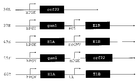
Figure 1: Schematic sections of the expression plasmids used for enhanced
CA 02544462 2006-05-02
WO 2005/042728 PCT/EP2004/052789
-12-
immortalization of primary duck cells (example 2). Polyadenylation signals are
omitted for clarity. The alphanumerics at the left are short identifiers for
the
plasmids. mPGK and hPGK, phosphoglycerate kinase promoters of mouse and
human, resp.; ad5, El-endogenous promoter of Ads; moCMV, mouse CMV
immediate early promoter; tk, herpes simplex virus thymidine kinase promoter;
orf 22 and gaml, CELO virus genes; E1A and E1B, adenovirus 5 El region
genes.
Figure 2: Phase contrast microscopy pictures as example of focus formation in
Ad5-E1 transfected duck embryonal liver cells (plasmid 49E). A, initial
magnification 4 x to depict a complete focus embedded in senescent primary
cells. B, initial magnification 20 x: perimeter of a large round focus of
small cells
arranged in a compact monolayer visible at the right of the panel, primary
cells
in advanced senescence towards the left.
Figure _ 3: Immunofluorescence assay for E1A and E1B 55K proteins (example 3).
Upper two rows, mix of plasmid 49E-immortalized and primary duck liver cells;
bottom two rows, 293 positive control cells. Left column, phase contrast
images;
middle column, immunostaining of E1A or E1B 55K proteins as indicated in the
images; right column, DAPI stain. The E1B 55K protein characteristically
localizes to the cytoplasm and accumulates in aggregates to yield an uneven,
spotty distribution. E1A is a nuclear protein. Note the compacted nuclei that
stain brightly with DAPI in the transformed duck cells.
Figure 4: Q-PERT assay (quantitative PERTassay) on cell supernatant for
detection of retroviral activity (example 4). Bold squares, CHO positive
control;
open squares, water negative control; bold diamonds, chicken embryonic
fibroblasts; bold triangles, 293 cell line negative control; grey circles,
substrate-
only negative control; open triangles, duck liver cells immortalized with
plasmid
49E; delta Rn, emission of the reporter dye over starting background
fluorescence.
Figure 5: MVA amplification on some of the described duck cell lines and CEFp
(example 5). Infection was performed with an MOI of 0.1. Titration was
performed on VERO cells 48 hours after infection (Example 2). CEFp, primary
CA 02544462 2006-05-02
WO 2005/042728 PCT/EP2004/052789
-13-
chicken embryonic fibroblasts.
Figure 6: serial passaging of MVA on duck retina cells immortalized with
plasmid
49E (example 5). Bold squares, burst size; bars, input virus adjusted to an
MOI
of 0.1. Input virus is given as reference to demonstrate that burst size is
independent of experimental fluctuations in cell numbers (which in turn define
input virus via MOI).
Sequence Listing - Free Text
SEQ ID NO: Description - free text
1 Primer VS182
2 Primer VS183
3 Primer VS184
4 Primer VS185
Primer VintSA-F
6 Primer VintSA-R
7 Plasmid pEFAd5E1A
8 Plasmid pEFAd5E1BSA
9 Plasmid 49E
Plasmid 25F
11 Primer V206
12 Primer V207
13 Primer V208
14 Primer V209
RT primer
16 Primer cDNA 1
17 Primer cDNA 2
18 Plasmid 60E
19 Plasmid 36E
Detailed Description of the Invention
"Immortalized", "immortalized cells" and "immortalized cell line" according to
CA 02544462 2006-05-02
WO 2005/042728 PCT/EP2004/052789
-14-
the present invention relates to a cell or cell line which has been
transfected/transformed by certain functional DNA sequences conferring the
potential for at least 200 passages, preferably unlimited number of passages,
i.e. immortality, to the respective starting cells.
A "gene cassette" of the present invention is to be understood as a DNA
sequence comprising a gene affecting the function of the retinoblasoma
protein,
i.e. which directly or indirectly (e.g. after expression) mediates the
disruption of
complexes between retinoblastoma proteins and E2F transcription factors, and
which in addition comprises a viral gene preventing induction of growth arrest
and apoptosis by p53 such as the adenovirus E1B 55K protein of all groups, the
E6 protein of papillomaviruses, preferably those of the low-risk human
papillomaviruses (HPV) (such as HPV1, HPV6 and HPV11, but not HPV16,
HPV18), or a cellular gene preventing growth arrest and apoptosis by p53 such
as mdm2.
In more detail, the above gene cassette comprises a "first gene" which in a
preferred aspect of (1) directly or indirectly (e.g. via cellular inducers)
mediates
the disruption of complexes between retinoblastoma proteins and E2F
transcription factors. This first gene may be a viral gene such as a
mastadenovirus E1A, gaml and orf22 of CELO or E7 of papillomaviruses,
preferably of the low-risk human papillomaviruses (such as HPV1, HPV6 and
HPV11, but not HPV16, HPV18), or a cellular gene such as a constitutively
active
CDK4 or an over-expressed D type cycline. The activity of the first gene
mediates cell cycle progression usually at the cost of induction of apoptosis
or
growth arrest with increased passaging.
A "second gene" is present in above gene cassette to counter this effect of
the
first gene. It prevents apoptosis or growth arrest and preferably acts by
inhibiting transcriptional activation by p53 via augmenting the degradation of
p53 or converting p53 from a trans-activator to a repressor of transcription.
Preferably the "second gene" is capable of preventing transcriptional
activation
by p53, including repression of the function of p53 and causing a decrease in
stability of p53. The "second gene" may be a viral gene such as the adenovirus
E1B 55K protein of all groups, orf22 of CELO, the E6 protein of
papillomaviruses,
CA 02544462 2006-05-02
WO 2005/042728 PCT/EP2004/052789
-15-
preferably of the low-risk human papiIlomaviruses (such as HPV1, HPV6 and
HPV11, but not HPV16, HPV18), or a cellular gene preventing growth arrest and
it
apoptosis by p53 such as mdm2. Preferably the "second gene" is orf22 of CELO
or adenovirus E1B 55k.
This is exactly opposite to the introduction of exogenous active wild type p53
which was associated with the generation of a chicken fibroblast line by an
unknown mechanism (US 5,879,924).
"Biologicals" in the context of present invention comprises therapeutic and
recombinant proteins, including antibodies, enzymes, hormones, receptors or
their ligands and fusions thereof. Prefererred biologicals are recombinant
proteins.
One preferred aspect of embodiment (1) is the use of a cell line derived from
embryonic or hatched chicken, duck, goose, quail or the like, preferably from
chicken or duck. In an especially preferred aspect of (1), additionally this
cell
line is free of reverse transcriptase activity, derived from immortalization
of a
primary cell originating from chicken embryos, hatched chicken, duck embryos
or hatched ducks, is derived from extraembryonic membrane and/or is
cultivated in a chemically defined medium. The medium is preferably free of
animal serum.
Another preferred aspect of embodiment (1) is that the cells subjected to
immortalization are primary cells including fibroblasts, cells from isolated
body
segments (somites) or separated individual organs including neuronal, brain,
retina, kidney, liver, heart, muscle and extraembryonic tissues and membranes
protecting the embryo. Most preferably, the cells are from extraembyonic
membranes or retina.
The immortalization leading to the cells of embodiment (1) is preferably
effected
by non-viral transfection, including, but not limited to, transfection
mediated by
liposomes, dendrimers or hydroxyapatite ("calcium phosphate") precipitates and
electroporation.
CA 02544462 2006-05-02
WO 2005/042728 PCT/EP2004/052789
-16-
Preferably, the first gene in embodiment (1) is a viral gene mediating
disruption
of complexes between retinoblastoma proteins and E2F transcription factors.
This includes, but is not limited to, an adenovirus E1A gene from
mastadenoviruses (preferably from mastadenoviruses of group C), an E7 protein
of papillomaviruses, preferably from low-risk human papilloma virus (HPV)
(such
as HPV1, HPV6 and HPV11, but not HPV16, HPV18), an orf 22 gene of avian
adenoviruses and/or E43 open reading frames from ovine attadenovirus.
Alternatively, the first gene of embodiment (1) is a cellular gene mediating
disruption of complexes between retinoblastoma proteins and E2F transcription
factors. This includes, but is not limited to, cyclin D1, cyclin D2, cyclin D3
and/or
a mutated CDK4 not susceptible to inactivation by p16INK4a.
The second gene of embodiment (1) is preferably a viral gene coding for a
protein preventing induction of growth arrest and apoptosis by p53. This
includes, but is not limited to, genes coding for the adenovirus E1B55K
protein
of all groups, GAM-1 of CELO, the E6 protein of papillomaviruses, preferably
those of the low-risk HPV (such as HPV1, HPV6 and HPV11, but not HPV16,
HPV18). Most preferred are genes coding for the adenovirus E1B55K protein and
GAM-1 of CELO. Alternatively, the second gene encodes a cellular protein
preventing growth arrest and apoptosis by p53 such as mdm2.
The first gene and second gene of embodiment (1) are preferably either
separated spatially by heterologous sequences or located on different nucleic
acid segments or plasmids.
In an especially preferred aspect of embodiment (1) the first gene is the E1A
and the second gene is the E1B region of an adenovirus from the genus
Mastadenovirus, preferably from adenovirus 5. Most preferably said E1A regions
have the sequence of bp 1193 to 2309, preferably bp 1239 to 2309, of SEQ ID
NO:7 or the sequence complementary to bp 4230 to 3113 of SEQ ID NO:9.
Furthermore most preferably said E1B regions have the sequence of bp 1145 to
3007, preferably bp 1197 to 2810, of SEQ ID NO:8 or the sequence
complementary to bp 2345 to 550 of SEQ ID NO:9.
In a further especially preferred aspect of embodiment (1) the first gene is
orf22
CA 02544462 2006-05-02
WO 2005/042728 PCT/EP2004/052789
-17-
and the second gene is GAM-1 from an adenovirus, preferably from the genus
aviadenovirus CELO, which preferably have the sequence represented by the
sequence complementary to bp 1252 to 635 of SEQ ID NO:10, and the sequence
complementary to bp 3138 to 2290 of SEQ ID NO:10.
In even a further especially preferred aspect of embodiment (1) and (2) the
plasmids 36E (SEQ ID NO:19), 37E (figure 1), 49E (SEQ ID NO:9), 25F (SEQ ID
NO: 10) or 60E (SEQ ID NO:18) are used for immortalization of the cells.
Furthermore, combinations of nucleic acids encoding E1A and/or E1B with GAM-
1 and/or Orf22 as defined above are preferred aspects of embodiment (1).
The cell line according to embodiment (1) may additionally carry non-natural
functional sequences including, but not limited to, transgenes such as genes
complementing deficient viruses (e.g. EBNA1, etc.), promoters (e.g. PGK-,
EF1.alpha-, CMV-promoter, El-promoters of Ad5, tk-promoter etc.), enhancers
(e.g. RSV-LTR), selection markers such as neomycin-resistance, puromycin-
resistance, etc.. In one preferred aspect the first and second gene are under
the
control of separate promoters selected independently from PGK-, CMV-, El- and
tk-promoters.
The cell line according to embodiment (1) is in one preferred aspect further-
more suitable for production of biologicals or viruses including vaccine
strains
(Marek's disease, infectious bursa] disease, Newcastle disease, turkey herpes,
chicken anemia, influenza, vaccinia (MVA), rubella, rabies viruses, etc.) and
recombinant viral vectors (e.g. recombinant MVA or alphaviruses). Most
preferred viruses for vaccination are MVA and influenza viruses. The most
preferred recombinant viral vector is MVA.
In one aspect of embodiment (1) the cell line is cell line 12A07-A10 (DSM
ACC2695) derived from immortalization of duck extraembryonal membrane cells
with plasmid 49E (example 2).
Furthermore preferred is the generation of the cell lines according to
embodiment (1) under cGMP conditions which renders them suitable for
CA 02544462 2011-01-11
WO 2005/042728 PCT/EP2004/052789
-18-
pharmaceutical application.
The method of embodiment (2) preferably comprises non-viral transfection of
the starting cell such as listed above. Most preferred is liposomal
transfection,
especially transfection by the EffecteneTM reagent.
A preferred use according to embodiment (3) is the use for the preparation of
a
vaccine or for gene therapy. A viral vaccine strain or gene therapy vector is
brought into contact with cells of a cell line according to embodiment (1) so
that
infection occurs and the virus is amplified by said cells. Continued passaging
of
virus (repeated cycles of infection and harvest of virus on said cells) will
lead to
attenuation or adaptation of virus to this particular host cell line. Thus, a
viral
vector or vaccine strain with lesser virulence for the intended vaccinee
(which is
not duck, preferably not avian) is generated. Attenuated viruses allow the
immune system of the vaccinee to launch a response that is more protective
than vaccination with fully inactivated particles, and that is less severe
than
infection with a wildtype (natural) pathogen. The preferred viruses for this
embodiment are measles and rabies viruses.
The method for producing viruses according to embodiment (4) preferably
comprises the contacting of said viruses with a cell line according to embodi-
ment (1) and/or the cultivation of said viruses on said cell line. Especially,
this
method can be used for producing a pox virus, preferably strain MVA, in a duck
cell line, preferably a cell line originating from duck somites or duck
neuronal
tissue, even more preferred from duck retina. Especially duck retina and
somite-
derived cells obtained by transfection of AdS-El region under cGMP conditions
stably support amplification of MVA with an efficiency comparable to or better
than primary chicken embryonic fibroblasts (Example 5).
The method for producing biologicals, especially recombinant proteins,
according
to embodiment (4) comprises the introduction of a gene coding for a
recombinant protein, operably linked to a promoter into a cell line according
to
embodiment (1), cultivating said modified cell line and harvesting the
recombinant protein.
CA 02544462 2006-05-02
WO 2005/042728 PCT/EP2004/052789
-19-
The method of embodiment (4) is used preferably for the production of viruses
and biologicals usable for vaccination or gene therapy.
Historically, chicken eggs and the respective cells (chicken fibroblasts) are
the
dominating substrate for the manufacturing of vaccines. For pharmaceutical
purposes chicken are available from pathogen-controlled environments with an
extensive monitoring system. A large body of literature suggests chicken eggs
as the primary target for cell line development. Therefore, chicken cells are
one
preferred source for starting cells of the invention. However, chicken-derived
cells and cell lines will be most likely RT positive. Literature data suggest
a low
risk for release of infectious virus. However, the absence of transmissible
virus
will have to be monitored for any cell line to be used in manufacturing.
Indeed,
most of the avian cell lines established so far are originating from chicken
(US
5,830,723, US 5,879,924). Although it was possible to breed a chicken lineage
(line 0) free of avian leucosis virus, endogenous avian retroviruses (EAV-HP)
(Boyce-Jacino et al., J. Virol 66(8):4919-29 (1992)) are present in chicken
cells
including line 0. EAVs provide an active reverse transcriptase, but expression
levels vary substantially. Therefore, even primary chicken cells and cell
lines
such as DF1 that tested RT negative in less sensitive assays (Crittenden et
al.,
Virology 57(1):128-38 (1974)) presumably will test positive in modern real
time
PCR approaches and may harbor retroviruses that are activated under certain
growth conditions.
Alternatively preferred avian species of this invention for cell line
development
are those which do not contain endogenous retroviruses or express reverse
transcriptase (RT). This includes ducks, which are suitable for two additional
reasons: Duck eggs are also available from pathogen free monitored stocks and
ducks are, in contrast to geese, less likely to develop spontaneous tumors.
While
it is known that many of the relevant vaccine strains replicate well in duck
(embryonal) cells as they do in chicken (embryonal) cells (e.g. Marek's
disease
virus (Witter, R.L., Avian Dis. 46:925-37 (2002)) or rubella (Rocchi, G.,
Salvadori, A., Nuovi Ann. Ig Microbiol. 21:336-40 (1970))), this remains to be
shown for virus strains of primary interest. For other vaccines such data is
not
available.
CA 02544462 2006-05-02
WO 2005/042728 PCT/EP2004/052789
-20-
To our knowledge it is a novel and unexpected finding of this invention that
the
highly attenuated pox virus strain MVA (modified vaccinia Ankara) replicates
in
duck cell lines at similar or higher efficiencies than in commonly used
primary
chicken embryonic fibroblasts. One intention of the inventors was to provide a
safe and robust alternative to primary cells for amplification of viruses that
require an avian host, or vaccine strains where a non-mammalian host is
preferred. An important virus for which convenient host cells are not
available is
MVA (modified vaccinia virus Ankara). MVA is a highly attenuated pox virus and
an extremely promising tool for therapeutic and protective vaccine
applications.
MVA will serve as a model virus for characterization of duck cells but should
not
be taken as an exclusive example: the described experiments can also be
performed with a range of other viruses, whether pathogens or therapeutic
vectors, such as measles, rubella, rabies, or influenza viruses.
Fibroblasts have been selected as the preferred cell type mainly for historic
and
practical reasons. Fibroblasts are the fastest growing primary cells from
mammalian as well as avian species. When a cell suspension from whole chicken
embryos is brought into culture, this is not the only but the predominant cell
type. However, fibroblasts grow strongly adherent and loose this feature only
after complete (tumorigenic) transformation. This process requires the
presence
of strong transforming genes such as v-ras interfering with signal
transduction
pathways. Early senescence of fibroblast cultures is in part caused by the
total
absence of telomerase activity in birds and man (Forsyth, N. R. et al.,
Differentiation 69 (4-5):188-97 (2002)).
Human primary fibroblasts are refractory to transformation with the El genes
of
adenovirus type 5 which do not directly interfere with these pathways
(personal
observation). Efficient immortalization and growth in suspension culture has a
higher chance to succeed for epithelial and neuronal cells. Moreover,
epithelia
instead of fibroblasts seem to be the primary site for virus replication
inside the
bird egg. Interestingly, in contrast to the human situation, bird kidney does
express telomerase throughout life which makes bird kidney cells a good target
for immortalization. Taken together, bird epithelial cells including kidney
epithelium and neuronal cells are considered the most promising targets to
develop a cell line of the required features.
CA 02544462 2006-05-02
WO 2005/042728 PCT/EP2004/052789
-21-
It is therefore only for the ease with which fibroblasts are obtained that
avian
cell line development has almost exclusively focused on these cells (Cowen, B.
S., Braune, M.O., Avian Dis 32(2):282-97 (1988); US 5,830,723). In some
cases whole embryos have been used (US 2001-0016348).
Viruses do not only exhibit species but also organ and tissue specificity
based on
receptor distribution and cellular factors supporting replication. Therefore,
in
contrast to the typical approach, a preferred way to perform present invention
is
the separation of organs prior to cultivation to obtain a most preferred host
cell.
For influenza virus, whose vaccine-adequate production is a major application
for the cell lines of present invention, the typical site of replication is
not the
embryo itself but extraembyonic membranes. Therefore, a specific aim was to
also develop cell lines from extraembryonic material, including protective
membranes of the embryo. Some tissue specific primary cultures including those
of the extraembryonic membranes have very short survival times compared to
fibroblasts. This further highlights the need for designed immortalization to
obtain optimized host cells. Successful immortalisation of multiple tissues in
a
limited time window requires the specific combination of genes used within
present invention.
It was not known which of the avian tissues has the highest replicative
potential
for pox viruses such as MVA or Canarypox. The typical manufacturing process
for MVA involves a mixture of cells from an embryo excluding the head which is
removed prior to disintegration. It is therefore completely unexpected that a
cell
line of neuronal origin, developed from the retina, has such a high capacitiy
for
MVA replication whereas other tissues have not.
The .same tissue specificity applies to protein production. The
transcriptional
capacity is dependent on the available set of transcription factors and even
strong ubiquitous viral and cellular promoters exhibit variable strength in
different tissues. Moreover, yields of secreted protein strongly depend on the
capability of a particular cell type to fold and process (e.g. glycosylate)
the
protein properly.
CA 02544462 2006-05-02
WO 2005/042728 PCT/EP2004/052789
-22-
The mechanisms leading to immortalization and transformation of primary cells
have been well described (Hahn, W.C. et al., Nature 400:464-8 (1999)).
Required elements interfere with (1) control of cell cycle progression, (2)
programmed cell death induced by the deregulated cell cycle, (3) growth factor
signal transduction and for human and avian cells (4) shortening of the
telomeres, the linear termini of the chromosomes. A large number of factors
are
known that can drive primary cells to an immortalized and transformed
phenotype but immortalization comes at the cost of inhibiting cellular
checkpoints that are responsible to minimize tumor formation in the host. It
is
therefore desired to select transforming factors that can effect experimental
generation of a cell line but pose a minimal risk of tumor induction in the
recipients of biologicals derived from the designer cells. This requirement
needs
to be balanced with the strength of the transforming factors: they should be
strong enough to cause transformation without the need for accumulation of
additional spontaneous mutations; that is, the molecular pathway leading to
the
resulting cell line should be known completely (categories I and II according
to
the FDA CBER Office of Vaccine's presentations at the May 2000 Advisory
Committee). It is furthermore desired to select a synergistic combination of
factors that individually cannot transform primary cells so that a concurrent
transfer of genetic material is required which further minimizes the risk of
inadvertent transformation in vaccinees or patients. Finally, it is desired
that the
transforming factor elicits an immune response in the recipient of biologicals
so
that immune tumor surveillance is activated in the unlikely event of tumor
formation due to product application. The last criterion can be realized if
non-
cellular but foreign, for example viral, transforming proteins are utilized.
It was now found that the El region from human adenovirus 5 (Ad5) is ideally
suited to transform avian cells so that the resulting designer cell complies
with
all of the above criteria.
The E1B region encodes two open reading frames on a bicistronic mRNA, the
21K and 55K proteins. The 55K protein binds to p53 and thus turns the pro-
apoptotic transcriptional activator into a repressor. The 21K protein comple-
ments this anti-apoptotic activity by binding to Bax, thus maintaining
integrity of
CA 02544462 2006-05-02
WO 2005/042728 PCT/EP2004/052789
-23-
the mitochondrial membrane and preventing the release of cytochrome C. This
protein is essential to drive adherent cells towards substrate independent
growth
and hence is essential to a fermentation process in suspension.
It has not been shown before whether human adenovirus E1B 55K can affect the
avian homologues of p53. Furthermore, the avian adenoviruses are not
equipped with genes resembling E1B so that inference also was not possible.
Contrary to all expectations, the inventors have found that E1B can provide
the
essential functions to allow immortalization by E1A.
A novel and crucial factor for the here described achievement was removal of
E1B from its weak natural context and placement under control of a strong,
recombinant promoter. This novel modification and combination allowed
efficient
immortalization of multiple tissues from duck and chicken by transfection
instead of retroviral transduction.
Although the underlying mechanism for transformation by El is complex one
hallmark is a most desirable feature: E1A is a strong inducer of cell
proliferation
and apoptosis whereas E1B proteins efficiently interfere with apoptosis but
cannot release restriction on cell cycle control.
Hence, not a single factor but the continuous presence of E1A and E1B proteins
are required to sustain the experimentally induced transformed phenotype.
Since the description of v-src in the 1970s (Brugge, J.S., Erikson, R.L.,
Nature
269:346-8 (1977)) a panoply of transforming factors have been discovered and
characterized. Indeed, it was the study of induction of tumors in birds by
alpharetroviruses that provided first molecular insights (Martin, G.S., Nature
227:1021-3 (1970)). The retroviral oncogenes are derived from cellular genes
with essential regulator domains mutated or deleted. Some of the factors that
have been identified in the course of these studies, such as v-myc or v-ras,
directly affect components of the RB and p53 pathways. Other proteins, such as
v-src or v-erbB, are constitutively activated (hence, dysregulated) signal
transducers that mimic impinging extracellular mitogens. The problem with
these
factors is that they target only one of several pathways required for
efficient
CA 02544462 2006-05-02
WO 2005/042728 PCT/EP2004/052789
-24-
transformation. The presence of v-src or v-myc predisposes the cell for
transformation and requires additional, spontaneous and unpredictable
alterations within the cell for full transformation. The risks for the patient
posed
by cells transformed with one of the retroviral oncogenes therefore is
difficult to
estimate.
Other DNA viruses such as papiIlomaviruses and polyomaviruses are also known
to transform cells in vitro. However, the selected transgenes should not be
too
aggressive to minimize the risk of tumor induction in the recipients of
biologicals
via inadvertently transferred cellular DNA. This criterion is especially
stringent for
vaccine production where a healthy population often is inoculated at a very
young age. Even with sophisticated modern purification methods polyomavirus
Large-T antigen is oonsidered too aggressive for use in cell lines generated
for
application in human medicine. Whereas 90% of cervix carcinomas carry
papillomavirus sequences (Munoz, N. et al., N. Engl., J. Med. 34816):518-27
(2003)) C-type adenoviruses (which include type 2 and type 5) are not
considered to induce tumors in vivo and adenoviral have not been detected in
human tumor tissue.
Based on the complementary features of the transforming genes shown above, it
was found that a combination of genes each interfering with single pathways in
the cell cycle and apoptosis is necessary to obtain a genetically stable cell
line
growing in suspension.
It was shown that the complete El region of adenovirus 5 can fulfill these
requirements. Whereas it was shown, that the 12S protein of E1A from Ad5 can
interact with avian RB (Guilhot, C. et al., Oncogene 8:619-24 (1993)) the
functional activity of 55K and 21K proteins in avian cells is demonstrated for
the
first time in present invention. It is not surprising that some clones of
quail cells
expressing the 12S protein of E1A exhibit transformed features (Guilhot,. C.
et
al., Oncogene 8:619-24 (1993)). The extremely efficient and stable
transduction
via retrovirus infection creates a large enough cell pool to allow individual
cells to
overcome the cell cycle block or induction of apoptosis by spontaneous genomic
changes. These required but unknown changes increase the medicinal risk and
the resulting cell line can not be considered a designer cell line, which
should be
CA 02544462 2006-05-02
WO 2005/042728 PCT/EP2004/052789
-25-
based on known genes. Moreover, transfection techniques are not sufficient to
create the large clone pool required for natural selection. Instead retrovirus
transduction was required. The transforming gene introduced via this approach
will be flanked by ITRs and can, therefore, be mobilized, even more in a cell
line
expressing reverse transcriptase.
Recently, an avian adenovirus, termed fowl adenovirus type 1 strain CELO (for
chick embryo lethal orphan), has been described in greater detail (Chiocca, S.
et
al., J. Virol. 70:2939-49 (1996)). Large, central genomic stretches of CELO
are
homologous to Ad5 but differ in important aspects - among others, CELO is not
equipped with an El-homologous region. Furthermore, CELO cannot
complement Ad5 mutagenized in E1A and, conversely, Ad5 El proteins cannot
trans-activate transcription of delayed-early CELO genes (Li, P. et al., J.
Gen.
Virol. 65(Pt 10):1817-25 (1984)). And yet, CELO is capable to transform
hamster cells in vitro (May, J.T. et al., Virology 68:483-9 (1975)). Genes
interfering with cell cycle and apoptosis, orf22 and GAM-1, have been
identified
in the CELO virus (Lehrmann, H., Cotton, M., J. Virol. 73:6517-25 (1999)).
orf22 encodes a protein that interact with RB, and GAM-1 interferes with
apoptosis in a fashion similar to the prototypical 21K protein (Chiocca, S. et
al.,
J. Virol. 71:3168-77 (1997)).
It was now found that the genes orf22 and GAM-1 from CELO virus are suitable
substitutes for E1A and E1B. The spectrum of available transgenes for
transformation of avian cells is therewith expanded. These proteins have not
been used previously to transform avian cells.
Furthermore, one of the viral genes may be replaced by a cellular gene.
Candidates for such replacement are E2F family members or D group cyclins for
the E1A region of adenovirus and mdm2 for the E1B region.
The following cell lines were deposited at the DMSZ, Deutsche Sammlung von
Mikroorganismen and Zellkulturen GmbH, Mascheroder Weg 1b, 38124
Braunschweig, Germany:
1. PBG04 as DSM ACC2577, deposited on September 18, 2002;
2. 12A07-A10 as DSM ACC2695, deposited on October 20, 2004.
CA 02544462 2006-05-02
WO 2005/042728 PCT/EP2004/052789
-26-
The invention will be explained in more detail by reference to the following
Examples, which are, however, not to be construed as to limit the invention.
Examples
Example 1: Immortalization of primary duck cells with Adenovirus 5 E1A,B
The adenovirus sequences for E1A and E1B were amplified from the culture of
passage 8 of the first generation (El deleted) adenovirus Admuc grown in HEK
293 which was heavily contaminated with wild type virus using provestart
polymerase (Qiagen).
The following primers were used:
VS182 ACTCGAGCTGACGTGTAGTGTATT (SEQ ID NO:1)
VS183 CACACGCAATCACAGGTT (SEQ ID NO:2)
to amplify the El A region and
VS184 ACTCGAGTCATGGAGGCTTGGGAGT (SEQ ID NO:3)
VS185 ACACATTTCAGTACCTCA (SEQ ID NO:4)
to amplify the El B region. Both fragments were first cloned into
pPCR4blunttopo
(Invitrogene).
The E1B construct misses the splice acceptor from the E1B message. It was
therefore replaced by a synthetic one amplified using primers from the leader
intron of a human immunoglobulin heavy chain. As template, the genomic DNA
from PBG04 (DMSZ ACC2577), a murine-human heterohybridoma was used.
Primers:
VintSA-F AAGGTACCCTCCCTAGTCCCAGTGA (SEQ ID NO:5)
VintSA-R CAATGTACAGAGTG GGCTCCTGTGG (SEQ ID NO:6)
This splice acceptor was directly cloned into pEFmyc, containing aEF1 alpha
promoter and the myc leader peptide to create fusion proteins. The E1A region
was removed from ptopoElA using EcoR I and Xho I sites and cloned into
CA 02544462 2011-01-11
WO 2005/042728 PCT/EP2004/052789
-27-
pEFmyc directly, removing the myc leader sequence and fusing the E1A to the
bovine growth hormone poly A. The E1B region was again removed with EcoR I
and Xho I restriction enzymes and cloned into pEFmycSA containing the
heterologous splice acceptor site. The resulting plasmids were named pEFAd5E1A
(SEQ ID NO:7) and pEFAd5E1BSA (SEQ ID NO:8).
Embryonated duck eggs were incubated at 37 C, 60% air humidity, for 12 days
(older embryos yielded more cells but also contained a higher number of
contaminating, differentiated fibroblasts). The shell was sterilized with 70%
isopropanol, opened at the large end, and the embryo was removed aseptically
to a sterile petri dish. The fetal brain and kidneys were removed, transferred
to
separate petri dishes filled with trypsin/EDTA and minced. After a brief
incubation
a suspension thereof was mixed with an excess of F12 medium
(Gibco/Invitrogen) supplemented with 10% fetal calf serum (Biochrom) and 2%
UltroserTM G (Ciphergen). This suspension was transferred into a petri dish
and
cultivation was performed at 37 C (which is lower than the 41.6 C
physiological
temperature of chicken) and 5% CO2. The culture medium with non-adherent
debris was replaced the following day and cultivation continued until at least
5 x
105 cells per 3.5 cm dishes were available for transfection of plasmids
pEFAd5E1A and pEFAd5E1BSA.
Initial experiments comparing liposomal (Effectene; Qiagen) and dendromeric
(Polyfect; Qiagen) transfection reagents suggested best efficiencies with
Effectene. Transfection there was performed using the Effectene reagent;
briefly:
2 pg of plasmid DNA was diluted in 200 pl EC Buffer containing 16 pl Enhancer.
After an incubation time of 5 min, 16 pl Effectene was added. After an
incubation
time of 10 min, supernatant was removed from the culture in 3.5 cm dishes and
replaced with 1 ml fresh medium containing the transfection mix. After an
incubation time of 2 hours at 37 C and 5% C02, additional 2.5 ml fresh medium
was added to the culture.
The transfected cells were allowed to reach confluency, trypsinated,
resuspended
in FCS/Ultroser G-supplemented F12 medium, and re-seeded into two 6 well
plates (corresponding to a 12-fold expansion). After 5 and 10 days, the medium
was replaced with F12 supplemented only with 5% FCS. The plates were scanned
CA 02544462 2006-05-02
WO 2005/042728 PCT/EP2004/052789
-28-
for the appearance of foci of cells with changed morphology (decrease in
overall
cell size, increased size of nucleus, increased visibility of plasma membranes
under phase contrast) and increased confluency.
Approximately 14 days post transfection, once the foci reached a diameter of 1-
3
mm the medium was aspirated and the culture washed twice with trypsin/EDTA
(Gibco). Trypsin-soaked cloning disks (Sigma) were placed on top of the
aspirated foci for 3 min, then transferred into wells of a 24-well plate
filled with
500 pi of F12 medium supplemented with 5% FCS.
The cloned, transformed cells were allowed to proliferate until confluency,
trypsinized, resuspended in F12 medium supplemented with 5% FCS and
transferred into 6-well plates. Once the culture reached confluency in the 6-
well
plate the cells were transferred to T25 flasks for continuous passaging.
For cryopreservation at defined intervals cells were trypsinized, resuspended
in
F12 medium containing 5% FCS, collected by centrifugation at 100 g for 10 min,
resuspended in F12 medium containing 50% FCS and 10% DMSO (Sigma) to a
concentration of approximately 3 x 10⁶ cells per ml, and placed in cryovials
in an
isopropanol-based cooling device at -75 C. The cooling device ensures a
constant cooling rate of 1 C per min. After 24 hours the cells were
transferred to
liquid nitrogen for permanent storage.
Example 2: Improved preparation of immortalized avian cell lines
a) Preparation of primary cells
The flock of origin for the duck eggs was certified to be free of Salmonella
enteritidis and S. typhimurium; Mycoplasma gallisepticum and M. synoviae;
cases of leucosis, reticulo-endotheliosis, psittacosis, avian influenza, duck
hepatitis, and Derzsy's disease. The animals intentionally were not vaccinated
against parvovirus and no cases of parvovirosis were detected. Animals in the
flock of origin have been vaccinated against S. enteritidis and S.
typhimurium;
Pasteurella multicodica; the metapneumovirus Turkey rhinotracheitis; and the
paramyxovirus causing Newcastle disease.
The eggs were allowed to equilibrate without agitation at room temperature and
CA 02544462 2006-05-02
WO 2005/042728 PCT/EP2004/052789
-29-
after two days were incubated at 38 C in a damp chamber, rotated frequently
by alternating +45 and -45 .
Duck embryos were sacrificed for isolation of primary cells after one or three
weeks of incubation. Eggs were transferred to a cGMP unit (a closed laboratory
performing as outlined by the Current Good Manufacturing Practices) and the
shell was sterilized by wiping with 70% isopropanol under a bminar flow hood.
All subsequent steps were performed in the GMP unit under sterile conditions
with defined solutions or media.
Eggs were opened carefully, embryos transfered to a large petri dish and
killed
immediately by decapitation. Samples from the following organs were removed:
brain, retina, liver, esophagus, heart, and extra-embryonic membranes.
In addition, cells from somites were prepared from an 8-day-old embryo.
All samples were rinsed with PBS (phosphate buffered saline; Gibco/Invitrogen,
USA), treated with trypsin (Gibco/Invitrogen, USA) for 1 to 10 min, and
triturated in DMEM:F12 culture medium (Gibco/Invitrogen, USA) supplemented
with 10% FCS (Biochrom AG, Germany) by repeated passaging through an 18G
syringe. The homogenized samples were cultivated at 37 C and 5% CO2. Debris
was removed from adherent cells by change of medium the following day.
b) Plasmid Constructions
Expression plasmids for E1A, E1B, 0rf22, and Gam1 were constructed by
extraction of the relevant target regions from the genomic DNA of adenovirus
serotype 5 or chicken embryo lethal orphan (CELO) wildtype virus,
respectively,
by PCR and insertion into vectors equipped with human or mouse
phosphoglycerate kinase (hPGK or mPGK), mouse CMV (moCMV) or tk
promoters (figure 1).
The adenovirus sequences for E1A and E1B were amplified from wild type virus
using ProofStart polymerase (Qiagen, Germany). The following primers were
used:
CA 02544462 2006-05-02
WO 2005/042728 PCT/EP2004/052789
-30-
VS182 ACTCGAGCTGACGTGTAGTGTATT (SEQ ID NO:1)
VS183 CACACGCAATCACAGGTT (SEQ ID NO:2)
to amplify the El A region and
VS184 ACTCGAGTCATGGAGGCTTGGGAGT (SEQ ID NO:3)
VS185 ACACATTTCAGTACCTCA (SEQ ID NO:4)
to amplify the El B region. Both fragments were first cloned into pPCR4-Blunt-
TOPO (Invitrogene, USA).
The E1B construct misses the splice acceptor from the E1B message. It was
therefore replaced by a synthetic one amplified using primers from the leader
intron of a human immunoglobulin heavy chain. As template, the genomic DNA
from PBG04 (DMSZ ACC2577), a murine-human hetero-hybridoma was used.
Primers used for amplification:
VintSA-F AAGGTACCCTCCCTAGTCCCAGTGA (SEQ ID NO: 5)
VintSA-R CAATGTACAGAGTGGGCTCCTGTGG (SEQ ID NO:6)
The genes GAM-1 and ORF-22 were amplified from wild type CELO virus with
primers
V206 AAC CTC GAG ACC CCC CTG TAC ATT CTA (SEQ ID NO: 11)
and V207 GCC GTT AAC TTC AGG GAT TGG TTA CAG (SEQ ID NO: 12), and
V208 CAC CTC GAG TCC GGA TTA AGA TGA ACG (SEQ ID NO:13)
and V209 CCA GTT AAC AGG TGA ACC ATT TAT ACA G (SEQ ID NO:14),
respectively.
Representative examples for the resulting plasmids are given with plasmid 49E
(adenoviral factors under control of human PGK and mouse CMV promoters;
SEQ ID NO:9), plasmid 25F (CELO factors under control of mouse and human
PGK promoters; SEQ ID NO:10), plasmid 60E (adenoviral factors under control
CA 02544462 2006-05-02
WO 2005/042728 PCT/EP2004/052789
-31-
of human PGK and tk promoters; SEQ ID NO:18) and plasmid 36E (CELO factor
under control of mouse PGK promoter; SEQ ID NO:19) (see also Figure 1).
Integrity of the expression plasmids was confirmed by sequencing. The plasmids
are not equipped to express resistance factors against antibiotics (such as
ampicillin) in eukaryotic cells.
c) Transfection
Primary cultures were transfected with expression plasmids for El or
0rf22/Gam1 shortly after isolation or after single subcultivation. Depending
on
the Experiment, plasmids were transfected as supercoils or after linearization
with the Sca I (New Englands Biolabs, USA) restriction enzyme. Initial
experiments comparing liposomal (Effectene; Qiagen, Germany) and
dendromeric (Polyfect; Qiagen, Germany) transfection reagents suggested best
efficiencies with Effectene. Transfection was performed as follows: 2 pg total
DNA was diluted into 200 pl provided EC buffer and mixed with 16 pl provided
enhancer. After an incubation for 2-5 min at room temperature 20 pl Effectene
reagent was added. After 5-10 min at room temperature this mixture was
applied to the cells in a 8 cm2 dish under 1 ml culture medium. After 2-5
hours
an additional 1.5 ml culture medium was added. On the following day, the
medium was replaced with 2 ml fresh culture medium, and thereafter once per
week. Successful transfection was confirmed in parallel experiments with a
reporter gene.
The cells were continously passaged in DMEM:F12 medium containing 10% FCS.
Twenty days after transfection changes of morphology in defined subpopulations
(foci; figure 2) of some cultures were observed; in other cultures foci did
not
appear or were not able to compete with robust proliferation of the primary
cells; again other cultures suffered massive cell death and senescence shortly
after transfection.
A large number of independent foci were expanded from plasmid 49E-
transfected cultures with cells derived from liver, retina and extra-embryonic
membrane. At passage 10, e.g., cell line 12A07-A10 derived from duck
CA 02544462 2006-05-02
WO 2005/042728 PCT/EP2004/052789
-32-
extraembryonal membrane cells transformed with plasmid 49E was isolated and
deposited at the DSMZ.
Foci were also obtained from plasmid 60E-transfected cultures with cells from
retina and somites.
In plasmid 49E, PGK and mouse CMV promoters drive expression of E1A and
E1B, respectively. Plasmid 60E (SEQ ID NO:18) also encodes the full Ad5-E1
region but expression of the protective E1B region is driven by tk, i.e. a
promoter that is not as strong as the mouse CMV promoter (but stronger than
the native E1B promoter). Consistent with the protective effect conferred by
E1B
far fewer foci in fewer cell samples were obtained with this construct when
compared to the results with plasmid 49E.
Formation of foci with both primary cell appearance and transformed phenotype
was also observed in cultures of liver transfected with CELO plasmids 36E (SEQ
ID NO:19) and 25F (SEQ ID NO:10).
Cultures with foci were expanded by treatment with trypsin for 2-3 min and
resuspension in DMEM:F12 medium for transfer to fresh culture vessels.
For cryopreservation at regular intervals cells were removed with trypsin,
resuspended in DMEM:F12 medium containing 10% FCS, collected by
centrifugation at 200 x g for 10 min, resuspended in DMEM:F12 medium
containing 50% FCS and 10% DMSO (Sigma, USA) to a concentration of
approximately 3 x 10⁶ cells per ml, and cooled with a rate of 1 C per min to
-80 C. After 24 hours, the cells were transferred to liquid nitrogen for
permanent storage.
Example 3: Immunofluorescence assay for stable transfection
Cultures of potentially immortalized cells were seeded on glass slides and
allowed to proliferate for several days before fixation with ice-cold methanol
for
min. The fixed cells were incubated with antibodies against E1A and E1B 55K
proteins, s--condary antibodies, and fluorescent dye specific against the
latter
according to standard immunofluorescene methods (Becton Dickinson, UK,
CA 02544462 2006-05-02
WO 2005/042728 PCT/EP2004/052789
-33-
#554155 antibody against E1A, diluted 1:30; Oncogene, USA, #DP08-1000G
antibody against E1B 55K, diluted 1:30; secondary antibody directed against
mouse or rat, respectively, and conjugated to biotin, both from Jackson Immuno
Research, USA, diluted 1:80; visualization with Jackson Immuno Research, USA,
#016-070-084 streptavidin-Texas Red conjugate, diluted 1:100). Primary cells
still abundant in early, not yet fully established immortalized cell lines and
readily distinguishable by morphology provided a convenient internal negative
control for antibody specificity. 293 cells (human embryonic kidney cells)
that
stably express the Ads El-region served as positive control. DAPI (4',6-
diamidino-2-phenylindol; Sigma, USA) to 1 pg/ml was added in the final
incubation step to stain the nuclei of the cells for orientation purposes.
A strong signal for E1A and 55K was observed only in cells that underwent
characteristic changes in morphology confirming successful immortalization by
the transfected plasmids (figure 3). Furthermore, spontaneous transformation,
a
formal possibility, was not observed as all cells with altered phenotype were
El-
positive. None of the cells with primary phenotype expressed El-proteins.
Although possible in transfections of supercoils where the linearization of
plasmid in the process of integration occurs at random positions none of the
examined foci exhibited E1A expression in absence of E1B expression, further
emphasizing the requirement for dual pathway disruption for immortalization.
Example 4: Assay for endogenous and exogenous retroviruses
A common problem encountered when vaccines are produced in primary chicken
fibroblasts is contamination with exogenous or endogenous retroviruses. The
diversity of the retrovirus family is too complex to predict whether a given
species is a carrier for retroviruses. Reports from the literature therefore
usually
are limited to a subset of the retrovirus family, for example EAV-HP/ALV
subgroup J (Smith, L.M. et al., J. Gen. Virol. 80(ptl):261-8 (1999)), and then
only to a subset of avian species.
A reliable confirmation of contamination with retroviruses therefore should
focus
on a common motif present in these viruses. Sequence diversity precludes
nucleic acid-based detection methods. However, common to all retroviruses is
the presence of the reverse transcriptase enzyme. The supernatant of expanded
CA 02544462 2011-01-11
WO 2005/042728 PCT/EP2004/052789
-34-
foci from duck liver cells immortalized with plasmid 49E was therefore assayed
by quantitative probe-based product enhanced PCR for reverse transcriptase (Q-
PERT) and compared to several controls, inter alia CHO as positive control and
293 cells as negative control (see below and figure 4), to detect both
endogenous retroviral activity or contamination with free retroviruses. The
assay
is a modification from the literature (Lovatt, A. et al., J. Virol. Methods
82(2):
185-200 (1999)). Briefly: retroviruses were enriched from culture supernatant
by ultracentrifugation with 100000 x g through a barrier of 20% sucrose in PBS
to remove cellular debris. Virions (if present) were resuspended into lysis
buffer
(50 mM Tris pH 7.8, 80 mM KCI, 2.5 mM DTT, 0.75 mM EDTA, 0.5% Triton X
1007M) and mixed with substrate buffer (10 mM each of dATP, dCTP, dGTP, and
dTTP; 15 pM specific primer [GCC TTT GAG AGT TAC TCT TTG; SEQ ID NO:15];
and 0.5 mg/ml fragmented herring sperm DNA [Promega Corp, #D1811])
containing a model RNA (5 pg/ml Brome Mosaic Virus RNA [Promega Corp, USA,
#D1541]) that is reverse transcribed if RT activity is present in the sample.
cDNA from the model RNA is amplified by PCR with primers (AAA CAC TGT ACG
GCA CCC GCA TT; SEQ ID NO:16) and (GCC TTT GAG AGT TAC TCT TTG; SEQ
ID NO:17) and detected via SYBR green fluorescence in an AB 7000TM Sequence
Detection System using the QPCR SYBRTM Green ROX Mix #AB-1163 from Abgene,
UK, according to the instructions of the manufacturer.
Figure 4 demonstrates strong RT activity in CHO cells as expected from reports
in the literature (for example, Anderson, K. P. et al., Virology 181(1): 305-
311
(1991)). With these cells as positive control and human 293 cells free of
retroviral activity as negative control a bracket is defined that allows
interpretation of unknown RT activity in the supernatant of cell cultures
(figure
4, bold squares and bold triangles).
We found moderate RT-activity in chicken embryo fibroblasts (figure 4, bold
diamond symbols).
The signal for RT activity in the duck cell supernatant was congruent with the
signal for RT activity in 293 cells, and both again congruent with a control
representing the detection limit for our assay consisting of model RNA not
incubated with RT (figure 4, compare curves with open and bold triangles and
CA 02544462 2006-05-02
WO 2005/042728 PCT/EP2004/052789
-35-
grey circles). Equivalent levels of signal intensity (delta Rn) were separated
by
at least two cycle numbers between samples from CHO cells and chicken
embryo fibroblasts (that for these experiments are derived from a source known
to be only weakly RT-positive) and by at least four cycle numbers between
samples from CHO cells and the 293 negative control and the duck cell culture.
Thus, contrary to chicken cells the described duck cells do not exhibit RT
activity
and thus fulfill an essential attribute for suitability in pharmaceutical
applications.
Example 5: Modified vaccinia virus Ankara (MVA)
Suitability of the expanded foci as substrate for amplification of MVA was
determined for liver, retina, somites and extra-embryonic membrane lines.
Table 1 and figure 5 show results obtained by infection of the cell lines with
an
inoculum prepared from a large scale preparation of MVA (ATCC #VR-1508) on
CEFp, primary chicken embryonic fibroblasts. The data in the table obtained by
infection with an MOI (multiplicity of infection or number of infectious
particles
per host cell) of 0.1 demonstrates that viral output of retina and somite
cells (in
plaque forming units per ml) are comparable to or even exceed the output
obtained with CEFp cells.
MVA yield in pfu/ml
(after 48 h, infection with MOI of 0.1)
CEFp 3.54 x 10
retina 2.06 x 10'
liver 3.20 x 10⁴
somite 4.60 x 10'
membrane 4.03 x 10³
Table 1: Comparison of virus titers obtained in parallel infections of 1 to 5
x 105
cells in cavities of 24-well plates. Input virus was adjusted for an MOI of
0.1.
CEFp, fresh primary chicken embryonic fibroblasts; membrane, extra-embryonic
membrane.
Plaque-forming units for MVA on duck cells were determined as follows: MVA
virus was recovered from infected cells after 48 hours from the supernatant
and
from adherent cells opened by repeated freeze-thawing. VERO (African green
monkey kidney) cells were seeded in 96 well plates (2 x 10⁴ cells per well)
and
CA 02544462 2011-01-11
WO 2005/042728 PCT[EP2004/052789
-36-
infected with serial 10-fold dilutions of MVA-containing suspension on the
following day. Two days thereafter, the cultures were fixed with methanol and
infected cells incubated with polyclonal vaccinia virus antibodies (Quartett,
Germany, #9503-2057, at 1:1000 dilution in PBS containing 1% fetal calf
serum) for 1 hour at 37 C. Two wash steps were performed with PBS containing
0.05% TweenTM 20 (Sigma Corp, USA) and secondary antibody to the vaccinia-
specific antibody is added at 1:1000 dilution in PBS containing 1% fetal calf
serum. This secondary antibody is coupled to the peroxidase enzyme that
catalyzes a color reaction upon incubation with AEC reagent (3-amino-9-ethyl-
carbozole; 0.3 mg/ml in 0.1 M acetate buffer pH 5.0 containing 0.015% F402).
Infected foci are identified by light microscopy and plaque forming units are
calculated from the maximum dilution of MVA suspension that yields a positive
dye reaction.
Figure 5 depicts the output of virus per cell. The output of virus per cell
correlates with permissiveness of a given host cell for a particular virus.
Permissiveness is influenced by biochemical properties such as receptor
density
or efficiency of processing of viral structural proteins. Figure 5
demonstrates
that the number of infectious particles released per retina cell or per somite
cell
compares favourably with the obtained infectious particles per chicken
embryonic fibroblast.
Division of "output virus per cell" by the "MOI" yields the burst size, the
ratio of
input virus to output virus. Burst size is equivialent to amplification of
virus and
thus important to estimate cost and required resources for large scale
production. The determined burst sizes in the described example are 374 for
CEFp, 513 for retina cells, and 1108 for somite-derived cells. Retina cells
and
somite cells yield better values than fresh primary chicken embryo fibroblasts
and thus should provide superior substrates for large scale production of MVA.
The unsatisfactory results for MVA amplification obtained with cells derived
from
liver or extra-embryonic membrane cannot be extended to other virus families:
it is evident to one familiar with the art that amplification of other
viruses, for
example vaccine-relevant influenza viruses; may be extremely successful on
these cells.
CA 02544462 2006-05-02
WO 2005/042728 PCT/EP2004/052789
-37-
It is conceivable that with subsequent passaging of virus on a given host cell
the
output titer decreases. Such events may occur if host cells support most but
not
all steps in the various stages of the infectious cycle. To address this
question
serial passage of MVA was performed on duck retina cells transformed with
plasmid 49E. The data in figure 6 demonstrate that MVA is not lost with
passaging on these cells: at similar levels of input virus adjusted to an MOI
of
0.3 (given by bars in figure 6) the burst size (bold squares) increases nine-
fold
from 35 to 315. The reason for the increase in burst size may be due to to
improved properties of the host cell as passage number increases.
In conclusion, duck retina and somite-derived cells obtained by transfection
of
Ad5-E1 region under cGMP conditions stably support amplification of MVA with
an efficiency comparable to or better than primary chicken embryonic
fibroblasts. Due to the highly attenuated nature of MVA conventional cell
lines
for large-scale production of viruses are not suitable. It is a surprising
finding
that designed duck cell lines performed better than primary chicken cells in
propagation of MVA and thus are able to provide novel production platforms for
this important vaccine candidate. The described cell lines were generated
under
cGMP conditions and are therefore suitable for pharmaceutical application.
DEMANDES OU BREVETS VOLUMINEUX
LA PRESENTE PARTIE DE CETTE DEMANDE OU CE BREVETS
COMPREND PLUS D'UN TOME.
CECI EST LE TOME 1 DE 2
NOTE: Pour les tomes additionels, veillez contacter le Bureau Canadien des
Brevets.
JUMBO APPLICATIONS / PATENTS
THIS SECTION OF THE APPLICATION / PATENT CONTAINS MORE
THAN ONE VOLUME.
THIS IS VOLUME 1 OF 2
NOTE: For additional volumes please contact the Canadian Patent Office.