Note : Les descriptions sont présentées dans la langue officielle dans laquelle elles ont été soumises.
CA 02548569 2009-11-02
METHODS FOR DETECTING DNA VIRUSES
BACKGROUND
Rapid and accurate methods are needed to detect agents which might be used in
a
bioterrorist attack, particularly "Category A" agents. Category A agents, as
defined by the
U.S. Centers for Disease Control and Prevention (CDC), include organisms that
pose a
public health risk because they can be easily disseminated or transmitted from
person to
person and that result in high mortality rates. Smallpox, a Category A agent,
is considered to
be a particularly dangerous threat, as fatality rates could be higher than 25
% from exposure
to the smallpox virus.
Earlier methods for detecting DNA viruses like the smallpox virus involve the
time-
consuming process of growing viable virus particles from a sample potentially
containing the
virus. More recent PCR- or antibody-based methods are potentially very rapid
and sensitive,
and can possibly be incorporated into portable devices. However, it is
necessary to know
enough about the viral agent of interest to design appropriate primers or to
generate an
antibody in order to use such methods. This means that a viral agent could be
genetically
engineered, or could naturally mutate, such that its genome or proteins would
not be detected
by these methods. In addition, PCR- and antibody-based assays do not
distinguish between
viable, infectious agents and non-viable or inactivated agents.
SUMMARY
The present methods allow the detection of viable, infectious DNA viruses in a
sample. In one embodiment, the present methods determine whether a DNA virus
is present
in a sample by contacting the sample with a first group of host cells capable
of being infected
by the DNA virus and transiently transfecting the host cells with a reporter
construct. The
CA 02548569 2006-06-07
WO 2005/076784 PCT/US2004/041455
2
reporter construct comprises a reporter sequence operably linked to a virus-
specific promoter
sequence that enhances the transcription of the reporter sequence when the
host cell is
infected by the DNA virus to be detected. The present method further includes
the step of
determining an expression level of the reporter sequence in the host cells,
thereby
determining whether the DNA virus is present in the sample. Transient
transfection can be
accomplished by transduction, electroporation, heat shock, or lipofection.
The present methods are particularly advantageous for detecting cytoplasmic-
replicating DNA viruses, including those of the Poxviridae family such as
vaccinia and
variola (smallpox), in which case the promoter sequence can be the sequence of
SEQ ID NO.
1. The present methods can also be used to detect viruses of the
Hepadnaviridae family, such
as the hepatitis B virus, as well as those of the Herpesviridae family such as
Herpes simplex,
Cytomegalovirus, and Epstein-Barr virus. Such viruses can be detected, e.g.,
in samples
comprising tissue from a human or non-human animal subject, such as blood,
plasma,
cerebrospinal fluid, or saliva. Alternatively, the sample can be derived from
a subject treated
with a therapeutic viral construct.
In some embodiments, the virus-specific promoter used in the present methods
enhances the transcription of the reporter sequence in the presence of a
plurality of DNA
viruses, generally viruses from the same family. This embodiment is
advantageous when a
positive control assay is performed together with the present methods. The
positive control
assay includes the steps of transiently transfecting a second group of host
cells with the
reporter construct, infecting the second group of host cells with a second DNA
virus, and
then determining an expression level of the reporter sequence in the second
group of host
cells, thereby determining that the presence of the DNA virus of interest in
the sample can be
detected.
In another embodiment, the present methods determine whether a poxvirus is
present
in a sample. In this embodiment, host cells capable of being infected by a
poxvirus are
contacted with a sample and transiently transfected with a reporter construct.
The reporter
construct comprises a reporter sequence operably linked to a poxvirus-specific
promoter
sequence. The expression level of the reporter sequence in the host cells is
then determined,
thereby determining whether the poxvirus is present in the sample. The
promoter sequence
used in this embodiment can be, for example, any of SEQ ID NO. 1, SEQ ID NO.2,
SEQ ID
CA 02548569 2008-02-07
NO. 3, or SEQ ID NO. 4. This embodiment can further comprise the step of
comparing the expression level of the reporter sequence to a calibration curve
in
order to quantitatively determine the amount of poxvirus in the sample, where
the
data points on the calibration curve are determined by contacting samples
having a
known titer of the poxvirus with respective groups of host cells capable of
being
infected by the poxvirus, transiently transfecting the host cells with a
reporter
construct comprising a reporter sequence operably linked to a poxvirus-
specific
promoter sequence, and determining an expression level of the reporter
sequence
in each of the groups of host cells.
In accordance with an aspect of the present invention there is provided a
method for determining whether a viable, infectious, cytoplasmic-replicating
first
DNA virus is present in a sample, comprising the steps of: (a) contacting the
sample with a first group of host cells capable of being infected by the first
DNA
virus; (b) transiently transfecting the first group of host cells with a
reporter
construct comprising a reporter sequence operably linked to a virus-specific
promoter sequence, wherein the virus-specific promoter sequence enhances the
transcription of the reporter sequence in host cells infected by the first DNA
virus;
(c) determining an expression level of the reporter sequence in the first
group of
host cells, thereby determining whether the first DNA virus is present in the
sample; and (d) performing a positive control assay, the positive control
assay
comprising the steps of: (i) transiently transfecting a second group of host
cells with
the reporter construct; (ii) infecting the second group of host cells with a
second
DNA virus, the second DNA virus being less virulent than the first DNA virus,
wherein the virus-specific promoter enhances the transcription of the reporter
sequence in the presence of the second DNA virus; and (iii) determining an
expression level of the reporter sequence in the second group of host cells,
thereby determining that the presence of the first DNA virus in the sample can
be
detected.
In accordance with another aspect of the present invention there is provided
a method for determining whether a viable, infections, cytoplasmic-replicating
first
DNA virus is present in a sample not known to contain the first DNA virus,
3
CA 02548569 2008-02-07
comprising the steps of: (a) contacting the sample with host cells capable of
being
infected by the first DNA virus; (b) transiently transfecting the host cells
with a
reporter construct comprising a reporter sequence operably linked to a virus-
specific promoter sequence, wherein the virus-specific promoter sequence:
(i) can enhance the transcription of the reporter sequence when a host cell is
infected by the first DNA virus; and (ii) can enhance the transcription of the
reporter
sequence when a host cell is infected by a second DNA virus, the second DNA
virus being in the same family as the first DNA virus and being less virulent
than
the first DNA virus; and (c) determining an expression level of the reporter
sequence in the host cells, thereby determining whether the first DNA virus is
present in the sample.
In accordance with another aspect of the present invention there is provided
a method for determining whether a poxvirus is present in a sample not known
to
contain the poxvirus, comprising the steps of: (a) contacting the sample with
host
cells capable of being infected by a first poxvirus; (b) transiently
transfecting the
host cells with a reporter construct comprising a reporter sequence operably
linked
to a poxvirus-specific promoter sequence, wherein the virus-specific promoter
sequence; (i) can enhance the transcription of the reporter sequence when a
host
cell is infected by the first poxvirus; and (ii) can enhance the transcription
of the
reporter sequence when a host cell is infected by a second poxvirus, the
second
poxvirus being in the same family as the first poxvirus and being less
virulent than
the first poxvirus; and (c) determining an expression level of the reporter
sequence
in the host cells, thereby determining whether the first poxvirus is present
in the
sample.
DRAWINGS
These and other features, aspects and advantages of the present invention
will become better understood with regard to the following description,
appended
claims, and accompanying figures where:
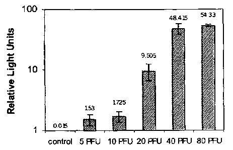
Figure 1 is a graph depicting the results of an experiment in which different
titers of vaccinia virus were detected using the present method.
3a
CA 02548569 2008-02-07
All dimensions specified in this disclosure are by way of example only and
are not intended to be limiting. Further, the proportions shown in these
Figures are
not necessarily to scale. As will be understood by those with skill in the art
with
reference to this disclosure, the actual dimensions of any device or part of a
device
disclosed in this disclosure will be determined by their intended use.
DESCRIPTION
Definitions
As used herein, the following terms have the meanings given below, unless
a different meaning is clearly intended by the context in which such term is
used.
"Cytoplasmic-replicating DNA virus"refers to a virus which stores genetic
information at least partially in deoxyribonucleic acid (DNA) and which
transcribes
such genetic information outside the nucleus of a host cell which it infects,
i. e. in
the cytoplasm of a host cell. Cytoplasmic-replicating DNA viruses include
viruses of
the Poxviridae family.
"E/L promoter"refers to a synthetic early-late pox virus promoter described
in Chakrabarti, S. , Sisler, J. R. , and Moss, B. ,"Compact, synthetic,
vaccinia virus
early/late
3b
CA 02548569 2006-06-07
WO 2005/076784 PCT/US2004/041455
4
promoter for protein expression," BioTechniques, 23:1094-1097 (1997) and
having the
sequence AAAAATTGAAATTTTATTTTTTTTTTTTGGAATATAAATA (SEQ ID NO. 1).
"Expression" of a nucleotide sequence refers to the transcription of the
sequence and
its subsequent translation into a polypeptide.
"Expression level," with reference to a reporter sequence (as defined below),
refers to
the abundance of the reporter sequence. The abundance of a reporter
corresponding to the
reporter sequence is normally detected in the present methods as a proxy for
the expression
level of the reporter sequence, and determining the expression level of a
reporter sequence
can comprise determining the abundance of the corresponding reporter in a host
cell or group
of host cells.
An "expression vector" is a nucleic acid construct, generated recombinantly or
synthetically, comprising DNA or other nucleic acids able to be recognized and
transcribed
by viral and cellular transcription factors in a host cell, in particular the
transcription factors
of a virus to be detected by the present methods. The expression vector can
be, for example,
part c: a plasmid or virus.
"Host cell" refers to a eukaryotic cell capable of being infected with a virus
in an
assay according to the present methods.
"Nucleotide sequence" refers to a chain of deoxyribonucleotides or
ribonucleotides,
i.e. oligonucleotides or polynucleotides, in either single- or double-stranded
form.
The term "operably linked" refers to functionally related nucleic acid
sequences.
When a promoter controls and/or enhances the transcription of a nucleotide
sequence, it is
said to be operably linked to the nucleotide sequence.
"Promoter" refers to a nucleotide sequence or sequences, usually comprising a
transcription factor binding site, that directs and/or enhances transcription
of another
nucleotide sequence.
"Reporter sequence" refers to a nucleotide sequence which can be transcribed
and
detected, or whose polypeptide translation product can be detected, such as by
spectroscopic,
photochemical, biochemical, immunochemical, luminescence, or chemical means.
"Reporter
construct" refers to an expression vector comprising a reporter sequence
operably linked to a
promoter. "Reporter" refers to a polypeptide translation product of a reporter
sequence.
CA 02548569 2006-06-07
WO 2005/076784 PCT/US2004/041455
"Transfection" refers to a process by which exogenous nucleotide sequences,
typically
DNA, enter a recipient host cell. For purposes of the present methods,
transfection includes
processes in which nucleotide sequences are physically or chemically
transferred into a cell,
such as through electroporation or lipofection, as well as virally mediated
processes, i.e.
5 transduction. "Transient transfection" refers to methods of transfection in
which the
exogeous DNA is not stably incorporated into the recipient host cell's
chromosomal DNA
and functions for only a limited time. Transiently transfected DNA is
generally located
predominantly within a cell's cytoplasm.
"Virus-specific promoter" refers to a promoter that directs and/or enhances
transcription of another nucleotide sequence only in the presence of
transcription factors or
other proteins encoded by a particular virus or by a limited number of viruses
of a particular
genus or family.
As used herein, the term "comprise" and variations of the term, such as
"comprising"
and "comprises," are not intended to exclude other additives, components,
integers or steps.
Methods
The present methods are cell-based assays which allow the detection of DNA
viruses,
in particular cytoplasmic-replicating DNA viruses. Prior art cell-based assays
for detecting
DNA viruses involved the creation of cells which were stably transfected with
a reporter
sequence under the control of a promoter specific to that virus, such that the
promoter and
reporter sequences were incorporated into a host cell's chromosomal DNA. This
approach
suffers from the tendency of reporter sequences in the nuclei of such stably
transfected cells
to be silenced over time, so that they are no longer transcribed in the
presence of the
appropriate transcription factors.
In the present methods, host cells are transiently transfected with a reporter
construct,
so that there is little opportunity for gene silencing to occur. Many more
copies of a reporter
sequence can also be placed into a cell using transient transfection, thus
increasing the
sensitivity of the present methods. With respect to cytoplasmic-replicating
DNA viruses,
transient transfection has the additional advantage of locating reporter
constructs
predominantly at the site of viral genome transcription, i.e. in the cytoplasm
of the host cell,
which further increases the sensitivity of the present assay.
CA 02548569 2006-06-07
WO 2005/076784 PCT/US2004/041455
6
To detect the presence of viable, infectious DNA viruses according to the
present
methods, host cells are placed into contact with a sample. The sample can be
any sample
suspected of containing a virus of interest. In one embodiment, the sample
comprises tissue
or fluid (such as blood, plasma, cerebrospinal fluid or saliva) derived from a
human or
animal subject. The sample can also be derived from an inanimate source, such
as a liquid or
solid particulate sample gathered from the environment, e.g. from the surface
of an object.
The sample should be in a condition that does not substantially interfere with
the growth or
metabolism of the host cells, however. For example, it should be at a
temperature conducive
to cellular viability and growth, and should not comprise substances that
would kill host cells
or inhibit the cellular mechanisms needed by a virus of interest to replicate
in the host cells.
In addition to contacting a group of host cells with a sample, additional
groups of host
cells are preferably exposed to other conditions in order to conduct negative
and positive
control assays. In order to verify that the carrier substance used to gather
the sample and/or
the growth medium used to grow the host cells does not contain a virus of
interest, a negative
control assay is performed. In the negative control assay, a group of host
cells is contacted
with the carrier and/or the growth medium instead of with the sample, and an
assay as
described herein is then conducted.
A positive control assay, using host cells exposed to a solution known to
contain a
specified amount of an appropriate virus, is also preferably performed in the
present
methods. The virus used as a positive control can be the same virus as the
virus of interest to
be detected, or can be another virus capable of effecting the transcription of
a reporter
sequence that is transiently transfected into the host cells. In a preferred
embodiment, the
virus used in the positive control assay is a different virus which is less
infectious to humans
and/or is less virulent than the virus to be detected in the present methods.
Most commonly,
the virus used in the positive control assay is from the same genus or family
as the virus to be
detected. For example, vaccinia virus can be used as a positive control for a
smallpox assay
as descri`~d herein, as long as the promoter in the reporter construct allows
expression by
both smallpox and vaccinia transcription factors. If the virus to be detected
is highly
infectious and/or virulent, such as smallpox virus, the use of less infectious
or virulent
viruses in the positive control assay has the advantage of reducing the risk
to technicians
conducting the present assay.
CA 02548569 2006-06-07
WO 2005/076784 PCT/US2004/041455
7
Host cells should be capable of being infected by the virus of interest as
well as by a
virus used as a positive control, if a different virus is used for the
positive control assay.
Host cells should also have the ability to express the reporter sequence
and/or the reporter at
easily detectable levels when infected by the virus of interest. Preferably,
host cells are used
which plate such that they are significantly confluent, such as 50%- 70%
confluent, at the
time of the assay. A majority of host cells are also preferably in log phase
when exposed to
the sample to be tested, i.e. are growing at a relatively constant and
generally exponential
rate. Depending on the method of analysis to be used, host cells can be plated
on glass cover
slips ;jr can be directly plated into plastic wells for convenience.
Using the foregoing criteria, one of skill in the art can choose an
appropriate host cell
to use in the present methods. When detecting DNA viruses capable of infecting
humans,
human or other mammalian cell lines are preferred. For example, the host cells
can be U2OS
cells, derived from human osteosarcoma cells, CV-1 monkey kidney cells,
Chinese hamster
ovary (CHO) cells, or baby hamster kidney (BHK) cells.
Host cells are preferably transiently transfected with a reporter construct in
the
present methods after an appropriate amount of time following exposure of the
host cells to a
sample, i.e. sufficient time to allow infection of the host cells to occur.
When assaying for
the presence of poxviruses, between approximately 30 and 60 minutes is
generally a sufficient
period of time. Transfection of host cells following contact with a sample
(and any viruses
contained therein) is preferred, as viral infection of such cells is believed
to facilitate the
transfer of the reporter construct into the cells.
Host cells can alternatively be transfected just prior to contact with a
sample. In this
embodiment, transfected host cells are preferably placed into contact with a
sample as soon
after transfection as is practicable, generally within about a week and/or
within about 10-15
cell divisions. Preferably, transfected host cells are exposed to a sample
within 96 hours
post-transfection and/or within 4-8 cell divisions. When a viral vector is
used to transfect the
host cells, the viral vector can be contacted with the host cells at the same
time as a sample is
placed into such contact.
Transient transfection can be accomplished in any manner known to the art,
including
infection with a viral vector, electroporation, heat shock, and lipofection.
In one
embodiment, the method of transfection used is lipofection, which can be
performed for
CA 02548569 2006-06-07
WO 2005/076784 PCT/US2004/041455
8
example with the FuGENE 6 Transfection Reagent (available from Roche
Diagnostics
Corporation, Roche Applied Science, P.O. Box 50414, 9115 Hague Road,
Indianapolis, IN).
In another embodiment, the assay is performed as a co-infection model, by the
insertion of
the reporter construct into a viral vector, such as an adenovirus or
retrovirus construct. An
advantage of the adenovirus-based approach is that high levels of the reporter
sequence can
be carried into the cytoplasm of a host cell, increasing the sensitivity of
the assay. Transient
transfection can also be accomplished through the use of a gene gun, such as
the Helios Gene
Gun System (available from Bio-Rad Laboratories, Hercules, California), which
bombards
cells with particles (typically gold particles) coated with nucleic acids.
In addition to transfecting a reporter construct, expression vectors
comprising positive
and negative controls are also preferably transfected into groups of host
cells that have been
placed in contact with the sample of interest. An expression vector serving as
a negative
control can comprise, for example, the reporter sequence used in the reporter
construct that is
not operably linked to a promoter. A positive control can comprise, e.g.; the
same reporter
sequence operably linked to a strong, constitutive promoter in the host cells,
such as a CMV
promoter.
The promoter used in the reporter construct in the present methods is specific
to the
virus of interest or to a limited group of viruses of the same genus or
family, so that the
reporter will be expressed in the presence of such virus or viruses. The
promoter can also be
specific to a stage of the life cycle of a virus or to the expression of a
particular viral gene.
In a preferred embodiment, a promoter is used which is active at different
stages of the life
cycle of a virus, in order to increase the expression level of the reporter
sequence and hence
the sensitivity of the assay.
Reporters used in the present assay are detectable moieties known to those of
skill in
the art. Examples of reporters include Green Fluorescent Protein (GFP),.
luciferase, beta-
galactosidase, and secreted alkaline phosphatase (SEAP). In a preferred
embodiment, the
reporter gene is Enhanced Green Fluorescent Protein (EGFP), which is a version
of GFP that
has been optimized for brighter fluorescence and higher expression in
mammalian cells. In
embodiments of the present assay used to quantitatively measure the presence
of a DNA virus
in a sample, the reporter is preferably luciferase.
CA 02548569 2006-06-07
WO 2005/076784 PCT/US2004/041455
9
The method of measuring a reporter depends on the reporter used, as will be
understood by those of skill in the art. For example, expression of EGFP can
be measured
by inverted fluorescence microscopy, using an instrument such as a Leica
fluorescence stereo
microscope (available from Leica Microsystems, Wetzlar, Germany) equipped with
a
mercury 100W lamp power supply connected to a CCD camera. In this case,
measurements
are preferably taken approximately twenty-four hours following transient
transfection of the
EGFP sequence into host cells contacted with a sample. Fluorescence emitted
from cells in a
96-well plate can also be measured with a microplate fluorimeter (such as the
FL600
Fluorescent Microplate Reader available from Bio-Tek Instruments, Inc.,
Highland Park,
P.O. Bo;: 998, Winooski, Vermont). When the reporter is detectable through
fluorescence,
the expression level of the reporter can be expressed as (magnitude of test
fluorescent signals)
/ (magnitude of reference fluorescent signals), where the reference signals
can, for example,
be derived from a negative control assay or a number of aggregated negative
control assays.
In another embodiment, measurement of the reporter can be performed using flow
cytometry, using an instrument such as the BD FACSCaliber System (available
from BD
Biosciences, 1 Becton Drive, Franklin Lakes, New Jersey). In another
embodiment, such as
when the reporter is EGFP, measurement can be performed by immunoblotting or
an ELISA
assay, using any of a number of commercially available antibodies specific for
EGFP.
Following sample contact and transfection, the expression of the reporter
sequence is
measured. After measuring the abundance of the reporter sequence or the
reporter, the
measurements are analyzed to determine whether the virus of interest is
present in the
sample. The analysis can include creating controls using appropriate samples
from the
general population (if the sample is a tissue sample), including positive
controls known to
contain the virus of interest and negative controls known not to contain the
virus, and using
measurements taken from those samples to calculate or estimate a number of
parameters in
the sample, such as virus presence and titer. The sensitivity, specificity,
positive predictive
value and negative predictive value of the assay are preferably also
calculated. These
statistical analyses allow the development of criteria for determining whether
a particular
measurement of reporter sequence expression is likely to indicate the presence
of a virus of
interest in a sample.
CA 02548569 2006-06-07
WO 2005/076784 PCT/US2004/041455
In one embodiment, the determination of whether a particular measurement of
reporter sequence expression indicates the presence of a virus of interest in
a sample is made
by comparing the expression level of the reporter sequence to a calibration
curve. The data
points on the calibration curve can be determined by first contacting samples
having a known
5 titer of the virus of interest with host cells capable of being infected by
the virus, transiently
transfecting the host cells with a reporter construct as described herein, and
then determining
the expression level of the reporter sequence in the host cells.
Results from the present methods are generally available within a matter of
hours to
days and are generally faster than culture-based methods, which is important
when dealing
10 with an outbreak of a contagious agent such as smallpox. Further, the
present methods can
distinguish between the presence of viable, infectious agents and non-viable
or inactivated
material. Additionally, the present methods are less susceptible to false
negative results due
to genetic engineering or mutations when compared with antibody or PCR-based
methods.
Since the methods of the present invention are cell-based, they are suitable
for use in
hospitals or other facilities with laboratory capabilities. Additionally, the
present methods
can be easily automated using available robotic equipment for high throughput
analysis, as
will be understood by those of skill in the art.
The present methods can, in addition to detecting viral contagions, be used to
monitor
human or non-human animal subjects treated with a therapeutic viral construct
based on a
DNA virus which is administered in the course of a gene therapy regimen. The
sample in
this case would comprise tissue from such a subject, and the assay would be
used to detect
the presence of infectious virus particles in the sample.
Promoters
DNA virus promoters known to the art can be used in the present methods. When
the
presence of a particular virus is to be assayed, a promoter that is specific
to that virus, or
which is specific to a limited number of viruses of a particular genus or
family, is chosen for
use. It is preferred that viral promoters having no significant homology with
promoters in
the host cell nucleus be used, in order to minimize background expression and
decrease the
incidence of false positives.
CA 02548569 2006-06-07
WO 2005/076784 PCT/US2004/041455
11
The present methods are particularly advantageous for the detection of
poxviruses.
Poxviruses shut off transcription of cellular genes in order to maximize
production of their
own proteins, and as a result, transcription of viral genes and translation of
virus proteins is
much higher (up to and beyond 1000-fold) than are those of the host cell. When
the virus to
be detected is smallpox, smallpox or vaccinia promoters are preferably
selected. The
smallpox virus has three classes of promoters, which are active, respectively,
in the early,
intermediate, and late stages of replication. A comparison of the sequences of
these three
classes of smallpox virus promoters is shown in Table 1 below (with
noncritical nucleotides
designated with an "N").
Table 1
Core Initiator
Early AAAANTGAAANNNTA (SEQ ID NO. 2) or A/G
AAAANTNGAAANNNTA (SEQ ID NO. 3)
Intermediate TNNNTTNAAANNAA (SEQ ID NO. 4) TAAA
(SEQ ID NO. 5)
Late A/T-rich TAAATG/A
(SEQ ID NO. 6)
The present methods can make use of naturally occurring promoters, such as
those
shown in Table 1, or alternatively can make use of a synthetic poxvirus-
specific promoter to
control expression of a reporter gene. A synthetic promoter that contains
elements of both
the early and late promoters, the E/L promoter, responds to poxviruses
(including vaccinia
and variola) at different stages of the viral life cycle. The use of such a
promoter increases
the sensitivity of the present assay and detects poxviruses throughout their
life cycle. This
promoter has no significant homology with promoters in mammalian host cells.
The use of a
promoter like the E/L promoter which is active with both smallpox and vaccinia
viruses has
the additional advantage of allowing the use of a vaccinia virus in the
positive control assay
rather than smallpox virus.
CA 02548569 2006-06-07
WO 2005/076784 PCT/US2004/041455
12
When detecting viruses of the Hepadnaviridae family, any of the four types of
promoters identified for such viruses,. i.e. the core, S1, S2 and X promoters,
or any
combination thereof can be used (see, e.g., Kramis and Kew, J. Viral Hepat.,
6:415-427
(1999); Moolla, N. et al, J. Viral Hepat, 9:323-331 (2002); Malpiece et al.,
"The Gene S
Promoter of Hepatitis B Virus Confers Constitutive Gene Expression", Nucleic
Acids Res.,
11:4645-4654 (1983)). The core promoter directs the synthesis of mRNA which
serves as a
template for the synthesis of core and polymerase proteins. The S1, S2 and X
promoters of
viruses of the Hepnaviridae family direct the synthesis of specific gene
products. Enhancers
of such promoters which direct liver-specific and differentiation state-
specific utilization of
these promoters, enhancer I (ENI) and enhancer II (ENII), can also be
incorporated into a
reporter construct used to detect hepatitis B virus in the present methods.
When detecting hepatitis B virus using the present methods, core promoter
sequences
such as those shown in Table 2 below can be used.
Table 2
GenBank Sequence
Accession No.
AY603446 GGGAGGAGAT TAGGTTAAAG GTCTTTGTAT TAGGAGGCTG
TAGGCATAAA TTGGTCTGCG C
(SEQ ID NO. 7)
AY489315 GGGGGAGGAG ATTAGGTTAA AGGTCTTTGT ATTAGGAGGC
TGTAGGCATA AATTGGTCTG CGCACCAACA TCATGCAACT
TTTTCACCTC TGCCTAATCA TCTCTTGT
(SEQ ID NO. 8)
AB099504 GATGATTAGG CAGAGGGGAA AAAGGTGCAT GGTGCTGGTG
AACAGACCAA TTTATGCCTA CAGCCTCCTA GTACAAAGAC
CTTTAACCTA GTCTCCTCCC CTAACTCCTC CCAGTCTTTA
AACAAACAGT CTTTGAAGTA TGCCTCAAGG TCGGTC
(SEQ ID NO. 9)
CA 02548569 2006-06-07
WO 2005/076784 PCT/US2004/041455
13
Example 1: Poxvirus Assay with U2OS Cells
An assay was performed to detect the presence of poxvirus in a sample. The
reporter
construct was made using a promoterless plasmid coding for EGFP, pEGFP1,
obtained from
Clontech (1020 East Meadow Circle, Palo Alto, California). The E/L promoter
was inserted
upstream of the EGFP sequence in the plasmid's multiple cloning site. This
plasmid was
designated pEGFP-1 E/L.
A second expression vector was constructed from the same plasmid, pEGFP1, in
order to serve as a positive control. A strong, constitutive CMV promoter was
inserted into
the multiple cloning site instead of the E/L promoter. This vector was
designated pEGFP-1
CMV. Candidate clones were identified using restriction endonuclease
digestions with
enzymes expected to' cut at the insertion sites, and identification of the
desired products was
confirmed by sequencing.
A U2OS cell line, obtainable from the American Type Culture Collection (HTB-
96),
was selected as the host cell line. These cells were 50%- 70% confluent at the
time of the
assay. The U2OS cells were plated one day prior to the assay on glass cover
slips in
individual wells of a 6-well plate, at a density of 2.5 x 10⁵ cells per well.
While in log stage growth, cells were exposed either to media (McCoys 5a
medium
supplemented with 1.5 mM L-glutamine, 90%; plus fetal bovine serum, 10%) alone
as a
negative control or to solutions containing the Lister strain of vaccinia
virus. Cells were
seeded with virus at an MOI of 1 (i.e., one virus per cell). 30 minutes after
the initial
infection, the cells were then transiently transfected using the FuGENE 6
Transfection
Reagent. The expression vectors transfected were either pEGFP-1 (as a negative
control),
pEGFP-1 CMV (as a positive control) or pEGFP-1 E/L (the experimental vector).
Next, infectious vaccinia virus was detected by measuring the expression of
the
reporter, EGFP, by inverted fluorescence microscopy, using a Leica
fluorescence stereo
microscope equipped with a mercury 100W lamp power supply that is connected to
a CCD
camera. Fluorescence was detected twenty-four hours following transient
transfection of the
reporter gene into host cells. Cells transfected with a plasmid coding for
EGFP under the
control of the E/L promoter exhibited strong fluorescence in the presence of
vaccinia virus,
as did those transfected with a plasmid coding for EGFP under the control of
the CMV
CA 02548569 2006-06-07
WO 2005/076784 PCT/US2004/041455
14
promoter in the absence of vaccinia virus. Only weak fluorescence was detected
in the
remaining wells.
Example 2: Poxvirus Assay using Different Viral Titers
CV-1 monkey kidney cells were plated in a 6-well microtiter plate, and the
next day
they were infected with vaccinia virus (strain Lister) with 10 plaque forming
units (PFU) per
well or 100 PFU per well, followed by transfection with a plasmid carrying the
GFP gene
under the control of the E/L promoter. Transfection was accomplished using a
lipid-based
transfection reagent, GENEPORTER Transfection Reagent (available from Gene
Therapy
Systems, Inc., San Diego, CA, USA). Negative control cells were transfected
with the
plasmid, but not infected with vaccinia virus.
Cells were visualized using a Carl Zeiss Axiovert 100TV fluorescence
microscope.
No fluorescent cells were detected in the negative control wells. However,
fluorescent cells
were detected on day 1 in wells inoculated with either 10 or 100 PFU, and the
number of
fluorescent cells increased significantly in both cases by day 2.
Example 3: Quantitative Poxvirus Assay
CV-1 cells were seeded into 96-well microtiter plates and then infected with
doses of
vaccinia virus comprising 5, 10, 20, 40, and 80 PFU per well. The cells were
then
transfected with a plasmid carrying the luciferase gene operably linked to the
E/L promoter.
On day 2 post-infection, cells were lysed and the bioluminescence of the
extracts was
measured in each well with a luminometer (detecting light intensity) in the
presence of a
luciferase substrate.
The results are shown in Figure 1. All control wells containing CV-1 cells
either
infected with the virus, or transfected with the plasmid (but not both),
emitted light at or
below 0.015 RLU (Relative Light Units). Increasing titers of virus in non-
control wells
corresponded to higher RLU readings.
Although the present invention has been discussed in considerable detail with
reference to certain preferred embodiments, other embodiments are possible.
The steps
disclosed for the present methods are not intended to be limiting nor are they
intended to
indicate that each step depicted is essential to the method, but instead are
exemplary steps
CA 02548569 2009-11-02
only. Therefore, the scope of the appended claims should not be limited to the
description of
preferred embodiments contained in this disclosure.