Note : Les descriptions sont présentées dans la langue officielle dans laquelle elles ont été soumises.
DEMANDES OU BREVETS VOLUMINEUX
LA PRESENTE PARTIE DE CETTE DEMANDE OU CE BREVETS
COMPREND PLUS D'UN TOME.
CECI EST LE TOME 1 DE 2
NOTE: Pour les tomes additionels, veillez contacter le Bureau Canadien des
Brevets.
JUMBO APPLICATIONS / PATENTS
THIS SECTION OF THE APPLICATION / PATENT CONTAINS MORE
THAN ONE VOLUME.
THIS IS VOLUME 1 OF 2
NOTE: For additional volumes please contact the Canadian Patent Office.
CA 02558666 2006-08-17
WO 2005/081867
PCT/US2005/005263
SALIVARY M RNA PROFILING, BIOMARKERS AND
RELATED METHODS AND KITS OF PARTS
[0001] This invention was made with Government support of grant U01-
DE15018 awarded by the NIH. The Government has certain rights on this
invention
FIELD OF THE DISCLOSURE
[0002] The present disclosure relates to profiling of biomarkers and to
method and kits using said biomarkers. In particular, the present disclosure
related to biomarkers for detection of cancer and in particular of Oral Cavity
and Oropharyngeal squamous Cell Carcinoma (OSCC).
BACKGROUND OF THE DISCLOSURE
[0003] Bionnarkers are molecular indicators of a specific biological property,
a biochemical feature or facet that can be used to measure the progress of
disease or the effects of treatment.
[0004] Proteins and nucleic acids are exemplary biomarkers. In particular, it
has been widely accepted that genomic messengers detected extracellularly
can serve as biomarkers for diseases [6]. In particular, nucleic acids have
been identified in most bodily fluids including blood, urine and cerebrospinal
fluid, and have been successfully adopted for using as diagnostic biomarkers
for diseases [28, 42, 49]. ,
[0005] Saliva is not a passive "ultrafiltrate" of serum [41], but contains a
distinctive composition of enzymes, hormones, antibodies, and other
molecules. In the past 10 years, the use of saliva as a diagnostic fluid has
been successfully applied in diagnostics and predicting populations at risk
for
a variety of conditions [47].
[0006] Specific and informative biomarkers in saliva are desirable to serve
for diagnosing disease and monitoring human health [30, 47, 6]. For example
biomarkers have been identified in saliva for monitoring caries,
periodontitis,
oral cancer, salivary gland diseases, and systemic disorders, e.g., hepatitis
1
CA 02558666 2006-08-17
WO 2005/081867
PCT/US2005/005263
ano I-11V ioi. Pt150 previous similes snow LnaL nurnali 1.11U1IldltSUIS
UtII IJC
identified in saliva and used for oral cancer detection [30, 36]. RNA is more
labile than DNA and is presumed to be highly susceptible to degradation by
RNases. Furthermore, RNase activity, is reported to be elevated in saliva,
which constitutes an inexpensive, non-invasive and accessible bodily fluid
suitable to act as an ideal diagnostic medium. In particular, RNAase activity
is
reported to be elevated in saliva of cancer patients [83]. It has, thus, been
commonly presumed that human mRNA could not survive extracellularly in
saliva. OSCC is the sixth most common cancer in the world, and affects
50,000 Americans annually. Worldwide, cancers of the oral cavity and
oropharynx represent a great public health problem. OSCC accounts for
nearly 50% of all newly diagnosed cancers in India and is a leading cause of
death in France [1].
[0007] Despite improvements in locoregional control, morbidity and mortality
rates have improved little in the past 30 years [2]. Therefore, early
detection
or prevention of this disease is likely to be most effective. Detecting OSCC
at
an early stage is believed to be the most effective means to reduce death and
disfigurement from this disease. The absence of definite early warning signs
for most head and neck cancers suggests that sensitive and specific
biomarkers are likely to be important in screening high risk patients.
SUMMARY OF THE DISCLOSURE
[0008] According to a first aspect, a method to detect a biomarker in a bodily
fluid including a cell phase and a fluid phase, wherein the biomarker is an
extracellular mRNA and bodily fluid is saliva, preferably unstimulated saliva,
is
disclosed. The method comprises: providing a cell-free fluid phase portion of
the bodily fluid; and detecting the extracellular mRNA in the cell-free fluid
phase portion of the bodily fluid.
[0009] In particular, detecting the extracellular mRNA can comprise:
isolating the extracellular mRNA from the cell-free fluid phase portion of the
bodily fluid, and amplifying the extracellular mRNA.
2
CA 02558666 2006-08-17
WO 2005/081867
PCT/US2005/005263
LUMUJ Accoraing to a secona aspect, rranscriprome analysis or a Doully
fluid, including a cell phase and a fluid phase, wherein the bodily fluid is
saliva, is disclosed. The method comprises: providing a cell-free fluid phase
portion of the bodily fluid; and detecting a transcriptome pattern in the cell-
free
fluid phase portion of the bodily fluid. The bodily fluid is preferably
unstimulated saliva.
[0011] In particular, detecting transcriptome pattern in the saliva
supernatant
is preferably performed by microarray assay, most preferably by high-density
oligonucleotide microarray assay. Detecting transcriptome pattern in the
saliva supernatant can also performed by quantitative PCR analysis or RT-
PCR analysis.
[0012] According to a third aspect, a method to detect genetic alterations in
an organ by analyzing a bodily fluid draining from the organ and including a
cell phase and a fluid phase, is disclosed. The bodily fluid is in particular
saliva, preferably unstimulated saliva and method comprises: providing cell-
free fluid phase portion of the bodily fluid; detecting aAranscriptome pattern
in
the cell-free fluid phase portion of the bodily fluid; and comparing the
transcriptome pattern with a predetermined pattern, the predetermined pattern
being indicative of a common transcriptome pattern of normal cell-free fluid
phase portion of the bodily fluid.
[0013] According to a fourth aspect, a method to detect genetic alteration of
a gene in an organ by analyzing a bodily fluid draining from the organ and
including a cell phase and a fluid phase, is disclosed. The bodily fluid is in
particular saliva and the method comprises: providing a cell-free fluid phase
portion of the bodily fluid; detecting an mRNA profile of the gene in the cell-
free fluid phase portion of the bodily fluid; and comparing the mRNA profile
of
the gene with a predetermined mRNA profile of the gene, the predetermined
mRNA profile of the gene being indicative of the mRNA profile of the gene in
normal cell-free fluid phase portion of the bodily fluid,.
[0014] According to a fifth aspect, a method to diagnose an oral or systemic
pathology disease or disorder in a subject, is disclosed. The method
3
CA 02558666 2007-06-05
comprises: providing a cell-free fluid phase portion of the saliva of the
subject,
detecting in the provided cell-free saliva fluid phase portion an mRNA profile
of a gene associated with the pathology, disease or disorder; and comparing
the RNA profile of the gene with a predetermined mRNA profile of the gene,
the predetermined mRNA profile of the gene being indicative of the presence
of the pathology, disease, or disorder in the subject.
[0015] In a first embodiment the pathology, disease or disorder is a cancer
of the oral cavity and/or of oropharynx, the bodily fluid is saliva and the
gene
is selected from the group consisting of the gene coding for IL8 (Interieukin
8),
IL1B (Interleukin 1, beta), DUSP1 (Dual specificity phosphatase 1), H3F3A
(H3 histone, family 3A), OAZ1 (Omithine decarboxyiase antizyme 1), 8100P
(8100 calcium binding protein P) and SAT (Spermidine/spermine N1-
acetyltransferase).
[0016] In a
second embodiment, the pathology, disease or disorder is a
cancer of the oral cavity and/or of oropharynx, the bodily fluid is blood
serum and
the gene is selected from IL6 (interleukin 6), H3F3A, TPT1 (Tumor protein
translationally controlled 1), FTH1 (Ferritin heavy polypeptide 1), NCOA4
(Nuclear receptor coactivator 4) and ARCR (Ras homolog gene family,
member A).
[0017] Diseases that can be diagnosed include oropharyngeal squamous
cell carcinoma and possibly other systemic diseases.
[0018] According to a sixth aspect, a method to diagnose an oral or
systemic pathology, disease or disorder in a subject is disclosed. The method
comprises: providing a cell-free fluid phase portion of the saliva of the
subject;
detecting in the provided cell-free fluid phase portion a transcriptome
pattern
associated with the pathology, disease or disorder; and comparing the
transcriptome pattern with a predetermined pattern, recognition in the
transcriptome pattern of characteristics of the predetermined pattern being
diagnostic for the pathology, disease or disorder in the subject.
4
CA 02558666 2006-08-17
WO 2005/081867
PCT/US2005/005263
LUITIUJ in an emooatmem, me pall-1 1 9y, cusease or atsoraer is a Gamut ut
the oral cavity and/or of oropharynx, and transcriptome include transcript is
selected from the group consisting of transcripts for IL8, IL1B, DUSP1,
H3F3A, OAZ1, S100P, SAT from saliva.
[0020] According to a seventh aspect, a method to diagnose an oral or
systemic pathology, disease or disorder in a subject is disclosed, the method
comprising: providing serum of the subject; detecting in the provided serum a
transcriptome pattern associated with the pathology, disease or disorder; and
comparing the transcriptome pattern with a predetermined pattern, recognition
in the transcriptome pattern of characteristics of the predetermined pattern
being diagnostic for the pathology, disease or disorder in the subject.
[0021] In an embodiment, the pathology, disease or disorder is a cancer of
the oral cavity and/or of oropharynx, and transcriptome include transcript is
selected from the group consisting of transcripts for IL6, H3F3A, TPT1, FTH1,
NCOA4 and ARCR from serum.
[0022] Diseases that can be diagnosed include oropharyngeal squamotis
cell carcinoma possibly other systemic diseases.
[0023] According to a eight aspect, a method for diagnosing a cancer, ini a
subject is disclosed. The method comprises: providing a bodily fluid of the
subject; detecting in the bodily fluid a profile of a biomarker, comparing the
profile of the biomarker with a predetermined profile of the biomarker,
recognition in the profile of the biomarker of characteristics of the
predetermined profile of the biomarker being diagnostic for the cancer.
[0024] Pathologies, diseases or disorders that can be diagnosed include
oropharyngeal squamous cell carcinoma and possibly other systemic
diseases. Biomarkers include IL8, IL1B, DUSP1, H3F3A, OAZ1, S100P, SAT,
IL6, H3F3A, TPT1, FTH 1, NCOA4 and ARCR.
[0025] In a first embodiment, the pathology, disease or disorder is
oropharyngeal squamous cell carcinoma, the biomarker is selected from the
group consisting of 18 ILA B, DUSP1, H3F3A, OAZ1, S100P, SAT, the bodily
5
CA 02558666 2006-08-17
WO 2005/081867
PCT/US2005/005263
num is saliva ana aetecting a profile or a =marker is perrormea Dy aetecting
the mRNA profile of the biomarker.
[0026] In a second embodiment, the pathology, disease or disorder is
oropharyngeal squamous cell carcinoma, the biomarker is selected from the
group consisting of 16, H3F3A, TPT1, FTH1, NCOA4 and ARCR the bodily
fluid is serum and detecting a profile of a biomarker is performed by
detecting
the mRNA profile of the biomarker.
[0027] In a third embodiment, the pathology, disease or disorder is
oropharyngeal squamous cell carcinoma, the biomarker is IL6, the bodily fluid
is blood serum and detecting a profile of a biomarker is performed by
detecting the protein profile of the biomarker
[0028] According to an eighth aspect, a kit for the diagnosis of an oral
and/or systemic pathology, disease or disorder is disclosed, the kit
comprising: an identifier of at least one biomarker in a bodily fluid, the
biomarker selected from the group consisting of IL8, ILI B, DUSP1, H3F3A,
OAZ1, S100P, SAT, IL6, H3F3A, TPT1, FTH1, NCOA4 and ARCR; and a
detector for the identifier.
[0029] Pathologies, diseases or disorders that can be diagnosed include
oropharyngeal squamous cell carcinoma, and possibly the other systemic
diseases.
[0030] The identifier and the detector are to be used in detecting the bodily
fluid profile of the biomarker according to the methods herein disclosed. In
particular, the identifier is associated to the biomarker in the bodily fluid,
and
the detector is used to detect the identifier, the identifier and the detector
thereby enables the detection of the bodily fluid profile of the biomarker.
[0031] According to a ninth aspect, a method to diagnose an oral and/or
systemic pathology disease or disorder, is disclosed. The method comprising:
using salivary and/or serum mRNAs as biomarkers for oral and/or systemic
pathology, disease or disorder.
6
CA 02558666 2014-02-27
[0032] In a preferred embodiment the mRNA codifies for at least one of the
biomarker
selected from the group consisting of IL8, ILI B, DUSP1, H3F3A, OAZ1, S100P,
SAT, IL6,
H3F3A, TPT1 , FTH1 , NCOA4 and ARCR.
[0033] Diseases that can be diagnosed include oropharyngeal squamous cell
carcinoma,
and possibly other systemic diseases.
[0034] According to a tenth aspect, a method to diagnose an oral and/or system
pathology, is disclosed. The method comprising: using salivary or serum
proteins as
biomarkers for oral and/or systemic pathology, disease or disorder, in
particular 1L6 protein
in serum and IL8 protein in saliva.
[0035] The methods and kits of the disclosure will be exemplified with the aid
of the
enclosed figures.
[0035A] Various embodiments of the invention provide a method to detect
mRNA in saliva, the saliva including a cell phase and a cell-free supernatant,
the method comprising: providing a saliva supernatant; isolating mRNA from
the saliva supernatant; and amplifying mRNA thereby detecting mRNA in the
saliva supernatant.
[0035B] Various embodiments of the invention provide a method to diagnose
oropharyngeal squamous cell carcinoma (OSCC) in a test subject, the method
comprising: providing a saliva supernatant from the test subject, detecting in
the saliva supernatant an mRNA profile of a gene; comparing the mRNA profile
of the gene with a predetermined mRNA profile of a gene, wherein the
predetermined mRNA profile is an OSCC mRNA profile, and wherein the gene
is selected from the group consisting of the gene coding for Interleukin 8
(1L8),
Interleukin-I beta (IL-113), Dual specificity phosphatase I (DUSP1), Histone
H3,
family 3A (H3F3A), Ornithine decarboxylase antizyme 1 (OAZ I), S100 calcium
binding protein P (S 100P) and Spermidine/spermine N 1-acetyltransferase
(SAT), wherein the expression of any one of these genes is elevated in a
subject with OSCC relative to a control and wherein a statistically
significant
correlation between the mRNA profile and the predetermined mRNA profile is
indicative of the presence of OSCC.
[0035C] Various embodiments of the invention provide a kit for the diagnosis
of
oropharyngeal squamous cell carcinoma (OSCC), the kit comprising: a
7
CA 02558666 2014-02-27
biomarker control profile, wherein the control profile is derived from a
subject
with OSCC and at least one binding reagent, wherein the binding reagent is a
polynucleotide and the binding reagent binds a biomarker, wherein the
biomarker is selected from the group consisting of: Interleukin 8 (IL8),
Interleukin-1 beta (IL1 B), Dual specificity phosphatase 1 (DUSPI), Histone
H3,
family 3A (H3F3A), Ornithine decarboxylase antizyme I (OAZ I), S100 calcium
binding protein P (S100P) and Spermidine/spermine NI-acetyltransferaase
(SAT), and a detector for the binding reagent.
[00350] Various embodiments of the invention provide a method to diagnose
oropharyngeal squamous cell carcinoma (OSCC) in a test subject, the method
comprising: providing a saliva supernatant from the test subject; detecting in
the provided saliva supernatant a transcriptome pattern, comparing the
transcriptome pattern with a predetermined pattern, wherein the predetermined
pattern is an mRNA pattern detected in subjects with OSCC, wherein the
predetermined pattern comprises a plurality of mRNA profiles, wherein the
plurality of mRNA profiles comprises a mRNA profile of each of Interleukin 8
(IL8), Interleukin-1 beta (IL IB), Ornithine decarboxylase antizyme I (OAZ 1),
and Spermidine/spermine N 1-acetyltransferase (SAT), wherein the expression
of these genes is elevated in a subject with OSCC relative to a control and
wherein a statistically significant correlation between the transcriptome
pattern
and the predetermined pattern is indicative of the presence of OSCC.
DESCRIPTION OF THE FIGURES
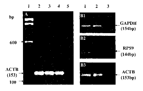
[0036] Figure 1A shows results of a RT-PCR typing for ACTB performed on RNA
isolated
from cell-free saliva supernatant from human beings after storage for 1 month
(lane 2), 3
months (lane 3) and 6 months (lane 4), with a 100bp ladder molecular weight
marker (lane
1) and a negative control (omitting templates) (lane 5). A molecular size
marker is
indicated on the left side of the Figure by arrows.
[0037] Figure 1B shows results of a RT-PCR performed on RNA isolated from cell-
free
saliva supernatant from human beings (lane 1) and typing GAPDH (81), RPS9 (82)
and
ACTB (83), with positive control (human total RNA, BD Biosciences Clontech,
Palo Alto,
7a
CA 02558666 2014-02-27
CA, USA) (lane 2) and negative controls (omitting templates) (lane 3). A
molecular size
marker is indicated on the left side of the Figure by arrows.
[0038] Figure 2A shows results of a capillary electrophoresis performed to
monitor RNA
amplification from RNA isolated from cell-free saliva supernatant from human
beings.
Lanes 1 to 5 show 1kb DNA ladder (lane 1), 5p1 saliva after RNA isolation
(undetectable)
(lane 2), 1 pl two round amplified cRNA (range from 200 bp to -4kb) (lane 3),
1 pl cRNA
after fragmentation (around
7b
CA 02558666 2006-08-17
WO 2005/081867
PCT/US2005/005263
uupp) vane ana Ammon uentury marker vane 0). Pk alUIOUUldl blLe
marker is indicated on the left side and right side of the Figure by arrows.
[0039] Figure 2B shows results of a PCR performed on RNA isolated from
cell-free saliva supernatant from human beings at various stage of
amplification and typing for ACTB. Lane 1 to 8 shows 100bp DNA ladder (lane
1), total RNA isolated from cell-free saliva (lane 2), 1st round cDNA (lane
3),
1st round cRNA after RT (lane 4), 2nd round cDNA (lane 5), 2nd round cRNA
after RT (lane 6), positive control (human total RNA, BD Biosciences
Clontech, Palo Alto, CA, USA) (lane 7) and negative control (omitting
templates) (lane 8). A molecular size marker is indicated on the left side of
the
Figure by arrows.
[0040] Figure 20 shows a diagram reporting results of the analysis of target
cRNA performed by Agilent 2100 bioanalyzer before hybridization on
microarray. On x axis, the molecular weight (bp) of the fragmented cRNA with
reference to the marker RNA, is indicated. On y axis, the quantity of the
fragmented cRNA (ug/ml) measurable by a Bioanalyzer, is indicated.
[0041] Figure 3 shows results of a RT-PCR performed on RNA isolated from
cell-free saliva supernatant from human beings (saliva) together with a ladder
(Mrkr) positive controls (Ctrl(+)) and negative controls (Ctrl(-)) and typing
for
IL6 (IL6), IL8 (IL8) and 13-Actin (6-Actin).
[0042] Figure 4 shows results of a PCR performed for the housekeeping [3-
actin on whole saliva, serum samples, and samples that had been centrifuged
at 0 xg (0 xg), 1,000xg (1,000xg), 2,600 xg (2,600 xg), 5,000 xg (5,000 xg)
and 10,000xg (10,000 xg) using genomic DNA as marker (Mrkr) for cell lysis
and spillage of intracellular compounds.
[0043] Figure 5A shows a diagram reporting the mean concentrations of
mRNA for IL8 detected in replicate samples by gRT-PCR in saliva from
patients with OSCC (Cancer) and normal subjects (Control). On x axis the
sample groups are reported. On y axis the number of copies detected is
reported.
8
CA 02558666 2006-08-17
WO 2005/081867
PCT/US2005/005263
[00441 Figure b1:3 shows a diagram reporting the mean concentrations or iLö
detected in replicate samples by ELISA in saliva from patients with OSCC
(Cancer) and normal subjects (Control). On x axis the sample groups are
reported. On y axis the concentration expressed in pg/ml, is reported.
[0045] Figure 6A shows a diagram reporting the mean concentrations of
mRNA for IL6 detected in replicate samples by gRT-PCR in serum from
patients with OSCC (Cancer) and normal subjects (Control). On x axis the
sample groups are reported. On y axis the number of copies detected is
reported.
[0046] Figure 6B shows a diagram reporting the mean concentrations of IL6
detected in replicate samples by ELISA in serum from patients with OSCC
(Cancer) and normal subjects (Control). On x axis, the sample groups are
reported. On y axis the concentration expressed in pg/ml, is reported
[0047] Figure 7A shows a diagram reporting the Receiver Operating
Characteristic (ROC) curve calculated for IL8 in Saliva. On the x axis 1-
specificity is reported. On y axis the sensitivity is reported.
[0048] Figure 7B shows a diagram reporting the ROC curve calculated for
IL6 in serum. On the x axis 1-specificity is reported. On y axis the
sensitivity is
reported.
[0049] Figure 7C shows a diagram reporting the ROC curve calculated for a
combination of IL8 in saliva and IL6 in serum. On the x axis 1-specificity is
reported. On y axis the sensitivity is reported.
[0050] Figure 8 shows results of a PCR reaction performed on serum
human mRNA phenotyping of salivary mRNAs for RPS9 (Lane 2, 3 and 4);
GAPDH (Lane 5, 6 and 7); B2M (Lane 8, 9 and 10) and ACTB (Lane 11, 12
and 13), together with DNA ladder, as a control (Lane 1).
[0051] Figure 9 shows a diagram reporting a ROC curve of the logistic
regression model for the circulating mRNA in serum. On the x axis 1-
specificity is reported. On y axis the sensitivity is reported.
9
CA 02558666 2006-08-17
WO 2005/081867
PCT/US2005/005263
LUUOZJ rigure -tu snows a aiagram Feporur iy LI IU Uli:AJIIIUGILILJI I cal iu
regression trees (CART) model assessing the serum mRNA predictors for
OSCC.
[0053] Figure 11 shows a diagram reporting a ROC curve of the logistic
regression model for the predictive power of combined salivary mRNA
biomarkers. On the x axis 1-specificity is reported. On y axis the sensitivity
is
reported.
[0054] Figure 12 shows a diagram reporting the classification and
regression trees (CART) model assessing the salivary mRNA predictors for
OSCC.
DETAILED DESCRIPTION OF THE PREFERRED EMBODIMENTS
[0055] A method to detect an extracellular mRNA in a bodily fluid, is
disclosed wherein the bodily fluid is saliva and the extracellular mRNA is
detected in a cell-free fluid phase portion of saliva. Presence of RNAs in the
cell-free fluid phase portion of saliva was confirmed by the procedures
extensively described in the Examples, the quality of the detected mRNA
meeting the demand for techniques such as PCR, qPCR, and nnicroarray
assays.
[0056] In the method, detecting extracellular mRNAs herein also informative
mRNAs, is performed in a bodily fluid, saliva, that meets the demands of an
inexpensive, non-invasive and accessible bodily fluid to act as an ideal
medium for investigative analysis.
[0057] Detecting informative mRNAs is in particular performed in a portion
of saliva (cell-free fluid phase) wherein presence of microorganisms and the
extraneous substances such as food debris is minimized, which allows
analyzing the molecules in simple and accurate fashion. Preferably, the cell-
free fluid phase portion of derived from unstimulated saliva.
[0058] In the method, the saliva can be collected according to procedures
known in the art and then processed to derive the cell-free fluid phase
thereof,
for example by centrifugation of the collected saliva, which results in a
CA 02558666 2006-08-17
WO 2005/081867
PCT/US2005/005263
penetea saliva cell pnase ana a cell-Tree saliva itutu pi idbu sUpelileLdilL.
ksee
procedures extensively described in Examples 1, 5 and 13)
[0059] According to the present disclosure, the conditions for separating the
cell-phase and the fluid phase of saliva are optimized to avoid mechanical
rupture of cellular elements which would contribute to the RNA detected in the
fluid cell-free phase.
[0060] In embodiments wherein the separation is performed by
centrifugation, optimization can be performed by testing housekeeping genes
on samples centrifuged at various speed and on whole saliva samples, using
DNA as a marker of cell lysis and spillage, to derive the optimized
centrifygation speed. (See procedure described in Example 5).
[0061] Detection of the extracellular mRNA in the cell-free saliva fluid phase
portion (salivary mRNA) can then be performed by techniques known in the
art allowing mRNA qualitative and/or a quantitative analysis, such as RT-
PCR, Q-PCR and Microarray. The detection can in particular be performed
according to procedures that can include isolation and an amplification of the
salivary mRNA and that are exemplified in the Examples.
[0062] Detection of the salivary mRNA in the method can be performed for
the purpose of profiling the salivary mRNA.
[0063] In a first series of embodiments, the expression of predetermined
genes, can be profiled in a cell-free fluid phase portion of saliva. In those
embodiments, detection of the mRNA profile can be performed by RT.-PCR or
any techniques allowing identification of a predetermined target mRNA.
Quantitative analysis can then be performed with techniques such as
Quantitative PCR (Q-PCR) to confirm the presence of mRNA identified by the
RT-PCR. A reference database can then be generated based on the mRNA
profiles so obtained. Exemplary procedures to perform such qualitative and
quantitative analyses of salivary mRNA are described in details in Examples
1, 4 and 9.
11
CA 02558666 2006-08-17
WO 2005/081867
PCT/US2005/005263
Lutioztj in a secona series or emoomments, a trarisuipturrie ar ialysts ui
saliva can be performed by detecting a transcriptome pattern in the cell-free
fluid phase portion of saliva. Detection of the transcriptome pattern can be
performed by isolating and linearly amplifying salivary mRNA, which can then
be profiled with techniques such as high-density oligonucleotide microarrays.
Quantitative analysis can then be performed with techniques such as Q-PCR
to confirm the presence of mRNA in the pattern identified by the microarray. A
reference database can then be generated based on the mRNA profiles so
obtained. Exemplary procedures to perform such qualitative and quantitative
analyses of salivary mRNA are described in details in Examples 2-3, 9-10 and
14-15.
[0065] Profiling salivary RNA can be performed to detect and/or monitor
human health and disease or to investigate biological questions, such as for
example, the origin, release and clearance of mRNA in saliva. The salivary
mRNA provides actual or potential biomarkers to identify populations and
patients at high risk for oral and systemic pathologies, diseases or
disorders.
[0066] Alterations of the salivary mRNA profiles and transcriptome patterns
characterizing the cell-free fluid phase portion of saliva or normal subjects
can
be indicative of pathologies, diseases or disorders of various origin.
Examples
of those pathologies, diseases or disorders are provided by the inflammatory
conditions of the oral cavity, OSCC or other conditions such as diabetes,
breast cancer and HIV.
[0067] Also comparison between the mRNA profiles and transcriptome
patterns of subject affected with a determined pathology, disease or disorder,
can result in the identification of informative biomarkers for the determined
pathology disease or disorder. In particular, salivary mRNA can be used as
diagnostic biomarkers for oral and systemic pathologies, diseases or
disorders that may be manifested in the oral cavity.
[0068] In particular, salivary mRNA can be used as diagnostic biomarkers
for cancer that may be manifested and/or affect the oral cavity. Sal iva-based
12
CA 02558666 2006-08-17
WO 2005/081867
PCT/US2005/005263
mRNA assays nave The needed specmcny ana sensitivity Tor teiiwie
diagnostics.
[0069] In case of various forms of cancer, alterations of the normal salivary
mRNA and transcriptome patterns can also reflect the genetic alterations in
one or more portions of the oral cavity which are associated with presence of
the tumor. For oral cancer patients, the detected cancer-associated RNA
signature is likely to originate from the matched tumor and/or a systemic
response (local or distal) that further reflects itself in the whole saliva
coming
from each of the three major sources (salivary glands, gingival crevicular
fluid,
and oral mucosa! cells). It is conceivable that disease-associated RNA can
find its way into the oral cavity via the salivary gland or circulation
through the
gingival crevicular fluid. A good example is the elevated presence of HER-2
proteins in saliva of breast cancer patients [87].
[0070] A common transcriptome of normal cell-free saliva, including
approximately 185 different human mRNAs, also defined as Normal Salivary
Core Transcriptome (NSCT) was identified in outcome of a transcriptome
analysis performed on cell-free fluid phase of saliva from normal subject (see
Example 2, Table 2).
[0071] Since the NSCT was identified using the probe sets on HG U1 33A
microarray representing only -19,000 human genes, and the human genome
composed of more than 30,000 genes [48], it is expected that more human
mRNAs will be identified in saliva by other methodologies and additional
salivary patterns are identifiable by the method herein disclosed.
[0072] The NSCT and/or other salivary transcriptome patterns in cell-free
saliva from normal populations can serve in a Salivary Transcriptome
Diagnostics (SlvTD), for potential applications in disease diagnostics as well
as normal health surveillance.
[0073] Accordingly, in a first embodiment of the SlvTD, a method to
diagnose an oral or systemic pathology disease or disorder in a subject, is
disclosed. The method comprises: providing a cell-free fluid phase portion of
13
CA 02558666 2006-08-17
WO 2005/081867
PCT/US2005/005263
tne saliva ot me subject; aetecting in me proviaea cell-tree saliva mita pnase
portion an mRNA profile of a gene associated with the disease; and
comparing the RNA profile of the gene with a predetermined mRNA profile of
the gene, the predetermined mRNA profile of the gene being indicative of the
presence of the disease in the subject.
[0074] In a second embodiment of the SlvTD, a method to diagnose an oral
or systemic pathology disease or disorder in a subject, is disclosed. The
method comprises: providing cell-free saliva supernatant of the subject;
detecting in the cell-free saliva supernatant a transcriptome pattern
associated with the pathology disease or disorder; and comparing the
transcriptome pattern with a predetermined pattern, recognition in the
transcriptome pattern of characteristics of the predetermined pattern being
diagnostic for the pathology disease or disorder in the subject.
[0075] In a third embodiment of the SlvTD, a method to identify a biomarker
associated with a predetermined pathology disease or disorder is disclosed.
The method comprises: detecting a first mRNA profiling of a predetermined
gene in cell-free fluid phase portion of saliva of a subject affected by the
pathology disease or disorder; detecting a second mRNA profiling of the
predetermined gene in cell-free fluid phase portion of saliva of a normal
subject; comparing the first mRNA profiling with the second mRNA profiling,
recognition of differences between the first mRNA profiling and the second
mRNA profiling, the differences validated by statistical analysis, being
indicative of the identification of the predetermined gene as a biomarker for
the predetermined pathology disease or disorder.
[0076] In particular the difference between the RNA profiling from one
disease category to one healthy category is analyzed by microarray statistical
methodologies. The algorithms used include MAS 5.0, DNA-Chip analyzer 1.3
and RMA 3Ø Preferably, the analysis is performed by a combination of these
methods to provide more powerful and accurate markers to test. The markers
identified by microarray will then be tested by conventional techniques such
as Q-PCR.
14
CA 02558666 2006-08-17
WO 2005/081867
PCT/US2005/005263
LUUt tj in a rourm emnaiment or me ivi u a atagnosuc memoa can De
performed, wherein the cell-free saliva is contacted with an identifier for
the
presence or expression of the biomarker, and the presence of the identifier
associated to presence or expression of the biomarker is detected, preferably
by means of a detector.
[0078] The SlvTD allow detection of diseases such as tumors at a stage
early enough that treatment is likely to be successful, with screening tools
exhibiting the combined features of high sensitivity and high specificity.
Moreover, the screening tool are sufficiently noninvasive and inexpensive to
allow widespread applicability.
[0079] The results of the above methods of the SlvTD can be integrated with
a corresponding analysis performed at an mRNA and/or protein level and/or in
other bodily fluid, such as blood serum.
[0080] Biomarkers, such as protein or transcriptome patterns detected in
serum can also serve in a Serum Transcriptome Diagnostics (SrmTD), for
potential applications in disease diagnostics as well as normal health
surveillance. Embodiments of the SrmTD include methods corresponding to
the ones reported above for the SlvTD, wherein the bodily fluid analyzed is
serum instead of cell-free saliva.
[0081] In particular, the results obtained following the SlvTD can be
combined with results obtained with the SrmTD, in a combined Salivary and
Serum Transicriptome approach (SSTD).
[0082] According to the SSTD a diagnostic method can be performed,
wherein the bodily fluid, serum and/or saliva is contacted with an identifier
for
the presence or expression of the biomarker, wherein the biomarker can be a
protein or an mRNA and the presence of the identifier associated to presence
or expression of the biomarker is detected, preferably by means of a detector.
[0083] Examples of the SlvTD, SrmTD and SSTD are herein provided with
reference to the OSCC. The person skilled in the art can derive the
CA 02558666 2006-08-17
WO 2005/081867
PCT/US2005/005263
appropriate modifications of the S I I.) nerein exemplified Tor diseases
amerent
than OSCC upon reading of the present disclosure.
[0084] Profiling of two specific cytokines, IL6 and IL8, was measured in the
cell-free fluid phase portion of saliva and serum of patients with OSCC
according to procedures extensively disclosed in Examples 4-8. IL8 was
detected at higher concentrations in the saliva of patients with OSCC (P <
0.01) and IL6 was detected at higher concentrations in the serum of patients
with OSCC (P < 0.01). These results were confirmed at both the mRNA and
the protein levels, and the results were concordant. The concentration of IL8
in saliva and IL6 in serum did not appear to be associated with gender, age,
or alcohol or tobacco use (P> 0.75). The data were subjected to statistical
analysis, in particular to ROC analysis, and were able to determine the
threshold value, sensitivity, and specificity of each biomarker for detecting
OSCC (see Example 8, Table 3). Furthermore, the inventors were able to
measure mRNA in salivary specimens.
[0085] A transcriptome analysis of unstimulated saliva collected from
patients with OSCC and normal subjects was performed as disclosed in
Examples 9-12 and in Examples 13-16.
[0086] RNA isolation was performed from the saliva supernatant, followed
by two-round linear amplification with T7 RNA polymerase. Human Genome
U133A rnicroarrays were applied for profiling human salivary transcriptome.
The different gene expression patterns were analyzed by combining a t test
comparison and a fold-change analysis on 10 matched cancer patients and
controls. Quantitative polymerase chain reaction (qPCR) was used to validate
the selected genes that showed significant difference (P < 0.01) by
microarray. The predictive power of these salivary mRNA biomarkers was
analyzed by receiver operating characteristic curve and classification models.
[0087] The results of a first set of microarray analysis showed that there are
1,679 genes exhibited significantly different expression level in saliva
between
cancer patients and controls (P < 0.05). Seven cancer-related mRNA
biomarkers that exhibited at least a 3.5-fold elevation in OSCC saliva (P <
16
CA 02558666 2006-08-17
WO 2005/081867
PCT/US2005/005263
u.ui) were consistently vailaatea oy qrL." on saliva samples
patients (n = 32) and controls (// =32). These salivary, RNA biomarkers are
transcripts of IL8, IL1B, DUSP1, H3F3A, OAZ1, SlOOP, and SAT. The
combinations of these biomarkers yielded sensitivity (91%) and specificity
(91%) in distinguishing OSCC from the controls. (see Examples 13-16)
[0088] The results of a second set of microarray analysis showed five of ten
up-regulated genes selected based on their reported cancer-association,
showed significantly elevated transcripts in serum of OSCC patient. These
RNA biomarkers are transcripts of H3F3A, TPT1, FTH 1, NCOA4 and ARCR.
The results validated by qPCR confirmed that transcripts of these five genes
were significantly elevated in the serum of OSCC patient (Wilcoxon Signed
Rank test, P < 0.05). (See Examples 9 to 12)
[0089] Using the described collection and processing protocols, the
presence of ACTB, B2W, GAPDH and RPS9 mRNAs (controls mRNA) were
confirmed in all serum (patients and controls) by RT-PR.
[0090] Accordingly, a method for diagnosing a cancer, in particular OSCC in
a subject, is disclosed. The method comprises: providing a bodily fluids of
the
subject; detecting in the bodily fluid a profile of a bit:, marker, the
biomarker
selected from the group consisting of IL8 IL1B, DUSP1, H3F3A, OAZ1,
S100P, SAT, IL6, H3F3A, TPT1, FTH1, NCOA4 and ARCR, comparing the
profile of the biomarker with a predetermined profile of the biomarker,
recognition in the profile of the biomarker of characteristics of the
predetermined profile of the biomarker being diagnostic for the cancer.
[0091] Also method to diagnose oral and/or systemic pathology, disease or
disorder, in particular OSCC, is disclosed. The method comprises using
salivary mRNAs as biomarkers for oral and/or systemic diseases, in particular
salivary mRNAs of selected from the group consisting of IL8 IL1B, DUSP1,
H3F3A, OAZ1, S100P and SAT.
[0092] Additionally a method to diagnose oral and/cDr systemic pathology,
disease or disorder, in particular OSCC, is disclosed. The method comprises:
17
CA 02558666 2006-08-17
WO 2005/081867
PCT/US2005/005263
using serum MKIWAS anscuor protein as olomarkers Tor orai am/or sysierniu
diseases, in particular serum mRNAs of selected from the group consisting of
IL6, H3F3A, TPT1, FTH1, NCOA4 and ARCR, and serum IL6 protein.
[0093] Given the multifactorial nature of oncogenesis and the heterogeneity
in oncogenic pathways use of combinations of salivary and/or serum
biomarkers, ensuring higher specificity and sensitivity, to detect the
disease,
is preferred. Multiple statistical strategies reported and risk models
described
in the examples can be used to identify combinations of biomarkers that can
identify OSCC patients samples and to facilitate assigning the appropriate
serum transcriptome-based diagnosis for patients' specific cancer risk.
[0094] Monitoring of profile of salivary mRNA in cell-free fluid phase portion
of saliva and/or in other bodily fluid such as blood serum, can be used in the
postoperative management of OSCC patients. It could potentially be used for
monitoring the efficacy of treatment, or disease recurrence after therapy has
concluded. Salivary mRNAs and in particular IL8 may also serve as
prognostic indicators to direct the treatment of patients-with oral cavity
cancer.
In perspective, high-risk patients Can be directed to more aggressive or
adjuvant treatment regimens.
[0095] The use of these biomarkers may also improve the staging of the
tumor. With traditional techniques, the presence of microscopic distant
disease is often under recognized. In recent years, there has been a shift
from locoregional failure to distant failure for patients treated for presumed
locoregional disease.[18] This in part is a reflection of subclinical distant
disease present prior to the initiation of therapy. Testing for the presence
of
biomarkers may allow the detection of small amounts of tumor cells in a
background of normal tissue. Salivary mRNAs as biomarkers specific for
head and neck tumors or a panel of such biomarkers may allow the detection
of distant microscopic disease. For oral cancer, one of the most important
applications of the STD approach in this respect is to detect the cancer
conversion of oral premalignant lesions.
18
CA 02558666 2006-08-17
WO 2005/081867
PCT/US2005/005263
11.1UUbj Framing OT salivary MKNAS can also De usea to investigate me role
of genes in the development of cancer, in particular whether the aberrant
expressions of these genes functionally contribute to the development of
human OSCC. The biological significance of differential expression of these
genes in head and neck/oral cancer should be determined. Identification of
cancer-associated genes that are consistently changed in cancer patients will
provide us not only with diagnostic markers but also with insights about
molecular profiles involved in head and neck cancer development.
Understanding the profile of molecular changes in any particular cancer will
be extremely useful because it will become possible to correlate the resulting
phenotype of that cancer with molecular events.
[0097] Kits of parts associated with the methods herein disclosed are also
disclosed. In an exemplary embodiment, a kit comprises: a identifier of a
biomarker in a bodily fluid, such as a salivary mRNA or protein, and serum
mRNA or protein, the biomarker selected from the group consisting of 18
IL1B, DUSP1, 1-13F3A, OAZ1, S100P, SAT, IL6, H3F3A, TPT1, FTH1,
NCOA4 and ARCR; and a detector for the identifier, the identifier and the
detector to be used in detecting the bodily fluid profile of the biomarker of
one
the methods herein disclosed, wherein the identifier is associated to the
biomarker in the bodily fluid, and the detector is used to detect the
identifier,
the identifier and the detector thereby enabling the detection of the bodily
fluid
profile of the biomarker.
[0098] The bodily fluid can be saliva, with the detection performed in the
cell-free fluid phase portion thereof, or another bodily fluid such as blood
serum.
[0099] The identifier and the detector able to detect the identifier, are
identifiable by a person skilled in the art. Other compositions and/or
components that may be suitably included in the kit and are also identifiable
by a person skilled in the art.
19
CA 02558666 2006-08-17
WO 2005/081867
PCT/US2005/005263
LUU1UUj I ne taentmer ana tne reagent can pe incluaea one or IIIUW
compositions where the identifier and/or the reagent are included with a
suitable vehicle, carrier or auxiliary agent.
[00101] In the diagnostic kits herein disclosed, the agents and identifier
reagents can be provided in the kits, with suitable instructions and other
necessary reagents, in order to perform the methods here disclosed. The kit
will normally contain the compositions in separate containers. Instructions,
for
example written or audio instructions, on paper or electronic support such as
tapes or CD-ROMs, for carrying out the assay, will usually be included in the
kit. The kit can also contain, depending on the particular method used, other
packaged reagents and materials (i.e. wash buffers and the like).
[00102] Further details concerning the identification of the suitable carrier
agent or auxiliary agent of the compositions, and generally manufacturing and
packaging of the kit, can be identified by the person skilled in the art upon
reading of the present disclosure.
[00103] The kit of parts herein disclosed can be used in particular for
diagnostic purpose. As a result a non-invasive diagnostic detection of
pathologies, diseases or disorder and in particular of oral cavity and
oropharyngeal cancer in patients, is disclosed.
[00104] The use of the fluid phase of saliva has unique advantages over the
use of exfoliated cells. Depending on the location of the tumor, one may not
be able to easily access and swab the tumor bed. Although salivary
biomarkers could not identify the site from which the tumor originated, they
could identify patients at risk. Such a saliva test could be ad ministered by
nonspecialists in remote locations as a screening tool to select patients for
referral for careful evaluation of the upper aerodigestive tract. Finding
early
stage, previously undetected disease may ultimately save lives. IVloreover,
the
use of easily accessible biomarkers may prove highly beneficial in large
populations or chemoprevention trials. This could be envisioned during routine
dental visits or targeted screening of individuals at high risk of development
of
the disease. A home test kit can also be envisioned.
CA 02558666 2006-08-17
WO 2005/081867
PCT/US2005/005263
LLIWIUOJ Also me use or blood test is envisioned in particular tor cancer
early
detection. Recovering the cell-free circulating mRNA or protein biomarkers in
the serum of cancer patients representing characteristics of tumor genetic
alteration, such as IL6 mRNA and protein, H3F3A, mRNA TPT1 mRNA ,
FTH1 mRNA, NCOA4 mRNA and ARCR mRNA diagnostic for OSCC, could
be envisioned as a screening test for presence of occult OSCC during routine
physician's visit with blood work or targeted screening of individuals at high
risk for oral cancer development. A home test kit can also be envisioned,
including preferably
[00106] In particular, peripheral blood can be obtained from subjects using
routine clinical procedures, and mRNA and proteins can be isolated,
preferably with an optimized procedures herein disclosed. Real time
quantitative PCR and ELISA for the respective cytokine will be performed for
one or biomarkers, such as IL6.
[00107] A perspective embodiments of the methods herein disclosed are
directed towards the eventual creation of micro-/nano-electrical mechanical
systems (MEMS/NEMS) for the ultrasensitive detection of molecular
biomarkers in oral fluid. RNA and protein expression for the validated OSCC
biomarkers will be selected as targets for cancer detection. The integration
of
these detection systems for the concurrent detection of mRNA and protein for
multiple OSCC biomarkers will result in an efficient, automated, affordable
system for oral fluid based cancer diagnostics.
[00108] Further details concerning reagents, conditions, compositions
techniques to be used in the method and kits of the disclosure are
identifiable
by a person skilled in the art upon reading of the present disclosure.
[00109] Also appropriate modifications of the STD methods and kits herein
disclosed and exemplified as associated to OSCC and/or HSNCC, for the
mRNA profiling and transcriptome analysis associated with investigation and
diagnosis of other pathology diseases and disorders can be made by a
person skilled in the art upon reading of the present disclosure.
21
CA 02558666 2006-08-17
WO 2005/081867
PCT/US2005/005263
LUU1 1(11 I ne totiowing examples are proviaea to aescnoe me invention in
further detail. These examples, which set forth a preferred mode presently
contemplated for carrying out the invention, are intended to illustrate and
not
to limit the invention
EXAMPLES
EXAMPLE 1: RNA ISOLATION, AMPLIFICATION AND GENE EXPRESSION
PROFILING FROM CELL-FREE SALIVA OF NORMAL DONORS
Normal subjects
[00111] Saliva samples were obtained from ten normal donors from the
Division of Otolaryngology, Head and Neck Surgery, at the Medical Center,
University of California, Los Angeles (UCLA), CA, in accordance with a
protocol approved by the UCLA Institutional Review Board. The following
inclusion criteria were used: age 30 years; no history of malignancy,
immunodeficiency, autoimmune disorders, hepatitis, HIV infection or smoking.
The study population was composed of 6 males and 4 females, with an
average age of 42 years (range from 32 to 55 years).
Saliva collection and processing to obtain the relevant fluid phase
[00112] Unstimulated saliva were collected between 9 am and 10 am in
accordance with published protocols [38]. Subjects were asked to refrain from
eating, drinking, smoking or oral hygiene procedures for at least one hour
prior to saliva collection. Saliva samples were centrifuged at 2,600 x g for
15
min at 4 C. Saliva supernatant was separated from the cellular phase. RNase
inhibitor (Superase-ln, Ambion Inc., Austin, TX, USA) and protease inhibitor
(Aprotinin, Sigma, St. Louis, MO, USA) were then added into the cell-free
saliva supernatant.
RNA isolation from cell-free saliva
[00113] RNA was isolated from cell-free saliva supernatant using the
modified protocol from the manufacturer (QIAamp Viral RNA kit, Qiagen,
22
CA 02558666 2010-01-05
Valencia, CA, USA). Saliva (560 pL), mixed well with AVL butter (2,240 pL),
was incubated at room temperature for 10 min. Absolute ethanol (2,240 pl..)
was added and the solution passed through silica columns by centrifugation at
6,000 x g for 1 min. The columns were then washed twice, centrifuged at
20,000 x g for 2 min, and eluted with 30 pl. RNase free water at 9,000 x g for
2 min. Aliquots of RNA were treated with RNase-free DNase (DNase 1-DNA-
free, Ambion Inc., Austin, TX, USA) according to the manufacturer's
instructions.
[00114] The stability of the isolated RNA was examined by RT-POR typing for
actin-13 (ACTB) after storage for 1, 3, and 6 months. The results reported on
Figure 1A show that the mRNA isolated could be preserved without significant
degradation for more than 6 month at -80 C.
[00115] The quality of isolated RNA was examined by RT-PCR for three
house-keeping gene transcripts: glyceraldehyde-3-phosphate dehydrogenase
(GAPDH), actin-13 (ACTB) and ribosomal protein S9 (RPS9). Primers were
designed using PR!MER3 software and were
synthesized commercially (Fisher Scientific, Tustin, CA, USA) as follows: the
primers having the sequence reported in the attached sequence listing as SEQ
ID NO: 1 and SEQ ID NO: 2 for GAPDH; the primers having the sequence
reported in the attached sequence listing as SEQ ID NO: 3 and SEQ ID NO: 4 for
ACTB; the primers having the sequence reported in the attached sequence
listing as SEQ ID NO: 5 and SEQ ID NO: 6 for RPS9. The quantity of RNA was
estimated using Ribogreene RNA Quantitation Kit (Molecular Probes, Eugene,
OR, USA). The results are shown in Figure 1B, wherein GAPDH (81), RPS9 (B2)
and ACTB (83) were detected consistently in all 10 cases tested, demonstrating
that all 10 saliva samples contain mRNAs that encode for house keeping genes:
GAPDH, ACTB and RPS9.
[00116] The mRNA of these genes could be preserved without significant
degradation for more than 6 months at -80 -C, (see results for ACTB reported
on Fig. 1A).
=
23
CA 02558666 2009-10-20
Target cRNA preparation
= [00117] Isolated RNA was then subjected to linear
amplification according to
published method from our laboratory (Ohyama H, Zhang X, Kohno Y, Alevizos I,
Posner M,
Wong DT, Todd R. Laser capture microdissection-generated target sample for
high-density
oligonueleotide array hybridization. Biotechniques, 2000 Sep;29(3):530-6.). In
brief reverse
transcription using T7-oligo-(dT)24 (SEQ ID NO:53) as the primer was performed
to synthesize the first strand cDNA. The first round of in vitro transcription
(IVT)
was carried out using T7 RNA polymerase (Ambion Inc., Austin, TX, USA). The
BioArrayTM High Yield RNA Transcript Labeling System (Enzo Life Sciences,
Farmingdale, NY, USA) was used for the second round IVT to biotinylate the
cRNA product; the labeled cRNA was purified using GeneChip Sample
Cleanup Module (Affymetrix, Santa Clara, CA, USA).
[00118] The quantity and quality of cRNA were determined by
spectrophotometry and gel electrophoresis. Exemplary results of agarose gel
electrophoresis test reported on Figure 2A show different quantities of
amplified cRNA at the different stages of the RNA amplification.
[00119] Also small aliquots from each of the isolation and amplification steps
were used to assess the quality by RT-PCR. Exemplary results reported in
Figure 2B show PCR typing ACTB Performed at the various stages of RNA
amplification, wherein the expected single band (153bp) can be detected in
every main step of the salivary RNA amplification process.
[00120] The quality of the fragmented cRNA (prepared as described by Kelly JJ,
Chernov BK,
Tovstanovsky I, Mirzabekov AD, Bavykin SG. Radical-generating coordination
complexes as
tools for rapid and effective fragmentation and fluorescent labeling of
nucleic acids for microchip
hybridization. Anal Biochem., 2002 Dec. 15;31 1(2):103-18.) was also assessed
by capillary
electrophoresis using the 2100 Bioanalyzer (Agilent Technologies, Palo Alto,
CA, USA).
Exemplary results reported in Figure 2C show one single peak in a narrow
range (50-200bp)
demonstrating proper fragmentation.
Gene expression profiling in the targeted cRNA preparation
[00121] Gene expression profiling was performed in cell free-saliva obtained
from ten normal donors, wherein on average, 60.5 + 13.1 ng (n=10) of total
RNA was obtained from 560 pL cell-free saliva samples. The results are
reported on Table 1.
24
CA 02558666 2006-08-17
WO 2005/081867
PCT/US2005/005263
Table 1.
Subject Gender Age RNA (ng)a cRNA Hg)'- Present Probesc Probe -%"
1 F 53 60.4 44.3 3172 14.24
2 M 42 51.6 40.8 2591 11.62
3 M 55 43.2 34.8 2385 10.70
4 M 42 48.2 38.0 2701 12.12
M 46 60.6 42.7 3644 16.35
6 M 48 64.8 41.8 2972 13.34
7 F 40 75.0 44.3 2815 12.63
8 M 33 77.8 49.3 4159 18.66
9 F 32 48.8 41.4 2711 12.17
F 32 79.8 44.4 4282 19.22
Mean SD 42 8.3 60.5 13.12 42.2 3.94 3143 665.0 14.11
2.98
[00122] The total RNA quantity is the RNA in 560p.L cell-free saliva
supernatant; the cRNA quantity is after two rounds of T7 amplification.
Number of probes showing present call on HG U133A microarray (detection
5 p<0.04). Present percentage (P%) = Number of probes assigned present
call /
Number of total probes (22,283 for HG U1 33A microarray).
[00123] After two rounds of T7 RNA linear amplification, the average yield of
biotinylated cRNA was 42.2 3.9 pg with A260/280=2.067 0.082 (Table1).
The cRNA ranged from 200 bp to 4 kb before fragmentation; and was
10 concentrated to approximately 100bp after fragmentation. The quality
of cRNA
probe was confirmed by capillary electrophoresis before the hybridizations.
ACTB mRNA was detectable using PCR/RT-PCR on original sample and
products from each amplification steps: first cDNA, first In Vitro
Transcription
(IVT), second cDNA and second IVT, with a resulting agarose electrophoresis
pattern comparable to the one shown in Fig. 2B.
EXAMPLE 2: MICROARRAY PROFILING OF MRNA FROM CELL-FREE SALIVA
OF NORMAL DONORS
[00124] Saliva was collected processed and the RNA isolated as reported in
Example 1. Also, stability, quality and quantity of the RNA was assessed are
reported in Example I.
CA 02558666 2010-01-05
HG-U1331A Microarray analysis
[00125] The Affymetrix Human Genome U1 33A Array, which contains 22,215 =
human gene cDNA probe sets representing -19,000 genes (i.e., each gene
may be represented by more than one probe sets), was applied for gene
expression profiling. The array data were normalized and analyzed using
Microarray Suite (MAS) software (Affymetrix). A detection p-value was
obtained for each probe set. Any probe sets with p < 0.04 was assigned
"present", indicating the matching gene transcript is reliably detected
(Affymetrix, 2001). The total number of present probe sets on each array was
obtained and the present percentage (P%) of present genes was calculated.
Functional classification was performed on selected genes (present on all ten
arrays, p < 0.01) by using the Gene Ontology Mining Tool,
[00126] Salivary mRNA profiles of ten normal subjects were obtained using
HG U133A array contains 22,283 cDNA probes. An average of 3,143 665.0
probe sets (p <0.04) was found on each array (n=10) with assigned present
calls. These probe sets represent approximately 3,000 different mRNAs. The
average present call percentage was 14.11 2.98% (n=10). A reference
database which includes data from the ten arrays was generated. The probe
sets representing GAPDH, ACTB and RPS9 assigned present calls on all 10
arrays. There were totally 207 probe sets representing 185 genes assigned
present calls on all 10 arrays with detection p < 0.01. These 10 genes were
categorized on the basis of their known roles in biological processes and
molecular functions. Biological processes and molecular functions of 185
genes in cell-free saliva from ten normal donors (data obtained by using Gene
Ontology Mining Tool) are reported on Table 2.
26
CA 02558666 2006-08-17
WO 2005/081867 PCT/US2005/005263
Table 2.
Biological processa Genes,nb Molecular functiona
Genes,nb
Cell growth and/or maintenance 119 Binding 118
Metabolism 93 Nucleic acid
binding 89
Biosynthesis 70 RNA binding 73
Protein metabolism 76 Calcium ion binding 12
Nucleotide metabolism 10 Other binding 23
Other metabolisms 18 Structural molecule 95
Cell organization and biogenesis 2 Ribosomal constituent 73
Homeostasis 3 Cytoskeleton constituent 17
Cell cycle 5 Muscle
constituent 2
Cell proliferation 11 Obsolete 15
Transport 5 Transporter 4
Cell motility 8 Enzyme 20
Cell communication 34 Signal transduction 10
Response to external stimulus 19 Transcription regulator 7
Cell adhesion 3 Translation regulator 5
Cell-cell signaling 5 Enzyme regulator 9
Signal transduction 17 Cell adhesion molecule 1
Obsolete 8 Molecular function unknown 6
Development 18 .
Death 2 .
Biological process unknown 11
' ___________________________________________________________________________
[00127] One gene may have multiple molecular functions or participate in
different biological processes. Number of genes classified into a certain
group/subgroup. The major functions of the 185 genes are related to cell
growth/maintenance (119 genes), molecular binding (118 genes) and cellular
structure composition (95 genes). We termed these 185 genes as "Normal
Salivary Core Transcriptome (NSCT)".
EXAMPLE 3: Q-PCR VALIDATION AND QUANTITATION ANALYSIS OF
MICROARRAY PROFILING FROM CELL-FREE SALIVA OF NORMAL DONORS
[00128] The Microarray analysis performed in Example 2 was validated
through a quantitative gene expression analysis by Q-PCR
27
CA 02558666 2009:10:20 "
¨
Quantitative gene expression analysis by Q-PCR
[00129] Real time quantitative PCR (Q-PCR) was used to validate the
presence of human mRNA in saliva by quantifying selected genes from the
185 "Normal Salivary Core Transcriptome" genes detected by the Microarray
profiling reported in Example 2. Genes MB, SFN and K-ALPHA-1, which
were assigned present calls on all 10 arrays, were randomly selected for
validation.
[00130] Q-PCR was performed using iCyclerTM thermal Cycler (Bio-Rad,
Hercules, CA, USA). A 2 pL aliquot of the isolated salivary RNA (without
amplification) was reverse transcribed into cDNA using MuLV Reverse
Transcriptase (Applied Biosystems, Foster City, CA, USA). The resulting cDNA
(3 pL) was used for PCR amplification using iQ SYBR Green Supermix (Bio-Rad,
Hercules, CA, USA). The primers were synthesized by Sigma-Genosys
(Woodlands, TX, USA) as follows: the primers having the sequence reported in
the attached sequence listing as SEQ ID NO: 7 and SEQ ID NO: 8 for interleukin
1, beta (IL1B); the primers having the sequence reported in the attached
sequence listing as SEQ ID NO: 9 and SEQ ID NO: 10 for stratifin (SFN); the
primers having the sequence reported in the attached sequence listing as SEQ
ID NO; 11 and SEQ ID NO: 12 for tubulin, alpha, ubiquitous (K-ALPHA-1). All
reactions were performed in triplicate with conditions customized for the
specific
PCR products. The initial amount of cDNA of a particular template was
extrapolated from a standard curve using the LightCyclerTM software 3.0 (Bio-
Rad,
Hercules, CA, USA). The detailed procedure for quantification by standard
curve
has been previously described (Ginzinger DG. Gene quantification using real-
time quantitative
PCR: an emerging technology hits the mainstream. Exp Hematol., 2002
Jun;30(6):503-12.).
[00131] Q-PCR results showed that mRNA of IL1B, SFN and K-ALPHA-1
were detectable in all 10 original, unamplified, cell-free saliva. The
relative
amounts (in copy number) of these transcripts (n=10) are: 8.68 x 10³ 4.15 x
103 for MB; 1.29 x 10⁵ 1.08 x 10⁵ for SFN; and 4.71 x 10⁶ 8.37 x 10⁵ for K-
ALPHA-1. The relative RNA expression levels of these genes measured by Q-
PCR were similar to those measured by the microarrays (data not shown).
28
CA 02558666 2006-08-17
WO 2005/081867
PCT/US2005/005263
tXAMPLE 4: ILb AND IL t5 MKNA ISOLATION AMPLIFICATION AND ANALY
OF THE EXPRESSION IN CELL-FREE SALIVA OF OSCC PATIENTS
Patients selection
[00132] Patients were recruited from the Division of Head and Neck Surgery
at the University of California, Los Angeles (UCLA) Medical Center, Los
Angeles, CA; the University of Southern California (USC) Medical Center, Los
Angeles, CA; and the University of California San Francisco (UCSF) Medical
Center, San Francisco, CA, over a 6 -month period.
[00133] Thirty-two patients with documented primary T1 or T2 squamous cell
carcinoma of the oral cavity (0C) or oropharynx (OP) were included in this
study. All patients had recently been diagnosed with primary disease, and
had not received any prior treatment in the form of chemotherapy,
radiotherapy, surgery, or alternative remedies. An equal number of age and
sex matched subjects with comparable smoking histories were selected as a
control comparison group.
[00134] Among the two subject groups, there were no significant differences
in terms of mean age (standard deviation, SD): OSCC patients, 49.3 (7.5)
years; normal subjects, 48.8 (5.7) years (Student's t test P > 0.80); gender
(Student's t test P> 0.90); or smoking history (Student's t test P> 0.75). No
subjects had a history of prior malignancy, immunodeficiency, autoimmune
disorders, hepatitis, or HIV infection. Each of the individuals in the control
group underwent a physical examination by a head and neck surgeon, to
ensure that no suspicious mucosal lesion was present.
Saliva Collection And Processing
[00135] Informed consent had been given by all patients. Saliva and serum
procurement procedures were approved by the institutional review board at
each institution: the University of California, Los Angeles (UCLA); the
University of Southern California (USC); and the University of California San
Francisco (UCSF).
29
CA 02558666 2006-08-17
WO 2005/081867
PCT/US2005/005263
[00136.1 Saliva trom 32 patients with uu or OH bUUA, ana jZ unattectea
age- and gender-matched control subjects were obtained for a prospective
comparison of cytokine concentration.
[00137] The subjects were required to abstain from eating, drinking, smoking,
or using oral hygiene products for at least one hour prior to saliva
collection.
Saliva collection was performed using the "draining (drooling)" method of
Navazesh and Christensen,[7] for a total donation of 5 cc saliva. Saliva
samples were subjected to centrifugation at 3500 rpm (2600xg) for 15 minutes
at 4 C by a Sorvall RT6000D centrifuge (DuPont, Wilmington, DE). The fluid-
phase was then removed, and RNAse (Superase-In, RNAse Inhibitor, Ambion
Inc., Austin, TX) and protease (Aprotinin, Sigma, St. Louis, MO;
Phenylmethylsulfonylfluoride, Sigma, St. Louis, MO; Sodium Orthovanadate,
Sigma, St. Louis, MO) inhibitors were then added promptly on ice. The
conditions for the separation of the cellular and fluid phases of saliva were
optimized to ensure no mechanical rupture of cellular elements which would
contribute to the mRNA detected in the fluid phase. All samples were
subsequently treated with DNAse (DNAsel-DNA-free, ;Ambion Inc., Austin,
TX). The cell pellet was retained and stored at ¨80 C.
RNA Isolation from cell-free saliva
[00138] 560 pL of saliva supernatant were then processed using the QIAamp
Viral RNA mini kit (QIAGEN, Chatsworth, CA) kit. RNA was extracted
according to the manufacturer's instructions. Samples were air-dried and
resuspended in water treated with diethyl pyrocarbonate and were kept on ice
for immediate usage or stored at ¨80 C. Aliquots of RNA were treated with
RNAse-free DNAse (DNAsel-DNA-free, Ambion Inc., Austin, TX) according to
the manufacturer's instructions. Concentrations of RNA were determined
spectrophotometrically, and the integrity was checked by electrophoresis in
agarose gels containing formaldehyde.
CA 02558666 2006-08-17
WO 2005/081867
PCT/uS2005/005263
Reverse I ranscriptase-Polymerase Uriain Keaction
[00139] Presence of IL6 and IL8 mRNA transcripts in the fluid phase in saliva
was tested by using reverse transcriptase-polymerase chain reaction (RT-
PCR).
[00140] RNA from each sample was reverse-transcribed in 40 pL of reaction
mixture containing 2.5 U of Moloney murine leukemia virus reverse
transcriptase (Applied Biosystems Inc.(ABI, Foster City, CA) and 50 pmol of
random hexanucleotides (ABI, Foster City, CA ) at 42 C for 45 minutes.
Based on the published sequences, oligonucletide primers were synthesized
commercially at Fisher Scientific (Tustin, CA) for PCR as follows: the primers
having the sequence reported attached sequence listing as SEQ ID NO: 13
and SEQ ID NO: 14 for p-actin; the primers having the sequence reported
attached sequence listing as SEQ ID NO: 15 and SEQ ID NO: 16 for IL8; and
the primers having the sequence reported attached sequence listing as SEQ
ID NO: 17 and SEQ ID NO: 18 for IL6.
[00141] Amplification of the complementary DNA (cDNA) was carried out
using 50 cycles at 95 C for 20 seconds, 60 C for 30 seconds, and 72 C for
30 seconds; followed by a final extension cycle ,of 72 C for 7 minutes.
Specificity of the PCR products was verified by the predicted size and by
restriction digestion. To establish the specificity of the responses, negative
controls were used in which input RNA was omitted or in which RNA was
used but reverse transcriptase omitted. As a positive control, mRNA was
extracted from total salivary gland RNA (Human Salivary Gland Total RNA,
Clontech, Palo Alto, CA). To ensure RNA quality, all preparations were
subjected to analysis of expression.
[00142] The RT-PCR studies so performed showed that saliva and serum
contained mRNA encoding for IL6 and IL8. Exemplary results reported in
Figure 3, show PCR products of the sizes (95 bp for IL6 and 88 bp for IL8)
that were expected from the selected primers. The same-sized products were
expressed in the positive control.
31
CA 02558666 2006-08-17
WO 2005/081867
PCT/US2005/005263
[00143] In order to ensure that the KNA ana protein analyzed were 'mom me
fluid phase of saliva only and to ensure the lack of contamination by
intracellular components, the centrifugation speed for the saliva samples was
optimized. PCR for the housekeeping genes [3-actin and ubiquitin on whole
saliva samples, and samples that had been centrifuged at various speeds
using DNA as a marker of cell lysis and spillage of intracellular components.
The results support an optimal centrifugation speed for saliva samples of
2,600 52 xg, with a preferred speed of 2,600 xg (see exemplary results
reported on Figure 4)
EXAMPLE 5: II
.-- AND 1L8 MRNA ISOLATION, AMPLIFICATION AND ANALYSIS
OF THE EXPRESSION IN SERUM OF OSCC PATIENTS
[00144] Patients recruited as reported in Example 4, where subjected to
analysis of presence of IL6 and 18 mRNA in blood serum.
Serum collection and processing
[00145] Serum from 19 patients with OC or OP SCCA, and 32 unaffected
age- and gender-matched control subjects were obtained for a prospective
comparison of cytokine concentration. Among the subject groups, there were
no significant differences in terms of age, gender, alcohol consumption, or
smoking history (P> 0.75).
[00146] Blood was drawn from control subjects and patients prior to
treatment. Sera were collected by centrifuging whole blood at 3000 rpm
(1000xg) for 10 minutes at 15 C by a Sorvall RT6000D centrifuge (DuPont,
Wilmington, DE). Serum was then separated, and RNAse (Superase-In,
RNAse Inhibitor, Ambion Inc., Austin, TX) and protease (Aprotinin, Sigma, St.
Louis, MO; Phenylmethylsulfonylfluoride, Sigma, St. Louis, MO; Sodium
Orthovanadate, Sigma, St. Louis, MO) inhibitors were then added promptly on
ice. All samples were subsequently treated with DNAse (DNAsel-DNA-free,
Ambion Inc., Austin, TX). The aliquots were stored at ¨80 C until further use.
32
CA 02558666 2006-08-17
WO 2005/081867
PCT/US2005/005263
t-Keverse I ranscrimase-rowmerase unain meauLtuti
[00147] Presence of IL6 and IL8 mRNA transcripts in the serum was tested
by using reverse transcriptase-polymerase chain reaction (RT-PCR)
performed as described in Example 4 above.
[00148] The RT-PCR studies so performed showed that serum contained
mRNA encoding for IL6 and IL8, with electrophoresis gel pattern comparable
to the one shown in Figure 3.
[00149] In order to ensure that the RNA and protein analyzed were from the
fluid phase of serum only and to ensure the lack of contamination by
intracellular components, the centrifugation speed for the serum samples was
optimized following the same approach described in Example 4 for saliva
samples. The results support an optimal centrifugation speed for saliva
samples of 1,000 20 xg with a preferred speed of 1,000 xg.
EXAMPLE 6: IL6 AND IL8 CYTOKINE LEVELS ANALYSIS IN SALIVA FROM
OSCC PATIENTS
[00150] On demonstrating that IL6 and IL8 mRNA transcripts were present in
the fluid phase in saliva, we prospectively examined and compared the levels
of IL6 and IL8 in the saliva of unaffected subjects and patients with OSCC
using quantitative real time PCR (qRT-PCR) and ELISA.
[00151] Saliva from 32 patients with OSCC, and 32 age- and gender-
matched control subjects were obtained. Among the subject groups, there
were no significant differences in terms of age, gender, alcohol consumption,
or smoking history (P> 0.75).
Real Time PCR for Quantification of IL6 and IL8 mRNA Concentrations in
Saliva from Patients and Normal Subjects
[00152] To analyze quantitatively the result of RT-PCR, quantitative real-time
PCR (Bio-Rad iCycler, Thermal Cycler, Bio-Rad Laboratories, Hercules, CA)
was used. Each sample was tested in triplicate. The amplification reactions
were carried out in a 20 pL mixture, using iQ SYBR Green Supermix (Bio-Rad
33
CA 02558666 2006-08-17
WO 2005/081867
PCT/US2005/005263
LcaLiul caw' UUIUS, HILUf !mai uunaturation at uo-u tor s minutes,
50 PCR cycles were performed at 60 C for 20 seconds, then 20 seconds at
72 C, then 20 seconds at 83 C, followed by 1 minute at 95 C, then followed
by a final 1 minute extension at 55 C. Aliquots were taken from each well and
checked by electrophoresis in agarose gels in order to ensure the specificity
of the products.
[00153] The RT-PCR results are illustrated by the diagram shown in Figure 5
A. Such results show that IL8 at both the mRNA and protein levels, was
detected in higher concentrations in the saliva of patients with OSCC when
compared with control subjects (t test, P< 0.01). There was a significant
difference in the amount of IL8 mRNA expression between saliva from OSCC
patients and disease-free controls. The mean copy number was 1.1 x 10³ for
the OSCC group, and 2.6 x 101 for the control group. The difference between
the two groups was highly statistically significant (P<0.0008).
[00154] No significant differences were instead found in the salivary
concentration of IL6 at the mRNA level. Within the sample size studies, the
inventors were also unable to detect differences between smoking and
nonsmoking subjects.
ELISA for Quantification of IL6 and IL8 Protein Concentrations in Saliva from
Patients and Normal Subjects
[00155] ELISA kits for IL6 and IL8 were used (Pierce Endogen, Rockford, IL)
according to the manufacturer's protocol. Each sample was tested in
duplicate in each of two replicate experiments. After development of the
colorimetric reaction, the absorbance at 450 nm was quantitated by an eight
channel spectrophotometer (EL800 Universal Microplate Reader, BIO-TEK
Instruments Inc., Winooski, VT), and the absorbance readings were converted
to pg/m1 based upon standard curves obtained with recombinant cytokine in
each assay. If the absorbance readings exceeded the linear range of the
standard curves, ELISA assay was repeated after serial dilution of the
supernatants. Each sample was tested in at least two ELISA experiments,
and the data were calculated from the mean of tests for each sample.
34
CA 02558666 2006-08-17
WO 2005/081867
PCT/US2005/005263
Luu I 001 I Ile CLIO/A rinaings are 111U511bILUU uy tile ulagrarn SI1OWF1 Ill
riuute
5B. The levels of IL8 in the saliva of OSCC patients were significantly higher
(720 pg/dL) than those in the saliva of the control group (250 pg/dL)
(P<0.0001). To ensure that the elevated levels of IL8 protein in saliva were
not due to an elevation of total protein levels in the saliva of OSCC
patients,
we compared the total protein concentrations in saliva among the two groups.
No significant differences were found (P> 0.05).
(00157] No significant differences were found in the salivary concentration of
IL6 at the protein level. Also in the ELISA analysis, no differences were
detected within the sample size studies between smoking and nonsmoking
subjects.
EXAMPLE 7: 1L6 AND 1L8 CYTOKINE LEVELS ANALYSIS IN SERUM FROM
OSCC PATIENTS
(00158] We also examined and compared the levels of IL6 and IL8 in the
serum of unaffected subjects and patients with OSCC using gRT- PCR and
ELISA. The patients were selected as described in Example 4 and the serum
processed as described in Example 5.
Real Time PCR for Quantification of IL6 and IL8 mRNA Concentrations in
Serum from Patients and Normal Subiects
(00159] To analyze quantitatively the result of RT-PCR, quantitative real-time
PCR was performed as described in Example 6.
[00160] The RT-PCR results are illustrated by the diagram shown in Figure 6
A. Such results show that IL6 at mRNA level was detected in higher
concentrations in the serum of patients with OSCC when compared with
control subjects (t test, P < 0.001). We noted a significant difference in the
amount of IL6 mRNA expression between serum from OSCC patients and
disease-free controls. The mean copy number was 5.2 x 10⁴ for the OSCC
group, and 3.3 x 10³ for the control group. The difference between the two
groups was highly statistically significant (P<0.0004).
CA 02558666 2006-08-17
WO 2005/081867
PCT/US2005/005263
LUUlbli No signmcant an-rerences were insteaa roma in me SUIUIII
concentration of IL8 at the mRNA level. Within the sample size studies, the
inventors were also unable to detect differences between smoking and
nonsmoking subjects.
ELISA for Quantification of IL6 and IL8 Protein Concentrations in Serum from
Patients and Normal Subjects
[00162] ELISA tests for quantification of IL6 and IL8 protein concentrations
in
serum were performed as described in Example 6.
[00163] The relevant ELISA findings are illustrated by the diagram shown in
Figure 6 B. The mean levels of IL6 in the serum of OSCC patients were
significantly higher (87 pg/dL) than those in the serum of the control group
(0
pgidL) (P<0.0001).
[00164] No significant differences were found in the serum concentration of
IL8 at the protein level. Also in the ELISA analysis, no differences were
detected within the sample size studies between smoking and nonsmoking
subjects. ,
,
EXAMPLE 8: ROC AN D SENSITIVITY/SPECIFICITY ANALYSIS
[00165] Statistical analysis of the data collected in outcome of the
experiments reported on Examples 1 to 7 above demonstrates the specificity
and sensitivity of these bionnarkers for HNSCC, and their predictive value.
Statistical Analysis
[00166] The distributions of patient demographics were calculated overall and
separately for OSCC cases and controls, and were compared between the
two arms with either the Student's t-test for continuous measures or two-by-
two Chi-square tables for categorical measures. The distributions of IL6 and
IL8 levels in saliva and serum were computed and compared between the
OSCC cases and controls using two independent group t-tests. Differences
were considered significant for P values less than 0.01. Due to the range of
the IL6 and IL8 levels, log transformations of these measures were also used
...
36
CA 02558666 2006-08-17
WO 2005/081867
PCT/US2005/005263
in me analyses. Data were expressea as me mean ou. /Age, yiiui, iiu
smoking history were controlled at the group level in the experimental design;
these patient factors were also adjusted in the analyses when comparing IL6
and IL8 through regression modeling.
[00167] Using the binary outcome of the disease (OSCC cases) and non-
disease (controls) as dependent variables, logistic regression models were
fitted to estimate the probability of developing OSCC as a function of each of
the potential biomarkers (IL6 or IL8), controlling for patient age, gender,
and
smoking history. Using the fitted logistic models, receiver operating
characteristic (ROC) curve analyses were conducted to evaluate the
predictive power of each of the biomarkers[8][9][10]. Through the ROC
analyses, we calculated sensitivities and specificities by varying the
criterion
of positivity from the least (cut at probability of 0) to the most stringent
(cut at
probability of 1). The optimal sensitivity and specificity was determined for
each of the biomarkers, and the corresponding cutoff/threshold value of each
of the biomarkers was identified. The biomarker that has the largest area
under the ROC curve was identified as having the strongest predictive power
for detecting OSCC.
Clinical Data
[00168] The mean (SD) age of the patients with OSCC was 49.3 (7.5) years
(range, 42-67 years) vs. 48.8 (5.7) years (range, 40-65 years) in the control
group; (Student's t test P> 0.80). Among the two subject groups, there were
no significant differences in terms of age (mean age): OSCC patients, 49.3
years; normal subjects, 48.8 years (Student's t test P > 0.80); gender
(Student's t test P> 0.90); or smoking history (Student's t test P> 0.75).
[00169] ROC (Receiver Operating Characteristic) curves, plots of sensitivities
versus 1-specificities, were generated for each of the potential biomarkers.
Age, gender, and smoking history were controlled as described above. The
areas under the ROC curves were calculated, as measures of the utility of
each biomarker for detecting OSCC.
37
CA 02558666 2006-08-17
WO 2005/081867 PCT/US2005/005263
juuiiuj riaure ana riaure lb snow me muL, curves Tor ILO Ill SallVd
dIlL1
IL6 in serum, respectively. The calculated ROC values (for predicting OSCC)
were 0.978 for IL8 in saliva; and 0.824 for IL6 in serum. Based on the
distribution of sensitivities and specificities, thresholds of biomarkers were
chosen for detecting OSCC. Based upon our data, for IL8 in saliva, a
threshold value of 600 pg/dL yields a sensitivity of 86% and a specificity of
97%. Similarly, for IL6 in serum, a threshold value of greater than 0 pg/dL
yields a sensitivity of 64% and a specificity of 81%.
[00171] The combination of biomarkers: IL-8 in saliva and IL-6 in serum holds
great potential for OSCC diagnostics as ROC analysis yields a sensitivity of
99% and a specificity of 90% as shown in Figure 7C.
[00172] The detailed statistics of the area under the ROC curves, the
threshold values, and the corresponding sensitivities and specificities for
each
of the potential biomarkers in saliva and in serum are listed in Table 3.
[00173] The detailed statistics of the area under the ROC curves, the
threshold values, and the corresponding sensitivities and specificities for
each
of the potential biomarkers in saliva and in serum are listed in table 3
below.
Table 3
Biomarker Area under ROC Threshold/Cutoff Sensitivity
Specificity
IL8 saliva protein 0.978 600 pg/mL 86% 97%
ILproteO serumin 0.824 > 0 pg/mL 57% 100%
IL8 saliva protein > 600 pg/ml
& IL6 serum 0.994 99% 90%
> 0 p/ml
protein
EXAMPLE 9: RNA ISOLATION, AMPLIFICATION AND GENE EXPRESSION
PROFILING FROM SERUM OF OSCC PATIENTS
Subject selection
[00174] Thirty-two OSCC patients were recruited from Medical Centers at
University of California, Los Angeles (UCLA) and University of Southern
38
CA 02558666 2006-08-17
WO 2005/081867
PCT/US2005/005263
uatirornia (i.)L.,), Los Angeles, U/-k. Aii patiems naa recently peen
alagnuseu
with primary T1/T2 OSCC, and had not received any prior treatment in the
form of chemotherapy, radiotherapy, surgery, or alternative remedies. Thirty-
five normal donors were recruited as controls from the general population at
School of Dentistry, UCLA. No subjects had a history of prior malignancy,
immunodeficiency, autoimmune disorders, hepatitis, or HIV infection. All
subjects signed the Institutional Review Board approved consent form
agreeing to serve as blood donors for this study.
[00175] Totally sixty-seven subjects were recruited, including 32 OSCC
patients and 35 normal subjects. Among the two subject groups, there were
no significant differences in terms of mean age (standard deviation, SD):
OSCC patients, 49.3 (7.5) years; normal subjects, 47.8 (6.4) years (Student's
t test P = 0.84). The gender distribution in OSCC group was 10:22 (female
number/male number) and in control group was 14:21 (Chi-square test P :==-=
1).
We matched the smoking history of these two groups by determining the
follows. All subjects were asked: (1) For how many years had they smoked?
(2) How many packs per day had they smoked? (3) How many years had
elapsed since they had quit smoking (if they had indeed quit)? (4) Did they
only smoke cigarettes, or did they also use cigars, pipes, chewing tobacco, or
marijuana? We then optimized the match between patients and controls in
terms of the above: (1) similar pack-year history (2) similar time lapse since
they had quit smoking (3) use of cigarettes exclusively. There was no
significant difference between two groups in the smoking history (Student's t
test P = 0.77).
Blood collection and processing.
[00176] Blood procurement procedure was approved by the institutional
review board at UCLA and USC. Blood was drawn from control subjects and
patients prior to treatment. The whole blood then underwent a centrifugation
by 1,000 x g for 10 minutes at 15 C by a Sorvall RT6000D centrifuge (DuPont,
Wilmington, DE). Serum was then separated, and 100U/mL RNase inhibitor
39
CA 02558666 2006-08-17
WO 2005/081867
PCT/US2005/005263
(superase-in, AMDI011 inc., Austin, I A) was aaaea prompuy to tne serum. i ne
aliquots were stored at ¨80 C until further use.
RNA isolation from serum.
[00177] RNA was isolated from 560 I serum using QIAamp Viral RNA kit
(Qiagen, Valencia, CA). Aliquots of isolated RNA were treated with RNase-
free DNase (DNasel-DNA-free, Am bion Inc., Austin, TX) according to the
manufacturer's instructions. The quality of isolated RNA was examined by RT-
PCR for four housekeeping gene transcripts: f3-actin (ACTB), 13-2-
microglobulin (B2M), glyceraldehyde-3-phosphate dehydrogenase (GAPDH),
and ribosomal protein S9 (RPS9). Based on the published sequences,
oligonucletide primers were designed and then synthesized (Sigma Genosis,
Woodlands, TX) for PCR. RT-PCR was performed to amplify the mRNAs'
coding region phenotyped in 3 segments using a common upstream primer
and three different downstream primers selected from the four housekeeping
gene transcripts for RT ¨PCR shown in Table 4.
Table 4
Name Accession no. Full length (bp) Primer
sequences Annplibon
(NCBI) (bp)
F: SEQ ID NO: 19
R1: SEQ ID NO: 20 195
ACTB X00351 1761
R2: SEQ ID NO: 21 705
R3: SEQ ID NO: 22 1000
F: SEQ ID NO: 23
R1: SEQ ID NO: 24 216
B2M NM 004048 987
_
R2: SEQ ID NO: 25 591
R3: SEQ ID NO: 26 848
F: SEQ ID NO: 27
R1: SEQ ID NO: 28 140
GAPDH M33197 1268
R2: SEQ ID NO: 29 755
R3: SEQ ID NO: 30 1184
F: SEQ ID NO: 31
R1: SEQ ID NO: 32 188
RPS9 NM 001013 692
_
R2: SEQ ID NO: 33 426
R3: SEQ ID NO: 34 614
CA 02558666 2006-08-17
WO 2005/081867
PCT/US2005/005263
[00178] In particular four serum human mRNAs were selected and coding
region phenotyped in 3 segments using a common upstream primer and three
different downstream primers dividing the coding region approximately into
three parts. 10 pl of each PCR reaction was electrophoresed on a 2%
agarose gel and stained with EtBr.
[00179] Specificity of all the PCR products was verified by the predicted size
comparing the positive control (Human Salivary Gland Total RNA, Clontech,
Palo Alto, CA). Negative controls were used in which input RNA was omitted
or in which RNA was used but reverse transcriptase omitted.
[00180] The serum phenotype of mRNA product from human was evaluated
by RT-PCR and electrophoresis. Exemplary results reported in Figure 8,
showed transcripts from four housekeeping genes (ACTB, B2M, GAPDH, and
RPS9) could be detected. In particular, amplicons for RPS9 with sizes of 188,
426 and 614bp were detected (see Figure 8 lane 2, 3 and 4 respectively);
amplicons for GAPDH with sizes of 140,755 and 1,184bp were detected (see
Figure 8 lane 5, 6 and 7 respectively); amplicons for B2M with sizes of
216,591 and 848bp were detected (see Figure 8 lane 8, 9 and 10
respectively); and amplicons for ACTB with sizes of 195,705 and 1,000bp
were detected (see Figure 8 lane 11, 12 and 13 respectively). Controls were
performed even if controls data are not shown in the Figure.
[00181] The longest PCR products we amplified covered 56.8% (ACTB),
85.9% (B2M), 93.4% (GAPDH) and 88.9% (RPS9) of the full length of the
corresponding mRNAs, according to the NCB! GenBank database. This result
also indicated there could be intact human mRNA circulating in blood in a cell-
free form.
EXAMPLE 10: MICROARRAY PROFILING OF mRNA OF SERUM FROM OSCC
PATIENTS
[00182] Serum from ten OSCC patients (8 male, 2 female, age=51 9.0) and
from ten gender and age matched normal donors (age=49 5.6) was
41
CA 02558666 2010-01-05
collected and processed as reported in Example 9 for use in microarray
analysis.
Microarray analysis
[00183] Isolated RNA from serum was subjected to linear amplification by
RiboAmpTM RNA Amplification kit (Arcturus, Mountain View, CA). Following
previously reported protocols [55], the Affymetrix Human Genome U133A
Array, which contains 22,215 human gene cDNA probe sets representing
¨19,000 genes (i.e., each gene may be represented by more than one probe
sets), was applied for gene expression profiling.
[00184] The raw data were imported into DNA-Chip Analyzer 1.3 (dChip)
software for normalization and model-based analysis [60]. dChip gives the
expression index which represents the amount of mRNA/Gene expression
and another parameter, called the present call of, whether or not the mRNA
transcript was actually present in the sample (14). S-plusTm 6.0 (Insightful,
Seattle, WA) was used for all statistical tests.
[00185] Three criteria were used to determine differentially expressed genes
between OSCCs and controls. First, genes that were assigned as "absent"
call in all samples were excluded. Second, a two-tailed student's 1 test was
used for comparison of average gene expression levels among the OSCCs
(n=10) and controls (n=10). The critical alpha level of 0.05 was defined for
statistical significance. Third, fold ratios were calculated for those genes
that
showed statistically significant difference (P < 0.05). Only those genes that
exhibit at least 2-fold change will be included for further analysis.
[00186] The HG U133A microarrays were used to identify the difference in
salivary RNA profiles between cancer patients and matched normal subjects.
Among the 14,268 genes included by the previously described criteria, we
identified 335 genes with P value less than 0.05 and a fold change 2.
Among these genes, there are 223 up-regulated genes and 112 down-
regulated genes in the OSCC group. According to Affymetrix, a gene that was
assigned with a present call indicates this gene is reliably detected in the
42
CA 02558666 2006-08-17
WO 2005/081867 PCT/US2005/005263
onginai sample. i ne number or genes 1.11al VVU1e assigriu presuiii izil IU
tile
present percentage on each array were shown in Table 5 reporting the human
mRNA expression profiling in serum.
Table 5
Normal OSCC
Subject ____________________________________________________________
Present Probe Present
Probe
Gender Age
Probes' p%b Gender Age
Probesa P%b
1 F 53 1564 7.02 F 55 1990 8.93
2 M 55 1600 7.18 M 61 2924 13.12
3 M 42 1600 7.18 M 42 2126 9.54
4 M 46 1716 7.7 M 46 3316 14.88
M 42 1845 8.28 M 42 2937 13.18
6 M 54 1854 8.32 M 52 1794 8.05
7 F 51 1903 8.54 F 67 2119 9.51
8 M 48 2032 9.12 M 46 2019 9.06
9 M 56 1823 8.18 M 61 4646 20.85
M 42 1979 8.88 M 44 2362 10.6
Mean 49 8.04 0. 51 9. 2623 86 11.8 3.
1792 165
SD 5.6 74 0 8* 90
_ ___________________________________________________________________
5
[00187] (a) Number of probes showing present call on, HG U133A microarray
(detection P < 0.04).
[00188] (b) Present percentage (P%) = Number of probes assigned present
call / Number of total probes (22,283 for HG U133A microarray).
10 [00189] * The arrays for OSCC have significant more probes assigned with
present call than those for control group (P .. 0.002, Wilcoxon test).
[00190] On average, there are 2623 868 probes in OSCC arrays and
1792 165 probes in control arrays that were assigned with present calls.
OSCC group have significant more present probes than control group (P 5_
0.002, Wilcoxon test).
[00191] Using a more stringent criterion that, for a certain gene, the present
call was assigned consistently to all arrays among all cancers (n=10) or all
controls (n=10), we identified 62 genes to be the candidates for further
43
CA 02558666 2007-06-05
analysis We noted that these 62 genes are all up-regulated in OSCC serum,
whereas there are no genes found down-regulated using the same filtering
criteria.
EXAMPLE 11: Q-PCR VALIDATION AND QUANTITATION ANALYSIS OF
MICROARRAY PROFILING FROM CELL-FREE SALIVA OF OSCC PATIENTS
[00192] qPCR was performed to quantify a subset of differently expressed
transcripts in saliva and to validate the microarray findings of Example 10,
on
an enlarged sample size including saliva from 32 OSCC patients and 35
controls.
Quantitative PCR (qPCR) assay.
[00193] Primer sets were designed by using PRIMER3 software (Table 2).
Using MuLV reverse transcriptase (Applied Biosystems, Foster City, CA) and
random hexamers as primer (ABI, Foster City, CA), cDNA was synthesized
from the original and un-amplified serum RNA. The qPCR reactions were
performed in an iCycleirm IQ real-time PCR detection system (Bio-Rad,
Hercules, CA, USA), using iQ SYBR Green Supermix (Bio-Rad, Hercules,
CA). All reactions were performed in triplicate with customized conditions for
specific products. The relative amount of cDNA/RNA of a particular template
was extrapolated from the standard curve using the LightCycler software 3.0
(Bio-Rad, Hercules, CA, USA). A two-tailed student's t test was used for
statistical analysis.
[00194] Ten significant up-regulated genes: H3F3A, TPT1, FTH1, NCOA4,
ARCR, THSMB (Thymosin beta 10), PRKCB1 (Protein Kinase C, beta 1), FTL1
(Ferritin Light polypeptide), COX411 (Cytochrome c oxidase subunit IV isoform
1)
and SERPI (stress associated endoplasmic reticulum protein 1; ribosome
associated membrane protein 4) were selected based on their reported cancer-
association as shown in Table 6, reporting ten genes selected for qPCR
validation.
44
CA 02558666 2006-08-17
WO 2005/081867
PCT/US2005/005263
Table 6
Probe set ID Accession No. qPCR P
(HG U133A) Gene name Symbol
(NCBI) (t test)
211940_x_at H3 histone, family 3A H3F3A BE869922 0.003
Tumor protein,
211943_x_at TPT1 AL565449 0.005
translationally-controlled 1
Ferritin, heavy polypeptide
200748_s_at FTH1 NM 002032 0.008
1
Nuclear receptor
210774_s_at NCOA4 AL162047 0.021
coactivator 4
Ras homolog ' ARCR BC001360
0.048
200059 s at
¨ ¨ gene family, member A
217733_s_at Thymosin, beta 10 THSMB NM
021103 0.318
209685_s_at Protein kinase C, beta 1 PRKCB1 M13975 0.615
208755_x_at Ferritin, light polypeptide FTL1 BF312331
0.651
Cytochrome c oxidase
200086 s at COX411 AA854966
0.688
¨ ¨ subunit IV isoform 1
Stress-associated
endoplasmic reticulum
200971_s_at protein 1; ribosome SERPI NM 014445 0.868
associated membrane
protein 4
[00195] Table 6 presents their quantitative alterations in serum from OSCC
patients, determined by qPCIR. The results confirmed that transcripts of
H3F3A, TPT1, FTH1, NCOA4 and ARCR were significantly elevated in the
saliva of OSCC patient (Wilcoxon Signed Rank test, P < 0.05). We did not
detect the statistically significant differences in the amount of the other
five
transcripts by qPCR.
CA 02558666 2006-08-17
WO 2005/081867
PCT/US2005/005263
EXAMPLE 12: ROC AND SENSITIVITY/SPECIFICITY ANALYSIS
[00196] Statistical analysis of the data collected in outcome of the
experiments reported on Examples 9 to 11 above demonstrates the specificity
and sensitivity of these biomarkers for H NSCC, and their predictive value.
Receiver Operating Characteristic Curve Analysis and Prediction Models.
[00197] Utilizing the qPCR results, multivariate classification models were
constructed to determine the best combination of the selected serum
transcripts for cancer prediction. Firstly, using the binary outcome of the
disease (OSCC) and non-disease (normal) as dependent variables, a logistic
regression model was constructed [61]. Age, gender and smoking history are
controlled in the data collection procedure.
[00198] Leave-one out cross validation was used to validate the logistic
regression model. The cross validation strategy first removes one observation
and then fits a logistic regression model from the remaining cases using all
markers. Stepwise model selection is used, for each of these models to
remove variables that do not improve the model. Subsequently, the observing
values for the case that was left out were used to compute a predicted class
for that observation. The cross validation error rate is then the number of
samples predicted incorrectly divided by the number of samples.
[00199] The Receiver operating characteristic (ROC) curve analysis was then
computed for the best final logistic model (S-plus 6.0), using the fitted
probabilities from the model as possible cut-points for computation of
sensitivity and specificity. Area under the curve was computed via numerical
integration of the ROC curve.
[00200] To demonstrate the utility of circulating mRNAs in serum for OSCC
discrimination, two classification/prediction models were observed. Using the
qPCR data, a logistic regression model was built compose of six serum
transcripts previously examined, ARHA, FTH1, H3F3A, TPT1, COX411 and
FTL1. Those six transcripts in combination provided the best prediction, which
was then validated by the leaving-one¨out validation. Out of 67 leaving-one-
46
CA 02558666 2006-08-17
WO 2005/081867
PCT/US2005/005263
out trial, 04 WI Yo) or tne Pest logistic moaeis was rouna to me same It
IUCIel
the one from the whole data and the validation error rate was 31.3% (21/67).
[00201] Results are reported in Figure 9, wherein the ROC curve computed
for this logistic regression model is shown.
[00202] Using a cut-off probability of 44% a sensitivity of 84% and a
specificity of 83% were obtained. The final model predicts correctly for 56
(83.5%) subjects out of 67 with 0.84 (27/32) sensitivity and 0.83 (29/35)
specificity and it misclassifies 6 subjects for control and 5 for OSCC. The
calculated area under the ROC curve was 0.88 for this logistic regression
model.
Tree-based classification model, classification and regression tree (CART),
[00203] Secondly, another prediction model utilizing the qPCR results was
built by a tree-based classification method. The classification and regression
trees (CART), was constructed by S-plus 6.0 using the serum transcripts as
predictors from qPCR result. CART fits the classification model by binary
recursive partitioning, where each step involves searching for the predictor
variable that results in the best split of the cancer versus the normal groups
[62]. CART used the entropy function with splitting criteria determined by
default settings for S-plus. By this approach, the parent group containing the
entire samples (n=67) was subsequently divided into cancer groups and
normal groups. Our initial tree was pruned to remove all splits that did not
result in sub-branches with different classifications.
[00204] A second model, the "classification and regression trees (CART)
model", was generated according to the diagram reported in Figure 10.
[00205] Our fitted CART model used the serum mRNA concentrations of
THSMB and FTH1 as predictor variables for OSCC. THSMB, chosen as the
initial split, with a threshold of 4.59E-17 M, produced two child groups from
the parent group containing the total 67 samples. 47 samples with the THSMB
concentration < 4.59E-17 M were assigned into "Normal-1", while 20 with
THSMB concentration 4.59E-17 M
were assigned into "Cancer-1". The
47
CA 02558666 2006-08-17
WO 2005/081867
PCT/US2005/005263
"Normal-1- group was turtlier partitionea oy 1- I hi witn a mresnom 01
0.4+4+c.-
16 M. The resulting subgroups, "Normal-2" contained 28 samples with FTH1
concentration < 8.44E-16 M, and "Cancer-2" contained 19 samples with FTH1
concentration 8.44E-16 M. Consequently, the 67 serum samples involved in
our study were classified into the "Normal" group and the "Cancer" group by
CART analysis.
[00206] The "Normal" group was composed of the samples from "Normal-2"
which included a total of 28 samples, 25 from normal subjects and 3 from
cancer patients. Thus, by using the combination of THSMB and FTH1 for
OSCC prediction, the overall specificity is 78% (25/35). The "Cancer" group
was composed of the samples from "Cancer-1" and "Cancer-2". There are a
total of 39 samples assigned in the final "Cancer" group, 29 from cancer
patients and 10 from normal subjects. Therefore, by u sing the combination of
these two serum mRNA for OSCC prediction, the overall sensitivity is 91%
(29/32, in cancer group) and specificity is 78% (25/35, in normal group).
EXAMPLE 13: RNA ISOLATION, AMPLIFICATION AND GENE EXPRESSION
PROFILING FROM SALIVA OF OSCC PATIENTS
Patient Selection.
[00207] OSCC patients were recruited from Medical Centers at University of
California, Los Angeles (UCLA); University of Southern California (USC), Los
Angeles, CA; and University of California San Francisco, San Francisco, CA.
[00208] Thirty-two patients with documented primary T1 or T2 OSCC were
included. All of the patients had recently received diagnoses of primary
disease and had not received any prior treatment in the form of
chemotherapy, radiotherapy, surgery, or alternative remedies.
[00209] An equal number of age- and sex-matched subjects with comparable
smoking histories were selected as a control group. Among the two subject
groups, there were no significant differences in terms of mean age: OSCC
patients, 49.8 7.6 years; normal subjects, 49.1 5.9 years (Student's t
test,
48
CA 02558666 2006-08-17
WO 2005/081867
PCT/US2005/005263
F> um); genaer > 0.90); or smoking nistory > u.to). NO SUDJUCLS nau
history of prior malignancy, immunodeficiency, autoimmune disorders,
hepatitis, or HIV infection. All of the subjects signed the institutional
review
board-approved consent form agreeing to serve as saliva donors for the
experiments.
Saliva Collection and RNA Isolation.
[00210] Unstimulated saliva samples were collected between 9 am. and 10
a.m. with previously established protocols [38]. Subjects were asked to
refrain
from eating, drinking, smoking, or oral hygiene procedures for at least 1 hour
before the collection. Saliva samples were centrifuged at 2,600 xg for 15
minutes at 4 C.
[00211] The supernatant was removed from the pellet and treated with
RNase inhibitor (Superase-In, Ambion Inc., Austin, TX). RNA was isolated
from 560 111, of saliva supernatant with QIAamp Viral RNA kit (Qiagen,
Valencia, CA). Aliquots of isolated RNA were treated with RNase-free DNase
(DNasel-DNA-free, Ambion Inc.) according to the manufacturer's instructions.
The quality of isolated RNA was examined by RT-PCR for three cellular
maintenance gene transcripts: glyceraldehyde-3-phosphate dehydrogenase
(GAPDH), actin-13(ACTB), and ribosomal protein S9 (RPS9). Only those
samples exhibiting PCR products for all three mRNAs were used for
subsequent analysis.
[00212] On average, 54.2 20.1 ng (n = 64) of total RNA was obtained from
560 lit of saliva supernatant. There was no significant difference in total
RNA
quantity between the OSCC and matched controls (t test, P = 0.29, n= 64).
RT-PCR results demonstrated that all of the saliva samples (n = 64) contained
transcripts from three genes (GAPDH, ACTB, and RPS9), which were used as
quality controls for human salivary RNAs [55]. A consistent amplifying
magnitude (658 47.2, n = 5) could be obtained after two rounds of RNA
amplification. On average, the yield of biotinylated cRNA was 39.3 6.0 lug (n
49
CA 02558666 2006-08-17
WO 2005/081867
PCT/US2005/005263
= LU). I nere were no signmcam thrrerences or me GrCINfrk quantity yietuuu
between the OSCC and the controls (t test, P = 0.31,n = 20).
EXAMPLE 14: MICROARRAY PROFILING OF mRNA OF SALIVA FROM OSCC
PATIENTS
[00213] Saliva from 10 OSCC patients (7 male, 3 female; age, 52 9.0
years) and from 10 gender- and age-matched normal donors (age, 49 5.6
years) was used for a microarray study. Isolated RNA from saliva was
subjected to linear amplification by RiboAmp RNA Amplification kit (Arcturus,
Mountain View, CA). The RNA amplification efficiency was measured by using
control RNA of known quantity (0.1 jig) running in parallel with the 20
samples
in five independent runs.
Microarray Analysis.
[00214] Following previously reported protocols [55], the Human Genome
U133A Array (HG U133A, Affymetrix, Santa Clara, CA) was applied for gene
expression analysis. The arrays were scanned and the fluorescence intensity
was measured by Microarray Suit 5.0 software (Affymetrix, Santa Clara, CA);
the arrays were then imported into DNA-Chip Analyzer software (http:
www.dchp.org) for normalization and model-based analysis [60]. S-plus 6.0
(Insightful, Seattle, WA) was used to carry out all statistical tests.
[00215] Three criteria were used to determine differentially expressed gene
transcripts. First, probe sets on the array that were assigned as "absent"
call
in all samples were excluded. Second, a two-tailed Student's t test was used
for comparison of average gene expression signal intensity between the
OSCCs = 10) and controls (1/ = 10). The critical level of 0.05 was defined
for statistical significance. Third, fold ratios were calculated for those
gene
transcripts that showed statistically significant difference (P < 0.05). Only
those gene transcripts that exhibited at least 2-fold change were included for
further analysis.
[00216] The HG U133A microarrays were used to identify the difference in
salivary RNA profiles between cancer patients and matched normal subjects.
CA 02558666 2006-08-17
WO 2005/081867
PCT/US2005/005263
Among me iu,iii transcripts incluaea oytne prevtousiy uesur IDULI iuieii, we
identified 1,679 transcripts with P value less than 0.05. Among these
transcripts, 836 were up-regulated and 843 were down-regulated in the OSCC
group. These transcripts observed were unlikely to be attributable to chance
alone (2 test, P <0.0001), considering the false positives with P <0.05..
Using
a predefined criteria of a change in regulation >3-fold in all 10 OSCC saliva
specimens and a cutoff of P value <0.01, 17 mRNA, were identified showing
significant up-regulation in OSCC saliva. 17 transcripts showed a change in
regulation >3-fold in all 10 OSCC saliva specimens, and a more stringent
cutoff of P value <0.01. It should be noted that these 17 salivary nnRINIA are
all up-regulated in OSCC saliva, whereas there are no mRNAs found down-
regulated with the same filtering criteria. The biological functions of these
genes and their products are presented in Table 7 showing Salivary mRNA
up-regulated (>3-fold, P < 0.01) in OSCC identified by microarray
51
CA 02558666 2006-08-17
WO 2005/081867
PCT/US2005/005263
Table 7
Gene Gene name GenBank Locus Gene functions
symbol accession
No.
B2M _ 13-2-microglobulin NM_04048 15q21- Antiapoptosis;
antigen
q22.2 presentation
DUSP1 Dual specificity NM_04417 5q34 Protein
modification; signal
phosphatase 1 transduction oxidative stress;
FTH1 Ferritin, heavy NM 02032 11q13 Iron ion transport; cell
polypeptide 1 proliferation
GOS2 Putative NM_015714 1q32.2- Cell growth and/or
lymphocyte GO-G1 q41 maintenance; regulation of cell
switch gene cycle
GADD45 Growth arrest and NM_015675 19p13.3 Kinase cascade; apoptosis
DNA-damage-
inducible [3
H3F3A H3 histone, family BE869922 1q41 DNA binding
activity
3A
HSPC016 Hypothetical BG167522 3p21.31 Unknown
protein HSPC016
IER3 Immediate early NM_003897 6p21.3
Embryogenesis;
response 3 morphogenesis; apoptosis; cell
growth and maintenance
ILI B Interleukin 13 M15330 2q14 Signal transduction;
proliferation; inflammation
apoptosis
IL8 Interleukin 8 NM_000584 4q13-q21 Angiogenesis;
replication;
calcium-mediated signaling
pathway; cell adhesion;
chemotaxis cell cycle arrest;
immune response
MAP2K3 Mitogen-activated AA780381 17q11.2 Signal
transduction; protein
protein kinase modification
kinase 3
OAZ1 Ornithine 087914 19p13.3 Polyamine biosynthesis
decarboxylase
antizyme 1
PRG1 Proteoglycan 1, NM_002727 10q22.1
Proteoglycan
secretory granule
RGS2 Regulator of G- NM 002923 1q31 Oncogenesis; G-protein
signal
protein signaling transduction
2, 24 kda
S100P S100 calcium NM_005980 4p16 Protein binding; calcium ion
binding protein P binding
SAT Spermidine/spermi NM_002970 Xp22.1 Enzyme,
transferase activity
ne N1-
acetyltransferase
EST highly similar BG537190 Iron ion homeostasis,
ferritin
ferritin light chain complex
52
CA 02558666 2011-04-21
=
[00217] The Human Genome U133A mlcroarrays were used to identify the
difference in RNA expression patterns in saliva from 10 cancer patients and
matched normal subjects. Using a criteria of a change in regulation >3-fold
in all 10 OSCC saliva specimens and a cutoff of P value <0.01, we identified
17 mRNA, showing significant up-regulation in OSCC saliva.
EXAMPLE 15: Q-PCR VALIDATION AND QUANTITATION ANALYSIS OF
MICROARRAY PROFILING FROM CELL-FREE SALIVA OF OSCC PATIENTS
[00218] Quantitative polymerase chain reaction (qPCR) was performed to
validate a subset of differently expressed transcripts identified by the
microarray analysis of Example 14.
Quantitative Polymerase Chain Reaction Validation
[00219] cDNA from the original and unamplified salivary RNA. was
synthesized using MuLV reverse transcriptase (Applied Biosystems, Foster
City, CA) and random hexamers as primer (Applied Biosystems). The qPCR
reactions were performed in an iCyder PCR system' with IQ SYBR Green
Supermix (Bio-Rad, Hercules, CAI. Primer sets were designed by using
PRIMER3 software.
[00220] All of the reactions were performed in triplicate with
customized conditions for specific products. The initial amount of cDNA/RNA of
a
particular template was extrapolated from the standard curve as described
previously [32]. This validation completed by testing all of the samples (n=
64)
including those 20 previously used for microarray study. Wilcoxon Signed
Rank test was used for statistical analysis.
[00221] Quantitative PCR was performed to validate the microarray findings
on an enlarged sample size including saliva from 32 OSCC patients and 32
matched controls. Nine candidates of salivary mRNA biomarkers: DUSP1,
GADD45B, H3F3A, !LIB, IL8, OAZ1, RGS2, SlOOP, and SAT were selected based
on their reported cancer association reported in Table 7. Table 8 presents the
53
I
CA 02558666 2006-08-17
WO 2005/081867
PCT/US2005/005263
quantitative alterations ot me above nine canataates in saliva -from uoLA.,
patients, determined by qPCR.
Table 8
Gene Primer sequence (5' to 3') Validated P Mean
symbol value fold
increas
DUSP1 F: SEQ ID NO: 35 Yes 0.039 2.60
R: SEQ ID NO: 36
H3F3A F: SEQ ID NO: 37 Yes 0.011 5.61
R: SEQ ID NO: 38
IL1B F: SEQ ID NO: 39 Yes 0.005 5.48
R: SEQ ID NO: 40
IL8 F: SEQ ID NO: 41 Yes 0.000 24.3
R: SEQ ID NO: 42
OAZ1 F: SEQ ID NO: 43 Yes 0.009 2.82
R: SEQ ID NO: 44
S100P F: SEQ ID NO: 45 Yes 0.003 4.88
R: SEQ ID NO: 46
SAT F: SEQ ID NO: 47 Yes 0.005 2.98
R: SEQ ID NO: 48
GADD4 F: SEQ ID NO: 49 No 0.116
5B
R: SEQ ID NO: 50
RGS2 F: SEQ ID NO: 51 No 0.149
R: SEQ ID NO: 52
Seven of the nine potential candidate were validated by qPCR (P < 0.05). *
Wilcoxon's Signed Rank test: if P < 0.05, validated (Yes); if P > 0.05, not
validated (No)
[00222] The results confirmed that transcripts of 7 of the 9 candidate mRNA
(78%), DUSP1, H3F3A, IL1B, IL8,0AZ1, S100P, and SAT, were significantly
elevated in the saliva of OSCC patient (Wilcoxon Signed-Rank test, P <0.05).
We did not detect the statistically significant differences in the amount of
RGS2 (P = 0.149) and GADD45B (P = 0.116) by qPCR. The validated seven
genes could be classified in three ranks by the magnitude of increase: high
up-regulated mRNA including IL8 (24.3-fold); moderate up-regulated mRNAs
54
CA 02558666 2006-08-17
WO 2005/081867
PCT/US2005/005263
'naming hirikk ko.en -Tom), ILib k0.40), b1FIU I Utir kii.00-101U), i:111U
IUVv up-
regulated mRNAs including DUSP1 (2.60-fold), OAZ1 (2.82-fold), and SAT
(2.98-fold).
EXAMPLE 16: ROC AND SENSITIVITY/SPECIFICITY ANALYSIS
[00223] Using the qPCR results, Receiver Operating Characteristic (ROC)
curve analyses was performed [82] by S-plus 6.0 to evaluate the predictive
power of each of the biomarkers identified in the Example 15.
Receiver Operating Characteristic Curve Analysis and Prediction Models.
[00224] The optimal cutpoint was determined for each biomarker by
searching for those that yielded the maximum corresponding sensitivity and
specificity. ROC curves were then plotted on the basis of the set of optimal
sensitivity and specificity values. Area under the curve was computed via
numerical integration of the ROC curves. The biomarker that has the largest
area under the ROC curve was identified as having the strongest predictive
power for detecting OSCC.
[00225] Next, multivariate classification models were constructed to
determine the best combination of salivary markers for cancer prediction.
Firstly, using the binary outcome of the disease (OSCC) and nondisease
(normal) as dependent variables, we constructed a logistic regression model
controlling for patient age, gender, and smoking history. The backward
stepwise regression [61] was used to find the best final model.
[00226] Leave-one-out cross-validation was used to validate the logistic
regression model. The cross-validation strategy first removes one observation
and then fits a logistic regression model from the remaining cases with all of
the markers. Stepwise model selection is used for each of these models to
remove variables that do not improve the model. Subsequently, the marker
values were used for the case that was left out to compute a predicted class
for that observation. The cross-validation error rate is then the number of
samples predicted incorrectly divided by the number of samples.
CA 02558666 2006-08-17
WO 2005/081867
PCT/US2005/005263
LUUZZij me KuL, curve, inustratea in rigure ii, was men uompuieu u le
logistic model by a similar procedure, with the fitted probabilities from the
model as possible cutpoints for computation of sensitivity and specificity.
[00228] The detailed statistics of the area under the receiver operator
characteristics (ROC) curves, the threshold values, and the corresponding
sensitivities and specificities for each of the seven potential salivary mRNA
biomarkers for OSCC are listed in Table 9 showing the ROC curve analysis of
OSCC-associated salivary mRNA biomarkers
Table 9
Biomarker Area under Threshold/cutoff Sensitivity Specificity
ROC curve (M) (%) (%)
DUSP1 0.65 8.35E-17 59 75
H3F3A 0.68 1.58E-15 53 81
IL1B 0.70 4.34E-16 63 72
1L8 0.85 3.19E-18 88 81
OAZ1 0.69 7.42E-17 100 38
SlOOP 0.71 2.11E-15 72 63
SAT 0.70 1.56E-15 81 56
[00229] Utilizing the qPCR results, we conducted ROC curve analyses to
evaluate the predictive power of each of the biomarkers. The optimal cutpoint
was determined yielding the maximum corresponding sensitivity and
specificity. The biomarker that has the largest area under the ROC curve was
identified as having the strongest predictive power for detecting OSCC.
[00230] The data showed IL8 mRNA performed the best among the seven
potential biomarkers for predicting the presence of OSCC. The calculated
area under the ROC curve for IL8 was 0.85. With a threshold value of 3.19E -
18 mol/L, 1L8 mRNA in saliva yields a sensitivity of 88% and a specificity of
81% to distinguish OSCC from the normal.
[00231] To demonstrate the utility of salivary mRNAs for disease
discrimination, two classification/prediction models were examined. A logistic
regression model was built based on the four of the seven validated
56
CA 02558666 2006-08-17
WO 2005/081867
PCT/US2005/005263
DIOITIariCeFS, IL I 0, lif-kL:1, W-N, I , dilU ILO, VVI IILI I III WI! Bill
Ictillil I 1-11 VVIUGu LI
best prediction (Table 10). Table 10 shows salivary for OSCC selected by
logistic regression model
Table 10
Biomarker Coefficient value SE P value
Intercept -4.79 1.51 0.001
IL1B 5.10E +19 2.68E+19 0.062
OAZ1 2.18E+20 1.08E+20 0.048
SAT 2.63E+19 1.10E+19 0.020
1L8 1.36E+17 4.75E+16 0.006
[00232] The logistic regression model was built based on the four of seven
validated biomarkers (IL1B, OAZ1, SAT, and IL8) that, in combination,
provided the best prediction. The coefficient values are positive for these
four
markers, indicating that the synchronized increase in their concentrations in
saliva increased the probability that the sample was obtained from an OSCC
subject.
[00233] The coefficient values are positive for these four markers, indicating
that the synchronized rise in their concentrations in saliva increased the
probability that the sample was obtained from an OSCC subject. The leave-
one-out cross-validation error rate based on logistic regression models was
19% (12 of 64). All but one (of the 64) of the models generated in the leave-
one-out analysis used the same set of four markers found to be significant in
the full data model specified in Table 10.
[00234] The ROC curve was computed for the logistic regression model.
Using a cutoff probability of 50%, we obtained a sensitivity of 91% and a
specificity of 91%. The calculated area under the ROC curve was 0.95 for the
logistic regression model (Fig. 11).
Tree-based classification model, classification and regression tree (CART),
[00235] A second model, a tree-based classification model, classification and
regression tree (CART) model," was generated. The CART model was
57
CA 02558666 2006-08-17
WO 2005/081867
PCT/US2005/005263
constructea by -pius 6.0 wrtn me valiciatea MKINH oiomancers as preu caul S.
CART fits the classification model by binary recursive partitioning, in which
each step involves searching for the predictor variable that results in the
best
split of the cancer versus the normal groups [62]. CART used the entropy
function with splitting criteria determined by default settings for S-plus. By
this
approach, the parent group containing the entire samples (II = 64) was
subsequently divided into cancer groups and normal groups. Our initial tree
was pruned to remove all splits that did not result in sub-branches with
different classifications.
[00236] Results are shown in the diagram of Fig. 12. Our fitted CART model
used the salivary mRNA concentrations of IL8, H3F3A, and SAT as predictor
variables for OSCC. IL8, chosen as the initial split, with a threshold of
3.14E _
18 mol/L, produced two child groups from the parent group containing the
total 64 samples. 30 samples with the 1L8 concentration <3.14E -18 mol/L
were assigned into "Normal-1," whereas 34 with 1L8 concentration > 3.14E -
18 were assigned into "Cancer-,1". The "Normal-1" group was further
partitioned by SAT with a threshold of 1.13E -14 mol/L.
[00237] The resulting subgroups, "Normal-2" contained 25 samples with SAT
concentration <1.13E - 14 mol/L, and "Cancer-2" contained 5 samples with
SAT concentration >1.13E - 14 mol/L. Similarly, the "Cancer-1" group was
further partitioned by H3F3A with a threshold of 2.07E - 16 mol/L. The
resulting subgroups, "Cancer-3" contained 27 samples with H3F3A
concentration >2.07E - 16 mol/L, and "Normal-3" group contained 7 samples
with H3F3A concentration <2.07E - 16 mol/L.
[00238] Consequently, the 64 saliva samples involved in our study were
classified into the "Cancer" group and the "Normal" group by CART analysis.
The "Normal" group was composed of the samples from "Normal-2" and those
from "Normal-3". There are a total of 32 samples assigned in the "Normal"
group, 29 from normal subjects and 3 from cancer patients.
[00239] Thus, by using the combination of IL8, SAT, and H3F3A for OSCC
prediction, the overall sensitivity is 90.6% (29 of 32). The "Cancer" group
was
58
CA 02558666 2010-01-05
= ,
composed of the samples from "Cancer-2" anci "Cancer-3". There are a total
of 32 samples assigned in the final "Cancer" group, 29 from cancer patients
and 3 from normal subjects. Therefore, by using the combination of these
three salivary mRNA biomarkers for OSCC prediction, the overall specificity is
90.6% (29 of 32).
[00240] In summary the present disclosure refers to a method to detect a
biomarker in saliva wherein the biomarker is an extracellular mRNA,
comprises detecting the extracellular mRNA in the cell-free saliva;
transcriptome analysis of saliva comprises detecting a transcriptome pattern
in the cell-free saliva; a method to detect genetic alterations in an organ or
in
a gene in the organ by analyzing saliva, comprises detecting a transcriptome
pattern and/or the mRNA profiling of the gene in cell-free saliva; a method to
diagnose an oral or systemic pathology disease or disorder in a subject,
comprises: detecting profile of a biomarker associated with the pathology
disease or disorder, in particular mRNA and/or protein, in cell-free saliva
and/or serum; kits comprising identifier for at least one biomarker for
performing at least one of the methods; and use of salivary biomarker salivary
and/or serum mRNAs as biomarkers for oral and/or systemic pathology,
disease or disorder.
[00241]
[00242] The present disclosure has been explained with reference to specific
embodiments. Other embodiments will be apparent to those of ordinary skill
in the art in view of the foregoing description. The scope of protection of
the
present disclosure is defined by the appended claims.
REFERENCES
[1] Parkin M, Pisani P, Ferlay J. Estimates of the worldwide incidence of 25
major cancers in 1990. Int J Cancer. 1999;80:827-841.
59
CA 02558666 2006-08-17
WO 2005/081867
PCT/US2005/005263
12] cioeptert h. squamous cen carcinoma or me neaa aria I ICUK. pabi
progress and future promise. CA Cancer J Clin. 1998;48:195-198.
[3] Sidransky D. Emerging molecular markers of cancer. Nat Reviews.
2002; 3:210-219.
[4] Alevizos I, Mahadevappa M, Zhang X, et al. Oral cancer in vivo gene
expression profiling assisted by laser-capture microdissection and microarray
analysis. Onco gene. 2001; 20:6196-6204.
[5] Chen Z, Malhotra PS, Thomas GR, et al. Expression of proinflammatory
and proangiogenic cytokines in patients with head and neck cancer. Clin
Cancer Res. 1999;5:1369-1379.
[6] Sidransky D. Nucleic acid-based methods for the detection of cancer.
Science. 1997; 278: 1054-1058.
[7] Navazesh M, Christensen CA. A comparison of whole mouth resting and
stimulated salivary measurements. J. Dent. Res. 1982; 61:1158-1162.
[8] Hanley JA, McNeil BJ. The meaning and use of the area under a receiver
operating characteristic (ROC) curve. Radiology. 1982; 143:29-36.
[9] Hanley JA, McNeil BJ. A method of comparing the areas under receiver
operating characteristic curves derived from the same cases. Radiology.
1983; 148:839-843.
[10] Liu HH, Wu TT. Estimating the area under a receiver operating
characteristic (ROC) curve for repeated measures design. Journal of
Statistical Software. 2003; 8:1-18.
[11] Norton JA, Peacock JL, Morrison SD. Cancer cachexia. Crit Rev Oncol
Hematol. 1987;7:289-327.
CA 02558666 2006-08-17
WO 2005/081867
PCT/US2005/005263
[12] Smith DR, Polverini PJ, Kunkel SL, et al. innibition ot inteneukin o
attenuates angiogenesis in bronchogenic carcinoma. J Exp
Med.
1994;179:1409-1415.
[13] Dong G, Loukinova E, Smith OW, Chen Z, and Van Waes C. Genes
differentially expressed with malignant transformation and metastatic tumor
progression of murine squamous cell carcinoma. J Cell Biochem Suppl.
1997;28/29:90-100.
[14] Pak AS, Wright MA, Matthews JP, et al. Mechanisms of immune
suppression in patients with head and neck cancer: presence of CD34+ cells
which suppress immune functions within cancers that secrete granulocyte-
macrophage colony-stimulating factor. Clin Cancer Res. 1995;1:95-103.
[15] Ueda T, Shimada E, Urakawa T. Serum levels of cytokines in patients
with colorectal cancer: possible involvement of interleukin-6 and interleukin-
8
in hematogenous metastasis. J Gastroenterol. 1994;29:423-429.
[16] Oka M, Yamamoto K, Takahashi M, et al. Relationship between serum
levels of interleukin 6, various disease parameters, and malnutrition in
patients with esophageal squamous cell : carcinoma. Cancer
Res.
1996;56:2776-2780.
[17] Wang PL, Ohura K, Fujii T, Oido-Mori M, Kowashi Y, Kikuchi M,
Suetsugu Y, Tanaka J. DNA
microarray analysis of human gingival
fibroblasts from healthy and inflammatory gingival tissues. Biochem Biophys
Res Commun. 2003; 305:970-973.
[18] Giannopoulou C, Kamma JJ, Mombelli A. Effect of inflammation,
smoking and stress on gingival crevicular fluid cytokine level. J Clin
Periodontol. 2003; 30:145-153.
[19] Sullivan Pepe M, Etzioni R, Feng Z, et al. Phases of biomarker
development for early detection of cancer. J
National Can Inst.
2001;93:1054-1061.
61
CA 02558666 2006-08-17
WO 2005/081867
PCT/US2005/005263
1:Zui van Houten vivi, iaaor iviv, van aen brercei ivivv, cu. ivitileuulai
for the diagnosis of minimal residual head-and-neck cancer: methods,
reliability, pitfalls, and solutions. Clin Cancer Res. 2000;6:3803-3816.
[21] Thompson ML, Zucchini W. On the statistical analysis of ROC curves.
Stat Med. 1989; 8:1277-1290.
[22] Hoffman HT, Karnell LH, Funk GF, Robinson RA, Menck HR. The
national cancer data base report on cancer of the head and neck. Arch
Otolaryngol Head Neck Surg. 1998;124:951-962.
[23] Khuri FR, Shin DM, Glisson BS, Lippman SM, Hong WK. Treatment of
patients with recurrent or metastatic squamous cell carcinoma of the head
and neck: current status and future directions. Semin Oncol. 2000;27:25-33.
[24] Spafford MF, Koch WM, Reed AL, et al. Detection of head and neck
squamous cell carcinoma among exfoliated oral cells by microsatellite
analysis. Clin Cancer Res. 2001;7:607-612.
[25] Bradford CR. Genetic markers of head and neck cancer: identifying new
molecular targets. Arch Otolaryngol Head Neck Surg. 2003;129:366-367,
[26] Koch WM. Genetic markers in the clinical care of head and neck cancer:
slow in coming. Arch Otolaryngol Head Neck Surg. 2003;129:367-368.
[27] Affymetrix, (2001). Affymetrix Technical Note: New Statistical Algorithms
for Monitoring Gene Expression on GeneChip0 Probe Arrays. Santa Clara,
CA: Affymetrix.
[28] Anker P, Mulcahy H, Chen XQ, Stroun M (1999). Detection of circulating
tumor DNA in the blood (plasma/serum) of cancer patients. Cancer
Metastasis Rev 18(1):65-73.
[29] Anker P, Mulcahy H, Stroun M (2003). Circulating nucleic acids in plasma
and serum as a noninvasive investigation for cancer: time for large-scale
clinical studies? Int J Cancer 103(2): 149-152.
62
CA 02558666 2006-08-17
WO 2005/081867
PCT/US2005/005263
[30] Bonassi 5, Neri M, Puntoni R (2001). validation ot momarKers as eany
predictors of disease. Mutat Res 480-481: 349-358.
[30] El-Naggar AK, Mao L, Staerkel G, Coombes MM, Tucker SL, Luna MA, et
al (2001). Genetic heterogeneity in saliva from patients with oral squamous
carcinomas: implications in molecular diagnosis and screening. J Mol Diagn
3(4): 164-170.
[31] Fleischhacker M, Beinert T, Ermitsch M, Seferi D, Possinger K,
Engelmann C, et al (2001). Detection of amplifiable messenger RNA in the
serum of patients with lung cancer. Ann NY Acad Sc! 945: i79-188.
[32]Ginzinger D (2002). Gene quantification using real-time quantitative PCR:
an emerging technology hits the mainstream. Exp Hemato 30: 503-5 12.
[33] Kelly JJ, Chernov BK, Tovstanovsky I, Mirzabekov AD, Bavykin SG
(2002). Radical-generating coordination complexes as tools for rapid and
effective fragmentation and fluorescent labeling of nucleic acids for
microchip
hybridization. Anal Biochem 311(2): 103-118.
[34] Kopreski MS, Benko FA, Kwak LW, Gocke CD (1999). Detection of tumor
messenger RNA in the serum of patients with malignant melanoma. Clin
Cancer Res 5:1961-1965.
[35] Lawrence HP (2002). Salivary markers of systemic disease: noninvasive
diagnosis of disease and monitoring of general health. J Can Dent Assoc
68(3):170-174.
[36] Liao PH, Chang YC, Huang MF, Tai KW, Chou MY (2000). Mutation of
p53 gene codon 63 in saliva as a molecular marker for oral squamous cell
carcinomas. Oral Oncol 36(3):272-276.
[37] Mercer DK, Scott KP, Melville CM, Glover LA, Flint HJ (2001).
Transformation of an oral bacterium via chromosomal integration of free DNA
in the presence of human saliva. FEMS Microbiol Lett 200(2): 163-167.
63
CA 02558666 2006-08-17
WO 2005/081867
PCT/US2005/005263
[361 Navazesn M (1 uu3). metnoas Tor colleuurig SIivd. Arm iv r frtuctu
694:72-77.
[39] Ohyama H, Zhang X, Kohno Y, Alevizos I, Posner M, Wong DT, et al
(2000). Laser capture microdissection-generated target sample for high-
density oligonucleotide array hybridization. Biotechniques 29(3): 530-536.
[40] Pusch W, Flocco MT. Leung SM, Thiele H, Kostrzewa M (2003). Mass
spectrometry-based clinical proteomics. Pharmacogenomics 4(4) :463-476.
[41] Rehak NN, Cecco SA, Csako G (2000). Biochemical composition and
electrolyte balance of "unstimulated" whole human saliva. Clin Chem Lab Med
3 8(4):33 5-343.
[42] Rieger-Christ KM, Mourtzinos A, Lee PJ, Zagha RM, Cain J, Silverman
M, et al (2003). Identification of fibroblast growth factor receptor 3
mutations in
urine sediment DNA samples complements cytology in bladder tumor
detection. Cancer 98(4):737-744.
[43] Sakki T, Knuuttila M (1996). Controlled study of the association of
smoking with lactobacilli, mutans streptococci and yeasts in saliva. Eur J
Oral
Sci 104(5-6):619-622.
[44] Horer, O.L. and G. Palicari, RNase activity in the capillary blood of
children. Virologie, 1986. 37(2): p. 111-114
[45] Sidransky D (2002). Emerging molecular markers of cancer. Nat Reviews
3:210-21 9.
[46] Stamey FR, DeLeon-Carnes M, Patel MM, Pellett PE, Dollard SC (2003).
Comparison of a microtiter plate system to Southern blot for detection of
human herpesvirus 8 DNA amplified from blood and saliva. J Virol Methods
108(2):1 89-193.
[47] Streckfus CF, Bigler LR (2002). Saliva as a diagnostic fluid. Oral Dis
8(2):69-76.
64
CA 02558666 2006-08-17
WO 2005/081867
PCT/US2005/005263
1.4t3.1 venter JU, Haams mu, iviyers VV,r. LI I-1/V,
IVILIFal U, OULLUI I ai
(2001). The sequence of the human genome (published erratum appears in
Science 292(5 523): 1838). Science 291(5507): 1304-135 1.
[49] Wong U, Lueth M, Li XN, Lau CC, Vogel H (2003). Detection of
mitochondrial DNA mutations in the tumor and cerebrospinal fluid of
medulloblastoma patients. Cancer Res 63(14):3866-3871.
[50] Xiang CC, Chen M, Ma L, Phan QN, Inman JM, Kozhich OA, et al (2003).
A new strategy to amplify degraded RNA from small tissue samples for
microarray studies. Nucleic Acids Res 31(9):e53
[51] Lipshutz, R.J., et al., High density synthetic oligonucleotide arrays.
Nat
Genet, 1999. 21(1 Suppl): p. 20-4.
[52] Lipshutz, R.J., Applications of high-density oligonucleotide arrays.
Novartis Found Symp, 2000. 229: p. 84-90; discussion 90-3.
[53] Barker, P.E., Cancer biomarker validation: standards and process: roles
for the National Institute of Standards and Technology (NIST). Ann N Y Acad
Sci, 2003. 983: p. 142-50.
[54] Juusola, J. and J. Ballantyne, Messenger RNA profiling: a prototype
method to supplant conventional methods for body fluid identification.
Forensic Sci Int, 2003. 135(2): p. 85-96.
[55] Li, Y., et al., RNA profiling of cell-free saliva using microarray
technology.
J Dent Res, 2004. 83(3): p. 199-203.
[56] Kopreski, M.S., F.A. Benko, and C.D. Gocke, Circulating RNA as a tumor
marker: detection of 5T4 mRNA in breast and lung cancer patient serum. Ann
N Y Acad Sci, 2001. 945: p. 172-178.
[57] Bunn, P.J., Jr., Early detection of lung cancer using serum RNA or DNA
markers: ready for "prime time" or for validation? J Clin Oncol, 2003. 21(21):
p. 3891-3893.
CA 02558666 2006-08-17
WO 2005/081867
PCT/US2005/005263
Lbrij vvong, et at., Lluanuricarion or plasma Dera-catenin rnrcivii HI
colorectal cancer and adenoma patients. Clin Cancer Res, 2004. 10(5): p.
1613-1617.
[59] Fugazzola, L., et al., Highly sensitive serum thyroglobulin and
circulating
thyroglobulin mRNA evaluations in the management of patients with
differentiated thyroid cancer in apparent remission. J Clin Endocrinol Metab,
2002. 87(7): p. 3201-8.
[60] Li, C. and W.H. Wong, Model-based analysis of oligonucleotide arrays:
expression index computation and outlier detection. Proc Natl Acad Sci U S A,
2001. 98(1): p. 31-36.
[61] Renger, R. and L.M. Meadows, Use of stepwise regression in medical
education research. Acad Med, 1994. 69(9): p. 738.
[62] Lemon, S.C., et al., Classification and regression tree analysis in
public
health: methodological review and comparison with logistic regression. Ann
Behav Med, 2003. 26(3): p. 172-181.
[63] Cancer facts and figures 2004. Atlanta: American Cancer Society, 2004.
[64] Wildt, J., T. Bundgaard, and S.M. Bentzen, Delay in the diagnosis of oral
squamous cell carcinoma. Clin Otolaryngol, 1995. 20(1): p. 21-25
[65] Fong, K.M., et al., Molecular genetic basis for early cancer detection
and
cancer susceptibility., in Molecular Pathology of Early Cancer, S.S. HD and G.
AF, Editors. 1999, IOS Press. p. 13-26.
[66] Epstein, J.B., L. Zhang, and M. Rosin, Advances in the diagnosis of oral
premalignant and malignant lesions. J Can Dent Assoc, 2002. 68(10): p. 617-
621.
[67] Mao, L., W.K. Hong, and V.A. Papadimitrakopoulou, Focus on head and
neck cancer. Cancer Cell, 2004. 5(4): p. 311-316
66
CA 02558666 2006-08-17
WO 2005/081867
PCT/US2005/005263
Ltitil U, Y., et al., .alitiaty rranscriprome UIdtIULIU iut vial uculuci
UGLGloIll111.
Clin Cancer Res, 2004. 10(24): p. 8442-8450.
[69] Ng, E.K., et al., Presence of filterable and nonfilterable mRNA in the
plasma of cancer patients and healthy individuals. Clin Chem, 2002. 48(8): p.
1212-1217
[70] Jung, K., et al., Increased cell-free DNA in plasma of patients with
metastatic spread in prostate cancer. Cancer Lett, 2004. 205(2): p. 173-180
[71] Wang, B.G., et al., Increased plasma DNA integrity in cancer patients.
Cancer Res, 2003. 63(14): p. 3966-3968.
[72] Lo, Y.M., et al., Quantitative analysis of cell-free Epstein-Barr virus
DNA
in plasma of patients with nasopharyngeal carcinoma. Cancer Res, 1999.
59(6): p. 1188-1191
[73] Chen, X.Q., et al., Telomerase RNA as a detection marker in the serum
of breast cancer patients. Clin Cancer Res, 2000. 6(10): p. 3823-3826.
[74] Silva, J.M., et al., Detection of epithelial messenger RNA in the plasma
of breast cancer patients is associated with poor prognosis tumor
characteristics. Clin Cancer Res, 2001. 7(9): p. 2821-2825.
[75] Dasi, F., et al., Real-time quantification in plasma of human telomerase
reverse transcriptase (hTERT) mRNA: a simple blood test to monitor disease
in cancer patients. Lab Invest, 2001. 81(5): p. 767-769
[76] Bernard, P.S. and C.T. Wittwer, Real-time PCR technology for cancer
diagnostics. Clin Chem, 2002. 48(8): p. 1178-1185.
[77] Jahr, S., et al., DNA fragments in the blood plasma of cancer patients:
quantitations and evidence for their origin from apoptotic and necrotic cells.
Cancer Res, 2001. 61(4): p. 1659-1665
67
CA 02558666 2006-08-17
WO 2005/081867
PCT/US2005/005263
jib] 6troun, IA., et al., iveopasuc cnaractenwic CJi Litu 1-avri luuliu III MG
plasma of cancer patients. Oncology, 1989.46(5): p. 318-322
[79] Hollstein M, Sidransky D, Vogelstein B, Harris CC. p53 mutations in
human cancers. Science (Wash DC) 1991;253:49 ¨53.
[80] Liu T, Wahlberg S, Burek E, Lindblom P, Rubio C, Lindblom
A.Microsatellite instability as a predictor of a mutation in a DNA mismatch
repair gene in familial colorectal cancer. Genes Chromosomes Cancer
2000;27:17-25.
[81] Groden J, Thliveris A, Samowitz W, et al. Identification and
characterization of the familial adenonnatous polyposis coli gene. Cell 1991;
66:589-600.
[82] Grunkemeier GL, Jin R. Receiver operating characteristic curve analysis
of clinical risk models. Ann Thorac Surg 2001;72:323¨ 326.
[83]. Kharchenko SV, Shpakov AA. [Regulation of the RNase activity of the
saliva in healthy subjects and in stomach cancer]. lzv Akad Nauk SSSR Biol
1989:58-63.
[84]. Myers LL, Wax MK. Positron emission tomography in the evaluation of
the negative neck in patients with oral cavity cancer. J Otolaryngol
1998;27:342-347.
[85] Mashberg A, Samit A. Early diagnosis of asymptomatic oral and
oropharyngeal squamous cancers. CA Cancer J Clin 1995;45:328 ¨51.
[86] Rosin MP, Epstein JB, Berean K, et al. The use of exfoliative cell
samples
to map clonal genetic alterations in the oral epithelium of high-risk
patients.
Cancer Res 1997;57:5258-5260.
[87] Streckfus C, Bigler L, Dellinger T, Dal X, Kingman A, Thigpen JT. The
presence of soluble c-erbB-2 in saliva and serum among women with breast
carcinoma: a preliminary study. Clin Cancer Res 2000;6: 2363-2370.
68
CA 02558666 2006-08-17
WO 2005/081867
PCT/US2005/005263
1.86.1 unoki to, Nakamura Y. (throwtn-suppressive euMAS UI DrAJL. d111-1
mvrµc,
two genes involved in the PTEN signaling pathway. Oncogene 2001;20:4457-
4465.
[89] Suzuki C, Unoki M, Nakamura Y. Identification and allelic frequencies of
novel single-nucleotide polymorphisms in the DUSP1 and BTG1 genes. J
Hum Genet 2001;46:155-157.
[90] Bettuzzi S, Davalli P, Astancolle S, et al. Tumor progression is
accompanied by significant changes in the levels of expression of polyamine
metabolism regulatory genes and clusterin (sulfated glycoprotein 2) in human
prostate cancer specimens. Cancer Res 2000;60: 28-34.
[91] ToreIli G, Venturelli D, Colo A, et al. Expression of c-nnyb
protooncogene
and other cell cycle-related genes in normal and neoplastic human colonic
mucosa. Cancer Res 1 987;47:5266 ¨5269.
[92] Tsuji T, Usui S, Aida T, et al. Induction of epithelial differentiation
and
DNA demethylation in hamster malignant oral keratinocyte by ornithine
decarboxylase antizyme. Oncogene 2001;20:24 ¨33.
[93] Gribenko A, Lopez MM, Richardson JM III, Makhatadze Gl. Cloning,
overexpression, purification, and spectroscopic characterization of human
S100P. Protein Sci 1998;7:211-215.
[94] Guerreiro Da Silva ID, Hu YF, Russo IH, et al. S100P calciumbinding
protein overexpression is associated with immortalization of human breast
epithelial cells in vitro and early stages of breast cancer development in
vivo.
Int J Oncol 2000;16:231-240.
[95] Mousses S, Bubendorf L, Wagner U, et al. Clinical validation of candidate
genes associated with prostate cancer progression in the CWR22 model
system using tissue microarrays. Cancer Res 2002;62: 1256-1260.
69
CA 02558666 2006-08-17
WO 2005/081867
PCT/US2005/005263
1.96,1 Mackay Ak, cones U, uexter I, et al. CUINfrk FflIU1OiiFd rldlySlb UI
yeiie
associated with ERBB2 (HER2/neu) overexpression in human mammary
luminal epithelial cells. Oncogene 2003;22:2680-2688.
[97] Logsdon CD, Simeone DM, Binkley C, et al. Molecular profiling of
pancreatic adenocarcinoma and chronic pancreatitis identifies multiple genes
differentially regulated in pancreatic cancer. Cancer Res 2003;63: 2649-2657.
[98] Crnogorac-Jurcevic T, Missiaglia E, Blaveri E, et al. Molecular
alterations
in pancreatic carcinoma: expression profiling shows that dysregulated
expression of S100 genes is highly prevalent. J Pathol 2003;201:63-74.
[99] Jablonska E, Piotrowski L, Grabowska Z. Serum Levels of IL-1b, IL6,
TNF-a, sTNF-RI and CRP in patients with oral cavity cancer. Pathol Oncol
Res 1997;3:126 ¨129.
[100] Chen OK, Wu MY, Chao KH, Ho HN, Sheu BC, Huang SC. T
lymphocytes and cytokine production in ascitic fluid of ovarian malignancies.
J
Formos Med Assoc 1999;98:24 ¨30.
[101] Hamajima N, Yuasa H. [Genetic polymorphisms related to interleukin- 1
beta production and disease risk]. Nippon Koshu Eisei Zasshi 2003;50:194 ¨
207.
[102] El-Omar EM, Rabkin CS, Gammon MD, et al. Increased risk of
noncardia gastric cancer associated with proinflammatory cytokine gene
polymorphisms. Gastroenterology 2003;124:1193-1201.
[103] Malamud D. Oral diagnostic testing for detecting human
immunodeficiency virus-1 antibodies: a technology whose time has come. Am
J Med 1997;IO2:9-14.
[104] Guven Y, Satman I, Dinccag N, Alptekin S. Salivary peroxidase activity
in whole saliva of patients with insulin-dependent (type-1) diabetes mellitus.
J
Clin Periodontol 1996;23:879-881.
CA 02558666 2006-08-17
WO 2005/081867
PCT/US2005/005263
IlJOI L-Ue JJ, nutly vvr., mitten-nail VVIN, eL I. rieuuLIlij LIII uuvuivpi I
to' IL
in oral leukoplakia: ten years of translational research. Clin Cancer Res
2000;6:1702-1710.
[106] Silverman S Jr, Gorsky M. Proliferative verrucous leukoplakia: a follow-
up study of 54 cases. Oral Surg Oral Med Oral Pathol Oral Radio! Endod
1997;84:154 ¨157.
[107] Sudbo J, Kildal W, Risberg B, Koppang HS, Danielsen HE, Reith A.
DNA content as a prognostic marker in patients with oral leukoplakia. N Engl J
Med 2001;344:1270-1278.
[108] Berta GN, Ghezzo F, D'Avolio A, et al. Enhancement of calcyclin gene
RNA expression in squamous cell carcinoma of the oral mucosa, but not in
benign lesions. J Oral Pathol Med 1997;26:206 ¨210.
[109] Watanabe H, lwase M, Ohashi M, Nagunno M. Role of interleukin-8
secreted from human oral squamous cell carcinoma cell lines. Oral Oncol
2002;38:670 ¨679.
71
DEMANDES OU BREVETS VOLUMINEUX
LA PRESENTE PARTIE DE CETTE DEMANDE OU CE BREVETS
COMPREND PLUS D'UN TOME.
CECI EST LE TOME 1 DE 2
NOTE: Pour les tomes additionels, veillez contacter le Bureau Canadien des
Brevets.
JUMBO APPLICATIONS / PATENTS
THIS SECTION OF THE APPLICATION / PATENT CONTAINS MORE
THAN ONE VOLUME.
THIS IS VOLUME 1 OF 2
NOTE: For additional volumes please contact the Canadian Patent Office.