Note : Les descriptions sont présentées dans la langue officielle dans laquelle elles ont été soumises.
CA 02572897 2007-01-04
WO 2006/004369 PCT/KR2005/002138
PHARMACEUTICALCOMPOSITION FOR THE
TREATMENT OF BONE FRACTURE
Technical Field
The present invention relates, in general, to a
pharmaceutical composition for the treatment of bone
fractures and, more particularly, to a pharmaceutical
composition comprising N-hydroxy-4-{5-[4-(5-isopropyl-2-
methyl-l,3-thiazol-4-yl)phenoxy]pentoxy}-benzamidine, 4-{5-
[4-(5-isopropyl-2-methyl-1,3-thiazol-4-yl)phenoxy]pentoxy}-
benzamidine or pharmaceutically acceptable salts.
Background Art
A bone fracture is a break or crack in a bone, with
complete or incomplete disruption of the continuity of a bone,
epiphyseal plate or articular surface. A bone fracture is
caused mostly by some type of trauma to a bone. This trauma
might occur as a result of a motor vehicle accident, an
accident in a workplace, physical abuse, repetitive stress
such as exercise, heavy lifting, etc. Normal, everyday
activities can cause bone fractures in people with diseases
that weaken the bones, such as osteoporosis, bone cancer, or
metabolic abnormalities. According to fracture line (line
along epiphyseal ends generated upon fracture), bone
1
CA 02572897 2007-01-04
WO 2006/004369 PCT/KR2005/002138
fractures are classified into fissured fractures, greenstick
fractures, transverse fractures, oblique fractures, spiral
fractures, segmental fractures, comminuted fractures,
avulsion fractures, compression fractures, depressed
fractures, etc.
Generally, upon a bone fracture, injury of blood
vessels occurs, incurring partial hemorrhage and blood clots.
In addition, the bone matrix around a fracture region is
broken down or ruptured, with the death of osteocytes.
During a fracture healing process, hence, the blood clots and
the injured osteocytes and bone matrix are removed by
macrophages while osteoprogenitor cells of the perilsteum and
endosteum around the fracture region actively proliferate to
form cellular tissue around the fracture region and are then
integrated with the fracture region. In the connective
tissue of the fracture region, either a bone tissue arises by
endochondral ossification from a small cartilage fragment or
an immature bone is formed by intramembranous ossification.
Accordingly, intramembranous ossification from mesenchymal
tissue and endochondral ossification are observed
concurrently in the connective tissue of a fracture region.
The trabecula of the immature bone irregularly formed in this
way temporarily connects ends of the fractured bone fragments,
resulting in the formation of a bony callus. The woven bone
of the bony callus formed in the fracture region is gradually
resorbed as the healing process progresses, and undergoes
2
CA 02572897 2007-01-04
WO 2006/004369 PCT/KR2005/002138
rearrangement resulting in the development of lamellar bone.
As a rule, fracture healing is largely divided into
three phases: inflammatory phase, bone reparative phase, and
remodeling phase.
In the inflammatory phase, inflammatory responses
occur since tissues around a fracture region are injured and
hematoma fills the fracture gap.
In the bone reparative phase, the hematoma is removed
from the fracture gap and substituted with granulation tissue
while soft callus is formed. According to the osteogenesis
mechanism, two processes proceed concurrently: endochondral
ossification, in which the soft callus is remodeled into hard
callus, and fibrous/i.ntramembranous ossification, in which a
new bone is formed by osteogenic cells.
In the remodeling phase, newly formed bone tissue is
extended over a long period of time by the orchestrated
action of osteoclastic bone resorption and osteoblastic bone
formation, with the correction of bone distortions and the
reinforcement of bone defects.
During the remodeling phase, patients with a bone
fracture conduct their lives without great difficulty because
the newly formed bone has become hard to some extent, but the
nascent bone tissue in the reparative phase is not hard
enough for patients to live their daily lives without
difficulty. In addition, the reparative phase is long. Thus,
it is clinically important for a fracture curative to have
3
CA 02572897 2007-01-04
WO 2006/004369 PCT/KR2005/002138
the function of shortening the reparative phase as well as
regenerating a fractured bone into a complete bone by
promoting the complex fracture healing process.
There are various promoters for fracture healing.
Peptides having physiologically active functions, such as
bone morphogenic proteins (BMPs) and transforming growth
factors (TGFs), are found to be involved in the fracture
healing process (Proc. Natl. Acad. Sci., USA, vol. 87, pp.
2220-2224 (1989)). Also, it has been studied that an
increase in intracellular cyclic AMP level by use of a
phosphodiesterase (PDE) inhibitor can lead to an increase in
bone mass. For example, it is reported that mice, into which
the general PDE inhibitor pentoxipylline or the selective
PDE4 inhibitor rolipram had been subcutaneously injected
every day, were observed to have the vertebrate and femur
increased in bone mineral density, and showed hyperplasia of
cortical bones (Bone, vol. 27., 6th edition, pp. 811-817
(2000)).
As mentioned above, attention has long been paid to
osteogenesis and fracture healing, and extensive studies on
fracture healing processes have been conducted from various
points of view, including genetic factors, adolescent
influence, hematopoietic effect, fixture effect, bone grafts,
other healing promoting factors, etc. (Kawamura, M and Urist
MR., Clin. Orthop., 236, 240-248, 1988).
Fracture healing requires a significant period of time
4
CA 02572897 2007-01-04
WO 2006/004369 PCT/KR2005/002138
and patients with osteoporosis tend to suffer more from bone
fractures according to the increase of an aged population.
Falling short of the expectation of usefulness in fracture
healing, currently available therapeutic agents for the
treatment of osteoporosis, such as calcium, estrogen,
calcitonin, active vitamin D, bisphosphonate, etc., are found
only to lower the risk of fracture by obstructing the
decrease of bone density, but to have no function of joining
fractured bones or generating bone tissues. The pathogenic
mechanism of osteoporosis can be explained by a subtle bone
matrix resulting from long maintenance of negat ive bone
homeostasis due to genetic or constitutional predispositions,
stagnant osteogenesis with normal bone resorption, and
increased bone resorption with normal osteogenesis. The
therapeutic agents for the treatment of osteoporosis are,
therefore, ineffective for the treatment of bone fracture
because the healing mechanism is quite different between
fractures and osteoporosis.
Due to the mechanism difference between fractures and
osteoporosis, anti-osteoporotic agents, having a function of
inhibiting bone resorption, may obstruct bone formation,
thereby actually retarding the fracture healing process. For
example, incadronate disodium, a bisphosphonate agent, is
reported to retard fracture healing in the femurs of rats
administered therewith (Li C et al., J. Bone Miner Res. 2001
Mar; 16(3):429-36). Also there is a report describing that
5
CA 02572897 2007-01-04
WO 2006/004369 PCT/KR2005/002138
whereas the pretreatment with incardronate has no influence
on fracture healing until 16 weeks after a bone fracture,
continuous treatment with incardronate increases bony callus,
but results in the retardation of the remodeling process (Li
J et al., J. Bone Miner Res. 1999 Jun; 14(6):969-79).
bFGF, known as a bone formation biomarker highly
associated with osteoporosis, is reported to have no relation
to fracture healing (Xu et al., Chin. J. Traumatol. 6,
160-166, 2003).
For these reasons, currently available therapeutic
agents for the treatment of osteoporosis are not adequate to
apply to bone fractures. Therefore, there is an urgent need
for a bone fracture curative that has great therapeutic
effect on bone fractures, regardless of association with
osteoporosis.
Leading to the present invention, intensive and
thorough study on fracture healing, conducted by the present
inventors, resulted in the finding that N-hydroxy-4-{5-[4-(5-
isopropyl-2-methyl-1,3-thiazol-4-yl)phenoxy]pentoxy}-
benzamidine and 4-{5-[4-(5-isopropyl-2-methyl-1,3-thiazol-4-
yl)phenoxy]pentoxy}-benzamidine, developed as a medicament
for the treatment of osteoporosis by the present inventors
(Korean Pat. Unexamined Publication No. 10-2003-8654), can
enhance the bone density and strength of the bony callus
formed during a fracture healing process and promote
6
CA 02572897 2007-01-04
WO 2006/004369 PCT/KR2005/002138
endochondral ossification and intramembranous ossification in
connective tissue, thereby exhibiting excellent healing
effects on fractures, in spite of great differences between
osteoporosis and fracture mechanisms.
Disclosure of the Invention
Accordingly, the present invention has been made
keeping in mind the above problems occurring in the prior
art, and an object of the present invention is to provide a
pharmaceutical composition for the treatment of bone
fractures, comprising N-hydroxy-4-{5-[4-(5-isopropyl-2-
methyl-l,3-thiazol-4-yl)phenoxy]pentoxy}-benzamidine, 4-{5-
[4-(5-isopropyl-2-methyl-1,3-thiazol-4-yl)phenoxy]pentoxy}-
benzamidine and pharmaceutically acceptable salts thereof.
Another object of the present invention is to provide
a method of treating bone fractures using the composition.
Brief Description of the Drawings
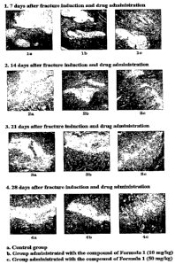
FIG. 1 an optical microphotograph showing sliced
tissue specimens of the 8t" rib extracted after fracture
induction, stained with Masson's trichrome.
Best Mode for Carrying Out the Invention
7
CA 02572897 2007-01-04
WO 2006/004369 PCT/KR2005/002138
The present invention pertains to a pharmaceutical
composition for the treatment of bone fractures, comprising a
benzamidine compound represented by the following chemical
formula 1 or a pharmaceutically acceptable salt thereof.
Chemical Formula 1
.~'
I. I
NHz
I I
.
wherein, R is a hydrogen atom or a hydroxyl group.
The benzamidine compound of Chemical Formula 1 may be
used in the form of pharmaceutically acceptable salts known
in the art. Preferable are acid addition salts prepared with
pharmaceutically acceptable free acids. Free acids suitable
for use in the present invention may be inorganic acids or
organic acids. Examples of the inorganic acids include
hydrochloric acid, bromic acid, sulfuric acid, phosphoric
acid, etc, and the organic acids may be exemplified by citric
acid, acetic acid, lactic acid, tartaric acid, fumaric acid,
formic acid, propionic acid, oxalic acid, trifluoroacetic
acid, methane sulfonic acid, benzene sulfonic acid, maleic
acid, benzoic acid, gluconic aicd, glycolic acid, succinic
acid, 4-morpholine ethane sulfonic acid, camphorsulfonic acid,
4-nitrobenzene sulfonic acid, hydroxyl-0-sulfonic acid, 4-
toluene sulfonic acid, galacturonic acid, embonic acid,
8
CA 02572897 2007-01-04
WO 2006/004369 PCT/KR2005/002138
glutamic acid and aspartic acid.
The benzamidine compound of Chemical Formula 1 may be
prepared according to known processes (Lee, Sung-Eun,
Synthesis and Biological Activity of Natural Products and
Designed New Hybrid Compounds for the Treatment of LTB4
Related Disease, Busan National University, a thesis for a Ph.
D degree, 1999. 8).
The term "bone fracture" as used herein means one of
various physical injuries of a bone, based on a complete or
incomplete disruption of the continuity of a bone , which are
classified according to anatomical location (epiphyseal,
metaphyseal, diaphyseal, intra-articular, proximal, midshaft,
distal, etc.), degree of fracture (complete, incomplete),
direction of fracture (transverse, oblique, spiral,
longitudinal), presence of open wound (open, closed), number
of fractures (simple, linear, segmental, comminuted, etc.),
stability of fracture (stable, unstable), displacement of
fracture, etc.
As compared to a non-treated group, a group treated
with the benzamidine compound of Chemical Formula 1 according
to the present invention was found to have the bony callus
significantly decreased in volume in a dose-dependent pattern,
but increased both in bone density and in bone strength, with
significance, in a dose-dependent pattern (p<0.01 or p<0.05).
Treatment with the benzainidine compound of Chemical
Formula 1 allowed the bony callus to significantly decrease
9
CA 02572897 2007-01-04
WO 2006/004369 PCT/KR2005/002138
in connective tissue and cartilage tissue while increasing
the content of a bone tissue with significance (p<0.01 or
p<0.05), compared to non-treatment. Both the decrease in
connective tissue and cartilage tissue and the increase in
bone tissue are dose-dependent.
In addition, the number of osteoclasts in a bony
callus increased significantly upon treatment with the
benzamidine compound of Chemical Formula 1, compared to non-
treatment, in the early phase of the fracture healing process
(p<0.01), and the increase pattern was dose-dependent.
In the late phase of the fracture healing process, a
group treated with the benzamidine compound of Chemical
Formula 1 had the bony callus decreased in the number of
osteoclasts with significance, compared to a non-treated
group (p<O.OI), which indicates that ossification was already
proceeding to some degree.
In summary, the benzamidine compound of Chemical
Formula 1 is an effective curative for bone fractures, with
functions of promoting the loss and ossification of the bony
callus formed during the fracture healing process. In more
detail, the benzamidine compound of the present invention
increases cellular components of bony callus in the early
phase of bone fracture healing process and promotes
endochondral ossification and intramembranous ossification in
the late phase in bone fracture healing process.
The composition of the present invention may further
CA 02572897 2007-01-04
WO 2006/004369 PCT/KR2005/002138
comprise at least one effective ingredients which are
equivalent or similar function to that of the benzamidine
compound of Chemical Formula 1 or its pharmaceutically
acceptable salt.
The composition of the present invention may further
comprise one or more pharmaceutically acceptable carriers. A
proper carrier may be selected from a group consisting of
saline, sterilized water, Ringer's solution, buffered saline,
a dextrose solution, a maltodextrin solution, glycerol,
ethanol, and combinations thereof, and may be, if necessary,
further supplemented with other typical additives such as an
antioxidant, a buffer, a static agent, etc. In combination
with a diluent, a dispersant, a surfactant, a binder, and a
lubricant, the composition of the present invention may also
be formulated into injectable dosage forms, such as aqueous
solutions, suspensions, emulsions, etc., pills, capsules,
granules, and tablets. Moreover, depending on the kind of
ingredient or disease, the formulation may be conducted using
methods known in the art or disclosed in Remington's
Pharmaceutical Science ((latest version), Mack Publishing
Company, Easton PA).
According to purposes, the composition of the present
invention may be administered orally or parenterally (e.g.,
intravenously, subcutaneously, intraabdominally, or
topically). The dosage amount of the composition of the
present invention varies depending on body weight, age,
11
CA 02572897 2007-01-04
WO 2006/004369 PCT/KR2005/002138
gender, health state, diet, administration time period,
administration route, excretion rate, disease severity, etc.
When all of these factors are taken into account, the
benzamidine compound of Chemical Formula 1 is administered
once or many times at a dose of approximately 10 to 1,000
mg/kg a day, and preferably at a dose of approximately 50 to
500 mg/kg a day.
For the prevention and treatment of physical injury of
bone comprising fracture, the composition of the present
invention can be used alone or in combination with surgery,
hormone therapy, chemical therapy, and/or a biological
response controller.
A better understanding of the present invention may be
obtained through the following examples which are set forth
to illustrate, but are not to be construed as the limit of
the present invention.
EXAWLE : Effect of Promoting Fracture Healing in Rib
Fracture-Induced Rat Model
The benzamidine compound of Chemical Formula 1 was
assayed for therapeutic effect on bone fracture in rat models
subjected to rib fracture. Starting from 2 days after the
induction of rib fracture, the administration of the
benzamidine compound was continued for one, two, three and
four weeks. Changes in (body weight, body weight gain,
12
CA 02572897 2007-01-04
WO 2006/004369 PCT/KR2005/002138
volume of bony callus, bone density, bone strength, and bone
histopathology were observed.
1. Experimental animals and Breeding managesnent
A total of 80 S.D. rats (10-week-old, BioGenomics,
Korea) was adapted to a laboratory environment for 12 days
before being used in experiments. While being housed at a
density of two or three to a plastic cage, the experimental
animals were kept in a breeding room under controlled
temperature (20 to 25 C) and humidity (30 to 35%). Under
light-dark cycles of 12 hours, the rats were allowed to have
free access to feedstuff and tap water.
2. Preparation and ac3ministration of sample
10 mg and 50 mg of N-hydroxy-4-{5-[4-(5-isopropyl-2-
methyl-1,3-thiazol-4-yl)phenoxylpentoxy}benzamidine were
completely dissolved in 5 ml of sterilized distilled water.
The benzamidine compound in the solutions was orally
administered at doses of 10 mg and 50 mg per kg of body
weight once a day for one, two, three and four weeks from day
2 of the surgery.
3. Induction of rib fracture
All the experimental animals were anesthetized with
ketamine hydrochloride and xylazine hydrochloride and
underwent an operation for inducing a fracture on the 8th and
13
CA 02572897 2007-01-04
WO 2006/004369 PCT/KR2005/002138
the 9th rib. In this regard, the ribs were transversely cut
with operation scissors. After the fracture induction, the
fractured ribs were assembled to be aligned with each other
and the wound cavity was closed through skin suture.
4. Change in body weight and weight gain
All the experimental animals were measured for body
weight one day before the operation, the day of the operation,
the day of administration, and 7, 14, 21 and 28 days after
administration. In order to reduce the difference among
individuals due to feedstuff intake, all experimental animals
were starved for 18 hours or more on the day of the
measurement. Also, to minimize the difference of change in
body weight of individual animals, body weight gain during
time periods from the day of the operation to 7, 14, 21 and
28 days after the administration were calculated.
The results are given in Table 1, below.
TABLE 1
Experimental Changes in Body Weight Gain (g)
Groups days after administration
7 days 14 days 21 days 28 days
Control 18.80 12.0740.20 25.0763.40 15.6871.60 15.82
Cpd. Of 10(mg/kg)16.00 13.5 44.40 14.5446.40 22.3 61.20 22.81
Chemical
Formula 1 50(mg/kg)14.80 08.8136.82 29.5268.60 16.6 84.40 23.37
As seen in Table 1, no significant changes in body
weight gain were observed over all -experimental periods,
14
CA 02572897 2007-01-04
WO 2006/004369 PCT/KR2005/002138
indicating that there were almost no errors attributable to
the administration of experimental substances or individual
differences between experimental animals.
S. Volume of bony callus
On the sacrificial day, the bony callus formed around
the fractured 8th and 9th ribs was separated from adjacent
tissues and taken out of all experimental animals. The
enucleated bony calluses were measured for long and short
diameters in millimeters. The volume of the bony callus was
calculated from the measurements using the following
mathematic formula 1.
Formula 1
Volume of Bony callus = 1/2 x (a x b2)
a: long diameter of bony callus,
b: short diameter of bony callus.
The results are given in Table 2, below.
TABLE 2
Changes in Bony Callus Volume(mm3)
Experimental
Groups Days After Administration
7 days 14 days 21 days 28 days
Control 35.35 7.96 19.09 3.11 11.69 4.15 9.25 3.00
Cpd. of 10 (mg/kg) 12.84 4.42* 5.47 1.81* 4.73 2.13* 3.96 2.41*
Chemical
Formula 50(mg/kg) 8.62 3.43* 4.36 1.44* 3.84 1.86* 3.37 0.79*
1
CA 02572897 2007-01-04
WO 2006/004369 PCT/KR2005/002138
*: significance compared to control (p<0.01)
As is apparent from Table 2, the volume of bony callus
according to fracture healing was significantly decreased in
the benzamidine compound-administered group, compared to the
non-treated control group (p<0.01), in a dose-dependent
pattern.
Thus, the benzamidine compound of Chemical Formula 1
is found to promote the loss of the bony callus formed during
the fracture healing process.
6. Histopathological observation
The 8th rib enucleated after the fracture induction
was fixed in 10% neutral formali.n, followed by
decalcification by changing a decalcification solution (2.24%
formic acid, 0.5N sodium hydroxide) with fresh solution once
a day for five days. After completion of the decalcification,
the rib was embedded in paraffin. The paraffin-embedded
tissue was sliced at a thickness of 3 to 4}un, stained with
hematoxylin-eosin or Masson's trichrome and observed through
an optical microscope.
The results are given in FIG. 1.
The benzamidine compound-administered group, as shown
in FIG. 1, was found to have increased bone tissue in bony
callus in all administration time periods, as opposed to the
none-treated group, and the increased behavior of the bone
16
CA 02572897 2007-01-04
WO 2006/004369 PCT/KR2005/002138
tissue was observed to be dose-dependent.
Hence, the benzamidine compound of Chemical Formula 1
can promote bone formation in the bony callus formed upon
fracture.
From the rib tissue specimen prepared above, the
amounts of the connective tissue, cartilage and bone tissue
in the bony callus were examined using an Analysis Image
processing system (SIS Germany) and are represented as
percentages in Tables 3 to 5, below.
Furthermore, the number of osteoclasts in the bony
callus, particularly, within an area of 200 }am2 on the
fracture surface at which endochondral ossification commenced,
was measured using an Analysis Image processing system (SIS
Germany)
The results are given in Table 6, below.
TABLE 3
Changes in Content of Connective Tissue
Experimental of Bony Callus Days after Administration
Groups (% relative to total bony callus)
7 days 14 days 21 days 28 days
Control 51.34 11.5519.43 2.0115.10 2.9 7.14 2.73
Cpd. Of 10(mg/kg) 33.19 3.06 6.28 0.72 5.55 1.42 3.20 0.89
Chemical
50(mg/kg)29.51 5.70* 6.06 0.44* 3.58 0.62*2.59 0.52
Formula 1
*: significance compared to control(p<0.01),
**: significance compared to control(p<0.05)
17
CA 02572897 2007-01-04
WO 2006/004369 PCT/KR2005/002138
As seen in Table 3, the benzamidine compound-
administered group decreased dose-dependently in the content
of connective tissue within the bony callus tissue, compared
to the non-treated group, with significance (p<0.01 or
p<0. 05) .
As a result, the benzamidine compound of Chemical
Formula 1 is identified to promote the substitution of bone
tissue for the connective tissue within the bony callus
formed upon fracture, that is, ossification.
TABLE 4
Changes in Content of Cartilage Tissue
Experimental in Bony Callus Days After Administration
Groups (o relative to total bony callus)
7 days 14 days 21 days 28 days
Control 43.28 4.66 39.49 2.79 24.93 4.13 17.78 2.30
Cpd. Of 10(mg/kg)24.79 5.43 23.77 3.44 18.51 2.29 6.59 2.02*
Chemical
Formula 1 50(mg/kg)22.42 5.45 20.09 6.38 11.49 2.31 5.37 1.38*
*: significance compared to control(p<0.01)
The cartilage tissue within the bony callus tissue, as
is apparent from Table 4, was significantly decreased in the
benzamidine compound-administered group, as compared to the
non-treated group, in a dose-dependent pattern (p<0.01).
Accordingly, the benzamidine compound of Chemical
Formula 1 is identified to promote the substitution of bone
tissue for the cartilage tissue within the bony callus formed
18
CA 02572897 2007-01-04
WO 2006/004369 PCT/KR2005/002138
upon fracture, that is, endochondral ossification.
TABLE 5
Changes in Content of Bone Tissue
Experimental in Bony Callus Days After Administration
Groups (% relative to total bony callus)
7 days 14 days 21 days 28 days
Control 1.92 0.70 38.21 4.92 54.49 6.04 66.88 5.68
Cpd. Of 10(mg/kg) 37.95 6.44 54.31 9.50 66.71 5.41 83.30 4.43
Chemical
50(mg/kg)39.24 14.12 55.94 8.38*74.07 8.43*87.27 8.97*
Formula 1
*: significance compared to control(p<0.01),
**: significance compared to control(p<0.05)
As seen in Table 5, the bone tissue within the bony
callus was significantly increased in the benzamidine
compound-administered group, compared to the non-treated
group(p<0.01 or p<0.05) in a dose-dependent pattern.
Thus, the benzamidine compound of Chemical Formula 1
is identified to promote the ossification of the bony callus
formed during the fracture healing process.
TABLE 6
Changes in Population of Osteoclasts
within
Experimental Bony Callus Days After Administration
Groups
(Counts present within 200 }iire of bony
callus)
7 days 14 days 21 days 28 days
Control 15.80 1.92 21.80 3.35 56.80 3.03 41.60 11.4
Cpd. Of Chemicall0(mg/kg)43.80 3.83 50.60 2.70 31.00 6.67 21.60 3.58
Formula 1 50(mg/kg)42.60 4.62 53.60 2.41 22.20 3.03 17.60 2.97*
19
CA 02572897 2007-01-04
WO 2006/004369 PCT/KR2005/002138
*: significance compared to control(p<0.01)
In the early phase of the facture healing process, as
seen in Table 6, the number of osteoclasts in the bony callus
was increased in the benzamidine compound-administered group,
compared to the non-treated group, with significance (p<0.01),
and the number of osteoclasts was found to increase as the
dosage increased. Thus, the administration of the
benzamidine compound of Chemical Formula 1 leads to a dose-
dependent increase in cellular components within bony callus
in the early phase of the fracture healing process.
In the late phase of the fracture healing process, a
group treated with the benzamidine compound of Chemical
Formula 1 had the bony callus decreased in the number of
osteoclasts with significance, compared to a non-treated
group (p<0.01), which indicates that ossification was already
proceeding to some degree.
In conclusion, the benzamidine compound of Chemical
Formula 1 is very useful as a curative for bone fractures,
with the function of promoting the ossification of the bony
callus formed upon fracture.
7. Measurement of bone density of bony callus
The 9th rib enucleated after the fracture induction
was measured for the bone density around the bony callus
using dual-energy x-ray absorptiometry (DEXA, PXlmus; Lunar
CA 02572897 2007-01-04
WO 2006/004369 PCT/KR2005/002138
Medison, WI) and the bone density is calculated in mg/cm2 in
Table 7.
TABLE 7
Changes in Bone Density of Bony Callus
Experimental
Days After Administration (mg/cm2)
Groups
7 days 14 days 21 days 28 days
Control 0.12 0.04 0.22 0.03 0.28 0.08 0.39 0.07
Cpd. Of 10(mg/kg)0.24 0.04 0.32 0.04 0.39 0.04 0.55 0.06
Chemical
50 (mg/kg) 0. 24 0 . 03 0. 32 0 . 04 0. 44 0 . 07* 0. 57 0. 04*
Formula 1
*: significance compared to control(p<0.01),
**: significance compared to control(p<0.05)
The benzamidine compound-administered group, as is
apparent from the data of Table 7, increased in the bone
density of the bony callus, compared to the non-treated
control, with significance (p<0.01 or p<0.05), and the bone
density increased as the dose increased.
Therefore, the benzamidine compound of Chemical
Formula 1 is identified to increase a bone density of the
bony callus formed upon fracture.
8. NSeasuresnent of bone strength of bony callus
The bone strength around the fracture face at which a
bony callus was formed in the 9th rib enucleated after the
fracture induction was determined from three point bending
tests using an Instron material testing system (Instron 6022;
21
CA 02572897 2007-01-04
WO 2006/004369 PCT/KR2005/002138
Instron, USA; speed 20 mm/min).
The results are given in Table 8, below.
TABLE 8
Changes in bone strength of bony callus
Experimental Days After Administration
Groups (Nos. of Impact applied)
7 days 14 days 21 days 28 days
Control 1.24 0.28 1.53 0.51 2.06 0.18 2.38 0.22
Cpd. Of 10(mg/kg)2.15 0.42 2.57 0.65 3.10 0.40 3.26 0.43
Chemical
50 (mg/kg) 2. 35 0. 47** 2. 84 0. 34* 3.23 0.35* 3. 35 0. 38**
Formula 1
significance compared to control (p<0.01),
**: significance compared to control (p<0.05)
As seen in Table 8, the benzamidine compound-
administered group increased in the bone strength, compared
to the non-treated group, with significance (p<0.01 or
p<0.05) in a dose-dependent pattern.
As a consequence, the benzamidine compound of Chemical
Formula 1 is identified to increase the bone strength in the
bony callus formed upon fracture.
9. Statistics
All numerals are represented as mean standard
deviation, and statistical significance of the differences
relative to the control was analyzed using Mann-Whitney U-
Wilcoxon Rank Sum test with the aid of SPSS (Release 6.1.3.,
SPSS Inc., USA).
22
CA 02572897 2007-01-04
WO 2006/004369 PCT/KR2005/002138
Likewise, methane sulfonic acid salts and hydrochloric
acid salts of N-hydroxy-4-{5-[4-(5-isopropyl-2-methyl-1,3-
thiazol-4-yl)phenoxy]pentoxy}benzamidine, and 4-{5-[4-(5-
isopropyl-2-methyl-l,3-thiazol-4-yl) phenoxy] pentoxy}
benzamidine and its methane sulfonic acid salts and
hydrochloric acid salts were found to exhibit the same
healing effects as above.
Preparation Example:
1. Preparation of powder
Benzamidine compound of Chemical Formula 1 2g
Lactose 0.5g
Mannitol 0.5g
The ingredients were mixed and filled in an airtight
sac to prepare a powder agent.
2. Preparation of tablet
Benzamidine compound of Chemical Formula 1 100mg
Corn Starch 50mg
Microcrystalline Cellulose 50mg
Lactose 100mg
Povidone 15mg
Magnesium Stearate 2mg
A mixture of the ingredients was prepared into a
tablet using a general tabletting method.
23
CA 02572897 2007-01-04
WO 2006/004369 PCT/KR2005/002138
3. Preparation of capsule
Benzamidine compound of Chemical Formula 1 100mg
Corn Starch 50mg
Microcrystalline Cellulose 50mg
Lactose 100mg
Povidone 15mg
Magnesium Stearate 2mg
A mixture of the ingredients was filled into a gelatin
capsule according to a typical procedure, so as to give a
capsule agent.
4. Preparation of soft capsule
Benzamidine compound of Chemical Formula 1 100 mg
Soybean Oil 400mg
Lecithin 20mg
Gelatin 200mg
A soft capsule was prepared from the mixture of the
ingredients, according to a typical procedured.
5. Preparation of injection
Benzamidine compound of Chemical Formula 1 10 ug/ml
Diluted Hydrochloric acid BP to pH 3.5
Injectable Sodium chloride BP 1 ml at most
A solution of the benzamidine compound of Chemical
Formula 1 in a proper volume of injectable sodium chloride BP
was adjusted to pH 3.5 with diluted hydrochloric acid BP and
24
CA 02572897 2007-01-04
WO 2006/004369 PCT/KR2005/002138
its volume was adjusted with injectable sodium chloride BP.
After being sufficiently mixed, the solution was filled in a
ml type I ampul made from transparent glass, which was then
molten so that the solution was packaged under the upper grid
5 of air. An injection was obtained by autoclaving at 120 C
for 15 min or longer.
Industrial Applicability
The composition of the present invention can
significantly reduce the volume of bony callus, increase
bony density and strength of bony callus, and decrease the
contents of connective tissue and cartilage tissue in bony
callus, and thus promote loss and ossification of the bony
callus formed during the fracture healing process.
Therefore, the composition of the present invention is
useful for the treatment of bone fracture.