Note : Les descriptions sont présentées dans la langue officielle dans laquelle elles ont été soumises.
DEMANDES OU BREVETS VOLUMINEUX
LA PRESENTE PARTIE I)E CETTE DEMANDE OU CE BREVETS
COMPREND PLUS D'UN TOME.
CECI EST LE TOME DE _2
NOTE: Pour les tomes additionels, veillez contacter le Bureau Canadien des
Brevets.
JUMBO APPLICATIONS / PATENTS
THIS SECTION OF THE APPLICATION / PATENT CONTAINS MORE
THAN ONE VOLUME.
THIS IS VOLUME 1 OF 2
NOTE: For additional volumes please contact the Canadian Patent Office.
CA 02576055 2007-02-05
WO 2006/031313 PCT/US2005/028012
METHODS AND COMPOSITIONS FOR CELL ACTIVATION
CROSS-REFERENCE
[0001] This application claims the benefit of U.S. Provisional Application No.
60/599,604, filed August 5, 2004, which application is incorporated herein by
reference in its entirety.
GOVERNMENT RIGHTS
[0002] This invention was made with government support under federal grant no.
5KO8 CA082176-04 awarded by the National Cancer Institute of the National
Institutes of Health. The United States Government may have certain rights in
this
invention.
BACKGROUND OF THE INVENTION
[0003] Telomeres, which define the ends of chromosomes, consist of short,
tandemly
repeated DNA sequences loosely conserved in eukaryotes. Human telomeres
consist
of many kilobases of (TTAGGG)N together with various associated proteins.
Small
amounts of these terminal sequences or telomeric DNA are lost from the tips of
the
chromosomes during the S phase of the cell cycle because of incomplete DNA
replication. Many human cells progressively lose terminal sequence with cell
division,
a loss that correlates with the apparent absence of telomerase in these cells.
The
resulting telomeric shortening has been demonstrated to limit cellular
lifespan, thereby
resulting in cellular senescence and inactivation.
[0004] Telomerase is a ribonucleoprotein (RNP) that uses a portion of its RNA
moiety
as a template for telomeric DNA synthesis. The catalytic core of telomerase is
comprised of two essential components: TERT, the telomerase reverse
transcriptase,
and TERC, the telomerase RNA component. Telomerase synthesizes telomeres
through reverse transcription of the template sequence encoded in TERC and
through
protein interactions that facilitate telomere engagement. Genetic studies in
yeast,
murine, and human cells have established that TERT and TERC are obligate
partners
in telomere synthesis; inactivation of either subunit abrogates enzymatic
activity and
prevents telomere addition, leading to progressive telomere shortening as a
consequence of the end replication problem. Telomere shortening ultimately
leads to
1
CA 02576055 2007-02-05
WO 2006/031313 PCT/US2005/028012
telomere uncapping, a change in telomere structure associated with loss of end
protection that results in both checkpoint activation and chromosomal end-to-
end
fusion.
[0005] According to this well-validated paradigm, telomerase functions
primarily to
prevent telomere uncapping through enzymatic extension of telomeres.
Telomerase is
thought to serve a similar function during tumor development where it prevents
telomere shortening and uncapping, thus enabling cancer cells to proliferate
in an
unlimited fashion.
[0006] A general need exists for the regulation and control of cell cycle
stages, e.g.,
control of progression of a cell from a quiescent state to an active state,
control of
progression of a cell from a non-proliferating state to a proliferating state,
and the like.
Regulation and control of cell cycle stage, e.g., from a quiescent state to an
active
state, is beneficial for a number of diseases or disorders related to cell
proliferative
capacity and senescence, wherein the disorder results from the cells entering
a
quiescent state (i.e., loss of proliferative capacity), and where activation
(i.e., a
proliferative state) will contribute to treatment of the disorder.
Accordingly, there
continues to be a need for development of such methods.
Relevant Literature
[0007] U.S. Patents of interest include: 6,166,178; 6,337,200; and 6,309,867.
Also of
interest are: Cheong et al., 2003, Exp. Mol. Med., 35(3):141-153; Gonzalez-
Suarez et
al., 2001, EMBO J., 20(11): 2619-2630; Ramirez et al., 1997, J. Invest.
Dermatol.,
108(1):113-117; Harle-Bachor et al., 1996, PNAS, 93(13):6476-6481; and Rochet
et
al., 1994, Cell, 76(6):1063-1073.
SUMMARY OF THE INVENTION
[0008] Methods and compositions for cell activation are provided. In
practicing the
subject methods, cell activation is achieved by conditionally increasing
expression of
either a telomerase reverse transcriptase (TERT) or a telomerase RNA component
..
(TERC). Also provided are transgenic animals and systems for practicing the
subject
methods.
FEATURES OF THE INVENTION
[0009] A feature of the present invention provides a method for activating a
cell by
conditionally increasing transcription of a coding sequence of either (e.g.,
only one of)
2
CA 02576055 2007-02-05
WO 2006/031313 PCT/US2005/028012
.. ... .... ..... ....... .. ...... ..... i ;....n ..n.. n....
a telomerase reverse transcriptase (TERT), or a telomerase RNA component
(TERC)
in the cell in a manner sufficient to activate the cell. In some embodiments,
the
subject method conditionally increases transcription of a TERT coding
sequence. In
other embodiments, the subject method conditionally increases transcription of
a
TERC coding sequence. Such a cell includes a hair follicle cell; a pancreatic
islet cell;
a neuronal cell; a bone marrow cell; and the like. Such a cell also includes a
stem cell
or progenitor cell in the hair follicle, bone marrow, pancreas, central
nervous system,
bone and cartilage, liver, and the like. The methods may be performed in vitro
or in
vivo. In some embodiments, the cell is present in a mammal, such as a human.
[0010] In some embodiments, the method includes introducing into the cell an
agent
that conditionally increases transcription of the coding sequence. In some
embodiments, the agent activates a conditional promoter system operably linked
to
the coding sequence. In other embodiments, the method includes introducing
into the
cell a nucleic acid vector including an expression system having a conditional
promoter system operably linked to the coding sequence. In further
embodiments, the
conditional promoter system includes a tetracycline inducible promoter.
[0011] Another feature of the present invention provides a method for
activating a cell
in a host by administering to the host an effective amount of an agent that
conditionally increases transcription of a coding sequence of either TERT or
TERC to
activate the cell. In some embodiments, the subject method conditionally
increases
transcription of a TERT coding sequence. In other embodiments, the subject
method
conditionally increases transcription of a TERC coding sequence. Such a cell
includes a hair follicle cell; a pancreatic islet cell; a neuronal cell; a
bone-marrow cell;
and the like. The methods may be performed in vitro or in vivo. In some
embodiments, the cell is present in a mammal, such as a human.
[0012] In some embodiments, the method includes introducing into the cell a
nucleic
acid vector including an expression system having a conditional promoter
system
operably linked to the coding sequence. In further embodiments, the
conditional
promoter system includes a tetracycline inducible promoter.
[0013] Yet another feature of the invention provides a method for activating a
hair
follicle cell in a host in vivo by administering to the host an effective
amount of an
agent that conditionally increases transcription of a coding sequence of
either TERT
3
CA 02576055 2007-02-05
WO 2006/031313 PCT/US2005/028012
.,,.... .
or TERC to activate the hair follicle cell. In some embodiments, the
activation of the
hair follicle cells results in hair growth.
[0014] .. In some embodiments, the subject method conditionally increases
transcription
of a TERT coding sequence. In other embodiments, the subject method
conditionally
increases transcription of a TERC coding sequence. The methods may be
performed
in vitro or in vivo. In some embodiments, the cell is present in a mammal,
such as a
human. In some embodiments, the method includes introducing into the cell a
nucleic
acid vector including an expression system having a conditional promoter
system
operably linked to the coding sequence. In further embodiments, the
conditional
promoter system includes a tetracycline inducible promoter.
[0015] Yet another feature of the invention provides a transgenic animal,
wherein the
transgenic animal conditionally transcribes either TERT or TERC. In some
embodiments, the transgenic animal includes a TERT transgene. In other
embodiments, the transgenic animal includes a TERC transgene. In such
embodiments, the transgenic animal is a mammal, such as a rodent.
[0016] In some embodiments, the conditional transcription is provided by a
conditional
promoter system operably linked to the TERT transgene or TERC transgene. In
further embodiments, the conditional promoter system is a tetracycline
inducible
promoter system.
[0017] Yet another feature of the invention provides a method for identifying
a
compound that is capable of modulating the activity of one of TERT or TERC, by
activating a cell by conditionally increasing transcription of a coding
sequence of either
TERT or TERC; administering a compound to the cell; and observing the effect
of the
compound on the cell. In some embodiments, the activating includes
conditionally
increasing transcription of a TERT coding sequence. In other embodiments, the
activating includes conditionally increasing transcription of a TERC coding
sequence.
In such methods, the cell may be in a mammal, such as rodent, such as a mouse.
In
such methods, the compound may be a polypeptide, a nucleic acid, or a small
molecule. In such methods, the modulating may be enhancing activity or
repressing
activity. In such embodiments, such activity may include active extension of
telomeric
repeat sequences at the ends of chromosomes, or may not include active
extension of
telomeric repeat sequences at the ends of chromosomes.
4
CA 02576055 2007-02-05
WO 2006/031313 PCT/US2005/028012
[0018] In some embodiments, the activating includes administering to the cell
an agent
that conditionally increases transcription of the coding sequence. In further
embodiments, the activating includes administering an agent that activates a
conditional promoter system operably linked to the coding sequence. In other
embodiments, method further includes introducing into the cell a nucleic acid
vector
including an expression system having a conditional promoter system operably
linked
to the coding sequence. In further embodiments, the conditional promoter
system
includes a tetracycline inducible promoter.
100191 Yet another feature of the invention provides a system for use in
identifying a
compound that is capable of modulating the activation of either TERT or TERC,
including transgenic animal conditionally transcribing either TERT or TERC,
and an
agent that activates conditional transcription of the transgene. In some
embodiments,
the conditional transcription is provided by a conditional promoter system
operably
linked to the TERT transgene or TERC transgene. In further embodiments,
conditional
promoter system is the tetracycline inducible promoter system. In such
systems, the
animal may be a mammal, such as rodent, such as mouse. In addition, in such
systems, the agent may be doxycycline or an analog thereof.
[0020] Yet another feature of the invention provides a conditional expression
vector
including a conditional promoter system operably linked to the coding sequence
of
either TERT OR TERC. In some embodiments, the conditional promoter system is a
tetracycline inducible promoter system.
[0021] Yet another feature of the invention provides a system for use in
producing a
conditional expression animal model including a conditional expression vector
that
includes a conditional promoter system operably linked to the coding sequence
of
either TERT or TERC, and an animal. In some embodiments, the conditional
promoter system is a tetracycline inducible promoter system. In further
embodiments,
the animal is a mammal, such as rodent.
BRIEF DESCRIPTION OF THE DRAWINGS
[0022] The invention is best understood from the following detailed
description when
read in conjunction with the accompanying drawings. It is emphasized that,
according
to common practice, the various features of the drawings may not be to-scale.
On the
contrary, the dimensions of the various features may be arbitrarily expanded
or
reduced for clarity. Included in the drawings are the following figures:
CA 02576055 2007-02-05
WO 2006/031313 PCT/US2005/028012
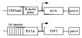
[0023] Fig. IA is a schematic depiction of actin-rtTA and tetop-TERT transgene
constructs.
[0024] Fig. 1 B is an image of a Northern blot showing expression of TERT mRNA
in
the skin of i-TERT Tg treated with doxycycline (dox) mice, but not in i-TERT
Tg (-dox)
mice or non-transgenic littermates (WT) at day 50.
[0025] Fig. 1C is an image showing the induction of telomerase activity in the
skin of i-
TERT Tg (+dox) mice as compared with i-TERT Tg mice (-dox) or WT mice at day
50.
[0026] Fig. 1 D is a diagram of anagen and telogen hair follicle cycle.
[0027] Fig. 1E is an image showing that telomerase activity is high during the
anagen
phase of the hair follicle and silenced during catagen and telogen phases in
hair
follicle cycling. Extracts are taken from skin at postnatal days 4 and 10
(anagen), 16
(catagen), 19 and 21 (telogen), 28 (anagen), 34 (catagen), and 52 (telogen).
[0028] Fig. 1 F is a photograph of i-TERT Tg mouse (+dox) (background) and i-
TERT
Tg (-dox) (foreground) at day 50, showing the disorganized fur and droopy
whiskers of
the +dox mouse.
[0029] Fig. 1G is a histological analysis showing that TERT activation,
beginning at
day 21, promotes changes in the state of the hair follicle from telogen to
anagen at
day 50. Follicles were appropriately in anagen at day 28 in both groups. i-
TERT Tg (-
dox) mice were indistinguishable from non-transgenic mice.
[0030] Fig. 1 H shows immunofluorescence sections of hair follicle epithelium
skin of i-
TERT Tg mice from day 50 following induction of TERT mRNA by doxycycline
treatment. Merging of the immunofluorescence images shows an overlap in
distribution pattern of TERT with keratin-14 protein.
[0031] Figs. 2A-2H shows intact differentiation and development in TERT
induced hair
follicles. In each panel, TERT-induced anagen (day 50), denoted Tg(+dox), is
compared to non-transgenic anagen (day 28) and age-matched non-transgenic mice
in telogen (day 50). Immunofluorescence showed normal patterns of: outer root
sheath differentiation by keratin-14 staining (Fig. 2A); inner layer of outer
root sheath
differentiation marked by keratin-6 (Fig. 2B); hair differentiation by AE13
staining (Fig.
2C); Normal inner root sheath differentiation marked by AE15 (Fig. 2D);
proliferation
in the matrix cells by Ki-67 staining (Fig. 2E). In situ hybridization
analysis showed:
normal, asymmetic pattern of Shh expression in the invaginating anagen hair
follicle in
both WT (day 28) and i-TERT Tg (day 50) (Fig. 2F); Lef1 is expressed in the
matrix
6
CA 02576055 2007-02-05
WO 2006/031313 PCT/US2005/028012
cells in both the WT and i-TERT Tg induced anagen hair follicle, but is absent
from the
telogen hair follicle (Fig. 2G); and Shh is absent from normal telogen (WT day
50)
(Fig. 2H).
[0032] Figs. 3A-3C shows that TERT triggers a rapid transition from telogen to
anagen. i-TERT Tg mice and non-transgenic littermates (WT) were treated with
doxycycline beginning at day 40, monitored through serial biopsies 0, 3, 9 and
12
days subsequently (day 0, 3, 9, 12). Fig. 3A shows that TERT mRNA expression
was
first detected at day 3, but increased substantially by day 9 via Northern
blot (left).
GAPDH was used as a loading control. Telomerase activity increased with
similar
kinetics seen by TRAP assay (right). Fig. 3B is histological data from the WT
and
iTERT TG groups showing that both groups were in telogen phase at the
initiation of
the experiment, age 40 days (day 0). After 9 days on doxycycline, follicles in
i-TERT
Tg mice entered early anagen (arrow), whereas controls remained in telogen-
(asterisk). Full anagen occured by 12 days on doxycycline in i-TERT mice. H&E,
20x.
Fig. 3C is a photograph of mice that were administered doxycycline in telogen
at age
45 days, shaved at age 55 days, and monitored for 14 days. Shaved hair briskly
grew
only in i-TERT Tg mice (+dox) (right), but not in i-TERT Tg mice (-dox)
(middle) or
non-transgenic littermates (left).
[0033] Figs. 4A-4B shows that TERT activates hair follicle stem cells
independent of
its function in telomere synthesis. TERC /- mice were backcrossed to the FVB/N
.
strain, then intercrossed with i-TERT Tg mice to generate cohorts of i-TERT Tg
mice
on TERC+/+, TERC+/- or TERC-/- backgrounds. Mice in each group were treated
with doxycycline beginning at day-21 and analyzed at day 50. Fig. 4A is
histological
analysis showing that induction of TERT facilitated transition from telogen to
anagen
in all TERC backgrounds, including TERC+/+, TERC+/-, and TERC-/-. Negative
controls remained in telogen including, i-TERT (-dox), single transgenic mice,
and non
transgenic mice in TERC +/+, TERC+/-, and TERC-/- backgrounds. Fig. 4B shows
that skin samples from i-TERT Tg and TERC-/- mice lacked telomerase activity
by
TRAP and TERC expression by RT PCR. The TERT transgene was induced similarly
in i-TERT Tg mice, irrespective of TERC genotype.
[0034] Figs. 5A-5C shows that telomeres remain stable and capped in i-TERT Tg
mice. Fig. 5A is a northern analysis showing induction of Tert in i-Tert Tg
MEFS
treated with doxycycline for 72 hours (left) or splenocytes treated with
doxycycline for
7
CA 02576055 2007-02-05
WO 2006/031313 PCT/US2005/028012
48 hours (right) as compared with controls. Fig. 5B shows images of metaphase
preparations from MEFs (left) and spienocytes (right), which showed no
increase in
chromosomal end-to-end fusions with TERT induction. Fig. 5C is a table
depicting the
average number of chromosomes, and number of fusions per metaphase found in
WT, i-TERT Tg(-dox), and i-Tert Tg(+dox) samples. No fusions were found in any
metaphases.
[0035] Figs. 6A-6D shows that induction of TERT does not lead to increased
apoptosis or anaphase bridge formation. Fig. 6A shows the results of a TUNEL
assay
that was performed on skin sections from i-Tert Tg(+dox) mice at day 50 as
well as
WT at day 50, WT at day 28, and late generation Tert-/- at day 28 as controls.
Increased number of TUNEL+ cells were only detected in the late generation
Tert-/-
sections. Anaphase bridges were detected in late generation Tert-/- skin
sections but
not in the i-Tert Tg(+dox) skin sections or WT controls. Fig. 6B is a bar
graph
depicting the average number of TUNEL positive cells per hair follicle. Fig.
6C is a
bar graph depicting the number of anaphase bridges per total number of
anaphases
surveyed. Fig. 6D is a table indicating the number of anaphases surveyed and
the
fraction that were bridges in each genotype. Anaphase bridges were only found
in the
late generation Tert-/- skin sections.
[0036] Figs. 7A-7B shows the conditional activation of TERC and the analysis f
the
hair follicle. Fig. 7A is a schematic depiction of actin-rtTA and tetop-TERC
transgene
constructs. Fig. 7B shows the results of a histological analysis from 50 day
old mice
showing that TERC activation promotes changes in the state of the hair
follicle from
telogen to anagen in the TERC Tg mice (+dox) (bottom) as compared to the TERC
Tg
(-dox) (middle) and non-trangenic littermates (top).
[0037] Fig. 8 is a photograph of mice that were administered doxycycline in
telogen at
age 45 days, shaved at age 55 days, and monitored for 14 days. Shaved hair
briskly
grew in iTERT Tg mice (+dox) (right) and iTERC Tg mice (+dox) (middle), but
not in
the wild type (non transgenic) littermates (left).
[0038] Fig. 9 shows tissue sections from i-TERC mice on doxycycline (right
panel) and
wild type controls (left panel) were hybridized with an anti-sense TERC probe.
As
shown in Fig. 9, transgenic TERC (red) was detected in the skin epithelium, in
a
pattern that overlaps with keratin-14 (green), a marker of the basal layer of
the
epidermis and the outer root sheath of the hair follicle.
8
CA 02576055 2007-02-05
WO 2006/031313 PCT/US2005/028012
[0039] Figs. IOA-IOF shows that TERT activates stem cells, depleting BrdU
label
from LRCs. Fig. IOA shows the maintenance of immunofluorescence for BrdU (red)
and CD34 (green) of LRCs in Non-Tg group, but dramatic loss of BrdU label in i-
TERT
mice after doxy treatment (pre-doxy = day 55, post-doxy= day 90). Fig. 10B is
a graph
showing the quantification of LRC data from Fig. IOA. The graph shows that the
number of BrdU+ cells/CD34+ cells. i-TERT (black bars, n=4 mice), Non-Tg (open
bars, n=3 mice), (-) indicates pre-doxy, (+) indicates post-doxy. Fig. IOC is
an LRC
analysis from whole mounts of epidermis from tail of mice labeled with BrdU at
day 10,
switched to doxy at day 40 and analyzed at day 65. (BrdU=red, K14=green). Fig.
10D
shows immunofluorescence using Ki-67 (red) to mark proliferating cells and K14
(green) to identify basal layer of skin. Fig. 10E is a graph showing the
quantitation of
proliferation index in Fig. 10D as Ki-67+ cells/100Nm length of basal layer
(n=2 mice
for each comparison). Fig. 10F shows a GFP epifluorescence costained with CD34
(inset, confocal microscopy) in skin section from an actin-GFP mouse. Fig. 10G
shows RNA in situ analysis for TERT mRNA in i-TERT(+doxy) mouse skin. The
inset
shows TERT mRNA expression (cytoplasmic) overlaps in bulge with LRCs, marked
by
BrdU (nuclear). Fig. 10H shows H&E sections from K5tTA+; tetop-TERT+ (-doxy)
(bottom) and Non-Tg (top) mice, 20X. Error bars indicate standard deviation. p
values
based on t-test. *=autofluorescence of hair.
DETAILED DESCRIPTION OF THE INVENTION
[0040] Methods and compositions for cell activation are provided. In
practicing the
subject methods, transcription of a coding sequence for either (i.e., one of)-
a -
telomerase reverse transcriptase (TERT) or a telomerase RNA component (TERC)
is
conditionally increased. Also provided are transgenic animals and systems for
practicing the subject methods.
[0041] Before the present invention is described further, it is to be
understood that this
invention is not limited to particular embodiments described, as such may, of
course,
vary. It is also to be understood that the terminology used herein is for the
purpose of
describing particular embodiments only, and is not intended to be limiting,
since the
scope of the present invention will be limited only by the appended claims.
[0042] Where a range of values is provided, it is understood that each
intervening
value, to the tenth of the unit of the lower limit unless the context clearly
dictates
otherwise, between the upper and lower limits of that range is also
specifically
9
CA 02576055 2007-02-05
WO 2006/031313 PCT/US2005/028012
disclosed. Each smaller range between any stated value or intervening value in
a
stated range and any other stated or intervening value in that stated range is
encompassed within the invention. The upper and lower limits of these smaller
ranges may independently be included or excluded in the range, and each range
where either, neither or both limits are included in the smaller ranges is
also
encompassed within the invention, subject to any specifically excluded limit
in the
stated range. Where the stated range includes one or both of the limits,
ranges
excluding either or both of those included limits are also included in the
invention.
[0043] Unless defined otherwise, all technical and scientific terms used
herein have
the same meaning as commonly understood by one of ordinary skill in the art to
which
this invention belongs. Although any methods and materials similar or
equivalent to
those described herein can be used in the practice or testing of the present
invention,
the preferred methods and materials are now described. All publications
mentioned
herein are incorporated_herein by reference to disclose and describe the
methods
and/or materials in connection with which the publications are cited.
[0044] It must be noted that as used herein and in the appended claims, the
singular
forms "a", "an", and "the" include plural referents unless the context clearly
dictates
otherwise. Thus, for example, reference to "a cell" includes a plurality of
such cells
and reference to "the agent" includes reference to one or more agents and
equivalents thereof known to those skilled in the art, and so forth._
[0045] The publications discussed herein are provided solely for their
disclosure prior
to the filing date of the present application. Nothing herein is to be
construed as an
admission that the present invention is not entitled to antedate such
publication by
virtue of prior invention. Furthermore, the dates of publication provided may
be
different from the actual publication dates which may need to be independently
confirmed.
[0046] The practice of the present invention will employ, unless otherwise
indicated,
conventional methods of chemistry, biochemistry, recombinant DNA techniques
and
virology, within the skill of the art. Such techniques are explained fully in
the literature.
See, e.g., Fundamental Virology, 2nd Edition, vol. I & II (B. N. Fields and D.
M. Knipe,
eds.); A. L. Lehninger, Biochemistry (Worth Publishers, Inc., current
addition);
Sambrook, et al., Molecular Cloning: A Laboratory Manual (3rd Edition, 2001);
Methods In Enzymology (S. Colowick and N. Kaplan eds., Academic Press, Inc.);
CA 02576055 2007-02-05
WO 2006/031313 PCT/US2005/028012
Oligonucleotide Synthesis (N. Gait, ed., 1984); A Practical Guide to Molecular
Cloning
(1984).
METHODS
[0047] As summarized above, the subject invention provides a method for
activating a
cell. By "activating" is meant that the cell state of the cell is progressed
or transitioned
from a first, quiescent state to a second non-quiescent state. As used herein
a
"quiescent state" means a non-proliferating and non-transcriptionally active
state, i.e.,
a state in which the cellular number of one or more cells is not increasing by
cellular
division, or increasing at a level below that of an actively proliferating
state. As used
herein a "non-quiescent state" means either a proliferating state, i.e., a
state in which
the cellular number of one or more cells is increasing by cellular division,
or a non-
proliferating and transcriptionally active state, i.e., a state in which the
transcription
rate of nucleic acid coding sequences within the cell is increased, e.g., by
at least
about 2-fold, as compared to the first non-transcriptionally active state, and
where the
cellular number of one or more cells is not increasing by cellular division,
or increasing
at a level below that of an actively proliferating state. The "non-quiescent
state" may
include active extension of telomeric repeat sequences at the ends of
chromosomes,
or may not include active extension telomeric repeat sequences at the ends of
chromosomes. In other words, "activating" a cell by the subject method to a
second
"non-quiescent state" does not require that active extension of telomeric
repeat
sequences at the ends of chromosomes occur during the second "non-quiescent
state".
[0048] In some embodiments, the subject method provides for activating a cell
by
progressing or transitioning a cell from a first state of non-proliferation to
a second
state of proliferation, wherein by a second state of proliferation is meant
that the
cellular number is increasing by cellular division as compared to the first
state of non-
proliferation. In further embodiments, the second state of proliferation also
includes
active extension of telomeric repeat sequences at the ends of chromosomes: In
other
embodiments, the second state of proliferation does not include active
extension of
telomeric repeat sequences at the ends of chromosomes.
[0049] In addition, with respect to undedicated progenitor cells (i.e.,
undifferentiated
stem cells), by activating is meant that the progenitor cell is moved from a
first
quiescent state to second non-quiescent state, where the first quiescent state
is
11
CA 02576055 2007-02-05
WO 2006/031313 PCT/US2005/028012
..,.. ...... _... ,. .._... .... _...
characterized by a state in which the cellular number is not increasing by
cellular
division, or increasing at a level below that of an actively proliferating
state, and the
second non-quiescent state is characterized by a state in which the cellular
number is
increasing by cellular division as compared to the first quiescent state, and
the cellular
progeny resulting from the cellular division develop into cells that further
differentiate
into specific cell types with distinctive characteristics as compared to the
undedicated
progenitor cells: As used herein-"proliferating" refers to the ability of a
target cell to
undergo cellular division where the daughter cells of such divisions are not
transformed, i.e., they maintain normal response to growth and cell cycle
regulation. In
such embodiments, the second non-quiescent state may also include active
extension
of telomeric repeat sequences at the ends of chromosomes, or may not include
active
extension of telomeric repeat sequences at the ends of chromosomes.
[0050] In further embodiments, with respect to undedicated progenitor cells
(i.e.,
undifferentiated stem cells), by activating is meant that the progenitor cell
is moved
from a first quiescent state to second non-quiescent state, where the first
quiescent
state is characterized by a state in which the cellular number is not
increasing by
cellular division, or increasing at a level below that of an actively
proliferating state,
and the second non-quiescent state is characterized by a state of self-
renewal. By
"self-renewal" is meant that the cellular number of the progenitor cell is
increasing by
cellular division as compared to the first quiescent state, and the cellular
progeny
resulting from the cellular division are not more developed, i.e., further
differentiated
into specific cell types with distinctive characteristics, as compared to
theparent
undedicated progenitor cells. In such embodiments, the second non-quiescent
state
may also include active extension of telomeric repeat sequences at the ends of
chromosomes, or may not include active extension of telomeric repeat sequences
at
the ends of chromosomes.
[0051] In other embodiments, the subject method provides for activating a cell
by
progressing or transitioning a cell from a first non=transcriptionally active
state to a
second transcriptionally active state, wherein by a second transcriptionally
active state
is meant that the transcription rate of nucleic acid coding sequences within
the cell is
increased as compared to the first non-transcriptionally active state, and
where the
cellular number of one or more cells is not increasing by cellular division,
or increasing
at a level below that of an actively proliferating state. In such embodiments,
the
12
CA 02576055 2007-02-05
WO 2006/031313 PCT/US2005/028012
~~ ,~.,.,, .. ..... .
second transcriptionally active state may also include active extension of
telomeric
repeat sequences at the ends of chromosomes, or may not include active
extension of
telomeric repeat sequences at the ends of chromosomes.
[0052] In certain embodiments in which the subject method provides for
activating a
cell by progressing or transitioning a cell from a first non-proliferating
state to a second
proliferating state, activation of a target cell can be determined by
detecting an
increase in the proliferative capacity of the target cell. The term
"proliferative capacity"
as used herein refers to the number of cellular divisions that a cell can
undergo in
response to a stimulus. In such embodiments an increase in the proliferative
capacity
of a target cell means an increase of at least about 1.2 to about 2 fold,
usually at least
about 5 fold and often at least about 10, 20, 50 fold or even higher, compared
to a
control. A suitable control for use in such methods is an untreated or mock-
treated
target cell, where the mock-treated cell is exposed to the same conditions as
the
treated target cell. Methods for measuring cellular proliferation are well
known in the
art and can be used in with the subject methods to assess activation of target
cell in
response to the subject methods.
[0053] Methods for measuring cell activation may be direct, such that the
increase in
~
actual daughter cells of the target cells are detected in the treated target
cells as
compared to control cells. In addition, methods for measuring cell activation
may be
indirect, e.g., such that an increase in cellular division mediating proteins
are detected,
or a decrease in cell cycle inhibitor proteins is detected in the treated
target cells as
compared to control cells.
[0054] In some embodiments, an increase in the proliferative capacity of a
target cell
may be determined by measuring the incorporation of a labeled nucleotide into
the
newly synthesized DNA of daughter cells during cellular division. Cells
incorporate the
labeled DNA precursors into newly synthesized DNA, such that the amount of
incorporation in the treated target cell as compared to control cells is a
relative
measure of cellular proliferation. A labeled nucleotide suitable for use with-
such
assays includes, but is not limited to, a radio-labeled nucleotide, such as
[3H]-
thymidine or [14C]-thymidine, where the incorporation of the radio-labeled
nucleotide
may be measured by liquid scintillation counting.
[0055] In other embodiments, an increase in the proliferative capacity of a
target cell
may be determined by measuring the incorporation of a fluorescent dye into the
13
CA 02576055 2007-02-05
WO 2006/031313 PCT/US2005/028012
,~,..,. õ ..
membranes of daughter cells of treated target cells. For example, an aliphatic
reporter molecule that acts as a plasma membrane dye and is incorporated into
the
plasma membranes of the daughters of replicating cells can be used to measure
the
relative number of daughter cells of treated target cells as compared to
control cells.
An example of such a cellular proliferation assay is the Cell Census PIusTM
System
(Sigma-Aldrich, St. Louis, MO) as described in U.S. Patent Nos. 4,783,401;
4,762,701; 4,859,584, incorporated here by reference.
[0056] In yet other embodiments, an increase in the proliferative capacity of
a target
cell may be determined by measuring an increase in the activity or the
expression of
cellular division mediating proteins, or a decrease in the activity or
expression of cell
cycle regulator proteins, such as cyclin-dependent kinase (CDK), in the
treated target
cells as compared to control cells. For example, a cyclin-dependant kinase
assay may
be used to measure the change in activity of treated target cells as compared
to
control cells. In addition, methods such as Western blot, ELISA, or .
immunocytochemistry can be used to quantify expression levels of such proteins
in
order to determine the proliferative capacity of a target cell.
[0057] In certain embodiments in which the subject method provides for
activating a
cell by progressing or transitioning a cell from a first non-transcriptionally
active state
to a second transcriptionally active state, cell activation may be determined
by for
example, and not limited to, measuring an increase or in the activity of
transcription
factors, an increase in the transcription of target nucleic acids, or a
decrease in the
activity of transcription repressors in the treated target cells as compared
to control
cells.
[0058] In some embodiments, an increase in the transcriptional activity of a
target cell
may be detecting an increase in the transcription of target nucleic acids in
the treated
target cells. For example, the coding sequence for a detectable protein, such
as
green-fluorescent protein or luciferase, may be used to detect activation of a
treated
target cell as compared to a control cell: In such embodiments an increase in
transcription means an increase of at least about 1.2 to about 2 fold, usually
at least
about 5 fold and often at least about 10, 20, 50 fold or even higher, compared
to a
control. A suitable control for use in such methods is an untreated or mock-
treated
target cell, where the mock-treated cell is exposed to the same conditions as
the
treated target cell.
14
CA 02576055 2007-02-05
WO 2006/031313 PCT/US2005/028012
[0059] In some embodirrients~' an"increase in the transcriptional activity of
a target cell
may be detecting the level of translocation of transcription factors in the
treated target
cells. For example, the level of translocation of a transcription factors,
such as NF-KB,
from the cytoplasm to the nucleus can be used to detect cell activation of a
target
treated cell as compared to a control cell. In such embodiments an increase in
the
level of translocation of a transcription factors from the cytoplasm to the
nucleus
means an increase of at least about 1.2 to about 2 fold, usually at least
about 5 fold
and often at least about 10, 20, 50 fold or even higher, compared to a
control. A
suitable control for use in such methods is an untreated or mock-treated
target cell,
where the mock-treated cell is exposed to the same conditions as the treated
target
cell.
[0060] As such, in certain embodiments, the subject methods provide for
activation of
a specific dedicated cell (i.e., non-progenitor cell), from a first quiescent,
non-
proliferating state, to a second non-quiescent, proliferating state,-wherein
the second
non-quiescent, proliferating state is characterized by an increase in cellular
number
resulting from cellular division, as compared to the first quiescent, non-
proliferating
state.
[0061] In other embodiments, the subject methods provide for activation of a
progenitor cell (i.e., non-dedicated cell), from a first quiescent, non-
proliferating state,
to a second non-quiescent, proliferating state, where the second non-
quiescent,
proliferating state is characterized by an increase in cellular number
resulting from
cellular division, as compared to the first quiescent state, and the cellular
progeny
resulting from the cellular division develop into cells that further
differentiate into
specific cell types with distinctive characteristics as compared to the
undedicated
progenitor cells
[0062] In such methods, a cell is activated by conditionally increasing
transcription
(e.g., expression) of a coding sequence of either (e.g., only one of) a
telomerase
reverse transcriptase component (TERT) or a telomerase RNA component (TERC) in
a manner sufficient to activate the cell. The subject methods of the present
invention
can be performed in vitro, where activation of the cells is achieved ex vivo
in for
example, tissue culture, or the methods can be performed in vivo, where
activation of
cells in achieved in an organism.
CA 02576055 2007-02-05
WO 2006/031313 PCT/US2005/028012
[0063] Thus, in one aspect the subject methods of the present invention
provide for
cell activation by conditionally increasing transcription (e.g., expression)
of a TERT
coding sequence. TERT is the catalytic protein component of telomerase. In
some
embodiments, a TERT coding sequence suitable for use in the subject methods is
human TERT (hTERT). The coding sequence for hTERT is provided in Genbank
Accession Nos. AF1 14847 and AF128893, and is further described in U.S. Patent
No.
6,166,178, incorporated herein-by reference.
[0064] In another aspect the subject methods of the present invention provide
for cell
activation by conditionally increasing transcription (e.g., expression) of a
TERC coding
sequence. TERC acts as a template for the addition of telomeric repeat
sequences at
the ends of chromosomes by telomerase. In some embodiments, a TERC coding
sequence suitable for use in the subject methods is human TERC (hTERC). The
coding sequence for hTERC is provided in Genbank Accession No. AF7544491, and
is further described in Feng et al., 1995, Science 269:1236-1241.
[0065] The subject methods of activating a cell can be performed by
introducing into a
cell an agent that conditionally increases transcription of a coding sequence
of either
TERT or TERC. As such, in some embodiments, the subject method is achieved by
contacting a cell (e.g., through administration to a host or subject that
includes the
cell) with an effective amount of an agent that conditionally increases
transcription of
an endogenous coding sequence for either TERT, or a fragment thereof, or TERC,
or
a fragment thereof, present in the genome of the subject cell. In such
embodiments,
the conditionally expressed TERT or TERC may be capable of extension of
telomere
ends, or may not be capable of extension of telomere ends.
[0066] In other embodiments, the subject method is achieved by introducing
into a cell
(e.g., through administration to a host or subject that includes the cell) a
nucleic acid
composition that encodes the coding sequence of either TERT, or a fragment
thereof,
or TERC, or a fragment thereof operably linked to a conditional promoter
system. In
such embodiments, the conditionally expressed TERT or TERC may be capable of
extension of telomere ends, or may not be capable of extension of telomere
ends.
[0067] By "conditional" is meant that the level of transcription of a coding
sequence is
modulated by the presence of an active regulatory agent, wherein the presence
of the
active regulatory agent either increases or decreases the level of
transcription of the
coding sequence, as compared to the level of transcription of the coding
sequence in
16
CA 02576055 2007-02-05
WO 2006/031313 PCT/US2005/028012
.. ~) ,: q,nP .,..,V .I,.,1i 1i ,: N..... Y,..l, ,L..P ...u., u.....
the absence of the active regulatory agent. In other words, the transcription
of a
coding sequence is conditional on the presence of an active regulatory agent,
wherein
the agent itself either directly increases transcription or indirectly
increases
transcription, e.g., by interacting and muting a repressive agent that acts by
decreasing or repressing transcription of the coding sequence. As such,
conditional is
the opposite of "constitutive" as that term is used in the art, i.e., to refer
to a gene
which is continuously expressed without any regulation (transcription can be
neither
suppressed nor encouraged).
[0068] By "increasing the transcription of a coding sequence" is meant that
the level of
transcription of the coding sequence is increased by at least about 2 fold,
usually by at
least about 5 fold and sometimes by at least 25, 50, 100, 150, 200 fold and in
particular about 300 fold higher, as compared to a control, i.e.,
transcription from an
expression system that is not subjected to the methods of the present
invention, or as
compared to transcription level of the coding sequence in the absence of the
active
regulatory agent. Alternatively, in cases where transcription of the coding
sequence in
the absence of the active regulatory agent is so low that it is undetectable,
transcription of the coding sequence is considered to be increased in the
presence of
the active regulatory agent if transcription is increased to a level that is
easily
detected.
[0069] As mentioned above, the subject methods can be achieved by introducing
into
the target cell an agent that conditionally increases transcription of an
endogenous
coding sequence for one of TERT or TERC. By endogenous is meant the naturally
existing coding sequence present in the genomic DNA of the target cell. As
such, in
some embodiments the agent acts by inhibiting the repression of transcription
from
the coding sequence of one of TERT or TERC. By inhibition of repression is
meant
that the repressive activity of a TERT or TERC coding sequence repressor
binding
site or repressor protein interaction with respect to TERT or TERC
transcription is
decreased by a factor sufficient to at least provide for the desired enhanced
level of
TERT or TERC transcription, as described above. Inhibition of transcription
repression may be accomplished in a number of ways, where representative
protocols
for inhibiting TERT or TERC transcription repression are provided below.
[0070] One representative method of inhibiting repression of transcription is
to employ
double-stranded, i.e., duplex, oligonucleotide decoys for the repressor
protein, which
17
CA 02576055 2007-02-05
WO 2006/031313 PCT/US2005/028012
decoys bind to the repressor protein and thereby prevent the repressor protein
binding
to its target site in the TERT or TERC promoter. Such duplex oligonucleotide
decoys
have at least a portion of the sequence of a repressor site required to bind
to the
repressor protein and thereby prevent binding of the repressor protein to the
repressor
site. In many embodiments, the length of such duplex oligonucleotide decoys
ranges
from about 5 to about 5000, usually from about 5 to about 500 and more usually
from
about 10 to about 50 bases. In using such oligonucleotide decoys, the decoys
are
placed into the environment of the repressor site and its repressor protein,
resulting in
de-repression of the transcription of the TERT or TERC coding sequence.
Oligonucleotide decoys and methods for their use and administration are
further
described in general terms in Morishita et al., Circ Res (1998) 82 (10):1023-
8.
[0071] Instead of the above-described decoys, other agents that disrupt
binding of a
repressor protein to the target repressor binding site and thereby inhibit its
transcription repression-activity may be employed. Other agents of interest
include,
among other types of agents, small molecules that bind to the repressor
protein and
inhibit its binding to the repressor region. Alternatively, agents that bind
to the
repressor sequence and inhibit its binding to the repressor protein are of
interest.
Alternatively, agents that disrupt repressor protein-protein interactions with
cofactors,
e.g., cofactor binding, and thereby inhibiting repression are of interest.
[0072] Naturally occurring or synthetic small molecule compounds of interest
include
numerous chemical classes, though typically they are organic molecules,
preferably
small organic compounds having a molecular weight of more than 50 and less
than
about 2,500 daltons. Candidate agents comprise functional groups necessary for
structural interaction with proteins, particularly hydrogen bonding, and
typically include
at least an amine, carbonyl, hydroxyl or carboxyl group, preferably at least
two of the
functional chemical groups. The candidate agents often comprise cyclical
carbon or
heterocyclic structures and/or aromatic or polyaromatic structures substituted
with one
or more of the above functional groups. Candidate agents are also found among
biomolecules including peptides, saccharides, fatty acids, steroids, purines,
pyrimidines, derivatives, structural analogs or combinations thereof. Such
molecules
may be identified, among other ways, by employing the screening protocols
described
below. Small molecule agents of particular interest include pyrrole-imidazole
polyamides, analogous to those described in Dickinson et al., Biochemistry
1999 Aug
18
CA 02576055 2007-02-05
WO 2006/031313 PCT/US2005/028012
17;38(33):10801-7. Other agents include "designer" DNA binding proteins that
bind
the repressor site (without causing repression) and prevent the repressor
proteins
from binding. - -
[0073] In yet other embodiments, expression of the repressor protein is
inhibited.
Inhibition of repressor protein expression may be accomplished using any
convenient
means, including administration of an agent that inhibits repressor protein
expression
(e.g., antisense agents), inactivation of the repressor protein gene, e.g.,
through
recombinant techniques, etc.
[0074] The anti-sense reagent may be antisense oligodeoxynucleotides (ODN),
particularly synthetic ODN having chemical modifications from native nucleic
acids, or
nucleic acid constructs that express such anti-sense molecules as RNA. The
antisense sequence is complementary to the mRNA of the targeted repressor
protein,
and inhibits expression of the targeted repressor protein. Antisense molecules
inhibit
gene expression through various mechanisms, e.g. by reducirig the amount of
mRNA
available for translation, through activation of RNAse H, or steric hindrance.
One or a
combination of antisense molecules may be administered, where a combination
may
comprise multiple different sequences.
[0075] Antisense molecules may be produced by expression of all or a part of
the
target gene sequence in an appropriate vector, where the transcriptional
initiation is
oriented such that an antisense strand is produced as an RNA molecule.
Alternatively,
the antisense molecule is a synthetic oligonucleotide. Antisense
oligonucleotides will
generally be at least about 7, usually at least about 12, more usually at
least about 20
nucleotides in length, and not more than about 500, usually not more than
about 50,
more usually not more than about 35 nucleotides in length, where the length is
governed by efficiency of inhibition, specificity, including absence of cross-
reactivity,
and the like. It has been found that short oligonucleotides, of from 7 to 8
bases in
length, can be strong and selective inhibitors of gene expression (see Wagner
et al.
(1996), Nature Biotechnol: 14:840-844). -
[0076] A specific region or regions of the endogenous sense strand mRNA
sequence
is chosen to be complemented by the antisense sequence. Selection of a
specific
sequence for the oligonucleotide may use an empirical method, where several
candidate sequences are assayed for inhibition of expression of the target
gene in an
19
CA 02576055 2007-02-05
WO 2006/031313 PCT/US2005/028012
in vitro or animal model. A combination of sequences may also be used, where
several regions of the mRNA'sequence are selected for antisense
complementation.
[0077] - Antisense oligonucleotides may be chemically synthesized by methods
known
in the art (see Wagner et al. (1993), supra, and Milligan et al., supra.)
Preferred
oligonucleotides are chemically modified from the native phosphodiester
structure, in
order to increase their intracellular stability and binding affinity. A number
of such
modifications have been described in the literature, which alter the chemistry
of the
backbone, sugars or heterocyclic bases.
[0078] Among useful changes in the backbone chemistry are phosphorothioates;
phosphorodithioates, where both of the non-bridging oxygens are substituted
with
sulfur; phosphoroamidites; alkyl phosphotriesters and boranophosphates.
Achiral
phosphate derivatives include 3'-O'-5'-S-phosphorothioate, 3'-S-5'-O-
phosphorothioate, 3'-CH2-5'-O-phosphonate and 3'-NH-5'-O-phosphoroamidate.
Peptide nucleic acids replace the entire ribose phosphodiester backbone with a
peptide linkage. Sugar modifications are also used to enhance stability and
affinity.
The a-anomer of deoxyribose may be used, where the base is inverted with
respect to
the natural b-anomer. The 2'-OH of the ribose sugar may be altered to form 2'-
O-
methyl or 2'-O-allyl sugars, which provides resistance to degradation without
comprising affinity. Modification of the heterocyclic bases must maintain
proper base
pairing. Some useful substitutions include deoxyuridine for deoxythymidine; 5-
methyl-
2'-deoxycytidine and 5-bromo-2'-deoxycytidine for deoxycytidine. 5-propynyl-2'-
deoxyuridine and 5-propynyl-2'-deoxycytidine have been shown to increase
affinity
and biological activity when substituted for deoxythymidine and deoxycytidine,
respectively.
[0079] As an alternative to anti-sense inhibitors, catalytic nucleic acid
compounds, e.g.
ribozymes, anti-sense conjugates, etc. may be used to inhibit gene expression.
Ribozymes may be synthesized in vitro and administered to the patient, or may
be
encoded on an expression vector, from which the ribozyme is synthesized in the
targeted cell (for example, see International patent application WO 9523225,
and
Beigelman et al. (1995), Nucl. Acids Res. 23:4434-42). Examples of
oligonucleotides
with catalytic activity are described in WO 9506764. Conjugates of anti-sense
ODN
with a metal complex, e.g. terpyridylCu(II), capable of mediating mRNA
hydrolysis are
described in Bashkin et al. (1995), Appl. Biochem. Biotechnol. 54:43-56.
CA 02576055 2007-02-05
WO 2006/031313 PCT/US2005/028012
[0080] As also noted above, the subject methods can be achieved by introducing
into
the target cell a nucleic acid composition, e.g., a nucleic acid vector
including an
expression system, where the- nucleic acid composition includes a coding
sequence
for one of TERT or TERC. Conditional regulation of a coding sequence may be
achieved by placing the coding sequence under conditional regulation of a
conditional
promoter system, such that there is no, or an undetectable level, of
transcription of the
coding sequence in the absence of an active regulatory agent (e.g., a
molecule) that
regulates transcription of the coding sequence through the conditional
promoter
system. As such, the active regulatory agent regulates transcription of the
coding
sequence through the conditional promoter system.
[0081] A suitable conditional promoter system for use with the subject methods
of the
invention is any sequence that may be regulated to alter transcription of an
associated
coding sequence. A conditional promoter system may be capable of regulating
gene
transcription at any step, including, for example, transcription initiation,
transcription
elongation, transcription termination, mRNA stability, RNA splicing, and
translation.
[0082] Regulatory agents and molecules that control gene transcription are
well known
in the art. Regulatable gene transcription inhibitor elements are generally
targets for
regulation by a corresponding regulatory agent or compound. For example,
regulatable gene transcription inhibitor elements include transcription
termination
sequences, transcription factor binding sites, ribozyme target sites, splice
acceptor
sites, dsRNAi target sequences, short interfering RNA (siRNA) target
sequences,
short hairpin RNA_(shRNA) target sequences, and antisense RNA targets. .
Regulatable gene transcription inhibitor elements of the invention may mediate
a
reduction in transcription of an associated coding sequence in the presence of
a
corresponding regulatory molecule or compound. Alternatively, gene
transcription
inhibitor elements of the invention mediate a reduction in expression of an
associated
gene upon removal of a regulatory compound.
[0083] Regulatory agents and compounds include any molecule or compound
capable
of regulating gene expression via the regulatable gene expression inhibitor
element,
either directly or indirectly. In certain embodiments, the active regulatory
agent
conditionally increases transcription of the coding sequence by directly
interacting with
the conditional promoter system, thereby increasing transcription. In other
embodiments, the active regulatory agent conditionally increases transcription
of the
21
CA 02576055 2007-02-05
WO 2006/031313 PCT/US2005/028012
coding sequence by indirectly interacting with the conditional promoter
system,
wherein the indirect interaction with the conditional promoter system is by
directly
interacting with an agent that is repressing (e.g., inhibiting) transcription
from the
conditional promoter system. In such embodiments, the active regulatory agent
increases transcription of the coding sequence by interacting with the
repressive
agent, thereby dissociating the repressive agent form the conditional promoter
system
and allowing transcription of the coding sequence. For example, a regulatory
agent
may be a binding partner for a molecule that interacts with the regulatable
gene
expression inhibitor element, or a regulatory agent may promote the release of
an
inhibitory molecule from a molecule that binds a regulatable gene expression
inhibitor
element. A regulatory agent may also, e.g., act by activating a second
molecule that
acts on the regulatable gene expression inhibitor element, or by altering
subcellular
localization of a molecule that acts directly on the regulatable gene
expression
inhibitor element.
[0084] In certain embodiments, the conditional promoter system suitable for
use with
the subject methods of the invention is the Ecdysone-Inducible Expression
System
(Invitrogen). The Ecdysone-Inducible expression system uses the steroid
hormone
ecdysone analog, muristerone A, to activate expression of a operably linked
coding
sequence via a heterodimeric nuclear receptor (No et al., 1996, PNAS,
93:3346). In
such embodiments, a coding sequence for one of TERT or TERC polypeptide is
cloned into an expression vector, which the expression vector contains five
modified
ecdysone response elements (E/GREs) upstream of a minimal heat shock promoter
and the multiple cloning site. Conditional transcription from the expression
vector is
then induced with the administration of an activating agent to the target
cells. In such
embodiments the activating agent suitable fir use with the ecodysone-inducible
expression system is muristerone A, wherein administration of muristerone A
results
in a conditional increase in transcription of the coding sequence.
[0085] In other embodiments, the conditional promoter system suitable for use
with the
subject methods is a tetracycline inducible promoter system, such as the Tet-
On and
Tet-off tetracycline regulated systems from Clontech. In such embodiment of
the
invention, a coding sequence for one of TERT or TERC polypeptide is
conditionally
transcribed using a tetracycline inducible promoter system, such as the Tet-on
and
Tet-off expression systems (Clontech) to provide regulated, high-level gene
22
CA 02576055 2007-02-05
WO 2006/031313 PCT/US2005/028012
,r . ,.,,. .. ..... ...... - . _
expression (Gossen et al., 1992, Proc. Natl. Acad. Sci. USA 89:5547; Gossen et
al.,
1995, Science 268:1766). In further embodiments, where the conditional
promoter
system is the a tetracycline inducible promoter system, such as the Tet-On and
Tet-off
tetracycline regulated systems, the active regulatory agent is tetracycline,
doxicycline,
or an analog thereof. The Tet-on and Tet-off expression system are further
described
in, for example, U.S. Patent Nos. 5.464,758, 5,650,298, and 6,133,027, the
disclosures of which herein incorporated by reference.
[0086] In yet other embodiments, the subject method is achieved by introducing
into a
cell (e.g., through administration to a host or subject that includes the
cell) TERC
ribonucleic acid, or a fragment, or mimetic thereof. In such embodiments, the
introduction of the TERC ribonucleic acid, or a fragment, or mimetic thereof,
may also
be accompanied by the conditional expression of endogenous coding sequence for
TERC, as further described above. In yet other embodiments, the subject method
is
achieved by introducing into a cell (e.g., through administration-to a host or
subject.
that includes the cell) polypeptides encoding TERT, or a fragment thereof.
[0087] The nucleic acids (e.g., expression vectors) for use in the subject
methods of
the invention may be introduced into a cell, tissue, organ, patient or animal
by a
variety of methods. The nucleic acid expression vectors (typically dsDNA) can
be
transferred into the chosen host cell by well-known methods such as calcium
chloride
transformation (for bacterial systems), electroporation, calcium phosphate
treatment,
liposome-mediated transformation, injection and microinjection, ballistic
methods,
virosomes, immunoliposomes, polycation:nucleic acid conjugates, naked DNA,
artificial virions, fusion to the herpes virus structural protein VP22 (Elliot
and O'Hare,
Cell 88:223), agent-enhanced uptake of DNA, and ex vivo transduction. Useful
liposome-mediated DNA transfer methods are described in U.S. Pat. Nos.
5,049,386,
4,946,787; and 4,897,355; PCT publications WO 91/17424, WO 91/16024; Wang and
Huang, 1987, Biochem. Biophys. Res. Commun. 147: 980; Wang and Huang, 1989,
Biochemistry 28:9508; Litzinger and Huang, 1992, Biochem: Biophys. Acta
1113:201;
Gao and Huang, 1991, Biochem. Biophys. Res. Commun. 179:280. Immunoliposomes
have been described as carriers of exogenous polynucleotides (Wang and Huang,
1987, Proc. Natl. Acad. Sci. U.S.A. 84:7851; Trubetskoy et al., 1992, Biochem.
Biophys. Acta 1131:311) and may have improved cell type specificity as
compared to
liposomes by virtue of the inclusion of specific antibodies which presumably
bind to
23
CA 02576055 2007-02-05
WO 2006/031313 PCT/US2005/028012
surface antigens on specific cell types (Behr et al., 1989, Proc. Natl. Acad.
Sci. U.S.A.
86:6982 report using lipopolyamine as a reagent to mediate transfection
itself, without
the necessity of any additional phospholipid to form liposomes.). Suitable
delivery
methods will be selected by practitioners in view of acceptable practices and
regulatory requirements (e.g., for gene therapy or production of cell lines
for
expression of recombinant proteins). It will be appreciated that the delivery
methods
listed above may be used for transfer of nucleic acids into cells for purposes
of gene
therapy, transfer into tissue culture cells, and the like.
[0088] The subject nucleic acids may be produced using any convenient
protocol,
including synthetic protocols, e.g., those where the nucleic acid is
synthesized by a
sequential monomeric approach (e.g., via phosphoramidite chemistry); where
subparts of the nucleic acid are so synthesized and then assembled or
concatamerized into the final nucleic acid, and the like. Where the nucleic
acid of
interest has a sequence that occurs in nature, the nucleic acid may be
retrieved,
isolated, amplified etc., from a natural source using conventional molecular
biology
protocols.
[0089] Also provided are constructs comprising the subject nucleic acid
compositions,
e.g., those that include the coding sequence of one of TERT or TERC operably
linked
to a conditional promoter system, inserted into a vector, where such
constructs may
be used for a number of different applications, including cell activation as
described
herein. Constructs made up of viral and non-viral vector sequences may be
prepared
and used, including plasmids, as desired. The choice of vector will depend on
the -.
particular application in which the nucleic acid is to be employed. Certain
vectors are
useful for amplifying and making large amounts of the desired DNA sequence.
Other
vectors are suitable for expression in cells in culture, e.g., for use in
screening assays.
Still other vectors are suitable for transfer and expression in cells in a
whole animal or
person. The choice of appropriate vector is well within the skill of the art.
Many such
vectors are available commercially. To prepare the constructs, the partial or
full-
length nucleic acid is inserted into a vector typically by means of DNA ligase
attachment to a cleaved restriction enzyme site in the vector. Alternatively,
the
desired nucleotide sequence can be inserted by homologous recombination in
vivo.
Typically this is accomplished by attaching regions of homology to the vector
on the
flanks of the desired nucleotide sequence. Regions of homology are added by
ligation
24
CA 02576055 2007-02-05
WO 2006/031313 PCT/US2005/028012
of oligonucleotides, or by polymerase chain reaction using primers comprising
both
the region of homology and a portion of the desired nucleotide sequence, for
example.
[0090] In the subject methods, the active agent(s) may be introduced into to
the
targeted cells using any convenient means capable of resulting in the desired
conditional enhancement of transcription of the coding sequence of one of TERT
or
TERC. Thus, the agent can be incorporated into a variety of formulations for
therapeutic administration. More particularly, the agents of the present
invention can
be formulated into pharmaceutical compositions by.combination with
appropriate,
pharmaceutically acceptable carriers or diluents, and may be formulated into
preparations in solid, semi-solid, liquid or gaseous forms, such as tablets,
capsules,
powders, granules, ointments (e.g., skin creams), solutions, suppositories,
injections,
inhalants and aerosols. As such, administration of the agents can be achieved
in
various ways, including oral, buccal, rectal, parenteral, intraperitoneal,
intradermal,
transdermal, intracheal, etc., administration.
[0091] In pharmaceutical dosage forms, the agents may be administered in the
form of
their pharmaceutically acceptable salts, or they may also be used alone or in
appropriate association, as well as in combination, with other
pharmaceutically active
compounds. The following methods and excipients are merely exemplary and are
in
no way limiting.
[0092] For oral preparations, the agents can be used alone or in combination
with
appropriate additives to make tablets, powders, granules or capsules, for
example,
with conventional additives, such as lactose, mannitol, corn starch or potato
starch;
with binders, such as crystalline cellulose, cellulose derivatives, acacia,
corn starch or
gelatins; with disintegrators, such as corn starch, potato starch or sodium
carboxymethylcellulose; with lubricants, such as talc or magnesium stearate;
and if
desired, with diluents, buffering agents, moistening agents, preservatives and
flavoring agents.
[0093] The agents can be formulated into preparations for injection by
dissolving,
suspending or emulsifying them in an aqueous or nonaqueous solvent, such as
vegetable or other similar oils, synthetic aliphatic acid glycerides, esters
of higher
aliphatic acids or propylene glycol; and if desired, with conventional
additives such as
solubilizers, isotonic agents, suspending agents, emulsifying agents,
stabilizers and
preservatives.
CA 02576055 2007-02-05
WO 2006/031313 PCT/US2005/028012
[0094] T~e agents can be util zed in aerosol formulation to be administered
via
inhalation. The compounds of the present invention can be formulated into
pressurized acceptable propellants such as dichlorodifluoromethane, propane,
nitrogen and the like.
[0095] Furthermore, the agents can be made into suppositories by mixing with a
variety of bases such as emulsifying bases or water-soluble bases. The
compounds of
the present invention can be administered rectally via a suppository. The
suppository
can include vehicles such as cocoa butter, carbowaxes and polyethylene
glycols,
which melt at body temperature, yet are solidified at room temperature.
[0096] Unit dosage forms for oral or rectal administration such as syrups,
elixirs, and
suspensions may be provided wherein each dosage unit, for example,
teaspoonful,
tablespoonful, tablet or suppository, contains a predetermined amount of the
composition containing one or more inhibitors. Similarly, unit dosage forms
for
injection or intravenous administration may comprise the inhibitor(s) in a
composition
as a solution in sterile water, normal saline or another pharmaceutically
acceptable
carrier.
[0097] The term "unit dosage form," as used herein, refers to physically
discrete units
suitable as unitary dosages for human and animal subjects, each unit
containing a
predetermined quantity of compounds of the present invention calculated in an
amount sufficient to produce the desired effect in association with a
pharmaceutically
acceptable diluent, carrier or vehicle. The specifications for the novel unit
dosage
forms of the present invention depend on the particular compound employed
andthe
effect to be achieved, and the pharmacodynamics associated with each compound
in
the host.
[0098] The pharmaceutically acceptable excipients, such as vehicles,
adjuvants,
carriers or diluents, are readily available to the public. Moreover,
pharmaceutically
acceptable auxiliary substances, such as pH adjusting and buffering agents,
tonicity
adjusting agents, stabilizers, wetting agents and the like; are readily
available to the
public.
[0099] Where the agent is a polypeptide, polynucleotide, analog or mimetic
thereof,
e.g. oligonucleotide decoy, it may be introduced into tissues or host cells by
any
number of routes, including viral infection, microinjection, or fusion of
vesicles. Jet
injection may also be used for intramuscular administration, as described by
Furth et
26
CA 02576055 2007-02-05
WO 2006/031313 PCT/US2005/028012
a/. (1992), Anal Biochem 205:365-368. The DNA may be coated onto gold
microparticles, and delivered intradermally by a particle bombardment device,
or
"gene gun" as described in the literature (see, for example, Tang et al.
(1992), Nature
356:152-154), where gold microprojectiles are coated with the DNA, then
bombarded
into skin cells. For nucleic acid therapeutic agents, a number of different
delivery
vehicles find use, including viral and non-viral vector systems, as are known
in the art.
[00100] Those of skill in the art will readily appreciate that dose levels can
vary as a
function of the specific compound, the nature of the delivery vehicle, and the
like.
Preferred dosages for a given compound are readily determinable by those of
skill in
the art by a variety of means.
[00101] A variety of cells can be activated with the subject methods of the
present
invention, such as for example, but not limited to, hair follicle cells,
pancreatic islet
cells, neurons, and stem cells, such as for example, but not limited to,
embryonic stem
cells, embryonic germ cells, adult stem cells, fetal stem cells, bone marrow
stem cells,
and neuronal stem cells.
[00102] A variety of hosts are treatable according to the subject methods.
Generally
such hosts are "mammals" or "mammalian," where these terms are used broadly to
describe organisms which are within the class mammalia, including the orders
carnivore (e.g., dogs and cats), rodentia (e.g., mice, guinea pigs, and rats),
and
primates (e.g., humans, chimpanzees, and monkeys). In many embodiments, the
hosts will be humans.
[00103] Practice of the subject methods, as described above, results in
activation of a
target cell or cells. The subject methods find use in a variety of different
applications,
representative applications of which are now reviewed in the following section
of the
application.
UTILITY
[00104] The subject methods of the present invention find use in a variety of
applications in which the. activation of a target cell is desired. As
previously noted,
activation of target cells according to the subject methods of the present
invention find
use in the treatment of disorders in which it is beneficial to progress a
target cell from
a first quiescent state to a second non-quiescent state. By treatment is meant
at least
an amelioration of the symptoms associated with the disease condition (or
other target
condition to be mediated) afflicting the host, where amelioration is used in a
broad
27
CA 02576055 2007-02-05
WO 2006/031313 PCT/US2005/028012
sense to refer to at least a reduction in the magnitude of a parameter, e.g.
symptom,
associated with the condition being treated. As such, treatment also includes
situations where the pathological condition, or at least symptoms associated
therewith, are completely inhibited, e.g. prevented from happening, or
stopped, e.g.
terminated, such that the host no longer suffers from the condition, or at
least the
symptoms that characterize the condition. Disorders or conditions of interest
include,
but are not limited to, situations in which cells have become inactive (i.e.,
quiescent),
as a result of a disease or premature cell cycle senescence, thereby resulting
in an
abnormal condition. Such conditions include, but are not limited to, hair loss
as a
result of hair follicle cell senescence, diabetic conditions as a result of
decreased
production of insulin by the pancreatic islet cells, neurodegenerative
disorders,
anemia, aplastic anemia, cancer, such as leukemia and myeloma, liver
cirrhosis,
degenerative joint disease, Alzheimer's disease, skin burns, wound healing,
and the
like.
[00105] In certain embodiments, the subject methods of the present invention
find use
in activation of hair follicle cells in order to progress the hair follicle
cells from a first
quiescent state to a second non-quiescent state, where the second non-
quiescent
state is characterized in an anagen growth phase, which anagen growth phase is
results in hair growth. In such embodiments, activation of the hair follicle
cells
typically results in an increase in hair growth of at least 1.2.to about 2
fold, usually at
least about 5 fold and often at least about 10, about 20, about 50 fold or
even higher,
compared to a control.
[00106] In other embodiments, the subject methods of the present invention
find use in
activation of pancreatic islet cells in order to progress the pancreatic islet
cells from a
first quiescent state to a second non-quiescent state, where the second non-
quiescent
state is characterized in an increase in cellular transcription activity of
pancreatic
polypeptides, such as insulin. In such embodiments, activation of the
pancreatic islet
cells typically results in an increase in hair growth of at least 1.2 to about
2 fold,
usually at least about 5 fold and often at least about 10, about 20, about 50
fold or
even higher, compared to a control, such as a target pancreatic islet cell
that had not
undergone activation according to the subject methods of the present
invention.
[00107] In other embodiments, the subject methods of the present invention
find use in
activation of stem cells. In such embodiments, the subject methods find use in
28
CA 02576055 2007-02-05
WO 2006/031313 PCT/US2005/028012
activation of stem cells in order to progress the stem cells from a first
quiescent state
to a second non-quiescent state, where the second non-quiescent state is
characterized in an increase in cellular proliferative capacity. In some
embodiments,
activation resulting in cellular proliferative capacity refers to the ability
of the stem cells
to undergo cellular division where the daughter cells of such divisions
develop into
cells that further differentiate into specific cell types and where such
daughter cells are
not transformed, i.e., they maintain normal response to growth and cell cycle -
regulation. In other embodiments, activation resulting in cellular
proliferative capacity
refers to the ability of the stem cells to undergo self-renewal, wherein self-
renewal is
an increase in the cellular number of the cell by cellular division as
compared to the
first quiescent state, and the cellular progeny resulting from the cellular
division are
not more developed, i.e., further differentiated into specific cell types with
distinctive
characteristics, as compared to the parent undedicated progenitor cells.
[00108] In such embodiments, an increase in proliferative capacity results in
an
increase in cellular division of at least 1.2 to about 2 fold, usually at
least about 5 fold
and often at least about 10, about 20, about 50 fold or even higher, compared
to a
control, such as a target neuronal stem cell that had not undergone activation
according to the subject methods of the present invention.
[00109] In other embodiments, the subject methods of the present invention
find use in
activation of neuronal stem cells. In such embodiments, the subject methods
find use
in activation of neuronal stem cells in order to progress the neurons from a
first
quiescent state to a second non-quiescent state, where the second non-
quiescent
state is characterized in an increase in cellular proliferative capacity. In
some
embodiments, activation resulting in cellular proliferative capacity refers to
the ability
of the neuronal stem cells to undergo cellular division where the daughter
cells of such
divisions develop into cells that further differentiate into specific cell
types and where
such daughter cells are not transformed, i.e., they maintain normal response
to growth
and cell cycle regulation. In other embodiments, activation resulting in
cellular
proliferative capacity refers to the ability of the neuronal stem cells to
undergo self-
renewal, wherein self-renewal is an increase in the cellular number of the
cell by
cellular division as compared to the first quiescent state, and the cellular
progeny
resulting from the cellular division are not more developed, i.e., further
differentiated
29
CA 02576055 2007-02-05
WO 2006/031313 PCT/US2005/028012
into specific cell types with distinctive characteristics, as compared to the
parent
undedicated progenitor cells.
[00110] In such embodiments, an increase in proliferative capacity results in
an
increase in cellular division of at least 1.2 to about 2 fold, usually at
least about 5 fold
and often at least about 10, about 20, about 50 fold or even higher, compared
to a
control, such as a target neuronal stem cell that had not undergone activation
according to the subject methods of the present invention.
[00111] In other embodiments, the subject methods of the present invention
find use in
activation of bone marrow stem cells. In such embodiments, the subject methods
find
use in activation of bone marrow stem cells in order to progress the bone
marrow
stem cells from a first quiescent state to a second non-quiescent state, where
the
second non-quiescent state is characterized in an increase in cellular
proliferative
capacity. In some embodiments, activation resulting in cellular proliferative
capacity
refers to the ability of the bone marrow stem cells to undergo
cellular.division where
the daughter cells of such divisions develop into cells that further
differentiate into
specific cell types and where such daughter cells are not transformed, i.e.,
they
maintain normal response to growth and cell cycle regulation. In other
embodiments,
activation resulting in cellular proliferative capacity refers to the ability
of the bone
marrow stem cells to undergo self-renewal, wherein self-renewal is an increase
in the
cellular number of the cell by cellular division as compared to the first
quiescent state,
and the cellular progeny resulting from the cellular division are not more
developed,
i.e., further differentiated into specific cell types with distinctive
characteristics, as
compared to the parent undedicated progenitor cells.
[00112] In such embodiments, an increase in proliferative capacity results in
an
increase in cellular division of at least about 1.2 to about 2 fold, usually
at least about
fold and often at least about 10, about 20, about 50 fold or even higher,
compared to
a control, such as a target bone marrow stem cell that had not undergone
activation
according to the subject methods of the present invention:
[00113] As indicated above, instead of a multicellular animal, the target may
be a cell or
population of cells, which are treated according to the subject methods and
then
introduced into a multicellular organism for therapeutic effect. As a non-
limiting
example of a target cell, the subject methods may be employed in bone marrow
stem
cell transplants for the treatment of anemia and cancer, such as leukemia and
CA 02576055 2007-02-05
WO 2006/031313 PCT/US2005/028012
myeloma. In these cases, cells are isolated from a human donor and then
cultured for
transplantation back into human recipients. During the cell culturing, the
cells
normally age and senesce, decreasing their useful lifespans. Bone marrow
cells, for
instance, lose approximately 40 % of their replicative capacity during
culturing. This
problem is aggravated when the cells are first genetically engineered (Decary,
Mouly
et al. Hum. Gene Ther. 7(11): 1347-50, 1996). In such cases, the therapeutic
cells
must be expanded from a single engineered cell: By the time there are
sufficient cells
for transplantation, the cells have undergone the equivalent of 50 years of
aging
(Decary, Mouly et al. Hum Gene Ther 8(12): 1429-38, 1997). Use of the subject
methods spares the replicative capacity of bone marrow cells during culturing
and
expansion and thus significantly improves the survival and effectiveness of
bone
marrow transplants. In such embodiments, activation of the bone marrow stem
cells
may also include extension of telomeres, or such activation will not include
extension
of telomeres. In embodiments, where activation of the bona marrow stem cells
does
not include extension of telomeres, such activation is characterized by self-
renewal of
the stem cells. Any transplantation technology requiring cell culturing can
benefit from
the subject methods, including ex vivo gene therapy applications in which
cells are
cultured outside of the animal and then administered to the animal, as
described in
U.S. Patent Nos. 6,068,837; 6,027,488; 5,824,655; 5,821,235; 5,770,580;
5,756,283;
5,665,350; the disclosures of which are herein incorporated by reference.
SCREENING ASSAYS
[00114] Also provided by the subject invention are screening methods and
assays for
identifying compounds that are capable of modulating the activity of one of
TERT or
TERC, e.g., enhancing or repressing the activity of one of TERT or TERC. The
conditions may be set up in vitro, e.g., in a cell that conditionally
expresses the coding
sequence for one of TERT or TERC, or in vivo, in an animal model that
conditionally
expresses the coding sequence of one TERT or TERC, as further described below.
The screening methods may be an in vitro or in vivo format, where both formats
are
readily developed by those of skill in the art.
Whether the format is in vivo or in vitro, the target cell is first activated
by
onditionally increasing transcription of a coding sequence for either TERT or
TERC,
n n the candidate agent is administered to the target cell, and the effect of
the
idate agent on the target cell is observed. In such embodiments, the cell is
31
CA 02576055 2007-02-05
WO 2006/031313 PCT/US2005/028012
activated by introducing into the target cell an agent that conditionally
modulates (i.e.,
increases or decreases) transcription of an endogenous coding sequence for
either
TERT or TERC by decreasing inhibition of transcription of the coding sequence,
as
described above.
[00116] In some embodiments, the cell is activated by introducing into the
target cell a
nucleic acid expression system, e.g., a plasmid, that includes a coding
sequence for
one of TERT or TERC operably linked to conditional promoter system, as
described
above. As summarized above, following introduction of the nucleic acid
expression
system, the transcription of the TERT or TERC is conditionally increased by
administering to the target cell an active regulatory agent. Once TERT or TERC
transcription is conditionally increased, a candidate agent is administered to
the cell
and the effect of the administration of the candidate agent is observed on the
target
cells, as compared to control cells that were not administered the candidate
agent.
[00117] A variety of different candidate agents may be screened by-the above
methods.
Candidate agents encompass numerous chemical classes, though typically they
are
organic molecules, preferably small organic compounds having a molecular
weight of
more than 50 and less than about 2,500 daltons. Candidate agents comprise
functional groups necessary for structural interaction with proteins,
particularly
hydrogen bonding, and typically include at least an amine, carbonyl, hydroxyl
or
carboxyl group, preferably at least two of the functional chemical groups. The
candidate agents often comprise cyclical carbon or heterocyclic structures
and/or
aromatic or polyaromatic structures substituted with one or more of the above
functional groups. Candidate agents are also found among biomolecules
including
peptides, saccharides, fatty acids, steroids, purines, pyrimidines,
derivatives,
structural analogs or combinations thereof.
[00118] Candidate agents are obtained from a wide variety of sources including
libraries of synthetic or natural compounds. For example, numerous means are
available for random and directed synthesis of a wide variety of organic
compounds
and biomolecules, including expression of randomized oligonucleotides and
oligopeptides. Alternatively, libraries of natural compounds in the form of
bacterial,
fungal, plant and animal extracts are available or readily produced.
Additionally,
natural or synthetically produced libraries and compounds are readily modified
through conventional chemical, physical and biochemical means, and may be used
to
32
CA 02576055 2007-02-05
WO 2006/031313 PCT/US2005/028012
produce combinatorial libraries. Known pharmacological agents may be subjected
to
directed or random chemical modifications, such as acylation, alkylation,
esterification,
amidification, etc. to produce structural analogs. -
[00119] Agents identified in the above screening assays that enhance the
activity of
one of TERT or TERC, by inhibiting the repression of TERT or TERC
transcription find
use in the methods described above, e.g., in the enhancement of TERT or TERC
transcription. Alternatively, agents identified in the above screening assays
that
enhance the activity of one of TERT or TERC find use in applications where an
increase in transcription of TERT or TERC, and the activation of the target
cell is
desired, e.g., in the treatment of disease conditions characterized by the
senescence
of the target cells, as described above.
ANIMAL MODELS
[00120] Also provided by -the subject invention are animal models for use in
the subject
screening methods described above. Such animal models for use in the subject
screening methods are capable of activation of target cells by the conditional
transcription of a coding sequence for either TERT or TERC.
[00121] In some embodiments, the conditional transcription animal model is
capable of
conditional transcription of a transgene, which transgene includes the coding
sequence of either TERT or TERC. In further embodiments the conditional animal
models of the present invention include a nucleic acid expression system,
e.g., a
plasmid, providing for the conditional transcription of TERT or TERc, where
the
nucleic acid vector includes the coding sequence for either TERT or TERC
operably
linked to a conditional promoter system, as described above. An example of a
conditional promoter system suitable for use with the subject conditional
transcription
animal models is the tetracycline inducible promoter system, such as the Tet-
On and
Tet-off tetracycline regulated systems, where the active regulatory agent is
tetracycline, doxicycline, or an analog thereof.
[00122] In other embodiments, the conditional transcription animal model is
capable of
conditional transcription of an endogenous coding sequence for either TERT or
TERC.
As further described above, the subject conditional transcription animal model
can be
achieved by introducing into the target cell of a subject animal an agent that
conditionally increases transcription of an endogenous coding sequence for one
of
TERT or TERC. As such, in some embodiments the agent acts by inhibiting the
33
CA 02576055 2007-02-05
WO 2006/031313 PCT/US2005/028012
repression of transcription from the coding sequence of one of TERT or TERC.
By
inhibition of repression is meant that the repressive activity of a TERT or
TERC coding
sequence repressor binding site or repressor protein interaction with respect
to TERT
or TERC transcription is decreased by a factor sufficient to at least provide
for the
desired enhanced level of TERT or TERC transcription, as described above.
Inhibition of transcription repression may be accomplished in a number of
ways,
where representative protocols for inhibiting TERT or TERC transcription
repression
are provided in the above methods.
[00123] Examples of animals suitable for use include nonhuman animals such as
apes,
monkeys, pigs and rodents, such a rats, mice, and guinea pigs.
SYSTEMS
[00124] Also provided by the subject invention are systems for use in the
subject
screening methods described above. Such systems include at least a conditional
transcription animal model that is capable of activation of target cells by
the
conditional transcription of the coding sequence for either TERT or TERC, as
described above, and an agent that activates the conditional transcription of
the
coding sequence. An example of an animal suitable for use with the subject
systems
is a non-human animal, such as a rat, mouse, guinea pig, and the like.
[00125] In some embodiments, the conditional transcription animal model is
capable of
conditional transcription of a transgene, which transgene includes the coding
sequence of either TERT or TERC. An example of a conditional promoter system
suitable for use with the subject conditional expression vector is the
tetracycline
inducible promoter system, such as the Tet-On and Tet-Off tetracycline
regulated
systems, where the active regulatory agent is tetracycline, doxicycline, or an
analog
thereof.
[00126] In other embodiments, the conditional transcription animal model is
capable of
conditional transcription of an endogenous coding sequence for either TERT or
TERC.
As further described above, the subject conditional transcription animal model
can be
achieved by introducing into the target cell of a subject animal an agent that
conditionally increases transcription of an endogenous coding sequence for one
of
TERT or TERC. As such, in some embodiments the agent that activates the
conditional transcription of the coding sequence acts by inhibiting the
repression of
transcription from the coding sequence of one of TERT or TERC. By inhibition
of
34
CA 02576055 2007-02-05
WO 2006/031313 PCT/US2005/028012
repression is meant that the repressive activity of a TERT or TERC coding
sequence
repressor binding site or repressor protein interaction with respect to TERT
or TERC
transcription is decreased by a factor sufficient to at least provide for the
desired
enhanced level of TERT or TERC transcription, as described above. Inhibition
of
transcription repression may be accomplished in a number of ways, where
representative protocols for inhibiting TERT or TERC transcription repression
are
provided in the above methods.
[00127] Also provided are systems that find use in producing a conditional
expression
animal model as described above. The systems for practicing the subject
methods at
least include a conditional expression vector, e.g., a plasmid, which vector
includes a
coding sequence for either TERT or TERC operably lined to a conditional
promoter
system; various buffers for use in carrying out the subject method of
producing a
conditional expression animal model; an animal; and the like. An example of a
conditional promoter system suitable for use with the subject conditional
expression
vector is the tetracycline inducible promoter system, such as the Tet-On and
Tet-Off
tetracycline regulated systems, where the active regulatory agent is
tetracycline,
doxicycline, or an analog thereof. An example of an animal suitable for use
with the
subject systems is a non-human animal, such as a rat, mouse, guinea pig, and
the
like.
[00128] Furthermore, additional items that are required or desired in the
protocol to be
practiced with the system components may be present, which additional items
include,
but are not limited to: means for delivering the expression vector to the
animal, e.g. a
syringe; one or more reagents necessary for preparation of the conditional
expression
animal model, such as reagents necessary for the induction of the expression
vector
into the animal, and the like; and instructions for carrying out the subject
methods.
EXAMPLES
[00129] The following examples are put forth so as to provide those of
ordinary skill in
the art with a complete disclosure and description of how to make and use the
present
invention, and are not intended to limit the scope of what the inventors
regard as their
invention nor are they intended to represent that the experiments below are
all or the
only experiments performed. Efforts have been made to ensure accuracy with
respect
to numbers used (e.g. amounts, temperature, etc.) but some experimental errors
and
CA 02576055 2007-02-05
WO 2006/031313 PCT/US2005/028012
deviations should be accounted for. Unless indicated otherwise, parts are
parts by
weight, molecular weight is weight average molecular weight, temperature is in
degrees Centigrade, and pressure is at or near atmospheric.
[00130] The following materials and methods are used in the examples below.
Transaenic Mice
[00131] TERT was placed under control of a tetracycline-inducible promoter by
subcloning a 3.5kb EcoRl fragment of the mouse TERT cDNA into the EcoRl site
of
pUHD10-3. To create actin-rtTA, an EcoRI-BamHl fragment of the rtTA cDNA was
subcloned into the EcoRl site of pCAGGS by blunt-ended ligation. Prokaryotic
sequences were excised from each plasmid and the gel-isolated DNA fragments
were
separately injected into pronuclei of FVB/N fertilized zygotes. Founder mice
were
screened by PCR and Southern blot. Actin-rtTA transgene positive mice were
intercrossed with tetop-TERT transgene positive mice to generate actin-rtTA
and_
tetop-TERT double transgenic mice for characterization (FIG. 1A).
[00132] TERC was placed under control of a tetracycline-inducible promoter by
subcloning a 4kb genomic fragment of the mouse TERC gene into the Stul/ApaLl
site
of pUHD10-3. To create actin-rtTA, an EcoRl-BamHl fragment of the rtTA cDNA
was
subcloned into the EcoRl site of pCAGGS by blunt-ended ligation. Prokaryotic
sequences were excised from each plasmid and the gel-isolated DNA fragments
were
separately injected into pronuclei of FVB/N fertilized zygotes. Founder mice
were
screened by PCR and Southern blot. Actin-rtTA transgene positive mice were
intercrossed with tetop-TERC transgene positive mice to generate actin-rtTA
and
tetop-TERC double transgenic mice for characterization (FIG. 1A).
[00133] Pups derived from these crosses were genotyped by PCR using the
following
oligonucleotide pairs:
actin-rtTA:
GTGCTGGTTGTTGTGCTGTC (SEQ ID NO.: 01)
GGCGAGTTTACGGGTTGT (SEQ ID NO.: 02)
36
CA 02576055 2007-02-05
WO 2006/031313 PCT/US2005/028012
Tetop-TERT:
GCTGGCTGCTCATTCTGTCATCTAC (SEQ ID NO.: 03)
TAAAAAACCTCCCACACCTCCCCC (SEQ ID NO:: 04).
Tetop-TERC
ATAAGCAGAGCTCGTTTAGTGAACC (SEQ ID NO.: 05)
CCCACAGCTCAGGTAAGACA (SEQ ID NO.: 06)
Histology
[00134] Skin biopsies were obtained from dorsal skin of mice under anesthesia.
Samples for Hematoxylin and Eosin (H&E) staining were fixed overnight in 10%
formalin then embedded in paraffin. Samples for immunohistochemistry and in
situ
were fixed overnight in 4% paraformaldehyde followed by overnight incubation
in 30%
sucrose. Tissues were then embedded in OCT freezing medium and frozen on an
isopropanol-dry ice slurry.
RNA In situ Analysis
1001351 Digoxygenin-labeled anti-sense RNA probes were synthesized in vitro
using
digoxygenin-UTP (Roche Applied Science). In situ analysis was performed on 10
M
frozen sections or 5 M paraffin sections. RNA in situs were developed either
by
indirect fluorescence using streptavidin-Cy3 (NEN Indirect Fluorescence) or by
chromagenic assay using streptavidin-horse radish peroxidase and DAB (NEN
Indirect Chromogenic Kit).
Immunohistochemistry
[00136] All assays were performed on 5 M Paraffin sections. Antigen was
retrieved
from sections using the Vector Unmasking Kit (Vector Laboratories) according
to
manufacturer's instructions. Mouse monoclonal primary antibodies were detected
using biotinylated anti-mouse IgG (MOM, Vector Laboratories) according to the
manufacturer's protocol. Rabbit antibodies were blocked with 10% NGS diluted
in
TBS-T, incubated in primary antibody overnight at 4 C and detected with FITC
conjugated-anti rabbit secondary antibody (Vector Laboratories, 1:200).
Primary
antibodies used included mouse anti-AE13 (Sun, 1:3), mouse anti-Ki-67
(Pharmingen,
1:100), and rabbit anti-K14 (Covance, 1:500), rabbit anti-K6 (Covance, 1:500),
rat-
anti-CD34 (Pharmingen), and rat anti-BrdU (BD). For BrdU detection, slides
were pre-
treated in 1 N HCL for 1 hour at 37 C.
37
CA 02576055 2007-02-05
WO 2006/031313 PCT/US2005/028012
Analysis of Label Retaining Cells
[00137] To label follicle stem cells, 10-day-old mice were injected with 250
pg of BrdU
every 12 hours for four injections to mark proliferating epidermal
keratinocytes. Skin
samples were obtained from the mice after an extended chase period of 45-90
days.
BrdU immunofluorescence was performed on frozen sections to visualize label
retaining cells, followed by co-staining for CD34.
Tail Wholemount Immunolabeling -
[00138] Wholemounts of tail epidermis were prepared and stained for BrdU and
K14 as
described in Braun et al., Development 130:5241-5255 (2003).
Northern Blots and Telomerase Activity Assays
[00139] Tissues were snap frozen in liquid nitrogen and then ground with
mortar and
pestle. RNA was isolated from organs or cells by means of homogenization in
Trizol.
pg of total RNA was fractionated on a .8% formaldehyde gel, transferred to
Hybond-
N membrane, and hybridized with TERT or GAPDH 32P-labeled DNA probes. For
telomerase repeat amplification protocol (TRAP) assays, protein was extracted
from
50-100 mg of tissue in CHAPS lysis buffer, and a standard TRAP reaction
performed
(TRAPeze).
EXAMPLE 1
Telomerase Activity is Tightly Regulated During
Mouse Postnatal Development and Hair Follicle Cycling
[00140] Telomerase is expressed in mouse stem and cancer cells and is
downregulated with differentiation (Caporaso et al., 2003, Mol. Cell.
Neurosci.,23:693-
702; Armstrong et al., 2000, Mech. Dev., 97:109-116; Holt et al., 1996, Mol.
Cell. Bio.,
16:2932-2939; Allsopp et al., 2003, Blood, 102:517-520). To determine if
telomerase
is subject to such regulation in whole tissues, TRAP assays were performed on
organs during postnatal development. During this period of development rates
of
proliferation diminish as morphogenesis is completed. Telomerase activity was-
.. --
readily detected in mouse kidney, brain, lung and skin at postnatal day 4.
Enzymatic
activity decreased markedly through days 10 and 21, reaching levels typical of
the
adult tissue by the three week timepoint.
[00141] Once down-regulated, telomerase can be reactivated in specific
cellular
contexts, a phenomenon well studied in lymphoid cells (Hodes et al., 2002,
Nat. rev.
Immunol. 2:699-706). For example, both B-cells and T-cells show elevated
38
CA 02576055 2007-02-05
WO 2006/031313 PCT/US2005/028012
telomerase levels when stimulated with antigen (Weng et al., 1996, J. Exp.
Med.
183:2471-2479; Ogoshi et al., 1997, J. Immunol., 158:622-628; Hathcock et al.,
1998,
J. Immunolo., 160:5702-5706; Weng et al., 1997, PNAS, 94:10827-10832; Hu et
al.,
1997, J. Immunol. 159:1068-1071). As hair follicle epithelium transitions from
telogen
to anagen, telomerase activity is elevated in the matrix cells of the bulb
(Ramirez et
al., 1997, J. Invest. Dermatol., 108:113-117).. This region harbors the highly
proliferative multi-potent progenitors that give rise to the cells of the hair
and inner root
sheath. In contrast, epithelium containing.the stem cells in the bulge showed
.
significantly lower, but measurable, levels of telomerase. These data indicate
that
telomerase levels increase as stem cells differentiate into progenitor cells.
[00142] To determine if telomerase is regulated similarly in hair follicles in
mice, we
performed TRAP assays on skin extracts. Hair follicle cycling in mice is
synchronized
throughout the skin for the first 60-80 days of life and the timing of these
cycles has
been well studied (Muller-Rover et al., 2001, J. Invest. Dermatol., 117:3-15).
Skin
biopsies were obtained from wild type mice and protein extracts derived from
these
biopsy specimens were used to program TRAP reactions. Telomerase activity in
mouse skin tracked closely with the anagen phase of the hair follicle cycle
(FIG. 1 E).
Telomerase activity was high at days 4 and 10, as follicle morphogenesis is
completed
during the first anagen, but decreased abruptly with regression of the
follicle during
catagen (day 16). Telomerase remained off during the first telogen (day 19)
and was
not reactivated until the second anagen (day 28). As the anagen follicle
regressed,
telomerase activity again declined (day 34) and remained off during the
protracted
resting phase of the second telogen (day 50; FIG. 1 G). Therefore, the results
show
that telomerase activity is tightly linked to the hair follicle anagen cycle,
a period of
intense progenitor cell proliferation and differentiation.
EXAMPLE 2
TERT is Conditionally Activated
in vivo in a Doxycycline-Dependent Manner
[00143] These observations reflect an association of telomerase with certain
developmental states characterized by proliferation; alternatively, telomerase
serves a
functional role in these developmental processes independent of its function
in
telomere synthesis. To determine if telomerase can modulate the
stem/progenitor cell
program, we engineered a transgenic system in which telomerase could be
39
CA 02576055 2007-02-05
WO 2006/031313 PCT/US2005/028012
conditionally activated in adult tissues using a tetracycline-inducible
approach
(Gossen et al., 1992, PNAS, 89:5547-5551; Furth et al., 1994, PNAS, 91:9302-
9306).
This conditional system is comprised of two transgenes, one in which the TERT
cDNA
is placed under the control of a tetracycline responsive promoter (tetop) and
a second
transgene which drives expression of the reverse tetracycline transactivator
(rtTA).
This configuration represents the tet-on approach in which the transgene is
silenced
until induced by treatment with the tetracycline analog, doxycycline. To drive
expression of rtTA we chose a CMV enhancer/beta-actin promoter because this
element was previously shown to be active in stem cells (Wright et al., 2001,
Blood,
97:2278-2285) and in a broad variety of epithelial tissues (Ventela et al.,
2000, Int. J.
Androl., 23:236-242; Okabe et al., 1997, FEBS Left., 407:313-319; Sawicki et
al.,
1998, Exp. Cell Res., 244:367-369; Akagi et al., 1997, Kidney Int., 51:1265-
1269; ).
[00144] Tetop-TERT+ mice were intercrossed with actin-rtTA+ mice to generate
Tetop-
TERT+; actin-rtTA+ (Double Tg) mice. Double Tg mice were bred off doxycycline
to
avoid potential adverse effects of telomerase induction on development. Based
on
our results showing that the adult pattern of telomerase expression is
established by
21 days of age (FIG. 1A), we weaned double Tg mice and controls into cages
with
doxycycline-drinking water at age 21 days to characterize expression of the
TERT
transgene.
[00145] To assess the regulation of TERT in Double Tg mice, RNA was isolated
from
tissues from non-transgenic mice and from age-matched Double Tg mice treated
with
or without doxycycline. Northern blot analysis revealed that TERT mRNA.was
induced in a doxycycline-dependent manner in several tissues including skin
(FIG.
1 B), as well as in kidney, liver, testis, and lung. TERT mRNA was
undetectable in
organs from both age-matched Double Tg mice off doxycycline and from non-
transgenic littermate controls. Endogenous TERT is expressed at very low
levels and
is not seen on Northern blots using unfractionated RNA. To determine if the
induced
TERT is enzymatically active, protein extracts from skin were assayed for
telomerase
activity by TRAP. Telomerase activity was strongly induced by doxycycline in
skin
from Double Tg mice, compared to Double Tg mice off doxycycline and non-
transgenic controls (FIG. 1 C). Therefore, the results show that both TERT
mRNA and
active telomerase enzyme are induced in vivo in a doxycycline-dependent manner
in
Double Tg mice.
CA 02576055 2007-02-05
WO 2006/031313 PCT/US2005/028012
EXAMPLE 3
Induction of TERT in the Skin Alters Normal Hair Follicle Cycling
[00146] Having demonstrated that TERT mRNA is induced in a doxycycline-
dependent
manner, we bred additional Double Tg mice to determine the phenotypic
consequences of activating TERT expression in adult mice. Double Tg mice were
weaned into cages with doxycycline-drinking water at age 21 days. Within three
to
four weeks of doxycycline treatment, the coats of Double Tg mice were altered.
The
hair appeared longer and less organized than controls (FIG. 1 F). In contrast,
Double
Tg mice off doxycycline, single Tg mice on doxycyclin and non-transgenic
littermates
remained unaffected. We noted that the appearance of Double Tg mice resembled
that of mice with spontaneous or engineered mutations that affected hair
follicle
cycling (Hebert et al., 1994, Cell, 78:1017-1025; Gat et al., 1998, Cell,
95:605-614;
Nakamura et al., 2001, Exp. Dermatol., 10:369-390). To investigate this
phenotype
further, we examined hair follicle histology after induction of TERT. Mice
undergo two
synchronized periods of hair follicle growth postnatally before entering a
prolonged
telogen phase at approximately forty days of age. To assess changes in hair
follicle
cycling, we analyzed skin biopsies from Double Tg mice on and off doxycycline
from
single transgenic mice and from non-transgenic littermates. At age 28 days,
induction
of TERT did not alter hair follicles; follicles in all cohorts were in anagen
and these
anagen follicles were histologically normal. In marked contrast, by age 50
days hair
follicles from double Tg mice on doxycyline were consistently in anagen (FIG.
1 G).
This effect was doxycycline-dependent, occurred with 100% penetrance (18/18)
and
was never seen in Double Tg mice off doxycline (0/6), actin-rtTA+ single Tg
mice on
or off doxycycline (0/6), non-transgenic littermates (0/2) or tetop-TERT+
single Tg
mice (0/3) (p=1.3x10⁻⁵ for double Tg on vs. off doxycycline by Chi square
analysis)
(see Table 1).
Table 1: Activation of TERt IN 1-tert Tg Mice Promotes Anagen at Day 50
Genot e- Dox c cline- Anagen Telogen Total
Non-Transgenic - 0 4 4
Tetop-TERT+ + 0 4 4
Actin-rtTA+ + 0 13 13
i-TERT Tg - 0 9 9
i-TERT T + 18 0 18
All Mice were administered doxycylcine starting day 21.
Statistical analysis was performed using chi-squared analysis.
41
CA 02576055 2007-02-05
WO 2006/031313 PCT/US2005/028012
[00147] To determine the expression pattern of transgenic TERT in skin, we
performed
a combination of RNA in-situ hybridization for TERT and immunohistochemistry
for
keratin-14, a marker of hair follicle outer root sheath. TERT mRNA was
specifically
detected in hair follicle epithelium and epidermis in Double Tg mice on
doxycycline.
TERT mRNA was detected neither in Double Tg mice off doxycycline nor in non-
transgenic littermates (FIG. 1 H). The distribution of TERT mRNA in Double Tg
mice
closely matched that of keratin-14, indicating that TERT is expressed in hair
follicle
epithelium upon doxycycline treatment. Together, these data show that
conditional
induction of TERT in hair follicle epithelium supports the anagen stage of the
hair
follicle cycle. Therefore, the results show that TERT causes this effect by
either
initiating a transition from telogen to anagen or by preventing an exit from
anagen.
EXAMPLE 4
Induction of TERC in the Skin Alters Normal Hair-Follicle Cycling
[00148] In addition to demonstrating induction of TERT in the skin alters
normal hair
follicle cycling, we bred additional Double Tg mice to determine the
phenotypic
consequences of activating TERC expression in adult mice. Double Tg mice were
weaned into cages with doxycycline-drinking water at age 21 days. To
investigate this
phenotype changes, we examined hair follicle histology after induction of
TERC. As
noted above, mice undergo two synchronized periods of hair follicle growth
postnatally
before entering a prolonged telogen phase at approximately forty days of age.
To
assess changes in hair follicle cycling, we analyzed- skin biopsies from
Double Tg
mice on and off doxycycline from single transgenic mice and from non-
transgenic
littermates. At age 50 days hair follicles from double Tg mice on doxycyline
were
consistently in anagen phase (FIG. 7B). These results demonstrate that the
induction
of only the TERC component of telomerase in the skin is capable of activating
normal
hair follicle stem cells.
EXAMPLE 5
Induction of TERT or TERC can Initiate
An Anagen Cycle and Facilitate Hair Growth
[00149] To distinguish between the possibility of TERT either initiating a
transition from
telogen to anagen or preventing an exit from anagen, TERT was induced in
Double Tg
42
CA 02576055 2007-02-05
WO 2006/031313 PCT/US2005/028012
mice after hair follicles had entered the prolonged second telogen (day 40).
Double
Tg mice and non-transgenic controls were treated with doxycycline beginning at
day
40. Skin biopsies were obtained at regular intervals to assess the hair
follicle cycle by
histology, TERT expression by Northern and telomerase activity by TRAP.
Histology
confirmed that follicles in Double Tg and non-transgenic mice were
consistently in
telogen at the time of initiating doxycycline treatment (FIG. 3B). TERT mRNA
was
detectable by Northern blot as early as three days after initiation of
doxycycline
treatment in Double Tg mice (FIG. 3A).. TERT.Ievels rose.incrementally from
day 3
through day 9 and telomerase activity increased during this time course with
similar
kinetics. Histological analysis revealed that follicles in Double Tg mice
remained in
telogen through day 6, but by day 9 initiation of the anagen program was
evident.
Hair follicles had entered mid anagen of the anagen cycle (FIG. 3B) (p=0.005
by chi-
squared analysis, see Table 2). By day 12, follicles in Double Tg mice were in
peak
anagen, as demonstrated by the presence of long follicles that penetrated the
adipocyte layer and closely abutted the paniculus carnosus, the thin
subcutaneous
muscle layer.
Table 2: Activation of TERT at Day 40 in i-TERT Tg Mice Triggers Hair
Follicles to
Enter Anagen by Day 50.
Genotype Dox ycycline Anagen Telogen Total
i-TERT T - 0 5 5
i-TERT T + 3 0 3
Three mice were administered doxycycline at day 40, when hair follicles were
in telogen. Serial
biopsies were taken at time intervals after doxycycline administration.
Anagen induction occurred in all three mice by day 50:
Statistical analysis was carried out by chi squared analysis.
[00150] Hair synthesis occurs exclusively in the anagen phase during which
actively
proliferating matrix cells in the bulb terminally differentiate to form the
keratinized cells
that comprise the hair shaft. Hair growth occurs as a result of this hair
formation at
the follicle base that progressively pushes the protruding hair shaft further
through the
skin.
[00151] To determine if conditional activation of TERT or TERC could promote
hair
growth, TERT and TERC Double Tg mice were treated with doxycycline beginning
in
telogen (day 45). After 10 days of treatment, TERT Double Tg mice on
doxycycline,
TERC Double Tg mice on doxycycline, and age-matched Double Tg mice off
43
CA 02576055 2007-02-05
WO 2006/031313 PCT/US2005/028012
doxycycline and as well as non-transgenic littermates were shaved dorsally.
These
mice were monitored for 14 days after shaving to assess rates of hair growth.
Neither
TERC Double Tg mice off doxycycline nor non-transgenic littermates showed
significant hair growth during this interval, as anticipated because this
period
comprises the extended second telogen phase. In marked contrast, induction of
the
anagen phase of the hair cycle was associated with brisk reconstitution of the
shaved
hair in the Double Tg mice on doxycycline. Differences between the two groups
were
evident within 7 days of shaving, and by 14 days the hair in the TERC Double
Tg mice
on doxycycline was similar in length to unshaved mice. (FIG. 3C). In addition,
by 14
days the hair in the TERT Double Tg mice on doxycycline was similar to the
hair
growth witnessed in the TERC Double Tg mice on doxycycline (FIG. 8).
Therefore,
these results show that induction of either one of TERT or TERC in hair
follicles in
adult mice initiates a rapid transition from the telogen phase to the anagen
phase of
the follicle cycle, facilitating hair growth.
[00152] TERC activates resting stem cells and initiates a new hair growth
cycle. RNA in
situ hybridization was used to determine what cell types express TERC in i-
TERC
transgenic mice. Tissue sections from i-TERC mice on doxycycline (right panel)
and
wild type controls (left panel) were hybridized with an anti-sense TERC probe.
As
shown in Fig. 9, transgenic TERC (red) was detected in the skin epithelium, in
a
pattern that overlaps with keratin-14 (green), a marker of the basal layer of
the
epidermis and the outer root sheath of the hair follicle. This is the layer
that harbors
the epidermal stem cells. Induction of TERC in this layer led to a rapid
transition form
telogen, the resting phase of the hair follicle cycle, to anagen, the active
phase. Note
the longer and much deeper hair follicles in the i-TERC + doxycycline samples.
All
controls including wild type mice and i-TERC mice off doxycycline remained in
telogen
during these experiments. To determine if activation of TERC could encourage
hair
growth, mice were shaved during the second postnatal telogen period and rates
of
hair growth were assessed. Induction of TERC in i-TERC mice caused rapid hair
growth. In contrast, control mice showed no hair growth during this period.
These
effects were similar to those of TERT. Therefore, the results show that
expression of
TERC activates quiescent stem cells, causes a rapid transition from telogen to
anagen
and facilitates hair growth. The effects of TERC on hair growth are similar to
those of
TERT as described above.
44
CA 02576055 2007-02-05
WO 2006/031313 PCT/US2005/028012
EXAMPLE 6
Induction of TERT Does Not Interfere With Normal Cell
Differentiation in the Hair Follicle
[00153] Hair follicle cycling is a complex signaling program involving self-
renewal,
proliferation, multilineage differentiation, and apoptotic regression. Many of
the
classical pathways that control hair follicle morphogenesis and cycling also
contribute
to proper differentiation of hair follicle keratinocytes. For example,
activation of the
Wnt/[3-catenin pathway can lead to induction of anagen, but alters
differentiation of the
inner root sheath. Prolonged activation can also lead to sever hyperplastic
hair
follicles and de novo hair follicle formation (Gat et al., 1998; Van Mater et
al., 2003,
Genes Dev., 17:1219-1224). In fact, separating the timing of hair follicle
cycling from
cell differentiation has been difficult. To determine if persistent TERT
expression
altered differentiation in TERT-induced anagen follicles, we assessed
keratinocyte
differentiation in these follicles through the use of well-established markers
that
identify specific cellular compartments of the hair follicle. TERT-induced
anagen
follicles in 50 day old Double Tg mice were compared to the second postnatal
anagen
in non-transgenic mice (day 28) and age-matched 50 day old non-transgenic mice
in
telogen. The pattern of expression of keratin-14 was identical in TERT-induced
anagen follicles and non-transgenic anagen hair follicles, indicating normal
differentiation of the outer root sheath (FIG. 2A). Similarly, expression
patterns for
keratin-6 (inner layer of the outer root sheath), AE-1 3 (hair keratins), and
AE-1 5 (outer
root sheath) were identical in both TERT-induced anagen and normal non-
transgenic
anagen follicles (FIGS. 2B, 2C and 2D). The dermal papilla was detected by
alkaline
phosphatase staining and was shown to have a location and structure similar in
TERT-induced anagen and non-transgenic anagen follicles. Finally, cell
proliferation
in TERT-induced anagen follicles was assessed using the Ki-67 marker that
identifies
cells in active phases of the cell cycle (FIG. 2E). The transit amplifying
matrix cells
comprised the majority of Ki-67+ cells in both normal anagen and TERT-induced
anagen follicies. Despite expression of transgenic TERT mRNA throughout the
epithelium of the follicle, active proliferation was restricted to the
progenitor cell
population in the bulb region. The absence of aberrant differentiation or
aberrant
proliferation in TERT-induced anagen follicles shows that TERT acts in this
setting by
altering the timing of hair follicle cycling.
CA 02576055 2007-02-05
WO 2006/031313 PCT/US2005/028012
EXAMPLE 7
TERT's Effect is Mediated by the Stem Cell Compartment
[00154] Because activation of bulge stem cells is integral to the initiation
of a new
anagen cycle (Cotsarelis, et al. Cell 61, 1329-37 (1990); Taylor, et al. Cell
102, 451-
61 (2000); and Tumbar, et al. Science 303, 359-63 (2004)), it was hypothesized
that
TERT's effects on the hair follicle cycle might be mediated through the stem
cell
compartment. To address this hypothesis, a label retaining technique was
employed
that has been used successfully to.mark hair follicle bulge stem cells by
repeated
injections of BrdU followed by a long chase period (Cotsarelis, et al. Cell61,
1329-37
(1990)). Cohorts of i-TERT mice and non-transgenic controls were injected with
BrdU
at 10 days of age. During the second telogen, mice in each group were
biopsied,
switched to doxycycline drinking water, and biopsied again between days 80 and
100.
Label retaining cells (LRCs) were visualized by double immunostaining with
antibodies
against BrdU and CD34 (Trempus, et al. J Invest Dermatol 120, 501-11 (2003);
Blanpain, et al. Cell 118, 635-48 (2004)). LRCs were present in similar
numbers in
both i-TERT and non-transgenic mice at age 55 days, before the switch to
doxycycline
water (approximately 0.6 BrdU+ cells/CD34+ cell). After five weeks of
doxycycline
treatment, BrdU label in CD34+ stem cells was retained in non-transgenic mice
at
comparable levels, consistent with previous observations that BrdU label
persists in
quiescent bulge cells for more than six months. In marked contrast, BrdU label
was
profoundly depleted in the CD34+ cell population in the bulge by induction of
TERT in
i-TERT mice (76% reduction in BrdU+ cells/CD34+ cell, p<0.0001) (FIGS. 9A and
9B).
Despite the loss of BrdU label, CD34+ cells in the bulge remained in similar
numbers,
indicating that, under the influence of TERT, stem cells divide but likely
self-renew to
maintain the CD34+ population. A similar reduction in LRCs in i-TERT mice was
seen
in epidermal wholemounts, corroborating the effects seen in dorsal skin
sections (FIG.
10C). These results show that TERT causes hair follicle bulge cells to
proliferate,
diluting BrdU label from this quiescent stem cell population:
[00155] To determine if TERT more broadly enhances keratinocyte proliferation,
the
proliferation index in the basal layer of the interfollicular epidermis was
measured
(FIG. 10D). Despite expression of transgenic TERT mRNA in this compartment,
proliferation was not substantially altered.in the basal layer in i-TERT mice
compared
to non-transgenic littermates in anagen (4.2 Ki-67+ cells/100Nm for i-TERT day
50
46
CA 02576055 2007-02-05
WO 2006/031313 PCT/US2005/028012
compared to 4.3 Ki-67+ cells/100pm for non-transgenic day 28) (FIG. 10E).
Furthermore, no changes in structure, differentiation, or signaling in either
hair follicle
or interfollicular epidermis in i-TERT mice were observed. Therefore, the
results show
that the principle effects of TERT in this system occur through activation of
quiescent
hair follicle stem cells.
[00156] To determine if these results are consistent with a direct effect of
TERT on
stem cells, the expression of transgenic TERT in the stem cell compartment was
assessed. It was found that the promoter element used to direct rtTA
expression is
strongly active in CD34+ bulge cells in actin-GFP mice (FIG. 10F).
Furthermore,
TERT mRNA was co-expressed with BrdU in LRCs in the bulge region in i-TERT
mice
(FIG. 10G). While induction of anagen can occur through signals from the
dermal
papilla (Sato, et al.. J Clin Invest 104, 855-64 (1999)), the lack of
detectable levels of
TERT mRNA in the dermal papilla makes it unlikely in this case. To confirm
that
TERT exerts its effect through the epithelium, tetop-TERT mice were
intercrossed with
a transgenic mouse in which the Keratin-5 promoter drives expression of the
tetracycline transactivator (tTA) in the basal layer and outer root sheath (K5-
tTA, tet
off configuration). Compound K5-tTA+; tetop-TERT+ mice were bred on
doxycycline
and weaned off doxycycline-drinking water at day 21 to induce the TERT
transgene.
Expression of TERT mRNA in skin epithelium (data not shown) induced anagen in
5/5
K5-tTA+; tetop-TERT+ mice, whereas all littermate control biopsies were in
telogen
(6/6, p=0.0009 by Chi square analysis)(FIG. 10H). These results show that
TERT's
effects in promoting anagen are intrinsic to the K5 compartment of the skin
epithelium,
the layer where the hair follicle stem cells reside.
EXAMPLE 8
Classical Anagen Signaling Pathways are
Active in the TERT-Induced Anagen Follicles
[00157] Although the signals that govern induction of anagen are incompletely
understood, reciprocal inductive interactions between the dermal papilla and
epithelium play a critical role. Secreted morphogens, including Wnts, Shh, and
noggin
proteins, contribute to both follicle morphogeneis and follicle cycling.
Because TERT
induced a transition from telogen to anagen, we wondered if conditional TERT
activation could substitute for any of these pathways. Shh is required for
hair follicle
47
CA 02576055 2007-02-05
WO 2006/031313 PCT/US2005/028012
morphogenesis and is expressed only during anagen at the base of the hair
follicle
(Oro et al., 2003, Dev. Biol., 255:238-248). Shh was expressed appropriately
by RNA
in-situ in an asymmetrical distribution of-TERT-induced anagen follicles (FIG.
2F).
Wnt/[3-catenin signaling is also critical for follicle morphogenesis and
follicle cycling.
Loss of (3-catenin or its partner, the transcription factor LEF-1, impairs
follicle
development (Huelsken et al., 2001, Cell, 105:533-545). In contrast
overexpression of
[3-catenin can induce anagen and de novo follicle morphogenesis (Gat et al.,
1998;
Van Meter et al., 2003; Van Genderen et al., Genes Dev., 8:2691-2703). We
therefore assessed the integrity of the [3-catenin pathway by assaying
expression of
LEF-1. Lef-1 was expressed in the bulb region of TERT-induced anagen follicles
and
this pattern was indistinguishable from its distribution in normal anagen
(FIG. 2G).
[00158] Finally, we examined two inhibitory pathways, FGF5 and BMP4. FGF5 is a
secreted protein expressed in the outer root sheath during the anagen VI phase
of the
hair growth cycle. Studies have shown that FGF5 functions as an inhibitor of
hair
elongation by contributing to the signal that instructs follicles to exit
anagen (Hebert et
al., 1994: Sundberg et al., 1997, Vet. Pathol., 34:171-179). BMP4 has been
implicated in inhibiting the induction of many ectodermal derivatives and is
thought to
be an inhibitor of anagen initiation and progression in postnatal skin by
antagonizing
the positive effects of noggin (Oro et al., 1998, Cell, 95:575-578). We
considered the
possibility that TERT could down regulate normal inhibitory signals and thus
lead to
hair follicles being trapped in anagen. However, expression levels and
patterns of
both BMP4 and FGF5 in TERT-induced anagen were similar to those in normal
anagen (FIG. 2H). Together these data show that conditional activation of TERT
can
initiate the anagen program, but that this program unfolds under the influence
of a
similar set of morphogens including Wnt and Shh.
EXAMPLE 9
Conditional TERT Activation Does Not Cause Telomere Uncapping-
[00159] Telomere uncapping can occur as telomeres progressively shorten and
the
shortest telomeres can no longer support the protected structure at the
chromosome
end. Long telomeres are also subject to uncapping, in the context of
overexpression
of some telomere binding proteins and telomerase components. EstlA and
dominant-
negative TRF2 each lead to rapid telomere uncapping when expressed in human
cells
48
CA 02576055 2007-02-05
WO 2006/031313 PCT/US2005/028012
(Reichenbach et al., 2003, Curr. Biol., 13:568-574; Smogorzewska et al., EMBO
J.,
21:4338-4348). In contrast, expression of TERT in culture results in telomere
synthesis and immortalization (Counter et al., 1992, EMBO J., 11:1921-1929).
Nonetheless, we wished to rule out an unanticipated effect of TERT on telomere
stability. The hallmark of telomere uncapping is chromosomal end-to-end fusion
(Mathieu et al., 2004, Cell. Mol. Life Sci., 61:641-656). To determine how
conditional
TERT activation affects telomere function, we derived mouse embryo fibroblasts
and
splenocytes from Double Tg mice and non-transgenic controls. TERT mRNA was
induced in a doxycycline-dependent manner in both MEFs and spienocyte cultures
(FIG. 5A). Analysis of metaphase preparations from MEFs and splenocytes showed
no increase in chromosomal end-to-end fusions with TERT induction (FIG. 5B).
To
determine if TERT induction caused telomere uncapping in the epithelium of the
hair
follicle, we measured rates of apoptosis in anagen follicles form Double Tg
mice and
non-transgenic controls. Telomere dysfunction in late generation telomerase-
deficient
mice causes significantly elevated rates of apoptosis in regenerating tissues
(Wong et
al., 2003, Nature, 421:643-648; Hemann et al., Mol. Biol. Cell, 12:2023-2030).
Apoptosis was therefore measured by TUNEL assay on TERT-induced anagen
follicles and on non-transgenic anagen follicles. The frequency of apoptotic
nuclei in
both groups was less than 0.5 per follicle. In contrast, anagen follicles from
late
generation TERT-/- mice showed a rate of 8 apoptotic nuclei per follicle (FIG.
5A and
FIG. 5B). Mitotic figures are abundant in anagen follicles because of the high
rates of
cell division in the matrix cell population. Fused chromosomes result in
anaphase
bridges during mitosis as dicentric chromosomes are pulled to opposite spindle
poles.
We measured rates of anaphase bridge formation in anagen follicles as an
independent measure of telomere dysfunction in this compartment. Anagen
follicles
from late generation TERT-/- mice showed frequent anaphase bridges. In
contrast,
anaphase bridges were seen neither in TERT-induced anagen follicles nor in non-
transgenic anagen follicles (FIGS. 5A, 5C, and 5D). Therefore, the results
show that
conditional activation of TERT does not result in telomere uncapping as
measured by
cytogenetics, rates of apoptosis, and frequency of anaphase bridge formation.
49
CA 02576055 2007-02-05
WO 2006/031313 PCT/US2005/028012
EXAMPLE 10
TERT Induces Anagen Through a
Telomerase RNA Component-Independent Mechanism
[00160] We hypothesized that the effects of TERT on the hair follicle could
occur
through one of two mechanisms. TERT activation could extend telomeres through
de
novo nucleotide addition to the telomere end. Enzymatic action at the
telomere, or
increased telomere length itself, could result in a signal that led to
activation of the
anagen program. Alternatively, the TERT protein may signal. hair follicle
activation
independent of its role in telomere synthesis. One way of distinguishing these
two
models genetically is to determine if the function of TERT in the hair
follicle requires
the telomerase RNA component. Telomere synthesis requires both TERT and TERC,
therefore if the effects of TERT are retained in a TERC-/- background,
telomere
extension cannot be required. Toward this end, TERC+/- mice in a mixed genetic
background were backcrossed to FVB/N for six generations. Once on a pure
background, TERC+/- mice were intercrossed with inducible TERT alleles to
derive
cohorts of Double Tg mice that are TERC+/+, TERC+/- and TERC-/-. Mice in each
group were treated with doxycycline at age 21 days and monitored
phentoypically and
through skin biopsies. After four weeks on doxycycline, Double Tg T TERC-/-
mice
showed longer, disorganized hair and were indistinguishable from their Double
Tg
TERC+/+ or Double Tg TERC +/- littermates. Histological analysis at day 50
revealed
that conditional activation of TERT induced anagen even in 5/5 Double Tg TERC -
/-
mice (FIG. 4A) (p=0.0003 for i-TERT Tg; TERC-/- plus doxycycline vs. i-TERT Tg
mice off doxyxyxline (0/8 in anagen), see Table 3). These results were
identical to
Double Tg mice in either TERC +/+ or TERC +/- backgrounds (6/6 in anagen),
showing that TERC is not required for TERT to induce anagen. Skin biopsies
from i-
TERT Tg TERC-/- mice were shown to lack TRAP activity and TERC RNA (FIG.
4B).These data prove that the effects of TERT in activating hair follicles to
transition
from telogen to anagen are independent of TERT's function in telomere
synthesis.
Table 3: TERT Can Induce an Anagen Phase in the Absence of TERC
Genotype Dox c cline Anagen Telogen Total
i-TERT Tg Controls - 0 8 8
i-TERT T; TERC+/- + 5 0 5
i-TERT Tg; TERC-/- + 5 0 5
Number of mice that were analyzed in TERC-/-, TERC+/-, or TERC+/+ backgrounds.
Mice were administered doxycycline starting day 21 and was biopsied at day 50.
Each biopsy
was categorized as anagen or telogen based on histology.
Statistical analysis was performed using chi squared analysis against
controls.
CA 02576055 2007-02-05
WO 2006/031313 PCT/US2005/028012
[00161) These observations define a new set of TERT activities that do not
involve
enzymatic action at the telomere end. The results in the hair follicle show
that these
non-canonical functions of TERT serve to activate resting stem cells in the
follicle
bulge region. TERT induction in this setting promotes the transition from
resting stem
cell to actively proliferating progenitor cell. In this way, TERT initiates
the anagen
program in the follicle. The data also show that once initiated, the follicle
relies on the
same set of intricate signaling programs that guide hair follicle development,
including
Shh and Wnt/[3-catenin. Therefore, the results show that TERT is capable of
initiating
a program of organ development that relies upon a complex set of morphogens
for its
accurate completion.
[00162] It is evident from the above results and discussion that the subject
invention
provides for highly efficient methods and compositions for activating a cell,
which can
be employed in the treatment of disorders in which it is beneficial to
progress a target
cell from a first quiescent state to a second non-quiescent state. As such,
the present
invention represents a significant contribution to the art.
[00163] The preceding merely illustrates the principles of the invention. It
will be
appreciated that those skilled in the art will be able to devise various
arrangements
which, although not explicitly described or shown herein, embody the
principles of the.
invention and are included within its spirit and scope. Furthermore, all
examples and
conditional language recited herein are principally intended to aid the reader
in
understanding the principles of the invention and the concepts contributed by
the
inventors to furthering the art, and are to be construed as being without
limitation to
such specifically recited examples and conditions. Moreover, all statements
herein
reciting principles, aspects, and embodiments of the invention as well as
specific
examples thereof, are intended to encompass both structural and functional
equivalents thereof. Additionally, it is intended that such equivalents
include both
currently known equivalents and equivalents developed in the future, i.e., any
elements developed that perform the same function, regardless of structure.
The
scope of the present invention, therefore, is not intended to be limited to
the
exemplary embodiments shown and described herein. Rather, the scope and spirit
of
present invention is embodied by the appended claims.
51
DEMANDES OU BREVETS VOLUMINEUX
LA PRESENTE PARTIE DE CETTE DEMANDE OU CE BREVETS
COMPREND PLUS D'UN TOME.
CECI EST LE TOME 1 DE 2
NOTE: Pour les tomes additionels, veillez contacter le Bureau Canadien des
Brevets.
JUMBO APPLICATIONS / PATENTS
THIS SECTION OF THE APPLICATION / PATENT CONTAINS MORE
THAN ONE VOLUME.
THIS IS VOLUME 1 OF 2
NOTE: For additional volumes please contact the Canadian Patent Office.