Note : Les descriptions sont présentées dans la langue officielle dans laquelle elles ont été soumises.
DEMANDE OU BREVET VOLUMINEUX
LA PRESENTE PARTIE DE CETTE DEMANDE OU CE BREVET COMPREND
PLUS D'UN TOME.
CECI EST LE TOME 1 DE 2
CONTENANT LES PAGES 1 A 70
NOTE : Pour les tomes additionels, veuillez contacter le Bureau canadien des
brevets
JUMBO APPLICATIONS/PATENTS
THIS SECTION OF THE APPLICATION/PATENT CONTAINS MORE THAN ONE
VOLUME
THIS IS VOLUME 1 OF 2
CONTAINING PAGES 1 TO 70
NOTE: For additional volumes, please contact the Canadian Patent Office
NOM DU FICHIER / FILE NAME:
NOTE POUR LE TOME / VOLUME NOTE:
CA 02591138 2007-06-18
WO 2006/073748 PCT/US2005/045706
METHOD OF TREATING AUTOIMMUNE DISEASE BY
INDUCING ANTIGEN PRESENTATION BY
TOLERANCE INDUCING ANTIGEN PRESENTING CELLS
CROSS REFERENCE TO RELATED APPLICATIONS
This application is a continuation-in-part of U.S. Patent Application Serial
No. 11/016647 filed December 17, 2004, which is a continuation-in-part of
PCT/USO4/06570 filed March 4, 2004, which claims priority to and the benefit
of
U.S. Provisional Application Serial No. 60/548,385 filed on February 28, 2004;
U.S. Provisional Application Serial No. 60/529,500 filed December 15, 2003;
and
U.S. Provisional Application Serial No. 60/451,816 filed March 4, 2003, the
entire
disclosures of all of which are incorporated herein by reference.
Technical Field
Developing and restoring natural immune tolerance to autoantigens to
treat or prevent autoimmune diseases.
Background of Related Art
T cell-mediated disease insulin-dependent diabetes mellitus ("T1 DM") is a
major health problem, affecting more than 1.5 million Americans. This
autoimmune disease results from the T cell-mediated destruction of insulin-
producing R-celis of the islets of Langerhans within the pancreas. Despite
treatment with insulin, deaths resulting from T1 DM have increased in the past
20
years, whereas mortality from cancer, cardiovascular disease and stroke have
decreased (Hurlbert et al, 2001). In addition, complications of treatment with
exogenous insulin including nephropathy, neuropathy and retinopathy are very
debilitating.
T1 DM is considered a Th1-mediated disease and early intervention which
shifts the immune response towards a Th2 type, for example by systemic
administration of IL-4, can prevent onset of disease (Cameron et aI, 1997).
The
CA 02591138 2007-06-18
WO 2006/073748 PCT/US2005/045706
balance of the effector T cells, Th1 and Th2, may be important in maintaining
immune tolerance, and shift in balance can result in autoimmunity. However,
protection from autoimmune disease is not an intrinsic property of Th2 cells
since
Th2 cell lines from NOD mice have also been shown to transfer disease (Pakkala
et al, 1997).
The immune system has evolved in complex ways to maintain self-
tolerance. The thymus provides an important initial selection of T cells. This
selection results in the export, to the periphery, of T-cells which are
tolerant to
self-antigens present in the thymus. However, many tissue-specific proteins
are
not expressed at sufficient levels to induce tolerance. For example, islet of
Langerhans-reactive T cells have been found in healthy subjects, though
presumably of low affinity (Lohman et al. 1996). Several mechanisms of
peripheral tolerance complement central tolerance mechanisms in the thymus to
keep autoreactive T cells under control. One of the key mediators of
peripheral
tolerance is the antigen presenting cell ("APC"). APCs such as dendritic cells
("DCs") and macrophages capture self antigens from other cells and present
them to autoreactive T cells to induce T cell tolerance by deletion, anergy
and/or
generation of regulatory T cells (Heath & Carbone, 2001). The current
hypothesis is that immature APCs, such as APCs in the steady-state immune
system, tolerize rather that activate T cells presumably due to a lack of co-
stimulatory molecules. Hawiger et al. have targeted antigen to the major
histocompatibility class II ("MHC II") pathway of DCs using antibodies to DEC-
205, a DC-restricted endocyte receptor (Hawiger et al., 2001). The antigen
presentation by these DCs prompted a short burst of CD4+ T cell proliferation,
followed by deletion and recipients were rendered tolerant to the antigen, as
shown by lack of response to subsequent peptide immunization. In contrast,
when antigen targeting was accompanied by a strong DC maturation stimulus
such as anti- CD40, immunity was induced.
Dendritic cells can also induce peripheral tolerance by generating
regulatory T cells that influence the functions of effector T cells through
suppressive cytokines or a contact-dependent mechanism (Roncarolo et al,
2001; Jonuleit et al, 2000; Dhodapkar & Steinman, 2001). A number of different
2
CA 02591138 2007-06-18
WO 2006/073748 PCT/US2005/045706
protocols for the induction of regulatory T cells have been developed,
generally
by means of "suboptimaP' T cell stimulation. Suboptimal stimulation of T cells
can be accomplished by antigen presentation in the absence of co-stimulation,
or
inflammation, or by partial blocking of the T cell receptor or its co-
receptors CD4
and CD8. The phenotype and mechanism of action of the regulatory T cells is
heterogeneous. Many suppressor cells are CD4+CD25+, however it is becoming
increasingly clear that in many situations CD4+CD25- cells are equally
effective.
Other markers identified in the regulatory T cell population include CD62L,
GITR
and CD103 (Lafaille & Lafaille, 2002), and CD8+ regulatory T cells have also
been reported (Dhodapkar & Steinman, 2002). Some regulatory T cells have
been shown to produce the immunosuppressive cytokine interieukin ("IL")-10
(Wakkach et al, 2001; Barrat et all 2002), while regulatory T cells induced by
oral
tolerance have been characterized by the production of Transforming Growth
Factor-R ("TGF-P"), in addition to the Th2 type cytokines IL-4 and IL-10
(Weiner,
2001). Contact-dependent suppressor cells have been generated by activating
CD4+CD45RA+ human peripheral T cells in the presence of TGF-R (Yamigawa
et al, 2001). While induction of regulatory T cells requires stimulation
through the
T cell receptor, their suppressive effect appears to be non-antigen specific
(Thorton & Shevach, 2000).
Immunoregulatory T cells have been shown to play a role in down
modulating the pathogenic autoreactive T cells in NOD mice. There is evidence
that prediabetic mice harbor immunoregulatory T cells and that a decrease in
their numbers, or their functional capacity, is a major contributing event in
the
disease progression (Sempe et al, 1994). Co-transfer experiments have shown
that CD4+ T splenocytes from prediabetic mice fully prevent disease transfer
by
diabetogenic cells into immuno-incompetent recipients (Boitard et al, 1989;
Hutchings & Cooke, 1990). Also, induction of regulatory T cells by immature
DCs correlated with disease prevention in the NOD mouse model (Huges et al,
2002).
In humans, autoreactive T cells responding to insulin, glutamic acid
decarboxylase ("GAD"), heat shock protein ("HSP") 60, or protein tyrosine
phosphatase-like molecule ("IA-2"), and other undefined R-cell antigens have
3
CA 02591138 2007-06-18
WO 2006/073748 PCT/US2005/045706
been described (Roep. et al, 1990; Atkinson et al, 1992; Honeyman et al, 1993;
Reijonen et al, 2002).
GAD is a biosynthetic enzyme of the inhibitory neurotransmitter gamma
animobutyric acid (Baekkeskov et al, 1990). Two distinct isoforms with 65%
homology, GAD65 and GAD67, have been cloned. Although GAD65 is the
predominant isoform in humans, whereas GAD67 is the major form in NOD mice,
antibodies against both isoforms are detected in humans (Kaufman et al, 1992).
In NOD mice, anti-GAD antibodies were detected before, or at the time of,
insulitis, and before antibodies to other P-cell antigens developed. This
timing
implies that GAD is the primary antigen that initiates R-cell autoimmunity in
this
model (Tisch et al, 1993). Further evidence for an important role of GAD in
diabetes comes from the observations by many laboratories that GAD-specific T
cells isolated from spleen or pancreas of diabetic mice can transfer disease
to
naive animals (Rohane et al, 1995; Wen et al, 1998; Zekzer et al, 1998).
Although there remains controversy with regard to the central role of GAD in
the
pathogenesis of T1 DM, evidence from animal experiments suggests at least an
important role of this protein.
Immunization with purified GAD65 at an early age either intrathymically or
intravenously can tolerize T cells against pancreatic P-cells in NOD mice,
thereby
preventing insulitis and diabetes (Tian et al, 1996; Ma et al, 1997).
Tolerization
against GAD could also prevent the development of immune reactions against
other antigens such as HSP65. Further studies addressed which GAD peptides
were capable of inducing tolerance (Tisch et al, 2001; Tisch et al, 1999;
Zechel et
al, 1998). Protection from diabetes onset can also be achieved by either
insulin
or HSP65 treatment via the intravenous, subcutaneous, oral or nasal route
(Elias
et al, 1991; Elias & Cohen, 1994; Elias et all 1997; Atkinson et all 1990).
While
antigen-specific therapies are highly effective in preventing disease onset
when
administered early, only few attempts were successful at controlling ongoing
disease (Elias & Cohen, 1994; Tian et al, 1996).
General peptide immunizations cannot control whether antigen presenting
cells present the peptides at a stage that induces immunity or by antigen
presenting cells that can shift the immune response towards tolerance, and
4
CA 02591138 2007-06-18
WO 2006/073748 PCT/US2005/045706
therefore can result in either immune stimulation or immune suppression.
Compromising the immune system can prevent the development of
diabetes. A vast array of general agents suppressing T cell function such as
FK506, anti- CD4, anti-CD8, anti-CTLA-4 and others have been shown to
prevent or delay diabetes onset in NOD mice (reviewed in: Atkinson & Leiter,
1999). However, none of these reagents is specific for diabetogenic T cells,
and
the majority of these can prevent onset of disease, but is ineffective once
disease
is established. General immunosuppressive agents such as cyclosporine tested
in clinical trials have been effective short-term (Feutren et al, 1988; Skyler
&
Rabinovitch, 1992). However, discontinuation of immunosuppression led to
prompt relapses, and side effects such as kidney toxicity preclude long-term
treatment (Parving et al, 1999).
Clinical trials have been initiated to assess the efficacy of antigen-specific
therapy in diabetes. The HSP6O p277 peptide (DiaPep277) was tested in early
onset diabetics (Raz et al, 2001). Multiple immunizations with the peptide
slowed
the disease progression and large-scale studies have been initiated to
validate
and extend the results. Clinical trials using the beta-chain of human insulin
in
combination with incomplete Freund's adjuvant, an altered peptide ligand of
insulin B9-23 and GAD, are underway. However, trials treating recently
diagnosed diabetics with oral insulin failed (Pozzili et al. 2000; Chaillous
et al.
2000) and parenteral insulin administration was unsuccessful in preventing
disease in high risk prediabetics (Diabetes Prevention Trial-Type 1(DPT) Study
Group, 2002). Failure could be due to several factors including choice of
antigen, antigen dose (Kurts et al., 1999), timing and route of
administration.
Also, antigen therapy can not control what type of immune cell takes up the
antigen. While mice are under controlled pathogen-free conditions, this is not
the
case in human trials. Priming, rather than tolerance can take place when there
are concurrent bacterial or viral infections. In animals, diabetes could be
induced
by antigen immunization under certain conditions (Blana et al. 1996; Bellmann
et
al. 1998).
Since the understanding of how the immune system maintains tolerance
to self-antigens has grown substantially in the past decade, current
therapeutic
5
CA 02591138 2007-06-18
WO 2006/073748 PCT/US2005/045706
strategies to, prevent or cure T1 DM aim at restoring immune tolerance to R-
cell
antigens. Current immunotherapy strategies are aimed at inducing tolerance to
R-cell antigens either by directly inactivating the autoreactive T cells
and/or
inducing T cells with regulatory capabilities. Induction of regulatory T cells
appears to be a promising approach for treatment of a number of autoimmune
diseases.
Summary
The present disclosure relates to a method of treating autoimmune
disease by inducing immune tolerance. The immune tolerance is induced by
presenting autoantigens onto antigen-presenting cells. The autoantigens are
linked to antibodies which recognize antigen- internalizing receptors. The
autoantigens are internalized by and presented on the antigen-presenting
cells,
causing an inhibition of autoreactive T cells.
In a particularly useful embodiment, the methods and compounds
described herein are used to treat diabetes mellitus by inducing an immune
tolerance to an autoantigen, which can be, inter alia, (3 cell antigens, GAD
or an
epitope thereof, insulin or an epitope thereof, HSP or an epitope thereof. The
autoantigen is linked to an antibody which recognizes DC-SIGNR, or a variation
of DC-SIGNR, which is an antigen-internalizing receptor. The autoantigen is
internalized into the target liver sinusoidal endothelial cells or other
tolerizing
APC's expressing DC-SIGNR on the surface. The autoantigen is presented on
the target liver sinusoidal endothelial cells and inhibits the proliferation
of
autoreactive T~ce{{s or activates suppressive effects of regulatory T cells.
In another aspect, antibody/peptide constructs are described which
contain an antibody to a receptor on an antigen presenting cell linked to a
peptide. Preferably the peptide is an antigen, more preferably an autoantigen.
In-
particularly useful embodiments, the antibody/autoantigen construct or portion
thereof is internalized by the antigen presenting cell and immune tolerance to
the
autoantigen is achieved. In some cases a toxin can be combined with the
antibodies of the present disclosure and administered to a patient. Where the
toxin is to, e.g., a tumor cell, the antibody of the present disclosure can be
6
CA 02591138 2007-06-18
WO 2006/073748 PCT/US2005/045706
utilized to direct the toxin to the tumor cell and thereby focus
administration of the
toxin to the tumor cell.
In another aspect, methods for recombinantly producing engineered
antibodies that contain an antibody.to a receptor or an antigen presenting
cell
linked to an autoantigen are described.
The present disclosure also relates to antibodies to DC-SIGNR which
interfere with the interaction of DC-SIGNR expressing cells and ICAM-
expressing
cells such as T cells. Blocking of such interaction might result in immune
stimulation. Furthermore, antibodies agonistic for L-SIGN might alter antigen
presentation properties of the targeted cell, which could result in either
immune
activation or suppression.
In another aspect, the antibodies to DC-SIGNR prevent entry of viruses
into cells including liver cells such as liver sinusoidal endothelial cells
and their
infection into other cells. The antibodies to DC-SIGNR may also be utilized to
prevent entry of viruses into T-cells and their infection into other cells. In
some
embodiments, the present disclosure includes the use of antibodies to DC-
SIGNR in vaccines.
In other embodiments, antibodies of the present disclosure may be utilized
to bind to DC-SIGN and/or L-SIGN, thereby blocking the binding, infection, and
transmission of infectious agents including, but not limited to, viruses such
as
HIV, HCV, Ebola, SARS, CMV and Sindbis
In yet another embodiment, the antibodies to DC-SIGNR may be utilized
to block binding, infection, and transmission of bacteria of the genus
Mycobacterium, including M. tuberculosis and M. bovis. In other embodiments,
the antibodies to DC-SIGNR may be utilized to block binding, infection, and
transmission of parasites such as Schistosoma mansoni.
In yet another embodiment, the antibodies or antibody/peptide. constructs
of the present disclosure can be labeled with a toxin to DC-SIGNR expressing
cells. Administration of the anti-DC-SIGNR antibodies or anti-DC-SIGNR
antibody/peptide constructs labeled with toxin can then be utilized to reduce
the
levels of DC-SIGNR expressing cells which, in some instances, can be
beneficial, such as in the treatment of autoimmune disease.
7
CA 02591138 2007-06-18
WO 2006/073748 PCT/US2005/045706
Antibodies to DC-SIGNR of the present disclosure may also be utilized as
routine diagnostics for tumor types associated with DC-SIGNR expression and,
in some embodiments, may be provided as part of diagnostic kits.
Antibodies to DC-SIGNR of the present disclosure may also be utilized as
therapeutics for the treatment of cancer and tumor types associated with. DC-
SIGNR expression.
Antibodies of the present disclosure may also be utilized to isolate DC-
SIGNR expressing cells from cells not expressing DC-SIGNR.
In some embodiments the antibodies to DC-SIGNR of the present
disclosure can be a humanized antibody. In other embodiments, the antibodies
to DC-SIGNR of the present disclosure can be an scFv.
In some embodiments, the present disclosure relates to: antibodies that
recognizes an L-SIGN receptor and blocks binding of HIVgp120 to L-SIGN;
antibodies that recognizes an L-SIGN receptor and blocks binding of Ebola
envelope protein to L-SIGN; antibodies that recognizes an L-SIGN receptor and
blocks binding of HIVgp120 to DC-SIGN; antibodies that recognizes. an L-SIGN
receptor and blocks binding of Ebola envelope protein to DC-SIGN; antibodies
that recognizes both an L-SIGN receptor and a DC-SIGN receptor and blocks
binding of HIVgp120 to DC-SIGN; and/or antibodies that recognizes both an L-
SIGN receptor and a DC-SIGN receptor and blocks binding of Ebola envelope
protein to DC-SIGN. The antibody may bind to the same epitope as the epitope
to which the Ebola envelope protein or the HIVgp120 binds.
In yet other embodiments, the present disclosure relates to chimeric
antibodies that include a non-human variable domain and a human IgG constant
region, wherein the non-human variable domain binds to a receptor on an
antigen presenting cell and, optionally a DC-SIGN receptor. In some
embodiments, the non-human variable domain recognizes an L-SIGN receptor
and the human constant region is an IgG1 region. Such antibodies may blocks
binding of HIVgp120 or Ebola envelope protein to L-SIGN and/or DC-SIGN. The
chimeric antibody may bind to L-SIGN and/or DC-SIGN better than the non-
human variable domain alone.
8
CA 02591138 2007-06-18
WO 2006/073748 PCT/US2005/045706
Further embodiments of the present disclosure relate to prophylactic
techniques as well as diagnostic techniques using the compositions and/or
embodying the methods as described above. Compositions comprising the
antibodies to DC-SIGNR of the present disclosure in a pharmaceutically
acceptable carrier are also provided.
Brief Description of the Drawings
Figure 1 schematically shows the interaction an antibody/autoantigen
construct in accordance with the present disclosure with an antigen presenting
cell (APC), and a T cell.
Figure 2A shows the light chain amino acid sequences (SEQ. ID NOS: 1-
6) and heavy chain amino acid sequences of rabbit anti-mSIGNR1 scFV
antibodies.
Figure 2B shows the heavy chain amino acid sequences (SEQ. ID NOS:
7-12) of rabbit anti-mSIGNR1 scFV antibodies.
Figure 3 is a schematic diagram of a portion of a vector for antibody
peptide construct production.
Figure 4 is a graphical depiction of the results of in vitro experiments in
accordance with the present disclosure showing the reactivity of IgG1 clones
with
human DC-SIGNR.
Figure 5 is a graphical depiction of the results of in vitro experiments in
accordance with the present disclosure showing the reactivity of IgG2a clones
with human DC-SIGNR.
Figure 6 is a graphical depiction of the results of in vitro experiments in
accordance with the present disclosure showing the reactivity of IgG1 clones
with
human DC-SIGNR and DC-SIGN.
Figure 7 is a graphical depiction of the results of in vitro experiments in
accordance with the present disclosure showing the reactivity of IgG2a clones
with human DC-SIGNR and DC-SIGN.
Figures 8A-8C shows the amino acid sequences of heavy chain clones
reactive with human DC-SIGNR (SEQ. ID NOS: 17-36).
Figures 9A-9B shows the amino acid sequences of light chain clones
9
CA 02591138 2007-06-18
WO 2006/073748 PCT/US2005/045706
reactive with human DC-SIGNR (SEQ. ID NOS: 37-55).
Figure 10 shows additional amino acid sequences of IgG1 heavy chain
clones reactive with human DC-SIGNR (SEQ. ID NOS: 63-82).
Figure 11 shows additional amino acid sequences of IgG1 light chain
clones reactive with DC-SIGNR (SEQ. ID NOS: 96-115).
Figure 12 shows additional amino acid sequences of IgG2a heavy chain
clones reactive with DC-SIGNR (SEQ. ID NOS.: 133-154).
Figure 13 shows additional amino acid sequences of lgG2a light chain
clones reactive with DC-SIGNR (SEQ. ID NOS: 169-189).
Figure 14 shows that six antibodies (clone names C7, D12, E4, E10, G3,
G10) exhibited very good binding with L-SIGN receptor, While three antibodies
(D12, G3 and E10) also reacted with DC-SIGN but at substantially lower level.
Figure 15 shows the affinity of various antibodies for the L-SIGN protein.
Figure 16 shows the epitope specificity of different antibodies
characterized by competing out L-SIGN specific monoclonal antibody (mab162)
binding to L-SIGNFc fusion protein in an ELISA.
Figure 17 shows the results of antibody internalization by liver sinusoidal
endothelial cells.
Figure 18 shows the results of fluorescent beads adhesion assay for
ligand blocking used to measure ICAM-1 and ICAM-3-mediated adhesion of
K562/LSIGN cells as measured by flow cytometry.
Figure 19A shows the adhesion of fluorescent beads coated with envelope
glycoproteins of Ebola K562/DC-SIGN and K562/L-SIGN. Fifty thousand K562/L-
SIGN cells and K562/DC-SIGN cells were incubated with fluorescent beads
coated with envelope glycoproteins of HIV and Ebola (20 beads/cell) for 30 min
in the absence of antibodies and the extent of binding by viral protein coated
beads was measured by flow cytometry in FL-3.
Figure 19B shows the ability of Fabs to block adhesion of HIVgp120
binding to L-SIGN and of Ebola gp binding. Fifty thousand K562/L-SIGN cells
were incubated with fluorescent beads coated with envelope glycoproteins of
HIV
and Ebola (20 beads/cell) in the presence of L-SIGN Fabs (20ug/ml) for 30 min
CA 02591138 2007-06-18
WO 2006/073748 PCT/US2005/045706
and the number of cells binding to viral protein coated beads was measured by
flow cytometry.
Figure 19C shows the ability of Fabs to block binding of both viral proteins
to DC-SIGN. Fifty thousand K562/DC-SIGN cells cells were incubated with
fluorescent beads coated with envelope glycoproteins of HIV and Ebola (20
beads/cell) in the presence of L-SIGN Fabs (20ug/mI) for 30 min and the number
of cells binding to viral protein coated beads was measured by flow cytometry.
Percent binding in Figures 19A-C is determined as 100 times the number of
cells
bound to protein coated beads with Fab divided by the number of cells bound to
protein coated beads without Fab. Bars represent mean + SD of two independent
experiments.
Figures 20A through C show binding of the full IgG version of Fabs E10
and G10 to the receptor and blocking viral protein adhesion. To generate the
data of Figure 20A, 5 x 10⁵ K562, K562/DC-SIGN and K562/L-SIGN cells were
incubated with purified Fabs (20 pg/mL) for one hour and the extent of their
binding was assessed by flow cytometry using PE conjugated goat anti-mouse
(Fab detection) or goat anti-human (IgG detection) secondary antibodies.
mAb162 (L-SIGN-specific) and mAb16211 (DC-SIGN/L-SIGN-cross reactive)
were used as positive controls. To generate the data of Figure 20B, fifty
thousand K562/L-SIGN and K562/DC-SIGN cells were incubated with fluorescent
beads coated with HIVgp120 (20 beads/cell) in the presence of Abs (20ug/ml)
for
min and the number of cells binding to viral protein coated beads was
measured by flow cytometry. To generate the data of Figure 20C, fifty thousand
K562/L-SIGN and K562/DC-SIGN cells were incubated with fluorescent beads
25 coated with Ebola envelope glycoprotein (20 beads/cell) in the presence of
Abs
(20ug/ml) for 30 min and the number of cells binding to viral protein coated
beads
was measured by flow cytometry. mAb162 (L-SIGN-specific) and mAbAZND1
(DC-SIGN-specific) were used as positive controls. Percent binding is
determined as 100 times the number of cells bound to protein coated beads with
30 Ab divided by the number of cells bound to protein coated beads without Ab.
Bars represent mean + SD of two independent experiments.
11
CA 02591138 2007-06-18
WO 2006/073748 PCT/US2005/045706
Detailed Description
The present methods induce immune tolerance to autoantigens, or self-
peptides, implicated in autoimmune disease.
Immunotolerance is induced in accordance with the present disclosure by
administering an antibody/autoantigen construct (sometimes referred to herein
as
an "engineered antibody") to a subject. The antibody/autoantigen construct
includes an autoantigen linked to an antibody.
The antibody component can be an antibody that binds to any receptor on
any antigen presenting cell. As those skilled in the art will appreciate,
types of
antigen presenting cells include dendritic cells, macrophages, endothelial
cells
Kupffer cells and B cells. Among the presently known receptors or antigen
presenting cells are DEC-205, mannose receptor, DC-SIGN, DC-SIGNR, MHC,
toll receptor, langerin, asialoglycoprotien receptor, beta-glucan receptor, C-
type
lectin receptor and dendritic cell immunoreceptor. In particularly useful
embodiments, the receptor is one that will internalize the STT antibody.
Whether
internalization occurs at a particular receptor can be determined
experimentally
using techniques known to those skilled in the art. Receptors or antigen
presenting cells that are presently known to provide internalization of
antibodies
include DEC-205, mannose receptor, DC-SIGN and DC-SIGNR.
The antibody component can be a natural antibody (isolated using
conventional techniques) or an antibody that is synthetically prepared by
recombinant methods within the purview of those skilled in the art. The
antibody
can be, for example, a fully human antibody, a non-human antibody, a
humanized antibody, a chimeric antibody or any of the foregoing types of
antibodies that have been manipulated in any way (e.g., site-specific
modifications or de-immunization). The antibody can be advantageously
selected from a library of antibodies using techniques known to those skilled
in
the art, such as, for example phage display and panning.
As used herein, "antibody " and "immunoglobulin" are used
interchangeably and refer to an entire immunoglobulin molecule or molecules
that contain immunologically active portions of whole immunoglobulin molecules
12
CA 02591138 2007-06-18
WO 2006/073748 PCT/US2005/045706
and includes Fab, F(ab')2, scFv, Fv, heavy chain variable regions and light
chain
variable regions.
Once selected, nucleic acid encoding the antibody can be amplified using
techniques known to-those skilled in the art such as, for example,
conventional
PCR or the amplification technique described in U.S. Patent Application Nos.
10/251,085 filed September 19, 2002 and 10/014,012 filed December 10, 2001,
respectively, the disclosures of which are incorporated herein by reference.
An autoantigen is linked to the antibody to prepare an
antibody/autoantigen construct in accordance with this disclosure. For
purposes
of the present disclosure, the terms "antibody/autoantigen construct" and
"antibody/peptide" are used interchangeably.
Any autoantigen can be employed. The autoantigen can be naturally
occurring and isolated using techniques known to those skilled in the art.
Alternatively, if the amino acid sequence of the autoantigen is known, it can
be
synthetically prepared using known techniques. Suitable autoantigens include
insulin, GAD, Hsp, nuclear antigens, acetylcholine receptor, myelin basic
protein,
myelin oligodendrocyte glycoprotein, proteolipid protein, myelin associated
glycoprotein, glomular basement membrane protein and thyrotropin receptor. In
particularly useful embodiments, the autoantigen is one that induces immune
tolerance upon presentation by a tolerizing antigen presenting cell.
The autoantigen can be linked to the antibody by any suitable method.
One particular method is set forth in the Examples, infra, however this
disclosure
is not limited to any particular method of making the antibody/autoantigen
construct.
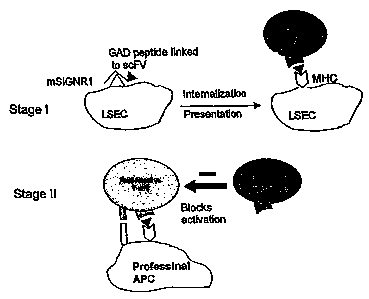
The present methods of inducing immune tolerance to autoantigens target
antigen-presenting cells ("APCs") and direct an autoantigen to those cells by
way
of an antibody. Figure 1 schematically shows the interaction of an
antibody/autoantigen construct in accordance with the present disclosure with
an
antigen presenting cell (APC), and a T cell. The antibody recognizes a
receptor
on the targeted cells. To direct delivery of the autoantigen via the antibody,
the
two are linked. This linking may be accomplished by any method, although this
disclosure delineates the use of vector cloning. The antibody targets and
binds
13
CA 02591138 2007-06-18
WO 2006/073748 PCT/US2005/045706
to the unique antigen-internalizing receptor only, thereby assuring delivery
of the
autoantigen to the desired cell type.
After the antibody is bound to the targeted antigen-internalizing receptor,
the linked autoantigen and the antibody are internalized in the antigen
presenting
cell. The autoantigen is presented on the surface of the APCs, presumably
through the autoantigen's interaction with major histocompatibility complex
("MHC") within the cell. Once an autoantigen is expressed on the surface of
the
APCs with co-stimulatory potential, naive autoreactive T cells can become
activated and target and react with their specific autoantigen. The absence of
a
co-stimulatory molecule in the surface of the APCs is most likely involved in
limiting the T cell response. Autoreactive effector T cells can kill only a
limited
number of antigen expressing tissue cells. After killing a few target cells,
the
effector cell dies. The autoantigen presenting cells are then tolerated.
Presentation of antigen by tolerizing antigen presenting cells to naive T
cells
induces regulatory T cells. Subsequently, the regulatory T cells prevent
activation of other potentially auto-reactive T cells by stimulatory antigen
presenting cells.
Thus, in some embodiments, the antibodies of the present disclosure
may be utilized to form antibody/autoantigen constructs capable of binding to
liver sinusoidal endothelial cells thereby stimulating proliferation of
regulatory
cells, or the antibodies of the present disclosure may be utilized to form
antibody/autoantigen constructs capable of binding to liver sinusoidal
endothelial
cells thereby suppressing the activity of auto-reactive T-cells.
Antibody/autoantigen constructs of the present disclosure may also be used to
deliver a vaccine antigen to sinusoidal endothelial cells, including those
found in
the lymph nodes, thereby stimulating proliferation of antigen specific T-
cells.
The present antibody/autoantigen construct can be administered in
accordance with known methods, e.g., injection or infusion by intravenous,
intraperitoneal, intracerebral, intramuscular, subcutaneous, intraocular,
intraarterial, intrathecal, inhalation or intralesional routes, topical or by
sustained
release systems as noted below. The antibody/autoantigen construct is
14
CA 02591138 2007-06-18
WO 2006/073748 PCT/US2005/045706
preferably administered continuously by infusion or by bolus injection. One
may
administer the antibody/autoantigen construct in a local or systemic manner.
The antibody/autoantigen constructs may be prepared in a mixture with a
pharmaceutically acceptable carrier. Techniques for formulation and
administration of the compounds of the instant application may be found in
"Remington's Pharmaceutical Sciences," Mack Publishing Co., Easton, PA, latest
edition. This therapeutic composition can be administered intravenously or
through the nose or lung, preferably as a liquid or powder aerosol
(lyophilized).
The composition may also be administered parenterally or subcutaneously as
desired. When administered systemically, the therapeutic composition should be
sterile, pyrogen-free and in a parenterally acceptable solution having due
regard
for pH, isotonicity, and stability. These conditions are known to those
skilled in
the art.
Briefly, dosage formulations of the present antibody/autoantigen construct
are prepared for storage or administration by mixing the compound having the
desired degree of purity with physiologically acceptable carriers, excipients,
or
stabilizers. Such materials are non-toxic to the recipients at the dosages and
concentrations employed, and may include buffers such as TRIS HCI,
phosphate, citrate, acetate and other organic acid salts; antioxidants such as
ascorbic acid; low molecular weight (less than about ten residues) peptides
such
as polyarginine, proteins such as serum albumin, gelatin, or immunoglobulins;
hydrophilic polymers such as polyvinylpyrrolidinone; amino acids such as
glycine,
glutamic acid, aspartic acid, or arginine; monosaccharides, disaccharides, and
other carbohydrates including cellulose or its derivatives, glucose, mannose,
or
dextrins; chelating agents such as EDTA; sugar alcohols such as mannitol or
sorbitol; counterions such as sodium and/or nonionic surfactants such as
TWEEN, PLURONICS or polyethylene glycol.
When used for in vivo administration, the antibody/autoantigen construct
formulation must be sterile and can be formulated according to conventional
pharmaceutical practice. This is readily accomplished by filtration through
sterile
filtration membranes, prior to or following lyophilization and reconstitution.
The
antibody ordinarily will be stored in lyophilized form or in solution. Other
vehicles
CA 02591138 2007-06-18
WO 2006/073748 PCT/US2005/045706
such as naturally occurring vegetable oil like sesame, peanut, or cottonseed
oil
or a synthetic fatty vehicle like ethyl oleate or the like may be desired.
Buffers,
preservatives, antioxidants and the like can be incorporated according to
accepted pharmaceutical practice.
Pharmaceutical compositions suitable for use include compositions
wherein one or more antibody/autoantigen constructs are contained in an amount
effective to achieve their intended purpose. More specifically, a
therapeutically
effective amount means an amount of antibody effective to prevent, alleviate
or
ameliorate symptoms of disease or prolong the survival of the subject being
treated. Determination of a therapeutically effective amount is well within
the
capability of those skilled in the art, especially in light of the detailed
disclosure
provided herein. Therapeutically effective dosages may be determined by using
in vitro and in vivo methods.
An effective amount of antibody/autoantigen construct to be employed
therapeutically will depend, for example, upon the therapeutic objectives, the
route of administration, and the condition of the patient. In addition, the
attending
physician takes into consideration various factors known to modify the action
of
drugs including severity and type of disease, body weight, sex, diet, time and
route of administration, other medications and other relevant clinical
factors.
Accordingly, it will be necessary for the therapist to titer the dosage and
modify
the route of administration as required to obtain the optimal therapeutic
effect.
Typically, the clinician will administer antibody/autoantigen construct until
a
dosage is reached that achieves the desired effect. The progress of this
therapy
is easily monitored by conventional assays.
For any antibody/autoantigen construct, the therapeutically effective dose
can be estimated initially from cell culture assays. For example, a dose can
be
formulated in animal models to achieve a circulating concentration range that
includes the EC50 as determined in cell culture (e.g., the concentration of
the test
molecule which promotes or inhibits cellular proliferation or
differentiation). Such
information can be used to more accurately determine useful doses in humans.
Toxicity and therapeutic efficacy of the antibody/autoantigen constructs
described herein can be determined by standard pharmaceutical procedures in
16
CA 02591138 2007-06-18
WO 2006/073748 PCT/US2005/045706
cell cultures or experimental animals, e.g., for determining the LD50 (the
dose
lethal to 50% of the population) and the ED50 (the dose therapeutically
effective
in 50% of the population). The dose ratio between toxic and therapeutic
effects is
the therapeutic index and it can be expressed as the ratio between LD50 and
ED50. Molecules which exhibit high therapeutic indices are preferred. The data
obtained from these cell culture assays and animal studies can be used iri
formulating a range of dosage for use in human. The dosage of such molecules
lies preferably within a range of circulating concentrations that include the
ED50
with little or no toxicity. The dosage. may vary within this range depending
upon
the dosage form employed and the route of administration utilized. The exact
formulation, route of administration and dosage can be chosen by the
individual
physician in view of the patient's condition. (See e.g., Fingl et aL, 1975, in
"The
Pharmacological Basis of Therapeutics", Ch. 1 p.1.)
Dosage amount and interval may be adjusted individually to provide
plasma levels of the antibody/autoantigen construct which are sufficient to
promote or inhibit cellular proliferation or differentiation or minimal
effective
concentration (MEC). The MEC will vary for each antibody/autoantigen
construct,
but can be estimated from in vitro data using described assays. Dosages
necessary to achieve the MEC will depend on individual characteristics and
route
of administration. However, HPLC assays or bioassays can be used to determine
plasma concentrations.
Dosage intervals can also be determined using MEC value.
Antibody/autoantigen construct molecules should be administered using a
regimen which maintains plasma levels above the MEC for 10-90% of the time,
preferably between 30-90% and most preferably between 50-90%.
In cases of local administration or selective uptake, the effective local
concentration of the antibody/autoantigen construct may not be related to
plasma
concentration.
A typical daily dosage might range from about 1 g/kg to up to 1000mg/kg
or more, depending on the factors mentioned above. Typically, the clinician
will
administer the antibody/autoantigen construct until a dosage is reached that
17
CA 02591138 2007-06-18
WO 2006/073748 PCT/US2005/045706
achieves the desired effect. The progress of this therapy is easily monitored
by
conventional assays.
Depending on the type and severity of the disease, from about 0.001
mg/kg to abut 1000 mg/kg, more preferably about 0.01 mg to 100 mg/kg, more
preferably about 0.010 to 20 mg/kg of the antibody/autoantigen construct might
be an initial.candidate dosage for. administration to the patient, whether,
for
example, by one or more separate administrations, or by continuous infusion.
For
repeated administrations over several days or longer, depending on the
condition, the treatment is repeated until a desired suppression of disease
symptoms occurs or the desired improvement in the patient's condition is
achieved. However, other dosage regimes may also be useful.
In a particularly useful embodiment, the disclosed methods can be used to
treat diabetes mellitus by inducing immune tolerance to insulin-producing (3
cells
of the islets of Langerhans within the pancreas. Autoantigens of these cells
are
linked to antibodies which recognize the desired antigen-internalizing
receptor.
Suitable autoantigens for use in this disclosure are R cell antigens, and
epitopes,
or peptides representing epitopes, of insulin, glutamic acid decarboxylase
("GAD") and heat shock protein ("HSP"). Linking a set of peptides covering
epitopes from insulin, GAD and hsp to an anti-DC-SIGNR antibody has the
potential to induce tolerance to all major antigens implicated in T1 DM. Other
autoantigens may be known and used, or discovered and used, by those skilled
in the art.
The antigen-internalizing receptor is presented on specialized APCs. For
this method, the antigen-internalizing receptor chosen is DC-SIGNR (dendritic
cell-specific intercellular adhesion molecule 3-grabbing nonintergrin related
receptor). DC-SIGNR is expressed by liver sinusoidal endothelial cells
("LSEC"),
which are liver-resident antigen presenting cells. ('Pohlmann et al, 2001). DC-
SIGNR belongs to the family of pathogen internalization receptors that
internalize
receptor bound protein and facilitate antigen presentation. ('Geijtenbeek et
al.,
2002). It has been shown that presentation of an antigen by LSECs results in
an
antigen- specific tolerance (Limmer et al., 2000). In contrast to other
dendritic
cell types that can mature from an immature tolerogenic state to an activating
18
CA 02591138 2007-06-18
WO 2006/073748 PCT/US2005/045706
state, liver sinusoidal cells can not be induced to develop into an activating
antigen presenting cell (2Knolle et al., 1999). The human DC-SIGNR (also
called
L-SIGN) homologue to human DC-SIGN shows 77% identity to DC-SIGN at the
amino acid level and has the typical domain for internalizing receptors
(Bashirova
et al, 2001; Soilleux et al, 2000). DC-SIGNR is highly expressed on LSEC and
is
also found on a sub-population of lymph node macrophage-like cells, but is not
expressed by DCs.
For purposes of the present disclosure, the terms "DC-SIGNR" and "L-
SIGN" are used interchangeably.
The C-type lectin mouse DC-SIGN (CD209) has recently been identified
as a DC-specific receptor. DC-SIGN mediates transendothelial migration of DCs,
which enables primary immune responses by initiating transient DC-T cell
interactions (3Geijtenbeek et al, 2000; 2Geijtenbeek et al, 2000). DC-SIGN
also
serves as an internalizing antigen receptor recognizing pathogens through
carbohydrate structures. Besides its prominent role in DC-T cell clustering
and
initiation of T cell responses, DC-SIGN is a major receptor involved in
infection of
DC and subsequent transmission to T cells of viruses such as HIV-1, HIV-2, SIV-
1, hepatitis C virus (HCV), Ebola virus, cytomegalovirus (CMV), and Dengue
virus; bacteria such as Helicobacter pylori, Klebsiella pneumonae, and
Mycobacteria tuberculosis; yeast such as Candida albicans; and parasites such
as Leishmania pifanoi and Schistosoma mansoni. The murine homologue of DC-
SIGNR, mSIGNRI, captures antigens that are rapidly internalized and targeted
for
lysozomes for processing ('Geijtenbeek et al, 2002). Based on amino acid
sequence; murine mSIGNR1 is equally homologous to human DC-SIGNR as it is
to human DC-SIGN and is therefore useful for animal modeling studies.
Thus, in another aspect, the present disclosure relates to anti-DC-
SIGNR (i.e., anti-L-SIGN) antibodies. As noted above, the term "antibody "as
used herein, refers to an entire immunoglobulin molecule or molecules that
contain immunologically active portions of whole immunoglobulin molecules and
includes Fab, F(ab')2, scFv, Fv; heavy chain variable regions and light chain
variable regions.
19
CA 02591138 2007-06-18
WO 2006/073748 PCT/US2005/045706
In one embodiment, the antibody of the present disclosure comprises a
light chain. As used herein, "light chain" means the smaller polypeptide of an
antibody molecule composed of one variable domain (VL) and one constant
domain (CL), or fragments thereof. In another embodiment, the portion of the
antibody comprises a heavy chain. As used herein, "heavy chain" means the
larger polypeptide of an antibody molecule composed of one variable domain
(VH) and three or four constant domains (CH1, CH2, CH3, and CH4), or
fragments thereof.
In another embodiment, the antibody comprises a Fab portion of the
antibody. As used herein, "Fab" means a monovalent antigen binding fragment
of an immunoglobulin that consists of one light chain and part of a heavy
chain.
It can be obtained by brief papain digestion or by recombinant methods. In
another embodiment, the portion of the antibody comprises a F(ab')2 portion of
the antibody. As used herein, "F(ab')2 fragment" means a bivalent antigen
binding fragment of an immunoglobulin that consists of both light chains and
part
of both heavy chains. It can be obtained by brief pepsin digestion or
recombinant
methods. In other embodiments, the antibody may be a Fab' fragment. Fab
expression libraries may for instance be obtained by the method of Huse et
al.,
Science 245: 1275 (1989).
Furthermore, "humanized" antibodies that bind to a receptor on an antigen
presenting cell may be used. Methods for humanization are disclosed, for
instance, in WO 98/49306, U.S. Patent Nos. 5,585,089; 5,225,539 and 5,693,761
and WO 90/07861, the disclosures of which are incorporated herein in their
entirety by this reference.. As used herein, "humanized" antibodies are those
antibodies wherein amino acids outside the CDR are replaced with
corresponding amino acids derived from human immunoglobulin molecules.
"CDR" or "complementarity determining region" means a highly variable
sequence of amino acids in the variable domain of an antibody. Recombinant
DNA technology can be used to produce a humanized antibody wherein the
CDRs of a variable region of one immunoglobulin are replaced with the CDRs
from an immunoglobulin with a different specificity such that the humanized
CA 02591138 2007-06-18
WO 2006/073748 PCT/US2005/045706
antibody recognizes the desired target but is not recognized in a significant
way
by the human subject's immune system.
In other embodiments, chimeric antibodies that include a non-human
variable domain that binds to any receptor on any antigen presenting cell
linked
to a human constant region are contemplated. The receptor to which the
variable domain binds may be, for example, L-SIGN or DC-SIGN and the
constant region may be a human IgG1 constant region. It has been surprisingly
found that in certain embodiments described more fully hereinbelow, such
chimeric antibodies exhibit better binding to the receptor on any antigen
presenting cell better than the variable domain alone.
For conversion of antibody clones into full IgGs, the coding regions for
both the light and heavy chains, or fragments thereof, can be separately
cloned
out of a bacterial vector and into mammalian vector(s). A single vector
system,
such as pDR1 or its derivatives, can be used to clone both light and heavy
chain
cassettes into the same plasmid. Alternatively, dual expression vectors where
heavy and light chains are produced by separate plasmids can be used.
Mammalian signal sequences need to be either already present in the final
vector(s) or appended to the 5' end of the light and heavy chain DNA inserts.
This can be accomplished by initial transfer of the chains into a shuttle
vector(s)
containing the proper mammalian leader sequences. Following restriction
enzyme digestion, the light chain and heavy chain regions, or fragments
thereof,
are introduced into final vector(s) where the remaining constant regions for
the
IgG are provided either with or without introns.
Antibodies in accordance with this disclosure may bind to both L-SIGN
and DC-SIGN, but bind preferentially to L-SIGN. That is, the binding to L-SIGN
may be stronger than the binding to DC-SIGN. The binding to L-SIGN can be,
for example, form 1.1 to 200 times stronger than the binding to DC-SIGN.
Alternatively, the binding to -SIGN can be1.2 to 10.0 times stronger than the
binding to DC-SIGN.
In some embodiments, the antibodies to DC-SIGNR modulate, i.e. inhibit
or enhance, the interaction of DC-SIGNR expressing cells with ICAM-expressing
cells. In one embodiment, the anti-DC-SIGNR antibodies bind to the DC- SIGNR
21
CA 02591138 2007-06-18
WO 2006/073748 PCT/US2005/045706
receptor site on the surface of an antigen presenting cell such as LSEC, and
impede the interaction(s) between the LSEC and a T cell. More specifically,
the
antibodies to DC-SIGNR reduce the adhesion between LSEC and T cells by
interfering with the adhesion between DC-SIGNR and an ICAM receptor on the
surface of a T cell. Blocking of this interaction can modulate the immune
response. For example, the antibodies to DC-SIGNR can modulate the immune
function by blocking L-SIGN-T-cell interaction, thereby causing proliferation
of
regulatory T-cells, including those found in the liver. In another embodiment,
the
antibodies to DC-SIGNR can modulate the immune function by blocking L-SIGN-
T-cell interaction, thereby suppressing proliferation auto-reactive of T-
cells,
including those found in the lymph nodes.
In some additional embodiments, the antibodies of the present
disclosure may be utilized to form antibody-peptide constructs capable of
binding
to liver sinusoidal endothelial cells thereby stimulating proliferation of
regulatory
cells, or the antibodies of the present disclosure may be utilized to form
antibody-
peptide constructs capable of binding to liver sinusoidal endothelial cells
thereby
suppressing the activity of auto-reactive T-cells. Antibody-peptide constructs
of
the present disclosure may also be used to deliver a vaccine antigen to
sinusoidal endothelial cells, including those found in the lymph nodes,
thereby
stimulating proliferation of antigen specific T-cells.
As used herein, "ICAM receptor(s)" means both the ICAM-2 and ICAM-3
receptor, especially the ICAM-3 receptor.
In some embodiments, the antibodies to DC-SIGNR of the present
disclosure do not bind to DC-SIGN. In other embodiments, the antibodies of the
present disclosure possess high affinity for DC-SIGNR and low affinity for DC-
SIGN. Antibodies of the present disclosure may be capable of binding both
linear and conformational epitopes on DC-SIGNR.
It is also contemplated that the present antibodies or antibody/peptide
constructs can be labeled with a toxin to DC-SIGNR expressing cells.
Administration of the anti-DC-SIGNR antibodies or anti-DC-SIGNR
antibody/peptide constructs labeled with toxin can then be utilized to reduce
the
levels of DC-SIGNR expressing cells which, in some instances, can be
22
CA 02591138 2007-06-18
WO 2006/073748 PCT/US2005/045706
beneficial, such as in the treatment of autoimmune disease, cancer or
inflammatory diseases. In this manner, the present antibodies or
antibody/peptide constructs can also be utilized to kill or ablate DC-SIGNR
expressing cells in vivo. This involves administering the antibodies or
antibody/peptide constructs bonded to a cytotoxic drug (e.g., a toxin or
radiation-
emitting compound) to a subject requiring such treatment. Since the antibodies
or
antibody/peptide constructs recognize DC-SIGNR expressing cells (e.g., cancer
cells or liver sinusoidal endothelial cells), any such cells to which the
antibodies
or antibody/peptide constructs bind are destroyed. In one embodiment, a
method of treating cancer in accordance with this disclosure involves
administering an effective cancer-cell killing amount of an anti-DC-SIGNR
antibody or anti-DC-SIGNR antibody/peptide construct having a toxin bound
thereto to a cancer patient. In another embodiment, a method of treating an
inflammatory disease in accordance with this disclosure involves administering
an effective DC-SIGNR expressing cell killing amount of an anti-DC-SIGNR
antibody or anti-DC-SIGNR antibody/peptide construct having a toxin bound
thereto to a patient suffering from an inflammatory disease.
In other embodiments, the antibodies to DC-SIGNR of the present
disclosure block entry of viruses into liver cells such as liver sinusoidal
cells and
their infection into other cells. The antibodies to DC-SIGNR of the present
disclosure may also block entry of viruses into T-celis and their infection
into
other cells.
In some embodiments, the antibodies to DC-SIGNR of the present
disclosure may be utilized to block infection by bacteria of the genus
Mycobacterium. Infections which may be blocked include those caused by M.
tuberculosis and M. bovis.
By interfering with the adhesion of T cells to antigen presenting cells, the
use of antibodies to DC-SIGNR will affect antigen presenting cell-T cell
clustering, T cell activation and other interactions that rely on contact
between
antigen presenting cells and T cells. These other interactions include both
direct
cell-to-cell contact or close proximity of antigen presenting cells and T
cells.
In other embodiments, the anti-DC-SIGNR antibodies of the present
23
CA 02591138 2007-06-18
WO 2006/073748 PCT/US2005/045706
disclosure are linked to peptides such as autoantigens, or self-antigen,
peptides.
These peptides can be linked to anti-DC-SIGNR antibodies by any suitable
method, including grafting a vector to an antibody fragment and cloning the
linked vector/antibody, or chemically linking. Methods of linking a vector,
cloning
or chemical linking are well known to those skilled in the art.
The peptides, preferably autoantigens, along with the linked antibody, are
then internalized into the LSEC. LSECs bear surface molecules necessary for
antigen presentation such as MHC II, CD80 and CD86 (Lohse et al, 1996;
Rubinstein et al, 1986). In addition to inducing a regulatory phenotype in
naive
CD4+T cells (Knolle et al, 1999), LSECs can induce tolerance in CD8+ T cells
by
.cross-presenting exogenous antigen (Limmer et al,.2000). LSECs respond to
stimuli as TNF-a and endotoxin by downregulation of MHC, and hindering
endosomal processing (2Knolle et al, 1999). Furthermore, LSECs do not migrate
out of the liver to lymph organs.
This internalization facilitates the presentation of self-antigen peptides to
the surface to the LSECs, mediated via MHC interactions. Once an autoantigen
is expressed on the surface of the LSECs which have co-stimulatory potential,
naive autoreactive T cells can become activated. The T cells target and react
with the linked autoantigen. The effector T cells kill few LSECs and die off
without co-stimulatory molecules. This presentation of an autoantigen by LSEC
results in autoantigen-specific tolerance.
The liver has a unique microenvironment with an abundance of
tolerogenic mediators such as IL-10 and TGF-R and specialized APCs that favor
the development of immunologic tolerance ('Knolle & Gerken, 2000).
Tolerogenic properties of the liver are supported by the finding that
allogeneic
liver transplants can be accepted across MHC barriers (Calne, 1969).
Furthermore, application of antigens via the portal vein is more likely to
lead to
tolerance than systemic application of the antigen (Kamei et al, 1990).
Draining
through the liver has been reported to be a prerequisite for oral tolerance
induction (Yang et al, 1994). Blood passing through the hepatic vessels first
comes into contact with Kupffer cells and LSECs. The blood flow through the
hepatic sinusoids is slow, allowing contact between the liver sinusoidal cell
24
CA 02591138 2007-06-18
WO 2006/073748 PCT/US2005/045706
populations and passing leukocytes. LSECs bear surface molecules necessary
for antigen presentation such as MHCII, CD80 and CD86 (Lohse et al, 1996;
Rubinstein et al, 1986). In addition to inducing a regulatory phenotype in
naive
CD4+T cells (3Knolle et al, 1999), LSECs can induce tolerance in CD8+ T cells
by cross-presenting exogenous antigen (Limmer et al, 2000). Klugewitz et al
(Klugewitz et al, 2002) demonstrated that injection of Th1, IFN-y producing
TCR-
transgenic cells into mice results after intravenous protein immunization in
suppression of IFN-y production by these cells in the liver and promotion of
Th2-
cells. In contrast to professional myeloid APC that can differentiate from an
immature, tolerogenic stage into a mature stage initiating immunity, LSECs
respond to stimuli as TNF-a and endotoxin by downregulation of MHC and
hindering endosomal processing (2Knolle et al, 1999). Furthermore, LSECs do
not migrate out of the liver to lymph organs. LSECs might not be the only APC
specialized on inducing tolerance. Pugliese et al recently identified a small
subset of spleen DCs that induced tolerance by presenting endogenously
expressed autoantigen (Puglise et al, 2001). Overall, LSECs appear to be a
favorable cell type for presenting P-cell antigens with the purpose of
tolerance
induction.
In addition, both DC-SIGN and L-SIGN have been shown to bind to a
number of viruses, e.g., HIV (Bashirova et al. 2001; 2Pohlmann et al. 2001),
HCV
(Gardner et al. 2003), Ebola (Alvarez et al. 2002; Simmons et al. 2003), SARS
(Jeffers et al. 2004), CMV (Halary et al. 2002), and Sindbis (Klimstra et al.
2003).
While these receptors act as an attachment point for HIV and HCV, transmitting
them in trans to other cells, e.g., T-ceNs (Bashirova et al. 2001; 2Pohlmann
et al.
2001; Cormier et al. 2004), they also bind to and permit the entry and
infection by
Ebola, SARS, CMV and Sindbis viruses into receptor expressing cells. In
addition, both DC-SIGN and L-SIGN serve as receptors for infection by
bacterial
pathogens of the Mycobacterium genus, including M. tuberculosis (aGeijtenbeek
et al. 2003; Koppel et al. 2004), as well as parasites such as Schistosoma
mansoni (Van Liempt et al. 2004).
Thus, in another embodiment, antibodies of the present disclosure may be
utilized to bind to DC-SIGN and/or L-SIGN, thereby blocking the binding,
CA 02591138 2007-06-18
WO 2006/073748 PCT/US2005/045706
infection,. and transmission of infectious agents including, but not limited
to,
viruses such as HIV, HCV, Ebola, SARS, CMV and Sindbis; bacterial pathogens
of the Mycobacterium genus, including M. tuberculosis and M. bovis; and
parasites such as Schistosoma mansoni.
Antibodies to DC-SIGNR of the present disclosure may also be utilized as
routine diagnostics for tumor types associated with DC-SIGNR expression. For
example, upregulation of DC-SIGNR in cancer samples could be utilized as the
basis for a diagnostic tool to evaluate whether a cancer has become exposed to
the immune system, since upregulation of immune receptors is expected to
happen only under pressure by the immune system. Using methods known to
those skilled in the art, including immunohistochemistry and/or FACS analysis,
tumor biopsies may be exposed to antibodies to DC-SIGNR and then analyzed
for the presence of bound antibody, which would be indicative of a cancer
associated with DC-SIGNR expression. In some embodiments the antibodies
may be provided as part of diagnostic kits for determining the presence of a
cancer expressing DC-SIGNR.
Antibodies to DC-SIGNR of the present disclosure may also be utilized as
therapeutics for the treatment of cancer. In one embodiment, the antibodies of
the present disclosure induce ADCC (antibody-dependent cellular cytotoxicity)
or
CDC (complement-dependent cytotoxicity) of tumor cells, thereby killing said
cells.
The present antibodies or antibody/peptide constructs can also be utilized
to kill or ablate cancerous cells in vivo. This involves administering the
antibodies
or antibody/peptide constructs bonded to a cytotoxic drug to a subject
requiring
such treatment. Since the antibodies or= antibody/peptide constructs recognize
cancer cells, any such cells to which the antibodies or antibody/peptide
constructs bind are destroyed.
The antibodies or antibody/peptide constructs of the present disclosure
may be used to deliver a variety of cytotoxic compounds. Any cytotoxic
compound can be fused to the present antibodies or antibody/peptide
constructs.
The fusion can be achieved chemically or genetically (e.g., via expression as
a
single, fused molecule). The cytotoxic compound can be a biological, such as a
26
CA 02591138 2007-06-18
WO 2006/073748 PCT/US2005/045706
polypeptide, or a small molecule. As those skilled in the art will appreciate,
for
small molecules, chemical fusion is used, while for biological compounds,
either
chemical or genetic fusion can be employed.
The antibodies or antibody/peptide constructs of the present disclosure
may be used to deliver a variety of cytotoxic drugs including therapeutic
drugs; a
compound emitting radiation; molecules of plant, fungal, or bacterial origin;
biological proteins; and mixtures thereof. The cytotoxic drugs can be
intracellularly acting cytotoxic drugs, such as short-range radiation
emitters,
including, for example, short-range, high-energy a-emitters. Enzymatically
active
toxins and fragments thereof are exemplified by diphtheria toxin A fragment,
nonbinding active fragments of diphtheria toxin, exotoxin A (from Pseudomonas
aeruginosa), ricin A chain, abrin A chain, modeccin A chain, a-sacrin, certain
Aleurites fordii proteins, certain Dianthin proteins, Phytolacca americana
proteins
(PAP, PAPII and PAP-S), Morodica charantia inhibitor, curcin, crotin,
Saponaria
officinalis inhibitor, gelonin, mitogillin, restrictocin, phenomycin, and
enomycin, for
example. Procedures for preparing enzymatically active polypeptides of the
immunotoxins are described in W084/03508 and W085/03508, which are
hereby incorporated by reference. Certain cytotoxic moieties are derived from
adriamycin, chlorambucil, daunomycin, methotrexate, neocarzinostatin, and
platinum, for example.
Procedures for conjugating the antibodies or antibody/peptide constructs
with the cytotoxic agents have been previously described.
Alternatively, the antibodies or antibody/peptide constructs of the present
disclosure can be coupled to high energy radiation emitters, for example, a
radioisotope, such as13'I, a y-emitter, which, when localized at the tumor
site,
results in a killing of several cell diameters. See, e.g., S. E. Order,
"Analysis,
Results, and Future Prospective of the Therapeutic Use of Radiolabeled
Antibody in Cancer Therapy", Monoclonal Antibodies for Cancer Detection and
Therapy, R. W. Baldwin et al. (eds.), pp. 303-316 (Academic Press 1985), which
is hereby incorporated by reference. Other suitable radioisotopes include a-
emitters, such as 212Bi, 213Bi, and 211At, and R-emitters, such as186Re and
90Y.
27
CA 02591138 2007-06-18
WO 2006/073748 PCT/US2005/045706
Radiotherapy is expected to be particularly effective in connection with
prostate
cancer, because prostate cancer is a relatively radiosensitive tumor.
In another embodiment, the antibodies to L-SIGN of the present disclosure
may be, utilized as therapeutics to treat tumors by binding to L-SIGN and
preventing negative regulation of the immune system through L-SIGN expressing
cancer cells. By preventing this negative regulation, the immune system may
proceed to eradicate the cancer cells.
The efficacy of the above treatments may be confirmed utilizing methods
known to those skilled in the art, including xenograft models.
Antibodies to L-SIGN in accordance with the present disclosure which are
utilized as cancer therapeutics. may also be combined with any other
immunomodulatory therapy, such as cancer vaccines, anti-CTLA-4, anti-CD25 or
cyclophosphamide to achieve increased therapeutic efficacy in the treatment of
cancer.
Antibodies to L-SIGN in accordance with the present disclosure may also
be used in some embodiments to isolate L-SIGN expressing cells found in a
mixture of cells which includes cells not expressing L-SIGN. For example,
human non-parenchymal liver cells, which contain a mixture of sinusoidal
endothelial cells, red blood cells, Kupffer cells and other minor cell
populations
may be readily obtained from commercial sources. To isolate L-SIGN expressing
cells, i.e., liver sinusoidal cells, from human non-parenchymal liver cells,
the red
blood cells may be exposed to an agent such as ammonium chloride which will
result in lysis of the red blood cells. The dead cells may be removed using
commercially available dead cell removal kits, including those sold by
Miltenyi
Biotech (Germany). The remaining cells may be counted and labeled with anti-L-
SIGN antibody. After washing the cells, they may be resuspended in an
appropriate buffer and the cells of interest, i.e., the L-SIGN expressing
cells, may
be isolated by exposing the remaining cells to anti-L-SIGN antibody which, in
some cases, may be conjugated to a bead or similar separation medium.
Commercially available separation media and methods for their use are known to
those skilled in the art and include commercially available beads from
Miltenyi.
In some particularly useful embodiments, the L-SIGN expressing cells may be
28
CA 02591138 2007-06-18
WO 2006/073748 PCT/US2005/045706
isolated from cells not expressing L-SIGN by using anti-LSIGN-conjugated
beads, e.g., anti-mouse IgG-conjugated beads, according to the manufacturer's
instructions (Miltenyi). Those cells binding to the anti-L-SIGN antibody are L-
SIGN expressing cells and are thereby isolated from the remaining cells not
expressing L-SIGN. Once isolated from the remaining cells, the L-SIGN
expressing cells may be removed from the anti-L-SIGN antibody using methods
readily known to those skilled in the art. The quality of the isolation may be
monitored by methods known to those skilled in the art, including FACS
analysis
using a panel of antibodies against molecules such as CD54, mannose receptor,
LYVE-1, CD40, asialoglycoprotein and others.
Practice of the present methods, including additional preferred aspects
and embodiments thereof, will be more fully understood from the following
examples, which are presented for illustration only and should not be
construed
as limiting in any way.
Example 1- Obtaining anti-mSIGNRI antibodies
Using phage display technology, a panel of single chain antibodies (scFv)
that recognize mSIGNR1 was identified. scFvs contain the variable light and
heavy chain region connected by a linker. Their short length makes these
antibody fragments very suitable for antigen linkage, and the capacity for
binding
to the receptor is preserved. Rabbits were immunized with recombinant
mSIGNR1, and a scFv antibody library was constructed using the phage display
vector pRL4 which is described in Published International Application No. WO
02/46436 A2 published on June 13, 2002, the disclosure of which is
incorporated
herein by reference. Antibody fragments in this system are displayed on the
gene III coat protein of the phage. Antibodies recognizing mSIGNR1 were
isolated by 4 rounds of solid phase panning on recombinant mSIGNRI. Six
different antibodies were, identified. The amino acid sequences of these six
antibodies are presented in Figures 2A and B (SEQ. ID NOS: 1-6 and 7-12,
respectively). All antibodies recognized mSIGNRI in solid phase ELISA, and no
cross-reactivity with mDC-SIGN, the murine homologue of human DC-SIGN, was
observed. The antibodies were epitope-tagged with HA and HIS6. Both
29
CA 02591138 2007-06-18
WO 2006/073748 PCT/US2005/045706
mSIGNR1 and DC-SIGNHIS were produced by 3T3 EBNA cells and purified over
a nickel column.
Example 2 - Identifying anti-mSIGNR1 antibodies that are internalized
upon binding to the cell surface receptor
Screen for cell lines expressina mSIGNR1
A panel of murine macrophage cell lines (P388D1, 1-13.35, WEHI-3 and
J774) are screened for expression of mSIGNR1 by RT-PCR by standard
methods. Primers are designed based on the mSIGNR1 Genbank sequence and
used these in RT-PCR of mouse organs. A cell line expressing mSIGNR1 on the
mRNA level is identified and surface expression is confirmed by FACS analysis.
5 x 10⁵ cells are incubated with 1 pg anti-mSIGNR1 antibody in PBS containing
1% BSA and 0.1 % NaN3 on ice for 15 minutes, conditions that do not allow for
antibody internalization. After 2 washes with PBS containing 1% BSA and 0.1 %
NaN3, bound anti-mSIGNR1 are detected by biotinylated anti-HA (Roche)
followed by PE-conjugated streptavidin (Becton Dickenson) and cells are
analyzed using FACSCalibur (Becton Dickinson, Mountain View, CA).
Alternatively, internalization is determined on primary cells known to express
mSIGNR1 such as liver sinusoidal endothelial cells. Expression of mSIGNR1 on
LSECs can also be confirmed by FACS as described above, but only 1 x 10⁵
cells is added per reaction.
Measurement of internalization
Once a mSIGNR1-expressing cell line or primary cell type has been
identified, internalization of the antibody panel is assessed by FACS
analysis. To
show that internalization is based on mSIGNR1 binding, a cell line that does
not
express mSIGNR1 such as JAWS1 mouse dendritic cells is included. Anti-
mSIGNRI detection using biotinylated anti-HA antibody followed by PE-
conjugated steptavidin on intact and permeabilized cells is compared as
described for anti-DEC-205 antibodies (Mahnke et al, 2000). 1.5 x 10⁶ cells
for
cell lines or 3 x 10⁵ cells for primary cells are incubated with 3 pg of
mSIGNR1 in
CA 02591138 2007-06-18
WO 2006/073748 PCT/US2005/045706
PBS containing 1 !o bovine serum albumin (BSA) for 20 minutes at 4 C to allow
for antibody binding to the surface without internalization. Unbound antibody
is
removed by washing 2 times with PBS containing 1% BSA at 4 C. Each sample
is divided into 3. One third is fixed with 4% paraformaldehyde and surface
antibody is detected as described above. The other two thirds are further
incubated for 30 minutes at 37 C to allow for internalization before being
fixed.
One half is directly detected with anti-HA and steptavidin, the other half is
permeabilized by incubating the cells with PBS containing 0.1 % (vol/wt)
saponin
(Sigma-Aldrich). The amount of internalized antibody is calculated by
subtracting
the mean fluorescence in fixed cells from that recorded with fixed and
permeabilized cells. The antibodies with the highest percentages of
internalization within 30 minutes are chosen for further studies linking
peptides to
the antibodies. An existing unrelated rabbit scFv is used as a negative
control,
the commercially available ER-TR9 antibody that has recently been shown to
bindmSIGNR1 ('Geijtenbeek et al, 2002) is used as a positive control. Also,
Fab
fragments of ER-TR9 are produced by papain digestion and tested for
internalization to verify that dimerization is not a requirement for
internalization. If
desired, the scFvs can be converted into Fab'2 or IgG.
In an alternative embodiment, a mSIGNRI library is panned for
internalizing antibody as described by the group of James D. Marks (Poul et
al.,
2000). A suitable process for this embodiment is outlined below.
Selection of internalizina antibodies from mSIGNR1 phage library
5 x 10⁶ cells identified as described above to express mSIGNRI are
incubated with 1 x 10¹² colony forming units of phage from a mSIGNRI library
presenting antibody fragments fused to gene 3 protein on their surface for 1.5
hours at 4 C to allow phage binding without internalization. After phage
binding,
the cells are washed 5. times with phosphate-buffered saline to remove non-
specifically or weakly bound phage. Cells are then incubated for 15 minutes at
37 C to allow endocytosis of surface-bound phage, but avoid phage degradation
within the cell. To remove phage bound to the surface of the cell, cells are
stripped by washing three times with a low pH glycine buffer. Then cells are
31
CA 02591138 2007-06-18
WO 2006/073748 PCT/US2005/045706
trypsinized and washed with PBS before being lysed with high pH triethylamine.
The cell lysate containing phage are used to infect E. coli to prepare phage
for
the next round of selection. A total of three rounds of selection are
performed.
The titer of phage bound to the cell surface (found in the first low pH
glycine
wash) and the number of phage recovered from within the cell are monitored for
each round. An increase in the number of endocytosed phage indicates a
successful selection of internalizing phage. antibody.
To determine whether any of the internalized scFv antibody fragments
bind to mSIGNR1, 500 clones from round 3 are selected using a robotic Qpix
(Genetix) system and grown in 96-well dishes in SB medium overnight in a
HiGrow shaker (Gene Machines). The next day, dishes are spun down and
supernatants tested in solid phase mSIGNR1 ELISA using a robotic Genesis
freedom 200 (Tecan) system. 96-well ELISA plates are coated with 1 pg
mSIGNR1/ml PBS overnight at 4 C. The next day, plates are blocked by 1%
BSA, followed by 3 washes with PBS containing 0.05% Tween. Control plates
are coated with 1% BSA. Supernatants containing antibody are added to
mSIGNR1 or BSA alone wells at concentrations between 0.05 - 5pg/ml in PBS
containing 1 % BSA. After 2 hours on a shaker at room temperature, plates are
washed 3 times with PBS containing 0.05% Tween. For detection of bound scFv,
anti-HA antibody (12CA5 mouse ascites, Strategic Biosolutions, DE) are added
at a 1:1,000 dilution in PBS with 1% BSA. After 2 hours on a shaker at room
temperature, plates are washed again and alkaline-phosphatase-conjugated anti-
mouse IgG (Sigma) are added for 2 hours. After 3 more washes, bound antibody
are detected using Sigma 104 substrate. The plates are read at various time
points at OD405 with an ELSA plate reader (Molecular Devices).
Clones giving a positive signal in ELISA are characterized by restriction
enzyme digest pattern. DNA are isolated using Qiagen's miniprep kit. 2pg of
DNA are digested with 5 U of EcoRII for 2 hours at 37 C and then the samples
are run on a 4% NuSieve agarose gel. Patterns are compared and sequences
are purified in small quantities (about 100 - 300 pg). scFvs are properly
assembled in the periplasmic space of bacteria and are secreted. scFvs can
either be isolated from the supernatant or the periplasmic space. Clones are
32
CA 02591138 2007-06-18
WO 2006/073748 PCT/US2005/045706
grown in 4 liter of SB to an OD600 of 0.8 and induced with 1 mM isopropyl-p-D-
thiogalactopyranoside (IPTG) for 3-4 hours at 30 C to produce optimum amounts
of scFv. To isolate single chain antibodies form the periplasmic space, cell
pellets
are resuspended in cold PBS with added Complete Mini (Roche) protease
inhibitor and are sonicated using a Sonics Vibra-cell VC750. Cellular debris
is
then pelleted and the supernatants are applied to Qiagen Ni-NTA columns using
an Akta FPLC (Pharmacia). Antibody is eluted with imidazole. This method
generally yields about 100-300 pg of purified antibody/liter. Endotoxin is
removed by filtration through Sartorius Q15 filters generally yielding
antibody
preparations containing less than 10 U/mI endotoxin as determined by LAL test
(an assay commercially available from Bio Whittaker). Antibodies are analyzed
again for internalization as described above as well as for binding to
recombinant
mSIGNRI in solid phase ELISA. The antibody with the highest percentage of
internalization within 30 minutes and a good signal in a solid phase ELISA (>
10D
after 1 hour at 1 pg/mi) are selected to make peptide-antibody constructs.
Example 3 - Link GAD peptides to the antibody
Vector and cloning strategy
Following identification of the best bacterially-produced scFv, a conversion
to a mammalian expression system is made. Mammalian expression allows for
the appropriate secondary modifications of the peptides and endotoxin-free
production. A vector (e.g., described in U.S. Patent No. 6,355,245, the
disclosure of which is incorporated herein by reference) with compatible
restriction sites as shown in figure 3 is used. DNA from the antibody of
interest in
pRL4 (described above) are cut with Sfi and inserted into the Apex 3P vector
containing a CMV promoter and mammalian antibody leader sequence. To insert
nucleotides encoding the peptides of interest, restriction sites are chosen
from
the sites available (MCS=Nael, Fsel, Xbal, EcoRl, Pstl, EcoRV, BSABI, BstXl,
Noti, BsrBI, Xho, PbvlOl, Sphl, Nsil, Xbal) that are not contained within the
antibody sequence. Oligonucleotides encoding peptides are synthesized by
Operon with the appropriate restriction sites at each end and are inserted
using
T4 DNA ligase. The resulting construct will contain the scFv followed by a
spacer
33
CA 02591138 2007-06-18
WO 2006/073748 PCT/US2005/045706
determined by the restriction enzyme chosen followed by a peptide and a HIS-
tag (Figure 3). Sequences are confirmed using standard techniques before
transfecting DNA into 393EBNA cells for antibody production.
Choice of peptide
While it should be understood that any of the peptides of the known
diabetes autoantigens insulin, hsp and GAD 65 and 67 can be used in the
processes described herein, for the following experiment GAD is chosen as the
peptide. GAD-reactive T cells are the first autoreactive T cells to be
detected in
the NOD mouse (Tisch et all 1993; Kaufman et al, 1993) and have been shown
to be important in the disease process. Furthermore, human and murine GAD
are 95% homologous. Epitopes recognized by splenic NOD T cells have been
extensively characterized (Kaufman et al, 1993; Tisch et all 1999; Zechel et
al.
1998) and many immunodominant peptides are similar in NOD mice and T1 DM
patients and have been used interchangeably in in vitro T cell assays (Kaufman
et all 1993). The initial immune response in NOD mice is directed against a
defined region in the carboxy-terminal region of GAD65 (peptide 509-528,
peptide 524-543 (Kaufman et all 1993). Later T cell responses are also
directed
against other regions between 200-300 as well as other autoantigens: One of
the
early CD4 GAD65 T cell epitopes, peptide 524-543
(SRLSKVAPVIKARMMEYGGT (SEQ. ID NO: 13), same sequence in mice and
humans) and two of the later occurring murine GAD65 epitopes, peptide 247-266
(NMYAMLIARYKMFPEVKEKG (SEQ. ID NO: 14), 1 amino acid difference
between human and mouse underlined), and peptide 290-309
(ALGIGTDSVILIKCDERGK (SEQ. ID NO: 15), same sequence in mice and
humans) are selected for use as the peptides in this experiment. All 3
epitopes
can induce a spontaneous proliferative response in NOD spienocytes.
Furthermore, peptide immunization with peptide 247-266 and peptide 290-309
have been shown to delay diabetes onset in NOD mice (Ma et all 1997; Tisch et
all 1999; Zechel et all 1998). In addition to CD4 T cell epitopes, tolerance
to CD8
T cell epitopes has also been reported to be important (Quinn et all 2001;
Bercovici et al, 2000). As a negative control, an antibody construct is made
with
34
CA 02591138 2007-06-18
WO 2006/073748 PCT/US2005/045706
hen egg lysozyme peptide 116-1 24 (KGTDVQAWI) (SEQ. ID NO: 16). The most
effective peptides from these in vitro studies are then linked in various
combinations with an antibody construct and tested in the NOD diabetes model.
Example 4 - Antibody-peptide construct production and purification
For T cell experiments, approximately 300 pg of each antibody construct is
produced in EBNA293 human embryonic kidney cells. Cells are grown in DMEM
with 10% FCS, 2 mM glutamine and 250 U/mI G418 (Sigma). Cells in T175 flasks
are transfected with DNA using Qiagen's Effectine reagent according to the
manufacturer's instruction..Medium is exchanged for serum-free medium after 3
days. Supernatant is collected at day 4 and day 8, cell debris is removed by
centrifugation and the cleared supernatant is loaded on a Ni-column using a
Akta-FPLC. Antibody is eluted with imidazole, dialyzed into PBS and correct
size
verified by running I pg on a SDS- gel.
Example 5 - Internalization of the antibody-peptide construct by LSEC
resulting
in peptide presentation and the effect of this presentation on T cells
Liver sinusoidal cells are targeted in vitro with the peptide-antibody
construct and it is determined whether these cells can induce a phenotypic
change in T cells derived from young NOD or Balb Ic mice.
Isolation of murine sinusoidal endothelial cells
Liver sinusoidal endothelial cells are isolated from 3 week old NOD or 4-6
week-old Balb/c mice. Cells are obtained by portal perfusion first with EGTA
to
chelate calcium and loosen cell-cell contacts followed by perfusion with 0.05%
collagenase A in Hank's buffer to degrade intercellular matrix as described by
Kretz-Rommel (Kretz-Rommel & Boelsterli, 1995). The perfused liver is removed
from the mouse and gently worked with a pair of angled forceps. The resulting
crude cell suspension is filtered through a series of metal sieves (30, 50, 80
mesh) to remove larger tissue fragments. Sinusoidal cells are separated from
parenchymal cells by density gradient centrifugation on a metrizamide gradient
(1.089 g/cm3) followed by 2 washing steps to remove cell debris (3Knolle et
al,
CA 02591138 2007-06-18
WO 2006/073748 PCT/US2005/045706
1999). At this point, a mixture of Kupffer cells and liver sinusoidal cells is
obtained. For FACS experiments this is sufficient, since Kupffer cells can be
distinguished from liver sinusoidal endothelial cells using the F4/80 antibody
that
recognizes Kupffer cells, but not liver sinusoidal cells. However, for co-
culture
experiments with T cells and peptide, Kupffer cells are removed by labeling
the
cells with PE-conjugated F4/80 (BD Pharmingen) followed by Miltenyi's anti-PE
microbeads and magnetic sorting of the labeled cells using MACS column and
separator according to the manufacturer's instructions. The remaining cell
population is seeded onto 96 well tissue culture plates in Dulbecco's modified
Eagle medium (DMEM) supplemented with 10% fetal bovine serum and 2%
glutamine. The purity of cell populations is investigated at day 3 after
isolation by
FACS staining for surface markers using anti-mSIGNR1 and anti-F4/80.
mSIGNRI is absent on Kupffer cells ('Geijtenbeek et al, 2002). 90% purity is
considered sufficient to proceed with the experiments. 2 mouse livers are used
per experiment with an expected yield of about 2 x 10⁷ cells (Knook &
Sleyster,
1976, extrapolated for mouse).
T cell phenotypic assays
T cell assays demonstrate if the peptide-antibody construct results in
presentation of peptide by liver sinusoidal endothelial cells and whether
peptide
presentation can induce a phenotypic change in T cells. Liver sinusoidal
endothelial cells are cultured in flat bottom microtiter plates at a density
of I x 105
cells/well. After maintaining the sinusoidal cells for 3 days, CD4+ T cells
will be
purified as described above from a 3-week old and an 8-week old NOD mouse or
a 4-6 week-old Balb/c mouse and are added at 104 or 105 cells/well. Also, each
antibody-peptide construct at concentrations of 0.1 -5 pg/well is added. As a
positive control, each GAD and control peptide by itself are included.
Peptides
are synthesized by SynPep (Dublin, CA). Negative control wells include either
T
cells alone or liver sinusoidal cells alone.
There are 4 possible outcomes of peptide presentation by LSECs to T
cells: 1) induction of regulatory T cells characterized by the production of
TGF-P
and/or IL-10 and IL-4 or the expression of CD4+CD25+CD62L, 2) deletion of T
36
CA 02591138 2007-06-18
WO 2006/073748 PCT/US2005/045706
cells or 3) a complete lack or response. 4) It is also conceivable that
peptide
presentation instead of inducing tolerance results in stimulation of Thi cells
producing IL-2. To distinguish among these possibilities, culture supernatants
(100 pl each) are collected at 24 and 48 hours and assayed for cytokine
production as described below. T cell responses of 3-week old are compared
with those of 8-week old NOD mice as well as with T cells derived from Balb/c
mice that do not show a spontaneous response to GAD. Assays are set up in
triplicate and repeated twice. Since a mixture of T cells is used, a cytokine
response in the supernatant might not be easily seen. However, a mixture of
cells reflects the situation in vivo and GAD-specific T cell responses are
seen
using total splenocytes (Tisch et al, 1993).
As a more sensitive measure for the induction of regulatory T cells, cells
are evaluated for the expression of typical surface markers such as CD25, CD4
and CD62L (Lafaille & Lafaille, 2002) by FACS analysis after 3 days in
culture.
All reagents are available from BD-Pharmingen. Also, IL-4 production is
analyzed
by FACS. A functional test of potential immunoregulatory properties of the
LSEC/peptide exposed T cells as described below are the ultimate test for
tolerance induction in this system. The possibility of peptide presented by
LSEC
inducing cell death are assessed in culture supernatants using Roche's cell
death ELISA kit according to the manufacturer's instructions.
Isolation of spienocytes and CD4+ T cells
Spleens from NOD of Balb/c mice are removed in a sterile environment
and put in PBS using techniques within the purview of those skilled in the
art.
Cells are separated using 18-21 gauge needles, and larger pieces are allowed
to
settle. Supernatant is removed and centrifuged at 200 g for 7 min. Red blood
cells are lysed using 5 ml 0.83% NH4C1 per spleen. Cells are washed twice in
PBS and then resuspended in medium. For certain experiments, total
splenocytes are used. For other experiments, CD4+ T cells are isolated using
Miltenyi's (Auburn, CA) CD4+ T cell isolation kit according to the
manufacturer's
instruction. Magnetic isolation of various cell populations is within the
purview of
one skilled in the art. In one process, isolation is based on depletion of non-
CD4+
37
CA 02591138 2007-06-18
WO 2006/073748 PCT/US2005/045706
T cells using a cocktail of biotin- conjugated monoclonal antibodies against
CDBa, CDI 1 b, CD45R, DX5 and Ter- 1 19. The purity of the isolated population
is assessed by staining of a mixture of FITC-conjugated anti-CD4, PE-
conjugated
anti-CD8, APC-conjugated anti-CD11 b and cy-5-conjugated CD45R (all
eBioscience, San Diego, CA). Expected purity is 90-95% with 70% yield. Since
at
least 1 x 10⁸ cells can be obtained from a mouse spleen and about 25% of
splenocytes are CD4+, about 1.75 x 10⁷ cells can be obtained, enough for 175
96-well microtiter plate wells.
Measurement of cytokine production
Presence of IL-10, TGF-P, IFN-y, IL-4 and IL-2 in supernatants of T
celi/LSEC co-cultures are determined by standard sandwich ELISA as described
(Kretz-Rommel & Rubin, 1997). All antibody pairs are available from BD
Pharmingen. A cytokine capture antibody is coated on the plate in PBS
overnight at 4 C. After 3 washes with PBS/0.05% Tween, culture supernatants
and a standard curve of mouse recombinant cytokine are added and incubated
for 2 hours on a shaker at room temperature. Plates are washed again and
bound cytokine is detected with an alkaline-phosphatase conjugated anti-
cytokine antibody. After 3 washes, Sigma 104TM substrate is added and the
plates are read at various timepoints at OD405 with an ELISA plate reader
(Molecular Devices).
Example 6 - Assess whether T cells exposed to GAD peptides presented on
LSECs can subsequently prevent activation of autoreactive T cells by
professional APCs presenting GAD
In a further experiment, whether T cells exposed to peptides on liver
sinusoidal endothelial cells for 3 days can negatively regulate activation of
autoreactive T cells by peptide presented on splenic professional APCs is
tested.
Feasibility of the induction of T cells with regulatory properties in vitro
has been
demonstrated by a number of laboratories (Wakkach et all 2001 ; Barrat et all
2002; Thorton & Shevach, 1998). Immunosuppressive properties can be tested
by adding the regulatory T cells to a culture system in which immune
stimulation
38
CA 02591138 2007-06-18
WO 2006/073748 PCT/US2005/045706
is normally observed. Addition of GAD peptide to spleen cells from a 7 week-
old
NOD mouse comprising both APCs and T cells provides such an
immunostimulatory system as seen by a strong proliferative response. Addition
of
regulatory T cells abrogates this response. 105 or 106 splenocytes are added
together with either 0.1, 1, or 10 pm peptide per well containing T cells
exposed
to LSEC and peptide-coupled antibody. Control wells include spienocytes with
peptide alone and LSEC + T cells alone. Furthermore, to exclude significant
contribution of proliferation by the presumed regulatory T cells, control
wells also
contain irradiated spienocytes (600 RAD, performed at UCSD irradiation service
facility by Joe Aguilera) and T cells previously exposed to LSEC and the
peptide-
antibody construct. Irradiated spienocytes can present antigen, but do not
proliferate. 1 NCi 3H-thymidine is added to each well during the last 16 hours
of a
72 hour culture period to label newly synthesized DNA as a readout for
proliferation. Cells are harvested using Packard's Universal Cell Harvester
and
incorporated 3H-thymidine is assessed using a Topcount (Packard). If 3H-
thymidine incorporation in cultures containing splenocytes and T cells
previously
exposed to LSEC+peptide is reduced compared to the spienocyte cultures, T
cells have been successfully induced with regulatory properties. Whether the
peptide-conjugated anti-mSIGNR1antibody can induce regulatory T cells in the
NOD mouse, and whether disease can be halted are also tested.
Example 7
Mouse anti human L-SIGN antibodies were identified using recombinant
phage technology. Mouse libraries (IgG1k and IgG2ak) derived from heavy and
light chain combination of mice immunized with recombinant human L-SIGN
were prepared by the methods disclosed in WO 03/025202, the contents of
which are incorporated by reference herein. Once prepared, the libraries were
first panned on human DC-SIGN to remove antibodies cross reactive with DC-
SIGN. The unbound supernatahts were used for selecting clones uniquely
reactive with L-SIGN. A total of ninety-five colonies (36/round) for each of
the
two libraries (IgG1 & IgG2a) were induced and antibody production and their
reactivity with L-SIGN were determined by a capture ELISA. Briefly, anti-human
39
CA 02591138 2007-06-18
WO 2006/073748 PCT/US2005/045706
Fc (Caltag) was coated on ELISA plates at 500 ng/mi overnight. The plates were
blocked with PBS containing 1% BSA followed by. the addition of recombinant L-
SIGN at 2 g/ml. After washing the plate with PBS, supernatants were added.
After a 12 hour incubation at room temperature, plates were washed 3 times and
an alkaline-phosphatase-conjugated anti-Fab antibody was added for 2 hours.
Signal after addition of SigmaS substrate was assessed using an ELISA reader
(Molecular Devices).
The majority of the clones showed good binding (OD405 >1.0) of the
antibody on the phage. Several clones from both IgG1 and IgG2a libraries
showed positive reactivity with human L-SIGN. Results are set forth in Figures
4
and 5: Figure 4 sets forth the IgG1 clones with human L-SIGN; Figure 5 sets
forth the reactivity of IgG2a clones with human L-SIGN.
Example 8
To identify clones uniquely reactive with human L-SIGN, all clones from
Example 7 with OD values of five-fold above background were selected to test
their reactivity with human DC-SIGN by ELISA. Anti-human Fc (Caltag) was
coated on ELISA plates at 500 ng/mi overnight. The plates were blocked with
PBS containing 1% BSA followed by the addition of recombinant DC-SIGN at 2
g/ml. After washing the plate with PBS, supernatants were added. After a 12
hour incubation at room temperature, plates were washed 3 times and an
alkaline-phosphatase-conjugated anti-Fab antibody was added for 2 hours.
Signal after addition of SigmaS substrate was assessed using an ELISA reader
(Molecular Devices).
Ten clones from the.IgG1 library and three clones from the IgG2a library
were found to uniquely react with human L-SIGN (five to ten fold higher OD
values vs. DC-SIGN). Results are set forth in Figures 6 and 7: Figure 6 sets
forth the reactivity of the IgG1 positive clones with L-SIGN and DC-SIGN;
Figure
7 sets forth the reactivity of the IgG2a clones with L-SIGN and DC-SIGN.
Example 9
CA 02591138 2007-06-18
WO 2006/073748 PCT/US2005/045706
Thirteen clones identified as reactive with only human L-SIGN and nine
clones strongly reactive with both L-SIGN and DC-SIGN as identified above in
Example 8 were sequenced to determine the number of unique clones.
Sequencing was determined by techniques known to those skilled in the art. The
sequences of these clones are set forth in Figures 8 and 9; sequences for
heavy
chain clones are set forth in Figure 8A-8C (SEQ. ID NOS. 17-36); sequences for
light chain clones are set forth in Figures 9A-9B (SEQ. ID NOS: 37-55).
Sequencing results demonstrated a more diverse group of heavy chains
compared with light chains. The diversity of the antibody clones was also
enhanced by cross pairing of different light chains with the same heavy chain.
A
total of five unique clones reactive with L-SIGN were identified and clones
were
grouped based upon the similarity of their amino acid sequences (see Table 1
below).
TABLE 1
Grouping of antibody clones reactive with human L-SIGN based on amino acid
sequence similarities
Ab clusters Light chain Heavy chain Unique L-sign
clones
1 B1;B2;D1;F1;H1 1;G1 a; B1(identical) 2
C2 (stop/CDR1) B2;C2;D1;F1;H1
1/FR1 vs. A1, B1, G1
2.1c B3;C3 B3 ;C3a 1
2.2 D3( 1/CDR2 ; H3 D3;H3 0
2.3 A3 (1/CDR2); A4 A3 a 1
(1/CDR3)
3.1 B4;E3 (stop/FR1) 134 ,E3 b 0
G3 (1/CDR3) G3 b 2/FR1;1/FR2
3.2 1 & G1 (stop/CDR2)
4 D2a 1
5 A2 A2 0
6 F3 0
7 Clb (incomplete seg) 0
aAntibody clones reactive with only L-SIGN
bAntibody clones reactive with both L- and DC-SIGN
c 2.1...2.3 designates similar light chains but unique heavy chains
The reactivities (OD values) of the clones selected for sequencing with L-
SIGN and DC-SIGN are set forth below in Table 2.
41
CA 02591138 2007-06-18
WO 2006/073748 PCT/US2005/045706
TABLE 2
IgG1k IgG1k IgG2ak IgG2ak
library libra library library
Seq. Well deep well# OD405 clones to Seq. Well deep well# OD405 clones to
# pudfy # purify
Al A5 3 A5. A3 E11 2.2 E11
B1 C5 3.2 B3 F11 2
C1 B6* 1.4 C3 F12 2 F12
Dl E6 3.2 D3 A3* 2
El D7 3.1 E3 D5* 2.9
Fl G9 3.1 F3 C6* 2.3
G1 H9 2.6 G3 D7* 2.9 D7
H1 C10 3.1 H3 E10* 2.2
A2 C11* 1.4 A4 All* 2.2
B2 H11 3.5 H11 B4 B12* 2.6
C2 B12 1.8
D2 C12 3.1 C12
*= Those clones selected for sequencing with L-SIGN and DC-SIGN
As set forth in Figure 8, heavy chain CDR3 regions of the antibodies that
bind to human DC-SIGNR were found have one of the following amino acid
sequences: LGGL (SEQ. ID NO: 56); EFTTKAMD (SEQ. ID NO: 57);
GLFYGYAWFN (SEQ. ID NO: 58). As set forth in Figure 9, light chain CDR3
regions of the antibodies that bind to human DC-SIGNR were found to have one
of the following amino acid sequences: QQYSSYPLT (SEQ. ID NO:59);
QQSNEDPRT (SEQ. ID NO: 60); QQNNEDPYT (SEQ. ID NO: 61);
LQNNEDPYT (SEQ. ID NO: 62).
Example 10
Additional clones from Example 7 above wer'e examined for their ability to
bind to L-SIGN utilizing the procedures described above in Example 7.
Sequencing was determined by techniques known to those skilled in the art. The
sequences for these additional clones are set forth in Figure 10 (SEQ. ID NOS:
63-82). As set forth in Figure 10, five additional heavy chain CDR3 regions of
the
antibodies that bind to human DC-SIGNR were found have one of the following
amino acid sequences: PSDNSYAWFA (SEQ. ID NO: 83); QATTTAFD (SEQ. ID
NO: 84); TATALSTMD (SEQ. ID NO: 85); NDYYWGFG (SEQ. ID NO: 86);
TATALYTMD (SEQ. ID NO: 87); and EFTTKALD (SEQ. ID NO: 88). The CDR2
regions of these clones that bound to human DC-SIGNR were found to have one
of the following amino acid sequences: MIDPSNSEARLNQRFKD (SEQ. ID NO:
42
CA 02591138 2007-06-18
WO 2006/073748 PCT/US2005/045706
89); TISSGGSFTFYPDSVKG (SEQ. ID NO: 90); NIDPYYGGTSYNQKFKG
(SEQ. ID NO: 91); VIWRGGNTDYNAAFMS (SEQ. ID NO: 92);
NFDPYYGVITYNQKFKG (SEQ. ID NO: 93); NIDPYYGGSSYNQKFKG (SEQ. ID
NO: 94); and TISSGGSFTYYPDNVKG (SEQ. ID NO: 95).
Table 3 shows additional IgG1 k antibody clones selected based on their
reactivity with cells expressing L-SIGN.
TABLE 3
Ab Light Heavy chain Unique clones
clusters chain
1 A10, H10 A10, B10, B5, D12, E9; F12, G10, H10 A10, H10
2 D8, F10 D8, E4, E7, F10 D8, F10
3 E12, H6 E12, H6 E12, H6
4 B7, C7 B7, C7 B7, C7
5 C8 C8 C8 sto /LC
6 D10 D10 D10
7 E10 E10 E10
8 G3 G3 G3 sto /LC
9 B5 - B5 sto /LC
B10 - B10
11 D12 - D12
12 E4 - E4
13 E7 - E7
14 E9 - E9
F12 - F12
16 G10 - G10
10 Table 4 below shows the reactivity of additional IgG1 k antibody clones
with cells expressing LSIGN (Geometric Mean fluorescence) and recombinant L-
SIGN and DC-SIGN proteins (OD values).
TABLE 4
I G1k Geo. Mean Fluorescence Geo. Mean Fluorescence OD405 OD405
Clone Name. K562 K562/L-SIGN L-SIGN DC-SIGN
A10 3.4 21.3 2.9 0.1
B5 1.5 10.5 2.1 0.1
B7 2.2 90.9 2.7 0.1
B10 2.0 53.1 3.5 0.1
C7 2.0 101.0 3.5 0.1
C8 1.7 13.4 0.1 0.1
D8 1.8 17.3 2.1 0.1
D10 1.6 10.2 0.9 0.5
43
CA 02591138 2007-06-18
WO 2006/073748 PCT/US2005/045706
D12 2.0 152.0 3.5 0.1
E4 1.9 50.4 3.5 0.1
E7 1.8 19.3 1.4 0.1
E9 1.9 25.5 2.9 0.1
E10 1.7 27.2 2.6 0.6
E12 3.0 22.9 2.9 0.1
110 2.4 13.8 0.8 0.1
F12 2.8 168.7 3.5 0.1
G1 2.1 14.1 2.0 0.1
G3 1.6 41.5 0.4 0.1
G10 2.0 86.1 2.0 0.1
H6 2.4 26.2 3.5 0.1
H10 3.4 12.3 2.7 0.1
As set forth in Figure 11, additional clones that bind human DC-SIGNR
were identified (SEQ. ID NOS: 96-115). These clones were found to have 1gG1k
light chain CDR3 regions with one of the following amino acid sequences: Q Y H
R S P Q T (SEQ. ID NO: 116); C Q Q F T S S P S (SEQ. ID NO: 117);QQYS
G Y P L T (SEQ. ID NO: 118); Q Q Y S G Y P G T (SEQ. ID NO: 119); H Q Y H
R S P P M T (SEQ. ID NO: 120); Q Q R S S Y P F T (SEQ. ID NO: 121); Q Q Y
S S Y P F T (SEQ. ID NO: 122); Q Q N N E D P P T (SEQ. ID NO: 123); Q Q Y S
GYSLT(SEQ. IDNO: 124); Q Q Y S G Y P L M L T (SEQ. ID NO: 125); Q Q Y
G G Y P L T (SEQ. ID NO: 126); Q Q N N E D P Y T(SEQ. ID NO: 127); Q Q Y
S G S P L T(SEQ. ID NO: 128). The CDR2 regions of these clones that bound
to human DC-SIGNR were found to have one of the following amino acid
sequences: S T S N L A S G (SEQ. ID NO: 129); L A S N L E S.G (SEQ. ID NO:
130); S T S N Q A P G(SEQ. ID NO: 131); W A S T R H T G(SEQ. ID NO: 132).
Table 5 shows additional IgG2ak antibody clones selected based on their
reactivity with cells expressing L-SIGN.
TABLE 5
Ab Light chain Heavy chain Unique clones
clusters
1 A12*, Bl 1, A12, C10, C12, H7 A12, C12, H7
C12*,C6, E12, E8*,
F10*, H7*
2 F6*, F12* F12, F6 F12, F6
3 C10,G10,G5 A3,F10,G5 A3
44
CA 02591138 2007-06-18
WO 2006/073748 PCT/US2005/045706
4 A4,B9 A4,C7,D12 A4
A3 A5, D8 C7
6 A5 C5* D12
7 C7 C6 F10
8 D8 B9 G5
9 D12 E12 C5*
H6 H6 H6
11 - G10 B9
12 - B11 E12
13 - E8 G10
14 B11
E8
16 C6
17 A5
18 D8
19 C10
* These sequences contain a stop codon
Table 6 shows the reactivity of additional IgG2ak antibody clones with
cells expressing LSIGN (Geometric Mean fluorescence) and recombinant L-
5 SIGN and DC-SIGN proteins (OD values).
TABLE 6
I G2ak Geo. Mean Fluorescence Geo. Mean Fluorescence OD405 OD405
Clone Name K562 K562/L-SIGN L-SIGN DC-SIGN
A3 2.8 15.4 3.5 1.0
A4 2.8 12.1 3.5 0.1
A5 1.8 59.8 3.5 0.1
A12 2.3 23.4 3.5 0.8
B9 3.5 31.4 3.5 0.1
B 11 3.4 14.2 3.5 0.6
C5 3.3 13.9 3.5 0.6
C6 2.6 13.2 3.5 0.6
C7 2.5 21.2 3.5 0.1
C10 2.8 25.8 3.5 0.7
C12 2.9 23.3 3.5 1.0
D8 3.2 17.3 3.5 0.1
D12 2.3 41.0 3.5 0.1
E8 2.9 11.2 3.5 0.4
E12 3.5 19.4 3.5 0.7
F6 2.7 13.8 3.5 0.6
F10 2.6 18.3 3.5 1.0
F12 2.0 9.5 3.5 0.5
G5 3.1 10.7 3.5 0.4
CA 02591138 2007-06-18
WO 2006/073748 PCT/US2005/045706
G10 2.6 21.5 3.5 0.7
H6 3.2 12.7 3.5 0.6
H7 2.1 11.5 3.5 1.0
As set forth in Figure 12, additional heavy chain clones that bind human
DC-SIGNR were identified (SEQ. ID NOS: 133-154). These clones were found
to have IgG2ak heavy chain CDR3 regions with one of the following amino acid
sequences: T R E F T T K A L D(SEQ. ID NO: 155); T R E F T T K A M D(SEQ.
ID NO: 156); A R T A T A L Y T M D (SEQ. ID NO: 157); L R T L P C I (SEQ. ID
NO: 158); SREFTTKAMD(SEQ.IDNO:159);ARQLXXYFXMD
(SEQ. ID NO: 160). The CDR2 regions of these clones that bound to human DC-
SIGNR were found to have one of the following amino acid sequences: T I S S G
GSFTYYPDNVKG(SEQ.IDNO:161);NIDPYYDSISYNQKFKG
(SEQ. ID NO: 162); N F D P Y Y G V I T Y N Q K F K G (SEQ. ID NO: 163); T I S
SGGSYTYYPDNVKG(SEQ.IDNO:164);XFXTDW.FYXT(SEQ.
IDNO: 165);NFDPYYGVISYNQKFKG(SEQ.IDNO:166);TISSG
G G F T Y Y P D N V K G (SEQ. ID NO: 167); X I Y P G T D N T Y Y N E X F K
G (SEQ. ID NO: 168).
As set forth in Figure 13, additional light chain clones that bind human DC-
SIGNR were identified (SEQ. ID NOS: 169-189). These clones were found to
have IgG2ak light chain CDR3 regions with one of the following amino acid
sequences: Q Q N N E D P Y T (SEQ. ID NO: 190); S G Y P L T F G S (SEQ. ID
NO:191); HRSPPMTFG(SEQ.IDNO:192);QQNNEDPFT(SEQ.ID
NO: 193); Y S G Y P L T F G(SEQ. ID NO: 194); N T L P L T F G(SEQ. ID NO:
195); Q Q S K E V P W T (SEQ. ID NO: 196); L Q N N E D P Y T F (SEQ. ID NO:
197). The CDR2 regions of these clones that bound to human DC-SIGNR were
found to have one of the following amino acid sequences: L A S N L E S (SEQ.
ID NO: 198); L A S N L E F (SEQ. ID NO: 199); N L A S G V P (SEQ. ID NO:
200); N L A S G V(SEQ. ID NO: 201); A A S N Q G S(SEQ. 1D NO: 202).
Example 11 - Selective reactivity of candidate soluble L-SIGN antibodies with
L-
SIGN receptor but not DC-SIGN receptor
46
CA 02591138 2007-06-18
WO 2006/073748 PCT/US2005/045706
Based on phage-Fab screening, DNA sequencing (Figures 10 and 11), six
antibodies (clone names C7, D12, E4, E10, G3, G10) having the best reactivity
with cells expressing L-SIGN receptor (Table 4) were subcloned to remove the
coat III protein. Each of these six candidate antibodies were expressed and
purified as soluble Fab moieties and tested again for their ability to
recognize the
L-SIGN receptor but not DC-SIGN on cells. As shown in Figure 14, all six
antibodies exhibited very good binding with L-SIGN receptor; while three
antibodies (D12, G3 and E10) also reacted with DC-SIGN but at substantially
lower level. All antibodies (20ug/ml) were incubated with 0.5 x10⁶ cells
(K562,
K562/DC-SIGN, K562/L-SIGN) in FACS buffer (DPBS with 1% BSA, 0.1% Azide)
for 1 hour at 4 C, washed with FACS buffer and incubated with phycoerythrin
(PE) conjugated goat-anti mouse antibody (Jackson Immunoresearch, West
Grove, PA), 1:50 dilution in FACS buffer at 4 C for 30 minutes, washed and
resuspended in 1% formaldehyde and analyzed on a BD FACSCalibur (Becton
Dickinson, Mountain View, CA).
Example 12 - Relative affinity and epitope characteristics of L-SIGN
antibodies
The six candidate antibodies were characterized further in terms of their
affinity for the L-SIGN protein and the nature of epitope recognized. As shown
in
Figure 15, while all antibodies react with the L-SIGN receptor with nanomolar
affinities, clone E10 exhibited binding even in the picomolar range. Epitope
specificity of different antibodies was characterized by competing out L-SIGN
specific monoclonal antibody (mab162) binding to L-SIGNFc fusion protein in an
ELISA. As shown in Figure 16, four antibody clones, C7, D12, G10 and E4
competed out mab162 in a concentration dependent manner, while clone G3 and
E10 did not compete. These data imply that clones C7, D12, G10 and E4 either
share the same epitope or bind to overlapping epitopes as indicated by their
subtle differences in the competition studies. To characterize the nature of
epitope (conformational vs. linear; monomeric vs. multimeric) bound by the six
antibodies, whole cell lysates of K562/L-SIGN cells prepared under denaturing
and reducing conditions were separated by SDS-PAGE and the membrane.
probed with individual antibodies. Two antibodies (clone D12 and E10)
47
CA 02591138 2007-06-18
WO 2006/073748 PCT/US2005/045706
recognized a protein band corresponding to the monomer, while clone G3
recognized a protein band corresponding to the trimeric form of the receptor.
These data also indicate epitopes recognized by clone D12, E10 and G3 are
linear, while epitopes recognized by antibody clones C7, E4 and G10 are
conformational. The nature of epitopes recognized by L-SIGN fabs was
delineated by western blofting studies. Briefly, one million K562/L-SIGN cells
were lysed in 50u1 lysis buffer (150mM NaCI; 25 mM Tris, pH 7.4; 2mM EDTA,
1 mM sodium orthvanadate, 10mM sodium fluoride; 1% Triton X-100, 0.5mM
PMSF, 10pg/ml Aprotinin and 10pg/ml Leupeptin). Lysis was achieved by gentle
rotation at 4 C for 20 minutes. Cell lysates were centrifuged (14,000xg,
10minutes) to remove cell debris and boiled for 5 minutes in SDS sample buffer
containing 1mM DTT. Protein lysates were resolved on 4-15% SDS-PAGE
gradient gels (Bio-Rad #116-1158), transferred to nitrocellulose membranes and
then probed individually with L-SIGN specific fabs (1 pg/mI). Protein transfer
was
monitored with pre-stained molecular weight standards (Bio-Rad #161-0324).
Immunoreactive bands were detected using HRP conjugated goat anti-mouse
IgG (Bio-Rad #170-6516) by enhanced chemiluminescence (Super-signal West
Pico kit, Pierce, Rockford, IL).
Example 13 - L-SIGN antibodies undergo internalization upon binding to the
receptor on human liver non-parenchymal cells
To deliver antigens into L-SIGN expressing sinusoidal endothelial cells
(LSECs), which are known to cause immune tolerance, the internalizing
potential
of the antibody was assessed by flow cytometry and confocal microscopy.
Freshly isolated human liver non-parenchymal cells were incubated with the six
candidate antibodies. As shown in Figure 17, three antibodies (C7, E10 and
G10) exhibited over forty-percent internalization in two hours. This level of
internalization was found to be statistically significant (p<0.05).
Furthermore,
antibody internalization was also confirmed directly by confocal microscopy
studies. All six antibodies were found inside K562/L-SIGN cells following a
ninety
minute incubation, while no uptake was observed with K562/DC-SIGN cells used
as control in this study. These studies demonstrate that while all of the L-
SIGN
48
CA 02591138 2007-06-18
WO 2006/073748 PCT/US2005/045706
antibodies are capable of being internalized, three antibodies (clones C7, E10
and G10) internalize most efficiently.
Antibody Internalization Assay - The assay was done as described by Takahara
K et al (International Immunology, 2004). Briefly, 0.5 x10⁶ cells (fresh human
LSECs or K562/L-SIGN) were incubated with 20 g/ml of L-SIGN fabs for 30
minutes at 4 C in DPBS/1 % BSA, washed off unbound antibody and then
incubated at 37 C for an additional 2 hours to enable internalization.
Duplicate
samples kept at 4 C in DPBS/1 % BSA/0.1 % Azide served as controls. At the end
of the incubation period, cells were incubated with phycoerythrin (PE)
conjugated
goat-anti mouse antibody for 30 minutes at 4 C in DPBS/1 % BSA/0.1 % Azide,
washed, fixed in 1% formaldehyde and analyzed on FACSCalibur (Becton
Dickinson).
Confocal Microscopy 105 K562 transfectants were incubated with 10 pg/ml of the
various antibodies for 90 minutes at 37 C in RPMI 1640 (GIBCO, Life
Technologies, Breda, The Netherlands) supplemented with 10% fetal calf serum.
Cells were then washed with PBS, fixed in PBS/4% paraformaldehyde, washed
again and adhered to poly-L-lysin coated coverslips (20 minutes at room
temperature). Cells were iricubated with blocking buffer, PBS/3% BSA (Sigma
Chemical Co., St. Louis, MO)/10mM glycine (Merck, Darmstadt, Germany)/0.1 %
saponin (Riedel de Haen, Seelze, Germany), for 1 hour at room temperature.
Subsequently, cells were incubated with 10 pg/ml rabbit anti-human CD55 (Santa
Cruz Biotechnology, Santa Cruz, CA) for 1 hour at room temperature, washed
with blocking buffer and incubated with 10 pg/mi goat anti-mouse IgG Alexa 647
(Molecular Probes, Leiden, The Netherlands) and 10 Ng/mI goat anti-rabbit IgG
Alexa 488 (Molecular Probes) in blocking buffer for 1 hour at room
temperature.
Cells were then washed with blocking buffer, with PBS and finally with 50 mM
Tris-HCI. Finally, coverslips were mounted onto glass-slides with Mowiol
(Calbiochem, Omnilabo International, Breda, The Netherlands).
Example 14 - L-SIGN antibodies block binding of ligand to the receptor
49
CA 02591138 2007-06-18
WO 2006/073748 PCT/US2005/045706
Several viruses e.g., Ebola, SARS, HIV and HCV have been shown to
utilize L-SIGN receptor to gain entry into cells. Both the envelope
glycoproteins
on the surface of the viruses and ICAM-3 on the surface of host T-cells are
known to interact with the lectin-binding domain of L-SIGN. To determine if
the
present antibodies are capable of blocking the L-SIGN receptor and ligand
interaction, ligand coated fluorescent bead-blocking assay was performed. As
shown in Figure 18, three antibodies (C7, G10 and E10) that internalized most
efficiently also blocked the binding of ICAM-3 to L-SIGN receptor the best (>
40%
inhibition was observed) without affecting the binding of ICAM-1 coated beads
used as control in the assay. (See Fig. 18.)
Fluorescent Beads Adhesion Assay for Ligand Blocking. Carboxylate-modified
TransFluorSpheres (488/645 nm, 1.0 pm; Molecular Probes) were coated with
ICAM-3 Fc protein (R & D systems, Minneapolis, MN) as was previously
described for ICAM-1 beads (5Geijtenbeek et al, 1999.). Briefly, streptavidin-
coated beads were incubated with biotinylated goat-antihuman anti-Fc Fab2
fragments for 2 hours at 37 C in PBS, 0.5% BSA. The beads were washed and
incubated with human IgG1 Fc fused ICAM-3 Fc (250 ng/mL) overnight at 4 C.
For adhesion to ICAM-3 beads, K562/L-SIGN cells were resuspended in Tris-
sodium-BSA (20 mmol/L Tris-HCI, pH 8.0, 150 mmol/L NaCl, 1 mmol/L CaC12,
2 mmol/L MgCI2, 0.5% BSA (wt/vol); 5 A- 106 cells/mL). Fifty thousand cells
were preincubated with or without L-SIGN fabs (20 Ng/mL) for 10 minutes at
room temperature in a 96-well V-shaped-bottom plate. The ligand-coated beads
(20 beads/cell) were added and the suspension was incubated for 30 minutes at
37 C. After washing, the cells were resuspended in Tris-sodium-BSA buffer.
ICAM-3-mediated adhesion of K562/LSIGN cells was measured by flow
cytometry using the FACSCalibur (Becton Dickinson). The percentage of cells
bound to ICAM-3 in absence of antibodies was set at 100 and the decrease in
binding in presence of L-SIGN antibodies expressed as ligand blocking.
CA 02591138 2007-06-18
WO 2006/073748 PCT/US2005/045706
Example 15 - A summary of biological activities mediated by L-SIGN specific
antibodies.
Based on the foregoing studies (see Table 7 below), at least three
antibody clones C7, G10 and E10 are excellent candidates for therapeutic
application in autoimmunity and infectious disease.
TABLE 7
Biological activities mediated by L-SIGN Fabs
Clone Receptor Internalization ( Jo)a Blocking (%)b of
Name S ecifici by
K562/L- ICAM- Ebola
SIGN LSECs 3 HIV 120
D12 L-SIGN/DC- 26 34
SIGN 25 31 48
C7 L-SIGN 26 47 45 62 70
E4 L-SIGN 27 39 35 47 22
G10 L-SIGN 32 42 45 50 22
E10 L-SIGN/DC- 64 71
SIGN 27 48 55
G3 L-SIGN/DC- 33 15
SIGN 46 37 39
a(%) Percent internalization was determined as (mean fluorescence intensity at
4 C -
mean fluorescence intensity at 37 C) /(mean fluorescence intensity at 4 C) X
100.
b(%) Percent blocking was determined as (number of cells bound to ligand with
Fab - number of
cells bound to ligand without Fab) / (number of cells bound to ligand without
Fab) X 100.
51
CA 02591138 2007-06-18
WO 2006/073748 PCT/US2005/045706
Example 16 - Selecting antibodies capable of blocking virus binding
by blocking ligand binding to the receptor
Several viruses, e.g., Ebola, SARS, HIV and HCV have been shown to
utilize DC-SIGN and L-SIGN receptors for gaining entry into cells. Both ICAM-3
on T-cells and envelope glycoproteins on viruses were found to interact with
the
carbohydrate recognition domain (CRD) of the SIGN receptors in an overlapping
but distinct manner. To determine if CRD reactive Fabs capable of blocking
ligand binding were isolated, a ligand coated fluorescent bead-blocking assay
was performed essentially in the manner described by 5Geijtenbeek et al.
(1999).
Briefly, fifty thousand K562/L-SIGN transfected cells are preincubated with or
without L-SIGN Fabs (20 pg/mL) for 10 minutes at room temperature in a 96-well
V-shaped bottom plate. Fluorescent beads (20 beads/cell) coated with viral
envelope proteins, e.g., HCV E1/E2 or HIV gp120 are added and the suspension
incubated for an additional 30 minutes at 37 C. After washing, the cells are
resuspended in Tris-sodium-BSA buffer. The extent of blocking by antibodies of
virus coated beads to K562/L-SIGN cells is measured using a FACSCalibur
(Becton Dickinson). The percentage of cells bound to the virus beads (negative
control) in the absence of antibodies is set at 100 and the decrease in
binding in
the presence of L-SIGN antibodies expressed as % blocking. Ligand coated
fluorescent beads not only mimic multimeric binding of the ligand to the cell
surface receptor but also allow for easy quantitation of ligand binding by
flow
cytometry. First, adhesion of fluorescent beads coated with envelope
glycoproteins of Ebola and HIV to K562/DC-SIGN and K562/L-SIGN was
assessed in absence of antibodies. As illustrated in Figure 19A, while Ebola
envelope glycoprotein bound equally well to both DC-SIGN and L-SIGN
expressing cells, the binding of HIV envelope glycoprotein to L-SIGN was much
weaker than binding to DC-SIGN expressing cells. These differences in viral
protein binding to the SIGN molecules had profound effects on the ability of
Fabs
to block adhesion of the viral proteins. While all six Fabs could block
HIVgp120
binding to L-SIGN (33% to 66%), only two clones, C7 and E10 showed significant
52
CA 02591138 2007-06-18
WO 2006/073748 PCT/US2005/045706
blocking (75% and 64% respectively) of Ebola gp binding (Figure 19B). Of the
three DC-SIGN cross-reactive clones, D12, E10 and G3, only E10 blocked
binding of both viral proteins to DC-SIGN. In contrast, the other three Fabs
uniquely reactive with L-SIGN had no blocking effect on ligand binding to DC-
SIGN (see Figure 19C). In addition, three Fabs, clones C7, E10 and G10, which
blocked the viral protein binding the best also prevented the binding of ICAM-
3 to
K562/L-SIGN cells the most (see Table 7).
Converting Fabs to IgGs Improves Receptor Bindina and Blocking of Viral
Protein Adhesion
Based on the receptor binding, internalization and ICAM-3 blocking
results, two Fab clones, E10 and G10 were converted into full IgGs by fusing
mouse variable domains of the Fabs to the constant region of human IgG1 using
conventional methods. These chimeric IgGs were evaluated for binding to the
receptor and blocking viral protein adhesion. As shown in Figure 20A, Fab to
IgG
conversion greatly improved receptor binding of both clones, E10 and G10. In
addition, while clone G10 did not show any binding to DC-SIGN as a Fab, it did
show some binding to DC-SIGN as IgG, most likely as a result of increased
avidity for the receptors. In addition, full IgGs demonstrated enhanced
blocking of
viral protein binding compared to their Fab counterparts (see Figures 19B and
19C). The most profound effects of Fab to IgG conversion were observed with
clone G10, where the full IgG produced over 90% blocking of Ebola envelope gp
to L-SIGN in contrast, little to no blocking was observed with the Fab
counterpart
(see Figure 19C). Conversion of Fab to IgG greatly improved the blocking
ability
of clone G10, but had more modest effects on clone E10, which similar to clone
C7 is a strong blocker as a Fab molecule.
The three Fab clones C7, E10 and G10 that internalized most efficiently
also blocked the ligand binding the best. Binding of the antibodies to the
carbohydrate recognition domain of the receptor makes them excellent
candidates for their use in modulating the immune response and preventing
viral
transmission. Clones C7 and E10 consistently and effectively blocked (p <0.05)
the binding of ICAM-3 and viral proteins to the L-SIGN receptor. Clone G10 was
53
CA 02591138 2007-06-18
WO 2006/073748 PCT/US2005/045706
able to block binding of ICAM-3 and HIVgp120 but not the binding of Ebola
envelope glycoprotein (summarized in Table 7, above). Based on the foregoing,
clone G10 blocks ligand binding by some mechanism of steric interference,
while
the blocking effects produced by clone C7 and clone E10 are due to direct
competition for the ligand-binding site.
Example 17 - Blocking viral entry.
After selecting antibodies capable of blocking virus binding to the receptor
in Example 16 above, the capacity of these antibodies to block viral entry is
tested. L-SIGN transfected K-562 cells are preincubated with antibodies for 30
minutes before adding reporter viruses expressing envelope proteins of
interest
or when feasible serum from virus+ or virus- donors. After 1 hour of
incubation at
37 C, cells are washed 5 times with phosphate buffered saline and viral RNA is
extracted from the cells using Qiagen's viral RNA mini spin kit. Viral RNA
thus
obtained is amplified by RT-PCR following the procedures of Gardner et al.
(2003) and a southern blot is performed.
Example 18 - Preventing viral transmission.
To test whether the antibodies identified in Example 16 can prevent
transfer of a virus from receptor positive endothelial cells to either human T-
cells
or liver cells, K562/L-SIGN cells or freshly isolated human liver sinusoidal
endothelial cells (L-SIGN+) or dendritic cells (DC-SIGN+) are incubated with
the
antibodies of Example 16 for 30 minutes before adding luciferase or green
fluorescent protein reporter viruses expressing envelope proteins of interest,
e.g.,
HCV-E2, HIV gp120, Ebola (Alvarez et al. 2002) or Sindbis (Klimstra et al.
2003).
After washing with culture medium, the cells are co-cultured with T-cells
(C8166)
or human liver cells (Huh-7). Reporter virus transmission is assessed either
by
measuring luciferase activity (relative light units) in target cell lysates or
by flow
cytometric analysis of GFP positive target cells in combination with suitable
surface marker double staining on target cells (e.g., CD3 on T-cells).
54
CA 02591138 2007-06-18
WO 2006/073748 PCT/US2005/045706
Example 19 - Assessing the role of antibodies in blocking infection from
Mycobacterium tuberculosis.
Mannosylated lipoarabinomannan (ManLAM), a carbohydrate rich
structure present on the surface of M. tuberculosis has been reported to
interact
with both DC-SIGN (4Geijtenbeek et al. 2003) and L-SIGN (Koppel et al. 2004).
High antibody titers against ManLAM are observed in people with active
tuberculosis and have been shown to reduce bacterial loads in passive
protection
experiments (Hamasur et al. 2004). Mycobacterial binding and infection are
inhibited.using L-SIGN antibodies or L-SIGN peptide mimics capable of binding
to ManLAM with high affinities. Strains of bacterium, e.g., M. bovis and M.
tuberculosis are labeled with fluorescein isothiocyanate (FITC) as detailed in
4Geijtenbeek et al. (2003). K562/L-SIGN cells are incubated with FITC
conjugated bacteria at a ratio of I to 20 in the presence or absence of
antibodies
of L-SIGN peptide mimics (50 Ng/mI). The extent of blocking (reduction in
fluorescence) by the antidotes or mimics is determined by flow cytometry
analysis.
Example 20 - Treatment of transplant patients with HCV infected liver.
If virus transmission is prevented, the antibodies can be used in a
transplant setting in which donors potentially have HCV infections. To test
this,
mildly HCV-infected human donor liver are transplanted into immunodeficient
mice such as NOD/SCID alongside with injection of primary blood lymphocytes
from a healthy, HLA matched human donor. Mice are treated with antibodies
over a period of one to 6 months. One to six months after transplantation, the
mice are sacrificed, and the extent of HCV infection in the liver is assessed.
Also, T cells are examined for infection with virus by PCR.
Example 21 - Identification of cancer types expressing L-SIGN.
Malignant tissues and matching normal tissues are collected and fixed in
para-formaidehyde or snap-frozen in OCT. Sections are prepared using a
microtome, and the sections are stained for the presence of L-SIGN using L-
SIGN antibodies either directly conjugated to a suitable fluorochrome, such as
CA 02591138 2007-06-18
WO 2006/073748 PCT/US2005/045706
FITC, or using a secondary fluorochrome-conjugated anti-mouse IgG. Staining
results for malignant tissue that are significantly stronger than staining of
normal
tissue are indicative of cancer types expressing L-SIGN.
Commercially available cell lines for those cancer types expressing L-
SIGN are then obtained and the presence of L-SIGN is evaluated by FACS
analysis. Briefly, one million cells are incubated with 1 pg of anti-L-SIGN
antibody in PBS containing 1%BSA and 0.1% NaN3. After 30 minutes
incubation, the cells are washed and incubated with fluorochrome-conjugated
anti-mouse IgG for another 30 minutes before analysis with a FACSCalibur
(Becton Dickinson).
Example 22 - Therapeutic use of anti-L-SIGN antibodies
Direct cell killing
Once an L-SIGN expressing tumor type has been identified as in Example
21 above, anti-L-SIGN antibodies are tested for their capacity to induce ADCC
or
CDC in vitro. For evaluating ADCC, target cells (tumor cells) are first
labeled
with 51Cr. Antibodies to L-SIGN are then added at concentrations between 1-50
g and target cell lysis by PBMC is determined after 4 hours at effector to
target
ratios of 1:10 -1:100. For evaluation of CDC, tumor cells are incubated with
human complement and L-SIGN antibodies. Cell killing isassessed by FACS
analysis after addition of propidium iodide, a reagent that can only enter
dead,
but not live, cells. For antibodies that do not induce ADCC or CDC, any
radiolabel or toxic reagent will remain conjugated to the antibody.
The capacity of either the naked or conjugated anti-L-SIGN antibodies to
halt tumor growth is assessed in xenograft models. Briefly, tumor cells
expressing L-SIGN are injected subcutaneously, intraperitoneally or
intravenously. Animals are treated with either control or anti-L-SIGN
antibodies.
Tumor growth is measured by size for subcutaneous treatment or survival time
for intraperitoneal or intravenous treatment. If tumor growth in the anti-L-
SIGN
treated groups is reduced by more than 30% compared to the control group, the
antibodies may be utilized as a cancer therapeutic.
56
CA 02591138 2007-06-18
WO 2006/073748 PCT/US2005/045706
Blocking of negative repulatory interaction of cancer cells with the immune
system
Antibodies blocking the interaction of L-SIGN with immune cells are
identified using the fluorescent bead assay as described in example 14. The
antibodies are evaluated for their therapeutic usefulness with regard to
allowing
the immune system to eradicate cancer cells by preventing negative regulation
of
the immune system through L-SIGN expressing cancer cells.
L-SIGN expressing tumor cells are implanted subcutaneously,
intraperitoneally or intravenously into immune-deficient mice such as
NOD/SCID.
Mice will also receive 2 million PBMC's from healthy donors (or any number of
PBMC's not sufficient to reject tumors by themselves). Tumor growth in the
presence or absence of anti L-SIGN antibody is compared with control antibody.
Tumor growth is measured by size for subcutaneous treatment or survival time
for systemic tumors. If tumor growth in the anti-L-SIGN treated groups is
reduced by more than 30% compared to the control group, the antibodies may be
utilized as a cancer therapeutic.
Example 23 - Use of L-SIGN antibodies to isolate L-SIGN expressing cells
Fresh human non-parenchymal liver cells are obtained from commercial
sources (e.g. CellzDirect, Tucson, AZ). Non-parenchymal liver cells contain a
mixture of sinusoidal endothelial cells, red blood cells, Kupffer cells and
other
minor cell populations. To isolate liver sinusoidal cells, red blood cells are
lysed
using ammonium chloride. Dead cells are removed using commercially available
dead cell removal kits (Miltenyi Biotech, Germany). Cells are counted and
labeled with 0.1 pg/million cells of anti-L-SIGN antibody. After washing the
cells,
they are resuspended in MACS buffer (Miltenyi) and the cells of interest are
isolated using anti-mouse IgG-conjugated beads according to the manufacturer's
instructions (Miltenyi). The quality of the isolation is monitored by FACS
analysis
using a panel of antibodies against molecules such as CD54, mannose receptor,
LYVE-1, CD40, asialoglycoprotein and others.
The following references are incorporated herein in their entirety by this
reference:
57
CA 02591138 2007-06-18
WO 2006/073748 PCT/US2005/045706
= Alvarez, C. P., F. Lasala, J. Carrillo, O. Muniz, A. L. Corbi, and R.
Delgado. 2002. C-type lectins DC-SIGN and L-SIGN mediate cellular entry
by Ebola virus in cis and in trans. J Virol 76:6841.
= Aspord C, Thivolet C (2002) Nasal administration of CTB-insulin induces
active toleranceagainst autoimmune diabetes in non-obese diabetic
(NOD) mice. Clin Exp Immunol 130:
= Atkinson MA, Kaufman DL, Campbell L, Gibbs KA, Shah SC, Bu DF,
Erlander MG, Tobin AJ, Maclaren NK (1 992) Response of peripheral-
blood mononuclear cells to glutamate decarboxylase in insulin-dependent
diabetes. Lancet 339: 458-459.
= Atkinson MA, Leiter EH (1999) The NOD mouse model of type I diabetes:
As good as it gets? Nat Med 6: 601-604.
= Atkinson MA, MacLaren NK, Luchetta R (1990) Insulitis and diabetes in
NOD mice reduced by prophylactic insulin therapy. Diabetes 39: 956-960.
= Bach JF (2001 ) Immunotherapy of insulin-dependent diabetes mellitus.
Curr Opin Immunol 13: 601-605.
= Baekkeskov S, Aanstoot HJ, Christgau S, Reetz A, Lolimena M, Cascalho
F, Folli F, Richter-Oelsen H, Decamilli P (1990) Identification of the 64 K
autoantigen in insulin-dependent diabetes as the GABA-synthesizing
enzyme glutamic acid decarboxylase. Nature 347: 151 -156.
= Barrat FJ, Cua DJ, Boonstra A, Richards DF, Crain C, Savelkouf HF, de
Waal-Malefyt R, Coffman RL, Hawrylowicz CM, O'Garra A (2002) In vitro
generation of interieukin 10-producing regulatory CD4(+) T cells is induced
by immunosuppressive drugs and inhibited by T helper type i(ThI) and
Th2-inducing cytokines. J Exp Med 195: 603-616.
= Bashirova AA, Geijtenbeek TBH, van Duijnhoven GCF et al (2001) A
dendritic cell-specific intercellular adhesion molecule 3-grabbing non-
integrin (DC-SIGN) related protein is highly expressed on human liver
sinusoidal endothelial cells and promotes HIV-1 infection. J Exp Med 193:
671-678.
. Bellmann K, Kolb H, Rastegar S Jee P, Scott FW (1998) Potential risk of
58
CA 02591138 2007-06-18
WO 2006/073748 PCT/US2005/045706
oral insulin with adjuvant for the prevention of type I diabetes: a protocol
effective in NOD mice may exacerbate disease in BB rats. Diabetologia
41: 844-847.
= Bercovici N, Heurtier A, Vizler C, Pardigon N, Cambouris C, Desreumaux
P, Liblau R (2000) Systemic administration of agonist peptide blocks the
progression of spontaneous CD8-mediated autoimmune diabetes
intrasgenic mice without bystander damage. J Immunol 165:202-210.
= Blana E, Carbone FR, Allison H, Miller JF, Heath WR (1996) Induction of
autoimmune diabetes by oral administration of autoantigen. Science 274:
1707-1709.
= Boitard C, Yasunami R, Dardenne M, Bach JF (1989) T cell mediated
inhibition of the transfer of autoimmune diabetes in NOD mice. J Exp Med
169: 1669-1680.
= Calne R (1969) Induction of immunological tolerance by porcine liver
allografts. Nature 223:427-476.
= Cameron MJ, Arreaza GA, Zucker P, Chensue SW, Strieter RM,
Chakrabarti S, Delovitch TL(1997) IL-4 prevents insulitis and insulin-
dependent diabetes mellitus in nonobese diabeticmice by potentiation of
regulatory T helper-2 cell function. J Immunol 159: 4686-4692.
= Chaillous L, Lefevre H, Thivolet C, Boitard C, Lahlou N, Atlan Gepner C,
Bouhanick B,Mognet A, Nicolino M, Carel J, Lecomte P, Marechaud R,
Bougneres P, Charbonnel B, Sai P(2000) Oral insulin administration and
residual beta-cell function in recent-onset type 1 diabetes: a multicentre
randomized controlled trial. Lancet 356: 545-549.
= Chatenoud L, Primo J, Bach JF (1997) CD3 antibody-induced self
tolerance in overtly diabetic NOD mice. J Immunol 158:2947-2954.
= Chatenoud L, Thervet E, Promo J, Bach JF (1994) Anti-CD3 antibody
induces long-term remission of overt autoimmunity in nonobese diabetic
mice. Proc Nati Acad Sci USA 89:3434-3438.
= Cormier, E. G., R. J. Durso, F. Tsamis, L. Boussemart, C. Manix, W. C.
Olson, J. P. Gardner, and T. Dragic. 2004. L-SIGN (CD209L) and DC-
59
CA 02591138 2007-06-18
WO 2006/073748 PCT/US2005/045706
SIGN (CD209) mediate transinfection of liver cells by hepatitis C virus.
Proc Natl Acad Sci USA.
= Daniel D, Gill RG, Schloot N, Wegmann D (1995) Epitope specificity,
cytokine production profile and diabetogenic activity of insulin-specific T
cell clones isolated from NOD mice. Eur. J Immunoi25: 1056-1062.
= Dhodapkar MV, Steinman RM (2001) Antigen-specific inhibition of effector
T cell function in humans after injection of immature dendritic cells. J Exp
Med 193:233-238.
= Dhodapkar MV, Steinman RM (2002) Antigen-bearing immature dendritic
cells induce peptide-specific CD8(+) regulatory. T cells in vivo in humans.
Blood 100:174-177.
= Diabetes Prevention Trial-Type I (DPT) Study Group (2002): Effects of
parental insulin in relatives at high risk of type 1 diabetes mellitus. N Eng
J
Med 346: 1685-1691.
= Elias D, Cohen IR (1994) Peptide therapy for diabetes in NOD mice.
Lancet 343: 704-706.
= Elias D, Meilin A, Ablamuntis V, Birk OS, Carmi P, Konen-Waisman S,
Cohen IR (1997) Hsp6O peptide therapy of NOD mouse diabetes induces
a Th2 cytokine burst and down regulates autoimmunity to various beta cell
antigens. Diabetes 46: 758-764.
= Elias D, Reshef T, Birk OS, Van der Zee R; Walker MD, Cohen IR (1991)
Vaccination against autoimmune mouse diabetes with a T cell epitope of
the human 65 kDa heat shock protein. Proc Natl Acad Sci USA 88: 3088-
3091.
= Engering A, Geijtenbeek TBH, van Vliet SJ et al (2002) The dendritic cell-
specific adhesion receptor DC-SIGN internalizes antigen for presentation
to T cells. J Immunol 168: 21 18-2126.
= Feutren G, Papoz L, Assan R et al (1988) Cyclosporin increases the rate
and length of remissions in insulin-dependent diabetes of recent onset.
Results of a multicenter double- blind trail. Lancet ii:119-124.
CA 02591138 2007-06-18
WO 2006/073748 PCT/US2005/045706
= Gardner, J. P., R. J. Durso, R. R. Arrigale, G. P. Donovan, P. J. Maddon,
T. Dragic, and W. C. Olson. 2003. L-SIGN (CD 209L) is a liver-specific
capture receptor for hepatitis C virus. Proc Nati Acad Sci USA 100:4498.
= 'Geijtenbeek TBH, Groot PC, Nolte MA, van Vliet SJ, Gangaram-Panday
ST, van Duijnhoven GCF, Kraal G, van Oosterhout AJM, van Kooyk Y
(2002) Marginal zone macrophages express a murine homologue of DC-
SIGN that captures blood-borne antigens in vivo. Blood 100: 2908-2916.
= 2Geijtenbeek TBH, Krooshoop DJEB, Bleijs DA et al (2000) DC-SIGN-
ICAM-2 interaction mediates dendritic cell trafficking. Nat Immunol 1: 252-
257.
= 3Geijtenbeek TBH, Torensma R, van Vliet SJ, van Duijnhoven GCF,
Adema GJ, van Kooyk Y, Figdor CG (2000) Identification of DC-SIGN, a
novel dendritic cell-specific CAM3 receptor that supports primary immune
responses. Cell 100: 575-585.
= 4Geijtenbeek, T. B., S. J. Van Viiet, E. A. Koppel, M. Sanchez-Hernandez,
C. M. Vandenbroucke-Grauls, B. Appelmelk, and Y. Van Kooyk. 2003.
Mycobacteria target DC-SIGN to suppress dendritic cell function. J Exp
Med 197:7.
= 5Geijtenbeek, T. B., Y. van Kooyk, S. J. van Vliet, M. H. Renes, R. A.
Raymakers, and C. G. Figdor. 1999. High frequency of adhesion defects
in B-lineage acute lymphoblastic leukemia. Blood 94:754.
= Halary, F., A. Amara, H. Lortat-Jacob, M. Messerle, T. Delaunay, C.
Houles, F. Fieschi, F. Arenzana-Seisdedos, J. F. Moreau, and J.
Dechanet-Merville. 2002. Human cytomegalovirus binding to DC-SIGN is
required for dendritic cell infection and target cell trans-infection.
Immunity
17:653.
= Hamasur, B., M. Haile, A. Pawlowski, U. Schroder, G. Kallenius, and S. B.
Svenson. 2004. A mycobacterial lipoarabinomannan specific monoclonal
antibody and its F(ab') fragment prolong survival of mice infected with
Mycobacterium tuberculosis. Clin Exp Immunol 138:30.
= Hawiger D, Inaba K, Dorsett Y, Guo M, Mahnke K, Rivera M, Ravetch JV,
61
CA 02591138 2007-06-18
WO 2006/073748 PCT/US2005/045706
Steinman RM, Nussenzweig MC (2001 ) Dendritic cells induce peripheral
T cell unresponsiveness under steady state conditions in vivo. J Exp Med
194:769-779.
= Heath WR, Carbone FR (2001 ) Cross-presentation, dendritic cells,
tolerance and immunity. Annu Rev Irnmuno{ 19: 47-64.
= Herold KC, Hagopian W, Auger JA, Poumian-Ruiz E, Taylor L, Donaldson
D, Gitelman SE, Harlan D, Xu D, Zivin RA, Bluestone JA (2002) Anti-CD3
monoclonal antibody in new-onset type I diabetes mellitus. N Eng J Med
346: 1692-1698.
= Homann D, Hoiz A, Bot A, Coon B, Wolfe T, Petersen J, Dyrberg TP,
Grusby MJ, von Herrath MG (1 999) Autoreactive CD4+ T cells protect
from autoimmune diabetes via bystander suppression using the IL-4/Stat6
pathway. Immunity 11: 463-472.
= Honeyman MC, Cram DS, Harrison LC (1993) Glutamic acid
decarboxyase 67-reactive T cells: a marker of insulin-dependent diabetes.
J. Exp. Med. 177:335-340.
= Huges S, Mougneau E Ferlin W, Jeske D, Hofman P, Homann D,
Beaudoin L, Schrike C, von Herrath M, Lehuen A et al (2002) Tolerance to
islet antigens and prevention from diabetes induced by limited apoptosis
of pancreatic beta cells. Immunity 16:169-181.
= Hurlbert MS, Stallard JF, Furlanetto RW (2001 ) Symposium summary,
Experimental Biology. J Investig Med 49:554-558.
= Hutchings PR, Cooke A (1 990) The transfer of autoimmune diabetes in
NOD mice can be inhibited or accelerated by distinct cell populations
present in normal spienocytes taken from young males. J Autoimmun 3:
175-1 85.
= Jeffers, S. A., S. M. Tusell, L. Gillim-Ross, E. M. Hemmila, J. E.
Achenbach, G. J. Babcock, W. D. Thomas, Jr., L. B. Thackray, M. D.
Young, R. J. Mason, D. M. Ambrosino, D. E. Wentworth, J. C. Demartini,
and K. V. Holmes. 2004. CD209L (L-SIGN) is a receptor for severe acute
respiratory syndrome coronavirus. Proc Nati Acad Sci USA 101:15748.
62
CA 02591138 2007-06-18
WO 2006/073748 PCT/US2005/045706
= Jonuleit H, Schmitt E, Schuler G, Knop J, Enk AH (2000) Induction of
interleukin 10-producing, nonproliferating CD4 T cells with regulatory
properties by repetitive stimulation with allogeneic immature human
dendritic.cells. J Exp Med 192: 121 3-1222.
= Kamei T, Callery MP, Flye MW (1990) Pretransplant portal venous
administration of donor antigen and portal venous allograft drainage
synergistically prolong rat cardiac allograft survival. Surgery 108: 41 5-
421.
= Kaufman DL, Claire-Salzler M, Tian J, Forsthuber T, Ting GSP, Robinson
P, Atkinson MA, Sercarz EE, Tobin AJ, Lehmann PV (1993) Spontaneous
loss of T-cell tolerance to glutamic acid decarboxylase in murine insulin-
dependent diabetes. Nature 366:69-72.
= Kaufman DL, Erlander MG, Clare-Salzer M., Atkinson MA, Maclaren NK,
Tobin AJ (1992) Autoimmunity to two forms of glutamate decarboxylase in
insulin-dependent diabetes mellitus. J. Clin. Invest. 89:283-292.
= King C, Mueller HR, Malo CM, Murali-Krishna K, Ahmed R, King E,
Sarvetnick N (2002) Interleukin-4 acts at the locus of the antigen-
presenting dendritic cell to counter-regulate cytotoxic CD8+ T cell
responses. Nat Med 7:206-214.
= Klimstra, W. B., E. M. Nangle, M. S. Smith, A. D. Yurochko, and K. D.
Ryman.. 2003. DC-SIGN and L-SIGN can act as attachment receptors for
alphaviruses and distinguish between mosquito cell- and mammalian cell-
derived viruses. J Virol 77:12022.
= Klugewitz K, Blumenthal-Barby F, Schrage A, Knolle PA, Hamann A,
Crispe IN (2002)Intramodulatory effects of the liver: deletion of activated
CD4+ cells and suppression of IFN-y producing cells after intravenous
protein immunization. J Immunol 169: 2407-241 3.
='Knolle P, Gerken G (2000) Local regulation of the immune response in
the liver. lmmunol Rev 174: 21-34.
= 2Knolle PA, Germann T, Treichel U, Uhrig A, Schmitt E, Hegenbarth S,
Lohse AW, Gerken G (1999) Endotoxin down-regulates T cell activation
63
CA 02591138 2007-06-18
WO 2006/073748 PCT/US2005/045706
by antigen-presenting liver sinusoidal endothelial cells. J Immunol 162:
1401 -1407.
= 3Knolle PA, Schmitt E, Jin S, Germann T, Duchmann R, Hegenbarth S,
Gerken G, Lohse AW (1999) Induction of cytokine production in naive
CD4 T cells by antigen-presenting murine liver sinusoidal endothelial cells
but failure to induce differentiation toward Th1 cells. Gastroenterology 1
16: 1428-1 440.
= Knook DL, Sleyster ECH (1976) Separation of Kupffer and endothelial
cells of the rat liver by centrifugal elutriation. Exp Cell Res 99: 444-449.
= Koppel, E. A., I. S. Ludwig, M. S. Hernandez, T. L. Lowary, R. R.
Gadikota, A. B.-Tuzikov, C. M. Vandenbroucke-Grauls, Y. van Kooyk, B. J.
Appelmelk, and T. B. Geijtenbeek. 2004. Identification of the
mycobacterial carbohydrate structure that binds the C-type lectins DC-
SIGN, L-SIGN and SIGNRI. Immunobiology 209:117.
= Kretz-Rommel A, Boelsterli UA (1995) Cytotoxic activity of T cells and
non-T cells from diclofenac-immunized mice against cultured syngeneic
hepatocytes exposed to diclofenca. Hepatology 22: 21 3-222.
= Kretz-Rommel A, Rubin RL (1997) A metabolite of the lupus-inducing drug
procainamide prevents anergy induction in T cell clones. J Immunol 158:
4465-4470.
= Kurts C, Sutherland RM, Davey G, Li M, Lew AM, Blanas E, Carbone FR,
Miller JFAP, Heath WR (1999) CD8 T cell ignorance or tolerance to islet
antigens depends on antigen dose. Proc Natl Acad Sci USA 96: 12703-
12707.
= Lafaille MACD and Lafaille JJ (2002) CD4+ regulatory T cells in
autoimmunity and allergy. Curr Opin lmmunol 14:771-778.
= Limmer A, Ohl J, Kurts C, Ljunggren HG, Reiss Y, Groettrup M, Momburg
F, Arnold B, Knolle PA (2000) Efficient presentation of exogenous antigen
by liver endothelial cells to CD8+ T cells results in antigen-specific T-cell
tolerance. Nat Med 6: 1348-1354.
= Lohman T, Leslie RGD, Londei M (1996) T cell clones to epitopes of
64
CA 02591138 2007-06-18
WO 2006/073748 PCT/US2005/045706
glutamic acid decarboxylase 65 raised from normal subjects and patients
with insulin-dependent diabetes. J Autoimmun 9: 385-389.
= Lohse AW, Knolle PA, Bilo K, Uhrig A, Waldmann C, Ibe M, Schmitt E,
Gerken G, Meyer Zum Buschenfelde KH (1 996) Antigen-presenting
function and B7 expression of murine sinusoidal endothelial cells and
Kupffer cells. Gastroenterology 11 0: 1 175-1 181.
= Ma SW, Zhao DL, Yin ZQ, Mukherjee R, Singh B, Qin HY, Stiller CR,
Jevnikar AM (1997) Transgenic plants expressing autoantigens fed to
mice to induce oral immune tolerance. Nat Med 3: 793-796.
= Mahnke K, Guo M, Lee S, Sepulveda H, Swain SL, Nussenzweig M,
Steinman RM (2000) The dendritic cell receptor for endocytosis, DEC-205,
can recycle and enhance antigen presentation via major histocompatiblity
complex class II-positive lysosomal compartments. J Cell Biol 151 : 673-
685.
= Miyazaki A et al (1985) Predominance of T lymphocytes in pancreatic
islets and spleen of pre-diabetic non-obese (NOD) mice: a longitudinal
study. Clin Exp Immunol 60:622-630.
= Notkins AL, LernmarkA (2001) Autoimmune type I diabetes: resolved and
unresolved issues. J Clin Invest 108: 1247-1252.
= O'Reilly LA, Hutchings PR, Crocker PR, Simpson E, Lund T, Kiousses D,
Takei F, Baird J, Cooke A (1991) Characterization of pancreatic islet cell
infiltrates in NOD mice: effect of cell transfer and transgene expression.
Eur J Immunol21:1171-1180.
= Pakkala SV, Kurrer MO, Katz JD (1997) T helper 2(Th2) cells induce
acute pancreatitis and diabetes in immune-compromised nonobese
diabetic (NOD) mice. J Exp Med 186:299-306.
= Park CG, Takahara K, Umemoto E et al(2001) Five mouse homologues of
the human dendritic cell C-type lectin, DC-SIGN. Int lmmunol 13: 1283-
1290.
= Parving HH, Tarnow L, Nielsen F, Rossing P, Mandrup-Poulsen T,
Osterby R, Nerup J (1999) Cyclosporine nephrotoxicity in type 1 diabetic
CA 02591138 2007-06-18
WO 2006/073748 PCT/US2005/045706
patients. A 7-year follow-up study. Diabetes Care 22: 478-483.
='Pohlmann S, Soilleux EJ, Baribaud F et al (2001) DC-SIGNR, a DC-SIGN
homologue expressed in endothelial cells, binds to human and simian
immunodeficiency viruses and activates infection in trans. Proc Natl Acad
Sci USA 98: 2670-2675.
= 2Pohlmann, S., G. J. Leslie, T. G. Edwards, T. Macfarlan, J. D. Reeves, K.
Hiebenthal-Millow, F. Kirchhoff, F. Baribaud, and R. W. Doms. 2001. DC-
SIGN interactions with human immunodeficiency virus: virus binding and
transfer are dissociable functions. J Virol 75:10523.
= Poul MA, Becerril B, Nielsen UB, Morisson P, Marks JD (2000) Selection
of tumor specific internalizing human antibodies from phage libraries. J
Mol Biol 301: 1149-1161.
= Pozzili P, Pitocco D, Visalli N, Cavall MG, Buzzetti R, Crino A, Spera S,
Suraci C, Multari G, Cervoni M (2000) No effect of oral insulin on residual
beta-cell function in recent-onset type I diabetes (the IMDIAB VII).
Diabetologia 43: 1000-1004.
= Pugliese A, Brown D, Garza D, Murchison D, Zeller M, Redondo M, Diez
J, Eisenbarth GS, Patel DD, Ricordi C (2001) Self-antigen presenting cells
expressing diabetes-associated autoantigens exist in both thymus and
peripheral lymphoid organs. J Clin Invest 107: 555- 564.
= Quinn A, Mclnerney MF, Sercarz EE (2001) MHC Class I-restricted
determinants on the glutamic acid decarboxylase 65 molecule induce
spontaneous CTL activity. J Immunol 167: 1748 1757.
= Raz I, Elias D, Avron A, Tamir M, Metzger M, Cohen IR (2001) P-cell
function in new-onset type 1 diabetes and immunomodulation with a heat-
shock protein peptide (DiaPep 277): a randomized, double-blind phase II
trail. Lancet 358: 1749-1 753.
= Reijonen H, Novak EJ, Kochik S, Henninger A, Liu AW, Kwok WW,
Nepom GT (2002) Detection of GAD65-specific T cells by major
histocompatibility complex class II tetramers in type I diabetic patients and
at-risk subjects. Diabetes 51: 1375-1382.
66
CA 02591138 2007-06-18
WO 2006/073748 PCT/US2005/045706
= Roep BO, Arden SD, de Vries RRP, Hutton JC.(1990) T cell clones from a
type-I diabetes patient respond to insulin secretory granule proteins.
Nature 345:632-634.
= Rohane PW, Shimada A, Kim DT, Edwards CT, Chariton B, Shultz LD,
Fathman CG (1995) Islet-infiltrating lymphocytes from prediabetic NOD
mice rapidly transfer diabetes to NOD-scid/scid mice. Diabetes 44: 550-
554.
= Roncarolo MG, Levings MK, Traversi C (2001) Differentiation of T
regulatory cells by immature dendritic cells. J Exp Med 193:F5-F9.
= Rubinstein D, Roska AK, Lipsky PE (1986) Liver sinusoidal lining cells
express class II major histocompatibility antigens but are poor stimulators
if fresh allogenic lymphocytes. J Immunol 137: 1803-3818.
= Sempe P, Richard MF, Bach JF, Boitard C (1994) Evidence of CD4+
regulatory T cells in the non-obese diabetic male mouse. Diabetologica
37:337-343.
= Simmons, G., J. D. Reeves, C. C. Grogan, L. H. Vandenberghe, F.
Baribaud, J. C. Whitbeck, E. Burke, M. J. Buchmeier, E. J. Soilleux, J. L.
Riley, R. W. Doms, P. Bates, and S. Pohlmann. 2003. DC-SIGN and DC-
SIGNR bind ebola glycoproteins and enhance infection of macrophages
and endothelial cells. Virology 305:115.
= Skyler J, Rabinovitch A (1992) Effects of cyclosporine in recent onset type
I diabetes mellitus. Effects on islet beta cell function. Miami Cyclosporine
Diabetes Study Group. J Diabetes 6: 77- 88.
= Smith JA, Tang Q, Bluestone JA (1 998) Partial TCR signals delivered by
Fc-R-nonbinding anti-CD3 monoclonal antibodies differentially regulate
individual Th subsets. J Immunol 160:4841-4849.
= Smith JA, Tso JY, Clark MR, Cole MS, Bluestone JA (1997) Nonmitogenic
anti-CD3 antibodies deliver a partial T cell receptor signal and induce
clonal anergy. J Exp Med 185: 1413-1422.
= Soilleux EJ, Batten R, Trowsdale J (2000) Cutting Edge: DC-SIGN; a
related gene, DC-SIGNR; and CD23 form a cluster on 19-13. J Immunol
67
CA 02591138 2007-06-18
WO 2006/073748 PCT/US2005/045706
165: 2937-2942.
= Thornton AM, Shevach EM (1998) CD4+CD25+ immunoregulatory T cells
suppress polyclonal T cell activation in vitro by inhibiting interleukin 2
production. J Exp Med 188: 287-296.
= Thorton AM, Shevach EM (2000) Suppressor function of CD4+CD25+
immunoregulatory T cells is antigen nonspecific. J lmmunol 164: 183-1 90.
= Tian JD, Clare-Salzler M, Herschenfeld A, Middleton B, Newman D,
Mueller R, Arita S, Evans C, Atkinson MA, Mullen Y, Sarvetnick N, Tobin
AJ, Lehmann PV, Kaufman DL (1996) Modulating autoimmune responses
to GAD inhibits disease progression and prolongs islet graft survival in
diabetes-prone mice. Nat Med 2: 1348-1 353.
= Tian J, Atkinson MA, Clare-Salzer M, Herschenfeld A, Forsthuber T,
Lehmann PV, Kaufman DL (1996) Nasal administration of glutamate
decarboxylase (GAD65) peptides induces Th2 responses and prevents
murine insulin-dependent diabetes. J Exp Med 183: 1561 -1 567.
= Tisch R., Yang XD, Singer SM, Liblau R, Fugger L, McDevitt H (1993)
Immune response to glutamic acid decarboxylase correlates with insulitis
in non-obese diabetic mice. Nature 366: 72-75
= Tisch R, Wang B, Atkinson MA, Serreze DV, Friedline R (2001) A glutamic
acid decarboxylase 65-specific Th2 cell clone immunoregulates
autoimmune diabetes in nonobese diabetic mice. J Immunol 166: 6925-
2936.
= Tisch R, Wang B, Serreze DV (1 999) Induction of glutamic acid
decarboxylase 65-specific Th2 cells and suppression of autoimmune
diabetes at late stages of disease is epitope dependent. J lmmunol 163:
1178-1187.
= Van Liempt, E., A. Imberty, C. M. Bank, S. J. Van Vliet, Y. Van Kooyk, T.
B. Geijtenbeek, and I. Van Die. 2004. Molecular basis of the differences in
binding properties of the highly related C-type lectins DC-SIGN and L-
SIGN to Lewis X trisaccharide and Schistosoma mansoni egg antigens. J
Biol Chem 279:33161.
68
CA 02591138 2007-06-18
WO 2006/073748 PCT/US2005/045706
= Wakkach A, Cottrez F, Groux H (2001) Differentiation of regulatory T cells
is induced by CD2 co-stimulation. J Immunol 167:3107-3113.
= Weiner HL (2001) Oral tolerance: immune mechanisms and the
generation of Th3-type TGF-beta secreting regulatory T cells. Microbes
Infect 3: 947-954.
= Wen L, Wong FS, Burkly L, Altieri M, Mamalaki C, Kioussis D, Flavell RA,
Sherwin RS (1998) Induction of insulitis by glutamic acid decarboxylase
peptide-specific and CD4(+) T cells from human DQ transgenic mice. J
Clin Invest 102:.947-957.
= Wong FS, Visintin I, Wen L, Flavell RA, Janeway CA Jr (1996) CD8 T cell
clones from young nonobese diabetic (NOD) islets can transfer rapid
onset diabetes in NOD mice in the absence of CD4 cells. J Exp Med 183:
67-76.
= Yagi H, Matsumoto M, Kunimoto K, Kawaguchi J, Makino S, Harada M
(1992) Analysis of parts of CD4+ and CD8+ T cells in autoimmune
diabetes of NOD mice using transfer to NOD athymic mice. Eur J
Immunol22: 2387-2393.
= Yamigawa S, Gray JD, Hashimoto S, Horwitz DA (2001) A role for TGF-
beta in the generation and expansion of CD4+CD25+ regulatory T cells
from human peripheral blood. J Immunol 166: 7282-7289.
= Yang R, Liu Q, Grosfeld JL, Pescovitz MD (1994) Intestinal venous
drainage through the liver is a prerequisite for oral tolerance induction. J
Pediatr Surg 29: 1145-1148.
= Zechel MA, Elliott JF, Atkinson MA, Singh B (1998) Characterization of
novel T-cell epitopes on 65 kDa and 67 kDa glutamic acid decarboxylase
relevant in autoimmune responses in NOD mice. J Autoimmun 1 :83-95.
= Zekzer D, Wong SF, Ayalon 0, Millet I, Altieri M, Shigeki S, Solimena M,
Sherwin R (1998) GAD-reactive CD4+Thl cells induce diab.gtes in
NOD/SCID mice. J Clin. Invest 101 :68-73.
= Zhang ZJ, Davidson L, Eisenbarth G, Weiner HL (1991) Suppression of
diabetes in nonobese diabetic mice by oral administration of porcine
69
CA 02591138 2007-06-18
WO 2006/073748 PCT/US2005/045706
insulin. Proc Natl Acad Sci USA 88:10252- 10256.
The above description should not be construed as limiting, but merely as
exemplifications of preferred embodiments. It will be understood that various
modifications may be made to the embodiments disclosed herein. For example,
as those skilled in the art will appreciate, the specific sequences described
herein
can be altered slightly without necessarily adversely affecting the
functionality of
the antibody or antibody fragment. For instance, substitutions of single or
multiple amino acids in the antibody sequence can frequently be made without
destroying the functionality of the antibody or fragment. Thus, it should be
understood that antibodies having a degree of homology greater than 70% to the
specific antibodies described herein are within the scope of this disclosure.
In
particularly useful embodiments, antibodies having a homology greater than
about 80% to the specific antibodies described herein are contemplated. In
other
useful embodiments, antibodies having a homology greater than about 90% to
the specific antibodies described herein are contemplated. Therefore, the
above
description should not be construed as limiting, but merely as
exemplifications of
preferred embodiments. Those skilled in the art will envision other
modifications
within the scope and spirit of this disclosure.
DEMANDE OU BREVET VOLUMINEUX
LA PRESENTE PARTIE DE CETTE DEMANDE OU CE BREVET COMPREND
PLUS D'UN TOME.
CECI EST LE TOME 1 DE 2
CONTENANT LES PAGES 1 A 70
NOTE : Pour les tomes additionels, veuillez contacter le Bureau canadien des
brevets
JUMBO APPLICATIONS/PATENTS
THIS SECTION OF THE APPLICATION/PATENT CONTAINS MORE THAN ONE
VOLUME
THIS IS VOLUME 1 OF 2
CONTAINING PAGES 1 TO 70
NOTE: For additional volumes, please contact the Canadian Patent Office
NOM DU FICHIER / FILE NAME:
NOTE POUR LE TOME / VOLUME NOTE: