Note : Les descriptions sont présentées dans la langue officielle dans laquelle elles ont été soumises.
CA 02593447 2007-07-06
WO 2006/072886
PCT/1B2006/000160
1
TRANSMITTED LIGHT FLUORESCENCE MICROSCOPE AND KIT FOR
ADAPTING A MICROSCOPE TO THE TRANSMITTED LIGHT
FLUORESCENCE WORKING MODE
TECHNICAL FIELD
The present invention relates to a transmitted
light fluorescence microscope and to a kit for adapting
a microscope to the transmitted light fluorescence
working mode.
BACKGROUND ART
It is known that fluorescence microscopy
(consisting in exciting, by means of a light beam of
predetermined spectral band, a sample, either self-
fluorescent or incorporating a fluorophore, and
detecting the fluorescent emission of the sample) calls
for a very intense illumination of a small portion of
the sample, where a high radiance must be obtained;
therefore, the known fluorescence microscopes employ
high-efficiency light sources, typically short-arc
discharge or halogen lamps.
In order to avoid the drawbacks related to the use
of this type of sources (in particular, high cost and
energy consumption; short life; bulky size; very wide
emission bands with consequent need to use heavy
filters; risks of deterioration of the samples and not
very satisfactory lighting efficiency), International
Patent Application WO 2004/088387 envisages the use of
a lighting assembly having a plurality of integrated
LED modules; the lighting assembly is arranged, as
however in most fluorescence microscopes with
traditional sources, either behind or by the side and
over the sample-holder mount, so as to send the light
from the top onto the sample (so-called "epi-
illumination" mode, in which the emission of the sample
is observed from the same side as which the excitation
CA 02593447 2007-07-06
WO 2006/072886
PCT/1B2006/000160
2
light is sent onto the sample).
With the currently available LEDs, however, the
illumination intensity obtainable on the sample with
this configuration may not be fully satisfactory.
Furthermore, since the excitation light reaches the
sample through the microscope objective, the
illumination field intensity depends on the type of
objective used: while the intensity may be sufficient
for objectives with magnification of approximately 40X
and higher, at lower magnifications (which are often
used for fluorescence analysis) intensity is clearly
insufficient.
On the other hand, traditional microscopes, called
"bright field" or "white light", in which a traditional
light source (typically a halogen lamp) it is arranged
underneath the sample and used in association with an
Abbe condenser for direct white light observation are
known. These microscopes, in their original
configuration, cannot be used alternatively for white
light direct observation and for fluorescence analysis.
Indeed, the traditional sources and in particular the
halogen lamps have continuous emission spectrums:
narrow band filters are therefore needed for
fluorescence analysis with consequent drastic reduction
of the available radiating power; thus, for the same
reason, since a high signal/noise ratio is required for
fluorescence, the excitation filter to be used also is
very heavy with consequent further reduction of the
available radiating power. Furthermore, fluorescence
analysis requires a much more concentrated beam that
white light observation; because the size of the
halogen lamps filament is considerable, these lamps are
not suitable to concentrate the excitation light in a
narrow high radiation density zone. For the same
reason, the Abbe condenser normally used in "white
light" microscopes cannot be used for fluorescence
CA 02593447 2012-11-07
3
analysis because, given its optical features, it does
not sufficiently concentrate the beam of light.
Furthermore, the optical components forming, such
condenser may also be fluorescent and therefore ,
noticeably worsen the signal/noise ratio during
observation. Therefore, also such condenser should be
modified or better replaced with one dedicated to
switch from one working mode to the other. With "white
light" microscopes in their original configuration',
switching to the fluorescence working mode is therefore
practically impossible.
Furthermore, since transmitted light fluorescence
analysis generally gives rise to a very high background
noise and therefore to a very low signal/background
ratio, it is commonly held, by whose skilled in the
art, that transmitted light fluorescence analysis is
not very efficient and/or requires the use of heavy
filters.
DISCLOSURE OF INVENTION
It is an object of the present invention to
provide a fluorescence microscope designed to eliminate
the aforementioned drawbacks of the known art.
In particular, it is an object of the invention to
provide a fluorescence microscope which is simple and
cost-effective to manufacture, compact, low-cost,
practical to use and has low consumption; it is a
further object of the invention to provide a
particularly versatile fluorescence microscope, which
is simply and effectively capable of alternatively
different working modes (particularly, direct white
light observation and fluorescence analysis).
In accordance with such objects, the preeent
invention relates to a transmitted light fluorescence
microscope and to a kit for adapting a microscope to
the transmitted light fluorescence working mode.
CA 02593447 2012-11-07
3a
In accordance with an aspect of the present invention
there is provided, a transmitted light fluorescence
microscope (1) , comprising a sample-holder mount (5), a
lighting assembly (10) and a condenser (11) interposed
between the lighting assembly (10) and the mount (5); the
microscope being characterised in that the lighting
assembly (10) comprises at least one LED (15) which emits
in a spectral band adapted to excite the fluorescence of
the sample (8) to be analysed and is arranged below the
mount (5) to light the sample (8) from underneath; and in
that at least one emission filter (37) is interposed
between the sample-holder mount (5) and an eyepiece (7) of
the microscope for filtering the fluorescent emission of
the sample (8).
In accordance with another aspect of the invention,
there is provided a kit (40) for adapting a microscope to
the transmitted light fluorescent working mode,
characterised by comprising a supporting unit (45) , which
carries a lighting assembly (10) with at least one
integrated lighting module (13) having a LED which emits in
a spectral band adapted to excite the fluorescence of a
sample, and releasable coupling means (46) of the unit (45)
to a base (3) of the microscope, the unit (45) being
insertable between the base and a sample holder device (5)
of the microscope for lighting said mount (5) from
underneath; the kit also comprising at least one emission
filter (37) insertable between the sample-holder mount (5)
and an eyepiece (7) of the microscope for filtering the
fluorescence emission of the sample (8).
CA 02593447 2012-11-07
4
The microscope according to the invention is
simple and cost-effective to manufacture, compact, low-
cost, practical to use and has low consumption; the
microscope of the invention is also extremely
versatile, because it can alternatively operate
according to different working modes (in particular,
direct white light observation and fluorescence
analysis), always efficiently and without requiring
interventions or complicated or demanding adjustments.
BRIEF DESCRIPTION OF THE DRAWINGS
Further features and advantages of the present in
invention will be apparent in the description of the
following non-limiting examples, with reference to the
accompanying drawings, in which:
- figure 1 is a schematic, simplified and partially
sectioned view of a first eWDodiment of a microscope
according to the invention;
- figure 2 shows a detail on magnified scale of the
microscope in figure 1;
- figures 3 and 4 are schematic views of a condenser
belonging to the microscope in figure 1, shown in
respective modes of use;
- figure 5 is a perspective view of a second
embodiment of the microscope according to the
invention, comprising a traditional microscope and a
fluorescence working mode adaptation kit;
- figure 6 is an exploded partial view of the
adaptation kit in figure 5;
- figure 7 is a schematic view of a detail of the
microscope in figure 5.
BEST MODE FOR CARRYING OUT THE INVENTION
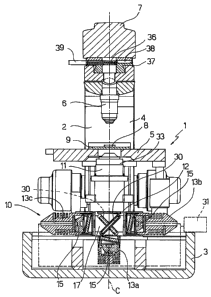
With reference to figures 1 and 2, a transmitted
light fluorescence microscope 1 comprises a base
structure 2, essentially known and having in particular
CA 02593447 2007-07-06
WO 2006/072886
PCT/1B2006/000160
an internally hollow base 3 from which vertically
extend a column 4, a sample-holder mount 5, one or more
objectives 6, and an eyepiece 7 (all known components
and neither described nor illustrated in detail for the
5 sake of simplicity). The sample 8 to be analysed is
carried for example by a transparent slide 9 placed on
the mount 5.
The microscope 1 also comprises a lighting
assembly 10, arranged underneath the mount 5, and a
condenser 11, arranged between the lighting assembly 10
and the mount 5.
The lighting assembly 10 comprises a box 12 and a
plurality of integrated lighting modules 13, which are
supported by the box 12 and are provided with
respective LEDs 15 (or other similar solid state light
sources); the LEDs 15 present respective emission bands
different one from the other and are arranged
underneath the mount 5 to illuminate from underneath
the sample 8 to be analysed on the mount 5; at least
one LED 15 emits a spectral band adapted to excite the
fluorescence of the sample.
Box 12 is releasably coupled, in a known way not
shown for the sake of simplicity, to the base 3, so
that the lighting assembly 10 is completely removable
from the base 3; the box 12 presents a plurality of
seats 16 for respective modules 13; the modules 13 face
a chamber 17 inside the box 12 presenting an exit
window 18 which is arranged in use in front of the
condenser 11 and is closed by a transparent plate 19.
In the non-limiting example shown in figures 1 and
2, the lighting assembly 10 comprises three modules 13
essentially arranged in a T; a central module 13a is
aligned with the condenser 11 essentially along an
optical axis C of the condenser 11, and two side
modules 13b, 13c are arranged and facing each over and
on opposite sides of the central module 13a.
CA 02593447 2007-07-06
WO 2006/072886
PCT/1B2006/000160
6
Each module 13 comprises a casing 25, inside which
are accommodated a LED 15, a collimator 20 and a filter
21 arranged aligned along an optical axis A of the
collimator 20; the LED 15 is carried by a plate 22
fastened to a thermal dissipator 23; the collimator 20
is arranged in close proximity to the LED 15 and is
overhangingly supported by stems 24 from the plate 22;
the filter 21 is an inferential filter, chosen
according to the emission band of the LED 15 with which
it is associated. The casing 25 is provided with
releasable fastening means 26 to a seat 16 and is
frontally closed, in front of the filter 21, by a clear
plate 27; the means 26 may be of any known type, for
example bayonet-joint means, threaded means or snap
means, and have the function of allowing the complete
removal of the module 13 from the box 12 and its
replacement with another similar module having a LED
with a different emission band.
The collimator 20 is a complex-surface
catadioptric collimator and, preferably, a total-
internal-reflection surface collimator and is shaped so
as to collect the emission of the LED 15 to which it is
associated and convey it into a beam of essentially
parallel light rays.
The filter 21 is arranged in front of the
collimator 20 on the opposite side of the LED 15 to
select a band to send onto the sample to be analysed.
The filter 21 is essentially disc-shaped and slanted
with respect to the optical axis A of the collimator
20, preferably at an angle from approximately 100 to
approximately 15 . The slant of the filter 21 avoids
the formation of so-called ghost images created by the
reflection of the sample emission on the (generally
highly reflecting) surfaces of the filter.
The chamber 17 also presents a side opening 28 and
a pair of guides 29 arranged in a cross and the
CA 02593447 2007-07-06
WO 2006/072886
PCT/1B2006/000160
7
lighting assembly 10 also comprises one or more foils
30 carried by sliders 31 sliding on the guides 29; each
foil 30 is removably accommodated in the chamber 17 and
interposed between the modules 13 and the condenser 11
and is interchangeable with another different foil. The
foils 30 can therefore be extracted from the side of
the chamber 17 to be replaced with different foils,
according to the module 13 (and therefore of the LED
15) used. The foils 30 are in particular reflecting,
dichroic or mirror foils according to needs.
The lighting assembly 10 then comprises an
electronic control unit 32 (known and only
schematically indicated with a dotted line in figure 1,
along with the connections to the modules 13) for the
management of the LEDs 15, which controls the selective
lighting of the LEDs 15 and optionally regulates the
emission intensity of the LEDs 15.
The condenser 11 is an Abbe condenser having a
casing 33 which accommodates two or more lenses: for
example, as shown in figures 3 and 4, three lenses 34.
In all cases, the condenser 11 has a focal distance
less than approximately 20 mm and preferably less than
approximately 15 mm, and numeric aperture higher than
approximately 0.8 and preferably higher than
approximately 0.9 (as known, the numeric aperture NA of
a condenser is the quantity which characterises the
maximum light collection angle, measured with respect
to the optical axis and defined as NA = n sen a, where
n is the refraction index of the means found at the
condenser outlet and a is the maximum output angle of
the beam measured with respect to the optical axis).
The focal distance and numeric aperture values are
understood as udry", that it with the condenser 11
working in air.
The condenser 11 is prepared for use, without the
need for changes or adjustments, according to both
CA 02593447 2007-07-06
WO 2006/072886
PCT/1B2006/000160
8
typical fluorescence microscopy working modes (shown in
figures 3 and 4):
- "dry" mode, that is when the means between the
condenser outlet 11 and the slide 9 on which the sample
8 is arranged is air (figure 3), and
- "immersion" mode, that is when a liquid 35 is used,
typically oil, between the condenser outlet 11 and the
slide 9 (figure 4).
With the help of an optional field diaphragm
(known and not shown), the condenser 11 allows also to
obtain an lighting system according to the Kohler
diagram.
The microscope 1 also comprises a filter assembly
36 have at least one emission filter 37 (figure 1)
arranged before the eyepiece 7 to filter the
fluorescent emission of the sample before it reaches
the eyepiece 7 (or another known detection device
capable of collecting the emission of the sample). The
emission filter 37 is selected according to the
emission of the LED 15 used; the emission filter 37 is
therefore extractable from a seat 38 formed in the
column 4 and interchangeable with another filter, or
selectable from a plurality of filters carried by a
filter holder mechanism 39 accommodated in the seat 38
(for example, in which the filters are carried by a
carousel rotating about the optical axis C or by a
slider shifting orthogonally to the optical axis C).
It is clear that microscope 1 may be provided with
various combinations of LEDs 15; in all cases, the
possibility of replacing at least one of the modules 13
further increases the versatility of the microscope 1.
A basic configuration of the microscope 1 envisages for
example a white light LED 15, arranged for example in
the module 13b, and two coloured light LEDs 15, for
example a blue light and a green light, arranged
respectively in modules 13a and 13c.
CA 02593447 2007-07-06
WO 2006/072886
PCT/1B2006/000160
9
When the white light LED is used, a mirror foil
30b (not necessarily a dichroic foil) is arranged in
the chamber 17; when coloured light LEDs are used
instead, a dichroic foil 30a is arranged in the chamber
17; the dichroic foil 30a also allows the simultaneous
use of the two coloured light LEDs, if required.
With reference to figures 5 and 6, in which
details similar or equal to those already described are
indicated with the same numbers, a transmitted light
fluorescence microscope 1 consists of a traditional
white light microscope la and a transmitted light
fluorescence working mode adaptation kit 40; the
microscope la is any known microscope found on the
market and has the same basic structure 2 already
described; the microscope la also comprises an
optical/lighting assembly 41 of the known type,
accommodated in a body 42 fitted on the base 3, and
provided with a traditional lamp (for example a halogen
lamp) and the respective optics (known and not shown).
The adaptation kit 40 comprises a supporting unit
45, which carries a lighting assembly 10 with at least
one integrated LED lighting module 13 and is insertable
between the base 3 and the mount 5 of the microscope
for lighting the mount 5 from underneath, releasable
coupling means 46 of the unit 45 to the structure 2 of
the microscope, a condenser 11, and a filter assembly
36.
The unit 45 presents a box 12 and the coupling
means 46 comprise supporting elements 47 which protrude
from the box 12 cooperating with respective portions 48
of the structure 2; in the non-limiting example shown
in figures 5 and 6, the elements 47 are formed by
respective legs which protrude vertically from the box
12 and are provided with shoulders 49 which rest on a
locator surface 50 of the base 3; the box 12 possibly
presents a lower centring portion (not shown) which
CA 02593447 2007-07-06
WO 2006/072886
PCT/1B2006/000160
cooperates with the body 42, for example a peripheral
upper end edge of the body 42, to provide a reference
for the assembly of the unit 45 on the microscope la.
The coupling means 46 also comprise fastening
5 members 53 of any known type (only one of which is
shown, only schematically, in figures 5 and 6 for the
sake of simplicity), fixed to the box 12 or to the
elements 47 and releasably fastened to the base 3 to
integrally fasten the unit 45 to the structure 2; in
10 the non-limiting example shown in figure 6, the
fastening members 53 comprise hooks 54 which hook onto
a lower edge 55 of the base 3 on opposite sides of the
base 3, and respective lever latches 56 which
integrally connect the latches 54 to the elements 47;
it is however understood that fastening members of any
other known type may be equally used, for example
elastic clips, tie-rods or straps.
The box 12 presents a inner chamber 17 having an
exit window 18, arranged in use in front of the
condenser 11 and closed by a transparent plate 19; the
chamber 17 comprises an inner through cavity 57, which
extends along an axis X and is arranged through the box
12 between the window 18 and a lower window 58, aligned
with the window 18; in use, when the unit 45 is fitted
on the microscope la, axis X essentially coincides with
optical axis C of the condenser 11 and with the optical
axis of the assembly 41 and the cavity 57 allows the
light emitted by the assembly 41 to cross the unit 45,
allowing therefore the use of the assembly 41, also
with unit 45 fitted on the microscope la. The chamber
17 also presents in this case a side opening 28
associated with a guide 29, formed in the chamber 17
and slanted with respect to axis X, and through which a
reflecting foil 30 fitted on a slider 31 sliding on the
guide 29 may be inserted and extracted. Different foils
30 (mirrors or possibly dichroic foils) are selectively
i
CA 02593447 2007-07-06
WO 2006/072886
PCT/1B2006/000160
11
usable in the chamber 17 according to the module 13
(and therefore of the LED 15) fitted on the unit 45.
The box 12 then presents at least one seat 16 for
a LED module 13 of the type already described above
(and therefore comprising again a casing 25 in which
are accommodated a LED 15, a collimator 20 and a filter
21, not shown in figures 5 and 6 for the sake of
simplicity, being however entirely similar to those
shown in figures 1 and 2).
The seat 16 is delimited by a peripheral edge 59
in which is insertable the casing 25 of the module 13
and is communicating with the chamber 17 so that the
module 13 is facing, once fitted in the seat 16, the
foil 30 in the chamber 17.
The casing 25 is provided with releasably
fastening means 26 to the seat 16; as already described
with reference to figures 1 and 2, also in this case
the means 26 can be of any known type and have the
function of allowing the complete removal of the module
13 from the box 12 and its replacement with another
similar module having a LED with a different emission
band. In the example of figures 5 and 6, the fastening
of the casing 25 in the seat 16 is obtained by means of
a threaded dowel 60 arranged through the casing 12 and
engaging a notch 61 formed on an outer surface of the
casing 25.
The seat 16 presents a pair of facing spring
contacts 64, cooperating with respective terminals 65
of the module 13 to ensure electrical powering and
electronic management of the module 13; for the sake of
simplicity, the electrical connections between the
contacts 64 and the power source (external mains or
battery) are not shown.
The condenser 11 that is part of the adaptation
kit 40 has already been described above and it is used
to replace the standard condenser of the microscope 1
CA 02593447 2007-07-06
WO 2006/072886
PCT/1B2006/000160
12
by using the same fastening system of the standard
condenser.
The filter assembly 36 comprises one or more
emission filters 37 to be used in combination with the
modules 13 (and selected according to the LED used); as
already shown with reference to figure 1, the filter
assembly 36 is inserted in a seat 38 (which is normally
prearranged on traditional microscopes upstream of the
eyepiece 7).
The adaptation kit 40 makes microscope la suitable
for transmitted light fluorescence analysis, without
requiring any structural modification or other type of
intervention on the microscope except for the
replacements of components which are already
prearranged to be interchangeable, such as the Abbe
condenser and the filters arranged upstream of the
eyepiece; the user may therefore fit the adaptation kit
on a commonly marketed microscope without at all
altering the functional components and electrical
connections of the microscope.
According to an important aspect of the invention,
the lighting assembly 10 included in the microscope 1
or belonging to the adaptation kit 40 comprises a
module 13 provided with a LED-UV which emits in the
ultraviolet; the collimator 20 associated to the LED-UV
is in this case made of a low UV absorbance material,
essentially not fluorescent by effect of UV radiation,
for example glass or polymeric material with low or no
fluorescent emission. Also the lenses 34 of the
condenser 11 are made of a low UV absorbance material,
essentially not fluorescent by effect of UV radiation,
particularly of glass.
In a preferred configuration, shown schematically
in figure 7, the module 13 with LED-UV is always of the
type described above and thus comprises a casing 25 in
which are housed a LED-UV 15 (which emits in the
CA 02593447 2007-07-06
WO 2006/072886
PCT/1B2006/000160
13
ultraviolet), a collimator 20 and a filter 21; the
collimator 20 associated to the LED-UV 15 consists of a
condenser 70 of the Abbe type, essentially equal to the
condenser 11 but used upside-down with respect to the
condenser 11, and that is with the LED-UV 15 arranged
in the frontal focus of the condenser 70; the system
constituted by two counterpoised condensers 20, 70 of
the Abbe type forms a high numeric aperture optical
system but above all a so-called "fully symmetric"
system in the optical design theory, where most of the
optical aberrations and mainly astigmatism and field
curvature are reduced or fully eliminated, with
consequent increase of excitation efficiency.
It is then clear that further changes and
variations can be made to the microscope described and
shown herein without departing from the scope of
protection of the annexed claims.
In particular, according to a further variation,
the lighting assembly 10 comprises a single "multichip"
LED capable of selectively emitting in different bands
of emission, instead of a plurality of LEDs 15 having
respective different emission bands; the band of
emission to be sent to the sample 8 to be analysed is
selected by means of unit 32. The lighting assembly 10
comprises in this case a filter holder device (for
example rotating carousel-type or shifting slider-type)
for selectively carrying an appropriate filter in axis
with the LED according to the selected emission band.