Note : Les descriptions sont présentées dans la langue officielle dans laquelle elles ont été soumises.
CA 02595891 2007-07-25
WO 2006/081407 PCT/US2006/002903
ADDING MICROSCOPIC POROSITY TO THE SURFACE OF A MICROCOIL
TO BE USED FOR MEDICAL IMPLANTATION
CROSS-REFERENCES TO RELATED APPLICATIONS
This application is based upon Provisional Patent Application Serial No.
60/647,516, filed January 26, 2005.
BACKGROUND OF THE INVENTION
Microcoils have been developed for implantability into aneurysms as a means of
promoting healing through the obstruction of pulsatile blood flow into the
center of the
promoting aneurysm. Such devices have become very successful in treatment of
cranial
aneurysms and as a method of treating and preventing strolce when such
malformations are
discovered.
Another embolic coil is known which includes a distal roughened, textured
surface
with poclcets having diameters of about 0.125 to 50 microns and depths of
about 0.25 to 20
microns to provide improved platelet adhesion and to promote clotting. Another
type of
removable occlusion system for treating the neck of an aneurysm includes a
mesh portion
with pores allowing blood to flow through the mesh portion. While such embolic
devices
have voids or pores that can promote clotting or allow blood flow through the
device, it
would be desirable to provide an embolic device that can further promote
healing of a
patient's vasculature.
One vasoocclusive coil is known that has an enhanced therapeutic strand
structure
that may be formed from or incorporate therapeutic or bioactive materials such
as
polyglycolic acid (PGA) or poly(D,L-lactic acid-co-glycolic acid) (PGLA), or
other
therapeutic materials. Another embolic device is known which includes
embolizing
elements made of a hydrophilic, macroporous, polymeric, hydrogel foam
material.
While the microcoil treatment of aneurysms is highly effective in improving
the
prognosis for recovery of those with such malformations, it is believed that
success rate of
the procedures would be enhanced if new and effective methods of treatment of
the micro
coils could be used to enhance the healing process once the microcoils are
placed. The
present invention resolves these and other limitations in prior art devices.
CA 02595891 2007-07-25
WO 2006/081407 PCT/US2006/002903
-2-
SUMMARY OF THE INVENTION
The present invention treats microcoils and other implantable devices, using
one or
more of a variety of etching methods, which can include plasma etching,
photolithography
and chemical etching, to create microscopic voids in the surface of a
microcoil which are
complex in shape and adapted to receive into the surface by pressure, melting
or deposition
one or more of a variety of therapeutic agents, therapeutic materials and
therapeutic plastic
agents which caii act to accelerate the healing process once the coil is in
place. At the
present time, agents which are believed to be appropriate for deposition in
the voids
created by the process include therapeutic drugs and agents such as PGA or
PGLA, among
others, which can act as an accelerant for the healing process.
The present invention accordingly provides for a vasoocclusive microcoil for
therapeutic treatment of a patient's vasculature, including a vasoocclusive
microcoil
having at least a portion having a surface defining a plurality of voids or
pores therein, and
a therapeutic or bioactive material disposed within the plurality of voids or
pores. The
present invention also provides for a method for occluding a patient's
vasculature,
involving the steps of providing a vasoocclusive microcoil having at least a
portion having
a surface defining a plurality of voids therein, and a therapeutic/bioactive
material
disposed within the plurality of voids, and introducing the vasoocclusive
microcoil into the
patient's vasculature, whereby the therapeutic/bioactive material can act to
accelerate a
healing process in the patient's vasculature. The present invention also
provides for a
method for delivering a therapeutic agent or material to a patient's
vasculature by such a
microcoil having at least a portion with a surface having a plurality of voids
or pores, and
for controlling the delivery of the therapeutic agent or material by
controlling the porosity
of the surface of the microcoil. The present invention also provides for a
method of
delivering a hydrogel to a patient's vasculature by a microcoil having at
least a portion with
a surface having a plurality of voids or pores carrying the hydrogel. The
present invention
also provides for a method for forming porosity in a surface of a microcoil
using one or
more of a variety of etching methods, which can include plasma etching and
sputtering.
The therapeutic agent or material may be a therapeutic drug or a therapeutic
plastic
agent which can act to accelerate the healing process once the coil is in
place, and in a
presently preferred aspect, the therapeutic agent or material may be
polyglycolic acid or
CA 02595891 2007-07-25
WO 2006/081407 PCT/US2006/002903
-3-
poly(D,L-lactic acid-co-glycolic acid), andlor a therapeutic drug. More
broadly, the
therapeutic material may be silk, collagen, elastin, polyglycolic acid,
polylactic acid,
poly(D,L-lactic acid-co-glycolic acid), poly(L-lactide), poly(L-lactide-co-D,L-
lactide),
poly(L-lactide-co-glycolide), poly(glycolide-co-trimethylene carbonate),
polyethylene
oxide, polydioxanone, polycaprolactone, hylauric acid, polyhydroxylbutyrate,
poly(phosphazene), poly(D,L-lactide-co-caprolactone), poly(glycolide-co-
caprolactone),
polyvinyl alcohol, polyanhydrides thereof, poly(ortho esters) thereof,
poly(phosphate
esters) thereof, poly(amino acids) thereof, poly(hydroxy butyrates) thereof,
copolymers
thereof, composites thereof, or coinbinations thereof. The therapeutic
material may also be
ethylene-octene copolynler, polypropylene, polyethylene, polyacrylate,
polyacrylamide,
poly(hydroxyethyl methacrylate), polyurethane, polysiloxane, copolymers
thereof,
composites thereof, or combinations thereof.
Other features and advantages of the present invention will become more
apparent
from the following detailed description of the preferred embodiments in
conjunction with
the accompanying drawings, which illustrate, by way of example, the invention.
BRIEF DESCRIPTION OF THE DRAWINGS
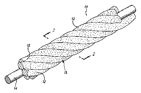
Figure 1 is a perspective of a portion of a microcoil according to the
invention.
Figure 2 is a cross-section at 2-2 of Figure 1.
Figure 3 shows a portion of a helical multi-stranded microcoil formed
according to
the invention.
Figure 4 shows a portion of a helical single-stranded microcoil foimed
according
to the invention.
DETAILED DESCRIPTION OF THE PREFERRED EMBODIMENTS
As is illustrated in the drawings, which are provided for the purposes of
illustration
and not by way of limitation, the invention is embodied in a vasoocclusive
microcoil
having at least a portion with a surface defining a plurality of voids or
pores therein, with a
therapeutic or bioactive material within the voids or pores. In a presently
preferred aspect
illustrated in Fig. 1, the vasoocclusive microcoil may be formed from one or
more flexible
strands 12 of a resilient material and/or super-elastic material, such as
nickel titanium
CA 02595891 2007-07-25
WO 2006/081407 PCT/US2006/002903
-4-
alloy, for example. The nickel titanium alloy is typically heat treated such
that the alloy is
highly flexible at a temperature appropriate for introduction into the body
via a catheter.
By choosing such a material for micro-coils and the like, the devices formed
from the
micro-cable can be relatively easily placed into the appropriate body cavity
and after
placement, the device will take on a shape designed to optimize the
therapeutic purposes
desired for the device.
As illustrated in Fig. 2, the vasoocclusive microcoil may also include a
centrally,
axially disposed radiopaque wire 14, which can be formed of platinum or gold,
for
example, or other similar suitable radiopaque metals, in order to provide a
radiopaque
marlcer of the deployed configuration of a device made of the vasoocclusive
microcoil
during vascular surgery.
One advantageous application of the invention is to vasooclusive devices
formed of
the vasoocclusive microcoil for insertion into aneurysms and other vascular
defects for the
purpose of occluding flow to the aneurysm. Fig. 3 illustrates a portion of a
helically
wound coil 16 of the vasoocclusive microcoil 10, which is formed to fit within
a micro-
catheter for insertion into an area upon which a therapeutic procedure is to
be performed.
Fig. 4 illustrates a portion of a helically wound coil 16' of the
vasoocclusive microcoil 10',
which is formed to fit within a micro-catheter for insertion into an area upon
which a
therapeutic procedure is to be performed. While helical coils are illustrated,
it will be
appreciated that numerous other shapes can be formed from the vasoocclusive
microcoil of
the invention.
As is illustrated in Figs. 1 and 4, the vasoocclusive microcoil includes at
least a
portion having a surface defining a plurality of microscopic voids or pores 18
in the
surface of the microcoil, which may be complex in shape. The voids or pores
may be
formed in the surface of the microcoil using one or more of a variety of
sputtering and
etching methods, which can include plasma etching, photolithography and
chemical
etching, for example, and any combination or combinations thereof. Other
methods of
creating microscopic voids or pores in the surface of an implantable device
can be
effective when combined with the ability to deposit, press, or melt a healing
accelerant
therapeutic and/or bioactive material into the surface.
CA 02595891 2007-07-25
WO 2006/081407 PCT/US2006/002903
-5-
The voids or pores are advantageously formed to receive and retain a variety
of
therapeutic and/or bioactive agents which can act to accelerate the healing
process once the
coil is in place. The agents may be deposited in the voids or pores by
pressure, melting or
deposition, or the lilce. At the present time, agents which are believed to be
appropriate for
deposition in the voids or pores created by the process include polyglycolic
acid (PGA)
and poly(D,L-lactic acid-co-glycolic acid) (PGLA), which can act as an
accelerant for the
healing process. Other therapeutic and/or bioactive agents that may be
deposited in the
voids or pores include polylactic acid or poly(D,L-lactide) (PLA), poly(D, L-
lactide-co-
glycolide) (PLA/PGA), poly(L-lactide) (PLLA), poly(L-lactide-co-D,L-lactide)
(PLLA/PLA), poly(L-lactide-co-glycolide) (PLLA/PGA), poly(glycolide-co-
trimethylene
carbonate) (PGA/PTMC), polyethylene oxide (PEO), polydioxanone (PDS),
polycaprolactone (PCL), hylauric acid, polyhydroxylbutyrate (PHBT),
poly(phosphazene),
poly(D,L-lactide-co-caprolactone) (PLA/PCL), poly(glycolide-co-caprolactone)
(PGA/PCL), polyvinyl alcohol (PVA), polyanhydrides (PAN), poly(ortho esters),
poly(phosphate ester), poly(amino acid), poly(hydroxy butyrate), copolymers of
these
materials as well as composites and combinations thereof; non-metallic fiber
material,
such as silk, collagen, elastin or other connecting proteins; plastic or other
polymers such
as an ethylene-octene copolymer, polypropylene, polyethylene, polyacrylate,
polyacrylamide, poly(hydroxyethyl methacrylate), polyurethane, polysiloxane
and their
copolymers. The therapeutic and/or bioactive non-metallic fiber material may
be
bioabsorbable or non-absorbable. The therapeutic and/or bioactive non-metallic
fiber
material may also be used for absorbing and releasing one or more therapeutic
agents.
It will be apparent from the foregoing that while particular forms of the
invention
have been illustrated and described, various modifications can be made without
departing
from the spirit and scope of the invention. Accordingly, it is not intended
that the
invention be limited, except as by the appended claims.
