Note : Les descriptions sont présentées dans la langue officielle dans laquelle elles ont été soumises.
DEMANDE OU BREVET VOLUMINEUX
LA PRESENTE PARTIE DE CETTE DEMANDE OU CE BREVET COMPREND
PLUS D'UN TOME.
CECI EST LE TOME 1 DE 2
CONTENANT LES PAGES 1 A 40
NOTE : Pour les tomes additionels, veuillez contacter le Bureau canadien des
brevets
JUMBO APPLICATIONS/PATENTS
THIS SECTION OF THE APPLICATION/PATENT CONTAINS MORE THAN ONE
VOLUME
THIS IS VOLUME 1 OF 2
CONTAINING PAGES 1 TO 40
NOTE: For additional volumes, please contact the Canadian Patent Office
NOM DU FICHIER / FILE NAME:
NOTE POUR LE TOME / VOLUME NOTE:
CA 02601671 2007-09-19
WO 2006/102569 PCT/US2006/010699
NUCLEIC ACID DETECTION
FIELD OF INVENTION
This invention relates to the field of nucleic acid detection.
BACKGROUND OF THE INVENTION
The detection of small differences in nucleic acid content is an important
task within
the field of molecular diagnostics.
The most coinmon method used to detect small quantities of nucleic acids is by
using
PCR or RT-PCR. These methods perform exceptionally well in determining whether
a
sequence of interest is present in a given sample or not, but in order to
determine if there is a
difference between the concentration of two sequences, approximately a 2-fold
difference is
needed.
Quantitative analysis of nucleic acid is used for example, in quality control,
gene
expression analysis, medical monitoring and diagnosis. Methods are described
herein that
significantly improve the accuracy of these measureinents, opening up new
possibilities
within science and medicine.
SUMMARY OF THE INVENTION
In one aspect, the invention provides improved methods for quantitating the
ainount
of a nucleic acid sequence in a nucleic acid sample. The claimed methods can
permit the
accurate detection of the amount of a nucleic acid having a given sequence in
a sample,
including accurate determination of differences of less than two-fold.
In one aspect, the methods rely upon a method of misbalancing a PCR reaction.
When the reaction is misbalanced in this manner, an asymmetry is created
between template
and complementary strands that permits subsequent differentiation between
desired and
undesired products in the reaction, which permits sensitive detection of even
small
differences in the amounts of nucleic acid sequences in nucleic acid samples.
1
CA 02601671 2007-09-19
WO 2006/102569 PCT/US2006/010699
In one embodiment, then, a method of misbalancing a PCR reaction is provided
that
comprises the steps of: a) generating products in a PCR reaction that include
the original
nucleic acid sequence of interest (template), its complement nucleic acid
sequence, and the
corresponding first and second PCR primers; and b) adding an excess of a
nucleic acid
disruption sequence that is complementary to one or the other of the first and
second PCR
primers. In the discussion that follows, the disruption sequence is
coinplementary to the first
primer. When the disruption sequence is allowed to hybridize in the next
annealing cycle, it
predominantly forms disruption sequence pairs by annealing to the first primer
and to one or
the other of the original (template) sequence or its complement, but not both
(depending upon
1o which of these two strands has the complement of the disruption sequence).
Products
generated by subsequent extension are misbalanced such that one strand, either
template or its
complement, is produced, but not both. That is, upon extension after
hybridization of the
disruption sequence(s), there is produced predominantly either: a) a double
stranded original
(template) sequence with a break in one of the strands and a single stranded
complement
sequence, OR b) a double stranded complement sequence with a break in one of
the strands
and a single stranded original (template) sequence. The identity of which
product is
produced in single or double-stranded form is determined by which of the two
PCR primers
is complementary to the disruption sequence. Detection of the single stranded
product
permits sensitive measurement of the original amounts of a target sequence.
In another aspect, a metliod is provided for blocking future PCR-based
ainplification
in a reaction mixture by locking double stranded DNA through cross-linking.
The cross-
linking prevents the amplification and thereby the detection of DNA that was
double-stranded
at the time of cross-linking. This blockage of amplification can permit more
sensitive
detection of the products that were not cross-linked. The cross-linking can be
accomplished
by, e.g., UV or chemical treatment.
In another aspect, a method is provided for determining the amount of a target
nucleic
acid relative to the amount of a reference nucleic acid in a nucleic acid
sample. The method
comprises performing PCR on a combined mixture of the reference and target
nucleic acid,
along with the forward and reverse PCR primers for both the reference and
target nucleic
acids. The reverse PCR primer for the reference contains a tail sequence that
is identical to a
sequence found on the target nucleic acid. Following a given number of cycles
of PCR, the
2
CA 02601671 2007-09-19
WO 2006/102569 PCT/US2006/010699
symmetry of the reaction is disrupted by adding excess amounts of two nucleic
acid
disruption sequences and letting them hybridize to the single stranded PCR
products. The
first disruption sequence is complementary to the target probe's reverse
primer, and the
second disruption sequence is complementary to the reference probe's reverse
primer but
not including the tail region that is identical to a sequence found on the
target nucleic acid.
Following disruption, remaining single stranded target or reference is
detected, such that the
amount of target in the original sample is determined relative to the
reference.
DESCRIPTION OF THE DRAWINGS
Figure 1 shows a schematic of controlled disruption of a PCR reaction to
create a
single stranded teinplate (ABC) and a double stranded complement (C'B'A'). The
first step
shows the original genomic DNA sequence (a.k.a. template, ABC) and the
respective forward
and reverse PCR primers (A and C', respectively). In the second step, the
products of several
PCR cycles are shown. After completion of the desired number of PCR steps, the
schematic
shows the addition of a disruption sequence (C) complementary to the reverse
primer (C') in
excess to the PCR primers. When cooled, the excess disruption sequence will
bind the reverse
primer, and the complement sequences. At the same time, the forward primer
will bind the
complement and extend, creating a double stranded complement with a break at
the junction
of B and C. The template (ABC) therefore, will remain single stranded.
Figure 2 shows a schematic of how to eliminate double stranded DNA from a
subsequent PCR or other detection reaction using cross-linking. Step 1 shows
the presence of
a sequence in both single stranded and double stranded form. Adding a cross-
linking agent
binds the double stranded DNA together permanently or semi-permanently,
preventing
complete dissociation during the high temperature step of the PCR cycle (step
3). This
prevents amplification of the double DNA e.g., by PCR, but does not limit the
single stranded
DNA amplification, selectively.
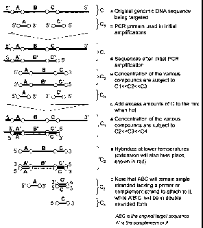
Figure 3 shows a schematic of a single tube relative amplification technique
that can
detect much smaller differences in nucleic acid content than previously
thought possible. Step
1 shows the starting materials: two genomic DNA sequences one would like to
compare, and
respective forward and reverse PCR primers, with the reverse primer of the
reference
sequence having a "tail" with partial B sequence. After performing several
cycles of PCR, the
3
CA 02601671 2007-09-19
WO 2006/102569 PCT/US2006/010699
products seen in step 2 will be produced. If after this step one adds excess C
and F primers to
the reactions, these can hybridize to the reverse primers and the complement
sequences,
leaving the template strands untouched by either the newly added C and F
primers, or the
previously added reverse primers. The single stranded reference and target
template can then
hybridize to each other and form a pair. Any unpaired reference or target
template will
remain single stranded. Step 4 shows what happens if the double stranded DNA
is cross-
linked. Cross-linking only has to happen on the C/C' and pB/pB' sequences. If
the cross-
linlcing has taken place permanently or semi-permanently, direct PCR can be
perforined using
primers that span a cross-linlcing site to detect only the single stranded
template (ABC as
shown).
Figure 4 shows a schematic of an embodiment where the cross-linking of the
double
stranded template pairs is followed by the detection of the single stranded
templates using
PCR. The figure shows a possible selection of PCR primers for further
analysis, where
forward primers A and D were already found in the final sample as seen in Step
4 of Figure 3,
and Rl and R2 where added later. As shown, the primers will only amplify the
single
stranded templates and will not be able to ainplify the cross-linked double
stranded template
shown on top.
DETAILED DESCRIPTION OF THE INVENTION
Definitions:
As used herein, a "polynucleotide" or "nucleic acid" refers to a covalently
linked
sequence of nucleotides (i.e., ribonucleotides for RNA and
deoxyribonucleotides for DNA) in
which the 3' position of the pentose of one nucleotide is joined by a
phosphodiester group to
the 5' position of the pentose of the next. The term "polynucleotide"
includes, without
limitation, single- and double-stranded polynucleotide. The term
"polynucleotide" as it is
employed herein embraces chemically, enzymatically or metabolically modified
forms of
polynucleotide comprising, e.g., DNA, RNA, PNA, combinations of these and/or
polymers
containing one or more nucleotide analogs. A "nucleotide analog", as used
herein, refers to a
nucleotide in which the pentose sugar and/or one or more of the phosphate
esters is replaced
with its respective analog. Exemplary phosphate ester analogs include, but are
not limited to,
alkylphosphonates, methylphosphonates, phosphoramidates, phosphotriesters,
4
CA 02601671 2007-09-19
WO 2006/102569 PCT/US2006/010699
phosphorothioates, phosphorodithioates, phosphoroselenoates,
phosphorodiselenoates,
phosphoroanilothioates, phosphoroanilidates, phosphoroamidates,
boronophosphates, etc.,
including any associated counterions, if present. Also included within the
definition of
"nucleotide analog" are nucleobase monomers which can be polymerized into
polynucleotide
analogs in which the DNA/RNA phosphate ester and/or sugar phosphate ester
backbone is
replaced with a different type of linkage. Further included within "nucleotide
analogs" are
nucleotides in which the nucleobase moiety is non-conventional, i.e., differs
from one of G,
A, T, U or C. For example, halogenated nucleotides such as bromodeoxyuridine
can be
employed. Generally a non-conventional nucleobase will have the capacity to
form hydrogen
lo bonds with at least one nucleobase moiety present on an adjacent counter-
directional
polynucleotide strand or provide a non-interacting, non-interfering base.
"Polynucleotide" also embraces a short polynucleotide, often referred to as an
oligonucleotide (e.g., a primer or a probe). A polynucleotide has a"5'-
terminus" and a "3'-
terminus" because polynucleotide phosphodiester linkages occur to the 5'
carbon and 3'
carbon of the pentose ring of the substituent mononucleotides. The end of a
polynucleotide at
which a new linkage would be to a 5' carbon is its 5' terminal nucleotide. The
end of a
polynucleotide at wliich a new linkage would be to a 3' carbon is its 3'
terminal nucleotide.
A terminal nucleotide, as used herein, is the nucleotide at the end position
of the 3'- or 5'-
terminus. As used herein, a polynucleotide sequence, even if internal to a
larger
polynucleotide (e.g., a sequence region within a polynucleotide), also can be
said to have 5'-
and 3'- ends.
As used herein, the term "chemically modified," when used in the context of a
nucleotide, refers to a nucleotide having a difference in at least one
chemical bond relative to
a standard ATP, CTP, GTP, UTP, dATP, dCTP, dGTP or dTTP nucleotide. The
"cheinical
modification" does not refer to the modification occurring when a nucleotide
is incorporated
into a polynucleotide by 5' to 3' phosphodiester linkage.
As used herein, the term "hybridization" is used in reference to the physical
interaction of coinplementary (including partially complementary)
polynucleotide strands by
the formation of hydrogen bonds between complementary nucleotides w11en the
strands are
arranged antiparallel to each other. Hybridization and the strength of
hybridization (i.e., the
5
CA 02601671 2007-09-19
WO 2006/102569 PCT/US2006/010699
strength of the association between polynucleotide strands) is impacted by
many factors well
known in the art including the degree of complementarity between the
polynucleotides, and
the stringency of the conditions involved, which is affected by such
conditions as the
concentration of salts, the presence of other components (e.g., the presence
or absence of
s polyethylene glycol), the molarity of the hybridizing strands and the G+C
content of the
polynucleotide strands, all of which results in a characteristic melting
temperature (Tm) of the
formed hybrid.
As used herein, when one polynucleotide is said to "hybridize" to another
polynucleotide, it means that the two polynucleotides form a hydrogen-bonded
antiparallel
hybrid under high stringency conditions. Hybridization requires partial or
complete sequence
complementarity between the polynucleotides that hybridize. When one
polynucleotide is
said to not hybridize to another polynucleotide, it means that there is
insufficient sequence
complementarity between the two polynucleotides to form a hydrogen-bonded
hybrid, or that
no hybrid forms between the two polynucleotides under high stringency
conditions. As used
herein, "specific hybridization" refers to the binding, duplexing, or
hybrization of a nucleic
acid molecule only to a target nucleic acid sequence and not to other non-
target nucleic acid
molecules in a mixture of both target and non-target nucleic acid sequence.
As used herein, the terms "low stringency," "medium stringency," "high
stringency,"
or, "very high stringency conditions" describe conditions for nucleic acid
hybridization and
washing. Guidance for performing hybridization reactions can be found in
Current Protocols
in Molecular Biology, John Wiley & Sons, N.Y. (1989), 6.3.1-6.3.6, which is
incorporated
herein by reference in its entirety. Aqueous and nonaqueous methods are
described in that
reference and either can be used. Specific hybridization conditions referred
to herein are as
follows: (1) low stringency hybridization conditions in 6X sodium
chloride/sodium citrate
(SSC) at about 45 C, followed by two washes in 0.2X SSC, 0.1% SDS at least at
50 C (the
temperature of the washes can be increased to 55 C for low stringency
conditions); (2)
medium stringency hybridization conditions in 6X SSC at about 45 C, followed
by one or
more washes in 0.2X SSC, 0.1% SDS at 60 C; (3) high stringency hybridization
conditions in
6X SSC at about 45 C, followed by one or more washes in 0.2X SSC, 0.1% SDS at
65 C;
6
CA 02601671 2007-09-19
WO 2006/102569 PCT/US2006/010699
and (4) very high stringency hybridization conditions are 0.5M sodium
phosphate, 7% SDS at
65 C, followed by one or more washes at 0.2X SSC, 1% SDS at 65 C.
As used herein, a polynucleotide "isolated" from a sample is a naturally
occurring
polynucleotide sequence within that sample which has been removed from its
normal cellular
or non-cellular environment. Thus, an "isolated" polynucleotide that was in a
normal cellular
environment may be in a cell-free solution or be placed in a different
cellular environment.
Similarly, an "isolated" polynucleotide that was in a normal non-cellular
environment may be
in a different cell-free solution or be placed in a cellular environment.
As used herein, the terms "blood," "plasma" and "serum" expressly encompass
fractions or processed portions thereof. Similarly, where a sample is taken
from a biopsy,
swab, smear, etc., the "sample" expressly encompasses a processed fraction or
portion
derived from the biopsy, swab, smear, etc.
As used herein in the context of a sample, a sample that is obtained "at least
partially"
from a given source coinprises at least one sample component obtained from
such a source.
"Complementary" sequences, as used herein, refer to sequences in which
antiparallel
alignment juxtaposes A residues on one strand with T or U residues and G with
C residues on
the other strand such that A:T, A:U, and G:C hydrogen-bonded base pairs can
form. These
are the standard "Watson-Crick" base pairs occurring in the vast majority of
DNA and RNA
hybrids in vivo. As used herein, and unless otherwise indicated, the term
"complementary,"
when used to describe a first nucleotide sequence in relation to a second
nucleotide sequence,
refers to the ability of an oligonucleotide or polynucleotide comprising the
first nucleotide
sequence to hybridize and form a duplex structure under certain conditions
with an
oligonucleotide or polynucleotide comprising the second nucleotide sequence,
as will be
understood by the skilled person. "Complementary" sequences can also include,
or be
formed entirely from, non-Watson-Crick base pairs and/or base pairs formed
from non-
natural and modified nucleotides, in as far as the above requirements with
respect to their
ability to hybridize are fulfilled.
The terms "complementary", "fully complementary" and "substantially
complementary" herein may be used with respect to the base matching between
the sense
7
CA 02601671 2007-09-19
WO 2006/102569 PCT/US2006/010699
strand and the antisense strand of a double-stranded nucleic acid hybrid. A
"fully
complementary" hybrid has every nucleotide on one strand base paired with its
juxtaposed
counterpart on the opposite strand. In a "substantially complementary" hybrid,
the two
strands can be fully complementary, or they can include one or more, but
preferably not more
than 10 mismatched base pairs upon hybridization, while retaining the ability
to hybridize
under the conditions used in the methods described herein.
A"chromosoinal abnormality", as used herein, refers to any deviation in the
DNA
composition or structure of a chromosome from that composition or structure
most
prevalent in a given population. This includes, but is not limited to,
deletions, mutations,
duplications, rearrangements, covalent modifications, uniparental disoiny, and
altered
chromatin structure. The metllods described herein are suited for detecting,
among others,
abnormal chromosome count (e.g. Down, Klinefelter, Patau, Edward, Turner,
Triple-X,
XYY, etc.) and abnormal sequence count (an abnormality where only a part of a
chromosome is present in abnormal quantities).
The term "oligonucleotide" is defined as a molecule coinprised of two or more
deoxyribonucleotides and/ or ribonucleotides, preferably more than three. Its
exact size
will depend upon many factors which, in turn, depend upon the ultimate
function and use of
the oligonucleotide. Oligonucleotides for use in the methods described herein
are most
often 15 to 600 nucleotides in length. The term "primer" as used herein refers
to an
oligonucleotide, whether occurring naturally as in a purified restriction
digest or produced
synthetically, which is capable of acting as a point of initiation of template-
dependent
nucleic acid synthesis. The primer may be either single-stranded or double-
stranded and
must be sufficiently long to prime the synthesis of the desired extension
product in the
presence of the chosen polymerase. The exact length of the primer will depend
upon many
factors, including hybridization and polymerization temperatures, source of
primer and the
method used. For example, for diagnostic applications, depending on the
coinplexity of the
target sequence, the oligonucleotide primer typically contains 15-25 or more
nucleotides,
although it may contain fewer or more nucleotides. The factors involved in
determining the
appropriate length of primer are readily known to one of ordinary skill in the
art.
8
CA 02601671 2007-09-19
WO 2006/102569 PCT/US2006/010699
As used herein, "an individual" refers to a human subject as well as a non-
human
subject such as a mammal, an invertebrate, a vertebrate, a rat, a horse, a
dog, a cat, a cow, a
chicken, a bird, a mouse, a rodent, a primate, a fish, a frog, a deer, a
fungus, a yeast, a
bacteria, and a virus. The examples herein are not meant to limit the
methodology of the
present invention to a human subject only, as the instant methodology is also
useful in the
fields of veterinary medicine, animal sciences, research laboratories and
such.
As used herein, "diagnosis" refers to the ability to demonstrate an increased
likelihood that an individual has a specific condition or conditions.
Diagnosis also refers to
the ability to demonstrate an increased likelihood that an individual does not
have a specific
condition. More particularly "diagnosis" refers to the ability to demonstrate
an increased
likelihood that an individual has one condition as compared to a second
condition. More
particularly "diagnosis" refers to a process whereby there is an increased
likelihood that an
individual is properly characterized as having a condition ("true positive")
or is properly
characterized as not having a condition ("true negative") while minimizing the
likelihood
that the individual is improperly characterized with said condition ("false
positive") or
improperly characterized as not being afflicted with said condition ("false
negative").
As used herein, the term "corresponding to" refers to a nucleotide in a first
nucleic
acid sequence that aligns with a given nucleotide in a reference nucleic acid
sequence when
the first nucleic acid and reference nucleic acid sequences are aligned.
Alignment is
performed, for example, by one of skill in the art using software designed for
this purpose. As
an example of nucleotides that "correspond," the T at position 11 of the
sequence 5'-
GTATCACTGA TAAAGGAGAA-3' (SEQ ID NO:1) "corresponds to" the T nucleotide at
position 27,091 of Gen Bank Accession # GI:1552506 of TCRB, and vice versa.
The term
"corresponding" also refers, for example, to the relationship between two
specific binding
partners - that is, one member of a binding partner pair "corresponds to" the
other member of
such pair.
As used herein, the phrase "close to the amount of reference or target
sequence
present" when used in reference to probe concentration means that the
concentration of the
discussed probe or probes is equal within 80% to the concentration of the
reference or the
target sequence, whichever might be discussed.
9
CA 02601671 2007-09-19
WO 2006/102569 PCT/US2006/010699
As used herein, a "probe" refers to a type of oligonucleotide having or
containing a
sequence which is complementary to another polynucleotide, e.g., a target
polynucleotide or
another oligonucleotide. The probes for use in the methods described herein
are ideally less
than or equal to 600 nucleotides in length, typically between 40-600
nucleotides.
s As used herein, the phrase "paired probes" refers to two probes that are
physically
associated with or bound to each other. Paired probes can be bound to each
other by the
association of two binding partner moieties as the term is defined herein,
including, but not
limited to binding via the formation of nucleic acid hybrids, binding via
covalent chemical
bonds, or binding via protein-protein interactions. The term "paired probes"
encompasses not
only probes that are paired in a 1:1 relationship, but also probes associated
in higher order
relationships, e.g., 1:2, 1:3, 1:4, 1:5, 1:6, 1:7, 1:8, etc. (i.e., one
molecule of one probe pairing
with 2, 3, 4, 5, 6, 7, or 8 molecules, etc. of a second probe), as long as the
ratio is known or at
least constant for a given set of probes. An "unpaired probe" is a probe
(e.g., a first probe)
that is not physically associated with or bound to another (e.g., a second)
probe. The
"pairing" can occur througll one or more adapter molecules.
As used herein, the phrases "rendering hybridized probes resistant to
detection" and
"rendering paired probes resistant to detection" refer to the treatment of
hybridized or paired
probes such that they are not substantially detected in the nucleic acid
detection method
employed to detect unpaired probe. By "not substantially detected" is meant
that hybridized
or paired probes treated to render them resistant to detection contribute less
than 10%, and
preferably less than 2% of the signal in the nucleic acid detection metliod
employed to detect
unpaired probe. The phrase "rendering hybridized probes resistant to
detection" is equivalent
to the terms "hiding" or "sequestering" when applied to probes. Non-limiting
examples of
treatments that render hybridized probes resistant to detection include
chemical and U.V.
cross-linking of probe to target or reference sequence or to another probe, or
the pliysical
removal of said lzybridized or paired probes.
As used herein, "binding partner" or "binding partner moiety" refers to a
member of a
specific binding pair. A specific binding pair is a pair of moieties that
specifically bind to
each other under a given set of conditions; "specific binding" refers to the
binding of one
CA 02601671 2007-09-19
WO 2006/102569 PCT/US2006/010699
member of the pair to the other member of the pair to the substantial
exclusion of the binding
of other moieties present in that environment.
As used herein, the phrase "conditions that permit a first binding partner
moiety to
interact with a second binding partner moiety" refers to those environmental
conditions that
favor the physical and/or chemical interaction of two members of a specific
binding pair.
Such conditions will vary depending upon the nature of the binding pair
interaction, but can
be determined by one of skill in the art. Exemplary conditions include
hybridizing conditions
as described herein or as known in the art, e.g., conditions of high
stringency or below, when,
for example, the binding partners are complementary nucleic acid sequences.
Such
conditions also include the substantial absence of competitor sequences,
including sequences
present in a nucleic acid sample for which the ainount of a target sequence is
to be
determined. Within the methods described herein, the step of placing binding
partner
moieties or probes comprising them under conditions that permit a first
binding partner
moiety to interact with a second binding partner moiety can be performed as a
separate step,
e.g., following contacting probes with sample nucleic acids, or it can occur
during such
contacting.
As used herein, the term "target nucleic acid" refers to a polynucleotide
whose
amount is to be determined in a sample, relative to a "reference nucleic
acid." A "target
nucleic acid" contains a known sequence of at least 20 nucleotides, preferably
at least 50
nucleotides, more preferably between 80 to 500 nucleotides but can be longer.
A "target
nucleic acid" of the invention can be a naturally occurring polynucleotide
(i.e., one existing
in nature without huinan intervention), or a recombinant polynucleotide (i.e.,
one existing
only with human intervention), including but not limited to genomic DNA, cDNA,
plasmid
DNA, total RNA, mRNA, tRNA, rRNA. The target polynucleotide also includes
amplified
products of itself, for exaiuple, as in a polyinerase chain reaction. As used
herein, a "target
polynucleotide" or "target nucleic acid" can contain a modified nucleotide
wllich can include
phosphorotllioate, phosphite, ring atom modified derivatives, and the like.
Target nucleic
acid sequence necessarily differs from reference nucleic acid sequence, such
that target and
reference nucleic acid sequences cannot hybridize to each other under
stringent conditions.
11
CA 02601671 2007-09-19
WO 2006/102569 PCT/US2006/010699
As used herein, the term "cross-linking" refers to covalent linkage of one
probe to
another, following a specific physical interaction between the two probes.
"Homology" or "identity" or "similarity" refer to sequence similarity between
two
nucleic acid sequences or between two polypeptide sequences. Homology can be
determined
by comparing a position in each sequence which may be aligned for purposes of
comparison.
When several positions of a compared sequence are occupied by the same bases
or amino
acids, then the molecules are homologous at that sequence. A degree of
homology between
sequences is a function of the number of matching or homologous positions
shared by the
sequences. An "unrelated" or "non-homologous" sequence shares less than 40%
identity,
though preferably less than 25% identity, with another sequence.
As used herein, the term "biological fluid" refers to a liquid taken from a
biological
source and includes, for example, blood, serum, plasma, sputum, lavage fluid,
cerebrospinal
fluid, urine, semen, sweat, tears, saliva, and the like.
As used herein, the phrase "resistant to nuclease cleavage" means that a given
nucleic
acid probe contains one or more chemical modifications or structural
attributes that render it
less susceptible to nuclease cleavage than a similar sequence without the
modification or
structural attribute. Non-limiting examples include changes to the
phosphodiester linkages,
e.g., the inclusion of a thiol linkage, and the presence of secondary
structure, e.g., double-
strandedness versus single strandedness over all or part of the probe
molecule. By "less
susceptible" is meant at least 10% fewer cleavage events relative to non-
modified probe
under the same nuclease cleavage conditions.
As used herein, the tenn "aneuploidy" refers to the state of having a
chromosome
number that is not a inultiple of the haploid number for the species. For
example, a diploid
cell or organism having a total number of chromosomes which is different from
(e.g., either
greater than or less than) a multple of two times the haploid number of
chromosomes would
be aneuploid.
As used herein, "polymerase chain reaction" or "PCR" refers to an in vitro
method for
amplifying a specific polynucleotide template sequence. The PCR reaction
involves a
repetitive series of temperature cycles and is typically performed in a volume
of 10-100 l.
12
CA 02601671 2007-09-19
WO 2006/102569 PCT/US2006/010699
The reaction mix comprises dNTPs (each of the four deoxynucleotides dATP,
dCTP, dGTP,
and dTTP), primers, buffers, DNA polymerase e.g., a thermostable DNA
polymerase, and
polynucleotide template. One PCR reaction may consist of, for example, 5 to
100 "cycles" of
denaturation and synthesis of a polynucleotide molecule.
A "hairpin sequence", as used herein, comprises two self-complementary
sequences
that may form a double-stranded stem region, separated by a loop sequence. The
two regions
of the oligonucleotide which coinprise the double-stranded stem region are
substantially
complementary to each other, resulting in self-hybridization. However, the
stem can include
one or more mismatches, insertions, sideloops, or deletions. The "hairpin
sequence", as used
herein, can additionally comprise single-stranded region(s) that extend from
the double-
stranded stem segment.
DESCRIPTION
MISBALANCING A PCR REACTION AT A FAVORABLE TIME TO ALLOW THE
REDIRECTION OF THE PROCESS
Although PCR is a coinmonly accepted methodology in molecular biology, the
basic
idea behind this methodology has changed very little in the past 20 years. The
primary reason
for this is that PCR has served the molecular biology community well. It is a
tool that can
detect extremely low quantities of nucleic acid sequences and it can even
provide a rough
quantitative analysis. One of the recurring problems with PCR, however, has
been around the
formation of a number of by-products in the final PCR mixture that make these
PCR products
unusable in many applications.
Described herein is a method by which the PCR reaction can be selectively
unbalanced. As used herein, the term "unbalancing" or "misbalancing" with
respect to PCR
refers to the use of PCR or a variant of PCR to produce an amplification
product containing
both single stranded and double stranded nucleic acids, i.e., to create either
a single stranded
template and a double stranded complement or to create a double stranded
teinplate and a
single stranded complement. The purpose of this misbalancing is to facilitate
differentiatiation between the template and its complement, thereby permitting
downstream
reactions with one, but not the other.
13
CA 02601671 2007-09-19
WO 2006/102569 PCT/US2006/010699
After several PCR cycles, a reaction mixture contains an assortment of
products. A'
way to misbalance the reaction at this stage is to introduce an excess of the
full or partial
complement to the forward or the reverse primers (or potentially, both) The
added excess
primer is referred to herein as the "disrupt sequence" or "disruption
sequence." If this disrupt
sequence is allowed to hybridize to the single stranded PCR products, half of
the products
will become double stranded, while the other half will remain single stranded
(Figure 1).
Note that the double stranded complement or template will not be a continuous
strand, but
rather will exhibit a break in the sequence where the disrupt sequence meets
the extension of
the original primer that is not coinplemented by the disrupt sequence.
Therefore, through this simple method of misbalancing the final PCR reaction,
the
method differentiates between the intended PCR products and their complements.
KNOCKING OUT NUCLEIC ACIDS FROM FUTURE DETECTION BY PCR OR
OTHER METHODS REQUIRING SINGLE STRANDED DNA
Once one has achieved a state where the desired products are single stranded
and the
undesired products are double stranded, one can lock the double stranded
nucleic acids by
permanent or semipermanent (i.e., stable) cross-linking (Figure 2). Cross-
linked double
stranded DNA will subsequently be an inert player in most reactions that
require a single
stranded form, such as nucleic acid hybridization, interaction with certain
fluorescent dyes
specific to single stranded nucleic acids, PCR and PCR's various modified
forms.
Essentially what has been achieved through the cross-linking, therefore, is
the
inhibition of further activity by the now unnecessary complements and the
isolation of the
desired product for further testing or other uses.
USING PCR MISBALANCING AND CROSS-LINKING TO DETECT SMALL
DIFFERENCES IN NUCLEIC ACID CONTENT
Detecting small differences in nucleic acid content or providing accurate
quantification of low concentration nucleic acids is an important goal for a
wide range of
diagnostic and research approaches. The method described below compares the
relative
concentration of two nucleic acids by eliminating the common background
through
stoichiometric pairing (which need not be one to one). The description that
follows discusses
14
CA 02601671 2007-09-19
WO 2006/102569 PCT/US2006/010699
the one to one pairing of nucleic acids and refers for illustration purposes
to Figure 3 and the
notation used in Figure 3.
For this approach, assume that the two nucleic acids, a target (ABC) and a
reference
(DEF) to be compared to each other are at low concentrations. By performing
PCR, the
quantity of the desired sequences can be increased. Now assume that this PCR
step is
performed such that the reverse primer (F') for the reference has been
modified to include a
partial sequence (pB) that is approximately equivalent to a region on the
target template (B).
By approximately equivalent, is meant that a complement of the approximately
equivalent
partial sequence (pB') would bind a desired region of the original sequence on
the target
template (B). This reverse primer for the reference template is referred to as
pBF'.
After several PCR steps e..g., 2, 3, 4, 5, 10, 15, etc., the products shown in
Step 2 of
Figure 3 will be found in the mixture. The difficulty lies in that many future
analyses are
prohibited by the presence of numerous fragments that can bind to our desired
product,
inhibiting many methodologies later on. This is where misbalancing the PCR
reaction
becomes important.
By adding excess complements (C, F) to both the reverse primer of the target
(C') and
the reverse primer of the reference (F') and establishing hybridization
conditions, the double
stranding of these primers is favored. If the conditions are right, a
subsequent extension on
the reverse primer will double strand the pB region with a pB' compleinent. C
and F are
referred to as disruption sequences. The disruption sequences will not stop
with double
stranding both reverse primers under hybridizing/extending conditions. They
will also bind to
the complement templates (C'B'A' and pBF'E'D'), and on the reference template
there will
be a short extension to double strand the pB sequence. The forward primers
will not remain
inactive either. They will eventually (although statistically later due to
relative
concentrations) find the complement templates as Well and by hybridization and
extension,
they will double strand the rest of the complement telnplate with a small
discontinuation in
the new double strand where the extension of the forward primers meets the
disruption
sequences.
Now that the higher concentration disruption primers and forward primers did
their
work, the original templates find time and space to bind to each other and
extend in a
CA 02601671 2007-09-19
WO 2006/102569 PCT/US2006/010699
predefined 1 to 1 ratio. What remains are shown in Step 3 of Figure 3. Due to
the 1 to 1
pairing of the original templates, if there is an excess of the target, there
will be single
stranded ABCs left over. If there is an excess of the reference, there will be
single stranded
DEFpB' left over. Even if the template pairing reaction is not perfect, the
relative difference
between the target and reference template will be increased by maintaining the
absolute
difference and reducing the common background by equal absolute amounts.
A next step is to "hide" the double stranded nucleic acids from further
reactions. This
can be accomplished, for exainple, by selectively, physically removing them,
enzymatically
degrading them, or cross-linking them. In Step 4 of Figure 3 the "hiding" of
the double
stranded nucleic acids by permanently or semi-permanently cross-linking them
to each other
is shown. If the detection method that follows is specific for single stranded
DNA or DNA
that is capable of becoming single stranded, one has successfully removed
interactions from
all double stranded entities.
One detection tool that works well after permanent or semi-permanent cross-
linking is
PCR. Large relative differences of even small quantities of nucleic acids can
be easily
detected by PCR, altliough it is necessary to prevent further cross-linking of
double stranded
molecules for PCR. In the preceding description, the forward primers for both
the target and
the reference templates (A and D, respectively) are already present. One need
only add new
reverse primers (Figure 4).
The above methodology could be simply modified such that the target and
reference
are reversed, or to focus on recovering the complements, rather than the
original template
sequences.
In another embodiment, one can create systems where the reference nucleic acid
that
the target is being compared to is part of a standard dilution with known
concentration. This
would allow for accurate bounding of nucleic acid concentrations by
determining which two
standard concentrations the target nucleic acid falls between and potentially
by looking at the
final relative amounts of target and reference, and estimating, for example,
the distance from
either or both standards.
16
CA 02601671 2007-09-19
WO 2006/102569 PCT/US2006/010699
NUCLEIC ACID SAMPLE:
The nucleic acid sample to which the methods described herein are applied can
be
from any source. Frequently, the sample can be a biological material which is
isolated from
its natural environment and contains a polynucleotide. A sample can consist of
purified or
isolated polynucleotide, or it can comprise a biological sample such as a
tissue sample, a
biological fluid sample, or a cell sample comprising a polynucleotide. A
biological fluid
includes, as non-limiting examples, blood, plasma, sputum, urine,
cerebrospinal fluid,
lavages, and leukophoresis samples. A nucleic acid sample can be derived from
a plant,
animal, bacterial or viral source. Samples can be obtained from differing
sources,
including, but not limited to, samples from different individuals, different
developmental
stages of the same or different individuals, different diseased individuals
(e.g., individuals
with cancer or suspected of having a genetic disorder), normal individuals,
different disease
stages of the same or different individuals, individuals subjected to
different disease
treatment, individuals subjected to different environmental factors, or
individuals with
predisposition to a pathology, or individuals with exposure to an infectious
disease agent
(e.g., HIV).
Samples can also be obtained from in vitro cultured tissues, cells, or otller
polynucleotide-containing sources. The cultured samples can be taken from
sources
including, but not limited to, cultures (e.g., tissue or cells) maintained in
different media
and conditions (e.g., pH, pressure, or temperature), cultures (e.g., tissue or
cells) maintained
for different periods of length, cultures (e.g., tissue or cells) treated with
different factors or
reagents (e.g., a drug candidate, or a modulator), or cultures of different
types of tissue or
cells.
Furtliermore, samples can be obtained as a product of polynucleotide
synthesis.
The sample preferably comprises isolated nucleic acid from a source as
described
above. Methods of isolating nucleic acids from biological sources are well
known and will
differ depending upon the nature of the source. One of skill in the art can
readily isolate
nucleic acid from a source as needed for the methods described herein. In some
instances,
17
CA 02601671 2007-09-19
WO 2006/102569 PCT/US2006/010699
it can be advantageous to fragment the nucleic acid molecules in the nucleic
acid sample.
Fragmentation can be random, or it can be specific, as achieved, for example,
using
restriction endonuclease digestion. Methods for random fragmentation are well
known in
the art, and include, for example, limited DNAse digestion, alkali treatment
and physical
shearing.
In one embodiment, the sample is collected from a pregnant female, for example
a
pregnant woman. In this instance, the sample can be analyzed using the methods
described
herein to prenatally diagnose chromosomal abnormalities in the fetus. The
sainple can be
collected from biological fluids, for exainple the blood, serum, plasma, or
some fraction
thereof. In a preferred embodiment, the sample consists of purified nucleic
acid isolated from
the blood of a pregnant woman.
Analysis of blood plasma DNA has revealed that it is composed mainly of short
DNA
fragments, and interestingly, the average fragment size was greater in
pregnant women than
in nonpregnant women. Furthermore, it seems that fetal fragments in pregnant
women's
plasma DNA were shorter on average than maternal fragments (Chan et al., 2004,
Clin.
Chem. 50: 88-92). Methods for the isolation of nucleic acid from blood, serum
or processed
fractions thereof are well known in the art. Methods of isolation of nucleic
acids from blood
or seruin are described in, for example Chen et al., 1996, Nature Med. 2: 1033-
1035 and Lo
et al., 1997, Lancet 350: 485-487. The Lo et al. reference specifically
recognized the
presence of fetal DNA in inaternal plasma and serum. Further, Dhallan et al.
(2004,
J.A.M.A. 291: 1114-1119) and WO 95/08646 describe methods to enrich for fetal
DNA from
maternal serum. While such enrichment is not necessary for the prenatal
diagnostic
embodiments described herein, the potential for such enrichment could be
advantageous in
some aspects of the methods described herein. Fetal cells can also be selected
and obtained
from the maternal circulation (see e.g., Bischoff et al., Hum. Reprod. Update
8, 493-500
(2002) and Merchant et al., Hum. Reprod. Update 8, 509-521 (2002), and can
serve as a
source for target and reference nucleic acid sequences. For example, sorted
fetal cells can
serve as the source for target and reference sequences obtained by PCR (see
e.g., Geifrnan-
Holtzman et al., Am. J. Obstet. Gynecol. 174:818-22 (1996)). If the fetus is
male, then
another approach is to use a reference sequence from the Y chromosome; such
sequences can
18
CA 02601671 2007-09-19
WO 2006/102569 PCT/US2006/010699
be obtained from cell-free fetal DNA in the maternal circulation (see e.g.,
Sekizawa et al.,
Am. J. Pharmacogenomics 1:111-7 (2001).
In addition to the early detection of birth defects, the methods described
herein can be
applied to the detection of any abnormality in the representation of genetic
sequences within
the genome. It has been shown that blood plasma and serum DNA from cancer
patients
contains detectable quantities of tumor DNA (Chen et al., 1996, Nature Med. 2:
1035;
Nawroz et al., 1996, Nature Med. 2: 1035-1037). Tumors are characterized by
aneuploidy, or
inappropriate numbers of gene sequences or even entire chromosomes. The
detection of a
difference in the amount of a given sequence in a sainple from an individual
can thus be used
in the diagnosis of cancer.
Target Nucleic Acid:
The metliods described herein facilitate the detection of differences in the
ainount of a
target nucleic acid versus a reference nucleic acid sequence. Target nucleic
acids include any
nucleic sequence that is associated with a difference in sequence
representation in healthy
versus diseased individuals. Genomic DNA is especially useful as a source of
target and
reference nucleic acids. Thus, a target nucleic acid sequence can be a
sequence on a
chromosome that is misrepresented in a disease, e.g., a sequence on a
chromosome noted in
Table 1.
Target sequences also include, for exainple, sequences known to exist in a
polymorphic state. Target sequences can also include, for example, sequences
known to be
amplified or over-represented not in the whole individual, but in certain
cells of the
individual, as is seen for example, in cells of some cancers.
Finally, target sequences also include sequences under investigation, for
example, for
differential gene expression. The amount of an RNA transcript can be measured
relative to a
reference sequence by applying the methods described herein to a sample
containing reverse-
transcription reaction products of the RNA source of interest.
19
CA 02601671 2007-09-19
WO 2006/102569 PCT/US2006/010699
Reference Nucleic Acid:
The reference nucleic acid called for in the methods described herein is a
sequence
against which the amount of a target sequence is compared. Most often, a
reference
sequence will be one having a known or expected representation in the nucleic
acid sample.
For genomic DNA, for example, a reference sequence can be a sequence that is
present in a
single copy per genome, e.g., in heterozygous individuals, or in two copies,
e.g., in
homozygous individuals. Where the target sequence is to be measured in RNA,
for
example to determine the level of expression of a given message, the reference
can be, for
example, a housekeeping gene sequence, e.g., GAPDH, actin or a histone
sequence, or
another sequence for which the level is lcnown, or at least which is known to
be relatively
invariant.
Most often, a reference sequence will be one that is already present in a
biological
sample, preferably at a known representation. For example, where one wishes to
investigate the amount of a sequence associated with a genetic disorder, such
as
1s chromosome 21 trisomy indicative of Down syndrome, the reference sequence
would be a
sequence not present on chromosome 21, while the target sequence would be a
sequence
present on chromosome 21. In this example, where the reference sequence is
present in two
copies (a homozygous sequence), if the target sequence is found to be more
abundant in
maternal serum than the reference sequence using the methods described herein,
the data
would be indicative of Down syndrome in the fetus.
Alternatively, the reference sequence can be one that is spiked into the
sample at a
known or constant amount and which differs from the target sequence. This
approach will
give results that indicate the amount of target sequence relative only to the
amount of
external spiked reference sequence, but can be used to normalize between
samples the
levels of another reference sequence that is internal to the sample. The use
of internal
standard sequences is especially useful for determining and correcting for
differences in
amplification efficiency, as is generally known in the art.
CA 02601671 2007-09-19
WO 2006/102569 PCT/US2006/010699
Probes:
Probes for use in the methods described herein will refer to the single
stranded
amplified target and reference template or complement, depending on which set
is under
further investigated. These probes could be exact PCR amplified copies or
complements of
the original sequences, or they could exhibit modifications introduced during
the initial PCR
steps. Target probe will correspond to the sequence under investigation that
was derived from
the target sequence. Reference probe will correspond to the sequence under
investigation that
was derived from the reference sequence.
Binding Partners:
In each instance, a probe will also comprise a region or moiety that permits
the
physical pairing of target and reference probes under certain conditions.
The region or moiety (referred to as a "binding partner moiety") that permits
physical
pairing will comprise a means of specifically binding one probe (under certain
conditions) to
a probe that binds another nucleic acid sequence. This ability of the target
probe to bind the
reference probe permits the "removal" or sequestration of a proportional
number of target and
reference probes. This "removal" permits the detection of non-paired target or
reference
sequence that is indicative of a difference in the amount of one sequence
versus the other in
the nucleic acid sample.
In a preferred aspect, a region or moiety for binding a reference probe to a
target
probe is made by incorporating a corresponding member of a specific binding
partner pair
into each of a target and a reference probe. Binding partners can interact by,
for example,
hybridization (involving hydrogen bonding), protein interactions, covalent
bonding, ionic
bonding, van der Waals interactions and hydrophobic interactions. The binding
partners will
necessarily bind to each other with a well-defined stoichiometry. This is not
to say that the
binding partners bind with 1:1 stoichiometry. Rather, what is important is
that the
stoichiometry be known. For example, avidin binds biotin with up to 8:1
stoichiometry.
However, the biotin:avidin stoichiometry actually observed can vary depending
upon the
influences of steric hindrances caused by the appended nucleic acid
sequence(s). For a given
21
CA 02601671 2007-09-19
WO 2006/102569 PCT/US2006/010699
biotinylated probe, however, the stoichiometry of avidin or streptavidin
binding is expected
to remain constant.
Binding partners useful in the methods described herein are preferably
conditionally
able to bind to each other. By "conditionally able to bind to each other" is
meant that the
binding of one partner to the other can be manipulated such that detectable
binding only
occurs when one wishes for it to occur. The conditional aspect can be
manipulated by, for
example, changing temperature, salt or some other physical or chemical
parameter of the
environment. For example, lowering the temperature of a solution below the
T,,, for a nucleic
acid binding pair renders the pair able to bind each other. Conditional
binding can also be
so achieved by competition for the binding sites by easily "removable"
competitors. By
"removable" competitors is meant molecules that compete for the binding of the
probes to
each other, but that can be either physically removed or made inert when it is
desired to
permit the probes to bind to each other.
Conditional binding can also be achieved through the addition of a catalyst
that causes
binding. For example, the exposure of complementary sequences comprising
halogenated
nucleosides to UV can result in the covalent cross-linking of the sequences.
Chemical cross-
linking agents are also known to those of skill in the art.
In one embodiment, the binding partners are substantially complementary
nucleic acid
sequences comprises by the respective probes. In this aspect, the binding
partner nucleic acid
sequence on one probe is able to hybridize to the binding partner nucleic acid
sequence on the
other probe under a given set of conditions.
Similar parameters to those considered in designing PCR primer sequences are
considered in designing the sequences of binding partner nucleic acid
sequences to include on
probes as described herein. For exainple, one of skill in the art will
consider the impact of
length and G+C content on the hybridization behavior of the binding partner
sequences.
Often, although not necessarily, the binding partner sequence of a probe that
uses a nucleic
acid as a binding partner will be of equal or shorter (e.g., at least one
nucleotide or more
shorter) length than that portion of a probe that binds the reference or
target sequence.
22
CA 02601671 2007-09-19
WO 2006/102569 PCT/US2006/010699
Binding partners can alternatively be respective members of any specific
binding pair
that is compatible with the environment required for nucleic acid
hybridization. That is, the
binding partner moieties can also interact through means other than
hybridization. For
example, the binding partner moieties can be a pair of moieties that bind to
each other
through covalent or non-covalent interactions. Examples of such binding
partner moieties
include but are not limited to: biotin-streptavidin, biotin-avidin, receptor-
ligand pairs,
heterodimerization motif pairs (e.g., complementary leucine zipper motifs,
complementary
helix-loop-helix motifs, etc.), antigen-antibody interactions, aptamer-ligand
interactions, or
multi-component chemical reactions. Metliods for the linkage of non-nucleic
acid binding
partners to probes are well known in the art. Further, one skilled in the art
can readily
determine whether the environment required for nucleic acid hybridization has
an adverse
effect on the binding partner moieties or their abilities to bind each other.
The binding partner moieties can also interact indirectly through an "adapter
molecule." As used herein, an adapter molecule is any molecule which is
capable of binding
specifically to the binding partner moieties, thereby bridging the reference
and target probe
sequences. In one embodiment, the adapter molecule comprises nucleic acid
sequences that
can hybridize to nucleic acid binding partner moieties of the first and second
probe. The
adapter molecule can be single-stranded, double-stranded or double-stranded
with one or
more overhangs. As one non-limiting example of an adapter, a double stranded
nucleic acid
with two different single-stranded overhangs could be used - one overhang
would be
substantially complementary to a binding partner sequence on the target probe,
and the other
would be substantially complementary to a binding partner sequence on the
reference probe.
The adapter molecule can also comprise multiple nucleic acids.
When using an adapter molecule, it is preferable that the sites which interact
with the
binding partner moieties are able to distinguish between the binding partner
moieties of the
first and second probe. It is also preferable that the ratio of first and
second probes with
which each adapter molecule can interact be a defined number. In one
embodiment, the
adapter molecule is able to bind the first and second probe at a ratio of 1:1.
It is not necessary that an adapter molecule interact with the first and
second probes at
a 1:1 ratio. In alternative embodiments, a single adapter molecule can bind to
multiple copies
23
CA 02601671 2007-09-19
WO 2006/102569 PCT/US2006/010699
(e.g., 2, 3, 4, 5, 6, 7, 8, etc.) of the first and second probes. For example,
the adapter
molecule can comprise a solid support containing a plurality of sites with
which the first and
second probes can specifically interact. As a non-limiting example, the
binding partner
moiety of the first probe may consist of a poly-A tail, and the binding
partner moiety of the
second probe may consist of a poly-C tail. The adapter molecule can comprise a
solid
support, for example a bead, comprising a plurality of poly-T and poly-G
oligonucleotides, to
which the first and second probe can specifically interact through their
binding partner
moieties, respectively. In another alternative embodiment, more than one
(e.g., 2, 3, 4, 5, 6,
7, 8, etc.) adapter molecule can be employed.
Sequestration or Removal of Target:Reference Probe complexes:
The methods described herein exploit the forination of complexes between a
reference
probe and a target probe. In order to detect non-complexed or "left over"
probe molecules
after the probes are bound to each otller, it can be advantageous to remove,
"hide" or
sequester the target:reference probe complexes. There are several ways to
accomplish this
removal, "hiding" or sequestration.
One approach is to "hide" the target:reference probe complexes from detection.
This
can be achieved by pennanent cross-linking of the target:reference probes in
such a way that
it interferes with the detection method. For example, if PCR is used for the
detection of
unpaired probes, complementary sequences on the target and reference probe can
be used to
bind them and this duplex can be cross-linked by chemical or physical means,
such as UV,
mitomycin C, or others described previously. If the primers for the detection
are designed to
overlap the perinanent crosslink site or can be found on opposite sides of the
crosslink sites,
PCR amplification of paired probes will be inhibited, thus only unpaired
probes will be
ainplified, and thus detected. .
Introducing halogenated nucleosides (e.g., to the PCR primers or disruption
sequences) can improve U.V. crosslinking efficiencies (see Qiagen website).
Other useful
chemical modifications to nucleosides or nucleotides include, as non-limiting
examples,
thiolation, amidation and biotinylation. More than one (e.g., 2, 3, 4 or more)
primer in the
reaction can be modified if desired, including different modifications to
different primers.
24
CA 02601671 2007-09-19
WO 2006/102569 PCT/US2006/010699
Chemical crosslinkers can also be used, such as mitomycin C (Bizanek et al.
Biochemistry
1992, 31, 3084-3091) or derivatives of it, nitric oxide (Caulfield et al. Chem
Res
Toxicology, 16(5):571-574, 2003), pyrrole/imidazole CPI conjugates (Bando et
al., J. Am.
Chem. Soc., 2003, 125, 3471-3485), carzinophilin, bizelesin, nitrogen mustard,
netropsin or
derivatives of these. As noted above, the cross-linked hybrids are not
effective templates
for detection by, for example, PCR. Therefore, PCR using primers that amplify
target
and/or reference probes or sequences will yield amplification products only
where there is
non-cross-linked template sequence. As discussed herein with regard to final
detection
methods, amplification primers should be designed so they will either
hybridize to the
region at which probes become cross-linked or so that the amplification
sequence would
contain the cross-linked region, thus inhibiting PCR strand extension. In
either instance the
presence of cross-linked probe will interfere with PCR amplification, and
therefore the
readout of the PCR will correspond to the sequences not crosslinked through
these
methods.
Detection of Unpaired Probes:
Following the physical pairing of target and reference probes in proportion to
the
amount of target sequence present, the methods described herein require the
detection of
unpaired probes. This detection can be performed by one of several different
approaches.
One method of detecting unpaired probe uses polymerase chain reaction (PCR)
amplification of probe molecules that are available to serve as amplification
templates. PCR
is well known in the art, and uses a thermostable template dependent
polymerase and
oligonucleotide primers that anneal to template nucleic acid on opposite
strands in cycles of
primer annealing, primer extension and strand separation to generate
exponentially increasing
numbers of duplicate copies of a template sequence. See, for example, Mullis
et al., U.S.
Patent No. 4,683,202.
PCR detection of unpaired probes can be performed through use of PCR primers
that
ainplify the unpaired probe sequences. PCR primers can be designed so as to
exploit the
nature of the unpaired probes. For example, where the target and reference
probes bind to
each other through hybridization of complementary sequence tags, one of the
primers used
for unpaired probe amplification can be designed to be complementary to the
sequence tag.
CA 02601671 2007-09-19
WO 2006/102569 PCT/US2006/010699
If, for example, the target and reference probes are cross-linked to each
other after
hybridization of the complementary sequence tags, the tags of the cross-linked
molecules will
not be available for amplification primer binding, which will exclude the
cross-linked probes
from amplification using a primer that hybridizes to the tag. Such an approach
would leave
only the unpaired probes available for amplification and subsequent detection.
The detection of PCR product indicative of unpaired probe and a difference in
the
amount of target nucleic acid can be by any means commonly used to detect PCR
products.
For example, PCR can incorporate a fluorescent or radiolabeled nucleotide or
primer, and
fluorescence or isotope detection can be used to obtain a read out.
Alternatively, a real time
io method such as the TaqManTM and Molecular Beacon methods, or related
methods, can be
used.
In the TaqMan assay (see e.g., U.S. Patent 5,723,591), two PCR primers flank a
central probe oligonucleotide. The probe oligoiiucleotide comprises two
fluorescent
moieties. During the polymerization step of the PCR process, the polymerase
cleaves the
probe oligonucleotide. The cleavage causes the two fluorescent moieties to
become
physically separated, which causes a change in the wavelength of the
fluorescent emission.
As more PCR product is created, the intensity of the novel wavelengtli
increases.
Molecular Beacons (see U.S. Patent Nos. 6,277,607; 6,150,097; 6,037,130) are
an
alternative to TaqMan. Molecular Beacons undergo a conformational change upon
binding
to a compleinentary template. The conformational change of the Beacon
increases the
physical distance between a fluorophore moiety and a quencher moiety on the
Beacon. This
increase in physical distance causes the effect of the quencher to be
diminished, thus
increasing the signal derived from the fluorophore.
Other applicable fluorescent and enzymatic PCR technologies, such as
ScorpionsTM
(Solinas et al., 2001, Nucleic Acids Res. 29: e96), SunriseTM primers
(Nazarenko et al., 1997,
Nucleic Acids Res., 25, 2516-2521), and DNAzymes can also be used.
PCR-based detection of unpaired probes can also use capillary electrophoresis
for
rapid detection. Generally, where capillary electrophoresis is used,
amplification of a
26
CA 02601671 2007-09-19
WO 2006/102569 PCT/US2006/010699
sequence incorporates a fluorescent nucleotide or primer that is then detected
as sample
passes tlirough the capillary.
Capillary electrophoresis can also be used without the need for PCR
amplification if
the signal from the unpaired probes is sufficient for a reliable signal.
Alternatively,
fluorescence tags or fluorescence tag "dockers" could be used that selectively
bind unpaired
probes. By fluorescence tag "dockers" are meant entities that can bind a
predetermined
number of fluorescent tags either directly or through adapter molecules to aid
in detection.
Yet another method is to add inactive enzymes that can be activated either
directly, or
through adapter molecules by the unpaired probes selectively. Enzyme activity
can then be
detected by a change in color, fluorescence or similar readout. Other
detection methods could
include radioactive tagging and other methods.
Chromosome Abnormalities and Disease:
In the methods described herein, deviations from a 1:1 ratio of target to
reference
gene indicates a likely chromosomal abnormality. Non-limiting examples of
chromosome
abnorinalities that are associated with disease and wllich can be evaluated
using the method
according to the methods described herein are provided in Table 1 below.
Table 1. Chromosome Abnormalities and Disease
Chromosome Abnormality Disease Association
X, XO Turner's Syndrome
Y
XXY Klinefelter syndrome
XYY Double Y syndrome
XXX Trisomy X syndrome
XXXX Four X syndrome
Xp21 deletion Duchenne's /Becker syndrome, congenital adrenal
hypoplasia, chronic granulomatus disease
Xp22 deletion steroid sulfatase deficiency
Xq26 deletion X-linked lymphproliferative disease
27
CA 02601671 2007-09-19
WO 2006/102569 PCT/US2006/010699
1 lp- (somatic) neuroblastoma
monosomy
trisomy
2 monosomy
trisomy 2q growth retardation, developmental and mental delay,
and minor physical abnormalities
3 monosomy
trisomy (somatic) non-Hodgkin's lyniphoma
4 monosoiny
trsiomy (somatic) Acute non lyrnphocytic leukaemia (ANLL)
5p- Cri du chat; Lejeune syndrome
5q- (somatic) myelodysplastic syndroine
monosomy
trisomy
6 monosomy
trisomy (somatic) clear-cell sarcoma
7q11.23 deletion William's syndrome
monosomy monosomy 7 syndrome of childllood; somatic: renal
cortical adenomas; inyelodysplastic syndrome
trisomy
8 8q24.1 deletion Langer-Giedon syndrome
8 monosomy
trisomy myelodysplastic syndrome; Warkany syndrome;
somatic: chronic myelogenous leukemia
9 monosomy 9p Alfi's syndrome
monosomy
28
CA 02601671 2007-09-19
WO 2006/102569 PCT/US2006/010699
9p partial trisomy Rethore syndrome
trisomy complete trisomy 9 syndrome; mosaic trisomy 9
syndrome
monosomy
trisomy (somatic) ALL or ANLL
11 1lp- Aniridia; Wilms tumor
11 q- Jacobson Syndrome
monosomy (somatic) myeloid lineages affected (ANLL, MDS)
trisomy
12 monosomy
trisomy (somatic) CLL, Juvenile granulosa cell tumor (JGCT)
13 13q- 13q- syndrome; Orbeli syndrome
13q14 deletion retinoblastoma
monosomy
trisomy Patau's syndrome
14 monsomy
trisomy (somatic) myeloid disorders (MDS, ANLL, atypical CML)
15q11-q13 deletion Prader-Willi, Angelman's syndrome
monosomy
trisomy (somatic) myeloid and lymphoid lineages affected, e.g., MDS,
ANLL, ALL, CLL)
16 16q13.3 deletion Rubenstein-Taybi
monosomy
trisomy (somatic) papillary renal cell carcinomas (malignant)
17 17p- (somatic) 17p syndrome in myeloid malignancies
17q11.2 deletion Smith-Magenis
29
CA 02601671 2007-09-19
WO 2006/102569 PCT/US2006/010699
17q13.3 Miller-Dieker
monosomy
trisomy (somatic) renal cortical adenomas
17p11.2-12 trisomy Charcot-Marie Tooth Syndrome type 1; HNPP
18 18p- 18p partial monosomy syndrome or Grouchy Lamy
Thieffry syndrome
18q- Grouchy Lamy Salmon Landry Syndrome
monosomy
trisomy Edwards Syndrome
19 monosomy
trisomy
20 20p- trisomy 20p syndrome
20p 11.2-12 deletion Alagille
20q- somatic: MDS, ANLL, polycythemia vera, chronic
neutrophilic leukemia
monosomy
trisomy (somatic) papillary renal cell carcinomas (malignant)
21 monosomy
trisoiny Down's syndrome
22 22q11.2 deletion DiGeorge's syndrome, velocardiofacial syndrome,
conotruncal anomaly face syndrome, autosomal
dominant Opitz G/BBB syndrome, Caylor cardiofacial
syndrome
monosomy
trisomy complete trisomy 22 syndrome
Generally, evaluation of chromosome or gene sequence dosage is performed in
conjunction with other assessments, such as clinical evaluations of patient
symptoms. For
CA 02601671 2007-09-19
WO 2006/102569 PCT/US2006/010699
example, prenatal evaluation may be particularly appropriate where parents
have a history of
spontaneous abortions, still births and neonatal death, or where advanced
maternal age,
abnormal maternal serum marker results, or a family history of chromosomal
abnormalities is
present. Postnatal testing may be appropriate where there are multiple
congenital
abnormalities, clinical manifestations consistent with known chromosomal
syndromes,
unexplained mental retardation, primary and secondary amenorrhea, infertility,
and the like.
DETECTING DISEASES AND DISORDERS
As described above, the methods described herein can detect differences
between
nucleic acid concentrations. As such, the methods can be used to detect
diseases and
disorders that are linked to a genetic iinbalance in DNA or RNA
concentrations. To do so,
one need only define what the deviation from normal is that describes the
disease. Given the
following possible scenarios, examples of potential solutions using the above
methods
follow:
The target concentration is normally equal to an internal reference, but in
case of
disease, the concentration of the target relative to the reference decreases
or increases:
The target and the reference in this case, once extracted from a biological
sample, is
equivalent to the target and reference, respectively, in the above analysis.
The starting
samples are prepared in such a manner that the relative amounts of target and
reference is
maintained, therefore allowing one to perform the steps described in the
previous section.
The final PCR detection step detects whether the target and the internal
reference are equal
or, alternatively, whether there is a deviation in the concentration of the
target as compared to
the reference. This knowledge is indicative of the underlying presence or
absence of a disease
or disorder.
The target concentration is normally less (more) than an internal reference,
but in case
of the disease, the concentration of the target becomes relative more (less)
than the
internal reference:
The target and the reference in this case, once extracted from a biological
sample, is
equivalent to the target and reference, respectively, in the above analysis.
The starting
samples are prepared in such a manner that the relative amounts of target and
reference are
maintained, therefore allowing one to perform the steps described in the
previous section.
31
CA 02601671 2007-09-19
WO 2006/102569 PCT/US2006/010699
The final PCR detection step detects whether the target concentration is
greater than or less
than that of the internal reference. This knowledge is indicative of the
underlying presence or
absence of a disease or disorder.
The target concentration is normally less (more) than a threshold
concentration X, but
in case of the disease, the concentration of the target becomes more (less)
than the
threshold concentration X:
The target in this case, once extracted from a biological sample, is
equivalent to the
target in the above analysis. The reference is an external standard added at
the threshold
concentration X. The starting samples are prepared in such a manner that the
relative amounts
of target and reference is maintained, therefore allowing one to perform the
steps described in
the previous section. The final PCR detection step detects whether the target
concentration is
greater than or less than that of the reference, which is at the threshold
concentration X. This
knowledge is indicative of the underlying presence or absence of a disease or
disorder.
32
CA 02601671 2007-09-19
WO 2006/102569 PCT/US2006/010699
DETECTING DOWN SYNDROME OR OTHER GENETIC DISORDERS FROM
FREE FETAL DNA FOUND IN MATERNAL SERUM OR PLASMA
1 in 20 babies are born with a genetic disorder. Down syndrome is the most
common
chromosomal disorder affecting about 1 in 750 births (Table 2). The incidence
of Down
syndrome is increasing with the increasing average age at which women are
bearing children.
Disorder I'nczderace Iiiherit(rsace
Down syndrome 1 in 750 births Trisomy 21
Edward syndrome 1 in 3,000 births Trisomy 18
, .... __._ .
Patau syndrome 1 in 5,000 births Trisomy 13
Klinefelter syndrome 1 in 1,000 births 47, XXY
u . . , ,.. _ __ . . . _.... _
Turner syn:drome l:, in 3,000 bxtths 45, XO
XYY syndrome 1 in 1,000 births 47, XYY
, .__ ,.,. ...._ . ,....
Triple-X syndrome 1 in 1,000 births 47~XXX Table 2. Incidence and inheritance
of fetal aneuploidy.
Down syndrome is caused by trisomy 21 - an occurrence of three instead of the
normal two copies of chromosome 21. Down patients suffer from mental
retardation, heart
defects, premature death, and anatomical deformities; most require a lifetime
of care. They
pose an iinmense emotional, physical and financial strain on the families and
society. Many
women therefore want a choice about bringing a child with Down's syndrome into
the world
or to prepare emotionally for the birth.
Down syndrome is an example of a disease in which early detection is
desirable. The
tests used today are amniocentesis, chronic villus sampling (CVS), umbilical
cord sampling,
fetal biopsy, and maternal serum, urine, and ultrasound screens.
Amniocentesis is an invasive test requiring an ultrasound-guided needle biopsy
of the
amniotic fluid surrounding the fetus, through the mother's abdomen. Fetal
cells from the
amniotic fluid are cultured, and the chromosomes are visualized by fluorescent
in-situ
33
CA 02601671 2007-09-19
WO 2006/102569 PCT/US2006/010699
hybridization (FISH). Results take 2-4 weeks. Amniocentesis is only
recommended between
15 and 18 weeks of pregnancy. It carries a 1% chance of miscarriage and a
slight increase in
risk of liinb disorders. Amniocentesis is estimated to have a sensitivity of
99.3% and a
specificity of 99.9%.
Serum screens for Down syndrome are non-invasive tests that measure the level
of
particalur serum markers. Markers include alpha-fetoprotein (AFP), human
clironic
gonadotropin (hCG), unconjugated estriol, inhibin A, and PAPP-A. Most markers
are tested
between the 16th and 18th week of pregnancy, and their combinations have less
than 75%
sensitivity at a 95% specificity.
In 1979 it was found that maternal blood contains fetal red blood cells
(fRBC), but no
commercial diagnostics to date have utilized this knowledge. In 1997, free
fetal DNA was
also found in maternal blood seruin and plasma (U.S. Pat. No. 5,641,268).
These fetal cell
and DNA, however, are diluted by significant amounts of maternal cells and DNA
(Lo et al.,
J. Huin Genet. 1998 62, 768-75), complicating the detection of fetal genetic
abnormalities.
Nevertheless, free fetal DNA found in maternal blood serum and plasma has been
hypothesized to be present in average relative concentrations (as compared to
maternal DNA)
of up to 20-25% on average (Dhallan et al, JAMA 291:1114-1119, 2004. Benachi,
Clin Chem
51:1-3, 2004. Lo et al, Am J Hum Genet 64:218-224, 1999.). A 20% fetal, 80%
maternal
DNA mixture of a healthy mother and a fetus with Down syndrome will result in
a 10%
greater chromosome 21 DNA content than a reference chromosome content, for
example,
chromosome 10 DNA content. This 10% difference can be detected by the above
methods.
Once the free DNA has been isolated from maternal blood serum or plasma, one
can
perforin a method as described herein upon it to determine the relative
amounts of target and
reference sequence. For detection of Down syndrome, for example, one would
choose a
target sequence on chromosome 21 and a reference sequence on any other
chromosome, e.g.,
chromosome 10. The operative question is whether the concentration of the
target sequence is
equal to the reference sequence concentration, indicative of a healthy fetus,
or whether the
target sequence can be found in excess as compared to the reference sequence,
which is
indicative of Down Syndrome. The results of the above methodology provide a
measurement
of this relative concentration, providing a method to detect fetal Down
syndrome from a
34
CA 02601671 2007-09-19
WO 2006/102569 PCT/US2006/010699
maternal blood sample. This approach is applicable to a wide range of such
genetic
disorders.
Description of additional methods and materials that further support the new
methods
described herein is provided in U.S. patent application No. 11/036,833, filed
January 14,
2005, and in U.S. patent application No. 60/622,522, filed October 27, 2004,
the entirety of
both of which is incorporated herein by reference.
CA 02601671 2007-09-19
WO 2006/102569 PCT/US2006/010699
EXAMPLES
Example 1. Detection of the amount of a Chromosome 18 sequence relative to a
Chromosome 10 sequence using biotin immobilization of the reference.
The methods described herein are applied to the detection of trisomy 21 in
maternal
serum as follows. The target and reference probes used are as follows
(nomenclature is as in
Figure 3):
Target sequence from 18q:
5' -
GAGGAGACCAGGGGCTCAAG
TGAGCCCCTCCGAGGGGATGGCTGTGCTGCAGCAGAGATATGACTAGAGACAACCCTCCT
GGGCCGACTGCTAGAGAACA
GCAGCGCCACTGTTGCGTCT
- 3' (SEQ ID NO:2)
wllere the first line will be equivalent to sequence A, second and third line
are
equivalent to sequence B, the third line is also equivalent to pB, and the
fourth line is
equivalent to C as shown in Figure 3.
Reference sequence from lOp:
5' -
ACAAGCTGCAAGCTCACGAC
TTACCATTCCGTAACGCTTTTATGGGCTCTGATGACCGAGGT
CTCAATGTCGATTGGGTGGT
- 3' (SEQ ID NO:3)
where the first line is equivalent to sequence D, the second line is
equivalent to
sequence E, and the third line is equivalent to sequence F as shown in Figure
3.
36
CA 02601671 2007-09-19
WO 2006/102569 PCT/US2006/010699
The above sequences are first amplified by PCR. The primers used are:
Target forward primer (A): 5' - GAGGAGACCAGGGGCTCAAG - 3'
(SEQ ID NO:4)
Target reverse primer (C'): 5' - AGACGCAACAGTGGCGCTGC - 3'
(SEQ ID NO:5)
Reference forward primer (D): 5' - ACAAGCTGCAAGCTCACGAC - 3'
(SEQ ID NO:6)
Reference reverse primer (pBF'): 5' - GGGCCGACTGCTAGAGAACA
ACCACCCAATCGACATTGAG - 3'
(SEQ ID NO:7)
The reference reverse primer's first half is a 20bp sequence that is
equivalent to the third line
sequence of the target sequence as shown above. Note that this is sequence pB
according to
Figure 3.
For this PCR amplification step, one starts with 5 1 of target and reference
sequences serving
as templates, then add 10 1 of primer solution with 450nM of each primer, and
a 15 1 of 2X
PCR mix (e.g. Advanced Biotechnologies buffer IV). Exemplary PCR cycling
conditions are
as follows:
a. 95 C for 15 minutes (activate enzyme if necessary)
b. cycle 10-20 times the following:
i) 94 C for 20 seconds
ii) 55 C for 30 seconds
iii) 72 C for 40 seconds
c. Hold at 4 C
37
CA 02601671 2007-09-19
WO 2006/102569 PCT/US2006/010699
After retrieving the above post-PCR mixture, one adds the disrupt sequences at
10 M final
concentration as described above:
Target disrupt sequence (C): 5' - GCAGCGCCACTGTTGCGTCT - 3'
(SEQ ID N0:8)
Reference disrupt sequence (F): 5' - CTCAATGTCGATTGGGTGGT - 3'
(SEQ ID NO:9)
This is followed by another heat cycle:
d. 95 C for 15 minutes (activate enzyme if necessary)
e. only once perform the following:
i) 94 C for 20 seconds
ii) 55 C for 30 seconds
iii) 72 C for 40 seconds
iv) 55 C for 1 hour
v) 72 C for 40 seconds
f. Hold at 4 C
To the misbalanced PCR mixture, one can then add a mitomycin dimer as shown
below
(picture of dimer taken from Paz et al., J Med Chem, 47:3308-3319, 2004) at a
final
concentration of 100 M:
~- 't;..} t:j'-.. y' y. ;~'#= j l t ..r;_Ã õ=k:.{..
37 ~Y R~.: ~'1 ~ Pt Y -4 ~l 4l ~ Jr
38
CA 02601671 2007-09-19
WO 2006/102569 PCT/US2006/010699
The mitomycin dimer is activated using acidic pH (pH 4.0) and the samples are
incubated for
3 hours to achieve maximum efficiency cross-linking. After the cross-linking
step, neutral
pH is reestablished to enable another PCR reaction.
Aliquots of the cross-linked samples from above are then used in another round
of
PCR. Since the previous PCR and hence the solution already contain the
necessary forward
primers, one need only to add two new reverse primers (Figure 4). These are,
e.g.:
R1 primer: 5' - AGGAGGGTTGTCTCTAGTCA - 3'
(SEQ ID NO:10)
R2 primer: 5' - GGGCCGACTGCTAGAGAACA - 3'
(SEQ ID NO:11)
Using the new reverse primers, the old forward primers and the sample aliquot
from the
cross-linked mixture, one then adds 2X PCR mix in equal volumes to perform a
round of
real-time PCR using multiplexed Taqman probes to detect the relative
concentration of the
target and the sequence probes. Again, a sample PCR heat cycle is:
a. 95 C for 15 minutes (activate enzyme if necessary)
b. Cycle 50 times the following:
i) 94 C for 20 seconds
ii) 55 C for 30 seconds
iii) 72 C for 40 seconds
c. Hold at 72 C for 15 seconds
d. Hold at 4 C
39
CA 02601671 2007-09-19
WO 2006/102569 PCT/US2006/010699
OTHER EMBODIMENTS
Other embodiments will be evident to those of skill in the art. It should be
understood
that the foregoing detailed description is provided for clarity only and is
merely exemplary.
The spirit and scope of the present invention are not limited to the above
examples, but are
encompassed by the following claims.
DEMANDE OU BREVET VOLUMINEUX
LA PRESENTE PARTIE DE CETTE DEMANDE OU CE BREVET COMPREND
PLUS D'UN TOME.
CECI EST LE TOME 1 DE 2
CONTENANT LES PAGES 1 A 40
NOTE : Pour les tomes additionels, veuillez contacter le Bureau canadien des
brevets
JUMBO APPLICATIONS/PATENTS
THIS SECTION OF THE APPLICATION/PATENT CONTAINS MORE THAN ONE
VOLUME
THIS IS VOLUME 1 OF 2
CONTAINING PAGES 1 TO 40
NOTE: For additional volumes, please contact the Canadian Patent Office
NOM DU FICHIER / FILE NAME:
NOTE POUR LE TOME / VOLUME NOTE: