Note : Les descriptions sont présentées dans la langue officielle dans laquelle elles ont été soumises.
CA 02640075 2012-01-31
1
METHODS FOR DETERMINING PEDICLE BASE CIRCUMFERENCE, PEDICLE
ISTHMUS AND CENTER OF THE PEDICLE ISTHMUS FOR PEDICLE SCREW OR
INSTRUMENT PLACEMENT IN SPINAL SURGERY
FIELD OF THE INVENTION
The present invention relates generally to the field of spinal surgery, to
computerized or
automated methods for the accurate placement of pedicle screws or instruments
in spinal
surgery and, more particularly, to methods for determining pedicle base
circumference, pedicle
isthmus and the center of the pedicle isthmus.
BACKGROUND OF THE INVENTION
Placement of screws into the human spine is a common surgical procedure to
allow for
a multitude of spinal surgeries to be performed. Screws are typically placed
into the pedicles
of individual vertebra in the lumbar and sacral spine. Given their
biomechanical advantages
over other modes of fixation, surgeons are expanding the areas of the spine in
which pedicle
screws are placed. However, adjacent to the spine are numerous vital
structures and organs, in
particular the cervical and thoracic spine regions, which have very low
tolerance for surgically
created injuries that may ultimately lead to significant morbidity and/or
mortality. For this
reason the majority of research focus on placement of pedicle screws is
centered on improving
accuracy to maintain a screw within a bony (intraosseous) environment.
Image guided systems are evolving which are increasingly user friendly to
assist a
surgeon in accurately placing a screw. The critical parameters for placing a
pedicle screw into
the human spine are diameter, length, trajectory and then actual placement of
the screw. To
date many of the image guidance systems allow for manual determination of
these parameters
to improve a surgeon's manual performance in screw placement. Up to the
present time, no
system is available which will automatically determine ideal pedicle screw
diameter, length
and trajectory for accurate placement of pedicle screws. The present invention
provides this
capability akin to a pilot who flies an airplane with computer controlled
aviation capabilities,
and allows for placement of pedicle screws using either an open or
percutaneous technique.
Patent Application Publication No. US 2004/0240715 Al, published on December
2,
2004, relates to methods and computer systems for determining the placement of
pedicle
CA 02640075 2008-07-23
WO 2007/087381 PCT/US2007/002001
2
screws in spinal surgery. It discloses a method wherein the minimum pedicle
diameter is first
established for determining the optimum screw trajectory and then the maximum
screw
diameter and length using the optimum trajectory for each pedicle. Two
dimensional
transverse slice data is stacked to form three dimensional data points to
determine optimum
trajectory by linear least squares solution to fit the data, requiring the
solution to go through the
overall minimum transverse pedicle widths. A disadvantage of this method is
that it allows for
eccentric trajectory determination, particularly for distorted pedicle
anatomy, with consequent
smaller maximum diameter and length screw determinations resulting in
biomechanically
inferior constructions. In contrast, the new and improved method of the
present invention
always places the trajectory concentrically through the pedicle by the
determination of
optimum trajectory by using the center point of the smallest cross sectional
area (isthmus) and
projecting with a computer a line normal to this circumscribed area in
opposite directions, as
described more particularly hereinafter. The new and improved methods of the
present
invention allow for maximum screw diameter and length determinations for
intraosseous
placement.
In Patent Application Publication No. 2005/0192575-AL, dated September 1,
2005,
relating to methodology for the determination of ideal pedicle screw diameter,
length and
trajectory there is a description of the transitional interface where the
pedicle is joined to the
vertebral body. This transitional interface describes the pedicle base
circumference (B) which
is identified radiographically on anteroposterior radiographic imaging as a
round like cortical
density seen on the cephalad lateral aspect of the vertebral body. An
essential feature of this
pedicle base circumference is that it is different from the pedicle isthmus
(X, the narrowest
region within a pedicle), but can on occasion be the same. The pedicle isthmus
is the rate
limiting step to maximizing the largest diameter pedicle screw without causing
a breach of the
cortical wall. To maximize the diameter of the pedicle screw within any given
pedicle the
pedicle isthmus must be determined. Subsequently, the center of the pedicle
isthmus allows
determination of the ideal trajectory to allow for concentric pedicle screw
placement along the
ideal trajectory.
The present application is directed to new and improved methods for
determining the
pedicle base circumference, pedicle isthmus and center of the pedicle isthmus.
CA 02640075 2012-01-31
3
SUMMARY OF THE INVENTION
In accordance with an aspect of the present invention, there is provided a
method of determining the pedicle base circumference and the pedicle isthmus
for
enabling optimum screw or instrument placement in a pedicle of a vertebral
body
during spinal surgery, comprising:
using computer imaging apparatus to create a three-dimensional image of
the vertebral body,
using computer imaging apparatus to create an image of the outer cortical
shell of the vertebral body by taking a section of the three-dimensional image
in a
transverse plane;
using computer imaging apparatus to provide on the transverse section a
series of first lines tangential to the outer cortical surface of the
vertebral body in
and near the pedicle;
using computer imaging apparatus to provide a series of second lines
substantially perpendicular to the series of first lines, said second lines
extending
through the vertebral body in and near the pedicle thereof;
using computer imaging apparatus to identify the pedicle base circumference
as the areas of the outer cortical surface where adjacent second lines are of
the
greatest angle with respect to one another; and
using computer imaging apparatus to identify the pedicle isthmus as the
areas of the outer cortical surface where said second lines that are opposed
to each
other are closest to being parallel to one another.
Preferably, in accordance with the methods of the present invention, serial
stacked images in any plane are obtained of the vertebral body in any suitable
manner. These images are then reconstructed to obtain a dimensionally true
three-
dimensional rendering of the vertebral body. The pedicle base circumference
and
pedicle isthmus are depicted in three-dimensional and two-dimensional images.
Preferably, once a true three-dimensional rendering of a vertebral body is
obtained, it is then sectioned in a transverse plane to visualize and obtain
an outer
CA 02640075 2012-01-31
3a
cortical shell. A series of first lines are then drawn tangentially along the
outer
cortical surface. A series of second lines are then drawn perpendicular to the
tangential lines lying on the outer cortical surface, with the second lines
lying within
the vertebral body.
Preferably, in the area of the pedicle and its transition into the vertebral
body, the second lines will define the pedicle base circumference and pedicle
isthmus. Specifically, the pedicle base circumference is defined as the region
in
which the adjacent second lines are at the greatest angle, non-linear or
discordant,
to one another. The pedicle isthmus is defined as the region in which the
opposing
second lines are most parallel to one another. Infinitesimal points on the
outer
cortical surface are utilized for the placement of the first tangential
surface lines and
their respective second perpendicular lines.
Preferably, once the pedicle isthmus is defined, it is then necessary to
define the center of the pedicle isthmus to allow for concentric trajectory
determination and pedicle cylinder building. Most pedicles are conceptualized
as
being cylindrical, although many pedicles have oval or irregular volumes. As
such, it
is essential to accurately determine the center of these pedicles. The method
of the
present invention utilizes the cross-sectional area defined by the pedicle
isthmus
and then identifies the center of this cross-sectional area as that point
which lies at
the intersection of two lines derived from the centers of infinitesimal
orthogonal
second perpendicular lines from the outer cortical surface. This methodology
allows
for pedicle isthmus center determination irrespective of pedicle
configuration.
CA 02640075 2008-07-23
WO 2007/087381 PCT/US2007/002001
4
BRIEF DESCRIPTION OF THE DRAWINGS
FIGURE 1A is a schematic drawing of a sagittal image of a vertebral body;
FIGURE 113 is a schematic drawing of a transverse image of a vertebral body;
FIGURE 2 is a schematic drawing of the vertebral body shown in Figure 1B;
FIGURE 3 is a schematic view of the pedicle portion of the vertebral body
shown in
Figure 2, showing infinitesimal tangential surface lines and their respective
perpendicular lines
as shown in Figure-2;
FIGURE 4A is a schematic drawing of a sagittal image of the vertebral body
showing
the location of the pedicle base circumference and pedicle isthmus determined
in accordance
with the methods of the present invention;
FIGURE 4B is a schematic drawing of a transverse image of the vertebral body
showing the pedicle base circumference and the pedicle isthmus determined in
accordance with
the methods of the present invention;
FIGURE 4C is a schematic drawing of a coronal image of the vertebral body
showing
the location of the pedicle base circumference and pedicle isthmus determined
in accordance
with the methods of the present invention;
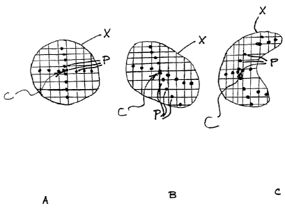
FIGURE 5A is a schematic cross-section of a pedicle isthmus illustrating the
center
thereof as determined in accordance with the methods of the present invention.
FIGURE 5B is a schematic view of the cross-section of a pedicle isthmus having
an
irregular shape showing the center thereof as determined in accordance with
the methods of the
present invention; and
FIGURE 5C is a schematic view of a pedicle isthmus cross-section having a
different
irregular shape showing the center thereof as determined in accordance with
the methods of the
present invention.
DESCRIPTION OF THE PREFERRED EMBODIMENTS
LPedicle Base Circumference and Pedicle Isthmus Determination.
In accordance with the methods of the present invention, serial stacked images
in any
plane are obtained of the vertebral body 10 in any suitable manner. These
images then are
reconstructed to obtain a dimensionally true three-dimensional rendering of
the vertebral body
10. The pedicle base circumference B and pedicle isthmus X are depicted in the
three-
dimensional and two-dimensional images as shown schematically in Figures 1A
and 1B.
CA 02640075 2008-07-23
WO 2007/087381 PCT/US2007/002001
Once a true three-dimensional rendering of the vertebral body 10 is obtained,
it is
sectioned in a transverse plane to visualize and obtain an outer cortical
shell. A series of first
lines T are then drawn tangentially to and along the outer cortical surface
12. A series of
second lines P are then drawn perpendicular to the first lines T lying on the
outer cortical
5 surface 12 with the second lines P lying within the vertebral body 10. This
is illustrated in
Figure 2 with respect to only two first tangential lines T and two second
perpendicular lines P
as an illustrative example.
In the area of the pedicle 14 and its transition into the vertebral body 10,
the series of
second perpendicular lines P will define the pedicle base circumference B and
pedicle isthmus
X. Specifically, the pedicle base circumference B is defined as that region in
which the
adjacent second perpendicular lines P are at the greatest angle A, nonlinear
or discordant to one
another. Conversely, the pedicle isthmus X is the region in which opposing
second
perpendicular lines P are most parallel to one another. Infinitesimal points
on the outer cortical
surface are utilized for placement of the first tangential lines T and their
respective second
perpendicular lines P. This is illustrated schematically in Figure 3.
The points from infinite transverse sections (TS1, TS2, TS3...) defining the
pedicle
base B and pedicle isthmus X are then collated to determine the anatomical
three-dimensional
location of the pedicle base circumference B and pedicle isthmus X, as shown
in Figures 4A,
4B and 4C. Figures 4B and 4C show the transverse section TS2 through the
center of the
pedicle 14 and its corresponding point on a transverse and coronal projection,
respectively.
Pedicle Isthmus Center Determination
Once the pedicle isthmus X is defined, the center of the pedicle isthmus C
must be
further defined. This is necessary to allow for concentric trajectory
determination and pedicle
cylinder building. Most pedicles are conceptualized as being cylindrical;
however, many
pedicles have oval or irregular volumes. As such, it is essential to determine
the center of
these pedicles. The new and improved method of the present invention utilizes
the cross-
sectional area defined by the pedicle isthmus X and then identifies the center
C of the cross-
sectional area as being that point which lies at the intersection of two lines
derived from the
centers of the infinitesimal orthogonal second perpendicular lines P as
illustrated in Figures
5A, 5B and 5C. This methodology allows for pedicle isthmus center
determination
irrespective of different pedicle configurations as shown in Figures 5A, 5B
and 5C.
It will be readily seen that the methods of the present invention provide for
simple and
reliable determination of pedicle base circumference, pedicle isthmus and the
center of the
CA 02640075 2012-01-31
6
isthmus to provide for concentric pedicle screw placement along the ideal
trajectory.
These methods can be effected in any suitable manner, such as visual imaging
through the use of a computer or the like, or manually from two-dimensional
sections.
While the invention has been described in connection with what is presently
considered to be the most practical and preferred embodiments, it is to be
understood that the invention is not to be limited to the disclosed
embodiments.
Indeed, the scope of the claims should not be limited by the preferred
embodiments
set forth in the examples, but should be given the broadest interpretation
consistent
with the description as a whole.