Note : Les descriptions sont présentées dans la langue officielle dans laquelle elles ont été soumises.
CA 02660035 2009-02-04
WO 2008/017704 PCT/EP2007/058263
1
METHOD OF PRODUCTION OF TRANSGENIC AVIAN USING
EMBRYONIC STEM CELLS
The present invention relates to avian biotechnology and in particular to a
method of
producing transgenic birds. The invention is particularly useful to generate
transgenic birds
expressing high amount of a recombinant protein of interest in egg.
With over 400 biomolecules in clinical development and a market of over 30
billions dollars,
the therapeutic field has witnessed an accelerated growth in the last fifteen
years. Most
recombinant proteins require specific post-translational modifications for
full biological
activities and must thus be produced in mammalian cells grown in large-scale
bio-reactors.
The cost associated with such production system, combined with important
quantitative
needs is responsible for increasing delays in the overall process of product
development. In
this context, transgenic animals could represent an economically attractive
alternative,
especially if the modification could be transmitted to the progeny through the
germ-line.
While rabbits, goats and cows have elicited plenty of interest, the hen has
long been
considered as the most appealing biological production system for fast and
cost-effective
production of recombinant therapeutic proteins in chicken eggs. The chicken
egg-laying
capabilities are indeed remarkable since commercial hen lays about 250 eggs a
year, each
egg containing nearly 4 grams of egg white proteins. If only 100 mg of
recombinant proteins
were to be produced in an egg, a small flock of 1000 hens would thus be able
to produce 25
kg of raw recombinant proteins material a year. In addition, the commercial
egg industry is
already highly automated and regulatory agencies are comfortable with egg-
derived products
since many human vaccines are produced in the chicken eggs since decades.
Likewise, the poultry industry may also have interest in using transgenesis
for accelerated
selection of genetic traits of commercial interest (i.e "meta-cloning
technology"). The idea
would be to bypass the classical selection scheme: [Pedigree -> GGP -> GP ->
PS ] to get
in a shorter time the desired chick. The most likely use of transgenic
technology in poultry
production could be to impart disease resistance which is a common practise in
transgenic
plants or to improve food assimilation . In addition, transgenic technology
could be a strategy
to conserve avian genetic resources. So far, the splitting into different
places of pedigree
animals, the most valuable animals in poultry industry, is the only way to
prevent the loss of
years of selection programs in case of troubles (eg. viral infections) in a
breeding facility.
Such an approach is expensive and does not guarantee against national sanitary
decisions
CA 02660035 2009-02-04
WO 2008/017704 PCT/EP2007/058263
2
enforcing the preventive elimination of all local poultry to check viral
infection, as in the
Netherlands in 2003.
The engineering of a modified animal implies first the stable introduction of
the transgene
into the genome of the animal. The different technologies to introduce DNA
investigated
since 25 years are : (i) the DNA micro-injection, (ii) the viral transduction,
(iii) the sperm-
mediated gene transfer, (iv) the nuclear transfer, the cell-based transfer
using (v) primordial
germ cells, or (vi) embryonic stem cells. In the chick, only the DNA micro-
injection approach
(Love et al, 1994 Biotechnology 12:60-63) and the viral transduction
(Bosselman et al, 1990
J. Reprod. Fertil. Suppl. 41: 183-195) have been validated for germ line
transmission of the
transgene. However, these two technologies suffer from several limitations;
they are
cumbersome and laborious and have a low efficacy, partly due to random
integration and
silencing of the transgene. Viral transduction technology has the additional
limitation of the
transgene size to be incorporated into the viral vector. These two
technologies did not allow
today to reach protein expression level compatible with commercial
developments.
Injection into the developing chick embryo of primordial germ cells (PGC) or
Embryonic Stem
(ES) cells, previously in vitro genetically engineered are among the promising
technologies
of avian transgenesis especially because these technologies allow targeting
transgene
integration to specific sites within the genome which should allow high
expression levels of
the transgene. However, in order to use this approach to produce transgenic
chicks, an
important prerequisite must be fulfilled: cells must survive to in vitro
manipulations, while still
maintaining their ability to be incorporated within a recipient embryo, to
colonize the germ-
line and then to transmit the modification to the progeny.
In the past, many attempts were made to overcome the different technical
hurdles at each
process steps and today, somatic and germ-line chimera had been obtained by
injection of
freshly isolated blastodermal cells isolated from un-incubated embryos into
the sub-germinal
cavity of freshly laid embryos. Donor and recipient cells contribution was
assessed in (i) the
melanocyte population through examination of black and white pigmentation
(Barred Rocks
or Brown Leghorns have a recessive allele at the I locus while the White
Leghorns have a
dominant allele at the I locus); (ii) the erythrocyte population through
analysis of DNA
fingerprints; (iii) the gonads through the analysis of the progeny with a
donor-derived
phenotype (Petitte et al, 1990 Development 108:185-189; Carsience et al, 1993
Development 117:669-675; Thoraval et al, 1994 Poultry Science 73:1897-1905;
Pain et al,
1996 Development 122:2339-2348). Chimera with contributions to both somatic
tissues and
the germline were observed when blastodermal cells were injected after 48
hours (Etches et
al, 1996 Mol. Reprod. Dev. 45:291-288) up to 7 days of culture (Pain et al,
1996). Etches et
CA 02660035 2009-02-04
WO 2008/017704 PCT/EP2007/058263
3
al, 1996, had demonstrated that significantly more somatic chimeras were
observed
following the injection of cells co-cultured with mouse fibroblasts. Pain et
al (1996) seeded
avian blastodermal cells on STO mouse fibroblast cell line. The ES status of
cells maintained
in culture relied on the expression of the ECMA-7 and SSEA-1 epitopes and the
telomerase
activity (Pain et al, 1996). Proliferation in the absence of differentiation
of blastodermal cells
was stimulated by the presence of Leukemia Inhibitory Factor (LIF) and other
factors, II-11,
SCF, bFGF, IGF-1 and differentiation was inhibited by exposure to anti-
retinoic acid
monoclonal antibody (Pain et al, 1996). It had been shown that colonization of
the embryo by
donor-derived cells was facilitated when the recipient embryo was compromised
by exposure
to irradiation prior to injection of the donor cells (Carsience et al, 1993).
However, blastodermal cells maintained in culture yielded fewer chimeras that
exhibit
reduced contributions to somatic tissues in comparison to the frequency and
extent of
somatic chimerism observed following injection of freshly prepared cells.
Moreover, even so
it was demonstrated that each of the component parts of the cell-based avian
transgenesis
strategy could be accomplished; no transgenic animal had been described that
were
obtained with the ES cell technology.
It remains a need for efficient methods of generating transgenic chickens.
This is the object
of the instant invention.
To achieve this object and in accordance with the purpose of the invention, as
embodied and
broadly described herein, the present invention provides a method of culturing
embryonic
stem (ES) cells of avian, comprising the steps of:
a) suspending ES cells originating from the blastoderm disk of fertilized,
preferably un-
incubated, avian egg(s) in a basal culture medium supplemented with:
- insulin-like growth factor-1 (IGF-1) and/or ciliary neurotrophic factor
(CNTF); and
- optionally, at least one growth factors selected in the group comprising
interleukin 6(II-6), interleukin 6 receptor (II-6R), stem cell factor (SCF),
fibroblast
growth factor (FGF), leukaemia inhibitory factor (LIF), interleukin 11 (II-
11),
oncostatin and cardiotrophin; and
- animal serum;
b) seeding the suspension of ES cells obtained in step a) on a layer of feeder
cells and
further culturing the ES cells for at least between 2 to 10 passages,
preferably
between 2 to 20 passages;
c) optionally removing at least one growth factors selected SCF, FGF, 11-6, II-
6R, LIF,
oncostatin, cardiotrophin and II-11 from the culture medium;
d) further culturing the ES cells in the medium of step c) on a layer of
feeder cells.
CA 02660035 2009-02-04
WO 2008/017704 PCT/EP2007/058263
4
In a preferred embodiment, the method of culturing embryonic stem (ES) cells
of avian of the
invention, comprises the steps of:
a) suspending ES cells originating from the blastoderm disk of fertilized,
preferably,
un-incubated avian egg(s) in a basal culture medium supplemented with insulin-
like
growth factor-1 (IGF-1), ciliary neurotrophic factor (CNTF), interleukin 6(II-
6),
interleukin 6 receptor (II-6R), stem cell factor (SCF) and fibroblast growth
factor (FGF)
and animal serum;
b) seeding the suspension of ES cells obtained in step a) on a layer of feeder
cells and
further culturing the ES cells for at least between 2 to 10 passages,
preferably
between 2 to 20 passages;
c) optionally removing at least one growth factor selected from the group
comprising
SCF, FGF, 11-6 and II-6R from the culture medium, and preferably removing the
growth
factors SCF, FGF, IL-6 and IL-6R from the culture medium;
d) further culturing the ES cells in the medium of step c) on a layer of
feeder cells.
In another preferred embodiment, the method of culturing embryonic stem (ES)
cells of avian
of the invention, comprises the steps of:
a) suspending ES cells originating from the blastoderm disk of fertilized,
preferably un-
incubated avian egg(s) in a basal culture medium supplemented with insulin-
like
growth factor-1 (IGF-1), ciliary neurotrophic factor (CNTF), interleukin 6(II-
6),
interleukin 6 receptor (II-6R), stem cell factor (SCF), fibroblast growth
factor (FGF) and
animal serum;
b) seeding the suspension of ES cells obtained in step a) on a layer of feeder
cells and
further culturing the ES cells for at least between 2 to 10 passages;
c) optionally removing at least one growth factor selected from the group
comprising
SCF and FGF from the culture medium; and preferably removing both growth
factors
SCF and FGF from the culture medium;
d) further culturing the ES cells in the medium of step c) on a layer of
feeder cells.
The optional step c) of the method of the invention is performed when signs of
mortality
and/or differentiation of ES cells are observed into culture. Examples of sign
of differentiation
are for example morphological changes of cells to a characteristic
morphological type, such
as for example epithelial cell type or fibroblast cell type. Another example
of differentiation
may be the absence or the decrease of expression of ES cells markers, such as
telomerase,
alkaline phosphatase, SSEA-1 antigen which are ES cells markers.
CA 02660035 2009-02-04
WO 2008/017704 PCT/EP2007/058263
In a more preferred embodiment, the method of culturing embryonic stem (ES)
cells of avian,
of the invention comprises the steps of:
a) suspending ES cells originating from the blastoderm disk of fertilized,
preferably un-
incubated, avian egg(s) in a basal culture medium supplemented with at least
insulin-
5 like growth factor-1 (IGF-1), ciliary neurotrophic factor (CNTF),
interleukin 6(II-6),
interleukin 6 receptor (II-6R), stem cell factor (SCF), fibroblast growth
factor (FGF) and
animal serum;
b) seeding the suspension of ES cells obtained in step a) on a layer of feeder
cells and
further culturing the ES cells in the medium of step a).
In another more preferred embodiment, the method of culturing embryonic stem
(ES) cells of
avian, of the invention comprises the steps of:
a) suspending ES cells originating from the blastoderm disk of fertilized un-
incubated
avian egg(s) in a basal culture medium supplemented with at least insulin-like
growth
factor-1 (IGF-1), ciliary neurotrophic factor (CNTF), interleukin 6(II-6),
interleukin 6
receptor (II-6R) and animal serum;
b) seeding the suspension of ES cells obtained in step a) on a layer of feeder
cells and
further culturing the ES cells in the medium of step a).
In another more preferred embodiment, the method of culturing embryonic stem
(ES) cells of
avian, of the invention comprises the steps of:
a) suspending ES cells originating from the blastoderm disk of fertilized un-
incubated
avian egg(s) in a basal culture medium supplemented with at least insulin-like
growth
factor-1 (IGF-1), ciliary neurotrophic factor (CNTF) and animal serum;
b) seeding the suspension of ES cells obtained in step a) on a layer of feeder
cells and
further culturing the ES cells in the medium of step a).
The term avian as used herein is intended to refer to any species,
subspecies or
race of organism of the taxonomic class ava , such as, but not limited to,
chicken, turkey,
duck, goose, quails, pheasants, parrots, finches, hawks, crows, ostrich, emu
and cassowary.
The term "avian, "bird", "aves" or "ava" as used herein is intended to have
the same
meaning, and will be used indistinctly. In a preferred embodiment, "birds"
refer to any animal
of the taxonomix order:
- "Anseriformes" (i.e duck, goose, swan and allies). The order Anseriformes
contains about
150 species of birds in three families: the Anhimidae (the screamers),
Anseranatidae (the
Magpie-goose), and the Anatidae, which includes over 140 species of waterfowl,
among
them the ducks, geese, and swans. All species in the order are highly adapted
for an aquatic
existence at the water surface. All are web-footed for efficient swimming
(although some
have subsequently become mainly terrestrial). The term includes the various
strains of
ducks, for example Pekin duck and Muscovy duck.
CA 02660035 2009-02-04
WO 2008/017704 PCT/EP2007/058263
6
- "Galliformes" (i.e chicken, quails, turkey, pheasant and allies). The
Galliformes is an order
of birds containing the chicken, turkeys, quails and pheasants. About 256
species are found
worldwide. The term includes the various strains of Gallus gallus, or
chickens, for example
S86N, Valo, White Leghorn, Brown Leghorn, Sussex, New Hampshire, Rhode Island,
Ausstralorp, Minorca, Amrox, California Gray, East Lansing, Italian-Partridge-
colored,
Marans, Barred Rock, Cou Nu Rouge (CNR), GF30, ISA as well as strains of
turkeys,
pheasants, quails, and other poultry commonly bred.
- "Columbiformes" (i.e Pigeon and allies). The bird order Columbiformes
includes the very
widespread doves and pigeons.
In a preferred embodiment, the avian cell of the present invention is a
chicken cell. The
chicken is preferably selected from the group of chicken strains comprising
S86N, Valo,
White Leghorn, Brown Leghorn, Sussex, New Hampshire, Rhode Island,
Ausstralorp,
Minorca, Amrox, California Gray, East Lansing, Italian-Partridge-colored,
Marans, Barred
Rock, Cou Nu Rouge (CNR), GF30, ISA. In a more preferred embodiment, the
chicken ES
cell of the present invention is a Barred-Rock strain. In another preferred
embodiment, the
avian cell of the present invention is a duck cell. In a more preferred
embodiment, the duck
ES cell of the present invention is a Pekin or Muscovy strain.
The cells of step a) are avian embryonic stem cells. Embryonic stem cells (ES
cells)
are stem cells which have the characteristic feature of being obtained from
culturing parts or
all of a very early embryo (e.g blastula stage). These ES cells exhibit in
vitro all the
characteristics of a stem cell, and in vivo the unique capacity of
contributing to the
morphogenesis of an embryo and of participating in germline colonization when
they are re-
implanted in any manner whatsoever in a recipient embryo. Primordial Germ
Cells (PGC)
which are the progenitors of the sperm or ovocyte cells that develop after
sexual maturity are
pluripotent ES cells and constitutes a subtype of ES cells. Preferably, avian
embryonic stem
cells of the invention are isolated from the blastoderm disk of fertilized
avian egg(s) at
development stage comprises between stages VI and XII of Eyal-Giladi &
Kochav's
classification, and more preferably around the stage X of of Eyal-Giladi &
Kochav's
classification (see EYAL-GILADI's classification: EYAL-GILADI and KOCHAV,
1976, From
cleavage to primitive streack formation : a complementary normal table and a
new look at
the first stages of the development in the chick ."General Morphology" Dev.
Biol. 49 : 321-
337). In a preferred embodiment, the cells of step a) are isolated from
freshly laid fertilized
eggs that is to say at a developmental stage named oviposition. According to
Sellier et al.
(2006, J. Appl. Poult. Res., 15:219-228), oviposition corresponds to the
following
development stages according to Eyal-Giladi's classification:
- Muscovy duck: stage VII
- Guinea fowl: stage VII - VII I
CA 02660035 2009-02-04
WO 2008/017704 PCT/EP2007/058263
7
- Turkey: stage VII-VIII
- Pekin duck: stage VIII
- Chicken: Stage X
- Japanese Quail: stage XI
- Goose: stage XI.
Preferably, the Pekin duck embryonic stem (ES) cells of step a) is obtained by
dissociating
embryo(s) at around stage VIII (oviposition) of Eyal-Giladi's classification.
Preferably, the Muscovy duck embryonic stem (ES) cells of step a) is obtained
by
dissociating embryo(s) at around stage VII (oviposition) of Eyal-Giladi's
classification.
Preferably, the chicken embryonic stem (ES) cells of step a) is obtained by
dissociating
embryo(s) at around stage X (oviposition) of Eyal-Giladi's classification.
More preferably, the eggs have never been incubated (i.e "un-incubated"). The
cells
isolated from blastoderm disk comprises avian embryonic cells, more
particularly avian
embryonic stem (ES) cells; these avian embryonic cells are totipotent or
pluripotent cells.
Blastoderma cells are tightly connected cells that grow as colonies; they
display a round-
shaped morphology, with a big nucleus and a small cytoplasm (see Figure 2).
The
morphology of the blastodermal cells evolves during the culture (steps Figure
2 b & c of the
method of the invention) from tightly connected cells to more dispersed cells
with looser
connections. Avian ES cells obtained at step c) preferably display looser
connections.
According to another embodiment, the cells of step a) are a population of
embryonic
stem cells enriched in primordial germ cells (PGC). More preferably, the avian
ES cells of
step a) are purified PGCs. In avian species, Primordial Germ Cells arise from
the central
region of the blastoderm (Ginsburg and Eyal-Giladi, 1987 Development
101(2):209-19;
Karagenc et al, 1996 Dev Genet 19(4):290-301; Petitte et al, 1997 Poult Sci.
76(8):1084-92).
Then they move to an anterior, extra-embryonic site, the germinal crescent
until collected by
the vasculature between 2.5 and 5 days of embryonic development to reach the
germinal
ridge. They colonize the germinal ridge where they eventually differentiate
into oocytes or
spermatocytes (Nieuwkoop and Sutasurya, 1979. The Migration of the primordial
germ cells.
In: Primordial germ cell in Chordates. London: Cambridge University Press p113-
127).
Methods for isolation of PGCs from donor avian embryos have been reported in
the literature
and can easily be performed by one skilled in the art (See, e.g. JP924997
published sept. 7,
1993 Pub. N 05-227947; Chang et al. 1992. Cell Biol. Int. 19(2): 143-149 ;
Naito et al. 1994
Mol. Reprod. Dev. 39: 153-161 ; Yasuda et al. 1992. J. Reprod. Fert. 96: 521-
528 ; Chang et
al. 1992 Cell Biol. Int. Reporter 16(9): 853-857). According to an embodiment,
PGCs are
collected from embryonic blood collected from the dorsal aorta of a chicken
embryo at stage
12-14 of Hamburger & Hamilton's classification (Hamburger & Hamilton 1951 A
series of
CA 02660035 2009-02-04
WO 2008/017704 PCT/EP2007/058263
8
normal stages in the development of chick embryo. J. Morphol. 88: 49-92). In
another
preferred embodiment, PGCs were collected from the germinal crescent by
mechanical
dissection of chicken embryo or from the gonads. However, as discussed above,
others
methods for isolating PGCs are known and can alternatively be used.
By "complete culture medium", it is meant a basal medium, preferably a basal
synthetic
medium, supplemented with at least one growth factor and animal serum. Example
of
complete culture medium is described in W003/076601, W005/007840, EP787180,
US6,114,168, US5,340,740, US6,656,479, US5,830,510 and in a Pain et al. (1996,
Development 122:2339-2348). According to the invention, "basal medium" meant a
medium
with a classical media formulation that allows, by itself, at least cells
survival, and even
better, cell growth. Examples of basal media are BME (basal Eagle Medium), MEM
(minimum Eagle Medium), medium 199, DMEM (Dulbecco's modified Eagle Medium),
GMEM (Glasgow modified Eagle medium), DMEM-HamF12, Ham-F12 and Ham-F10,
Iscove's Modified Dulbecco's medium, MacCoy's 5A medium, RPMI 1640. Basal
medium
comprises inorganic salts (for examples: CaCIZ, KCI, NaCI, NaHCO3, NaH2PO4,
MgSO4,...),
amino-acids, vitamins (thiamine, riboflavin, folic acid, D-Ca
panthothenate,...) and others
components such as glucose, beta-mercapto-ethanol, sodium pyruvate. Preferably
basal
medium is a synthetic medium. Most preferred basal medium of the invention is
DMEM-
HamF12 that are complemented with 2 mM L-glutamin, 1 mM sodium pyruvate, 1%
non-
essential amino-acids, 0.16 mM beta-mercapto-ethanol.
Alternatively, the complete culture medium is a conditioned medium, preferably
BRL
conditioned medium. By way of example, BRL conditioned media is prepared
according to
art-recognized techniques, such as described by Smith and Hooper (1987, Dev.
Biol. 121: 1-
9). BRL cells are available from ATCC accession number CRL-1442. Conditioned
medium
may be supplemented with exogenous growth factors as described below.
The term "growth factor" as used herein meant exogenous growth factor added to
the culture medium necessary for the survival and the growth of the avian
cells in culture. It
is possible to schematically distinguish two families of growth factors: the
cytokines and the
trophic factors. The cytokines are mainly cytokines whose action is through a
receptor which
is associated with the gp130 protein. Thus, leukemia inhibitory factor (LIF),
interleukin 11,
interleukin 6, interleukin 6 receptor, Ciliary Neurotrophic factor (CNTF),
oncostatin and
cardiotrophin have a similar mode of action with the recruitment at the level
of the receptor of
a specific chain and the combination of the latter with the gp130 protein in
monomeric or
sometimes hetero-dimeric form. The trophic factors are mainly Stem cell Factor
(SCF),
Insulin Growth factor 1 (IGF-1) and Fibroblast Growth Factor (FGF), preferably
basic FGF
(bFGF) or human FGF (hFGF).
CA 02660035 2009-02-04
WO 2008/017704 PCT/EP2007/058263
9
The complete culture medium used in step a) of the process of invention
comprises
basal medium, preferably basal synthetic medium, and at least one cytokine
whose action is
through a receptor which is associated with the gp130 protein and/or at least
one trophic
factors. Preferably, the complete culture medium according to the invention
comprises basal
medium and at least one growth factor selected in the group consisting of
Leukemia
Inhibitory factor (LIF), oncostatin, cardiotrophin, Insulin Growth factor 1
(IGF-1), Ciliary
Neurotrophic factor (CNTF), Interleukin 6 (IL-6), interleukin 6 receptor (IL-
6R), Stem cell
Factor (SCF), Fibroblast Growth Factor (FGF), interleukin 11 (IL-11).
According to a first preferred embodiment, the complete culture medium is
basal
medium supplemented with animal serum and with at least IGF-1 and CNTF.
According to a second preferred embodiment, the complete culture medium is
basal
medium supplemented with animal serum and at least IGF-1, CNTF, IL-6 and IL-
6R.
According to a third preferred embodiment, the complete culture medium is
basal
medium supplemented with animal serum and at least IGF-1, CNTF, IL-6, IL-6R,
SCF, FGF.
According to another embodiment, the complete culture medium is a conditioned
culture medium comprising growth factors (i.e expressed by BRL cells for
example) and
optionally supplemented with at least one exogenous growth factors selected in
the group
comprising: Leukemia Inhibitory factor (LIF), Insulin Growth factor 1(IGF-1),
Ciliary
Neurotrophic factor (CNTF), interleukin 6 (IL-6), interleukin 6 receptor (IL-
6R), Stem cell
Factor (SCF), Fibroblast Growth Factor (FGF), interleukin 11 (IL-11).
The concentration of growth factors IGF-1, CNTF, IL-6, IL-6R, SCF, FGF, IL-11
in
the basal medium or in the conditioned culture medium is comprised between
about 0.01 to
10 ng/ml, preferably, 0.1 to 5 ng/ml, and more preferably about 1 ng/ml.
The avian embryonic stem cells of the invention are cultured on a layer of
feeder cells.
Feeder cells can either be cells or cell lines cultured for the purpose of
obtaining ES cells.
Alternatively, the feeder cells could be substituted with extra-cellular
matrix plus bound
growth factors. Feeder matrix will thereafter refers to either feeder cells or
extra-cellular
matrix. A feeder matrix as used herein is constructed in accordance with
procedures known
in the art. As noted above, it is preferred that the feeder matrix be
preconditioned. By the
term "preconditioned" it is meant that the feeder matrix is cultured in the
presence of media
for a period of time prior to the depositing of cells originating from the
blastoderm disk
fertilized avian eggs in contact with the feeder matrix, e.g. a time
sufficient to initiate and
establish production of, for example, growth factors or other factors by the
feeder matrix;
usually a feeder matrix is preconditioned by culturing the feeder matrix by
itself for one to two
days prior to the depositing of cells originating from the blastoderm disk
fertilized avian eggs
in contact with the feeder matrix. The feeder cells preferably comprises mouse
fibroblast
cells. STO fibroblasts are preferred, but primary fibroblasts are also
suitable. Also while the
CA 02660035 2009-02-04
WO 2008/017704 PCT/EP2007/058263
present invention has been described with respect to the use of mouse cell
feeder matrices,
it is contemplated that feeder matrices comprising cells from other murine
species (e.g. rat);
other mammalian species (e.g; ungulate, bovine, porcine species); or avian
species (e.g.
Gallinacea, chicken, turkey, duck, goose, quail, pheasant) may also be used.
In another
5 embodiment, feeder cells of the invention may be transfected with expression
vector(s)
allowing for example the constitutive expression of growth factors such as
avian SCF in STO
cells. Thus, this "feeder" produces the factor in a form which is soluble
and/or attached in the
plasma membrane of the cells. Thus, the culturing process of the present
invention may
optionally comprise establishing a monolayer of feeder cells. Feeder cells are
mitotically
10 inactivated using standard techniques. For example, the feeder cells may be
exposed to X or
gamma radiation (e.g. 4000 Rads of gamma radiation) or may be treated with
Mitomycin C
(e.g. 10 g/ml for 2-3 hours). Procedures for mitotically inactivating cells
are also detailed in
the information typically sent with cells from the American Type Culture
Collection (ATCC),
10801 University Boulevard, Manassas, Va. 20110-2209 (e.g. STO feeder cells
are available
under ATCC accession number 1503). Mono-layers may optionally be cultured to
about 80%
confluency, preferably to about 90% confluency, and more preferably about 100%
confluency. While configuration of the feeder cells as a monolayer is the
preferred
configuration for the culture, any suitable configuration is contemplated to
be within the
scope of the present invention. Thus, for example, layers, mono-layers,
clusters, aggregates
or other associations or groupings of feeder cells are contemplated to fall
within the scope of
the present invention and are particularly contemplated to fall with the
meaning of the term
"matrix".
The culture medium of the invention is supplemented with animal serum. The
animal serum
preferably used is fetal animal serum. Fetal bovine serum is preferred. Also
while the present
invention has been described with respect to the use of fetal bovine serum, it
is
contemplated that animal serum comprising serum from other animal species
(e.g. chicken,
horse, porcine, ungulate, etc...) may also be used. The final concentration of
animal serum
in the culture medium is comprises between approximately 1 to 25%, preferably
between 5%
to 20%, more preferably between 8% and 12%. In the preferred embodiment, the
final
concentration of animal serum in the culture medium is approximately 10%.
According to a
preferred embodiment, the culture medium comprises approximately 10% of fetal
calf serum.
The culture medium of the invention may comprise in addition antibiotics, such
as for
example penicilline and streptomycine, to prevent bacterial contamination.
According to another embodiment, the invention provides a method of
genetically modifying
avian embryonic stem cells, comprising the steps of:
CA 02660035 2009-02-04
WO 2008/017704 PCT/EP2007/058263
11
a) transfecting ES cells obtained and cultured according to the method above,
with a
vector;
b) selecting transfected ES cells, preferably by addition of a selection agent
in the
medium, such as for example antibiotics, amino-acids, hormones.
c) screening and amplification of resistant ES clones genetically modified,
d) culturing said genetically modified ES cell of step c) on a layer of feeder
cells in a
culture medium as previously described. According to a first embodiment, said
culture
medium of step d) comprises animal serum and at least one growth factor
selected in
the group comprising IGF1, CNTF, IL-6, IL-6R, II-11, LIF, FGF, SCF,
oncostatin,
cardiotrophin. According to a preferred embodiment, said culture medium of
step d)
comprises animal serum and IGF1 and CNTF. According to another embodiment,
said
culture medium of step d) comprises animal serum and IGF1 and CNTF and
optionally
at least one growth factor selected in the group comprising IL-6, IL-6R, II-
11, LIF, FGF,
SCF, oncostatin and cardiotrophin. According to a third preferred embodiment,
said
culture medium of step d) comprises animal serum and IGF1, CNTF, IL-6 and IL-
6R.
According to a fourth preferred embodiment, said culture medium of step d)
comprises
animal serum and IGF1, CNTF, IL-6, IL-6R, SCF and FGF.
ES cells of step c) are genetically modified. Genetic modification are
performed either
by transient or stable transfection with the vector in ES cells. According to
a preferred
embodiment the ES are stably transfected with the vector according to
techniques well
known by the man skilled in the Art. According to a first embodiment, the
vector is inserted
randomly into the genome of ES cells. According to a preferred embodiment, the
vector is
inserted by homologous recombination into the genome of ES cells. W003/043414
described protocols and expression vectors to genetically modified ES cells by
homologous
recombination.
ES cells of step a) may be maintained and cultured for a long period of time
in vitro
prior to their introduction into recipient embryo. This long period of time
allows to genetically
modified said cells. According to a preferred embodiment, the cells have been
in vitro
cultured for at least 5 days, at least 10 days, at least 14 days, at least 25
days, at least 50
days, at least 75 days, at least 100 days.
The terms "vector" as used herein refer to a natural or synthetic single or
double
stranded plasmid or viral nucleic acid molecule that can be transfected into
cells and
replicate independently of, or within, the host cell genome. A circular double
stranded
plasmid can be linearized by treatment with an appropriate restriction enzyme
based on the
nucleotide sequence of the plasmid vector. A nucleic acid can be inserted into
a vector by
cutting the vector with restriction enzymes and ligating the pieces together.
The nucleic acid
CA 02660035 2009-02-04
WO 2008/017704 PCT/EP2007/058263
12
molecule can be RNA or DNA. The term "plasmid" as used herein refers to a
small, circular
DNA vector capable of independent replication within a bacterial or yeast host
cell. The
nucleic acid vector further includes at least one regulatory sequence operably
linked to a
nucleotide sequence coding for a "polypeptide of interest". Regulatory
sequences are well
recognized in the art and may be selected to ensure good expression of the
linked
nucleotide sequence without undue experimentation by those skilled in the art.
As used
herein, the term "regulatory sequences" includes promoters, enhancers, and
other elements
that may control expression. Standard molecular biology textbooks such as
Sambrook et al.
eds "Molecular Cloning: A Laboratory Manual" 2nd ed. Cold Spring Harbor Press
(1989) and
Lodish et al. eds., "Molecular Cell Biology," Freeman (2000) may be consulted
to design
suitable expression vectors, promoters, and other expression control elements.
It should be
recognized, however, that the choice of a suitable expression vector depends
upon multiple
factors including the choice of the host cell to be transformed and/or the
type of protein to be
expressed. Also useful for various applications are tissue-selective (i.e.
tissue-specific)
promoters, i.e., promoters from which expression occurs preferentially in
cells of a particular
kind of tissue, compared to one or more other types of tissue. An exemplary
tissue-specific
promoter is a chicken oviduct-specific promoter that is naturally associated
with the proteins
of avian egg whites including ovalbumin, lysozyme, ovomucoid, conalbumin and
ovomucin
and the like. Useful promoters also include exogenously inducible promoters.
These are
promoters that can be "turned on" in response to an exogenously supplied agent
or stimulus,
which is generally not an endogenous metabolite or cytokine. Examples include
an antibiotic-
inducible promoter, such as a tetracycline-inducible promoter, a heat-
inducible promoter, a
light-inducible promoter, or a laser inducible promoter. (e.g., Halloran et
al., 2000,
Development 127(9): 1953-1960; Gemer et al., 2000, Int. J. Hyperthermia 16(2):
171-81;
Rang and Will, 2000, Nucleic Acids Res. 28(5): 11205; Hagihara et al., 1999,
Cell
Transplant. 8(4): 4314; Huang et al., 1999, Mol. Med. 5(2): 129-37; Forster,
et al., 1999,
Nucleic Acids Res. 27(2): 708-10; and Liu et al., 1998, Biotechniques 24(4):
624-8, 630-2
(1998)). As used herein the term "polypeptide of interest" or "protein of
interest" refer to a
polymer of amino acids of three or more amino acids in a serial array, linked
through peptide
bonds. The term "polypeptide" includes proteins, protein fragments, protein
analogues, oligo-
peptides, peptides and the like. The term "polypeptide" contemplates
polypeptides as
defined above that are encoded by nucleic acids, produced through recombinant
technology,
isolated from an appropriate source or are synthesized. Non limiting examples
of
polypeptides are growth hormones, cytokine, interleukine, interferon, enzymes,
immunoglobulins or fragments thereof. The terms "transfection" or
"transfected" as used
herein refer to the process of inserting a nucleic acid into a host cell (i.e
the avian ES). Many
techniques are well known to those skilled in the art to facilitate
transfection of a nucleic acid
into a prokaryotic or eukaryotic organism. These methods involve a variety of
techniques
CA 02660035 2009-02-04
WO 2008/017704 PCT/EP2007/058263
13
including, but not limited to, treating the cells with high concentrations of
salt such as, but not
only, a calcium or magnesium salt, an electric field (i.e. electroporation),
detergent, or
liposome mediated transfection (i.e. lipofection, etc...), to render the host
cell competent for
the uptake of the nucleic acid molecules.
The invention also provide a method of obtaining a chimeric chick comprising
the
steps of:
a) introducing avian ES cells obtained and cultured by the method of the
invention into
the sub-germinal cavity of a recipient avian embryo; and
b) incubating the embryo obtained in step a) to hatch as a chick;
c) selecting said chimeric chick comprising heterologous cells having
colonized said
chick.
The term "chick" as used herein meant a young bird, and it includes young
chicken. The term
"subgerminal cavity" meant the space between the blastoderm and the yolk. This
space is
created when the blastoderm cells absorb fluid from the albumin and secrete it
between
themselves and the yolk.
It is also an object of the invention to provide a method of obtaining a
genetically modified
chimeric chick comprising the steps of:
a) introducing genetically modified ES obtained and cultured by the method of
the
invention into the sub-germinal cavity of a recipient avian embryo; and
b) incubating the embryo obtained in step a) to hatch as a chick;
c) selecting said chimeric chick comprising genetically modified heterologous
cells
having colonized said chick.
The selection of said chimeric chick may be performed either by phenotypic or
genotypic analysis. According to a preferred embodiment, the chimeric chick is
selected by
phenotypic analysis of plumage.
According to another embodiment, the method of obtaining chimeric chick of the
invention may comprise the additional step of determining the sex of recipient
embryo prior
the introduction of ES cells.
According to another embodiment, the recipient embryo derives from a freshly
laid
un-incubated egg and comprises between around 5,000 to around 70,000 cells.
Preferably,
said recipient embryo is at stage comprised between stages VI and XII of Eyal-
Giladi &
Kochav's classification, preferably:
- around the stage X of of Eyal-Giladi & Kochav's classification when the
embryo is a
chicken;
- around the stage VII of of Eyal-Giladi & Kochav's classification when the
embryo is a
Muscovy duck;
CA 02660035 2009-02-04
WO 2008/017704 PCT/EP2007/058263
14
- around the stage VIII of of Eyal-Giladi & Kochav's classification when the
embryo is a
Pekin duck;
- around the stage XI of of Eyal-Giladi & Kochav's classification when the
embryo is a
Japanese quail or a goose;
- around the stage VII-VIII of of Eyal-Giladi & Kochav's classification when
the embryo is
a Guinea fowl or a turkey;
Preferably, at least 1000 ES cells, at least 10,000 ES cells, at least 15,000
ES cells,
at least 30,000 ES, at least 45,000 ES cells, at least 65,000 ES cells, at
least 85,000 ES
cells or at least 100,000 ES cells are introduced into the sub-germinal cavity
of the recipient
avian embryo. According to a preferred embodiment, at least 30,000 ES cells
are introduced
into the sub-germinal cavity of the recipient avian embryo.
ES cells introduced into the sub-germinal cavity of the recipient avian embryo
may be
a mixed population of female and male ES cells. According to another
embodiment, the
recipient embryo and donor ES cells are previously sexed before introduction.
According to a
preferred embodiment, female ES cells are introduced into the sub-germinal
cavity of a
female recipient avian embryo. According to another preferred embodiment, male
ES cells
are introduced into the sub-germinal cavity of a male recipient avian embryo.
The method of obtaining chimeric chick of the invention comprises the optional
step of
slightly irradiating the recipient embryo with X or gamma irradiation prior to
the introduction
of ES cells into the sub-germinal cavity of said recipient embryo. According
to a preferred
embodiment, the chicken recipient embryo is irradiated with between 3 to 6
gray of X rays,
preferably around 4 gray of X-rays. According to a preferred embodiment,
around 15,000
chicken ES cells are introduced into the sub-germinal cavity of the recipient
chicken embryo,
previously X-irradiated with around 4 gray. According to another preferred
embodiment, at
least 30,000 chicken ES cells are introduced into the sub-germinal cavity of
the a non-
irradiated recipient chicken embryo.
According to a first embodiment, the recipient chicken embryo is of White
Leghorn
strain and the donor chicken ES cells derived from a strain selected in the
group composed
of barred rock strain, Marans strain, S86N strain. According to second
embodiment, the
recipient chicken embryo is of a chicken strain selected in the group
comprising barred rock
strain, Marans strain and S86N strain and the donor chicken ES cells derived
from White
Leghorn strain.
The selection of the chimeric chick of the invention comprising heterologous
cells comprises
the steps of:
a) obtaining a sample of genetic material from said chimeric chick;
b) assaying for the presence of a polymorphism in a sequence of avian leucosis
virus integrated in the avian genome, and wherein the polymorphism is
identifiable
CA 02660035 2009-02-04
WO 2008/017704 PCT/EP2007/058263
by an amplification by a set of primers selected in the group consisting of
the set of a
forward primer 5'-GGTGTAAATATCAAAATTATC-3' (SEQ ID n 1) and a reverse
primer 5'-CGGTTAAAATACGAATAGAGA-3' (SEQ ID N 2) and the set of a forward
primer 5'-CTATGAGCAGTTACGAGGGTC-3' (SEQ ID N 3) and a reverse primer 5'-
5 CGGACCAACAGGCTAGTCTC-3' (SEQ ID N 4). According to a preferred
embodiment, said chicken ES cells are derived from barred rock species and
said
recipient embryo is of White Leghorn species. Said amplification is performed
by
polymerase chain reaction (PCR) or reverse-transciptase PCR and wherein said
analysis comprises the digestion of PCR amplified DNA with the restriction
enzyme
10 Hincll.
The method of identifying the presence or absence of a polymorphism is
selected
from a group consisting of: restriction fragment length polymorphism (RFLP)
analysis,
heteroduplex analysis, single strand conformational polymorphism (SSCP),
denaturating
gradient gel electrophoresis (DGGE) temperature gradient gel electrophoresis
(TGGE),
15 allele-specific oligonucleotide (ASO) an dideoxy-fingerprinting (ddF).
The step of assaying for the presence of said polymorphism comprise the steps
of:
a) digesting said genetic material with Hincll restriction enzyme;
b) separating the fragments obtained from said digestion;
c) detecting the restriction pattern generated by said fragments; and
d) optionally comparing said pattern with at least one restriction pattern
obtained by
digestion of White Leghorn, Marans, Barred Rock, S86N genetic material using
Hincll restriction enzyme, wherein difference in restriction patterns detected
in step
c) and d) is indicative of chimeric chick comprising heterologous cells.
The invention also includes the method of obtaining a progeny of the chimeric
chick
wherein said method comprises the following steps:
a) allowing the selected chimeric chick obtained by the method of the
invention to
mature as an adult bird;
b) breeding said adult bird having heterologous cells herein, thereby
producing a bird
progeny; and
c) selecting the birds in the progeny comprising heterologous cells. The
selection of
said birds may be performed by phenotypic analysis of plumage or when possible
by
genotypic analysis by assaying for the presence of a Hincll polymorphism in a
sequence of avian leucosis virus integrated in the bird genome.
The method may comprise the additional step of expressing the heterologous
polypeptide
encoded by the vector comprised in genetically modified heterologous cells.
Preferably, the
heterologous polypeptide is delivered into biological fluids of the bird, such
as blood, sperm,
CA 02660035 2009-02-04
WO 2008/017704 PCT/EP2007/058263
16
urine, or the white of a developing avian egg produced by a female of the
genetically
modified bird.
The present invention also relates to a culture medium for genetically and non-
genetically modified avian embryonic stem (ES) cells, preferably chicken and
duck ES cells,
supplemented with animal serum and comprising at least one growth factors
selected in the
group consisting of insulin-like growth factor-1 (IGF-1), ciliary neurotrophic
factor (CNTF),
Interleukin 6(II-6), Interleukin 6 receptor (II-6R), interleukin 11, Stem cell
factor (SCF),
fibroblast growth factor (FGF), leukaemia inhibitory factor (LIF), oncostatin
and cardiotrophin
wherein said medium is sufficient for the maintenance of said chicken
embryonic stem cells
into culture for 10 days at least, for 30 days at least, preferably for 100
days at least and
more preferably for infinite period.
According to a preferred embodiment, the present invention also relates to a
basal
culture medium for genetically or non-genetically modified avian embryonic
stem (ES) cells,
preferably chicken and duck ES cells, supplemented with animal serum and
supplemented
with insulin-like growth factor-1 (IGF-1) and ciliary neurotrophic factor
(CNTF).
According to a second preferred embodiment, the present invention relates to a
basal culture medium for genetically or non-genetically modified avian
embryonic stem (ES)
cells, preferably chicken and duck ES cells, supplemented with animal serum
and
supplemented with insulin-like growth factor-1 (IGF-1), ciliary neurotrophic
factor (CNTF),
Interleukin 6(II-6) and Interleukin 6 receptor (II-6R).
According to a third preferred embodiment, the present invention relates to a
basal
culture medium for genetically or non-genetically modified avian embryonic
stem (ES) cells,
preferably chicken and duck ES cells, supplemented with animal serum and
supplemented
with insulin-like growth factor-1 (IGF-1), ciliary neurotrophic factor (CNTF),
Interleukin 6(II-
6), Interleukin 6 receptor (II-6R), Stem cell factor (SCF), Fibroblast growth
factor (FGF).
Said media are sufficient for the maintenance of said avian embryonic stem
(ES)
cells, preferably chicken and duck ES cells into culture for at least 7 days,
for at least 14
days, for at least 30 days, for at least 50 days, and preferably for at least
100 days.
The culture medium of the invention may further comprise optionally at least
one
compound selected in the group comprising Interleukin-11, cardiotrophin,
oncostatin and/or
LIF. The culture medium of the invention may further comprise a layer (i.e
lawn) of feeder
cells.
The invention also provides a method of genetic polymorphism analysis to
distinguish between White Leghorn chicken strain from another chicken strain,
wherein said
method comprises the steps of:
a) obtaining a sample of genetic material from said White Leghorn chicken
strain,
and a sample of genetic material from said other chicken strain;
CA 02660035 2009-02-04
WO 2008/017704 PCT/EP2007/058263
17
b) assaying for the presence of a polymorphism in a sequence of avian leucosis
virus integrated in the chicken genome, and wherein the polymorphism is
identifiable
by an amplification by a set of primers selected in the group consisting of
the set of a
forward primer 5'-GGTGTAAATATCAAAATTATC-3' (SEQ ID n 1) and a reverse
primer 5'-CGGTTAAAATACGAATAGAGA-3' (SEQ ID N 2) and the set of a forward
primer 5'-CTATGAGCAGTTACGAGGGTC-3' (SEQ ID N 3) and a reverse primer 5'-
CGGACCAACAGGCTAGTCTC-3' (SEQ ID N 4).
In the method of genetic polymorphism analysis according to the invention, the
amplification
is preferably performed by polymerase chain reaction (PCR) or reverse-
transciptase PCR.
The step of assaying for the presence of said polymorphism comprises the steps
of:
a) digesting PCR amplified DNA with the Hincll restriction enzyme;
b) separating the fragments obtained from said digestion;
c) detecting the restriction pattern generated by said fragments; and
d) comparing said pattern obtained by digestion of White Leghorn genetic
material
using Hincll restriction enzyme and said pattern obtained by digestion of
genetic
material of said other chicken strain, wherein the presence of Hincll
restriction site is
indicative of White Leghorn strain, and the absence of Hincll restriction site
is
indicative that it is not a White Leghorm strain.
The method of identifying the presence or absence of a polymorphism is
selected from a
group consisting of: restriction fragment length polymorphism (RFLP) analysis,
heteroduplex
analysis, single strand conformational polymorphism (SSCP), denaturating
gradient gel
electrophoresis (DGGE), temperature gradient gel electrophoresis (TGGE),
allele-specific
oligonucleotide (ASO) an dideoxy-fingerprinting (ddF).
The examples below explain the invention in more detail. The following
preparations and
examples are given to enable those skilled in the art to more clearly
understand and to
practice the present invention. The present invention, however, is not limited
in scope by the
exemplified embodiments, which are intended as illustrations of single aspects
of the
invention only, and methods which are functionally equivalent are within the
scope of the
invention. Indeed, various modifications of the invention in addition to those
described herein
will become apparent to those skilled in the art from the foregoing
description and
accompanying drawings. Such modifications are intended to fall within the
scope of the
appended claims. For the remainder of the description, reference will be made
to the legend
to the figures below.
CA 02660035 2009-02-04
WO 2008/017704 PCT/EP2007/058263
18
FIGURES
Figure 1: cell culture kinetics of avian ES cells
Chicken ES cells were grown on a mouse feeder layer in culture medium
supplemented with
10% fetal calf serum and with IGF1, CNTF, IL6, IL6R, SCF, FGF.
Number of cells was determined at each dissociation. Numbers of cells obtained
at each
dissociation were added to determine the cumulative coefficient of
proliferation
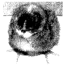
Figure 2: Morphology of chicken ES cells
Morphologies of blastodermal cells:
(A) BR22p2 and (B) BR22p3: round-shaped cells with big nucleus and small
cytoplasm.
( C) BR29p12: dispersed with looser connections blastodermal cells.
Figure 3: Expression of cell-specific marker SSEA-1
Expression of ES cell-specific markers SSEA-1 was tested on blastodermal cells
maintained
in vitro for different culture period. Left panel A: phase contrast. Right
panel B: antibody
staining.
Figure 4: Alcaline phosphatase activity of ES cells
Endogenous alkaline phosphatase activity of blastodermal cells. Cells were
stained for
alkaline phosphatase activity for different culture period.
Figure 5: Evolution of expression of markers of differentiation VASA, THY1,
BRACHYURY
while cells are maintained in culture.
Markers and culture. 1 : Bone Marrow, 2 : Embryo (head); 3 : Male gonads, 4
embryo
(stade X); 5 : feeder (STO); 6 : feeder (SN); 7 : BR 4-5; 8: BR4-6; 9 : BR 5-
2; 10 : BR 8-7;
11 : BR 8-8; 12 : BR 8-9; 13 : BR 8-12; 14 : V 9-19; 15 : BR 15-0; 16 : BR 15-
1; 17 : BR 15-2;
18: BR 15-3; 19 : BR 15-5; 20 : BR 15-6; 21 : BR 15-7; 22 : BR 15-8; 23 : BR
15-8; 24 : BR
15-9; 25 : BR 15-10; 26 : BR 15-11; 27 : BR 18-2; 28 : BR 18-3; 29 : BR 18-4;
30 : BR 18-5;
31 : BR 19-1; 32 : BR 19-2; 33 : BR 19-3; 34 : BR 19-4; 35: BR 20-2; 36: BR 20-
3; 37 : S1
p20; 38 : gDNA Barred Rock; 39 : water; 40 : water.
CA 02660035 2009-02-04
WO 2008/017704 PCT/EP2007/058263
19
Expression of Vasa, Thy-1 and Brachuyry (T) markers during different culture
period.
GAPDH control marker. Cells were maintained in culture for several weeks. Cell
pellets were
frozen at each dissociation. RNA extraction and RT-PCR were performed
concomitantly for
each cell pellets.
Figure 6: PCR RFLP profile analyis of the donor and recipient chicken strains
and of a
chimera
PCR RFLP profile of the donor and recipient chicken strains and of a chimera.
7
embryonated freshly laid eggs from donor and recipient chicken strains were
incubated for 5
days. DNA was extracted from total embryos. Somatic chimera: embryo injected
with donor
cells were incubated for 18 days. DNA was extracted from blood, heart, liver,
spleen and
gonads.
Figure 7: Chimera Birds
Donor chicken cells were injected into recipient chicken embryos. Recipient
embryos were
incubated until hatch. Chimeric birds were raised until adulthood.
Figure 8: F1 and F2 progeny
8A: F1 Bird with a typical Barred-Rock plumage pigmentation
8B: F2 birds from 5664-5665 hen. Hens were inseminated with semen from Barred
Rock
roosters. Eggs were incubated until hatch.
Figure 9: Morphology of duck ES cells
Duck ES cells were grown on a mouse feeder layer in DMEM culture medium
supplemented
with 10% fetal calf serum and with IGF1, CNTF, IL6, IL6R, SCF, FGF.
EXAMPLES
EXAMPLE 1: Materials and methods
Blastodermal cells
Embryos were collected from freshly laid un-incubated eggs. A sterile filter
paper ring was
laid over the embryo with blastodisc in center of cutout area. The yolk
membrane was cut
around outside of the disk. The disk was flipped over and transfer to a petri
dish of PBS at
CA 02660035 2009-02-04
WO 2008/017704 PCT/EP2007/058263
room temperature. Excess yolk was carefully washed out. The entire blastoderm
was
removed by gentle aspiration with a Pasteur pipette and transfer in PBS. An
average of 200
embryos was pooled together. The cells were centrifuged twice at 300g. The
cell pellet was
mechanically dissociated in culture medium. Cells in complete culture medium
were seeded
5 on an inactivated STO feeder layer. Blastodermal cells were maintained at 39
C in 7.5%
COZ. The cells were dissociated by incubation at 39 C in a solution of pronase
(5 to 2 %
w/v). Dissociated cells were seeded on new feeder layer cells in the complete
medium.
Between passages 3 and 5 cells were seeded in the minimal medium, on a STO
feeder
layer.
Culture medium
The complete culture medium was composed of DMEM-F12 base supplemented with 10
%
foetal calf serum (JRH), 0.16 mM beta-mercaptoethanol (SIGMA), 1% non
essential amino
acids (Biowhittaker), 1 mM sodium pyruvate (Biowhittaker), 2 mM L-Glutamine
(Biowhittaker), 1 ng/ml IGF1 (Tebu), 1 ng/ml CNTF (Eurobio), 1 ng/ml 11-6
(Eurobio), 1 ng/ml
II-6R (Tebu), 1 ng/ml SCF (Tebu), 1 ng/ml bFGF (Peprotech), 1%
penistreptomycine
(Biowhittaker).
Alternatively, the complete culture medium was composed of DMEM-F12 base
supplemented with 10 % foetal calf serum (JRH), 0.16 mM b-mercaptoethanol
(SIGMA), 1%
non essential amino acids (Biowhittaker), 1 mM sodium pyruvate (Biowhittaker),
2 mM L-
Glutamine (Biowhittaker), 1 ng/ml IGF1 (Tebu), 1 ng/ml CNTF (Eurobio), 1 ng/ml
11-6
(Eurobio), 1 ng/ml II-6R (Tebu), 1% penistreptomycine (Biowhittaker).
Alternatively, the complete culture medium may also be composed of DMEM-F12
base
supplemented with 10 % foetal calf serum (JRH), 0.16 mM b-mercaptoethanol
(SIGMA), 1%
non essential amino acids (Biowhittaker), 1 mM sodium pyruvate (Biowhittaker),
2 mM L-
Glutamine (Biowhittaker), 1 ng/ml IGF1 (Tebu), 1 ng/ml CNTF (Eurobio), 1%
penistreptomycine (Biowhittaker).
Preparation of feeder cells
The mouse fibroblast STO cell line (ATCC) was maintained at 37.5 C, 7.5 % COZ
in DMEM
(Cambrex) supplemented with 4% foetal calf serum (JRH) and 1% glutamine
(Biowhittaker).
At sub-confluence STO cells were dissociated with pronase (Roche) 1X, washed
with PBS
and irradiated with a gamma source at 45 grays. Feeder cells were seeded at
1.5x10⁶ to
2x10⁶ cells in fresh medium in 100-mm dishes.
CA 02660035 2009-02-04
WO 2008/017704 PCT/EP2007/058263
21
Transfection
STO cell lines: pGPARa and pMEHCS were given by Dr. Bertrand Pain. pClNeo was
purchased from Promega. Plasmid DNA was prepared using alkaline lysis and PEG
purification. For transfection, cells were seeded in the morning at 0.5x10⁶
cells per 100-mm
dish. They were transfected in the evening with 3.0 g of circular pClNeo, 15
g pGPARa or
pMEHCS using FuGENE 6 (Roche) lipofectant. The day after, cells were washed
and the
medium was changed. Selection with neomycine, 0.3 mg/ml began at Dl and was
applied
for 8 days. Resistant cells were amplified and frozen in liquid nitrogen.
Alkaline phosphatase reaction:
Cells were washed twice with PBS and fixed in 1.5% formaldehyde, 0.5%
glutaraldehyde,
0.1% Igepal for 10 to 20 minutes at 4 C. After washing cells were incubated at
37 C in the
alkaline phosphatase staining solution composed of 100 mM NaCI, 100 mM Tris pH
9.5, 50
mM MgC12, 1 mg/ml NBT, 0.1 mg/ml BCIP. The reaction was stopped by addition of
PBS lx
or H2O.
Immunofluorescence analysis:
SSEA-1 antibodies were purchased from the Developmental Studies Hybridoma Bank
of the
University of IOWA.
Cells were fixed either 20 minutes at 4 C in 1.5% formaldehyde, 0.5%
glutaraldehyde, or in
0.2% glutaraldehyde, 0.01% IGEPAL, or 10 minutes at room temperature in
paraformaldehyde 4% or methanol.
Cells were incubated in PBS-BSA 0.1 % for 2h to 4h. The first diluted antibody
was added
overnight. After washing, the second FITC-labelled anti-IgG antibody was added
for 1 h at
4 C. Reaction was stopped by removing the antibody and washing the cells with
PBS. Cells
were observed with a fluorescent microscope.
PCR RFLP:
DNAs from tissues of embryos incubated for 18 days were purified according to
the
instructions of QlAamp DNA mini-kit. A fragment of avian leucosis virus
integrated in the
genome was amplified by PCR using the following couples of primers:
Oli_pos (157-158):
Forward : 5'- GGTGTAAATATCAAAATTATC (SEQ ID N 1)
CA 02660035 2009-02-04
WO 2008/017704 PCT/EP2007/058263
22
Reverse : 5' -CGGTTAAAATACGAATAGAGA (SEQ ID N 2)
O1i_pos (78-81):
Forward : 5'-CTATGAGCAGTTACGAGGGTC (SEQ ID N 3)
Reverse : 5'-CGGACCAACAGGCTAGTCTC (SEQ ID N 4)
Amplicons are respectively 3106 and 1004 bp. The specificity of primer 157 is
higher for
DNA from Barred Rock chicken than DNA from White Leghorn. The resulting
amplicons are
cut with the Hincll enzyme.
RT PCR analysis:
RNAs were purified according to the instructions of Promega SV total RNA
isolation kit.
Head RNA were extracted from embryos after 5 days of incubation, bone marrow
from
thighbone embryos incubated for 15 days, testis from adult chicken. RNA from
BR15 cells
was extracted according to the instructions of Qiagen RNeasy kit. RNAs were
reverse
transcribed according to Promega Random primers and AMV Reverse transcriptase
kits'
instructions. PCR amplification was realized using the following primers.
Oligos vasa:
Vasa forward: TTTGGTACTAGATGAAGCAGACC (SEQ ID N 5)
Vasa reverse: GTTCCCTATCTCCATGAATGC (SEQ ID N 6)
Oligos Brachyury:
Brachyury forward: CACAAAGACATGATGGAGGAAG (SEQ ID N 7)
Forward 2: TGAAGTCCTCTCCAAAACCATT (SEQ ID N 8)
Brachyury reverse: CATAAGTTCGGGTACTGACTGG (SEQ ID N 9)
Reverse 2: CACAAAATCATTCTGCGGTAAA (SEQ ID N 10)
Oligos GAPDH :
GAPDH forward : AGGTGCTGAGTATGTTGTGGAGTC (SEQ ID N 11)
GAPDH reverse : AGAACTGAGCGGTGGTGAAGA (SEQ ID N 12)
Oligos Thy-1:
Forward: AGGACAACAGGAAGCACATCAT (SEQ ID N 13)
Reverse: GTTCTGGATCAAGAGGCTGAAG (SEQ ID N 14)
CA 02660035 2009-02-04
WO 2008/017704 PCT/EP2007/058263
23
Cells injections into recipient embryos:
When irradiated, recipient embryos were prepared by exposing freshly laid, un-
incubated
eggs to 4 Grays of X irradiation from an accelerator source. The embryos were
accessed
through a window cut into the long axis of the egg. The shell was removed by
grinding. The
shell membrane was maintained wet by addition of a drop of PBS. The shell
membrane was
cut to expose the embryo just before the injection of cells. 3 l of cells in
culture medium
were injected into the sub-germinal cavity of the recipient embryo using a
micropipette.
The window was closed by aligning two pieces of shell membrane previously
dipped into
albumen. When the shell membranes were dried the windows were tightly sealed
with
surgical tape. Eggs were incubated in conventional incubators maintained at
37.5 C and
50% relative humidity and turned through 90 every hour for 18 days. Eggs were
then
transferred to a conventional hatcher at 37 C and 85% relative humidity until
hatch.
Phenotypic and somatic chimerism were evaluated in embryos after 18 days of
incubation.
Germ-line contribution of donor cells was assessed by mating injected birds to
Barred Rock
chickens.
EXAMPLE 2: Isolation and amplification of chicken ES cells
Chicken embryonic Stem cells were isolated from freshly laid eggs. An average
of 200
embryos were pooled together and seeded on a feeder layer of irradiated mouse
fibroblasts.
Actually, Etches et al. (1996 Mol. Reprod. Dev. 45:291-288), had demonstrated
that
significantly more somatic chimeras were observed following the injection of
chicken
blastodermal cells co-cultured with mouse fibroblasts. From initial seeding to
passages 3 to
5, blastodermal cells were grown in the complete culture medium composed of
DMEM-F12
base supplemented with 10% foetal calf serum (JRH), 0.16 mM beta-
mercaptoethanol
(SIGMA), 1% non-essential amino acids (Biowhittaker), 1 mM sodium pyruvate
(Biowhittaker), 2 mM L-Glutamine (Biowhittaker), 1 ng/ml IGF-1 (TEBU), 1 ng/ml
CNTF
(Eurobio), 1 ng/ml IL-6 (Eurobio), 1 ng/ml IL-6R (TEBU), 1 ng/ml SCF (TEBU), 1
ng/ml
bovine FGF (Peprotech), 1 % penistreptomycine (Biowhittaker). Then after
several
passages, some growth factors were removed and cells were grown in minimal
culture
medium to avoid differentiation. The growth factors that were removed are
either (SCF and
FGF), or (SCF, FGF, IL-6, IL-6R).
The culture medium to grow chicken ES cells to avoid differentiation,
comprises basal
medium (i.e DMEM-F12) supplemented with 10% foetal calf serum (JRH), 0.16 mM
beta-
mercaptoethanol (SIGMA), 1% non-essential amino acids (Biowhittaker), 1 mM
sodium
pyruvate (Biowhittaker), 2 mM L-Glutamine (Biowhittaker), and supplemented
with 1 ng/ml
IGF-1, 1 ng/ml CNTF, 1 ng/ml IL-6, 1 ng/ml IL-6R, 1 % penistreptomycine.
Alternatively, the
CA 02660035 2009-02-04
WO 2008/017704 PCT/EP2007/058263
24
culture medium to grow chicken ES cells to avoid differentiation, comprises
basal medium
(i.e DMEM-F12) supplemented with 10% foetal calf serum (JRH), 0.16 mM beta-
mercaptoethanol (SIGMA), 1% non-essential amino acids (Biowhittaker), 1 mM
sodium
pyruvate (Biowhittaker), 2 mM L-Glutamine (Biowhittaker), and supplemented
with 1 ng/ml
IGF-1, 1 ng/ml CNTF, 1 % penistreptomycine (Biowhittaker).
Chicken ES cells could be isolated and expanded from different chicken strains
(table 1).
nb of nb of
strain experiments isolates %success
S86 N 127 33 26
Valo 17 5 29
Brown
Leghorn 53 4 7
GF30 20 4 20
White Leghorm 14 1 7
Cou Nu Rouge 11 4 36
Marans 29 16 55
Barred Rock 49 35 72
ISA 3 1 33
Table 1: Isolation of ES cells from various chicken strains.
The characterization of the ES status of the in vitro isolated and amplified
cells relied on a
series of biological criteria that have been demonstrated to be specific for
mouse and human
ES cells: the self-renewal property, the cell morphology, the expression of
stem cells specific
markers and the totipotency of the cells, ie. their ability to differentiate
in vitro in different
lineages and in vivo to contribute to the constitution of an embryo. The
growth characteristics
of most of the isolates were identical to the ones observed with chicken
blastodermal cells
maintained for more than a year in culture (figure 1), indicating their
potentiality of indefinite
self renewal. Blastodermal cells grew as colonies. They were round-shaped
cells, with a big
nucleus and a small cytoplasm (figure 2, BR22p2 & BR22p3). Interestingly, it
was observed
that the morphology of the blastodermal cells evolved during the culture
period from tightly
connected cells (figure 2 BR22p2 & BR22p3) to more dispersed cells with looser
connections (figure 2, BR29p12). The cells with looser connections could be
stabilized on
long term culture in the minimum medium. Cells of the different morphologies
have been
further characterized for the expression of stem cells specific markers and
for their ability to
contribute to the constitution of an embryo.
CA 02660035 2009-02-04
WO 2008/017704 PCT/EP2007/058263
ES cells are classically defined by the expression of different markers, ECMA-
7 (Kemler et
al. 1981 J. Embryol. Exp. Morphol. 64:45-60), SSEA-1 (Solter and Knowles, 1978
Proc. Natl.
Acad. Sci. USA 75(11):5565-5569), EMA-1 (Hahnel and Eddy, 1986 J. reprod.
Immunol.
10(2)89-110); the presence of specific enzymatic activities, telomerase and
alkaline
5 phosphatase activity and by the absence of markers of differentiated cells
like TROMA-1.
The expression of EMA-1 and SSEA-1 markers and alkaline phosphatase activity
was
assessed on the in vitro amplified blastodermal cells. The morphology of the
blastodermal
cells changing during the culture period, markers specific to the germ
(Tsunekawa et al,
2000 Development 127:2741-2750), mesoderm (Wilkinson et al, 1990, Nature
343:657-659)
10 and hematopoietic (Uchida et al, 1994 Blood 83:3758-3779) lineages were
added to
complete the analysis. Since antibodies against Brachyury, Thy-1 and vasa were
not
available, transcription of corresponding genes was assessed by RT PCR. This
analysis was
performed throughout an extended culture period and with respect to the
different
morphologies. SSEA-1 and EMA-1 remained expressed throughout a long culture
period
15 (figure 3). Alkaline phosphatase activity remained also stable throughout
the same culture
period (figure 4).
Figure 5 illustrates the evolution of expression of markers of differentiation
while cells are
maintained in culture. Vasa mRNA was strongly present in male gonads as
expected (figure
20 5 lane 3). It was not detected in stage X embryos (lane 4), although germ
cells are present in
stage X embryos. Vasa gene was weakly but reproducibly expressed in cells
maintained in
culture (lane 18 to 36). The absence of Vasa transcripts in stage X embryos
might be
explained by a poor sensitivity of the RT PCR. The expression of vasa gene in
cells
amplified in vitro might mean either that these cells have germ-line
competence or that germ
25 cells, that are present in stage X embryos, have been amplified in vitro.
Brachyury mRNA, a
marker of mesoderm lineage, was already detected in stage X embryos (embryos
from
freshly laid eggs) (figure 5, lane 4). Expression increased while cells are
maintained in
culture (fig.5, BR8 lane 12 & 13; BR 15 lane 19 to 26; BR18 lane 27 to 30 and
BR19 lane 32
to 34). Hematopoietic lineage originated from the mesoderm. Thy-1 mRNA, a
marker of the
hematopoietic lineage, was also detected in cells maintained in culture (lanes
7, 8; 11, 12,
13; 16 to 24; 27 to 34). HNK1 a marker of neural crest cells in chick was
expressed by
blastodermal cells. The expression of HNK1 remained stable while cells were
maintained in
culture (data not shown). Markers of undifferentiated cells were expressed
throughout the all
culture period (data not shown). Cultured blastodermal cells expressed markers
of
undifferentiated cells as well as neural crest cells and mesoderm markers.
Surprisingly, Vasa
gene which was thought to be germ cells specific is transcribed in ES cells.
CA 02660035 2009-02-04
WO 2008/017704 PCT/EP2007/058263
26
The totipotency of the in vitro amplified chicken cells was check by
evaluating their ability to
contribute in vivo to the reconstitution of an embryo.
Experiment were set-up with different number of chicken ES cells injected into
a recipient
chicken embryo to determine the effects on level of chimerism. The impact of
the irradiation
of the recipient embryo on the efficiency of colonization has also been
studied. Four
parameters were selected to monitor the effect of the injections and the
radiation: (i) the
viability of the embryos; (ii) the percentage of phenotypic chimera
(percentage of embryos
with colored feathers); (iii) the extend of phenotypic chimerism (percentage
of colored
feathers); (iv) the percentage of somatic chimerism (chimerism in other
tissues than the
feather).
300, 5,000, 15,000 or 30,000 ES donor cells were injected in the sub-germinal
cavity of
freshly laid recipient embryos either previously compromised by gamma-
radiation or not
irradiated. Injected eggs were incubated according to standard conditions.
Cultured
blastodermal cells were injected every week between 14 and 43 days of culture
to determine
the evolution of the potentialities of colonization with the culture time. All
the embryos were
analysed at 18 days of incubation.
The cultured blastodermal cells (donor cells) were from the colored Barred
Rock or S86N
strains, which are homozygous recessive (ii) at the dominant white locus 1.
Donor cells were
injected into recipient embryos from the White Leghorn strain (recipient
strain) which is
homozygous dominant at the I locus. Somatic chimera could be identified at
hatch by the
presence of black down. Nevertheless, a PCR RFLP approach was developed to
discriminate the genetic fingerprints from donor cells and recipient embryos
(figure 6). The
PCR RFLP pattern of the recipient strain is a 1 or 2 bands profile depending
on the
individual. The PCR RFLP pattern of the donor strain is a 3 bands profile.
Chimeric tissues
are identified by a 3 bands profile typical of the donor. It was then possible
to determine
whether the injected cells possessed the ability to colonize other tissues
than the feathers.
As was done for the expression of specific markers, the ability of in vitro
colonization was
studied while cells were maintained in long term culture and also according to
the cell
morphologies.
Several phenotypic (ie animals with feathers colonised by cells from the donor
strain) and
somatic chicken chimerae (ie animals with tissues other than the feathers
colonised by cells
from the donor strain) have been identified. The donor cells were shown to be
present in the
derivative of all three germ layers including the gonads. This results
demonstrate that in vitro
expanded chicken ES cells, obtained according to the method of the invention,
could engraft
and reconstitute various tissues of the recipient embryo including the gonads.
CA 02660035 2009-02-04
WO 2008/017704 PCT/EP2007/058263
27
Surprisingly, the inventors demonstrate that injection of a larger amount of
cells compared to
amount recommended in the prior art (i.e around 300 to 500 irradiated cells),
does not
impact the viability of the embryos. Moreover, the percentage of phenotypic
chimerism is
significantly increased when large amount of cells (that is to say more than
15000 and
eventually around 30000) are injected regardless of the presence or absence of
the step of
radiation. 30000 cells injected is not an upper limitation, and one considers
injecting more
cells, the only limitation being that the recipient embryo is able to survive
and develop after
having received these cells. The inventor demonstrate that injection of 15000
cells in
irradiated recipient embryos and the injection of 30000 cells in non
irradiated embryos give
the best results, and specifically give the higher percentage of embryos
chimeric in the
gonads..
The inventors demonstrate that the used range of irradiation does not impact
the viability of
the chicken embryos. Moreover, the colonisation potentiality did not seem to
be dependent
of the time length of blastodermal chicken cells in culture (data not shown).
Altogether the data support the notion that the in vitro isolated and
amplified ES cells are
true embryonic stem cells.
EXAMPLE 3: Germ-line transmission by chicken ES cells
With the best methodologies of injection developed, 9% of the injected birds
were chimeric in
the gonads (data not shown). To address the issue of the germ-line
transmission, a flock of
584 animals was generated by injection of cultured cells into recipient
embryos according to
the best methodologies shown to allow an optimal colonization of the gonads.
The
phenotypic chimerism of the birds extended from a few feathers at hatch, that
were lost later
on, to more than 95% at adulthood (figure 7 and table 2).
................................... ......................
...................................................
..........................................................
...........................................................
hE!er#::~ : :Fr.ra!a~~~~~:: :~~ntF~ :~l::>::~1
............. ..........X......... ..................... 41
....................... .......... ............ . 90
.......................... . ................
2940 ...Ø0025 ........ ..............
1730 X 28 0 2.4
1692 X 21 <5 0.064
4164 no 70 <5 < 10-3
4316 no 46 at hatch only < 10-3
4542 X 59 0 2.7x10 8
Table 2: extend of phenotypic chimerism of injected hens and roosters
Hens injected with Barred Rock cells were mated with Barred Rocks roosters to
assess the
contribution of the donor lineage to the germ-line. The germ-line transmission
was assessed
through examination of the distribution of black and yellow offspring. The
progeny of 221
hens have been examined. Of 16006 Fl birds, 5 chicks exhibited variable
percentage of
CA 02660035 2009-02-04
WO 2008/017704 PCT/EP2007/058263
28
white feathers (one was dead before hatch) and 1 chick presented plumage
pigmentation
typical from that of a Barred Rock (figure 8A). Description of the hens
producing progeny
with plumage pigmentation Barred-Rock-like and description of the conditions
of injection are
presented in the table 3. Even after 59 days of culture, cells remained able
to colonize the
germ-line. Germ-line transmission did not correlate with a high percentage of
phenotypic
chimerism of the hen. Indeed the mother of the Barred Rock-like chick was
white. The
potential number of PGC injected was estimated assuming that 50 PGC (Eyal-
Giladi et al,
1976) are present in stage-X embryos. The final number of PGC was calculated
according to
the dilution factor resulting from the successive passages of the cells and
the concentration
and volume of cell suspension injected to the recipient embryo.
h ~.,.... :...:.:::> :::::::> ::: of., >
~:u ~ :>::::o f:::<::~~~i~t: :~~ ~ a ..........!~o.. 0 n 0t.. P. 1 e
:1 M e r a
ei
...
:~.:::::::::::::.:::::::::::::::::::::::::::::: Y::::::::::::::::::::::::::.
14 2.3 0
22 6.4 0
29 11.1 0
37 11.6 0
43 6 1.3
(heart, liver, spleen, gonads)
Table 3: influence of ES cells culture on in vivo colonization
[% of phenotypic chimera: number of embryos with colored
feathers/nb alive 18 days embryos; % of gentotypic chimera: nb
of embryos chimeric in other tissues than the feathers/nb of alive
18 days embryos].
Four (4) out 5 chicks of Fl progeny (16006 birds) survived until adulthood.
These 4 hens
were inseminated with semen from Barred Rock roosters. The progeny of the hen
5664-
5665 was Barred Rock like (fig.8B and table 4). Among the progeny of the other
3 hens were
Barred Rocked-like and White Leghorn-like chicks (table 4).
,:.:: ;:::.::.;:.>::
>::::>ll:e:ri::::l:tte:ri:tlf~:~a:ri::>:::>::::>::::::::>::::;:.t+l:.;:.;:
:~n. .;:..
:::X :::.9~:
5664-5665 (Barred Rock) 6 Barred Rock
5660-5661 13 5 Barred Rock
8 White Leghorn
5650-5651 8 3 Barred Rock
5 White Leghorn
5662-5663 13 7 Barred Rock
6 White Leghorn
Table 4: Progeny of Fl birds. Hens resulting from the
mating of injected chimera with Barred Rock birds were
inseminated with Barred Rock semen. Eggs were incubated
until hatch. The phenotype of the progeny was recorded.
CA 02660035 2009-02-04
WO 2008/017704 PCT/EP2007/058263
29
The inventors provide a method of culture chicken ES cells. Amplification of
blastodermal
cells had been documented only for White Leghorn and Barred Rock chicken
strains (Pain et
al, 1996 Dev. 122(8) 2339-2348), Petitte et al,1990 108(1):185-189, Zhu et al,
2005 Nat.
Biotechnol. 23(9):1159-1169). The culture process developed in this study, in
particular the
combination of specific growth factors and its evolution during time allowed
reproducibly
isolating and amplifying cells from different chicken strains. The efficacy of
amplification was
strain dependant, a result consistent with the strain difference in
establishment of ES cells
lines described for the mouse (Kawase et al, 1994 Int.J.Dev.Biol. 38(2):385-
390).
Interestingly and unlike mouse, totipotency of chick ES cells maintained in
long term culture
is supported by a deprivation of several growth factors. That is to say that
chicken ES cells
do preserve totipotency during long term culture in complete culture medium,
and also in
complete medium deprived with growth factors such as IL-6, IL-6R, SCF and FGF.
Of note,
human ES cells cannot be either maintained in the culture conditions
established for mouse
ES cells.
Pain et al (1996) described, in chicken blastodermal long-term cultures (more
than 160
days), large colonies of small cells, tightly packed in nests with typical "ES-
like"
morphological features similar to that of mouse ES cells. By contrast, the
morphology of
chicken blastodermal cells that the inventor maintained in long-term culture
did not remain so
stable and changed from cells with a round shape that were tightly connected
to cells with
looser connections. This morphological evolution was consistent and
independent of the
chicken strain. This change in morphology did not affect the self-renewal
property specific of
ES cells. The potential commitment of the cells in different lineages was
addressed by (i) the
biochemical characterization of the cells by immunofluorescence or RT PCR
analysis; (ii) a
transgenesis methodology to access to the biological property of the cultured
cells. Cells
have been characterized with respect to markers specific of all three germ
layers, ectoderm,
mesoderm and endoderm. Markers indicative of a very early differentiation were
selected.
None of the morphologies did present a specific combination of markers.
Markers of
undifferentiated cells, markers of all three germ layers could be detected as
well as a marker
of the germ-line like vasa. There was no evolution of markers expression
specific of a
change of morphology. Even cells with the most differentiated morphology
expressed
markers specific of ES cells and did not express markers of differentiation.
In vitro amplified cells suspensions were injected into recipient embryos. The
feather
pigmentation results from a transfer of pigments from the melanocytes to the
shaft of the
feathers. Melanocytes derived from neural crest cells of the trunk of the
embryo. The
differentiation of neural crest cells happens very early in the development of
an embryo.
CA 02660035 2009-02-04
WO 2008/017704 PCT/EP2007/058263
Pigmentation of the plumage of white recipient embryos injected with cells
from a black
strain of chicken might not reflect the totipotency of the injected cells but
might indicate that
cells committed to the melanocyte lineage were able to join the melanoblastic
lineage of the
recipient. Thus the study of the contribution of the injected cells to the
recipient embryo was
5 not restricted to the observation of the plumage but included tissues
derivatives of all three
germ layers. Phenotypic and somatic chimera were obtained with each of the
main
morphologies of cells observed in culture (cords, clumps and comets)
supporting the
undifferentiated status of the cells regardless of the morphology.
Interestingly the gonads
were also colonized by the injected cells.
The ultimate proof of the ES status of a cell is the germ-line transmission. 1
FO (injected) hen
(0.3%) supported germ-line transmission. Over the 16 chicks progeny of this
hen, 1 Fl chick
was Barred Rock-like (6.25%). All the F2 chicks from the Fl hen presented the
Barred Rock
phenotype which indicates that the acquisition of the donor traits is stable
through several
generations. Half of the progeny of the Fl hens that presented variable
percentage of black
feathers was Barred Rock-like and the other half had a White Leghorn phenotype
as
expected from Mendel inheritance rules.
Thus the inventors demonstrate the germ-line competence of chicken
blastodermal cells.
Chicken blastodermal cells are thus useful to develop avian transgenesis.
EXAMPLE 4: Isolation and amplification of duck ES cells:
4.1 - Raw Material
Duck Eggs
Duck eggs from Pekin strains GL30 were obtained from GRIMAUD FRERES SELECTION
(La Corbiere, Roussay France). The parent ducks were vaccinated against
Escherichia Coli
(Autogenous vaccine Coli 01 & 02), Pasteurella multocida (Landavax), Duck
viral hepatitis
(Hepatovax), Erysipelothrix rhusiopathiae (Ruvax), Avian metapneumovirus
(Nemovac),
Salmonella typhimurium & Enteridis (Autogenous vaccine), Riemerella
antipestifer
(Autovaccine Riemerella), Avian metapneumovirus (Nobilis RTV inactive) and
Erysipelothrix
rhusiopathiae (Ruvax). After receipt, fertilized Pekin duck eggs were
submitted to a
disinfection in an hypochloryde bath followed by a decontamination with
Fermacidal
(Thermo) to avoid any risk of contamination linked to dusts attached on the
shell.
CA 02660035 2009-02-04
WO 2008/017704 PCT/EP2007/058263
31
Feeder cells
Cells from murine origin (STO cells) were used as feeder layer to maintain the
pluripotency
of duck stem cells. Those feeder cells are mitotically inactivated by gamma
irradiation (45 to
55 Grays) before seeding on plastic. This dose of irradiation is a sub-lethal
dose that induces
a definitive arrest of the cell cycle but still permits the production of
growth factors and
extracellular matrix, necessary for the promotion of the cell growth of non
differentiated cells.
The STO cell line was derived by A. Bernstein, Ontario Cancer Institute,
Toronto, Canada
from a continuous line of SIM (Sandos Inbred Mice) mouse embryonic fibroblasts
and it was
supplied by the American Type Culture Collection (ATCC) (STO Product number:
CRL-1503,
Batch number 1198713). Fresh feeder layers were prepared twice a week.
Exponentially
cells were dissociated and counted. A part of cells were seeded for
maintenance of viable
cultures and another part was irradiated. For irradiation, we prepared a cell
suspension at
10x106 cells/mL in tubes. Cells were exposed to a 45 to 55 grey dose and were
seeded on
plastic. After seeding, dishes or plates coated with inactivated feeder cells
were used during
a maximum of 5 days.
Medium
Medium GTM-3 (Sigma, Cat n G9916)
DMEM- HamF12 (Cambrex, Cat n BE04-687)
Additives
Glutamine (Cambrex, Cat n BE17-605E)
Antibiotics: Pencillin/streptomycin (Cambrex, Cat n BE17-602E))
Non essential Amino Acids (Cambrex, Cat n BE13-114E)
Sodium pyruvate (Cambrex, Cat n BE13-115 )
Vitamins (Cambrex, Cat n 13-607C)
Beta Mercapto Ethanol (Sigma, Cat n M7522)
Yeastolate (SAFC, Cat n 58902C)
Factors
Six different recombinant factors were used:
O Recombinant Human Ciliary Neurotrophic Factor (CNTF) (Peprotech Inc, Cat n
450-13)
O Recombinant Human Insulin Like Factor I (IGF1) (Peprotech Inc, Cat n 100-
11)
O Recombinant Human Interleukin 6(IL6) (Peprotech Inc, Cat n 200-06)
O Recombinant Human soluble Interleukin 6 receptor (sIL6r) (Peprotech Inc, Cat
n 200-06
R)
71 Recombinant Human Stem Cell Factor (SCF) (Peprotech Inc, Cat n 300-07)
CA 02660035 2009-02-04
WO 2008/017704 PCT/EP2007/058263
32
O Recombinant Human basic Fibroblast Growth Factor (bFGF) (Peprotech Inc, Cat
n 100-
18B)
All those factors, excepted IL6r, are produced in E. Coli bacteria. Soluble
IL6r is expressed
in transfected HEK293 cells.
Fetal Bovine Serum
Non irradiated Fetal Bovin Serum (FBS) (JRH, Cat n 12003)
The non irradiated serum used in the program was collected and produced in
Australia.
Animals used for collection were USDA inspected and acceptable for slaughter.
It was added
in the medium during avian stem cells culture. This batch was not submitted to
irradiation to
avoid the destruction of critical proteins or components that may be essential
for the
maintenance of stem cells in culture.
Irradiated serum (JRH, Cat n 12107)
The irradiated batch used in this program was collected in United States. This
irradiated
batch was added as supplement in the DMEM medium used for the culture of STO
cells
(feeder cells). Those cells do not require as stem cells a specific quality of
serum for growth
and maintenance in culture. To minimize high concentration of serum in the
medium we
have adapted the STO cells to grow in presence of 4 % of FBS only.
4.2 - Process of isolation and culture of duck ES cells
Around 360 Fertilized duck eggs were opened, the yolk were separated from the
albumen
during the opening. The embryos were removed from the yolk with the aid of a
small
absorbent filter paper (Whatmann 3M paper), cut out beforehand in the form of
a perforated
ring with the aid of a punch. The diameter of the perforation is about 5 mm.
These small
rings were sterilized using dry heat for about 30 minutes in an oven. In
practice, during the
step of embryo collection, a small paper ring is deposited on the surface of
the yolk and
centered on the embryo which is thus surrounded by the paper ring. The latter
is then cut out
with the aid of small pairs of scissors and the whole removed is placed in a
Petri dish, filled
with PBS. The embryos thus carried away by the ring were cleaned of the excess
yolk in the
medium and the embryonic disk, thus free of the excess vitellin, were
collected with a
Pasteur pipette.
The duck embryos were placed in 50 mL tubes containing PBS 1X The duck embryos
were
then mechanically dissociated, washed with PBS, and seeded on an inactivated
layer of
feeder STO cells into complete culture medium at 39 C, 7,5 % COZ. The feeder
cells were
seeded in 6 well plates or dishes at around 2,7 x10⁴ cell/cmZ. The complete
culture medium
is composed of serum free medium DMEM-Ham F12 supplemented with 10 % fetal
bovine
CA 02660035 2009-02-04
WO 2008/017704 PCT/EP2007/058263
33
serum, with IGF1, CNTF, and optionally 11-6, II-6R, SCF and Bovine FGF, at a
final
concentration of 1 ng/ml, and with 1 % non-essential amino acids, with 1 % of
mixture of
vitamins of commercial origin, with sodium pyruvate at a final concentration
of 0,1 mM, with
beta-mercapto-ethanol at a final concentration of 0.5 mM, glutamine at a final
concentration
of 2,1 mM, penicillin at a final concentration of 100 U/ml, streptomycin at a
final
concentration of 100 g/ml and yeastolate 1X. Rapidly at the passage 4, the
mixture of
antibiotics is no longer added to the medium.
The duck ES cells were cultured in the DMEM-Ham F12 complete culture medium up
to passage 4. After passage 4, the base medium is modified and DMEM-Ham F12
complete medium is replaced either by:
- the GTM-3 medium supplemented with 10 % fetal bovine serum and with IGF1 and
CNTF at a final concentration of 1 ng/ml, with 1 % non-essential amino acids,
with 1 %
of mixture of vitamins of commercial origin, with sodium pyruvate at a final
concentration of 0,1 mM, with beta-mercapto-ethanol at a final concentration
of 0.5
mM, glutamine at a final concentration of 2,1 mM and yeastolate 1X; or
- the GTM-3 medium supplemented with 10 % fetal bovine serum and with IGF1,
CNTF, 11-6, II-6R, SCF, FGF at a final concentration of 1 ng/ml, with 1 % non-
essential
amino acids, with 1 % of mixture of vitamins of commercial origin, with sodium
pyruvate at a final concentration of 0,1 mM, with beta-mercapto-ethanol at a
final
concentration of 0.5 mM, glutamine at a final concentration of 2,1 mM and
yeastolate
1 X.
The duck ES cells were further cultured during at least 14 passages in this
new medium of
culture without differentiation.