Note : Les descriptions sont présentées dans la langue officielle dans laquelle elles ont été soumises.
CA 02662785 2009-03-05
WO 2008/030842 PCT/US2007/077586
IMPLANTS WITH TRANSITION SURFACES AND RELATED PROCESSES
[0001] This application claims the benefit of the following U.S. Provisional
Applications: Serial No. 60/828158, filed October 4, 2006, titled
Instrumentation for Bicompartmental Knee; Serial No. 60/824696, filed
September 6, 2006, titled Instrumentation for Bicompartmental Knee; and
Serial No. 60/825533 filed September 13, 2006, titled Variable Transition
Referencing Guide, the entire contents of each of which are hereby
incorporated by reference.
FIELD OF THE INVENTION
[0002] The invention relates to implants and processes for use in joint
surgery, particularly knee replacement surgery. In certain embodiments,
methods are provided for locating and using a transition point on the femur
for proper positioning of resections that are intended to receive a femoral
component during a surgical procedure. Implants are provided according
to certain embodiments that replace the medial condyle and part of the
patellofemoral channel of the femur, but preferably do not replace portions
of the lateral condyle that articulate with respect to the tibia. According to
certain embodiments, a resection guide that includes a guide surface for
performing a transition resection can be positioned relative to the resections
on the bone formed using the transition point. The resection guide can then
be moved on the resection surfaces to position the transition resection guide
surface to form a transition resection that allows implant external surfaces
to
transition smoothly to portions of the lateral condyle that articulate with
respect to the tibia.
CA 02662785 2009-03-05
WO 2008/030842 PCT/US2007/077586
BACKGROUND
[0003] Knee arthritis and trauma in various forms can cause loss of joint
cartilage, including for example, osteoarthritis, excessive wear or sudden
trauma, rheumatoid arthritis, or infectious arthritis. When joint cartilage is
worn away, the bone beneath the cartilage is left exposed, and bone-on-
bone contact can be very painful and damaging. Other types of problems
can occur when the bone itself becomes diseased. One conventional
solution for these types of joint problems takes the form of total knee
replacements. In a total knee replacement (TKR), the proximal end of the
tibia is replaced with a tibial component, the distal end of the femoral bone
is replaced with a femoral component, and the patella is replaced with a
patellar component. Such procedures often require sacrifice of the anterior
and posterior cruciate ligaments.
[0004] However, many patients who develop knee arthritis experience
issues isolated to the medial (inner) compartment and the patellofemoral
(knee cap) part of the joint, while the lateral (outer) compartment of the
joint
remains healthy. The conventional treatment for such patients is either the
combination of a unicompartmental knee in conjunction with a
patellofemoral implant or the use of a total knee implant, which requires
removal of the healthy lateral condyle. However, one recent solution is a
hybrid femoral component that preserves the healthy lateral condyle as well
as the anterior and posterior cruciate ligaments, and only replaces the
medial compartment and patellofemoral joint. (Such a hybrid femoral
component may be used in conjunction with a unicompartmental tibial tray,
which only requires resurfacing of part of the tibia as well). A hybrid
femoral
component requires a smaller incision and preserves ligaments that can help
the knee retain its natural kinematics. It can be implanted using a procedure
called a bicompartmental knee replacement.
2
CA 02662785 2009-03-05
WO 2008/030842 PCT/US2007/077586
[0005] A bicompartmental knee replacement is a procedure that
replaces only the medial (inner) parts of the femoral and tibial components.
It does not resurface or resect the lateral parts of the knee (including the
distal femoral articular cartilage), and as such, can allow the anterior and
posterior cruciate ligaments to be retained. Bicompartmental knee
replacements have a number of advantages over total knee replacements.
Because the outer lateral portion of the joint is not resurfaced, the incision
made may be smaller, resulting in less pain, quicker recovery time, and less
blood loss. Also, because certain ligaments do not need to be sacrificed, a
greater stability of the knee can be maintained.
[0006] The femoral component used in such a replacement is often
called a monolithic implant. It has an anterior portion and a medial condyle
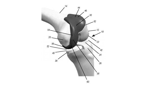
portion, without a lateral condyle portion (again, because as much of the
lateral bone as possible is retained). As with most typical femoral implants,
the component may be made of titanium, stainless steel, cobalt-chrome,
zirconium, oxinium, any combination thereof, or any other appropriate
material that has sufficient strength and biocompatibility for use in knee
replacement surgery.
[0007] While performing bicompartmental knee replacement with a
monolithic implant, it is necessary to locate the implant on the bone
properly, in order, among other things, to achieve proper articulation in both
the medial and lateral compartments of the knee between femur and tibia,
as well as proper articulation between the patella and the femur or femoral
component interface. For example, the surgeon wants to retain as much
healthy bone as possible while removing the diseased bone, but also needs
to consider the depth of the medial condyle portion of the implant in order
to ensure that there is a smooth transition from the implant to the bone and
to maintain proper performance of the reconstructed knee in flexion and
extension.
3
CA 02662785 2009-03-05
WO 2008/030842 PCT/US2007/077586
[0008] With conventional patellofemoral replacements, one popular
current method for preparing the bone to receive an implant is to use an
osteotome in conjunction with a trochlea trial to mark the boundary of the
transition between the implant and the bone. However, there is no known
solution or method for marking the boundary for bicompartmental knee
replacement. Accordingly, such surgeries are conventionally performed
using traditional total knee replacement instrumentation, without any
additional components that help identify certain reference points. For
example, recessing the implant to the cartilage on the lateral side is
important, and without specific instrumentation or techniques for this type of
procedure, the surgeon is left to estimate the cuts that are needed.
SUMMARY
[0009] Implants and processes for installing them are provided for
replacing the medial condyle of the femur and portions of the
patellofemoral channel, preferably without replacing portions of the lateral
condyle which have not been subject to degradation. According to some
such processes, instrumentation may be used which allows for an anterior
resection and a distal resection of the femur that are properly located and
oriented so that proper positioning of the implant to ensure smooth transition
between bone and implant on lateral outer surfaces of the femur, as well as
proper functioning of the reconstructed knee in flexion and extension, can
be reduced to determining the proper medial / lateral position of the implant
on those resections.
[0010] In some cases, an anterior resection instrument can be used to
form an anterior resection that is properly located in the anterior /
posterior
dimension and in interior / exterior rotation relative to the femur. A
transition
point can then be chosen, which can correspond if desired to the distal-most
point on a lateral portion of the anterior resection, for proper proximal /
distal
or superior / inferior location and valgus / varus rotation of a distal
resection.
A distal resection guide, of a type which can be used with cutting devices
4
CA 02662785 2009-03-05
WO 2008/030842 PCT/US2007/077586
such as saws, or of a type which can be used with milling devices, or a type
which can be used with both, and which can be positioned and oriented
relative to the transition point may be used to perform this distal resection
of
the medial condyle. Alternatively, a single such instrument can be used to
perform the anterior resection and the distal resection.
[0011] In some cases, an additional resection guide can be used which
can be positioned properly on the anterior resection and the distal resection
and then slid or otherwise manipulated medially or laterally to determine
proper location of a transition resection which will help form the transition
between implant and bone on outer surfaces of lateral portions of the femur.
Alternatively, one or more of the transition resection guide surface, the
distal
resection guide surface, and also the anterior resection guide surface can
be included in one instrument or resection guide.
[0012] In some cases, implants adapted to be installed on such
resected femurs feature a transition surface which corresponds to the
transition resection that has been controllably located and oriented relative
to the femur as mentioned above. Such a transition when properly located
aims to create a smooth transition from implant surface to bone surface by,
among other things, reducing surface discontinuity such as implant and / or
bone overhang. Preferably, the transition between bone and implant in
such cases is located so that only anatomical lateral condyle surfaces
articulate relative to the tibia in the knee joint in which the implant has
been
installed.
[0013] Accordingly, there is provided: A process for conducting knee
surgery on a knee comprising a tibia and a femur, the femur in turn
comprising a lateral condyle and a medial condyle, comprising: Resecting
an anterior portion of a distal portion of the femur, thereby creating an
anterior resection, the anterior resection positioned at a predetermined
depth and oriented at a predetermined angle relative to the femur in
internal / external rotation; Selecting a transition point relative to the
anterior
CA 02662785 2009-03-05
WO 2008/030842 PCT/US2007/077586
resection, wherein the transition point is located in the vicinity of the
distal-
most point of a lateral portion of the anterior resection; Performing a distal
resection on a portion of the medial condyle, the distal resection
intersecting
a point in the vicinity of the transition point, and oriented at a
predetermined
angle relative to the femur in varus / valgus rotation; Performing a
transition
resection on a lateral portion of the femur at an angle relative to both the
anterior resection and the distal resection, the transition resection
intersecting
the anterior resection and distal resection so that an implant installed on
the
bone does not project beyond lateral external surfaces of the femur and so
that external surfaces of lateral portions of the implant form a smooth
transition to lateral external surfaces of the femur; and Installing an
implant
on the resected femur, the implant having inner surfaces which substantially
fit the resections, which implant replaces distal articulating surfaces of the
medial condyle and patellofemoral articulating surfaces, but does not
replace surfaces of the lateral condyle which articulate with the tibia.
[0014] There is also provided: A femoral implant for implantation on
resected distal portions of a patient's femur, comprising: An anterior portion
which comprises an anterior medial articulating surface, an anterior lateral
articulating surface, an anterior patellofemoral channel and an anterior inner
surface adapted to correspond to an anterior resection on the femur; A
transition portion which comprises a transition medial articulating surface, a
transition patellofemoral channel, a transition lateral articulating surface,
and
a transition inner surface adapted to correspond to a transition resection on
the femur, the transition inner surface intersecting the transition medial
articulating surface to form a truncation of the transition medial
articulating
surface; A distal medial portion which comprises a distal articulating surface
and a distal inner surface adapted to correspond to a distal resection on the
femur; and A posterior medial portion which comprises a posterior medial
articulating surface and at least one posterior medial inner surface adapted
to correspond to at least one posterior resection of the femur.
6
CA 02662785 2009-03-05
WO 2008/030842 PCT/US2007/077586
[0015] There is also provided: An instrument adapted to perform a distal
resection a patient's femur comprising; An instrument index adapted to
correspond to a transition point on an anterior resection of the femur; A
distal
resection guide surface and a rod connection portion, the rod connection
portion adapted to adjustably connect to an intermedullary or
extramedullary rod, whereby orientation of the distal resection guide surface
is adjustable in varus / valgus rotation relative to the transition point on
the
femur by maintaining the index substantially aligned with the transition point
while adjusting orientation of the instrument relative to the rod; Whereby the
distal resection guide surface is positioned and oriented properly to form a
distal resection on the medial condyle of the femur when the index
corresponds to the transition point and the instrument is properly oriented in
varus / valgus relative to the rod.
[0016] There is also provided: A resection guide adapted to resect a
patient's femur, comprising: A distal resection abutment surface adapted to
contact a distal resection on the femur; An anterior resection abutment
surface adapted to contact an anterior resection on the femur; A transition
resection guide surface for guiding a transition resection of the femur; An
index located on the resection guide at a position that corresponds to a
lateral extremity of an implant that corresponds in size to the resection
guide;
[0017] Whereby the transition resection guide surface is adapted to be
properly positioned relative to the distal resection and the anterior
resection
for performing a transition resection by contacting the abutment services of
the resection guide with the anterior resection and the distal resections, and
moving the guide in a medial or lateral direction on the femur to a point
where the index indicates proper medial / lateral alignment of the transition
resection guide surface.
BRIEF DESCRIPTION
7
CA 02662785 2009-03-05
WO 2008/030842 PCT/US2007/077586
[0018] FIG. 1 is a front view of an implant according to certain
embodiments of the invention.
[0019] FIG. 2A is a front view of the implant of FIG. 1 in place on a model
of a human knee.
[0020] FIG. 2B is a navigational rose showing translational and rotational
axes which may constitute useful references in positioning and orienting
body parts, instruments and implants of certain embodiments of the
invention.
[0021] FIG. 2C is a front view corresponding generally to FIG. 2A with the
knee shown in approximately full extension.
[0022] FIG. 2D is a front view of the knee of FIGs. 2A and C with the
knee shown in approximately ninety degrees flexion.
[0023] FIG. 2E is a perspective lateral view of an implant according to
one embodiment of the invention made for a left knee.
[0024] FIG. 3 is a front view of a human femur on which has been
performed an anterior resection according to one embodiment of the
invention.
[0025] FIG. 4 is a perspective view of an anterior resection guide
according to one embodiment of the invention in place on a patient's femur
to perform an anterior resection such as shown in FIG. 3.
[0026] FIG. 5 is a perspective view of the anterior resection guide of FIG.
4 in place where the anterior resection has been performed.
[0027] FIGs. 6A - 6F are schematic distal and front views of human
femurs on which anterior resections according to one embodiment of the
invention have been performed, and which show effect of depth of the
anterior resection on its shape and size.
[0028] FIGs. 7A - 7F are schematic distal and front views of human
femurs on which anterior resections according to one embodiment of the
invention have been performed, and which show effect of internal / external
rotation of the anterior resection on its shape.
8
CA 02662785 2009-03-05
WO 2008/030842 PCT/US2007/077586
[0029] FIG. 8A is a front view of a distal resection guide according to
one embodiment of the invention in place on a human femur, to perform a
distal resection on the medial condyle according to one embodiment of the
invention.
[0030] FIG. 8B is a front view of a distal resection guide according to
one embodiment of the invention in place on a human femur, with a shim, to
perform a distal resection on the medial condyle according to one
embodiment of the invention.
[0031] FIG. 9 is a front view of a human knee, with the femur in
approximately ninety degrees flexion, showing the distal part of the femur
after a distal resection to the medial condyle according to one embodiment
of the invention has been made.
[0032] FIG. 10 is a perspective front view of an anterior / posterior
resection guide according to one embodiment of the invention.
[0033] FIG. 1 1 is a perspective medial view showing the resection guide
of FIG. 10 in place on a human femur, in contact with the anterior resection
and the medial condyle distal resections, so that it can be positioned (as by
sliding) medially or laterally on the femur in contact with those resections,
to
position the transition cutting surface of the resection guide in order to
yield
a smooth transition between implant and bone on the lateral side of the
knee.
[0034] FIG. 12 is a perspective posterior view of the resection guide of
FIGs. 10 and 1 1 in place on a human femur.
[0035] FIG. 13 is a perspective medial side view of the resection guide of
FIGs. 10 - 12 in place on a human femur.
[0036] FIG. 14 is a perspective medial front view of a resection guide
according to another embodiment of the invention positioned on a human
femur.
[0037] FIG. 15 is a perspective top view of the resection guide of FIG. 14
positioned on a human femur.
9
CA 02662785 2009-03-05
WO 2008/030842 PCT/US2007/077586
[0038] FIG. 16 is a perspective lateral front view of the resection guide of
FIG. 14 positioned on a human femur.
[0039] FIG. 17 is a perspective medial front view showing a human
femur on which anterior, distal, chamfer and transition resections have been
made according to one embodiment of the invention, using resection
guides according to certain embodiments of the invention.
[0040] FIG. 18 is a perspective medial front view showing an implant
according to one embodiment of the invention in place on a femur.
[0041] FIG. 19 is a front view of a resection guide according to an
alternate embodiment of the invention, for use with milling devices for
forming resections on the femur.
[0042] FIG. 20 is a superior view of the guide of FIG. 19 showing certain
milling devices.
[0043] FIG. 21 is a superior view of the guide of FIG. 19 without an
intramedullary rod.
[0044] FIG. 22 is another superior view of the guide of FIG. 19.
[0045] FIG. 23 is a side view of the guide of FIG. 19.
[0046] FIG. 24 is a perspective view of the guide of FIG. 19.
[0047] FIG. 25 is a side perspective view of the guide of FIG. 19.
[0048] FIG. 26 is a superior view of a guide according to another
alternate embodiment of the invention.
[0049] FIG. 27 is a perspective view of the guide of FIG. 26.
[0050] FIG. 28 is a superior view of the guide of Fig. 26.
[0051] FIG. 29 is a superior view of the guide of FIG. 26.
[0052] FIG. 30 is a superior view of the guide of FIG. 26.
[0053] FIG. 31 is a side view of the guide of FIG. 26.
[0054] FIG. 32 is a perspective view of the guide of FIG. 26.
[0055] FIG. 33 is a side view of a guide according to another alternate
embodiment of the invention.
[0056] FIG. 34 is a perspective view of the guide of FIG. 33.
CA 02662785 2009-03-05
WO 2008/030842 PCT/US2007/077586
[0057] FIG. 35 is a superior view of the guide of FIG. 33.
[0058] FIG. 36 is a superior view of the guide of FIG. 33.
[0059] FIG. 37 is a perspective view of the guide of FIG. 33.
[0060] FIG. 38 is a perspective view of a milling guide used with a milling
apparatus which rotates about a medial/lateral axis according to an
alternate embodiment of the invention.
[0061] FIG. 39 is a perspective view of a collet 182 for use in connection
with a guide 180 according to another alternate embodiment of the
invention.
[0062] FIGS. 40 A and B are side and front views, respectively, of the
collet of FIG. 39.
[0063] FIG. 41 is a perspective view of a resection guide according to
another alternate embodiment of the invention.
[0064] FIG. 42A and FIG. 42B are side and front views, respectively, of
the guide of FIG. 41.
[0065] FIG. 43 is a perspective view of the guide of FIG. 41.
[0066] FIGS. 44A and 44B are front and side views of the guide of FIG.
41.
[0067] FIG. 45 is a perspective view of the guide of FIG. 41.
[0068] FIGS. 46A and 46B are front and side views of the guide of FIG.
41.
[0069] FIGS. 47A and 47B are side views of the guide of FIG. 41.
[0070] FIGS. 49A and 49B show a femur resected using the guide of FIG.
41.
DETAILED DESCRIPTION
[0071] FIGS. 1 and 2A are front views of an implant 10 according to an
embodiment of the invention. Implant 10 is adapted to be installed on the
distal portion 12 of a human femur 14. The femur can be that of a human or
11
CA 02662785 2009-03-05
WO 2008/030842 PCT/US2007/077586
other being with appropriate hinge joints. FIG. 2A shows an implant 10
placed on a sawbones model of a human femur 14. Anatomically, the
femur 14 cooperates with the tibia 16 to form the knee joint 18. The distal
portion 12 of the femur 14 includes two condyles, a medial condyle 20 and a
lateral condyle 22. These condyles articulate (move in gross motion, whether
rotational or translational or both) relative to the tibial plateau 24 which
is a
surface on the proximal portion 26 of tibia 16. Not shown is a patella which
is
connected to a patella tendon, also not shown, which in turn inserts on the
tibia and attaches to the head of quadricep muscles to apply traction for
extension of the knee joint. The patella tracks, as by sliding, in the
patellofemoral channel 30. Patellofemoral channel 30 of implant 10 shown in
FIG. 2A replicates the patellofemoral channel in the anatomical knee, which
is a channel on anterior and distal surfaces of the femur between condyle 20
and lateral condyle 22 for tracking of the patella during flexion and
extension
of the knee 18. Ordinarily, the femur 14 and tibia 16 do not contact each
other but instead each bear against menisci (not shown) which are
interposed between condyles 20, 22 on the one hand and tibial plateau 24
on the other hand. An anterior cruciate ligament (not shown) and a
posterior cruciate ligament (not shown) are among two of the ligaments
which are connected to both the femur 14 and the tibia 16. One of the
primary purposes of these ligaments is to control translation of the femur 14
and the tibia 16 relative to each other and in an anterior / posterior
direction.
These two ligaments in particular are important for knee stability and it is
often preferred to preserve them if possible during knee surgery.
[0072] FIG. 2B is a navigational rose that corresponds to FIG. 2A. It
shows the three degrees of translational freedom and the three degrees of
rotational freedom that define the six degrees of potential freedom of
motion in a knee such as the one shown in FIG. 2A. Translationally, the
degrees of freedom are lateral / medial, anterior / posterior and superior /
inferior. Rotationally, the degrees of freedom are flexion / extension,
internal
12
CA 02662785 2009-03-05
WO 2008/030842 PCT/US2007/077586
/ external and varus / valgus. In that respect, FIGS. 2C AND 2D show a knee
18 with an implant 10 according to an embodiment of the invention installed
on the femur with the knee at essentially zero degrees of flexion, and
approximately 90 degrees of flexion, respectively.
[0073] FIG. 1 shows an implant 10 according to an embodiment of the
invention together with a tibial implant 38 and a corresponding insert 40
which together form a prosthesis for reconstructing a portion of the knee 18.
The implant 10 preferably does not replace some portions of the lateral
condyle 22 that articulate against the menisci in the lateral compartment 42,
and thus indirectly tibia 16. However, it does replace portions of the knee 18
such as those discussed above that are often found to be more prone to
osteoarthritis -- the portions of the medial condyle 20 that articulate
against
medial compartment menisci and thus indirectly against tibia 16 (for the
prostheses installed) and the patellofemoral channel 30. Such a structure is
beneficial for a number of reasons, including that the lateral compartment
42 of the knee 18 (which includes portions of the lateral condyle 22 and
lateral portions of tibia 16) is preserved with multiple beneficial effects.
In
addition to improved kinematics and greater stability, such partial knee
replacements can reduce contact of soft tissue connecting the femur 14
and the tibia 16 or lateral and medial sides of the knee with the implant 10,
and thus lesser wear, particularly on the lateral side of knee 18.
Additionally,
the implant can be installed using minimally invasive surgical procedures to
shorten the hospital stay, simplify the surgical procedure, and improve
therapy prospects and long-term results, among other benefits. Furthermore,
the implant can be installed without sacrificing the anterior cruciate
ligament
34 and the posterior cruciate ligament 36 (not shown).
[0074] Implant 10 and tibial implant 38 may be made of conventional
metallic or other materials conventionally used for knee prosthetics,
including
without limitation cobalt-chrome alloys, alloys which have been treated with
zirconium oxide or other treatments, stainless steel materials and other
metals
13
CA 02662785 2009-03-05
WO 2008/030842 PCT/US2007/077586
or materials. Insert 40 may be formed of conventional ultra high molecular
weight polyethylene of the sort conventionally used to form inserts in knee
prosthetics, or it may be formed of any desired material.
[0075] FIG. 2D is a front view of the anterior portion of tibia 16 with knee
18 in approximate 90 degrees of flexion. The distal portion of femur 14 is
evident, with lateral condyle 22 intact and the implant 10 replacing portions
of the medial condyle 20 and the patellofemoral channel 30. (The femoral
head 50, which forms part of the hip socket, can also be seen in this view
and can give some degree of intuitive appreciation for why it may be that
medial compartment 52 of the knee is sometimes more prone to
osteoarthritis and other wear than is lateral compartment 42.)
[0076] As shown in FIG. 2D, distal portion 54 of implant 10 generally
corresponds to the portion of the implant 10 between the anterior portion 44
and the posterior medial condylar portion 56 of implant 10. It also
corresponds generally to distal regions of the medial condyle 20 and
patellofemoral channel 30 of the femur 14. On the medial side of the knee
18, portions of distal articulating surfaces 58 of implant 10 articulate
against
tibial insert 40 which itself is positioned relative to tibial implant 30 on
proximal
portions of the tibia 16 where the tibial implant 38 and insert 40 are used.
(In
circumstances where the tibial components are not used, distal articulating
surfaces 58 of implant 10 can articulate against menisci and tibial plateau
24). On the lateral side of the knee, FIG. 2D makes evident a beneficial
result
of implant 10, that the lateral distal surfaces of the femur 14 and the tibia
16
remain in place to articulate relative to each other. According to this
embodiment, the lateral compartment of the knee 42 is left in place so that
the implant 10 does not articulate with the tibia 16 in that compartment.
Rather, the transition 62, discussed below, between the implant 10 and the
lateral articulating surfaces of the femur 14 is angled and is located
sufficiently anterior on the lateral side of the femur 14 to reduce chances of
such articulation, while yet providing sufficient replacement of portions of
the
14
CA 02662785 2009-03-05
WO 2008/030842 PCT/US2007/077586
patellofemoral channel 30 of the femur 16 which often suffer arthritic or
other
degradation when the medial condyle 20 does.
[0077] As shown in FIG. 2D, posterior medial articulating surfaces 60 of
implant 10 articulate against insert 40 at greater degrees of knee 18 flexion.
In circumstances where implant 38 and insert 40 are not used, the posterior
medial articulating surfaces 60 articulate against menisci and thus tibia 16
indirectly.
[0078] FIG. 2D shows, on the lateral side of the knee 18, a transition
portion of implant 10 of this disclosed embodiment of the invention which
includes transition 62. The structure of this implant 10 aims to create a
smooth transition from the natural bone lateral condyle 22 material to the
implant 10 material. A transition 62 can be considered smooth if it does not
suffer undue implant 10 or bone surface overhang or discontinuity between
implant 10 and bone. Additionally, the transition 62 with its angled resection
of bone does not require any resection of the anterior cruciate ligament or
posterior cruciate ligament. The reasons for this include that resections
required for implant 10 do not require cutting of those tissues during
minimally
invasive surgery or otherwise, and that no portions of the implant 10
interfere
with those tissues when the implant 10 is inserted into the knee 18 and
positioned on the femur 14 during minimally invasive surgery. Other
advantages of the structure and shape of implant 10 are evident to a person
of ordinary skill in the art from FIG. 2D (as well as other figures and other
portions of this document) and bearing in mind how the implant 10 is
installed during surgical procedure. Additionally, as mentioned above, the
transition 62 feature provides an implant 10 structure where the lateral
meniscus preferably does not come into contact with the femoral implant,
but rather articulates preferably only against natural bone of the lateral
condyle 22.
[0079] Accordingly, FIG. 2D shows a distal view of a femoral implant
which differs from implants such as conventional implants used in
CA 02662785 2009-03-05
WO 2008/030842 PCT/US2007/077586
bicompartmental knee arthroplasty, because (among other things) it omits
lateral condylar distal and proximal portions and instead truncates the
lateral
structure with transition 62.
[0080] FIG. 2E shows a perspective view of the implant 10 of FIG. 1 from
another perspective which is helpful in understanding the transition 62 and
other geometric and navigational aspects and features of certain
embodiments of the invention. Among other things, the inner surfaces of the
implant 10 are shaped and oriented in a manner that allows precise and
accurate positioning of implant 10 on femur 14 in order, among other things,
to replicate motion of the natural knee and optimize the benefits of
maintaining natural bone in the lateral condyle 22 using transition 62 or
similar
constructs and related geometry and structures, while producing a smooth
transition from bone to implant across transition 62.
[0081] FIG. 2E shows a navigational rose which is helpful in
understanding the orientation of various surfaces of implant 10. Anterior
articulating surfaces 46, distal articulating surfaces 58 and posterior medial
articulating surfaces 60 are evident. A transition portion of implant 10
including transition 62 is also evident. A number of surfaces are shown in
FIG.
2E as cooperating to form inner surfaces of implant 10. As is known to those
who design and install femoral implants, these surfaces are formed with a
view to fitting to distal areas of the femur 14 which have been resected to
correspond to the surfaces. Some or all of the surfaces may be cemented to
the bone or may contain bone in-growth material such as sintered beads or
wires or other porous or similar material which enhances growth of bone into
the surface of the implant, or they may feature any desired surface
characteristics. In the particular implant shown in FIG. 2E, all of these
surfaces on the inner side of implant 10 are substantially planar, that is
generally flat in the shape of a plane but including the possibility of
discontinuities such as bone ingrowth material, indentations, raised areas,
pegs, openings and other surface discontinuities which could otherwise
16
CA 02662785 2009-03-05
WO 2008/030842 PCT/US2007/077586
technically be said to remove a surface from the strict category of being
substantially in a plane or being planar. However, implants according to the
invention can also feature one or more interior surfaces which are curved, to
fit resected surfaces which have been formed by resection guides of the
present invention that resect curved surfaces onto bone as by using milling,
grinding, routing, machining, or similar apparatus which is capable of forming
curved surfaces on materials (hereinafter "milling" devices or apparatus).
[0082] In the particular implant 10 shown in FIG. 2E, anterior inner
surface 64, distal inner surface 66, posterior chamfer surface 68 and
posterior
inner surface 70 are intended substantially to abut corresponding portions of
resected bone or shims or inserts which are interposed between bone and
implant to compensate for undue bone loss or for other reasons. Anterior
inner chamfer surface 72 is disposed between distal inner surface 66 and
anterior inner surface 64 to intersect, preferably as a line, anterior
intersection
line 74.
[0083] Additionally, transition surface 76 which is also preferably but not
necessarily substantially planar, extends along lateral portions of implant 10
to intersect anterior inner chamfer surface 72, preferably in a line, the
lateral
intersection line 78. In this particular structure of this embodiment of the
invention shown in FIG. 2E, the anterior inner surface 64, anterior inner
chamfer surface 72, and transition surface 76 intersect at a point on lateral
portions of the implant 10, the convergence point 80. As a corollary in this
construct, anterior intersection line 74 and lateral intersection line 78
intersect
at convergence point 80. In a similar fashion, planes of the anterior inner
surface 64, transition surface 76 and distal inner surface 66 intersect at
implant point 83. Implant point 83 in some embodiments is located laterally,
when the implant 10 is installed on femur 14, to transition point 82.
[0084] In the particular implant shown in FIG. 2E, transition surface 76,
like other inner surfaces, is planar, although it can be curved in other
implants
according to other embodiments of the invention. A primary aim of some
17
CA 02662785 2009-03-05
WO 2008/030842 PCT/US2007/077586
embodiments of the invention is to define and use a reference or navigation
point on the bone for positioning and orienting resections and therefore
implant 10. So long as a navigational point such as a transition point on the
bone can be designated to properly form resections that will permit an
implant to be properly positioned and oriented on the femur for good knee
kinematics and performance, the particular shape of the resected surfaces
and corresponding implant surfaces, whether curved or planar, and how the
resections are formed, whether by sawing, milling or otherwise, matter less
and can be accommodated within the principles of the invention.
[0085] FIG. 3 is a front view of distal portions of a femur 14 which shows
an anterior resection 84 and a transition point 82 designated on the bone
that can be used to position and orient a distal resection 100 (discussed
below) of the femur 14 that, in combination with the anterior resection,
ultimately allow positioning of an implant such as shown in FIGS. 1 and 2 on
the bone. Accordingly, among other things, the implant can be located
and oriented properly relative to mechanical axes of the anatomy and
otherwise for proper flexion / extension and other kinematics and functioning
of the knee, and also to allow the transition from bone to implant 10 across
transition 62 to be smooth, so that for instance it suffers minimal
discontinuities
such as overhang of implant or bone.
[0086] In the femur 14 shown in FIG. 3, anterior portions of the femur 14
have been resected to form anterior resection 84 using instrumentation that
corresponds to the implant shown in FIGS. 1 and 2, as discussed more fully
below. Anterior resection 84 will correspond to anterior portion inner surface
64 of implant 10 when the implant 10 is installed on femur 14. Anterior
resection 84 is often hourglass in shape with a lateral lobe 86 and a medial
lobe 88. The transition point 82 can be chosen as the distal-most point of
lateral portions of anterior resection 84, which in the drawing of FIG. 3 is
the
distal-most point on the lateral lobe 86 of anterior resection 84. What point
is
distal-most for purposes of determining the location of the transition point
82
18
CA 02662785 2009-03-05
WO 2008/030842 PCT/US2007/077586
on the bone can be considered as intersection of a line that is parallel to a
line connecting distal-most portions of the medial and lateral condyles 20,
22.
Alternatively, location of transition point 82 can be at another
location inside or outside of anterior resection, or at any other desired
point
on the bone. What matters primarily is anterior resection 84 be formed
properly on the femur 14 in the anterior / posterior dimension and in internal
/
external rotation (see FIGs. 6A - 6F) and that a transition point can be
designated relative to which a distal resection 100 (discussed below) can be
formed properly in the superior / inferior dimension relative to the anterior
resection 84 and oriented properly in varus / valgus rotation. Proper
positioning of an implant 10 with corresponding surfaces can then be
achieved so that among other things, the implant can be located and
oriented properly relative to mechanical axes of the anatomy and otherwise
for proper flexion / extension and other kinematics and functioning of the
knee, and also to allow the transition from bone to implant 10 across
transition 62 to be smooth, so that for instance it suffers minimal
discontinuities
such as overhang of implant or bone.
[0087] FIG. 4 shows an anterior resection instrument 90 according to one
embodiment of the invention for performing an anterior resection 84 on
femur 14 to accommodate the implant 10 of FIGS. 1 and 2. Instrument 90 is
coupled to an intramedullary rod 92 which has been inserted into the distal
portion 12 of femur 14. An extramedullary rod can be used instead of the
intramedullary rod. Before instrument 90 is coupled to intramedullary rod 92,
a template or other device may be employed to mark geometry on the
femur 14, such as the anterior-posterior line and / or a line perpendicular to
it.
The instrument 90 may be coupled to the intramedullary rod and aligned
with such indicia to ensure that anterior resection 84 is properly oriented
and
located. The instrument 90 shown in FIGS. 4 and 5 includes a body 94 to
which may be connected in sliding fashion for adjustment in the anterior-
posterior direction, an anterior resection guide surface 96. Body 94 may be
19
CA 02662785 2009-03-05
WO 2008/030842 PCT/US2007/077586
connected to intramedullary rod 92 or extramedullary rod with a collar or
other desired structure to allow for translational and / or rotational freedom
as desired. In the embodiment shown in FIG. 4, body 94 can be controllably
constrained from rotating in any direction relative to the rod, although the
rod itself may be rotated in bone to align the body 94 with the indicia
marked on the femur 14. However, body 94 can move in the anterior-
posterior direction relative to the rod, and the guide surface 96 can move
relative to body 94 in the same direction. Body 94 is also able to slide
relative
to the rod in the superior / inferior direction. In the particular embodiment
shown in FIGS. 4 and 5, the body 94 is constrained from translating in the
medial / lateral direction, although that need not necessarily be the case. A
paddle 98, with or without other components connected to body 94, can be
used to determine the appropriate size of implant 10 and thus, in some
aspects of the invention, in some cases, size of certain instrumentation which
will be used to install the implant 10. Once the instrument 90 and
particularly
body 94 and anterior resection guide surface 96 have been properly
positioned, guide surface 96 may be used to create anterior resection 84.
[0088] FIGS. 6A - F show effects of moving the guide surface 96 in the
anterior-posterior direction to perform the anterior resection 84. FIGS. 6A
and
6B show an anterior resection 84 made with the guide surface 96 position in a
"neutral" anterior-posterior position. If the guide surface 96 is positioned
posteriorly to that "neutral" position, FIG. 6D shows how the shape and size
of
anterior resection 84 changes and enlarges, respectively. If the guide
surface 96 is positioned more anterior to the "neutral" position, FIG. 6F
shows
that the anterior resection 84 diminishes in size and changes shape.
Although the shape of each of the particular anterior resections 84 shown in
FIGS. 6B, 6D and 6F are hourglass and feature lateral lobes 86 and medial
lobes 88, it is possible that at some point the shape could be other than
hourglass such as if the guide surface 96 is positioned sufficiently posterior
of
the "neutral" position to make it more heart shaped, or if it is positioned
CA 02662785 2009-03-05
WO 2008/030842 PCT/US2007/077586
sufficiently anterior of the "neutral" position to cause the anterior section
84
to take the form of two ovals or other rounded closed areas.
[0089] FIGS. 7A-F show effects of internal and external rotation of the
guide surface 96 relative to intramedullary or extramedullary rod 92 to
perform the resection. FIG 7B shows the anterior resection 84 formed when
the guide surface 96 is positioned in a neutral internal-external rotational
orientation. FIG. 7D shows the anterior resection 84 when the guide surface
96 has been positioned with two degrees of internal rotation relative to
intramedullary rod 92. The size of the lateral lobe 86 has diminished and the
size the medial lobe 88 has increased. As shown in FIG. 7F, two degrees of
external rotation of the guide surface 96 relative to intramedullary rod 92 to
form the anterior resection 84 causes the opposite effect: the lateral lobe 86
increases in size and the medial lobe 88 decreases in size.
[0090] FIGS. 6 and 7 show that positioning of the anterior resection
guide surface 96 and the anterior-posterior translational and the internal-
external rotational direction can change the size and shape of the anterior
resection 84 and therefore in some embodiments the location of the bone
transition point 82 that is employed to create the right distal resection 100
/
anterior resection 84 location and orientation to allow proper positioning of
implant 10 as shown in FIGS. 1 and 2.
[0091 ] After the anterior resection 84 has been performed using this
particular embodiment of the invention, instrument 90 may be removed from
the intramedullary rod 92 and a distal resection instrument102 coupled to
that intramedullary rod 92 for performing a distal resection 100 on the medial
condyle of the femur 14. FIGS. 8A and 8B show one such distal resection
instrument 102 according to this embodiment of the invention.
[0092] Distal resection instrument 102 shown in FIGS. 8A and 8B includes
a distal resection guide surface 104 and structure for connecting it to the
intramedullary rod 92. Preferably, that structure allows distal resection
guide
104 to be adjusted in at least varus / valgus rotational and superior /
inferior
21
CA 02662785 2009-03-05
WO 2008/030842 PCT/US2007/077586
translational directions relative to the rod 92. The structure connecting the
distal resection guide surface 104 and the intramedullary rod 92 can include,
for example, a collet 106 and a body 108. The collet 106 can be positioned
on the intramedullary rod 92 in sliding relationship and connected directly or
indirectly to body 108 which can be connected directly or indirectly to
resection guide surface 104. For example, guide surface 104 can be
connected in sliding relationship to body 108 so that it can move relative to
body 108 in anterior / posterior direction but be constrained in the other
degrees of freedom with respect to body 108. Collet 106 can include indicia
to select and / or indicate magnitude of rotation of guide 104 in the varus /
valgus direction. One form of such indicia 110 can be seen on the top
surface of collet 106 and FIG. 8B. Alternately, a series of collets can be
provided for selection by the surgeon to accommodate various angles of
varus / valgus. Distal resection guide surface 104 can also contain a
plurality
of openings 112 to receive pins for pinning it to the bone when properly
positioned, for example by pinning it to the anterior resection 84.
[0093] A distal resection 100 can be performed on the medial condyle
20 such as by using instrument 102 as follows. Other instrumentation can also
be used, and can suffice if it allows a distal resection to be made to the
medial condyle 20 which substantially passes through or is navigated relative
to transition point 82 and is correctly oriented in the varus / valgus
direction.
With reference to FIGS. 8A and 8B, distal resection instrument 102 can be
placed on intramedullary rod 92 and positioned by sliding so that body 108 is
positioned correctly to locate distal resection guide surface 104 so that it
can
be positioned and oriented relative to the bone transition point 82 and
rotated in varus / valgus so that a distal resection 100 may be made using
resection guide surface 104 which passes through, near or suitably relative
to,
transition point 80 and is properly oriented in varus / valgus. It may be
desirable to position the resection guide surface 104 so that the distal
resection 100 can pass proximal to the transition point 82 or, if desired,
distal
22
CA 02662785 2009-03-05
WO 2008/030842 PCT/US2007/077586
to it. Once the distal resection guide surface 104 has been properly
positioned relative to intramedullary, extramedullary or other rod 92, it can
be pinned to anterior resection 84, if desired, to perform the distal
resection
100. Accordingly, the rod 92, body 108 and collet 106 can be removed from
the bone to leave distal resection guide surface 104 retained in place by the
pins. Resection can also be performed without pins if desired, by relying on
rod 92 and the other structure of instrumentation 102 to retain the resection
guide surface 104 in place while resection 100 is being performed.
[0094] To serve as a distal resection guide surface 104 index 114, a
portion of the flat surface of the resection guide surface 104 can be
employed to visually align the distal resection guide surface 104 with the
transition point 82, or to place this portion of the resection guide surface
104
near, such as proximal or distal relative to, the transition point 82 so that
distal
resection 100 will pass through, near or suitably relative to, transition
point 82.
Alternatively, index 114 can include a physical indicium (not shown) such as
a mark, engraving, raised portion, or other desired indicium on any portion of
the distal resection guide surface 104.
[0095] After the distal resection 100 has been performed, distal
resection guide surface 104 can removed from the bone (as can
instrumentation 102 and intramedullary rod 92 if they were left in place).
[0096] FIG. 9 shows a distal view of a distal resection 100 of the medial
condyle 20 of a femur 14 performed using instrumentation as shown in FIGS.
8A and 8B. At this stage, after the distal resection 100 has been performed,
the position and orientation of an implant 10 have been defined in at least
four degrees of freedom by resecting in accordance with certain
embodiments of the invention as disclosed above:
[0097] anterior / posterior translation as defined by the anterior
resection 84;
[0098] superior / inferior translation as defined by the distal resection
100;
23
CA 02662785 2009-03-05
WO 2008/030842 PCT/US2007/077586
[0099] internal / external rotation as defined by the anterior resection
84; and
[00100] varus / valgus rotation as defined by the distal resection 100.
[00101] Thus, essentially all that remains for determining proper location
and orientation of the implant 10 on the femur 14 is medial / lateral
positioning on the anterior resection 84 and distal resection 100.
[00102] For such medial / lateral positioning, a transition resection guide
116 according to an embodiment of the invention as shown in FIGS. 10 - 13,
or other desired instrument, can be used. Among other things, the resection
guide 116 shown in those FIGs. can be used to create transition resection 118
and anterior chamfer resection 120. Essentially, any structure is sufficient
to
perform these resections if a transition resection 118 can be performed using
the instrumentation which positions properly the transition surface 76 of
implant 10 or other implant according to the invention with reference to
location and orientation of both anterior resection 84 and distal resection
100.
[00103] Resection guide 116 as shown in FIGs. 10 - 13 can include a finger
or other index 122 for aligning guide 116 with transition point 82. Index 122
can correspond to a relevant landmark on the implant 10, such as a lateral
outer extremity of the implant 10, or with a predetermined lateral / medial
and / or superior / inferior offset distance, to a point located relative to
the
implant point 81. The index 122 may be of any particular structure or shape,
including virtual if desired rather than physical. It can be connected to body
124 of resection guide 116 as by a flange 126 which has, on its posterior side
as seen best in FIG. 13, an anterior resection alignment surface 128. Anterior
resection alignment surface 128 can be used to position resection guide 116
as by positioning alignment surface 128 flat against anterior resection 84.
[00104] The body 124 or any other desired portion of resection guide 116
can include a distal resection alignment surface 130 which can be used to
position resection guide 116 as by positioning it flat against distal
resection
24
CA 02662785 2009-03-05
WO 2008/030842 PCT/US2007/077586
100. Resection guide 116 may thus be positioned against the femur 14 for
proper resection of transition resection 118 and chamfer resection 120 by
moving anterior resection alignment surface 128 on anterior resection 84 and
distal resection alignment surface 130 on distal resection 112 while aligning
or
positioning index 122 medially or laterally to position transition resection
guide
134 properly for a smooth transition of bone to implant across transition 62
which, for instance, features minimal discontinuities such as overhang of
implant or bone. One way to achieve that result using the guide 116 shown
in FIGs. 10 - 12 is to position index 122 laterally / medially to an extent
that
shows the surgeon where the lateral extremity of the implant 10 will be
positioned relative to the bone, if a transition resection 120 is performed
using transition resection guide surface 134 on guide 116 with guide 116 in
that position. Condyle marks 125 on posterior surfaces of guide 116 (see FIG.
12) corresponding, for example, to condyle width, can also be used in
combination with the index 122 for this purpose. Marks 125 or index 122 may
be used independently, or guide 116 can include any other marks or indices
for helping the surgeon determine where best to position the guide 116 and
thus implant 10 laterally / medially for performing the transition resection
118
at a location that causes minimal surface discontinuity across transition 62
between implant 10 and bone.
[00105] Transition resection guide 116 can also contain a chamfer
resection guide surface 132 for forming an anterior chamfer surface on the
bone corresponding to chamfer surface 72 of the implant 10 (see FIG. 2E)
and a drill guide bore 136 that is tangent to chamfer resection guide surface
132 and transition resection guide surface 134, or otherwise corresponds to
their intersection. Guide 116 can if desired include a drill guide bore 136
which can operate as follows: Once the resection guide 116 has been
properly positioned on the femur 14 as disclosed above, a drill may be
aligned through drill guide bore 136 to form a bore 138 in the bone of the
femur 14 that will correspond to lateral intersection 78 on the inner surface
of
CA 02662785 2009-03-05
WO 2008/030842 PCT/US2007/077586
implant 10 that extends from convergence point 80 in an angular fashion to
help form the intersection between transition surface 76 of the implant and
anterior inner chamfer surface 72 of implant 10 (see FIG. 2E). Transition
resection 118 can then be performed using transition resection guide surface
134, and chamfer resection 120 can then be performed using chamfer
resection guide surface 132. These resections can be performed without
using drill guide bore 136 to form a bore 138, and drill guide bore 136 can be
omitted from guide 116 if desired.
[00106] Anterior / posterior resection guide 116 may also include a
posterior resection guide surface 137 for forming a posterior resection 139
that corresponds to posterior inner surface 70 of the implant 10. Similarly,
resection guide 116 can include a posterior chamfer resection guide surface
140 for forming a posterior chamfer resection 142 on the bone that
corresponds to posterior chamfer inner surface 68 of implant 10. These latter
resections are shown in FIG. 13.
[00107] FIGS. 14-16 show an alternative form of resection instrumentation
144 which uses a single instrument 144 for performing both a anterior
resection 84 and the distal resection 100. Anterior resection guide surface
147 can be used to perform an anterior resection 100 after instrument 144
has been adjusted so that anterior resection guide surface 147 is properly
located in the anterior / posterior dimension and in internal / external
rotation. Distal resection guide surface 146 is connected through a structure
which allows it to be positioned relative to intramedullary or extramedullary
rod 92 so that resection guide surface 146 can be oriented correctly relative
to transition point 82 on the bone and oriented in varus / valgus to form the
distal resection 100. Such structure in the embodiment shown in FIGS. 14-16
include a collet 148 and body 150. The collet includes indicia 152 to indicate
desired varus / valgus orientation of the resection guide surface 146. Similar
to the way in which distal resection instrumention 102 may be used, the
alternate distal resection guide surface 146 can be positioned in the superior
26
CA 02662785 2009-03-05
WO 2008/030842 PCT/US2007/077586
/ inferior direction relative to intramedullary rod 92 by sliding collet 148
on the
rod. It can be adjusted in varus / valgus by using the indicia on the collet
148. As in the case of distal resection guide surface 104, the alternate
distal
resection guide surface 146 surface itself, without any markings or special
physical distinctions, can serve as an index 154 for positioning of the
alternate distal resection guide surface 146 so that the distal resection 100
passes through or near or relative as desired to the transition point 82. As
with
the case of distal resection guide surface 104, alternate distal resection
guide surface 146 or other portion of alternate distal resection
instrumentation 144 can contain indica (not shown) or other desired markings
or features to serve as an index 154 for such proper alignment so that the
distal resection 100 extends through, near or suitably relative to the
transition
point 82 and is correctly positioned in varus / valgus.
[00108] Implant sizing markings 156 can also be included, as shown in
FIGS. 14-16 to allow this instrumentation 144, in a manner similar to anterior
resection instrument 90 and / or distal resection instrumentation 102, to show
or suggest to the surgeon what size of implant 10, and what size of transition
resection guide 116, will be needed.
[00109] FIG. 17 shows distal portion 12 of femur 14 with what is left of
anterior resection 84 after performing a transition resection 118 and chamfer
resection 120 according to one embodiment of the invention. This view is
taken before posterior resection 138 and posterior chamfer resection 142
have been performed.
[00110] FIG. 18 shows implant 10 installed on femur 14, with the knee in
approximately 65 degrees of flexion. The posterior medial articulating
surfaces 60 of implant 10 are articulating against tibial insert 40 of the
medial
compartment 52 of the knee, while natural bone of the lateral condyle 22 of
the femur 14 and the tibial plateau 24, form the lateral compartment 42 of
the knee 18.
27
CA 02662785 2009-03-05
WO 2008/030842 PCT/US2007/077586
[00111] FIGS. 19-25 show a resection guide 158 according to an alternate
embodiment of the invention, which can resect bone so that implants 10
having one or move curved inner surfaces can be installed. Accordingly, an
anterior resection 84, which may be flat or curved, can be formed using any
desired resection device or guide, such as those discussed above, or milling
apparatus with appropriately positioned guide. As in the devices discussed
above, a bone transition point 82 which may be designated as desired,
including the distal most point on the lateral portion of anterior resection
84.
Upon designation of the transition point 82, guide 158 may be positioned on
the distal portion of femur 12. In the structure shown in FIGS. 19-25, guide
158
features an anterior paddle 160 which may be substantially flat or curved as
appropriate to correspond to anterior resection 84. The paddle or other
portion of the guide 158 can also include a transition point index 162 for
helping locate guide 158 relative to transition point 82. Transition point
index
162 can be any desired physical or other marker or structure on guide 158 as
desired. Also, helping position guide 158 relative to femur 14 is a collet 164
which is connected, preferably in adjustable relationship, to an
intramedullary or extramedullary rod 166. Collet 164 could also be in the
form of an adjustable structure with indica as can be the case with resection
guides discussed above, or a series of collets 164 each corresponding to a
particular desired varus/valgus angle, may be employed, one of the collets
164 being selected for a particular application. Thus, guide 158 can be
properly navigated and located relative to distal portion of femur 12 using
the transition point 82 to help regulate the depth of the distal resection or
distal surface to be formed by a milling device operating relative to guide
158, and proper navigation and location in varus/valgus and otherwise to
cause guide 158 properly to guide milling or other resection devices to form
curved surfaces, straight surfaces, or combinations, in proper orientation and
position for proper kinematics of the reconstructed knee.
28
CA 02662785 2009-03-05
WO 2008/030842 PCT/US2007/077586
[00112] FIG. 20 shows guide 158 properly located on femur 14 to form a
curved distal resection 112 and posterior resection 139, together with
transition resection 118 (not shown in FIGS. 20-25, but similar in location
and
orientation to the transition resection 118 discussed in connection with
resection guides disclosed above.) As shown in FIGS. 20-23, one or more
medial condyle milling devices 168 can be guided by guide 158 to form
distal resection 112 and posterior resection 139, both of which are curved
and preferably meet in curved continuous fashion in the particular
embodiment shown in FIGS. 20-23. Guide 158 can be constructed to use
only one medial condyle milling device 168, multiple such devices, or as
otherwise desired. Guide 158 can also be structured to allow the devices 168
to be positioned in order to rotate about a medial / lateral axis rather than
as
shown in FIGS. 20-23. A transition resection milling device 170 can be used to
track within guide 158 to form the transition resection 118. FIGS. 24 and 25
show a shim 172 which may be coupled to guide 158 to help position guide
158 relative to medial condyle 20.
[00113] FIGS. 26-32 show a version of the guide 158 with a paddle 160
adapted to correspond to a curved anterior resection 84.
[00114] FIGS. 33-38 show a guide 174 according to another embodiment
of the invention adapted to be navigated relative to the transition point 82
on a flat or curved anterior resection 84, and for forming a flat posterior
resection 139 on medial condyle 20. Guide 174 can be navigated relative to
the femur 14 using the transition point 82 on the femur 14 which has been
designated as disclosed above, and relative to an intramedullary or
extramedullary rod 166 using a collet 176. The collet 176 can be of the same
sort as disclosed above in connection with guide 158. Once the guide 174
has been properly navigated and located, including if desired, like guide
158, being pinned to the bone in conventional fashion, the distal resection
112 can be formed using medial condyle milling devices in a fashion similar
to that disclosed in connection with guide 158. Alternatively, surfaces of
29
CA 02662785 2009-03-05
WO 2008/030842 PCT/US2007/077586
guide 174 can be used to guide a milling device whose rotational axis is in
the medial lateral direction, as shown in FIG. 38 by way of example.
Medial/lateral rotational milling device 176 can be wider than that shown in
FIG. 38, if desired, and used with a guide 174 which uses slots or other
desired
structure to allow milling device 176 to rotate against bone on the medial
condyle 20 to shape it appropriately, and for device 176 to be guided by
and manipulated relative to guide 174. A transition milling device 170, not
shown, may be used as in the guide 158, to form transition resection 118.
[00115] Guides 158 or 174 may be configured and structured as desired
in order to guide one or more medial condyle milling devices 168 or 176 to
form distal resection 112 and/or posterior resection 139 in a continuous
curved fashion, with or without flat portions, or as otherwise desired. Guide
174 like guide 158 can be used in connection with flat or curved anterior
resections 84 which resections may be formed using cutting blocks or milling
guides.
[00116] FIGS. 39-49 show a resection guide 180 according to another
alternate embodiment of the invention. Such a guide can incorporate
functionality for forming not only the distal resection 112, transition
resection
118 and posterior resection 139, but also anterior resection 84. The
particular
guide 180 shown in these figures is adapted to be positioned on a generally
tubular collet 182. Collet 182 can be located and positioned on
intramedullary or extramedullary rod 92 so that collet 182 and guide 180 may
be properly positioned and then locked in place as desired relative to the
rod 92. Any other collet can be used, whether or not adjustable or provided
in a series to accommodate various angles of varus/varus. In the particular
structure shown in these FIGS. 39 - 49, guide 180 can slide and then be
locked in place relative to collet 180 in an anterior/posterior direction, as
well
as rotated and then locked into place relative to collet 182 to adjust guide
180 in a varus/valgus rotation as desired relative to femur 14. Accordingly,
guide 180 can be positioned relative to intramedullary or extramedullary rod
CA 02662785 2009-03-05
WO 2008/030842 PCT/US2007/077586
92 in a varus/valgus and interior/exterior rotational direction, and in a
superior/inferior and anterior/posterior translational direction, and then
locked in place as desired in each of these rotations or translations. Guide
180 contains an anterior resection guide surface 184, a distal resection guide
surface 186, a posterior resection guide surface 188, a transition resection
guide surface 190, an anterior chamfer guide surface 192 and a posterior
chamfer guide surface 194. A shim 196 can be used to help position guide
180 for proper distal and other resections. Shim 196 is shown in FIG. 43.
[00117] In use, intramedullary or extramedullary rod 92 is placed and the
guide 190 of FIGS. 39 - 49 properly positioned relative to it on collet 182 to
form an anterior resection 84 in accordance with the principles discussed in
connection with the embodiment shown in FIGS. 5-8. Transition point 82 is
then designated and a positioner 198 as shown in FIG. 43 can be connected
to guide 190 to abut anterior resection 84 or otherwise referenced to it, and
also reference positioner 198 and guide 180 relative to transition point 82 so
that a distal resection 112 can be formed at proper depth to achieve proper
flexion extension of the reconstructed knee. Positioner 198 can also contain
the distal resection guide surface 186 for forming distal resection 112. Guide
180 and positioner 198 are shown properly navigated and located into place
on the femur 14 for forming the distal resection 112. The other resections,
including transition resection 118, posterior resection 139, anterior chamfer
resection 120, posterior chamfer resection 142 can be formed using the
respective guide surfaces 188, 190, 192, and 194. FIG. 49 A and B show the
resections formed on the bone using guide 180: anterior resection 84; distal
resection 112, posterior resection 139, anterior chamfer resection 120 and
posterior chamfer resection 142 and transition resection 118.
[00118] An implant such as that shown in FIGS. 1 and 2 can be installed
on the femur 14 so resected.
31