Note : Les descriptions sont présentées dans la langue officielle dans laquelle elles ont été soumises.
CA 02677361 2014-07-21
PROGRESSIVELY SIZED ANNULOPLASTY RINGS
Field of the Invention
The present invention refers to a prosthetic annuloplasty ring or set of
rings,
in particular for the mitral annulus, that are progressively proportioned at
different
orifice sizes.
Background of the Invention
The human heart has four valves; the aortic valve, the mitral valve, the
pulmonary valve and the tricuspid valve. Various diseases and certain genetic
defects of the heart valves can impair the proper functioning of the valves.
Improper
functioning of a valve can be severely debilitating and even fatal if left
untreated,
particularly if the diseased valve is the aortic valve (between the left
ventricle and
the aorta) or the mitral valve (between the left atrium and left ventricle).
The
common defects and diseases affecting each of these valves, and the treatments
thereof, are typically different.
The mitral valve and, less frequently, the tricuspid valve, are prone to
deformation, such as dilation of the valve annulus, tearing of the chordae
tendineae
and leaflet prolapse, which results in valvular insufficiency wherein the
valve does
not close properly and allows for regurgitation or back flow from the left
ventricle
into the left atrium. Deformations in the structure or shape of the mitral or
tricuspid
valve are repairable. Thus, because prosthetic valves have certain
disadvantages that
can have serious effects (e.g., mechanical valves carry the risk of
thromboembolism
and require anticoagulation treatment, and biological valves have limited
durability), an improper functioning mitral or tricuspid valve is ideally
repaired
rather than replaced.
1
4195606 vi
CA 02677361 2014-07-21
The mitral annulus is a pliable junctional zone of fibrous and muscular tissue
joining the left atrium and left ventricle that anchors the peripheral hinge
portion of
the anterior and posterior mitral leaflets. The annulus has two major
collagenous
structures: (1) the right fibrous trigone, which is part of the central
fibrous body and
is located at the intersection of the atrioventricular membranous septum, the
mitral
and tricuspid valves, and the aortic root; and (2) the left fibrous trigone at
the
junction of the mitral valve and left coronary cusp of the aortic valve. The
mitral
valve has two major leaflets, the much larger anterior (or aortic) leaflet and
the
smaller posterior (or mural) leaflet. The anterior mitral leaflet spans the
distance
between the commissures (including the trigones) and is in direct fibrous
continuity
with most of the left and noncoronary aortic valve cusps. The posterior one-
half to
two-thirds of the annulus, which subtends the posterior leaflet, is primarily
muscular with little or no fibrous tissue, and usually contains three (or
sometimes
more) scallops separated by fetal clefts or "subcommissures."
During systolic contraction of the heart, the free margins of the mitral
leaflets
appose each other and close the respective atrial-ventricular passage. The
chordae
tendineae and papillary muscles hold the leaflets in this position throughout
the
systole cycle to prevent the leaflets from bulging into and opening within the
left
atrium. The functional competence of the mitral valve relies on proper,
coordinated
interaction of the mitral annulus and leaflets, chordae tendineae, papillary
muscles,
left atrium, and left ventricle. However, when the valve or its leaflets are
misshapen
or enlarged, for example, when the annulus is dilated, the edges of the
leaflets fail to
meet each other, leaving an opening therebetween. This opening may involve
lateral
separation of the valve leaflets and/or elevation of one valve leaflet with
respect to
the other. In either case, the ineffective closure of the valve during
ventricular
contraction results in regurgitation or leakage of blood back into the atrium,
and
ultimately in reduced pumping efficiency. To compensate for such inefficiency
in the
mitral valve, the left ventricle must work harder to maintain the requisite
cardiac
2
4195606 vi
CA 02677361 2014-07-21
output. Over time, this compensatory mechanism typically results in
hypertrophy of
the heart followed by dilation, i.e., an enlarged heart, which can lead to
congestive
heart failure.
Mitral regurgitation is one of the most common valvular malfunctions in the
adult population, and typically involves the elongation or dilation of the
posterior
two-thirds of the mitral valve annulus, the section corresponding to the
posterior
leaflet. The most common etiology of systolic mitral regurgitation in patients
undergoing surgical evaluation is myxomatous degeneration, also termed mitral
valve prolapse (29% to 70% of cases), or in gross terms, at least 5 to 10
percent of the
population in the U.S. Women are affected about twice as often as men. Mitral
valve
prolapse has been diagnosed as Barlow's syndrome, billowing or balloon mitral
valve, floppy mitral valve, floppy-valve syndrome, myxomatous mitral valve,
prolapsing mitral leaflet syndrome, or systolic click-murmur syndrome. The
syndrome of mitral valve prolapse includes palpitations, chest pain, syncope
or
dyspnea, and a midsystolic click (with or without a late systolic murmur of
mitral
regurgitation). These latter findings are typically seen in patients with
Barlow's
syndrome, where extensive hooding and billowing of both leaflets are the rule.
Some
forms of mitral valve prolapse seem to be hereditary, though the condition has
been
associated with Marfan's syndrome, Grave's disease, and other disorders.
Myxomatous degeneration involves weakness in the leaflet structure, leading
to thinning of the tissue and loss of coaptation. Barlow's disease is
characterized by
myxoid degeneration and appears early in life, often before the age of fifty.
In
Barlow's disease, one or both leaflets of the mitral valve protrude into the
left atrium
during the systolic phase of ventricular contraction. The valve leaflets are
thick with
considerable excess tissue, producing an undulating pattern at the free edges
of the
leaflets. The chordae are thickened, elongated and may be ruptured. Papillary
muscles are also occasionally elongated. The annulus is dilated and sometimes
3
4195606 vi
CA 02677361 2014-07-21
calcified. Of course, some of these symptoms present in other pathologies, and
therefore the present application will refer to mitral valve prolapse as a
catch-all for
the various diagnoses, including Barlow's syndrome.
Other causes of mitral regurgitation include ischemic heart disease with
ischemic mitral regurgitation (IMR), dilated cardiomyopathy (in which the term
"functional mitral regurgitation" [FMR] is used), rheumatic valve disease,
mitral
annular calcification, infective endocarditis, idiopathic chordal rupture
(usually
associated with fibroelastic deficiency [FED]), congenital anomalies,
endocardial
fibrosis, and collagen-vascular disorders. IMR is a specific subset of FMR,
but both
are usually associated with morphologically normal mitral leaflets.
It will therefore be apparent that the types of valve disease that lead to
regurgitation are varied and present vastly differently. For instance, Figures
1-8
show first a normal mitral valve anatomy and then the causes of pure mitral
regurgitation from a number of pathologies. Figures 1A-1B show a normal mitral
anatomy with the mitral leaflet 20 spread out flat in Figure 1A, and Figure 1B
shown
as a section through one papillary muscle 22. The chordae 24 connect the lower
edges of the leaflet 20 to the papillary muscles 22 within the left ventricle.
Figures 2A-2B illustrate a condition diagnosed as infective endocarditis,
either
active or healed. Vegetation or growths 30 may occur on the leaflet 20, and
sometimes a perforation 32. Often the chordae 24 rupture, such as at 34.
Figures 3A-3B illustrate floppy mitral valve which causes prolapse. The
leaflets 20 is distended, increasing the annulus area, leaflet area, and
causing
buckling.
4
4195606 vi
CA 02677361 2014-07-21
Figures 4A-4B show advanced floppy mitral valve which causes the chordae
to rupture, such as seen at 40.
Figures 5A-5B illustrate rheumatic heart disease. Diffuse fibrous thickening
forms at the lower edge of the mitral leaflet 20 and the chordae 24 exhibit
focal
thickening.
Figures 6A-6B illustrate papillary muscle dysfunction (coronary), in which
one or more of the muscles is scarred and atrophied, such as at 50. Possible
effects
may be severe coronary artery narrowing and acute or healed infarct.
Figures 7A-7B illustrate papillary muscle dysfunction (infiltrative), in which
typically both muscles are infiltrated with foreign bodies, possibly amyloid,
sarcoid,
infection or neoplasm.
Finally, Figures 8A-8B annular calcification. Calcific deposits 60 produce
leaflet protrusion toward the atrium.
As is clear from the illustrations 2-8, many conditions lead to regurgitation.
However, it is understood that four general types of structural changes of the
mitral
valve apparatus may produce regurgitation: leaflet retraction from fibrosis
and
calcification, annular dilation, chordal abnormalities (including rupture,
elongation,
shortening, or apical tethering or "tenting" as seen in FMR and IMR), and
possibly
papillary muscle dysfunction.
Carpentier's functional classification of the types of leaflet and chordal
motion
associated with mitral regurgitation may be seen with reference to Figures 9A-
9D. In
Type I, Figure 9A, the leaflet motion is normal. Type II (seen in Figure 9B)
mitral
regurgitation is due to leaflet prolapse or excessive motion. Type III
(restricted leaflet
5
4195606 vi
CA 02677361 2014-07-21
motion) is subdivided into restriction during diastole Type Illa (Figure 9C)
or systole
Type Illb (Figure 9D). Type Mb (Figure 9C) is typically seen in patients with
ischemic mitral regurgitation. The course of the leaflets during the cardiac
cycle is
represented by the dotted lines. (Derived from Carpentier A: Cardiac valve
surgery:
the "French correction." J Thorac Cardiovasc Surg 86: 323, 1983.)
Various surgical techniques may be used to repair diseased or damaged
mitral and tricuspid valves. These include but are not limited to annuloplasty
(i.e.,
contracting the valve annulus to restore the proper size and shape of the
valve),
quadrangular resection of the leaflets (i.e., removing tissue from enlarged or
misshapen leaflets), commissurotomy (i.e., cutting the valve commissures to
separate
the valve leaflets), shortening and transposition of the chordae tendoneae,
reattachment of severed chordae tendoneae or papillary muscle tissue, and
decalcification of valve and annulus tissue.
In patients with degenerative mitral valve disease, valve repairs using mitral
valvuloplasty valve reconstruction, or annuloplasty have been the standards
for
surgical correction of mitral regurgitation and have provided good long-term
results. A rigid support ring (e.g., Carpentier-Edwards Classic ), a semi-
flexible ring
(e.g., Carpentier-Edwards Physio ), or a flexible ring (e.g., Cosgrove-Edwards
)
may be used. These rings are typically D-shaped with a minor/major axis ratio
of
about 3:4. Some rings are flat or planar, while others exhibit three-
dimensional bows,
typically along the anterior segment. Not all physicians agree which ring is
appropriate for any one condition.
Despite accepted treatments for correcting mitral regurgitation, there is a
need
for a simpler and more effective approach that takes into account more of the
common pathologies.
6
4195606 vi
CA 02677361 2014-07-21
Summary of the Invention
The present invention provides, in one aspect, a set of mitral annuloplasty
rings each comprising a ring body arranged around a flow axis having an upward
direction and a downward direction. The downward direction corresponds to the
direction of blood flow through the mitral valve annulus when the annuloplasty
ring
is implanted. In accordance with a preferred embodiment, the ring body defines
a
minor axis extending between and bisecting the anterior segment and posterior
portion and a major axis extending perpendicularly thereto, the major and
minor
axes being generally perpendicular to the flow axis and each having dimensions
across the ring body.
The set of rings is progressively sized to take into account more of the
common pathologies. More specifically, the proportional shapes of each ring as
the
orifice size changes are not the same. In a preferred embodiment, the larger
rings
have larger minor axis dimensions relative to their major axes.
Brief Description of the Drawings
Features and advantages of the present invention will become appreciated as
the same become better understood with reference to the specification, claims,
and
appended drawings wherein:
Figure 1A is a diagram of a normal mitral annulus, leaflets, and connected
chordae and papillary muscles shown laid flat or unrolled;
Figure 1B is a "radial" sectional view through one of the papillary muscles of
Figure 1A;
Figures 2-8 are diagrams from the same viewpoints as Figures 1A and 1B
demonstrating various causes of pure mitral regurgitation as follows:
Figures 2A-2B illustrate infective endocarditis;
7
4195606 v1
CA 02677361 2014-07-21
Figures 3A-3B illustrate floppy mitral valve;
Figures 4A-4B illustrate floppy mitral valve with ruptured chordae;
Figures 5A-5B illustrate rheumatic heart disease;
Figures 6A-6B illustrate papillary muscle dysfunction (coronary);
Figures 7A-7B illustrate papillary muscle dysfunction (infiltrative); and
Figures 8A-8B illustrate annular calcification;
Figures 9A-9D illustrate Carpentier's functional classification of mitral
regurgitation, namely: Type I: normal leaflet motion. Type II: increased
leaflet
motion (leaflet prolapse). Type III: restricted leaflet motion; Ma,
restriction in
diastole and systole; Mb, restriction in systole;
Figures 10 and 11 are plan and section views of an exemplary annuloplasty
ring of the present invention;
Figure 12 is a graph showing the changing minor/major axis proportion of
the exemplary ring; and
Figures 13-18 show plan and side views of several different sized rings of the
present invention.
Detailed Description of the Preferred Embodiments
The present invention provides a novel set of annuloplasty rings for
correcting pathologies resulting in mitral regurgitation.
Figures 10 and 11 are plan and section views of an exemplary annuloplasty
ring 70 of the present invention. The ring 70 is shown with a fabric covering
72 over
a structural interior support or body 74. Typically a suture-permeable
interface 76
fills the space between the covering 72 and interior body 74.
The ring 70 in the plan view of Figure 10 has a minor axis dimension B and a
major axis dimension A. Figure 11 shows preferred heights above a datum plane,
8
4195606 v1
CA 02677361 2014-07-21
with the center of the anterior segment rising to height C and the center of
the
posterior segment rising to height D. The preferred ratio of C/D is about 3:1,
with
the smallest rings rising to 3 mm on the anterior side and the largest to
about 6 mm.
The interior body 74 of the present invention in one embodiment is desirably
made of material(s) that are "generally rigid" and will initially resist
distortion when
subjected to the stress imparted thereon by the mitral valve annulus of an
operating
human heart. In this sense, "distortion" means substantial permanent
deformation
from a predetermined or manufactured shape; the opposite concept of which is
"elastic" meaning the ability to recover the ring shape in the absence of an
external
force. A number of "generally rigid" materials can be utilized that will
perform this
function, including various bio-compatible polymers and metals and/or alloys.
Certain polyesters that resist distortion and also rapid degradation within
the body
may be used (a material that degrades slowly may provide the required initial
support). In a preferred embodiment, at least an inner core or body of the
annuloplasty ring of the present invention is made of a suitable metal, such
as
titanium or its alloys, or ELGILOY made by Elgiloy, L.P. of Elgin, Ill.,
U.S.A. The
core or ring body may be one piece, or may include a plurality of concentric
or
otherwise cooperating elements.
The interface 76 is a molded silicone tube or band around the ring body 74
and the fabric covering on the exterior of the ring is desirably Dacron
(polyethylene
terephthalate). The tubular fabric covering around the silicone sleeve provide
an
interface for securing the annuloplasty ring to the mitral annulus, although
other
interfaces are contemplated. For example, rings having outward hooks or barbs
are
known in the art.
Typical annuloplasty support rings have a long or major dimension and a
short or minor dimension, with the conventional ratio of the minor to major
9
4195606 vi
CA 02677361 2014-07-21
dimension being at most 3:4 (75%), and typically less. The present invention
provides an armuloplasty ring that has a gradually increasing minor axis
dimension
B to major axis dimension A ratio. The dimensions A and B are measured to the
inner edge of the body 74. This increasing dimensional ratio provides rings in
the
larger sizes that are more suited to correcting conditions where the mitral
leaflet is
floppy, such as the conditions shown in Figures 2-4, and in general for Type
II
pathologies seen in Figure 9B. Typically, larger patients exhibit this general
condition leading to regurgitation as opposed to smaller patients, for which
rings
having more conventional B/A ratios are more appropriate.
The following table indicates the actual values of the major and minor axes as
measured across the interior of the ring body 74 (dimensions A and B,
respectively,
in Figure 10) for nine different exemplary rings, and also gives the ratios of
the
minor axis to the major axis. The ring sizes are given in even 2 mm increments
as
measured across the major axis. Such rings will have distinct packaging so as
to be
labeled with the particular size.
Ring size Major axis Minor Axis B/A ratio
(mm) (mm) (mm)
24 24.0 16.5 0.6875
26 26.0 17.7 0.6808
28 28.0 18.9 0.6750
30 30.0 20.4 0.6800
32 32.0 21.9 0.6844
34 34.0 23.5 0.6912
36 36.0 25.5 0.7083
38 38.0 28.5 0.7500
40 40.0 32.0 0.8000
4195606 vi
CA 02677361 2014-07-21
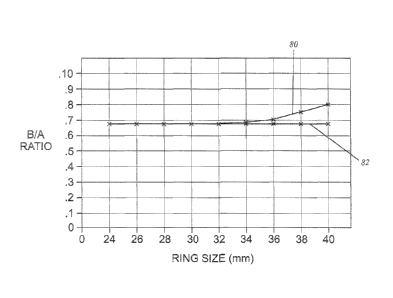
Figure 12 is a graph showing the changing minor/major axis proportion of
the exemplary ring along line 80 as compared with a line 82 for a prior art
ring, the
Carpentier-Edwards Physio ring. This shows the divergence of the ring
proportions starting at around the 32 mm ring.
Figures 13-18 show plan and side views of several different sized rings of the
present invention for comparison. Figures 13-14 show a 24 mm ring, Figures 15-
16
show a 32 mm ring, and Figures 17-18 show a 40 mm ring. The overall "look" of
the
rings are the same though the B/ A ratio increases in the larger rings.
While the invention has been described in its preferred embodiments, it is to
be understood that the words which have been used are words of description and
not of limitation. Therefore, changes may be made within the appended claims
without departing from the true scope of the invention.
11
4195606 v1
