Note : Les descriptions sont présentées dans la langue officielle dans laquelle elles ont été soumises.
CA 02678544 2009-08-12
WO 2008/101220 PCT/US2008/054174
Non-Invasive, Bedside Intra-Cranial Pressure Monitoring
System Utilizing Early On-Set Auditory Evoked Responses
Related Application
This application is the non-provisional filing of provisional U.S. Application
Serial No. 60/890,116 filed February 15, 2007.
Background of the Invention
This invention relates to monitoring intracranial pressure, and in particular
to non-invasive intracranial monitoring using waveforms evoked from a patient.
The invention provides a system capable of monitoring intra-cranial
pressure (1CP), using early onset auditory brainstem response (ABR), modified
auditory brainstem response (MABR) and electrocochleography (ECochG)
methods. The invention is used to estimate when ICP is increased, or has
increased compared to the patient's earlier baseline value. This nurse-
friendly,
monitoring and warning system constitutes an important bedside surveillance
system for a high risk patient group. It is fully automated in both the
presentation
of auditory stimuli and immediate analysis of the recorded potentials ¨ not
requiring that a neurologist, neurosurgeon or neurophysiologist be present for
the
test or its interpretation.
Increased ICP is commonly seen in conditions such as brain tumors, head
injury, stroke, or cerebral fluid (CSF) build up in hydrocephalus. The
management of increased intra-cranial pressure remains a major obstacle to the
successful treatment of many patients with life-threatening intra-cranial
space-
taking lesions. At the present time, the measurement of ICP requires an
invasive
procedure ¨ a hole must be drilled through the skull and often the cerebrum
must
be punctured. Various medical or surgical measures may be used to alleviate
increased ICP if detected in a timely fashion. Patients with headaches or
certain
findings on clinical examination such as drowsiness or focal neurological
signs or
brain scans that suggest increased ICP, are usually seen in an emergency room
CA 02678544 2009-08-12
WO 2008/101220 PCT/US2008/054174
and closely observed in the intensive care unit (ICU) Unfortunately, even
today,
patients with brain masses may rapidly deteriorate as lesions enlarge, and the
urgency of surgical or medical measures to combat increased ICP can be
misjudged. Nurses and physicians may be short staffed or busy with other
patients, and neurological status may be clouded by sedative medications given
for headache or restlessness.
It has been known for many years that increased ICP is frequently associated
with fullness in the ears, mild or moderate usually low tone hearing
impairment, and
dizziness or imbalance. The cause is likely related to the cochlear aqueduct,
a distinct
channel in the basal skull that interfaces CSF with perilymph destined for the
cochlea.
Animal studies bear out direct increased CSFACP pressure transmission to the
inner
ear and associated damping of electrocochleography (EcochG) potentials, EcochG
has not previously been used in patients with increased ICP. Some comparison
has
been made to Meniere's disease or 'endolymphatic hydrops¨typified by episodic
symptoms of vertigo, progressive sensorineural hearing loss tinnitus, and
fullness in
the ear with disturbed EcochG potentials recorded from symptomatic patients.
Early-onset or short latency auditory evoked responses (ECochG, ABR,
MABR) are robust, reliably recorded potentials largely refractory to the
presence of
depressant and anesthetic medications or the patient's level of consciousness -
making these responses an ideal choice in the intensive care setting. Wave V -
the
most prominent waveform of the ABR and MABR, and the chosen target for
automated analysis, is generated from the critical midbrain region of the
brainstem.
This same region is highly vulnerable to the effects of transtentorial brain
herniation,
the most common and fatal form of deterioration in patients with intracranial
mass
lesions and increased ICP. Thus the ABR and MABR Wave V can capture the early
phases of this devastating deterioration associated with increasing ICP.
Many studies have demonstrated abnormalities in the conventional or
standard click-evoked auditory brainstem response (ABR) in patients with
increased
ICP, and reversal of these abnormalities with normalization of ICP. The
standard
ABR is well known to be sensitive to brainstem lesions or compression, as
found in
2
CA 02678544 2009-08-12
WO 2008/101220 PCT/US2008/054174
later stages of increased ICP. However, the invention mirrors rises in ICP
compared
to a patient's earlier baseline, and captures mild or moderate increases in
ICP, and
also the late stages of actual brainstem shift.
Published reviews in this field have yielded the knowledge that the
conventional or routine-click evoked ABR, without actual midbrain shift, may
reflect
moderately increased ICP in less than one-half of patients, but often with
only non-
specific abnormalities. This led the present inventor to develop the MABR to
further
challenge the cochlea yet keep the test practical and require minimal time.
However,
the results of these studies could not be accessed in a timely manner, as
required to
be useful to a critically ill patient under observation, and necessitated a
neurologically
trained physician or clinical neurophysiologist to interpret the results.
Ordinarily,
evoked potential studies require such a professional for interpretation.
Summary of the Invention
The invention is a user-friendly automated system that samples and
automatically analyzes early auditory responses, and produces a timely warning
' signal to alert nursing staff or others of changes reflecting increased 1CP.
In one form
of the invention, it is directed to an intracranial pressure monitoring
system,
comprising an auditory stimulation and recording unit, which includes a
stimulation
controller, a memory for storing at least one of established patient baseline
waveform
data and normative range waveform data, a device for generating a comparison
by
comparing received waveform data with established patient baseline waveform
data
or normative range waveform data, and an alarm which is operable based upon
that
comparison.
At least one cranial electrode is provided, which is attachable to a patient.
An
audible stimulation device is included, operable by the stimulation controls.
In accordance with the preferred form of the invention, the auditory
stimulation
device includes at least one ear stimulation instrument and an auditory
stimulator
connected to the ear stimulation instrument. Preferably, there is a pair of
ear
stimulation instruments, and each ear stimulation instrument comprises an
acoustic
ear insert.
3
CA 02678544 2009-08-12
WO 2008/101220 PCT/US2008/054174
Preferably there is a plurality of the cranial electrodes, for judicious
placement
cranially on a patient. Between three and five electrodes may be used.
The alarm may be audible, visual or a combination of audible and visual.
The method according to the invention comprises the steps of auditorially
stimulating a patient to evoke a received waveform data indicative of
intracranial
pressure, then generating a comparison by comparing the received waveform data
with one of established patient baseline waveform data and established
normative
waveform range data, and, finally, generating an alarm responsive to that
comparison.
BRIEF DESCRIPTION OF THE DRAWINGS
The invention is described in greater detail in the following description of
examples embodying the best mode of the invention, taken in conjunction with
the drawing figures, in which:
Figure 1 is a block diagram of a system according to the invention.
Figure 2 is an example of use of the invention with ABR click or pure tone,
and
Figure 3 illustrates use of the invention with MABR.
DESCRIPTION OF EXAMPLES EMBODYING
THE BEST MODE OF THE INVENTION
Patients would greatly benefit if a safe, non-invasive bedside method
existed to automatically sample and interpret physiologic signals that reflect
increasing ICP in a timely manner. The system of the invention is used to
monitor ICP utilizes MABR or/and EcochG methodology, and is not significantly
affected by patients taking depressant or paralytic medications, or under
general
anesthesia. The system should greatly impact patient care, save lives, and
lead
to fewer invasive ICP monitoring procedures. The system should be a valuable
back-up safety measure to existing medical and surgical management, including
invasive ICP monitors which can fail about 7% of the time.
4
CA 02678544 2009-08-12
WO 2008/101220 PCT/US2008/054174
As explained in greater detail below, the invention may utilize conventional
components, such as Bio-logic (Natus/Bio-logic, Mundelein, IL) instrumentation
and accessories. A commercially available Navigator Pro laptop based unit can
be used to perform stimulation, recording, amplification, averaging, and
display of
waveforms. A separate component stimulator and preamplifier is attached
directly to the patient. Auditory stimulation is delivered by soft foam ER 3A
insert
headphones placed just within the external ear canal, and all recordings by
noninvasive skin surface stick-on or gel electrodes. A Biologic TM (tympanic
membrane) electrode is exclusively used for ECochG. Natus / Bio-logic is
additionally a leader in the manufacture and distribution of automated, nurse
friendly ABR devices used routinely world-wide as a hearing screen in
neonates.
Electrocochleography (ECochG) - Analysis of electrical signals generated
by the cochlea which- require proximity to the inner car to be reliably
recorded
following moderately loud (100-105 dBpeSPL) auditory click stimulation
delivered
by insert headphones. An adequate eighth nerve action potential (AP) voltage
of
about 1 microvolt (uV) is recorded from the tympanic membrane (TM) electrode
referred to the contralateral mastoid skin surface (nasion ground) with a
latency
of about 1.5 millisecond after the auditory stimulation, in addition to the
AP, are
two earlier cochlear hair-cell receptor potentials whose onset begins with the
auditory stimulation ¨ the cochlear microphonic (CM), and summating postential
(SP).
Auditory Brainstem Response (ABR) ¨ consists of five positive vertex
scalp recorded waves generated by the auditory nerve and 4 auditory brainstem
nucleii or tracts, recorded within 6 to 7 milliseconds. Foam insert headphones
deliver a moderately loud (100-105 dBpeSPL) auditory click stimulus at
approximate rates between 11-22 per second. Wave V (and following Vn) are
usually most prominent with a voltage (amplitude) approaching 1/2 microvolt
(uV). For ABR the active skin surface electrode is placed at the frontal
vertex
(Fz) and referenced at the ipsilateral mastoid skin surface. A surface ground
electrode is placed at the nasion. The ABR, most notably Wave V, can also be
generated by an insert headphone that delivers a pure tone burst stimulus, and
is
CA 02678544 2009-08-12
=
WO 2008/101220
PCT/US2008/054174
recorded with identically placed recording electrodes. In some instances, this
tonal ABR may have more promise than the conventional click ABR in capturing
ICP.
Modified Auditory Brainstem Response (MABR) ¨is elicited by a rapid click
stimulation rate of about 40-70 per second and binaural (bilateral
simultaneous)
presentation to both ears, both modifications augment the amplitude of the
prominent Wave V (and Vn) which are the major waveforms of interest. The
frontal
vertex (Fz) referred to C2 neck linkage also augments Wave V amplitude. A
ground
is placed at the nasion. This augmentation is necessary since the MABR is
performed at 4 moderate loudness intensities (i.e. 85,75,72,65 dBpeSPL), all
well
below that of the standard ABR (100-105 dBpeSPL). These maneuvers stress the
cochlea, yet yield a robust Wave V (approximately 1 uV) for automated Wave V
recognition, Wave V latency/intensity and Wave V amplitude/intensity curves
for
analysis, display if desired, and warning. An MABR wave V (and Vn) can also be
generated by a pure tone
This invention is for a bed-side auditory stimulation and surface scalp
recording device that can use tympanic membrane recorded electrocochleography
(ECochG), the conventional click-evoked, or pure tone burst auditory brainstem
response (ABR), and modified click or tone burst evoked ABR (MABR) tests
involving bilateral (binaural) or unilateral- rapid stimulation rates of
diminishing
stimulation intensities to create Wave V latency/intensity and Wave V
amplitude/intensity decay curves. Easily tolerated soft foam insert headphones
deliver the stimuli and simple skin surface electrodes are used for recording
the
potentials. Wave V, the most prominent ABR waveform, can be windowed and
captured (peak picking) with software facilitating automated wave form
recognition
and analysis. Software can also handle waveforms derived at diminishing
intensities
and create the above mentioned latency/intensity and amplitude/intensity
curves.
When these curves reach critical values compared to an earlier baseline in the
same
patient or curves derived from normals, a warning tone and light alerts
hospital staff
of the concern for increasing ICP in the patient. The early-onset evoked
response
6
CA 02678544 2009-08-12
WO 2008/101220 PCIVUS2008/054174
battery can be automatically set to be administered every 10 or 20 minutes
(etc), as
determined by the nursing staff or physicians.
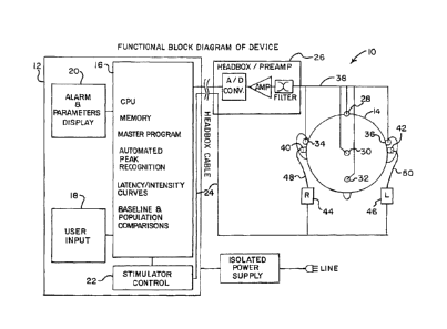
A non-invasive, bedside intra-cranial pressure monitoring system 10
according to the invention is generally illustrated in block form in Figure 1.
The
system 10 includes an auditory stimulation and recording unit 12 which may, as
explained below, be a single unit or a series of individual elements joined as
a unit.
The auditory stimulation and recording unit 12 is used for monitoring the ICP
of a
patient 14, as also explained further below.
The auditory stimulation and recording unit 12 includes a CPU 16, which may
be a general purpose computer, as identified above, and which includes all
software
and memory needed in order to perform not only storage of waveform data, but
also
analysis required by the invention, The CPU 16 thus includes, as indicated on
the
CPU 16, memory, the master program necessary for operation, automated peak
recognition for analyzing waveform data received from the patient 14,
latency/intensity curves which provide normative range waveform data, and
baseline
and population comparisons. The baseline can include patient baseline waveform
data collected from the patient 14, and the population comparisons can include
waveform data gathered from patients with known levels of increased ICP. A
user
input 18, which may be as simple as a keyboard, is used to import data into
the CPU
16.
The unit 12 also includes alarm and parameters display 20. The display 20
can be as simple as an audible alarm, or a visual display, or a combination of
both
audible and visual displays to provide an indication relative to comparison of
waveform data received from the patient 14 with data stored in the CPU 16.
The unit 12 also includes a stimulator control 22. The stimulator control 22
is
used to send stimulating signals to the patient 14 via a cable 24, or
wirelessly if
wireless connections are used.
For appropriate connection to electrodes placed on the patient 14, the
auditory stimulation and recording unit 12 is connected through a typical
preamplifier
26. Depending on the system being used to obtain waveform data from the
patient
7
CA 02678544 2009-08-12
WO 2008/101220 PCT/US2008/054174
14, electrodes 28 through 36, which may be non-invasive skin surface stick on
or gel
electrodes, are employed. The electrodes 28 through 36 are connected via
cables
38 to the preamplifier 26 and then to the auditory stimulation and recording
unit 12.
For auditory stimulation, ear inserts 40 and 42 are used. The inserts 40 and
42 may be standard soft foam insert headphones which are placed just within
the
external ear canal of the patient 14. Each of the ear inserts 40 and 42 is
activated
by a respective conventional auditory stimulator 44 and 46 through a
respective
acoustic tube 48 and 50.
Figure 2 illustrates the invention, using auditory brainstem response (ABR).
For this purpose, the electrode 30 is placed at the frontal vertex and the
electrode 32
is placed at the nasion as a surface ground electrode. The electrodes 34 and
36 are
mastoid electrodes from which waveform data may also be obtained.
Figure 3 illustrates the use of the invention with MABR. The electrode 30 is
connected to the frontal vertex and the electrode 32 is connected at the
nasion as a
ground. The electrode 28 is connected at the neck to augment the wave V
amplitude.
Initiation of an alarm at the display 20 depends on set limits that are set in
the
unit 12. Intensive care unit monitoring of early-onset (short latency)
auditory evoked
responses is similar to intra-operative monitoring, and if there is a fifty
percent drop
in the wave V amplitude, or ten percent increase in wave V latency, compared
to the
patient's baseline waveform data, the CPU 16 can be set to issue a warning via
the
display 20. Other limits can also be set, such as a wave V latency shift or
wave V
amplitude drop beyond 2.5 standard deviations can trigger a warning by the
display
20.
While the invention has been described with respect to comparison of patient
waveform data with either the patient's baseline waveform data or normative
range
waveform data, it can also be compared with other waveform data, such as
waveform data from a group of patients with known levels of increased ICP.
8
CA 02678544 2015-07-14
Even more rapid rates of auditory stimulation (100 or more clicks or tone
bursts per second ¨ requiring maximum-length sequence techniques) may bring
out
first and higher order nonlinear responses, which may prove more sensitive to
changes In ICP. A stimulator and preamplifier component may be attached
directly
to the patient, held by a neck band or pocket, and this portable component
(the site
of a deck of cards) communicating wirelessly with the near-by bedside unit 12.
The
patient could return from tests without a need to remove the electrodes or ear
inserts, and once again be within range of the base unit for monitoring.
9