Note : Les descriptions sont présentées dans la langue officielle dans laquelle elles ont été soumises.
MODULATION OF NKT CELL ACTIVITY WITH ANTIGEN-LOADED
CD I d MOLECULES
Background Of The Invention
Field of the Invention
[0001] The invention generally relates to the field of immunology.
Background Art
[0002] The natural immune system strikes a complex balance between highly
aggressive, protective immune responses to foreign pathogens and the need to
maintain tolerance to normal tissues. In recent years there has been
increasing
recognition that interactions among many different cell types contribute to
maintaining this balance. Such interactions can, for example, result in
polarized
responses with either production of pro-inflammatory cytokines (e.g.,
interferon-
gamma) by TH1 type T cells or production of interleukin-4 (IL-4) by TH2 type T
cells that suppress TH1 activity. In a number of different animal models, T
cell
polarization to Till has been shown to favor protective immunity to tumors or
infectious pathogens whereas T cell polarization to TH2 can be a critical
factor in
preventing development of cell-mediated autoimmune disease. The conditions
that
determine whether immune stimulation will result in aggressive cell-mediated
immunity or in down regulation of such responses are highly localized in the
sense
that each tissue is comprised of a distinctive set of antigen presenting cells
(APC) and
lymphocyte lineages that interact to favor different immune responses. For
example,
under optimal conditions, the dendritic cells (DC) localized in a normal
tissue may
represent predominantly a lineage and stage of maturation that favors
tolerogenic
interactions and serves as a barrier to cell-mediated autoimmunity whereas a
tumor or
site of infection will attract mature myeloid dendritic cells that stimulate
potent cell-
mediated immune responses.
[0003] CD1d-restricted NKT cells are a unique class of non-conventional T
cells that
appear to play an important role in defining the outcome of immune stimulation
in the
CA 2678618 2018-07-09
CA 02678618 2009-08-17
WO 2008/103392 PCT/US2008/002256
- 2 -
local environment. They share with the larger class of NKT cells the
expression of
markers of both the T cell and natural killer (NK) cell lineages. As such, NKT
cells
are considered as part of innate immunity like NK cells and in humans their
frequency
in normal individuals can be as high as 2.0% of total T lymphocytes (Gumperz
et al.,
2002. J Exp Med 195:625; Lee etal., 2002. J Exp Med 195:637).
[0004] CD1d-restricted NKT cells are distinguished from other NKT cells by
their
specificity for lipid and glycolipid antigens presented by the monomorphic MEC
class lb molecule, CD1d (Kawano et al., Science 278 (1997), pp. 1626-1629).
CD1d
is a non-MHC encoded molecule that associates with 02-microglobulin and is
structurally related to classical MEC class I molecules. CD1d has a
hydrophobic
antigen-binding pocket that is specialized for binding the hydrocarbon chains
of lipid
tails or hydrophobic peptides (Zeng et al., Science 277 (1997), pp. 339-345).
CD1d is
known to bind a marine sponge derived a-glycosylated sphingolipid, a-
galactosylceramide (a-GalCer), and related molecules such as ceramide-like
glycolipid antigens with a-linked galactose or glucose but not mannose (Kawano
et
al., Science 278 (1997), pp. 1626-1629; and Zeng etal., Science 277 (1997),
pp. 339-
345). As discussed below, the ability to activate CD id-restricted NKT cells
by
stimulation with a-GalCer or related molecules bound to CD 1 d of antigen
presenting
cells has greatly facilitated functional analysis of this non-conventional T
cell subset.
In the absence of inflammation, CD id-restricted NKT cells have been shown to
localize preferentially in certain tissues like thymus, liver and bone marrow
(Wilson
et al., 2002. Trends Mol Med 8:225) and antitumor activity of NKT cells has
been
mainly investigated in mouse liver metastasis.
[0005] NKT cells have an unusual ability of secreting both TH1 and TH2
cytokines
and potent cytotoxic as well as regulatory functions have been documented in
inflammation, autoimmunity and tumor immunity (Bendelac et al., (1995) Science
268:863; Chen and Paul. 1997. J Immunol 159:2240; and Exley et a/. ,1997. J
Exp
Med 186:109).
[0006] Among the CD1d-restricted NKT cells is a subset, referred to herein
as INKT
cells," that express a highly conserved cci3T cell receptor (TCR). In man this
invariant
TCR is comprised of Va24Ja15 in association with vp11 whereas in mice the
CA 02678618 2009-08-17
WO 2008/103392 PCT/US2008/002256
- 3 -
receptor comprises the highly homologous Va. 14Jcc18 and Vf38.2. Other CD1d-
restricted NKT cells express more variable TCR. Both TCR invariant and TCR
variant classes of CD1d-restricted T cells can be detected by binding of CD1d-
tetramers loaded with a-GalCer (Benlagha et at., J Exp Med 191 (2000), pp.
1895-
1903; Matsuda et al., J Exp Med 192 (2000), pp. 741-754; and Karadimitris et
al.,
Proc Nat! Acad Sci USA 98 (2001), pp. 3294-3298). CD1d-restricted NKT cells,
as
defined in this application (CD1d-restricted NKT), include cells that express
either
invariant or variant TCR and that bind or are activated by CD1d loaded with
either a-
GalCer or with related ceramide-like glycolipid antigens. CD id-restricted NKT
cells,
as defined in this application (CD1d-NKT), include cells that express either
invariant
or variant TCR and that bind or are activated by CD1d loaded with either a-
GalCer or
with related sphingolipids that have a-linked galactose or glucose including
molecules such as OCH, which differs from a-GalCer by having a shortened long-
chain sphingosine base (C5 vs. C14) and acyl chain (C24 vs. C26) (Miyamoto et
al.,
Nature 2001 413:531-4).
100071 CD Id-restricted NKT have been shown to have direct cytotoxic
activity
against targets that express CD1d. It is likely, however, that the effect of
CD1d-
restricted NKT on immune responses is amplified through recruitment of other
lymphocytes either by direct interaction or, perhaps even more importantly, by
indirect recruitment through interaction with DC. CD1d-restricted NKT have the
unique ability to secrete large quantities of IL-4 and IFN-y early in an
immune
response. Secretion of IFN-y induces activation of DC which produce
interleukin-12
(IL-12). IL-12 stimulates further IFN-y secretion by NKT cells and also leads
to
activation of NK cells which secrete more IFN-y.
100081 Since CD1d-restricted NKT are able to rapidly secrete large amounts
of both
IL-4 and IFN-y, the polarization of immune responses will depend on whether
the
effect of pro-inflammatory IFN-y or anti-inflammatory IL-4 cytokines
predominate.
This has been reported to be, in part, a function of the relative frequency of
different
subsets of CD1d-restricted NKT. These subsets include (i) an invariant CD1d-
restricted NKT population that is negative for both CD4 and CD8 and that gives
rise
CA 02678618 2009-08-17
WO 2008/103392 PCT/US2008/002256
- 4 -
to predominantly a TH I type response including secretion of pro-inflammatory
IFN-y
and TNF-a and (ii) a separate population of CD id-restricted NKT that is CD4+
and
that gives rise to both a TH1 type and TH2 type response including secretion
of the
anti-inflammatory Th2-type cytokines IL-4, IL-5, IL-10 and IL-13 (Lee et al.,
J Exp
Med 2002; 195 :637-41; and Gumperz et al., J Exp Med 2002; 195 :625-36). In
addition, NKT cell activity is differentially modulated by depending on the
particular
ceramide-like glycolipid bound to CD1d (see, e.g., US Patent Application
Publication
No. 2006/0052316). Local factors that influence activation of CD1d-restricted
NKT
subsets include the cytokine environment and, importantly, the DC that are
recruited
to that environment.
[0009] A number of indirect mechanisms contribute to the protective effect
of CD1d-
restricted NKT cells. Activation of NKT cells by administration of a-GalCer in
vivo
results in concomitant activation of NK cells (Eberl and MacDonald, Eur. J.
Immunol.
30 (2000), pp. 985-992; and Caniaud et al., J. Immunol. 163 (1999), pp. 4647-
4650).
In mice deficient in NKT cells, a-GalCer is unable to induce cytotoxic
activity by
NK cells. NKT cells also enhance the induction of classical MHC class I
restricted
cytotoxic T cells (Nishimura et al., Int Immunol 2000; 12 :987-94; and Stober
et al., J
Immunol 2003; 170:2540-8).
[0010] The availability of a defined antigen, e.g., a-GalCer and related
antigens, that
can be employed to specifically activate CD1d-restricted NKT cells has made it
possible to examine the role of these non-conventional T cells in a variety of
immune
responses.
[0011] Indeed, a-GalCer has significant promise as a therapeutic agent or
adjuvant.
For example, a-GalCer administration has a dramatic effect on a number of
different
microbial infections, including protective effects in murine malaria, fungal
and
hepatitis B virus infections (Kakimi et al, J Exp Med 192 (2000), pp. 921-930;
Gonzalez-Aseguinolaza et al., Proc Natl Acad Sci USA 97 (2000), pp. 8461-8466;
and Kawakami et al., Infect Inunun 69 (2001), pp. 213-220). Dramatic effects
of
administration of a-GalCer have also been observed in animal models of tumor
immunity. For example, stimulation with a-GalCer suppresses lung and liver
CA 02678618 2009-08-17
WO 2008/103392 PCT/US2008/002256
- 5 -
metastases in an NKT dependent manner (Smyth et al., 2002. Blood 99:1259). In
addition, cc-GalCer has been shown to have a protective effect against certain
autoimmune diseases, including type 1 diabetes a,d experimental autoimmune
encephalomyelitis (EAE, a well-known model system for multiple sclerosis)
(Hong S,
et al. Nat. Med. 2001;7:1052-1056 and Miyamoto K. et al. Nature. 2001;413:531-
534).
[0012] However, NKT cells, upon restimulation with a-GalCer, become
unresponsive, e.g., reduced in their capacity to proliferate, produce
cytokines,
transactivate other cell types, and prevent tumor metastasis. Parekh, VV, et
al. J. Clin.
Invest. 115:2572-2583 (2005). Accordingly, there remains a need in the art for
methods of stimulating NKT cells multiple times without causing the NKT cells
to
become nonresponsive.
Summary Of The Invention
[0013] In one
embodiment, the present invention is directed to a method of
modulating an immune response in an animal, comprising administering to an
animal
in need of immune modulation a composition comprising: (a) non-specific CD1d
complex which comprises: (i) an isolated soluble CD1d polypeptide sufficient
to
associate with 112-microglobulin and bind a ceramide-like glycolipid antigen;
(ii) an
isolated polypeptide comprising 132-microglobulin or a fragment thereof
associated
with the CD1d polypeptide; and (iii) a ceramide-like glycolipid antigen bound
to the
CD1d polypeptide; and (b) a carrier; wherein the non-specific CD1d complex is
administered in an amount sufficient to affect the activity of NKT cells in
the animal.
[0014] In another embodiment, the present invention is directed to a
method of
treating a disease in an animal, comprising administering to an animal with
said
disease a composition comprising: (a) non-specific CD1d complex which
comprises:
(i) an
isolated soluble CD1d polypeptide sufficient to associate with 132-
microglobulin and bind a ceramide-like glycolipid antigen; (ii) an isolated
polypeptide comprising f32-microglobulin or a fragment thereof associated with
the
CD1d polypeptide; and (iii) a ceramide-like glycolipid antigen bound to the
CD1d
CA 02678618 2009-08-17
WO 2008/1(13392 PCT/US2008/002256
- 6 -
polypeptide; and (b) a carrier; wherein said composition is administered in an
amount sufficient to alter the progression of the disease.
[0015] In another embodiment the present invention is directed to a
method of
inhibiting an anergic effect of a ceramide-like glycolipid antigen on NKT cell
activity,
comprising: stimulating NKT cells with the ceramide-like glycolipid antigen as
part
of a CD1d complex, where the complex comprises: (a) an isolated soluble CD1d
polypeptide sufficient to associate with 132-microglobu1in and bind a ceramide-
like
glycolipid antigen; (b) an isolated polypeptide comprising 02-microglobulin or
a
fragment thereof associated with the CD 1 d polypeptide; and (c) the ceramide-
like
glycolipid antigen bound to the CD1d polypeptide; and restimulating said NKT
cells
one or more times with said complex; wherein said NKT cells are activated in
response to said stimulation, and wherein said NKT cells are reactivated in
response
to said restimulation by said complex.
[0016] In yet another embodiment, the present invention is directed to
a method of
modulating an immune response to an immunogen in an animal, comprising
administering to an animal in need thereof a composition comprising: (a) an
immunogen; (b) a CD1d complex, said complex comprising: (i) an isolated
soluble
CD1d polypeptide sufficient to associate with 132-microglobulin and bind a
ceramide-
like glycolipid antigen; (ii) an isolated polypeptide comprising 132-
microglobulin or a
fragment thereof associated with the CD1d polypeptide; and (iii) a ceramide-
like
glycolipid antigen bound to the CD1d polypeptide; and (c) a carrier; wherein
the
CD1d complex is administered in an amount sufficient to modulate the immune
response against the immunogen relative to administration of the immunogen in
the
absence of the CD1d complex.
[0017] In yet another embodiment the present invention is directed to a
method of
treating a disease in an animal, comprising administering to an animal in need
thereof
a composition comprising: (a) an immunogen; (b) a CD1d complex which
comprises:
(i) an
isolated soluble CD1d polypeptide sufficient to associate with 132-
microglobulin and bind a ceramide-like glycolipid antigen; (ii) an isolated
polypeptide comprising [32-microglobulin or a fragment thereof associated with
the
CD1d polypeptide; and (iii) a ceramide-like glycolipid antigen bound to the
CD1d
CA 02678618 2009-08-17
WO 2008/103392 PCT/US2008/002256
- 7 -
polypeptide; and (c) a carrier; wherein an immune response against the
immunogen is
effective in treating the disease, and wherein the CD complex is administered
in an
amount sufficient to modulate the immune response against the immunogen
relative
to administration of the immunogen in the absence of the CD1d complex.
[0018] In yet another embodiment the present invention is directed to a
method of
preventing a disease in an animal, comprising administering to an animal in
need
thereof a composition comprising: (a) an immunogen; (b) a CD1d complex which
comprises: (i) an isolated soluble CD1d polypeptide sufficient to associate
with 132-
microglobulin and bind a ceramide-like glycolipid antigen; (ii) an isolated
polypeptide comprising 132-microglobulin or a fragment thereof associated with
the
CD1d polypeptide; and (iii) a ceramide-like glycolipid antigen bound to the
CD1d
polypeptide; and (c) a carrier; wherein an immune response against the
immunogen is
effective in treating the disease, and wherein the CD complex is administered
in an
amount sufficient to modulate the immune response against the immunogen
relative
to administration of the immunogen in the absence of the CD1d complex.
[0019] The present invention is further directed to a composition
comprising any
CD1d complex described herein.
Brief Description of the Figures
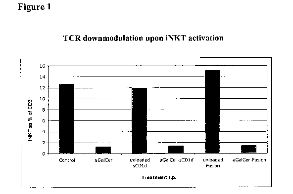
[0020] Figure 1: In vivo biological activity of aGalCer-loaded sCD1d and
CD1d-anti
HER2 fusion protein shown by the transient disappearance of liver iNKT cells
20
hours after i.p. injection with PBS (control), aGalCer 5p.g, or aGalCer/sCD1d
20pg
or aGalCer/CD1d-anti-HER2 fusion 4014 (loaded or unloaded). Frequency of iNKT
cells was measured by flow cytometry using CD 1 d-Tetramer-Extravidin-PE and
anti
CD3 FITC.
[0021] Figure 2: a Sustained IFNy production by liver and spleen iNKT cells
after
several injections of CD1d/anti-HER2 fusion. Liver and spleen lymphocytes were
isolated 20 minutes after the sixth injection of either PBS (control, white
bar),
aGalCer 0.41.tg (grey bar), or aGalCer/CD1d-anti-HER2 fusion protein 40[tg
(black
bar) and cultured for 1 hour in presence of Golgi Plug reagent. NKT cells were
then
CA 02678618 2009-08-17
WO 2008/103392 PCT/US2008/002256
- 8 -
stained with anti NK1.1-PE and anti CD3-FITC antibodies, fixed and stained for
intracellular IFN7 with anti lFNy-APC. Graph shows percentage of IFNy
producing
NKT cells (gated on NK1.1+ CD3+ cells). b Sustained IFNI, production by liver
NKT
cells after several injections i.v. and in vitro rechallenge with aGalCer or
aGalCer
loaded recombinant CD1d molecules. Liver lymphocytes were isolated after 5
injections i.v. of either PBS (Control), aGalCer, aGalCer/CD1d-anti-HER2
fusion
protein (Fusion) or aGalCer/sCD1d (sCD1d) and stimulated in vitro for 6 hours
in
presence of Golgi Plug as indicated. (PBS, white; aGalCer, light grey;
aGalCer/CD1d-anti-HER2 fusion, black; aGalCer/sCD1d, dark grey) Cells were
stained for FACS analysis as described in a.
[0022] Figure 3: iNKT expansion in blood during systemic treatment with
aGalCer
loaded recombinant CD1d molecules. Mice were bled after the third injection of
either PBS (control) aGalCer (0.4i.tg), aGalCer/CD1d-anti-Her2 fusion protein
(Fusion, 40 g), or aGalCer/sCD1d (sCD1d, 20pg). NKT cells were stained in
PBMCs using the CD1d-Tetramer-PE and anti CD3 FITC antibody. a representative
dot blot of one mouse from each group. b graph representing several mice per
group
expressed as CD1d tetramer positive and CD3+ cells as percentage of total
PBMC.
[0023] Figure 4: Sustained iNKT activation in vivo due to repeated
injections of
aGalCer/sCD1d prevents formation of lung metastases. Mice were treated 5 times
with PBS (Control, white), aGalCer (0.4pg, light grey), or aGalCer/sCD1d
(sCD1d,
20 g, black) and then grafted with 700,000 B16 wild type cells plus co-
injection of
the respective treatment. 2 naive groups were included that had no
pretreatment and
got only tumor cell graft + co-injection of aGalCer (striped grey) or
aGalCer/sCD1 d
(sCD1d, dark striped grey) respectively. Lung metastases formation was
analysed 2
weeks after graft with the ImageJ k-means clustering program and results are
expressed as percent of black metastatic surface over total lung surface. P
values for
aGalCer/sCD1d pretreated and naive+ocGalCer groups compared to control *P <
0.04, and compared to aGalCer pretreated group *P < 0.02.
[0024] Figure 5: Precoating experiment. B16-HER2 (a) and B16 wild type (B16
wt)
cells (b) were precoated for 1 hour with 0.4 g/m1 aGalCer, 40 g/m1
aGalCer/CD1d-
CA 02678618 2009-08-17
WO 2008/103392 PCT/US2008/002256
- 9 -
Her2 Fusion protein (Fusion), lOug/m1 Herceptin, or 2011g/m1 aGalCer/sCD1d
(sCD1d) and, with or without a previous wash, cells were injected i.v. Lung
metastases were analyzed 3 weeks after graft with the ImageJ k-means
clustering
program and results are expressed as percentage of black metastatic surface
over total
lung surface. * P <0.005 compared to PBS control.
10025] Figure 6: In vivo anti tumor activity - Systemic Treatment. a Mice
were
grafted i.v. with 700.000 B16-HER2 cells and i.v. treatment was started 48
hours
later. Mice were injected i.v. 5 times every 2 to 3 days with PBS (control),
aGalCer
(0.414), aGalCer/sCD1d (sCD1d, 2514), or aGalCer/CD1d-anti-HER2 fusion
(Fusion, 401.1s). Lung metastases were analysed after 3 weeks by the ImageJ k-
means
clustering program and expressed as percent metastatic surface over total lung
surface. Treatment with aGalCer/CD1d-anti-HER2 fusion protein significantly
inhibited the metastases formation. *P < 0.005 versus control, *P < 0.06
versus
aGalCer b Treatment was started 6 days after graft. Mice were injected i.v.
with
700,000 B16-HER2 cells and treatment with PBS (Control), aGalCer, or
aGalCer/CD1d-anti-HER2 fusion protein (Fusion) was started 6 days after.
graft.
Mice were treated 3x i.v. every 2-3 days and lung metastases were analyzed
after 3
weeks as described above. Only treatment with the CD id-anti HER2 fusion
protein
significantly reduced metastatic growth. *P < 0.01 versus control.
100261 Figure 7: Transactivation of NK, DCs and T cells by aGalCer/sCD1d or
aGalCer/CD1d-anti-HER2 fusion protein activated iNKT cells. a Increase of
liver
NK cell numbers 20 hours after i.p. treatment with either PBS (control, white
bar),
5[tg aGalCer (light grey bar), or 201.tg aGalCer-loaded sCD1d or 4014
aGalCer/CD1d-anti-HER2 fusion protein (aGalCer-sCD1d, dark grey and aGalCer-
Fusion, black bars, respectively). Cells were stained with anti NK1.1-PE and
anti
CD3-FITC and analysed by flow cytometry. NK cells are reported as NK1.1
positive/
CD3 negative population. b c Induction of DC maturation and T cell
proliferation by
aGalCer/sCD1d activated NKT cells in vivo. Splenocytes were isolated after
five
treatments i.v. with either PBS (control), aGalCer, or aGalCer/sCD1d and
cultured
for 4 days with GM-CSF and then 3 more days with either PBS (control, white
bars),
CA 02678618 2009-08-17
WO 2008/103392 PCT/US2008/002256
- 10 -
aGalCer (grey bars), or aGalCer/sCD1d (sCD1d, black bars). Cells were then
stained
with anti CD11c-FITC and biotinylated anti CD40 and Streptavidin-PE to detect
double positive mature DCs (c), or with anti NK1.1-PE and anti CD3-FITC to
detect
CD3 single positive T cells (d) by flow cytometry.
[0027] Figure 8: aGalCer loaded sCD ld acts as a strong adjuvant for the
expansion
of antigen-specific T cells upon active immunization. a kinetics of expansion
of H-
2Kb/OVA specific CTLs. Mice were primed with 200ug ovalburnin either as such
(i.v.) or with Montanide adjuvant (3:7 ratio, s.c.) or with 1 [tg aGalCer or
20 g
aGalCer/sCD1d (i.v.). Mice were bled every 5-7 days and PBMC were stained with
H-2Kb/OVA tetramer+anti-CD8. Results are expressed as tetramer+ and CD8+ NKT
cells as a percentage of total CD8+ T cells. Mice were boosted with OVA
peptide
(20 g) on day 22 employing the same adjuvants and route of injection as for
primary
immunization. b Ten days later, mice were sacrificed and splenocytes were
cultured
another five days either with no stimuli or with the same stimuli and
adjuvants as for
the in vivo boost. Frequency of OVA specific T cells was measured as in a. c
The
presence of mature DC in the same cultures as in b was analyzed by surface
staining
with anti CD11 c and anti CD40 antibodies.
Detailed Description Of The Invention
[0028] The present invention provides compositions and methods which are
useful for
modulating, i.e., either eliciting, inhibiting, or stimulating, an immune
response. The
compounds comprise one or more CD1d complexes comprising a ceramide-like
glycolipid antigen bound to a soluble CD1d polypeptide fragment associated
with
beta-2 microglobulin. In certain embodiments, the soluble CD1d complexes of
the
present invention are non-specific, i.e., they are not targeted to any
particular tissue,
cell, or cell surface marker. Soluble CD1d complexes for use in the methods of
the
present invention modulate an immune response by affecting the activity of
CD1d-
restricted natural killer T ("NKT") cells. Soluble CD1d complexes as described
herein are useful for stimulating desirable immune responses, for example,
immune
responses against infectious agents or cancer; or for inhibiting undesirable
immune
CA 02678618 2009-08-17
WO 2008/103392 PC T/US2008/002256
- 11 -
responses, such as allergic responses, allograft rejections, and autoimmune
diseases.
In certain embodiments, soluble CD1d complexes of the present invention are
administered with an immunogen and function as an adjuvant by, for example,
increasing or modulating the immune response to the immunogen.
Definitions
[0029] It is to be noted that the term "a" or "an" entity refers to one or
more of that
entity; for example, "a vector" is understood to represent one or more
vectors. As
such, the terms "a" (or "an"), "one or more," and "at least one" can be used
interchangeably herein.
[0030] As used herein, the term "polypeptide" is intended to encompass a
singular
"polypeptide" as well as plural "polypeptides," and refers to a molecule
composed of
monomers (amino acids) linearly linked by amide bonds (also known as peptide
bonds). The term "polypeptide" refers to any chain or chains of two or more
amino
acids, and does not refer to a specific length of the product. Thus, peptides,
dipeptides, tripeptides, oligopeptides, "protein," "amino acid chain," or any
other term
used to refer to a chain or chains of two or more amino acids, are included
within the
definition of "polypeptide," and the term "polypeptide" may be used instead
of, or
interchangeably with any of these terms. The term "polypeptide" is also
intended to
refer to the products of post-expression modifications of the polypeptide,
including
without limitation glycosylation, acetylation, phosphorylation, amidation,
derivatization by known protecting/blocking groups, proteolytic cleavage, or
modification by non-naturally occurring amino acids. A polypeptide may be
derived
from a natural biological source or produced by recombinant technology, but is
not
necessarily translated from a designated nucleic acid sequence. It may be
generated
in any manner, including by chemical synthesis.
[0031] A polypeptide of the invention may be of a size of about 3 or more,
5 or more,
or more, 20 or more, 25 or more, 50 or more, 75 or more, 100 or more, 200 or
more, 500 or more, 1,000 or more, or 2,000 or more amino acids. Polypeptides
may
have a defined three-dimensional structure, although they do not necessarily
have
such structure. Polypeptides with a defined three-dimensional structure are
referred to
CA 02678618 2009-08-17
WO 2008/103392 PCT/US2008/002256
- 12 -
as folded, and polypeptides which do not possess a defined three-dimensional
structure, but rather can adopt a large number of different conformations, and
are
referred to as unfolded. As used herein, the term glycoprotein refers to a
protein
coupled to at least one carbohydrate moiety that is attached to the protein
via an
oxygen-containing or a nitrogen-containing side chain of an amino acid
residue, e.g.,
a serine residue or an asparagine residue.
[00321 By an "isolated" polypeptide or a fragment, variant, or derivative
thereof is
intended a polypeptide that is not in its natural milieu. No particular level
of
purification is required. For example, an isolated polypeptide can be removed
from
its native or natural environment. Recombinantly produced polypeptides and
proteins
expressed in host cells are considered isolated for purposed of the invention,
as are
native or recombinant polypeptides which have been separated, fractionated, or
partially or substantially purified by any suitable technique.
[0033] Also included as polypeptides of the present invention are
fragments,
derivatives, analogs, or variants of the foregoing polypeptides, and any
combination
thereof. The terms "fragment," "variant," "derivative" and "analog" when
referring to
polypeptides of the present invention include any polypeptides that retain at
least
some of the biological, antigenic, or immunogenic properties of the
corresponding
native polypeptide. Fragments of polypeptides of the present invention include
proteolytic fragments, as well as deletion fragments, in addition to other
specific
fragments discussed elsewhere herein. Variants of polypeptides of the present
invention include fragments as described above, and also polypeptides with
altered
amino acid sequences due to amino acid substitutions, deletions, or
insertions.
Variants may occur naturally or be non-naturally occurring. Non-naturally
occurring
variants may be produced using art-known mutagenesis techniques. Variant
polypeptides may comprise conservative or non-conservative amino acid
substitutions, deletions or additions. Derivatives of polypeptides of the
present
invention, are polypeptides which have been altered so as to exhibit
additional
features not found on the native polypeptide. Examples include fusion
proteins.
Variant polypeptides may also be referred to herein as "polypeptide analogs."
As
used herein a "derivative" of a polypeptide refers to a subject polypeptide
having one
CA 02678618 2009-08-17
WO 2008/103392 PCT/US2008/002256
- 13 -
or more residues chemically derivatized by reaction of a functional side
group. Also
included as "derivatives" are those peptides which contain one or more
naturally
occurring amino acid derivatives of the twenty standard amino acids. For
example, 4-
hydroxyproline may be substituted for proline; 5-hydroxylysine may be
substituted
for lysine; 3-methylhistidine may be substituted for histidine; homoserine may
be
substituted for serine; and ornithine may be substituted for lysine.
[0034] The term "polynucleotide" is intended to encompass a singular
nucleic acid as
well as plural nucleic acids, and refers to an isolated nucleic acid molecule
or
construct, e.g., messenger RNA (mRNA), virally-derived RNA, or plasmid DNA
(pDNA). A polynucleotide may comprise a conventional phosphodiester bond or a
non-conventional bond (e.g., an amide bond, such as found in peptide nucleic
acids
(PNA)). The term "nucleic acid" refers to any one or more nucleic acid
segments,
e.g., DNA or RNA fragments, present in a polynucleotide. By "isolated" nucleic
acid
or polynucleotide is intended a nucleic acid molecule, DNA or RNA, which has
been
removed from its native environment. For example, a recombinant polynucleotide
encoding a therapeutic polypeptide contained in a vector is considered
isolated for the
purposes of the present invention. Further examples of an isolated
polynucleotide
include recombinant polynucleotides maintained in heterologous host cells or
purified
(partially or substantially) polynucleotides in solution. Isolated RNA
molecules
include in vivo or in vitro RNA transcripts of the present invention, as well
as positive
and negative strand forms, and double-stranded forms, of pestivirus vectors
disclosed
herein.
[0035] Isolated polynucleotides or nucleic acids according to the present
invention
further include such molecules produced synthetically. In addition, a
polynucleotide
or a nucleic acid may be or may include a regulatory element such as a
promoter,
ribosome binding site, or a transcription terminator.
[0036] As used herein, a "coding region" is a portion of nucleic acid which
consists of
codons translated into amino acids. Although a "stop codon" (TAG, TGA, or TAA)
is
not translated into an amino acid, it may be considered to be part of a coding
region, if
present, but any flanking sequences, for example promoters, ribosome binding
sites,
transcriptional terminators, introns, 5' and 3' non-translated regions, and
the like, are
CA 02678618 2009-08-17
WO 2008/103392 PCT/US2008/002256
- 14 -
not part of a coding region. Two or more coding regions of the present
invention can
be present in a single polynucleotide construct, e.g., on a single vector, or
in separate
polynucleotide constructs, e.g., on separate (different) vectors. Furthermore,
any
vector may contain a single coding region, or may comprise two or more coding
regions, e.g., a vector of the present invention may encode one or more
polyproteins,
which are post- or co-translationally separated into the final proteins via
proteolytic
cleavage. In addition, a vector, polynucleotide, or nucleic acid of the
invention may
encode heterologous coding regions, either fused or unfused to a first or
second
nucleic acid encoding of the
invention, or variant or derivative thereof.
Heterologous coding regions include without limitation specialized elements or
motifs, such as a secretory signal peptide or a heterologous functional
domain.
100371 In certain embodiments, the polynucleotide or nucleic acid is
DNA. In the
case of DNA, a polynucleotide comprising a nucleic acid, which encodes a
polypeptide normally may include a promoter and/or other transcription or
translation
control elements operably associated with one or more coding regions. An
operable
association is when a coding region for a gene product, e.g., a polypeptide,
is
associated with one or more regulatory sequences in such a way as to place
expression
of the gene product under the influence or control of the regulatory
sequence(s). Two
DNA fragments (such as a polypeptide coding region and a promoter associated
therewith) are "operably associated" if induction of promoter function results
in the
transcription of mRNA encoding the desired gene product and if the nature of
the
linkage between the two DNA fragments does not interfere with the ability of
the
expression regulatory sequences to direct the expression of the gene product
or
interfere with the ability of the DNA template to be transcribed. Thus, a
promoter
region would be operably associated with a nucleic acid encoding a polypeptide
if the
promoter was capable of effecting transcription of that nucleic acid. The
promoter
may be a cell-specific promoter that directs substantial transcription of the
DNA only
in predetermined cells. Other transcription control elements, besides a
promoter, for
example enhancers, operators, repressors, and transcription termination
signals, can
be operably associated with the polynucleotide to direct cell-specific
transcription.
Suitable promoters and other transcription control regions are disclosed
herein.
CA 02678618 2009-08-17
WO 2008/103392 PCT/US2008/002256
- 15 -
[0038] A variety of transcription control regions are known to those
skilled in the art.
These include, without limitation, transcription control regions, which
function in
vertebrate cells, such as, but not limited to, promoter and enhancer segments
from
cytomegaloviruses (e.g., the immediate early promoter, in conjunction with
intron-A),
simian virus 40 (e.g., the early promoter), and retroviruses (such as, e.g.,
Rous
sarcoma virus). Other transcription control regions include those derived from
vertebrate genes such as actin, heat shock protein, bovine growth hormone and
rabbit
13-globin, as well as other sequences capable of controlling gene expression
in
eukaryotic cells. Additional suitable transcription control regions include
tissue-
specific promoters and enhancers as well as lymphokine-inducible promoters
(e.g.,
promoters inducible by interferons or interleukins).
[0039] Similarly, a variety of translation control elements are known to
those of
ordinary skill in the art. These include, but are not limited to ribosome
binding sites,
translation initiation and termination codons, and elements derived from viral
systems
(particularly an internal ribosome entry site, or IRES, also referred to as a
CITE
sequence).
[0040] In other embodiments, a polynucleotide of the present invention is
RNA, for
example, in the form of messenger RNA (rnRNA). RNA of the present invention
may
be single stranded or double stranded.
[0041] Polynucleotide and nucleic acid coding regions of the present
invention may
be associated with additional coding regions which encode secretory or signal
peptides, which direct the secretion of a polypeptide encoded by a
polynucleotide of
the present invention. According to the signal hypothesis, proteins secreted
by
mammalian cells have a signal peptide or secretory leader sequence which is
cleaved
from the mature protein once export of the growing protein chain across the
rough
endoplasmic reticulum has been initiated. Those of ordinary skill in the art
are aware
that polypeptides secreted by vertebrate cells generally have a signal peptide
fused to
the N-terminus of the polypeptide, which is cleaved from the complete or "full
length"
polypeptide to produce a secreted or "mature" form of the polypeptide. In
certain
embodiments, the native signal peptide, e.g., an immunoglobulin heavy chain or
light
chain signal peptide is used, or a functional derivative of that sequence that
retains the
- 16 -
ability to direct the secretion of the polypeptide that is operably associated
with it.
Alternatively, a heterologous mammalian signal peptide, or a functional
derivative
thereof, may be used. For example, the wild-type leader sequence may be
substituted
with the leader sequence of human tissue plasminogen activator (TPA) or mouse
13-
glucuronidase.
[0042] The term "construct" refers to an engineered vector.
100431 The term "artificial" refers to a synthetic, or non-host cell
derived
composition, e.g., a chemically-synthesized oligonucleotide.
100441 As discussed in more detail below, a functional antigen-loaded
soluble
fragment of a CD1d polypeptide, including both CD1d and 13-2 microglobulin
subunits, is referred to herein as a "soluble CD I d complex." The antigen to
be loaded
onto the CD1d polypeptide is a glycolipid, typically a ceramide-like
glycolipid, e.g.,
an alpha-galctosylcerainide, e.g., a-GalCer. "Ceramide-like glycolipids," as
referred
to herein include glyeolipids with a-linked galactose or glucose. Examples of
glycolipid antigens which bind to CD1d are found, e.g., in Porcelli, U.S.
Patent Appl.
Publ. No. 2006/0052316, Tsuji, U.S. Patent Appl. Publ. No. 2006/0211856,
Jiang,
U.S. Patent Appl. Publ. No. 2006/0116331, Hirokazu et al., U.S. Patent Appl.
Publ.
No. 2006/0074235, Tsuji et al, U.S. Patent Appl. Publ. No. 2005/0192248,
Tsuji, U.S.
Patent Application No. 2004/0127429, and Tsuji et al., U.S. Patent Application
No.
2003/0157135.
[0045] The term "non-specific soluble CD1d complex" refers to a soluble
CD1d
complex which has not been engineered to be targeted to any specific organ,
tissue,
cell, or cell-surface molecule. A "non-specific soluble CD1d complex" is,
however,
capable of interacting with NKT cells, in a way similar to that in which a
cell-surface-
expressed CD1d molecule, when loaded with antigen, would interact. In contrast
to a
"non-specific soluble CDl d complex" is a "targeted CD1d complex," which is
fused
or conjugated to an antibody or other binding molecule, thus targeting the
complex to
a specific organ, tissue, cell, or cell-surface marker. Targeted CD1d
complexes exert
their effect on NKT cells locally, e.g., in the vicinity of a tumor. See,
e.g., Bruno et
al. U.S. Patent Appl. Publ. No. 2006/0269540.
CA 2678618 2018-07-09
CA 02678618 2009-08-17
WO 2008/103392 PCT/US2008/002256
- 17 -
[0046] Antibodies are constructed of one, or several, units, each of which
consists of
two heavy (H) polypeptide chains and two light (L) polypeptide chains. The H
and L
chains are made up of a series of domains. The L chains, of which there are
two
major types (ic and X,), consists of two domains. The H chains are of several
types,
including , 8, and y (of which there are several subclasses), a and c. In
humans,
there are eight genetically and structurally identified antibody classes and
subclasses
as defined by heavy chain isotypes: IgM, IgD, IgG3, IgG1, IgG2, IgG4, IgE, and
IgA.
Further, for example, "IgG" means an antibody of the G class, and that, "IgG1
" refers
to an IgG molecules of subclass 1 of the G class. IgG1 antibodies, like all
antibodies
of the IgG class, are comprised of 4 domains, one of which is variable and the
other 3
are constant. An Fab antibody fragment is comprised of an intact light chain
and a
truncated heavy chain that each comprise two domains, one variable and one
constant.
[0047] As used herein, the term "antibody" (Ab) or "monoclonal antibody"
(MAb) is
meant to include intact molecules as well as antibody portions (such as, for
example,
Fab and F(ab')2 portions and Fv fragments) which are capable of specifically
binding
to a cell surface marker. Such portions are typically produced by proteolytic
cleavage, using enzymes such as papain (to produce Fab portions) or pepsin (to
produce F(ab')2 portions). Especially preferred in the compounds of the
invention are
Fab portions. Alternatively, antigen-binding portions can be produced through
the
application of recombinant DNA technology.
[0048] In addition, the immunoglobin may be a single chain antibody
("SCA"). These
may consist of single chain Fv fragments ("scFv") in which the variable light
("V[L]")
and variable heavy ("V[H]") domains are linked by a peptide bridge or by
disulfide
bonds. Also, the immunoglobulin may consist of single V[H]domains (dAbs) which
possess antigen-binding activity. See, e.g., G. Winter and C. Milstein, Nature
349:295
(1991); R. Glockshuber et al., Biochemistry 29:1362 (1990); and, E. S. Ward et
al.,
Nature 341:544 (1989).
[0049] Also preferred for use in the present invention are chimeric
monoclonal
antibodies, preferably those chimeric antibodies having specificity toward a
tumor
associated surface membrane antigen, a surface membrane antigen of a tissue or
organ
affected by autoimmune disease, or an antigen of a pathogen infected cell. As
used in
- 18 -
this example, the term "chimeric antibody" refers to a monoclonal antibody
comprising a variable region, i.e. binding region, from one source or species
and at
least a portion of a constant region derived from a different source or
species, usually
prepared by recombinant DNA techniques.
100501 Encompassed by the term "chimeric antibody" is the concept of
"humanized
antibody", that is those antibodies in which the framework or
"complementarily"
determining regions ("CDR") have been modified to comprise the CDR of an
imrnunoglobulin of different specificity as compared to that of the parent
imrnunoglobulin. In certain embodiments, a murine CDR is grafted into the
framework region of a human antibody to prepare the "humanized antibody". See,
e.g., L. Riechmann et al., Nature 332:323 (1988); M. S. Neuberger et at,
Nature
314:268 (1985).
(0051] In other embodiments, fully human antibodies or fragments thereof
are used in
the compositions and methods of the invention, preferably those fully human
antibodies having specificity toward a tumor associated surface membrane
antigen , a
surface membrane antigen of a tissue or organ affectedhy autoimmune disease,
or an
antigen of a pathogen infected cell. Methods have been described for selection
of
fully human antibodies in human innnunoglobulin transgenic mice, from
libraries of
human immunoglobulin genes constructed in phage and expressed in bacteria or
constructed in a mammalian viral expression vector for expression in mammalian
cells, and from human hybridoma cells. A method for selection of fully human
antibodies from libraries of human irnmunoglobulin genes constructed in
vaccinia
virus is described in Zauderer, M. et at. WO 01/72995, published 4 October
2001.
[0052] In certain embodiments, targeted CD1d complexes of the present
invention
comprise, instead of, or in addition to an antibody, a specific binding
molecule, e.g., a
receptor or ligand that has a matching or counterpart ligand or receptor
expressed on a
cell surface of a target cell. In these embodiments, the targeted CD1d complex
comprises a ligand or receptor specific for a cell surface marker. Examples
include:
CD4 coupled to CD1d for interaction with HIV infected cells; chemokine or
CA 2678618 2018-07-09
CA 02678618 2009-08-17
WO 2008/103392 PC T/US2008/002256
- 19 -
chemokine receptor coupled to CD 1 d for interaction with DC subset; or
heregulins
coupled to CD1d for interaction with ErbB2 positive tumor cells.
[0053] In one embodiment, the antibody is specific for a cell surface
marker of a
tumor cell. In another embodiment, the antibody is specific for a cell surface
marker
of a CD id-restricted NKT cell. In another embodiment, the antibody is
specific for a
cell surface marker of a target tissue of autoimmune disease or inflammatory
response. In another embodiment, the antibody is specific for an infectious
agent or a
cell surface marker of an infected cell or tissue.
[0054] In another embodiment, the antibody is specific for a cell surface
marker of a
professional antigen presenting cell, e.g., a dendritic cell.
[0055] The term "antigen" and the related term "antigenic" as used herein
refers to a
substance that binds specifically to an antibody or to a T-cell receptor.
[0056] The term "immunogen" and the related term "immunogenic" as used
herein
refers to the ability to induce an immune response, including an antibody
and/or a
cellular immune response in an animal, preferably a mammal. It is quite likely
that an
immunogen will also be antigenic, but an "antigen," because of its size or
conformation, may not necessarily be an "immunogen." An "immunogenic
composition" induces an immune response in a subject, e.g., antibodies that
specifically recognize one or more antigens, contained within that
"immunogenic
composition."
[0057] The term "immune response" is meant to include any activity of cells
of the
immune system in response to an antigen or immunogen. Such activities include,
but
are not limited to production of antibodies, eytotoxicity, lymphocyte
proliferation,
release of cytokines, inflammation, phagocytosis, antigen presentation, and
the like.
An immune response which is highly specific to a given antigen or immunogen,
e.g.,
production of specific antibodies or production of specific T lymphocytes is
referred
to herein as an "adaptive immune response." An immune response which is not
specific to a given antigen, e.g., release of cytokines by NK and NKT cells,
is referred
to herein an "innate immune response." Examples of immune responses include an
antibody response or a cellular, e.g., cytotoxic T-cell, response.
CA 02678618 2009-08-17
WO 2008/103392 PCT/US2008/002256
- 20 -
[0058] The terms "protective immune response" or "therapeutic immune
response"
refer to an immune response to an immunogen which in some way prevents or at
least
partially arrests disease symptoms, side effects or progression. By
"protective" is
meant that the immune response is induced in a subject animal which has not
contracted a disease, where the immune response alleviates, reduces, moderates
or, in
some cases fully prevents disease symptoms if the animal later contracts or is
suceptible to that disease. By "therapeutic" is meant that the immune response
is
induced in a subject animal which has the disease, where the immune response
alleviates, reduces, moderates, or in some cases fully eliminates disease
symptoms.
100591 The term "modulating an immune response" is meant to refer to any
way in
which a given immune response is increased, decreased, or changed by a
composition
or treatment relative to the immune response without that composition or
treatment.
For example, use of an adjuvant to increase an immune response to an antigen
is
considered modulation of that immune response. Decrease in an immune response,
e.g., prevention of autoimmunity, is also a modulation. In addition, changing
an
immune response, e.g., from a TH2 response to a TH1 response, is a modulation
of an
immune response.
[0060] The term "anergy" refers to a specific kind if immune modulation, in
which
certain cells of the immune system are rendered non-responsive to antigen
stimulus.
An example would be the ability of free a-GalCer, upon multiple
administrations to
an animal, to render the NKT cells of that animal non-responsive to stimulus,
e.g.,
unable to proliferate or produce cytokines.
[0061] The term "adjuvant" refers to any material having the ability to (1)
alter or
increase the immune response to a particular antigen or (2) increase or aid an
effect of
a pharmacological agent. In certain embodiments, a soluble CD1d complex of the
present invention, e.g., a non-specific soluble CD1d complex, functions as an
adjuvant upon administration with an immunogen. Other suitable adjuvants
include,
but are not limited to, cytokines and growth factors; bacterial components
(e.g.,
endotoxins, in particular superantigens, exotoxins and cell wall components);
aluminum-based salts; calcium-based salts; silica; polynucleotides; toxoids;
serum
CA 02678618 2009-08-17
WO 2008/103392 PCT/US2008/002256
- 21 -
proteins, viruses and virally-derived materials, poisons, venoms,
imidazoquiniline
compounds, poloxamers, and cationic lipids.
[0062] A great variety of materials have been shown to have adjuvant
activity through
a variety of mechanisms. Any compound which may increase the expression,
antigenicity or immunogenicity of the polypeptide is a potential adjuvant.
Potential
adjuvants include, but are not limited to: inert carriers, such as alum,
bentonite, latex,
and acrylic particles; pluronic block polymers, such as TiterMax (block
copolymer
CRL-8941, squalene (a metabolizable oil) and a microparticulate silica
stabilizer),
depot formers, such as Freunds adjuvant, surface active materials, such as
saponin,
lysolecithin, retinal, Quil A, liposomes, and pluronic polymer formulations;
macrophage stimulators, such as bacterial lipopolysaccharide; alternate
pathway
complement activators, such as insulin, zymosan, endotoxin, and levamisole;
and non-
ionic surfactants, such as poloxamers, poly(oxyethylene)-poly(oxypropylene)
tri-
block copolymers.
[0063] In certain embodiments, the adjuvant is a cytokine. A composition of
the
present invention can comprise one or more cytokines, chemokines, or compounds
that induce the production of cytokines and chemokines. Examples include, but
are
not limited to granulocyte macrophage colony stimulating factor (GM-CSF),
granulocyte colony stimulating factor (G-CSF), macrophage colony stimulating
factor
(M-CSF), colony stimulating factor (CSF), erythropoietin (EPO), interleukin 2
(IL-2),
interleukin-3 (IL-3), interleukin 4 (IL-4), interleukin 5 (IL-5), interleukin
6 (IL-6),
interleukin 7 (IL-7), interleukin 8 (IL-8), interleukin 10 (IL-10),
interleukin 12 (IL-
12), interleukin 15 (IL-15), interleukin 18 (IL-18), interferon alpha (1FNa,),
interferon
beta (IFNI3), interferon gamma (IFNT), interferon omega (IFN(o), interferon
tau
(IFNr), interferon gamma inducing factor I (IGIF), transforming growth factor
beta
(TGF-13), RANTES (regulated upon activation, normal T-cell expressed and
presumably secreted), macrophage inflammatory proteins (e.g., MIP-1 alpha and
MEP-1 beta), Leishmania elongation initiating factor (LEIF), and Flt-3 ligand.
[0064] The term "vertebrate" is intended to encompass a singular
"vertebrate" as well
as plural "vertebrates" and comprises mammals and birds, as well as fish,
reptiles, and
amphibians.
CA 02678618 2009-08-17
WO 2008/103392 PC T/US2008/002256
- 22 -
[0065] The term "mammal" is intended to encompass a singular "mammal" and
plural
"mammals," and includes, but is not limited to humans; primates such as apes,
monkeys (e.g., owl, squirrel, cebus, rhesus, African green, patas, cynomolgus,
and
cercopithecus), orangutans, baboons, gibbons, and chimpanzees; canids such as
dogs
and wolves; felids such as cats, lions, and tigers; equines such as horses,
donkeys, and
zebras, food animals such as cows, pigs, and sheep; ungulates such as deer and
giraffes; ursids such as bears; and others such as rabbits, mice, ferrets,
seals, whales.
In particular, the mammal can be a human subject, a food animal or a companion
animal.
[0066] The term "bird" is intended to encompass a singular "bird" and
plural "birds,"
and includes, but is not limited to feral water birds such as ducks, geese,
terns,
shearwaters, and gulls; as well as domestic avian species such as turkeys,
chickens,
quail, pheasants, geese, and ducks. The term "bird" also encompasses passerine
birds
such as starlings and budgerigars.
Soluble CDId Complexes
[0067] As mentioned above, soluble CD I d complexes of the present
invention can be
used both to prevent a disease, and also to therapeutically treat a disease.
In
individuals already suffering from a disease, the present invention is used to
further
stimulate or modulate the immune system of the animal, thus reducing or
eliminating
the symptoms associated with that disease or disorder. As defined herein,
"treatment"
refers to the use of one or more compositions of the present invention to
prevent, cure,
retard, or reduce the severity of given disease symptoms in an animal, and/or
result in
no worsening of the disease over a specified period of time in an animal which
has
already contracted the disease and is thus in need of therapy. The term
"prevention"
refers to the use of one or more compositions of the present invention to
generate
immunity in an animal which has not yet contracted a disease, thereby
preventing or
reducing disease symptoms if the vertebrate is later disposed to develop that
disease.
The methods of the present invention therefore may be referred to as
therapeutic
methods or preventative or prophylactic methods. It is not required that any
composition of the present invention provide total immunity to a disease agent
or
CA 02678618 2014-12-10
- 23 -
totally cure or eliminate all disease symptoms. As used herein, an "animal in
need of
therapeutic and/or preventative immunity" refers to an individual for whom it
is
desirable to treat, i.e., to prevent, cure, retard, or reduce the severity of
certain disease
symptoms, and/or result in no worsening of disease over a specified period of
time.
[00681 The present invention provides methods of modulating an immune
response
comprising administering to an animal a composition which comprises an antigen-
loaded soluble CD1d molecule, which interact with, and thereby affects, the
activity
of CD1d-restricted NKT cells. The monomorphie CD1d molecule is suitable for
activation of a broad spectrum of CD1d-restricted NKT in an entire species.
Soluble
CD1d molecules, which include both CD1d and D-2 microglobulin subunits, are
loaded with a ceramide-like glycolipid antigen, for example, a-GalCer, to
produce a
soluble CD1d complex. Soluble CD1d complexes of the present invention, e.g.,
non-
specific soluble CD1d complexes, can be the primary or only active ingredient
in a
composition of the present invention, for example for treatment of cancer. hi
other
embodiments, soluble CD1d complexes of the present invention may be used as an
adjuvant in combination with a specific immunogen, thereby, stimulating,
increasing,
modulating, or otherwise altering an immune response to that immunogen
relative to
administration of the immunogen without the soluble CD Id complex. The present
invention further encompasses pharmaceutical compositions which comprise an
immunogen and a soluble CD1d complex adjuvant.
[00691 Moreover, soluble CD1d complexes of the present invention may be
used as a
= diagnostic or therapeutic agent not only for cancer and infectious
diseases but also for
a large class of autoimmune and inflammatory diseases that result from a
failure to
down modulate cell-mediated immune responses.
10070] Soluble CD1d complexes for use in the methods of the present
invention
comprise a soluble fragment of a CD1d polypeptide sufficient to bind 32-
microglobulin as well as a ceramide-like glycolipid antigen, a 132-
microglobulin
polypeptide, and a ceramide-like glycolipid antigen, e.g., a-GalCer. The
ceramide-
hke glycolipid antigen is bound in the antigen binding groove of the CD1d
molecule.
100711 As taught by WO 9964597, published 16 December 1999,
it is possible to introduce mutations into 132-mieroglobolin that
CA 02678618 2009-08-17
WO 2008/103392 PCT/US2008/002256
- 24 -
increase affinity for the class I heavy chain so as to facilitate assembly and
increase
stability of the CD1d complex in the fusion protein. In certain embodiments, a
soluble CD1d polypeptide is linked to p2-microglobulin as a fusion protein. In
certain
embodiments, the P2-microg1obulin polypeptide is linked via its C-terminus to
the N-
terminus of the soluble CD polypeptide. Such fusion constructs can be made
using
conventional recombinant nucleic acid techniques. The fusion may be direct or
may
contain spacers. A short linker amino acid sequence may be inserted between
the
CD1d polypeptide and the 02-microglobulin polypeptide. If a linker sequence is
included, this sequence will preferably contain at least 3 and not more than
30 amino
acids. More preferably, the linker is about 5, 6, 7, 8, 9, 10, 11, 12, 13, 14,
15, 20, or
25 amino acids long. Generally, the linker consists of short glycine/serine
spacers, but
any known amino acid may be used. Examples of linkers known to those skilled,
in
the art include (G1y4Ser)3 (SEQ ID NO:3) and (Gly4Ser)2Gly3AlaSer (SEQ 1D
NO:4).
[0072] Alternatively, the CD1d and 32-microglobu1in polypeptides may be
chemically linked. A number of reagents capable of cross-linking proteins are
known
in the art, illustrative entities include: azidobenzoyl hydrazide, N44-(p-
azidosalicylamino)buty1]-3'42'-pyridyldithio] propionamide), bis-
sulfosuccinimidyl
suberate, dimethyladipimidate, disuccinimidyltartrate, N-y-
maleimidobutyryloxysuccinimide ester, N-hydroxy
sulfosuccinimidy1-4-
azidobenzoate, N-succinimidyl [4-azidopheny1]-1,3'-dithiopropionate, N-
succinimidyl
[4-iodoacetyl]aminobenzoate, glutaraldehyde, formaldehyde and succinimidyl 44N-
maleimidomethyl] cyclohexane-l-carboxylate.
[0073] In certain embodiments, multiple CD complexes are linked
together through
a multivalent compound. The CD1d complexes may be linked to the multivalent
compound through any site. In a preferred embodiment soluble CD1d polypeptides
are linked to the multivalent compound through the CD1d carboxyl terminus.
These
compounds typically comprise 2 or more CD 1 d complexes. The compounds may
comprise 2, 3, 4, 5, 6, 7, 8, 9 or 10 CD1d complexes.
[0074] Examples of multivalent compounds are chicken avidin or
streptavidin (Shin,
S.U. et al., I Immunology 158: 4797-4804 (1997)) to which biotinylated CD1d
CA 02678618 2009-08-17
WO 2008/103392 PCT/US2008/002256
- 25 -
complexes are bound (Altman, J. et al, Science 274:94-96 (1996); Boniface,
J.J. et al.,
Immunity 9:459-66 (1998)); or a leucine zipper system.
[0075] Alternatively, CD 1 d and 132-microglobulin polypeptides can be
genetically
modified by including sequences encoding amino acid residues with chemically
reactive side chains such as Cys or His. Such amino acids with chemically
reactive
side chains may be positioned in a variety of positions on the CD1d and 32-
microglobulin polypeptides, preferably distal to the site(s) where p2-
microglobulin
and CD1d interact. Suitable side chains can be used to chemically link two or
more
assembled CD complexes to a suitable dendrimer particle. Dendrimers are
synthetic
chemical polymers that can have any one of a number of different functional
groups
on their surface (D. Tamalia, Aldrichimica Acta 26:91:101 (1993)). Exemplary
dendrimers for use in accordance with the present invention include e.g. E9
starburst
polyamine dendrimer and E9 combburst polyamine dendrimer, which can link
cysteine residues. The CD 1 d and/or 132-microglobulin polypeptides are
modified to
introduce a cysteine residue at the carboxyl terminus. Following synthesis in
eukaryotic cells, a complete cysteine modified CD1d complex is assembled in
vitro.
Cysteine modified CD1d and/or f32-microglobulin polypeptides will react with
the
maleimide groups on the various peptide backbones with either two, three, or
four
modified lysine residues for formation of CD1d dimers, trimers, and tetramers.
[0076] Cochran, J.R. et al., Immunity /2:241-50 (2000) describe the use of
chemically
synthesized peptide-based cross-linking reagents in which two or more thiol-
reactive
maleimide groups are linked to lysine side chains in a flexible peptide of 8
to 19
residues containing glycine, serine, and glutamic acid in addition to the
modified
lysine residues. Isolated CD1d and/or 32-microglobulin polypeptides are
modified to
introduce a cysteine residue at the carboxyl terminus. Cysteine modified CD1d
and/or 132-microglobulin polypeptides react with the maleimide groups on the
various
peptide backbones with either two, three, or four modified lysine residues for
formation of dimers, trimers, and tetramers.
[0077] Another means of assembling polymeric CD1d complexes is to exploit
the
observation that defined amino acid substitutions in the GCN4 leucine zipper
dimerization domain results in formation of highly stable trimeric and
tetrameric
CA 02678618 2009-08-17
WO 2008/103392 PCT/US2008/002256
- 26 -
structures of the synthetic peptide (Harbury, P.B. et al., Science 262:1401-7
(1993)).
For example, multivalent CD1d complexes are constructed by attaching a
modified
GCN4-zipper to the carboxyl terminus of soluble CD1d or 02-microglobulin
polypeptides. Tetravalent CD1d complexes could be assembled from a mixture of
CD1d complexes each separately fused to a modified GCN4-zipper motif.
[0078] The attachment site(s) on a soluble CD1d complex for binding to a
multivalent
compound may be naturally occurring, or may be introduced through genetic
engineering. The site will be a specific binding pair member or one that is
modified to
provide a specific binding pair member, where the complementary pair has a
multiplicity of specific binding sites. Binding to the complementary binding
member
can be a chemical reaction, epitope-receptor binding or hapten-receptor
binding where
a hapten is linked to the subunit chain.
[0079] In a preferred embodiment, the CD1d and/or P2 microglobulin contain
an
amino acid sequence which is a recognition site for a modifying enzyme.
Modifying
enzymes include BirA, various glycosylases, farnesyl protein transferase, and
protein
kinases. The group introduced by the modifying enzyme, e.g. biotin, sugar,
phosphate,
farnesyl, etc. provides a complementary binding pair member, or a unique site
for
further modification, such as chemical cross-linking, biotinylation, etc. that
will
provide a complementary binding pair member.
[0080] For example, the CD1d molecule may be engineered to contain a site
for
biotinylation, for example a BirA-dependent site. The multivalent compound can
be
avidin or can be linked to avidin either directly or indirectly.
100811 Both the soluble CD1d and p2-microglobulin polypeptides useful in
the
present invention may be autologous to any mammalian or avian species, for
example,
primates (esp. humans), rodents, rabbits, equines, bovines, canines, felines,
etc. 02-
microglobulin is typically not inflammatory in vivo. However, it is preferable
to
employ 132-microglobulin derived from the same species as is to be treated so
as to
reduce the risk of a xenogeneic immune response.
- 27 -
Soluble CD1d Polypeptides
[0082] In certain embodiments, the non-specific CD1d complex comprises
soluble
CD1d polypeptides and polypeptide fragments, which associates with P2-
microglobulin and binds antigen, e.g., ceramide-like glycolipid. The CD1d
molecule
is a member of the family of major histocompatibility complex (MN+HC) antigen-
like glycoproteins which associate with 132-microglobulin and are expressed at
the
surface of cortical thymocytes, B cells, dendritic cells, Langerhans cells in
the skin,
and gastrointestinal epithelial cells. CD1d is mainly expressed on dendritic
cells or
epithelial cells of the gastrointestinal tract. The CD1 family members are
involved in
the presentation of glycolipids as antigens. In particular, CD1d regulates
cytokine
tone through activation of a distinct subset of T-lymphocytes, namely NK1 T
cells
which secrete IL-4 and INF-y. All of the CD1 glycoproteins have been cloned
and
analyzed. For a detailed discussion of CD1 glycoproteins, and in particular
CD1d,
see, e.g., Balk et al., Proc. Natl. Acad. Sci. USA 86:252-256 (1989); Kojo at
al.,
Biochem. Biophy. Res. Comm. 276:107-111(2000); Kojo et al., J. Rheumatology
30:2524-2528 (2003); Kang and Cresswell, Nature Immunology 5:175-181(2004); Im
et al., J. Biol. Chem. 279:299-310 (2004); Dutronc and Porcelli, Tissue
Antigens
60:337-353 (2002).
Domains of CD1d
[0083] Full-length CD1d consists of a signal sequence, an extracellular
domain, a
transmembrane domain and a cytoplasmic domain. The full-length CD1d
polypeptide
is 335 amino acids in length.
[0084] The following polypeptide sequence was reported as the human CD1d
sequence and has the accession number NP _001757 in Genbank.
[0085] Full-Length Human CD1d (SEQ ID NO:1):
MGCLLFLLLW ALLQAWGSAE VPQRLFPLRC LQISSFANSS WTRTDGLAWL
GELQTHSWSN DSDTVRSLKP WSQGTFSDQQ 'WETLQH1FRV YRSSFTRDVK
EFAICMLRLSY PLELQVSAGC EVHPGNASNN FFHVAFQGICD ILSFQGTSWE
PTQEAPLWVN LAIQVLNQDK WTRETVQWLL NGTCPQFVSG LLESGKSELK
KQVKPKAWLS RGPSPGPGRL LLVCHVSGFY PICPVWVICWMR
CA 2678618 2018-07-09
_ . .
- 28 -
GEQEQQGTQP GDILPNADET WYLRATLDVV AGEAAGLSCR VICHSSLEGQD
1VLYWGGSYTSMGLIALAVL ACLLFLLWG FTSRF1CRQTS YQGVL
[0086] A variant of human CD1d includes, but is not limited to, a
polypeptide with
the following mutation:T64S.
[0087] The sequence of mouse CD1d can be found on Genbank with the
following
accession number: NP_031665. The sequence of rat CD can be found on Genbank
with the following accession number: NP 058775. The sequence of sheep CD1d can
be found on Genbank with the following accession numbers: 062848 and Q29422.
The sequence of chimpanzee CD1d can be found on Genbank with the following
accession number: N13_001065272. The sequence of rabbit CD1d can be found on
Genbank with the following accession number: P23043.
[0088] The accession number was reported as the mouse CD1d: NP_031665 in
Genbank.
[0089] The extracellular domain of CD1d consists of three domains: the al
domain,
the a2 domain, and the a3 domain. The al and a2 domains comprise the antigen
binding sites. The a3 domain includes a 132-microglobulin association site.
[0090] The CD ld domain designations used herein are defined as follows:
Table 1. CD1d domains
Domain CD1d (human)
Signal Seq. 1-19
Extracellular 0-301
at domain 0-108
¨cat2 domain 109-201
a3 domain 1202-295
r2-322
ransmembrane
ytoplasmic 23-335
CA 2678618 2018-07-09
CA 02678618 2009-08-17
WO 2008/103392 PCT/US2008/002256
-29-
100911 As one of skill in the art will appreciate, the beginning and ending
residues of
the domains listed above may vary depending upon the computer modeling program
used or the method used for determining the domain.
100921 Some embodiments of the invention provide a CD1d complex, e.g., a
non-
specific CD1d complex, which comprises a soluble CD1d polypeptide or
polypeptide
fragment. Specifically, soluble CD1d polypeptides of the present invention
include
fragments, variants, or derivative thereof of a soluble CD1d polypeptide.
Table 1
above describes the various domains of the CD1d polypeptide. Soluble CD1d
polypeptides of the invention generally comprise a portion or all of the
extracellular
domain of the polypeptides, including the al, a2, and a3 domains. Soluble CD1d
polypeptides generally lack some or all of the transmembrane domain and
cytoplasmic domain. As one of skill in the art would appreciate, the entire
extracellular domain of CD ld may comprise additional or fewer amino acids on
either
the C-terminal or N-terminal end of the extracellular domain polypeptide.
[0093] Soluble human CD 1 d polypeptides for use in the methods of the
present
invention include, but are not limited to, a soluble CD ld polypeptide
comprising,
consisting essentially of, or consisting of an amino acid sequence identical
to a
reference amino acid sequence, except for up to twenty amino acid
substitutions,
wherein said reference amino acid sequence is selected from the group
consisting of
amino acids a to 295 of SEQ ID NO:1, amino acids 21 to b of SEQ ID NO:1, and a
to
b of SEQ ED NO:1, wherein a is any integer from 1 to 100, and b is any integer
from
201 to 301, and wherein said soluble CD1d polypeptide associates with P2-
microglobulin and binds a ceramide-like glycolipid antigen. In one embodiment,
the
soluble CD 1 d polypeptide comprises amino acids 21 to 295 of SEQ ID NO: . In
another embodiment, the soluble CD1d polypeptide comprises amino acids 20-295,
20-296, 20-297, 20-298, 20-299, 20-300 and 20 to 301 of SEQ ID NO:1.
[0094] By "a reference amino acid sequence" is meant the specified sequence
without
the introduction of any amino acid substitutions. As one of ordinary skill in
the art
would understand, if there are no substitutions, the "isolated polypeptide" of
the
invention comprises an amino acid sequence which is identical to the reference
amino
acid sequence.
CA 02678618 2009-08-17
WO 2008/103392 PCT/US2008/002256
- 30 -
[0095] Soluble CD1d polypeptides described herein may have various
alterations
such as substitutions, insertions or deletions. Exemplary amino acids that can
be
substituted in the polypeptide include amino acids with basic side chains
(e.g., lysine,
arginine, histidine), acidic side chains (e.g., aspartic acid, glutamic acid),
uncharged
polar side chains (e.g., glycine, asparagine, glutamine, serine, threonine,
tyrosine,
cysteine), nonpolar side chains (e.g., alanine, valine, leucine, isoleucine,
proline,
phenylalanine, methionine, tryptophan), beta-branched side chains (e.g.,
threonine,
valine, isoleucine) and aromatic side chains (e.g., tyrosine, phenylalanine,
tryptophan,
histidine).
[0096] Corresponding fragments of soluble CD1d polypeptides at least 70%,
75%,
80%, 85%, 90%, or 95% identical to the polypeptides and reference polypeptides
described herein are also contemplated.
[0097] As known in the art, "sequence identity" between two polypeptides is
determined by comparing the amino acid sequence of one polypeptide to the
sequence
of a second polypeptide. When discussed herein, whether any particular
polypeptide
is at least about 70%, 75%, 80%, 85%, 90% or 95% identical to another
polypeptide
can be determined using methods and computer programs/software known in the
art
such as, but not limited to, the BESTFIT program (Wisconsin Sequence Analysis
Package, Version 8 for Unix, Genetics Computer Group, University Research
Park,
575 Science Drive, Madison, WI 53711). BESTFIT uses the local homology
algorithm of Smith and Waterman, Advances in Applied Mathematics 2:482-489
(1981), to find the best segment of homology between two sequences. When using
BESTFIT or any other sequence alignment program to determine whether a
particular
sequence is, for example, 95% identical to a reference sequence according to
the
present invention, the parameters are set, of course, such that the percentage
of
identity is calculated over the full length of the reference polypeptide
sequence and
that gaps in homology of up to 5% of the total number of amino acids in the
reference
sequence are allowed.
[0098] In methods of the present invention, a soluble CD1d polypeptide or
polypeptide fragment of the invention may be administered directly as a
preformed
polypeptide. In certain embodiments, however, the soluble CD1d polypeptide or
,
31 -
fragment thereof is associated with 132-microglobulin, and is bound to a
ceramide-like
glycolipid antigen.
p2-microglobulin polypeptides
100991 In certain embodiments, a CD1d complex of the invention comprises
a N-
microglobulin polypeptide, which associates with a soluble CD1d polypeptide or
polypeptide fragment. P2-mioroglobulin is present on the surface of all
nucleated cells
as the small extracellular subunit of the major histocompatibility complex
(MEC)
class I molecule and actively participates in the immune response. For a
detailed
discussion of P2-microglobulin, see, e.g., Peterson et al., Adv. Cancer Res.
24:115-163.
(1977); Sege et al., Biochemistty 20:4523-4530 (1981)e.
02-microglobulin domains
[0100] Full-length [32-microglobulin is a secreted protein which
comprises a signal
sequence and Ig-like domain. The full-length CD1d polypeptide is 119 amino
acids
in length.
[0101] The following polypeptide sequence was reported as the human 112-
mieroglobulin sequence and has the accession number N13_004039 in Genbank.
[01021 Full-Length Human 32-microglobulin (SEQ ID NO:2):
MSRSVALAVL ALLSLSGLEA IQRTPKIQVY SRHPAENGKS
NFLNCYVSGF HPSDIEVDLL ICNGERIEKVE HSDLSFSICDW
SFYLLYYTEF TPTEKDEYAC RVNTIVTLSQP ICIVKWDRDM
[0103] Variants of human 02-microglobulin include, but are not limited
to,
polypeptides with one or more of the following mutations: A20G, P52Q, S55V,
and
Y86YS.
[01041 The sequence of mouse 132-microglobulin can be found on Genbank
with the
following accession number: NP_033865. The sequence of pig 02-microglobulin
can
be found on Genbank with the following accession number: NP_999143. The
sequence of rat P2-mieroglobulin can be found on Genbank with the following
accession number: NP_036644. The sequence of chimpanzee 02-mieroglobulin can
CA 2678618 2018-07-09
CA 02678618 2009-08-17
WO 2008/103392 PC T/US2008/002256
- 32 -
be found on Genbank with the following accession number: NP_001009066. The
sequence of rabbit 132-microglobulin can be found on Genbank with the
following
accession number:P01885. The sequence of sheep 132-microglobu1in can be found
on
Genbank with the following accession number: NP_001009284.
[0105] The 132-microglobulin domain designations used herein are defined as
in
Table 2:
Table 2. 132-microglobulin domains
Domain 132-microglobulin (human)
Signal Seq. 1-20
132-microglobulin 21-119
Ig domain 25-113 or 22-116
[0106] As one of skill in the art will appreciate, the beginning and ending
residues of
the domains listed above may vary depending upon the computer modeling program
used or the method used for determining the domain.
[0107] Some embodiments of the invention provide a CD1d complex, e.g., a
non-
specific CD1d complex, which comprises a 132-microglobulin polypeptide or
polypeptide fragment. 132-microglobu1in polypeptides of the present invention
include
fragments, variants, or derivative thereof of a f32-microglobulin polypeptide.
Table 2
above describes the various domains of the 132-microglobulin polypeptide. 132-
microglobulin polypeptides of the invention generally comprise a portion or
all of the
secreted portion of the polypeptides.
[0108] Human. 132-microglobulin polypeptides for use in the methods of the
present
invention include, but are not limited to, a 02-microglobulin polypeptide
comprising,
consisting essentially of, or consisting of an amino acid sequence identical
to a
reference amino acid sequence, except for up to twenty amino acid
substitutions,
wherein said reference amino acid sequence is selected from the group
consisting of
amino acids a to 119 of SEQ ID NO:2, amino acids 21 to b of SEQ ID NO: 2, and
a to
b of SEQ ID NO:2,wherein a is any integer from 15 to 25, and b is any integer
from
100 to 119, wherein said 132-microglobulin polypeptide associates with CD1d
and
CA 02678618 2009-08-17
WO 2008/103392 PCT/US2008/002256
- 33 -
supports binding of ceramide-like glycolipid antigens. In one embodiment, the
132-
microglobulin polypeptide comprises amino acids 21 to 113 of SEQ ID NO:2. In
one
embodiment, the 132-microglobu1in polypeptide comprises amino acids 21 to 119
of
SEQ ID NO:2.
[0109] By "a reference amino acid sequence" is meant the specified sequence
without
the introduction of any amino acid substitutions. As one of ordinary skill in
the art
would understand, if there are no substitutions, the "isolated polypeptide" of
the
invention comprises an amino acid sequence which is identical to the reference
amino
acid sequence.
[0110] 02-microglobu1in polypeptides described herein may have various
alterations
such as substitutions, insertions or deletions. Exemplary amino acids that can
be
substituted in the polypeptide include amino acids with basic side chains
(e.g., lysine,
arginine, histidine), acidic side chains (e.g., aspartic acid, glutamic acid),
uncharged
polar side chains (e.g., glycine, asparagine, glutamine, serine, threonine,
tyrosine,
cysteine), nonpolar side chains (e.g., alanine, valine, leucine, isoleucine,
proline,
phenylalanine, methionine, tryptophan), beta-branched side chains (e.g.,
threonine,
valine, isoleucine) and aromatic side chains (e.g., tyrosine, phenylalanine,
tryptophan,
histidine).
[0111] Corresponding fragments of 132-microglobulin polypeptides at least
70%, 75%,
80%, 85%, 90%, or 95% identical to the polypeptides and reference polypeptides
described herein are also contemplated.
[0112] In methods of the present invention, a 132-microglobulin polypeptide
or
polypeptide fragment of the invention is typically administered directly as a
preformed polypeptide. In certain embodiments, the 02-microglobulin
polypeptide or
fragment thereof is associated with a soluble CD1d polypeptide.
[0113] A soluble CD1d polypeptide may contain some or all of the amino
acids from
the transmembrane domain, provided that the polypeptide is still capable of
remaining
soluble in an aqueous, e.g., a physiological solution. Preferably, not more
than about
20, 19, 18, 17, 16, 15, 14, 13, 12, 11, 10, 9, 8, 7, 6, 5, 4, 3, 2 or 1, and
preferably none
of the amino acids of the transmembrane domain will be included.
CA 02678618 2009-08-17
WO 2008/103392 PCT/US2008/002256
- 34 -
[0114] Additionally, fragments of [32-microglobulin are useful in the
present
invention. To be useful in the present invention, the fragment of 02-
microglobulin
would have to retain the ability to associate with the CD1d molecule.
Preferably, not
more than about 20, 19, 18, 17, 16, 15, 14, 13, 12, 11, 10, 9, 8, 7, 6, 5, 4,
3, 2 or 1, and
preferably none of the amino acids of P2-microglobulin will be deleted.
[0115] One may wish to introduce a small number of amino acids at the
polypeptide
termini of either the soluble CD 1 d polypeptide or the 132-microglobulin
polypeptide,
usually not more than 20, 19, 18, 17, 16, 15, 14, 13, 12, 11, 10, 9, 8, 7, 6,
5, 4, 3, 2 or
1. The deletion or insertion of amino acids will usually be as a result of the
needs of
the construction, providing for convenient restriction sites, addition of
processing
signals, ease of manipulation, improvement in levels of expression, or the
like. In
addition, one may wish to substitute one or more amino acids with a different
amino
acid for similar reasons, usually not substituting more than about 10, 9, 8,
7, 6, 5, 4, 3,
2 or 1 amino acids in any one domain.
[0116] The soluble CD 1 d polypeptide and p2-microglobulin polypeptide may
be
separately produced and allowed to associate to form a stable heteroduplex
complex,
or both of the subunits may be expressed in a single cell.
[0117] Soluble CD1d polypeptides and 132-microglobulin polypeptides for use
in the
methods and compositions of the present invention may be isolated from a
multiplicity of cells, e.g., transformed cell lines JY, BM92, WlN, MOC, and
MG, and
CHO using a variety of techniques known to those skilled in the art.
[0118] Additionally, the amino acid sequences of CD 1 d and 02-
microglobulin from a
variety of species are known, and the polynucleotides encoding these
polypeptides
have been cloned, therefore, the polypeptides can be made using recombinant
methods. The coding regions for the CD1d and 132 microglobulin chains or their
fusion products are inserted into expression vectors, expressed separately in
an
appropriate host, such as E. coli, yeast, insect cells, mammalian cells or
other suitable
cells, and the recombinant proteins obtained are recombined in the presence of
a
ceramide like glycolipid antigen (e.g. a-GalCer).
CA 02678618 2009-08-17
WO 2008/103392 PCT/US2008/002256
- 35 -
Fusion Proteins, Modified Proteins and Conjugated Polypeptides
101191 Some embodiments of the invention involve the use of a soluble CD1d
polypeptide and/or p2-microg1obulin polypeptide fused to a heterologous
polypeptide
moiety to form a fusion protein. Such fusion proteins can be used to
accomplish
various objectives, e.g., increased serum half-life, improved bioavailability,
in vivo
targeting to a specific organ or tissue type, improved recombinant expression
efficiency, improved host cell secretion, ease of purification, and higher
avidity.
Depending on the objective(s) to be achieved, the heterologous moiety can be
inert or
biologically active. Also, it can be chosen to be stably fused to the soluble
CD1d
polypeptide or p2-microglobulin polypeptide of the invention or to be
cleavable, in
vitro or in vivo. Heterologous moieties to accomplish these other objectives
are
known in the art.
[0120] As an alternative to expression of a fusion protein, a chosen
heterologous
moiety can be preformed and chemically conjugated to the soluble CD1d
polypeptide
or P2-microglobulin polypeptide of the invention. In most cases, a chosen
heterologous moiety will function similarly, whether fused or conjugated to
the
soluble CD1d polypeptide or P2-microglobulin polypeptide. Therefore, in the
following discussion of heterologous amino acid sequences, unless otherwise
noted, it
is to be understood that the heterologous sequence can be joined to the
soluble CD1d
polypeptide or 132-microglobulin polypeptide in the form of a fusion protein
or as a
chemical conjugate.
[0121] Soluble CD1d polypeptides or P2-microglobulin polypeptides for use
in the
treatment methods disclosed herein include derivatives that are modified,
i.e., by the
covalent attachment of any type of molecule such that covalent attachment does
not
prevent the soluble CD1d polypeptide and 132-microglobulin polypeptide from
associating and binding antigen to form a CD1d complex, e.g., a non-specific
CD1d
complex. For example, but not by way of limitation, the soluble CD1d
polypeptides
and/or 02-microglobulin polypeptides of the present invention may be modified
e.g.,
by glycosylation, acetylation, pegylation, phosphylation, phosphorylation,
amidation,
derivatization by known protecting/blocking groups, proteolytic cleavage,
linkage to a
cellular ligand or other protein, etc. Any of numerous chemical modifications
may be
CA 02678618 2009-08-17
WO 2008/103392 PCT/US2008/002256
- 36 -
carried out by known techniques, including, but not limited to specific
chemical
cleavage, acetylation, formylation, metabolic synthesis of tunicamycin, etc.
Additionally, the derivative may contain one or more non-classical amino
acids.
[0122] Soluble CD1d polypeptides or P2-microglobulin polypeptides for
use in the
treatment methods disclosed herein can be composed of amino acids joined to
each
other by peptide bonds or modified peptide bonds, i.e., peptide isosteres, and
may
contain amino acids other than the 20 gene-encoded amino acids. Soluble CD1d
polypeptides and/or P2-microglobulin polypeptides may be modified by natural
processes, such as posttranslational processing, or by chemical modification
techniques which are well known in the art. Such modifications are well
described in
basic texts and in more detailed monographs, as well as in a voluminous
research
literature. Modifications can occur anywhere in the soluble CD1d polypeptides
or P2-
microglobulin polypeptide including the peptide backbone, the amino acid side-
chains
and the amino or carboxyl termini, or on moieties such as carbohydrates. It
will be
appreciated that the same type of modification may be present in the same or
varying
degrees at several sites in a given soluble CD1d polypeptides or [32-
microglobulin
polypeptide. Also, a
given soluble CD1d polypeptides or P2-microglobulin
polypeptide may contain many types of modifications. Soluble CD1d polypeptides
or
132-microglobulin polypeptides may be branched, for example, as a result of
ubiquitination, and they may be cyclic, with or without branching. Cyclic,
branched,
and branched cyclic soluble CD1d polypeptides or 132-microglobulin
polypeptides
may result from posttranslational natural processes or may be made by
synthetic
methods. Modifications include acetylation, acylation, ADP-ribosylation,
amidation,
covalent attachment of flavin, covalent attachment of a heme moiety, covalent
attachment of a nucleotide or nucleotide derivative, covalent attachment of a
lipid or
lipid derivative, covalent attachment of phosphotidylinositol, cross-linking,
cyclization, disulfide bond formation, demethylation, formation of covalent
cross-
links, formation of cysteine, formation of pyroglutamate, formylation, gamma-
carboxylation, glycosylation, GPI anchor formation, hydroxylation, iodination,
methylation, myristoylation, oxidation, pegylation, proteolytic processing,
phosphorylation, prenylation, racemization, selenoylation, sulfation, transfer-
RNA
CA 02678618 2009-08-17
WO 2008/103392 PCT/US2008/002256
- 37 -
mediated addition of amino acids to proteins such as arginylation, and
ubiquitination.
(See, for instance, Proteins - Structure And Molecular Properties, T. E.
Creighton, W.
H. Freeman and Company, New York 2nd Ed., (1993); Posttranslational Covalent
Modification Of Proteins, B. C. Johnson, Ed., Academic Press, New York, pgs. 1-
12
(1983); Seifter et al., Meth Enzymol /82:626-646 (1990); Rattan et al., Ann NY
Acad
Sci 663:48-62 (1992)).
[0123] A heterologous polypeptide to which the soluble CD1d polypeptides or
P2-
microglobulin polypeptide is fused may be useful therapeutically or useful to
target
the soluble CD1d polypeptides and/or 132-microglobulin polypeptide. Soluble
CD1d
fusion polypeptides and/or f32-microglobulin fusion proteins can be used to
accomplish various objectives, e.g., increased serum half-life, improved
bioavailability, in vivo targeting to a specific organ or tissue type,
improved
recombinant expression efficiency, improved host cell secretion, ease of
purification,
and higher avidity. Depending on the objective(s) to be achieved, the
heterologous
moiety can be inert or biologically active. Also, it can be chosen to be
stably fused to
the soluble CD polypeptides or 02-microglobulin polypeptide or to be
cleavable, in
vitro or in vivo. Heterologous moieties to accomplish these various objectives
are
known in the art.
[0124] Various heterologous amino acid sequences, i.e., polypeptide
moieties or
"carriers," for increasing the in vivo stability, i.e., serum half-life, of
therapeutic
polypeptides are known. Examples include serum albumins such as, e.g., bovine
serum albumin (BSA) or human serum albumin (HSA).
[0125] Due to its long half-life, wide in vivo distribution, and lack of
enzymatic or
immunological function, essentially full-length human serum albumin (HSA), or
an
HSA fragment, is commonly used as a heterologous moiety. Through application
of
methods and materials such as those taught in Yeh et al., Proc. Natl. Acad.
Sci. USA,
89:1904-08 (1992) and Syed et al., Blood 89:3243-52 (1997), HSA can be used to
form a fusion protein or polypeptide conjugate that displays pharmacological
activity
of a CD1d complex, e.g., a non-specific CD1d complex of the invention, while
displaying significantly increased in vivo stability, e.g., 10-fold to 100-
fold higher.
The C-terminus of the HSA can be fused to the N-terminus of the soluble CD1d
CA 02678618 2009-08-17
WO 2008/103392 PCT/US2008/002256
- 38 -
polypeptides or 02-microglobulin polypeptide moiety. Since HSA is a naturally
secreted protein, the HSA signal sequence can be exploited to obtain secretion
of the
fusion protein into the cell culture medium when the fusion protein is
produced in a
eukaryotic, e.g., mammalian, expression system.
[0126] Some embodiments of the invention employ a soluble CD1d polypeptide
or
132-microglobulin polypeptide moiety fused to a hinge and Fc region, i.e., the
C-
terminal portion of an immunoglobulin heavy chain constant region.
[0127] Potential advantages of a soluble CD1d polypeptide-Fc fusion or a
132-
microglobulin polypeptide-Fc fusion include solubility, in vivo stability, and
multivalency, e.g., dimerization. The Fc region used can be an IgA, IgD, or
IgG Fc
region (hinge-CH2-CH3). Alternatively, it can be an IgE or IgM Fc region
(hinge-
CH2-CH3-CH4). An IgG Fc region is generally used, e.g., an IgG1 Fc region or
IgG4
Fc region. Materials and methods for constructing and expressing DNA encoding
Fc
fusions are known in the art and can be applied to obtain fusions without
undue
experimentation. Some embodiments of the invention employ a fusion protein
such
as those described in Capon et al., U.S. Patent Nos. 5,428,130 and 5,565,335.
[0128] Fully intact, wild-type Fc regions display effector functions that
normally are
unnecessary and undesired in an Fc fusion protein used in the methods of the
present
invention. Therefore, certain binding sites typically are deleted from the Fc
region
during the construction of the secretion cassette. For example, since
coexpression
with the light chain is unnecessary, the binding site for the heavy chain
binding
protein, Bip (Hendershot et al., Immunol. Today 8:111-14 (1987)), is deleted
from the
CH2 domain of the Fc region of IgE, such that this site does not interfere
with the
efficient secretion of the immunofusion. Transmembrane domain sequences, such
as
those present in IgM, also are generally deleted.
[0129] The IgG1 Fc region is most often used. Alternatively, the Fc region
of the
other subclasses of immunoglobulin gamma (gamma-2, gamma-3 and gamma-4) can
be used in the secretion cassette. The IgG1 Fc region of immunoglobulin gamma-
1 is
generally used in the secretion cassette and includes at least part of the
hinge region,
the CH2 region, and the CH3 region. In some embodiments, the Fc region of
immunoglobulin gamma-1 is a CH2-deleted-Fc, which includes part of the hinge
CA 02678618 2009-08-17
WO 2008/103392 PCT/US2008/002256
- 39 -
region and the CH3 region, but not the CH2 region. A CH2-deleted-Fc has been
described by Gillies et al., Hum. Antibod. Hybridomas 1:47 (1990). In some
embodiments, the Fc region of one of IgA, IgD, IgE, or IgM, is used.
[0130] Soluble CD1d polypeptide-Fc fusion proteins or P2-microglobulin-
polypeptide-moiety-Fc fusion proteins can be constructed in several different
configurations. In one configuration the C-terminus of the soluble CD1d or Pr
microglobulin polypeptide moiety is fused directly to the N-terminus of the Fc
hinge
moiety. In a slightly different configuration, a short polypeptide, e.g., 2-10
amino
acids, is incorporated into the fusion between the N-terminus of the soluble
CD1d or
02-microglobulin polypeptide moiety and the C-terminus of the Fc moiety. In
the
alternative configuration, the short polypeptide is incorporated into the
fusion
between the C-terminus of the soluble CD or P2-microglobulin polypeptide
moiety
and the N-terminus of the Fc moiety. Such a linker provides conformational
flexibility, which may improve biological activity in some circumstances. If a
sufficient portion of the hinge region is retained in the Fc moiety, the
soluble CD1d
polypeptide-Fc fusion or P2-microglobulin-polypeptide-moiety-Fc fusion will
dimerize, thus forming a divalent molecule. A homogeneous population of
monomeric Fc fusions will yield monospecific, bivalent dimers. A mixture of
two
monomeric Fc fusions each having a different specificity will yield
bispecific,
bivalent dimers.
[0131] Soluble CD1d or P2-microglobulin polypeptides of the invention can
be fused
to a polypeptide tag. The term "polypeptide tag," as used herein, is intended
to mean
any sequence of amino acids that can be attached to, connected to, or linked
to a
soluble CD1d or 132-microglobulin polypeptide and that can be used to
identify, purify,
concentrate or isolate the soluble CD1d or 32-microglobulin polypeptide. The
attachment of the polypeptide tag to the soluble CD 1 d or 132-microglobulin
polypeptide may occur, e.g., by constructing a nucleic acid molecule that
comprises:
(a) a nucleic acid sequence that encodes the polypeptide tag, and (b) a
nucleic acid
sequence that encodes a soluble CD1d or [32-microglobulin polypeptide.
Exemplary
polypeptide tags include, e.g., amino acid sequences that are capable of being
post-
translationally modified, e.g., amino acid sequences that are biotinylated.
Other
CA 02678618 2009-08-17
WO 2008/103392 PCT/US2008/002256
- 40 -
exemplary polypeptide tags include, e.g., amino acid sequences that are
capable of
being recognized and/or bound by an antibody (or fragment thereof) or other
specific
binding reagent. Polypeptide tags that are capable of being recognized by an
antibody
(or fragment thereof) or other specific binding reagent include, e.g., those
that are
known in the art as "epitope tags." An epitope tag may be a natural or an
artificial
epitope tag. Natural and artificial epitope tags are known in the art,
including, e.g.,
artificial epitopes such as FLAG, Strep, or poly-histidine peptides. FLAG
peptides
include the sequence Asp-Tyr-Lys-Asp-Asp-Asp-Asp-Lys (SEQ ID NO:5) or Asp-
Tyr-Lys-Asp-Glu-Asp-Asp-Lys (SEQ ID NO:6) (Einhauer, A. and Jungbauer, A., J.
Biochem. Biophys. Methods 49:1-3:455-465 (2001)). The Strep epitope has the
sequence Ala-Trp-Arg-His-Pro-Gln-Phe-Gly-Gly (SEQ ID NO:7). The VSV-G
epitope can also be used and has the sequence Tyr-Thr-Asp-Ile-Glu-Met-Asn-Arg-
Leu-Gly-Lys (SEQ ID NO:8). Another artificial epitope is a poly-His sequence
having six histidine residues (His-His-His-His-His-His (SEQ ID NO:9).
Naturally-
occurring epitopes include the influenza virus hemagglutinin (HA) sequence Tyr-
Pro-
Tyr-Asp-Val-Pro-Asp-Tyr-Ala-Ile-Glu-Gly-Arg (SEQ ED NO:10) recognized by the
monoclonal antibody 12CA5 (Murray etal., Anal. Biochem. 229:170-179 (1995))
and
the eleven amino acid sequence from human c-myc (Myc) recognized by the
monoclonal antibody 9E10 (Glu-Gln-Lys-Leu-Leu-Ser-Glu-Glu-Asp-Leu-Asn (SEQ
ID NO:11) (Manstein etal., Gene 162:129-134 (1995)). Another useful epitope is
the
tripeptide Glu-Glu-Phe which is recognized by the monoclonal antibody YL 1/2.
(Stammers et al. FEBS Lett. 283:298-302(1991)).
[0132] In certain embodiments, the soluble CD1d or p2-microglobulin
polypeptide
and the polypeptide tag may be connected via a linking amino acid sequence. As
used
herein, a "linking amino acid sequence" may be an amino acid sequence that is
capable of being recognized and/or cleaved by one or more proteases. Amino
acid
sequences that can be recognized and/or cleaved by one or more proteases are
known
in the art. Exemplary amino acid sequences include, but are not limited to,
those that
are recognized by the following proteases: factor VIIa, factor IXa, factor Xa,
APC, t-
PA, u-PA, trypsin, chymotrypsin, enterokinase, pepsin, cathepsin B,H,L,S,D,
cathepsin G, renin, angiotensin converting enzyme, matrix metalloproteases
CA 02678618 2009-08-17
WO 2008/103392 PCT/US2008/002256
- 41 -
(collagenases, stromelysins, gelatinases), macrophage elastase, Cir, and Cis.
The
amino acid sequences that are recognized by the aforementioned proteases are
known
in the art. Exemplary sequences recognized by certain proteases can be found,
e.g., in
U.S. Patent No. 5,811,252.
[0133] Polypeptide tags can facilitate purification using commercially
available
chromatography media.
[0134] By fusing a soluble CD1d or 132-microglobulin polypeptide moiety at
the
amino and carboxy termini of a suitable fusion partner, bivalent or
tetravalent forms
of a soluble CD1d or 132-microglobulin polypeptide or polypeptide fragment of
the
invention can be obtained. For example, a soluble CD1d or 132-microglobulin
polypeptide moiety can be fused to the amino and carboxy termini of an Ig
moiety to
produce a bivalent monomeric polypeptide containing two soluble CD1d or 132-
microglobulin polypeptide moieties. Upon dimerization of two of these
monomers,
by virtue of the Ig moiety, a tetravalent form of a soluble CD or 132-
microglobulin
polypeptide is obtained. Such multivalent forms can be used to achieve
increased
binding affinity for the target. Multivalent forms of a soluble CD1d or 132-
microglobulin polypeptide or polypeptide fragment of the invention also can be
obtained by placing soluble CD1d or 02-microglobulin polypeptide moieties in
tandem to form concatamers, which can be employed alone or fused to a fusion
partner such as Ig or HSA.
Conjugated Polymers (other than polypeptides)
[0135] Some embodiments of the invention involve a soluble CD1d or 132-
microglobulin polypeptide or polypeptide fragment of the invention wherein one
or
more polymers are conjugated (covalently linked) to the soluble CD1d or 132-
microglobulin polypeptide. Examples of polymers suitable for such conjugation
include polypeptides (discussed above), sugar polymers and polyalkylene glycol
chains. Typically, but not necessarily, a polymer is conjugated to the soluble
CD1d or
132-microglobulin polypeptide or polypeptide fragment of the invention for the
purpose
of improving one or more of the following: solubility, stability, or
bioavailability.
CA 02678618 2009-08-17
WO 2008/103392 PCT/US2008/002256
-42 -
[0136] The class of polymer generally used for conjugation to a soluble
CD1d or 132-
microglobulin polypeptide or polypeptide fragment of the invention is a
polyalkylene
glycol. Polyethylene glycol (PEG) is most frequently used. PEG moieties, e.g.,
1, 2,
3, 4 or 5 PEG polymers, can be conjugated to each soluble CD1d or 132-
microglobulin
polypeptide to increase serum half life, as compared to the soluble CD or P2-
microglobulin polypeptide alone. PEG moieties are non-antigenic and
essentially
biologically inert. PEG moieties used in the practice of the invention may be
branched or unbranched.
[0137] The number of PEG moieties attached to the soluble CD1d or 02-
microglobulin polypeptide and the molecular weight of the individual PEG
chains can
vary. In general, the higher the molecular weight of the polymer, the fewer
polymer
chains attached to the polypeptide. Usually, the total polymer mass attached
to a
soluble CD1d or 02-microglobulin polypeptide or polypeptide fragment is from
20
kDa to 40 kDa. Thus, if one polymer chain is attached, the molecular weight of
the
chain is generally 20-40 kDa. If two chains are attached, the molecular weight
of
each chain is generally 10-20 kDa. If three chains are attached, the molecular
weight
is generally 7-14 kDa.
[0138] The polymer, e.g., PEG, can be linked to the soluble CD1d or 132-
microglobulin polypeptide through any suitable, exposed reactive group on the
polypeptide. The exposed reactive group(s) can be, e.g., an N-terminal amino
group
or the epsilon amino group of an internal lysine residue, or both. An
activated
polymer can react and covalently link at any free amino group on the soluble
CD1d or
32-microglobulin polypeptide. Free carboxylic groups, suitably activated
carbonyl
groups, hydroxyl, guanidyl, imidazole, oxidized carbohydrate moieties and
mercapto
groups of the soluble CD or P2-microglobulin polypeptide (if available) also
can be
used as reactive groups for polymer attachment.
[0139] In a conjugation reaction, from about 1.0 to about 10 moles of
activated
polymer per mole of polypeptide, depending on polypeptide concentration, is
typically employed. Usually, the ratio chosen represents a balance between
maximizing the reaction while minimizing side reactions (often non-specific)
that can
impair the desired pharmacological activity of the soluble CD1d or 02-
microglobulin
CA 02678618 2009-08-17
WO 2008/103392 PCT/US2008/002256
- 43 -
polypeptide moiety.
Preferably, at least 50% of the biological activity (as
demonstrated, e.g., in any of the assays described herein or known in the art)
of the
soluble CD1d or 02-microglobulin polypeptide is retained, and most preferably
nearly
100% is retained.
[01401 The polymer can be conjugated to the soluble CD1d or 02-
microglobulin
polypeptide using conventional chemistry. For example, a polyalkylene glycol
moiety can be coupled to a lysine epsilon amino group of the soluble CD1d or
B2-
microglobulin polypeptide. Linkage to the lysine side chain can be performed
with an
N-hydroxylsuccinimide (NHS) active ester such as PEG succinimidyl succinate
(SS-
PEG) and succinimidyl propionate (SPA-PEG). Suitable polyalkylene glycol
moieties include, e.g., carboxymethyl-NHS and norleucine-NHS, SC. These
reagents
are commercially available. Additional amine-reactive PEG linkers can be
substituted
for the succinimidyl moiety. These
include, e.g., isothiocyanates,
nitrophenylcarbonates (PNP), epoxides, benzotriazole carbonates, SC-PEG,
tresylate,
aldehyde, epoxide, carbonylimidazole and PNP carbonate. Conditions are usually
optimized to maximize the selectivity and extent of reaction. Such
optimization of
reaction conditions is within ordinary skill in the art.
[0141] PEGylation can be carried out by any of the PEGylation reactions
known in
the art. See, e.g., Focus on Growth Factors, 3: 4-10, 1992 and European patent
applications EP 0 154 316 and EP 0 401 384. PEGylation may be carried out
using an
acylation reaction or an alkylation reaction with a reactive polyethylene
glycol
molecule (or an analogous reactive water-soluble polymer).
[01421 PEGylation by acylation generally involves reacting an active
ester derivative
of polyethylene glycol. Any reactive PEG molecule can be employed in the
PEGylation. PEG esterified to N-hydroxysuccinimide (NHS) is a frequently used
activated PEG ester. As used herein, "acylation" includes without limitation
the
following types of linkages between the therapeutic protein and a water-
soluble
polymer such as PEG: amide, carbamate, urethane, and the like. See, e.g.,
Bioconjugate Chem. 5: 133-140, 1994. Reaction parameters are generally
selected to
avoid temperature, solvent, and pH conditions that would damage or inactivate
the
soluble CD1d or B2-microglobulin polypeptide.
CA 02678618 2009-08-17
WO 2008/103392 PCT/US2008/002256
- 44 -
[0143] Generally, the connecting linkage is an amide and typically at least
95% of the
resulting product is mono-, di- or tri-PEGylated. However, some species with
higher
degrees of PEGylation may be formed in amounts depending on the specific
reaction
conditions used. Optionally, purified PEGylated species are separated from the
mixture, particularly unreacted species, by conventional purification methods,
including, e.g., dialysis, salting-out, ultrafiltration, ion-exchange
chromatography, gel
filtration chromatography, hydrophobic exchange chromatography, and
electrophoresis.
[0144] PEGylation by alkylation generally involves reacting a terminal
aldehyde
derivative of PEG with a soluble CD1d or 132-microglobulin polypeptide or
polypeptide fragment of the invention in the presence of a reducing agent. In
addition, one can manipulate the reaction conditions to favor PEGylation
substantially
only at the N-terminal amino group of the soluble CD1d or 32-microglobulin
polypeptide, i.e. a mono-PEGylated protein. In either case of mono-PEGylation
or
poly-PEGylation, the PEG groups are typically attached to the protein via a -
CH2-
NH- group. With particular reference to the -CH2- group, this type of linkage
is
known as an "alkyl" linkage.
[0145] Derivatization via reductive alkylation to produce an N-terminally
targeted
mono-PEGylated product exploits differential reactivity of different types of
primary
amino groups (lysine versus the N-terminal) available for derivatization. The
reaction
is performed at a pH that allows one to take advantage of the pKa differences
between
the epsilon-amino groups of the lysine residues and that of the N-terminal
amino
group of the protein. By such selective derivatization, attachment of a water-
soluble
polymer that contains a reactive group, such as an aldehyde, to a protein is
controlled:
the conjugation with the polymer takes place predominantly at the N-terminus
of the
protein and no significant modification of other reactive groups, such as the
lysine
side chain amino groups, occurs.
[0146] The polymer molecules used in both the acylation and alkylation
approaches
are selected from among water-soluble polymers. The polymer selected is
typically
modified to have a single reactive group, such as an active ester for
acylation or an
aldehyde for alkylation, so that the degree of polymerization may be
controlled as
CA 02678618 2009-08-17
WO 2008/103392 PCT/US2008/002256
- 45 -
provided for in the present methods. An exemplary reactive PEG aldehyde is
polyethylene glycol propionaldehyde, which is water stable, or mono Cl-C10
alkoxy
or aryloxy derivatives thereof (see, e.g., Harris et al., U.S. Pat. No.
5,252,714). The
polymer may be branched or unbranched. For the acylation reactions, the
polymer(s)
selected typically have a single reactive ester group. For reductive
alkylation, the
polymer(s) selected typically have a single reactive aldehyde group.
Generally, the
water-soluble polymer will not be selected from naturally occurring glycosyl
residues,
because these are usually made more conveniently by mammalian recombinant
expression systems.
[0147] Methods for preparing a PEGylated soluble CD1d or 132-microglobulin
polypeptide of the invention generally include the steps of (a) reacting a
soluble CD1d
or B2-microglobulin polypeptide or polypeptide fragment of the invention with
polyethylene glycol (such as a reactive ester or aldehyde derivative of PEG)
under
conditions whereby the molecule becomes attached to one or more PEG groups,
and
(b) obtaining the reaction product(s). In general, the optimal reaction
conditions for
the acylation reactions will be determined case-by-case based on known
parameters
and the desired result. For example, a larger the ratio of PEG to protein,
generally
leads to a greater the percentage of poly-PEGylated product.
[0148] Reductive alkylation to produce a substantially homogeneous
population of
mono-polymer/soluble CD 1 d or 32-microglobulin polypeptide generally includes
the
steps of: (a) reacting a soluble CD 1 d or f32-microglobulin polypeptide or
polypeptide
fragment of the invention with a reactive PEG molecule under reductive
alkylation
conditions, at a pH suitable to permit selective modification of the N-
terminal amino
group of soluble CD1d or 132-microglobulin; and (b) obtaining the reaction
product(s).
[0149] For a substantially homogeneous population of mono-polymer/soluble
CD1d
or f32-microglobulin polypeptide, the reductive alkylation reaction conditions
are those
that permit the selective attachment of the water-soluble polymer moiety to
the N-
terminus of a soluble CD1d or 132-microglobulin polypeptide or polypeptide
fragment
of the invention. Such reaction conditions generally provide for pKa
differences
between the lysine side chain amino groups and the N-terminal amino group. For
CA 02678618 2009-08-17
WO 2008/103392 PCT/US2008/002256
- 46 -
purposes of the present invention, the pH is generally in the range of 3-9,
typically
3-6.
[0150] In some embodiments, the polyalkylene glycol moiety is coupled to a
cysteine
group of the soluble CD1d or p2-microglobulin polypeptide. Coupling can be
effected
using, e.g., a maleimide group, a vinylsulfone group, a haloacetate group, or
a thiol
group.
[0151] Optionally, the soluble CD1d or 132-microglobulin polypeptide is
conjugated to
the polyethylene-glycol moiety through a labile bond. The labile bond can be
cleaved
in, e.g., biochemical hydrolysis, proteolysis, or sulfhydryl cleavage. For
example, the
bond can be cleaved under in vivo (physiological) conditions.
[0152] The reactions may take place by any suitable method used for
reacting
biologically active materials with inert polymers, generally at about pH 5-8,
e.g., pH
5, 6, 7, or 8, if the reactive groups are on the alpha amino group at the N-
terminus.
Generally the process involves preparing an activated polymer and thereafter
reacting
the protein with the activated polymer to produce the soluble protein suitable
for
formulation.
Vectors
[0153] Vectors comprising nucleic acids encoding soluble CD1d or 132-
microglobulin
polypeptides may be used to produce CD1d complexes, e.g., non-specific CD1d
complexes for use in the methods of the invention. The choice of vector and
expression control sequences to which such nucleic acids are operably linked
depends
on the functional properties desired, e.g., protein expression, and the host
cell to be
transformed.
[0154] Expression control elements useful for regulating the expression of
an
operably linked coding sequence are known in the art. Examples include, but
are not
limited to, inducible promoters, constitutive promoters, secretion signals,
and other
regulatory elements. When an inducible promoter is used, it can be controlled,
e.g.,
by a change in nutrient status, or a change in temperature, in the host cell
medium.
[0155] The vector can include a prokaryotic replicon, i.e., a DNA sequence
having
the ability to direct autonomous replication and maintenance of the
recombinant DNA
CA 02678618 2009-08-17
WO 2008/1(13392 PCT/US2008/002256
-47 -
molecule extra-chromosomally in a bacterial host cell. Such replicons are well
known
in the art. In addition, vectors that include a prokaryotic replicon may also
include a
gene whose expression confers a detectable marker such as a drug resistance.
Examples of bacterial drug-resistance genes are those that confer resistance
to
ampicillin or tetracycline.
[0156] Vectors that include a prokaryotic replicon can also include a
prokaryotic or
bacteriophage promoter for directing expression of the coding gene sequences
in a
bacterial host cell. Promoter sequences compatible with bacterial hosts are
typically
provided in plasmid vectors containing convenient restriction sites for
insertion of a
DNA segment to be expressed. Examples of such plasmid vectors are pUC8, pUC9,
pBR322 and pBR329 (BioRade Laboratories), pPL and pICK223 (Pharmacia). Any
suitable prokaryotic host can be used to express a recombinant DNA molecule
encoding a protein used in the methods of the invention.
[0157] For the purposes of this invention, numerous expression vector
systems may
be employed. For example, one class of vector utilizes DNA elements which are
derived from animal viruses such as bovine papilloma virus, polyoma virus,
adenovirus, vaccinia virus, baculovirus, retroviruses (RSV, MMTV or MOMLV) or
SV40 virus. Others involve the use of polycistronic systems with internal
ribosome
binding sites. Additionally, cells which have integrated the DNA into their
chromosomes may be selected by introducing one or more markers which allow
selection of transfected host cells. The marker may provide for prototrophy to
an
auxotrophic host, biocide resistance (e.g., antibiotics) or resistance to
heavy metals
such as copper. The selectable marker gene can either be directly linked to
the DNA
sequences to be expressed, or introduced into the same cell by
cotransformation. The
neomycin phosphotransferase (neo) gene is an example of a selectable marker
gene
(Southern et al., J. Mol. Anal. Genet. /:327-341 (1982)). Additional elements
may
also be needed for optimal synthesis of mRNA. These elements may include
signal
sequences, splice signals, as well as transcriptional promoters, enhancers,
and
termination signals.
[0158] Of course, any expression vector which is capable of eliciting
expression in
eukaryotic cells may be used in the present invention. Examples of suitable
vectors
CA 02678618 2009-08-17
WO 2008/103392 PCT/US2008/002256
- 48 -
include, but are not limited to plasmids pcDNA3, pHCMV/Zeo, pCR3.1, pEF1/His,
plND/GS, pRc/HCMV2, pSV40/Zeo2, pTRACER-HCMV, pUB6N5-His, pVAX1,
and pZeoSV2 (available from Invitrogen, San Diego, CA), and plasmid pCI
(available
from Promega, Madison, WI). Additional eukaryotic cell expression vectors are
known in the art and are commercially available. Typically, such vectors
contain
convenient restriction sites for insertion of the desired DNA segment.
Exemplary
vectors include pSVL and pKSV-10 (Pharmacia), pBPV-1, pm12d (International
Biotechnologies), pTDT1 (ATCC 31255), retroviral expression vector pMIG and
pLL3.7, adenovirus shuttle vector pDC315, and AAV vectors. Other exemplary
vector systems are disclosed e.g., in U.S. Patent 6,413,777.
[0159] In general, screening large numbers of transformed cells for those
which
express suitably high levels of a soluble CD1d or I32-microglobulin
polypeptide is
routine experimentation which can be carried out, for example, by robotic
systems.
[0160] Frequently used regulatory sequences for mammalian host cell
expression
include viral elements that direct high levels of protein expression in
mammalian
cells, such as promoters and enhancers derived from retroviral LTRs,
cytomegalovirus
(CMV) (such as the CMV promoter/enhancer), Simian Virus 40 (SV40) (such as the
SV40 promoter/enhancer), adenovirus, (e.g., the adenovirus major late promoter
(Adm1P)), polyoma and strong mammalian promoters such as native immunoglobulin
and actin promoters. For further description of viral regulatory elements, and
sequences thereof, see e.g., Stinski, U.S. Pat. No. 5,168,062; Bell, U.S. Pat.
No.
4,510,245; and Schaffner, U.S. Pat. No. 4,968,615.
101611 The recombinant expression vectors may carry sequences that regulate
replication of the vector in host cells (e.g., origins of replication) and
selectable
marker genes. The selectable marker gene facilitates selection of host cells
into which
the vector has been introduced (see, e.g., Axel, U.S. Pat. Nos. 4,399,216;
4,634,665
and 5,179,017). For example, typically the selectable marker gene confers
resistance
to a drug, such as G418, hygromycin or methotrexate, on a host cell into which
the
vector has been introduced. Frequently used selectable marker genes include
the
dihydrofolate reductase (DHFR) gene (for use in dhfr- host cells with
methotrexate
selection/amplification) and the neo gene (for G418 selection).
CA 02678618 2009-08-17
WO 2008/103392 PCT/US2008/002256
- 49 -
[0162] Vectors encoding soluble CD1d or r32-microglobulin polypeptides can
be used
for transformation of a suitable host cell. Transformation can be by any
suitable
method. Methods for introduction of exogenous DNA into mammalian cells are
well
known in the art and include dextran-mediated transfection, calcium phosphate
precipitation, polybrene-mediated transfection, protoplast fusion,
electroporation,
encapsulation of the polynucleotide(s) in liposomes, and direct microinjection
of the
DNA into nuclei. In addition, nucleic acid molecules may be introduced into
mammalian cells by viral vectors.
[0163] Transformation of host cells can be accomplished by conventional
methods
suited to the vector and host cell employed. For transformation of prokaryotic
host
cells, electroporation and salt treatment methods can be employed (Cohen et
al., Proc.
Natl. Acad. Sci. USA 69:2110-14 (1972)). For transformation of vertebrate
cells,
electroporation, cationic lipid or salt treatment methods can be employed.
See, e.g.,
Graham et al., Virology 52:456-467 (1973); Wigler et al., Proc. Natl. Acad.
Sci. USA
76:1373-76 (1979).
[0164] The host cell line used for protein expression may be of mammalian
origin;
those skilled in the art are credited with ability to determine particular
host cell lines
which are best suited for the desired gene product to be expressed therein.
Exemplary
host cell lines include, but are not limited to NSO, SP2 cells, baby hamster
kidney
(BHK) cells, monkey kidney cells (COS), human hepatocellular carcinoma cells
(e.g.,
Hep G2), A549 cells DG44 and DUXB11 (Chinese Hamster Ovary lines, DHFR
minus), HELA (human cervical carcinoma), CVI (monkey kidney line), COS (a
derivative of CVI with SV40 T antigen), R1610 (Chinese hamster fibroblast)
BALBC/3T3 (mouse fibroblast), HAK (hamster kidney line), SP2/0 (mouse
myeloma), P3x63-Ag3.653 (mouse myeloma), BFA-1c1BPT (bovine endothelial
cells), RAJI (human lymphocyte) and 293 (human kidney). Host cell lines are
typically available from commercial services, the American Tissue Culture
Collection
or from published literature.
[0165] Expression of polypeptides from production cell lines can be
enhanced using
known techniques. For example, the glutamine synthetase (GS) system is
commonly
used for enhancing expression under certain conditions. See, e.g., European
Patent
- 50 -
Nos. 0 216 846, 0 256 055, and 0 323 997 and European Patent Application No.
89303964.4.
Ceramide-like Glycolipid Antigens
101661 Ceramide like
glycolipid antigens useful within the present invention include
any which are capable of modulating an immune response in an animal when
presented in conjunction with a CD1d molecule. The antigens may be derived
from
foreign antigens or from autoantigens. Further, the antigens may be synthetic.
Suitable antigens are disclosed, e.g., in Porcelli, U.S. Patent Appl. Pub!.
No.
2006/0052316, Tsuji, U.S. Patent Appl. Publ. No. 2006/0211856, Jiang, U.S.
Patent
Appl. Publ. No. 2006/0116331, Hirokazu et al., U.S. Patent Appl. Pub!. No.
2006/0074235, Tsuji et al., U.S. Patent Appl. Pub!. No. 2005/0192248, Tsuji,
U.S.
Patent Application No. 2004/0127429, and Tsuji et al., U.S. Patent Application
No.
2003/0157135_ In certain
embodiments,
the ceramide-like glycolipid antigen is a-GalCer.
10167] Other Ceramide-like glycolipid antigens for use in the present
invention
include, but are not limited to, the antigens in Table 3.
CA 2678618 2018-07-09
- 51 -
o
t..)
=
Q0
TABLE 3
,
.
=
,..,
c,)
t..)
Compound Name CHO N-linked sphingoid
Activity Activ-
MW Structure
ity in Comments
Bronx UKJother
group group base
in vitro
vivo
KRN7000. Strong agonist,
DB04-1 C18 \`'" - 0
a-D-Gal C26:0 858.32 "
mixed IL-4 and IFNy
--4 H-
+++ -H-+
(KRN7000) aminotriol
t ::: response both in vivo and (-)
in vitro.
0
Similar to KRN7000, but
IV
01
-.3
C18
slightly less potent. Strong c0
01
DB01-1 a-D-Gal C24:0 830.27 --;1' " H9
A-H- +-F agonist, mixed IL-4 and I-.
co
aminotriol
' OH- IFNy response both in vivo IV
0
0
and in vitro.
,
.
Moderate to weak iNKT
c0
,
I-.
agonist in vitro and in vivo.
...,
C18
. \---,õ)
Gives reasonably strong
DB02-1 a-D-Glu C24:0 830.27 0 HY Hg
+ +
aminotriol
early IL-4 response, with
OH
reduced late IFNy like
DB03-4 or OCH.
"Type 2 Cytokine bias":
-:
n
0 Strong inducer of iNKT
C20; 11,14 C18 - -
DB03-4 a-D-Gal 770.13 ,76""), 1 P.?.
+++ +++ cell IL-4 response, with ci)
t..,
cis dienoic aminotriol
=
OH blunted IFNy and NK cell g,
transactivation.
=
t=J
"
'A
C'
-52-
0
t.)
c,
00
Activ-
o
Compound Name CHO N-linked sphingoid MW Structure
w
w
Structure
ity in Comments
.. ,
Bronx UK/other group group base in
vitro
vivo
C20; 5,8,
"Type 2 Cytokine bias":
11,14 cis ?(... 0
Strong inducer of iNKT
C18 HN ¨ ¨ ¨ ¨
DB03-5 oc-D-Gal tetraenoic 766.10 Ho
S QH ++ -- -- cell IL-4 response, with
(arachidona aminotriol
OH
blunted IFNy and NK cell
te)
transactivation. a
Weak iNKT agonist
2
activity in vitro. No serum
01
0
-.3
C18 Hy. ,4g
+
"II- cytokine response in vivo, CD
al
I--, DB03-8 PI-11 oc-L-Fuc C24:0 814.27
9...--r...,..--.......,
but exacerbates SLE in
CD
aminotriol
Ho a
IV
NZB/W Fl mice. Possible
0
0
antagonist/partial agonist.
lO
1
0
Similar to DB03-4. Most
c ,
1--,
C20, 11,14 C18 I, Fit( .\--;^--''
active among the 3-Man -..,
DB04-9 PI-14 p cis dienoic aminotriol . ot . 770.13 "H`="c.-
0õ,y..,..... ++ ++
analogues tested to date
(7/30/05)
OH on 0
A potent Th2-biased
C18:2 (10t, C18 H06.-.)
DB05-9 PI-19 a-D-Gal 742.08 H
HtI HQ -H- ND agonist in vitro. Not tested
12c, conj) aminotriol
n
OH
yet in vivo (122206)
*o
.i
' t'.6:.))
C18:3 0
Similar to DBOS-9,
(7,C18 HO
¨ ./. _
DB05-10 P1-20 a-D-Gal (9c,11t, 740.06 H
HY Hp. -H- ND possibly slightly more
aminotriol 0 -
c,
13c, conj) OH
potent oe
4=
t..)
t..)
u,
C,
- 53 -
0
4=
=
00
Compound Name
Activ- ...
CHO N-linked
sphingoid MWActivity ity in w
Structure
Comments w
Bronx UK/other
group group base in
vitro
t.,
vivo
A potent Th2-biased
H 01..1,&1 0
C18:2 (9c, C18 ¨ ¨
analogue similar to DB05-
DB05-11 PI-21 oc-D-Gal 742.08 O.-.6'1 Hv Hg
-H- ND
11c, conj) aminotriol 0 , -
9, possibly more active in
OH proliferation assay
C20:2 H O.F._,.,&.,.4 0
Similar to DB05-11, but Q
C18
DB05-12 PI-22 a-D-Gal (11c,13t, 770.13 -.1.--OH Htl
Hg -1-+ ND possibly less potent and
aminotriol 0 , -
0
K,
conj) OH
less Th2-bias 0,
-.3
C18:3 0.,s_.3) 0
01CD
H / / ¨
Very active in prolif,
DB05-14 PI-24 a-D-Gal (8tJatJ2c,iiirlrmtrioi 740.06 H 1119 1
1 i ND co
0....--r...---......--....w...--
strong IL-4 secretion bias K,
conj) OH
0
0
l0
C18:2
1
0
C18,....) WI _ -H-
c0
DB05-15 PI-26 a-D-Gal (9c,11t, 742.08 OH HO
ND Similar to DB05-14
1 '
-.
aminotriol
conj) OH
C18:2 . 7)
C18 0
-.... -....
DB05-16 PI-27 a-D-Gal (9t,1 1t, 742.08 H '''' iv ++
ND Similar to DB05-14
aminotriol
conj) OH
Weak in splenocyte prolif, .0
r)
C18:3
but moderately strong in
H 0( ) 'ql o
DB05-17 PI-29 a-D-Gal (9c,11t,13t, 2notrioi 740.06 H0 I Pi? ++
ND cytokine secretion assays (7,
t.)
conj) OH
with moderate IL-4 ='
ce
predominance
=
t..)
t..)
u.
cA
-54-
0
w
oe
,
Activ-
Compound Name CHO N-linked sphingoid
Activity w
MW Structure
ity in Comments w
,z
group group base in
vitro w
Bronx UK/other
vivo
.............................=
Weakly active in
splenocyte stimulation in
vitro. Minimal or no
0 prolif, but moderate IL-4
DB06-14 a C18 -L-Fuc C26:0 842.32
aminotriol +
ND secretion at high
..,,,2_10õ OH
a
.. HO
concentration with weak 0
EFNg (needs to be
"
0
compared directly to
0
0
DB03-8)
F'
CD
IV
Moderately active in
0
C20:2 (cis C18
OH 0
¨ ¨
splenocyte prolif; Weak 0
w
,
DB06-15 a-D-Glu 770.13 nu o +
ND
11,14) arninotriol
IL-4 and even weaker INFg T
OH
F"
secretion
RIVIN 3- C9 HEr)a
AH04-1 a-D-Gal C24:0 704.03 - Hu iv ++
-H- Identical to OCH
84 arninotriol
' OH
Similar activity to OCH in
B6 splenocyte assay.
v
r)
About a half log more
AH04-2 a-D-Gal C24:0
C9 688.03 ..
potent than OCH in
- Hu ++
ND v)
w
aminodiol
' OH hybridoma stimulation =
assay. No info on
oe
'a
recognition by human
k.)
w
iNKT cells.
(A
01
-55-
0
r.)
c'
cio
Compound Name
Activity Activ- 1--,
o
CHO N-linked sphingoid
w
MW Structure
ity in Comments w
group group base in
vitro
Bronx UK/other
vivo
OH cm
EXTREMELY active when
C18
ND presented to mouse iNKT
YTC03-15 a-D-Gal C18:0 746.11 OH WI iig +
aminotriol 0 , -
hybridoma by human
OH CD1d+ HeLa cells
.
.
40 OH
Very strong agonist in a
Biphenylac C18 H0 0 ip
some in vitro studies, but
0
YTC03-17 a-D-Gal 705.96 HO 'r -H-
- iv
etate aminotriol ; OH i'lil
OHO . 7 no activity detectable in 01
-.3
vivo (cytokine stimulation).
CD
al
OH I--,
CD
dimethoxyp C18 = 5) . so
relative to TFNy in vitro 0
.....
Possible enhanced IL-4 I.,
0
0
YTC03-24 oc-D-Gal henyl 657.83 HO OH I-II OH +
- l0
aminotriol 0 - -
with splenocytes from 1
acetate
CD
OH NZB/W Fl mice 1
1--,
-..,
Possible enhanced IL-4
relative to EFNy in vitro
fluorepheny C18 H0 .,011 0 /10
NZB/W Fl mice.
YTC03-30 a-D-Gal 1 615.77 HO F
with splenocytes from
' ) Hu OH Ho , 7 '
aminotriol
EXTREMELY active when
acetate
*o
OH presented to mouse iNKT n
hybridoma by human
CD Id+ HeLa cells
,
methoxyph C18 . 0 4,-, . 0 0
i
YTC03-33 a-D-Gal enyl 627.81 HO-751 1-12. OH +
ND t..)
t..)
aminotriol 0 - -
u,
acetate c,
D.
-56-
0
k..)
g
zo
-,
Compound Name CHO N-linked sphingoid
Activity Activ- =
t..,
MW Structure ity in Comments c.)
Bronx UK/other
group group base in vitro
vivo "
Moderate activity with
mouse iNKT hybridoma.
Possible TH2 skewing of
OH OH
0 0 C18
cytokine response? Active
1-10-- )\----/-
YTC03-34 a-D-Gal C10:0 633.90
OH1--) HI HQ + + in vivo with good IL-4
aminotriol 0 - -
a
production assoc. with
OH
0
weak TENgamma and weak
N)
o,
-.,
IL-12 p70 (very interesting
CO
61
compound!)
1--,
co
S-glycoside of KRN7000
0
0
from D. Bundle. No
l0
I
0
agonist activity in iNKT
CO
I
hybridoma assay.
1--,
...,
C18 .F,H;
SICRN7000 a-D-Gal C26:0 874.39 0. H iv
. . - - ND However, seemed to reduce
arninotriol
OH autoreactivity of iNKT cell
hybridoma, so might be
worth evaluating as an
antagonist.
-o
n
C-glycoside of KRN7000.
,-,
Reported to be strong
c4
t..)
C19 2õ)". .
agonist, with enhanced S
RF03-1 a-D-Gal C26:0 856.35 OH H. H9 +/-
ND oc
aminodiol ,,c
IFNy production in vivo.
OH
May have slower kinetics
l:t
41
of activation.
,
-57-
0
Compound Name
Activ-
CHO N-linked sphingoid MW Structure Structure
ity in Comments
Bronx UK/other group group base in
vitro
vivo
Famous "TH2-skewing"
analogue. Weaker agonist
than DB03-4 in our hands,
0,1 OH
and skewing of cytokine
C9
OCH (AH04-1) a-D-Gal C24:0 704.03 -11.0, _HY H -H-
-H- response not very
aminotriol
T
a
impressive in most assays.
0
Also, seems NOT to be
recognized by human
01
iNKT cells.
CO
0
0
OD
Cl)
!Ji
CA 02678618 2009-08-17
WO 2008/103392 PCT/US2008/002256
- 58 -
[0168] Ceramide-like glycolipid antigens are bound or associated with
soluble CD1d
polypeptides by standard methods known to those of ordinary skill in the art.
For
example, a preparation of purified CD1d protein (including both the CD1d and
P2-
microglobulin subunits is mixed with a 2.5 molar excess of a-GalCer, insuring
that
every CD1d protein is bound with antigen. Excess a-GalCer is then removed,
e.g., by
chromatographic methods, e.g., size exclusion FPLC.
[0169] In certain embodiments, compositions for use in the methods of the
present
invention further comprise another component, e.g., a polypeptide with
immunological activity. Preferably, the protein with immunological activity is
a
costimulatory molecule, such as a Saponin, a toll-like receptor ("TLR"), B7.1
or B7.2.
"B7" is used herein to generically refer to either B7.1 or B7.2. In one
embodiment, a
costimulatory molecule, e.g., the extracellular domain of B7-1 (CD80) or B7-2
(CD86) that interacts with CD28 on T- and NK-cells, is administered as an
amino
terminal fusion to f32-microglobulin incorporated into the structure of a
soluble CD 1 d
complex for use in the present invention. See, e.g., WO 9964597, published 16
Dec
1999. Alternatively, a costimulatory molecule is administered as an amino-
terminal
fusion to the CD1d heavy chain. In certain embodiments, incorporation of a
costimulatory molecule, e.g., a B7 signaling molecule in the compositions of
the
invention allows more effective and prolonged activation of CD id-restricted
NKT
cells by the soluble CD1d complex.
[0170] In other embodiments, the compositions for use in the methods of the
present
invention farther comprise adjuvant components, e.g., Toll-like receptor (TLR)
agonists. Examples of TLR agonist adjuvants which may be effective, include,
but
are not limited to: N-acetylmuramyl-L-alanine-D-isoglutamine (MDP),
lipopolysaccharides (LPS), genetically modified and/or degraded LPS, alum,
glucan,
colony stimulating factors (e.g., EPO, GM-CSF, G-CSF, M-CSF, PEGylated G-CSF,
SCF, IL-3, IL6, PIXY 321), interferons (e.g., 'y-interferon, a-interferon),
interleukins
(e.g., IL-2, IL-7, IL-12, IL-15, IL-18), saponins (e.g., QS21), monophosphoryl
lipid A
(MPL), 3 De-0-acylated monophosphoryl lipid A (3D-MPL), unmethylated CpG
sequences, 1-methyl tryptophan, arginase inhibitors, cyclophosphamide,
antibodies
that block immunosuppressive functions (e.g., anti-CTLA4 antibodies), lipids
(such as
CA 02678618 2009-08-17
WO 2008/103392 PCT/US2008/002256
- 59 -
palmitic acid residues), tripalmitoyl-S-glycerylcystein lyseryl-serine (P3
CSS), and
Freund's adjuvant.
[0171] Alternatively or additionally, compositions of the present invention
my further
comprise a lymphokine or cytokine that modulates immune cell activation such
as
interleukins IL-1, IL-2, m-3, IL-4, IL-5, IL-6, IL-10, IL-12, IL-15, IL-18;
granulocyte-macrophage colony stimulating factor (GM-CSF); transforming growth
factor (TGF, e.g., TGFa and TGF13); a interferons (e.g. IFNa); p interferons
(e.g.
IFNI3); y interferons (e.g. IFNy) or lymphocyte function-associated protein,
such as
LFA-1 or LFA-3; or an intercellular adhesion molecule, such as ICAM-1 or ICAM-
2.
[0172] Compositions of the present invention may contain a homogenous or
heterogeneous population of antigens and/or costimulatory molecules. That is,
each
soluble CD1d polypeptide in the composition may be linked to the same ceramide-
like glycolipid antigen or soluble CD1d polypeptides may be linked to
different
antigens. Likewise, various soluble CD1d complexes may be associated with the
same costimulatory molecules or different costimulatory molecules.
[0173] The soluble CD1d complexes of the present invention, or compositions
comprising same may be labeled, so as to be directly detectable, or will be
used in
conjunction with secondary labeled immunoreagents which will specifically bind
the
compound for example, for detection or diagnostic purposes. Labels of interest
may
include dyes, enzymes, chemiluminescers, particles, radioisotopes, or other
directly or
indirectly detectable agent. Alternatively, a second stage label may be used,
e.g.
labeled antibody directed to one of the constituents of the compound of the
invention.
[0174] Examples of suitable enzyme labels include malate dehydrogenase,
staphylococcal nuclease, delta-5-steroid isomerase, yeast-alcohol
dehydrogenase,
alpha-glycerol phosphate dehydrogenase, triose phosphate isomerase,
peroxidase,
alkaline phosphatase, asparaginase, glucose oxidase, beta-galactosidase,
ribonuclease,
urease, catalase, glucose-6-phosphate dehydrogenase, glucoamylase, and
acetylcholine esterase.
, , 2
[0175] Examples of suitable radioisotopic labels include 3H, 111Jn 1251
1311 32/3, 35s,
14C, 57
To, 58Co, 59Fe, 75Se, 152Eu, 90Y, 67Cu, 217Ci, 211At, 212Pb, 47Sc,
- 60 -
109Pd, etc. Examples of suitable non-radioactive isotopic labels include
1570d, Mn,55
I62Dy,52Tr, and 56Fe.
[0176] Examples of suitable fluorescent labels include an I52Eu label, a
fluorescein
label, an isothiocyanate label, a rhodamine label, a phycoerythrin label, a
phycocyanin
label, an allophycocyanin label, an o-phthaldehyde label, and a fluorescamine
label.
[0177] Examples of suitable toxin labels include diphtheria toxin,
ricin, and cholera
toxin.
[0178] Examples of chemiluminescent labels include a luminal label, an
isoltuninal
label, an aromatic acridinium ester label, an imidazole label, an acridinium
salt label,
an oxalate ester label, a luciferin label, a luciferase label, and an aequorin
label.
[0179] Examples of nuclear magnetic resonance contrasting agents include
heavy
metal nuclei such as Gd, Mn, and Fe.
[0180] Typical techniques for binding the above-described labels to
polypeptides are
provided by Kennedy et al., Clin. Chim. Acta 70:1-31 (1976), and Schurs et al,
Clin.
Chim. Acta 81:1-40 (1977). Coupling techniques mentioned in the latter are the
glutaraldehyde method, the periodate method, the dimaleimide method, the m-
maleimidobenzyl-N-hydroxy-succinimide ester method.
Immunogenic And Therapeutic Molecules
[0181] An "immunogenic polypeptide" is meant to encompass any antigenic
or
immunogenic polypeptides including poly-aminoacid materials having epitopes or
combinations of epitopes. As used herein, an immunogenic polypeptide is a
polypeptide which, when introduced into a vertebrate, reacts with the immune
system
molecules of the vertebrate, i.e., is antigenic, and/or induces an immune
response in
the vertebrate, i.e., is immunogenic. It is quite likely that an immunogenic
polypeptide
will also be antigenic, but an antigenic polypeptide, because of its size or
conformation, may not necessarily be immunogenic. Examples of antigenic and
immunogenic polypeptides include, but are not limited to, polypeptides from
infectious agents such as bacteria, viruses, parasites, or fungi, allergens
such as those
CA 2678618 2018-07-09
CA 02678618 2009-08-17
WO 2008/103392 PCT/US2008/002256
- 61 -
from pet dander, plants, dust, and other environmental sources, as well as
certain self
polypeptides, for example, tumor-associated antigens.
[0182] Antigenic and immunogenic polypeptides of the present invention can
be used
to prevent or treat, i.e., cure, ameliorate, lessen the severity of, or
prevent or reduce
contagion of viral, bacterial, fungal, and parasitic infectious diseases, as
well as to
treat allergies.
[0183] In addition, antigenic and immunogenic polypeptides of the present
invention
can be used to prevent or treat, i.e., cure, ameliorate, or lessen the
severity of cancer
including, but not limited to, cancers of oral cavity and pharynx (i.e..,
tongue, mouth,
pharynx), digestive system (i.e.., esophagus, stomach, small intestine, colon,
rectum,
anus, anal canal, anorectum, liver, gallbladder, pancreas), respiratory system
(i.e..,
larynx, lung), bones, joints, soft tissues (including heart), skin, melanoma,
breast,
reproductive organs (i.e.., cervix, endometirum, ovary, vulva, vagina,
prostate, testis,
penis), urinary system (i.e.., urinary bladder, kidney, ureter, and other
urinary organs),
eye, brain, endocrine system (i.e.., thyroid and other endocrine), lymphoma
(i.e.,
hodgkin's disease, non-hodgkin's lymphoma), multiple myeloma, leukemia (i.e.,
acute lymphocytic leukemia, chronic lymphocytic leukemia, acute myeloid
leukemia,
chronic myeloid leukemia).
[0184] Examples of viral antigenic and immunogenic polypeptides include,
but are
not limited to, adenovirus polypeptides, alphavirus polypeptides, calicivirus
polypeptides, e.g., a calicivirus capsid antigen, coronavirus polypeptides,
distemper
virus polypeptides, Ebola virus polypeptides, enterovirus polypeptides,
flavivirus
polypeptides, hepatitis virus (AE) polypeptides, e.g., a hepatitis B core or
surface
antigen, herpesvirus polypeptides, e.g., a herpes simplex virus or varicella
zoster virus
glycoprotein, immunodeficiency virus polypeptides, e.g., the human
immunodeficiency virus envelope or protease, infectious peritonitis virus
polypeptides, influenza virus polypeptides, e.g., an influenza A
hemagglutinin,
neuraminidase, or nucleoprotein, leukemia virus polypeptides, Marburg virus
polypeptides, orthomyxovirus polypeptides, papilloma virus polypeptides,
parainfluenza virus polypeptides, e.g., the hemagglutinin/neuraminidase,
paramyxovirus polypeptides, parvovirus polypeptides, pestivirus polypeptides,
CA 02678618 2009-08-17
WO 2008/103392 PCT/US2008/002256
- 62 -
picoma virus polypeptides, e.g., a poliovirus capsid polypeptide, pox virus
polypeptides, e.g., a vaccinia virus polypeptide, rabies virus polypeptides,
e.g., a
rabies virus glycoprotein G, reovirus polypeptides, retrovirus polypeptides,
and
rotavirus polypeptides.
[0185] Examples of bacterial antigenic and immunogenic polypeptides
include, but
are not limited to, Actinomyces polypeptides, Bacillus polypeptides, e.g.,
immunogenic polypeptides from Bacillus anthracis, Bacteroides polypeptides,
Bordetella polypeptides, Bartonella polypeptides, Borrelia polypeptides, e.g.,
B.
burgdorferi OspA, Brucella polypeptides, Campylobacter polypeptides,
Capnocytophaga polypeptides, Chlamydia polypeptides, Clostridium polypeptides,
Corynebacterium polypeptides, Coxiella polypeptides, Dermatophilus
polypeptides,
Enterococcus polypeptides, Ehrlichia polypeptides, Escherichia polypeptides,
Francisella polypeptides, Fusobacterium polypeptides, Haemobartonella
polypeptides, Haemophilus polypeptides, e.g., H influenzae type b outer
membrane
protein, Helicobacter polypeptides, Klebsiella polypeptides, L form bacteria
polypeptides, Leptospira polypeptides, List eria polypeptides, Mycobacteria
polypeptides, Mycoplasma polypeptides, Neisseria polypeptides, Neorickettsia
polypeptides, Nocardia polypeptides, Pasteurella polypeptides, Peptococcus
polypeptides, Peptostreptococcus polypeptides, Pneumococcus polypeptides,
Proteus
polypeptides, Pseudomonas polypeptides, Rickettsia polypeptides, Rochalimaea
polypeptides, Salmonella polypeptides, Shigella polypeptides, Staphylococcus
polypeptides, Streptococcus polypeptides, e.g., S. pyogenes M proteins,
Treponema
polypeptides, and Yersinia polypeptides, e.g., Y. pestis Fl and V antigens.
[0186] Examples of tumor-associated antigenic and immunogenic polypeptides
include, but are not limited to, tumor-specific immunoglobulin variable
regions, GM2,
Tn, sTn, Thompson-Friedenreich antigen (TF), Globo H, Le(y), MUC1, MUC2,
MUC3, MUC4, MUC5AC, MUC5B, MUC7, carcinoembryonic antigens, beta chain
of human chorionic gonadotropin (hCG beta), C35, HER2/neu, CD20, PSMA,
EGFROH, KSA, PSA, PSCA, GP100, MAGE 1, MAGE 2, TRP 1, TRP 2, tyrosinase,
MART-1, PAP, CEA, BAGE, MAGE, RAGE, and related proteins.
CA 02678618 2009-08-17
WO 2008/103392 PCT/US2008/002256
- 63 -
[0187] Compositions of the present invention may further comprise other
therapeutic
agents. The therapeutic agent or agents may be linked to or otherwise
associated with
the soluble the CD1d complex. Examples of therapeutic agents include, but are
not
limited to, antimetabolites, alkylating agents, anthracyclines, antibiotics,
and anti-
mitotic agents. Antimetabolites include methotrexate, 6-mercaptopurine, 6-
thioguanine, cytarabine, 5-fluorouracil decarbazine. Alkylating agents include
mechloretharnine, thioepa chlorambucil, melphalan, carmustine (BSNU) and
lomustine (CCNU), cyclothosphamide, busulfan, dibromomannitol, streptozotocin,
mitomycin C, and cis-dichlorodiamine platinum (II) (DDP) cisplatin.
Anthracyclines
include daunorubicin (formerly daunomycin) and doxorubicin (also referred to
herein
as adriamycin). Additional examples include mitozantrone and bisantrene.
Antibiotics
include dactinomycin (formerly actinomycin), bleomycin, mithramycin, and
anthramycin (AMC). Antimytotic agents include vincristine and vinblastine
(which
are commonly referred to as vinca alkaloids). Other cytotoxic agents include
procarbazine, hydroxyurea, asparaginase, corticosteroids, mytotane (0,P'-
(DDD)),
interferons. Further examples of cytotoxic agents include, but are not limited
to, ricin,
doxorubicin, taxol, cytochalasin B, gramicidin D, ethidium bromide, etoposide,
tenoposide, colchicin, dihydroxy anthracin dione, 1-dehydrotestosterone, and
gluco corticoid .
[0188] Analogs and homologs of such therapeutic and cytotoxic agents are
encompassed by the present invention. For example, the chemotherapuetic agent
aminopterin has a correlative improved analog namely methotrexate. Further,
the
improved analog of doxorubicin is an Fe-chelate. Also, the improved analog for
1-
methylnitrosourea is lomustine. Further, the improved analog of vinblastine is
vincristine. Also, the improved analog of mechlorethamine is cyclophosphamide.
NKT Activity Assays
[0189] The ability of a composition of the present invention to modulate an
immune
response can be readily determined by an in vitro assay. NKT cells for use in
the
assays include transformed NKT cell lines, or NKT cells which are isolated
from a
mammal, e.g., from a human or from a rodent such as a mouse. NKT cells can be
CA 02678618 2009-08-17
WO 2008/103392 PC T/US2008/002256
- 64 -
isolated from a mammal by sorting cells that bind CD 1 d:a-GalCer tetramers.
See, for
example, Benlagha et al., J Exp Med 191 (2000), pp. 1895-1903; Matsuda et al.,
J
Exp Med 192 (2000), pp. 741-754; and Karadimitris et al., Proc Natl Acad Sci
USA
98 (2001), pp. 3294-3298. A suitable assay to determine if a compound of the
present invention is capable of modulating the activity of NKT cells is
conducted by
coculturing NKT cells and antigen presenting cells, adding the particular
compound
of interest to the culture medium that targets either the antigen presenting
cells or the
NKT cells directly, and measuring IL-4 or IFN-y production. A significant
increase or
decrease in EL-4 or IFN-y production over the same co-culture of cells in the
absence
of the compound of the invention or, preferably, in the presence of a compound
of the
invention with a non-targeting antibody indicates stimulation or inhibition of
NKT
cells.
[0190] The NKT cells employed in the assays are incubated under conditions
suitable
for proliferation. For example, an NKT cell hybridoma is suitably incubated at
about
37 C and 5% CO2 in complete culture medium (RPMI 1640 supplemented with 10%
FBS, penicillin/streptomycin, L-glutamine and 5x10⁻⁵ M 2-mercaptoethanol).
Serial
dilutions of the compound can be added to the NKT cell culture medium.
Suitable
concentrations of the compound added to the NKT cells typically will be in the
range
of from 10-12 to 10-6 M. Use of antigen dose and APC numbers giving slightly
submaximal NKT cell activation is preferred to detect stimulation or
inhibition of
NKT cell responses by the compounds of the invention.
[0191] Alternatively, rather than measurement of an expressed protein such
as IL-4 or
IFN-y, modulation of NKT cell activation can be determined by changes in
antigen-
dependent T cell proliferation as measured by radiolabelling techniques as are
recognized in the art. For example, a labeled (e.g., tritiated) nucleotide may
be
introduced to an assay culture medium. Incorporation of such a tagged
nucleotide into
DNA serves as a measure of T cell proliferation. This assay is not suitable
for NKT
cells that do not require antigen presentation for growth, e.g., NKT cell
hybridomas.
A difference in the level of T cell proliferation following contact with the
compound
of the invention indicates the complex modulates activity of the T cells. For
example,
a decrease in NKT cell proliferation indicates the compound can suppress an
immune
CA 02678618 2009-08-17
WO 2008/103392 PCT/US2008/002256
- 65 -
response. An increase in NKT cell proliferation indicates the compound can
stimulate
an immune response.
[0192] Additionally, the 51Cr release assay, described below, can be used
to determine
cytotoxic activity.
[0193] These in vitro assays can be employed to select and identify soluble
CD1d
complexes and compositions comprising same that are capable of modulating an
immune response. Assays described above, e.g., measurement of IL-4 or 1FN-y
production or NKT cell proliferation, are employed to determine if contact
with the
compound modulates T cell activation.
[0194] In vivo assays also may be suitably employed to determine the
ability of a
composition of the invention to modulate the activity of NKT cells. For
example, a
composition of interest can be assayed for its ability to stimulate NKT cell
activation
or inhibit tumor growth. For example, a composition of the invention can be
administered to a mammal such as a mouse, before or after challenge with a
tumorigenic dose of transformed cells and the presence or size of growing
tumors
may be monitored.
[0195] Compositions of the present invention further comprise a suitable
carrier. Such
compositions comprise a therapeutically effective amount of the soluble CD ld
complex and a pharmaceutically acceptable carrier or excipient. Such a carrier
includes but is not limited to saline, buffered saline, dextrose, water,
glycerol, ethanol,
and combinations thereof. The formulation should suit the mode of
administration.
Methods Of Treatment
[0196] The present invention also includes a method of modulating, i.e.,
either
stimulating or inhibiting an immune response, comprising administering to an
animal
an effective amount of a composition comprising a soluble CD1d complex loaded
with a ceramide-like glycolipid antigen as described herein.
[0197] The present invention further provides a method of treating a
disease in an
animal, comprising administering to an animal with that disease, or prone to
contract
that disease, a composition comprising a soluble CD1d complex loaded with a
ceramide-like glycolipid antigen as described herein.
CA 02678618 2009-08-17
WO 2008/103392 PCT/US2008/002256
- 66 -
[0198] According to these methods, a composition if the present invention
is
administered in an amount sufficient to alter the progression of said disease.
[0199] Certain embodiments of the present invention include a method of
reducing or
eliminating the anergic response of NKT cells to multiple administrations of
ceramide-like glycolipid antigens administered by themselves, which are
therefore
presented to NKT cells in the context of cell-bound CD1d. It has been shown
that
multiple administrations of a-GalCer, administered by itself, causes NKT cells
to
become non-responsive for an extended period of time. The present invention,
in
which glycolipids such as a-GalCer are administered as part of a soluble CD1d
complex, protects NKT cells from anergy in response to antigen, and allows for
a
prolonged response upon multiple administrations. Accordingly, NKT cells are
activated in response to stimulation with soluble CD 1 d complexes loaded with
a
ceramide-like glycolipid antigen of the present invention and furthermore, NKT
cells
can be reactivated in response to restimulation by soluble CD1d complexes
loaded
with a ceramide-like glycolipid antigen of the present invention.
[0200] In certain embodiments, soluble CD1d complexes for use in the
methods
described herein are "non-specific" soluble CD1d complexes, i.e., they are not
targeted to a specific organ, tissue, cell, or cell surface marker, rather
they are
administrated systemically.
[0201] According to the methods of the present invention, a composition
comprising
a soluble CD1d complex is administered to modulate an immune response in an
animal, e.g., a vertebrate, e.g., a mammal, e.g., a human. In certain
embodiments, the
the methods of the present invention result in the enhancement of an immune
response, e.g., to an immunogen delivered before, after, or concurrently with
a soluble
CD1d complex. Administration of soluble CD1d complexes of the invention, e.g.,
with an immunogen, may typically result in the release of a cytokines from
immune
cells, e.g., NKT cells or NK cells. Cytokines released in response to
administration of
compositions of the invention may be those associated with a TH1-type immune
response, e.g., interferon gamma and TNF-alpha. Alternatively, or in addition,
administration of compositions of the present invention may result in the
release of
cytokines associated with a TH2-type immune response, e.g., IL-4, IL-5, IL-10,
or IL-
.
CA 02678618 2009-08-17
WO 2008/103392 PC T/US2008/002256
- 67 -
13. Alternatively, or in addition, administration of compositions of the
present
invention may result in the release of other cytokines, e.g., IL-2, IL-113, IL-
12, IL-17,
IL-23, TNF-p/LT, MCP-2, oncostatin-M, and RANTES. Methods to modulate the
type of cytokines released include varying the ceramide-like glycolipid
antigen of the
soluble CD1d complex. Choosing and testing various ceramide-like glycolipid
antigens for their effect on cytolcine release from NKT or other immune cells
may be
performed using in vitro assays described elsewhere herein and in Porcelli,
U.S.
Patent Appl. Publ. No. 2006/0052316, as well as by additional methods well-
known
by those of ordinary skill in the art. Administration of soluble CD1d
complexes of the
present invention and compositions comprising same may further modulate an
immune response by inducing proliferation of NKT cells, and also by inducing
recruitment and or activation of other immune cells including, but not limited
to NK
cells, CTLs, other T lymphocytes, e.g., CD8+ or CD4+ T lymphocytes, dendritic
cells, B lymphocytes, and others.
[0202] In certain embodiments, administration of soluble CD1d complexes of
the
present invention and compositions comprising same results in the suppression
or
inhibition of an undesired immune response, e.g., inflammation or
autoimmunity.
[0203] In certain embodiments, administration of soluble CD1d complexes of
the
present invention and compositions comprising same affects one or more NKT
cell
activities such as, but not limited to cell proliferation, the production of
one or more
cytokines, or recruitment and/or activation of non-NKT immune system cells
including, but not limited to NK cells, CTLs, other T lymphocytes, e.g., CD8+
or
CD4+ T lymphocytes, dendritic cells, B lymphocytes, and others.
[0204] Certain embodiments of the present invention involve use of soluble
CD1d
complexes of the invention as adjuvants, i.e., to modulate an immune response
to a
specific immunogcn. Accordingly, the present invention provides a method of
modulating an immune response to an itnmunogen in an animal, where the method
comprises administering to an animal in need of such modulation a composition
comprising an immunogen, a soluble CD1d complex loaded with a ceramide-like
glycolipid antigen as described elsewhere herein, and a suitable carrier.
According to
this embodiment, the soluble CD1d complex is administered in an amount
sufficient
CA 02678618 2009-08-17
WO 2008/103392 PCT/US2008/002256
- 68 -
to modulate the immune response against the immunogen relative to
administration of
the immunogen without the soluble CD1d complex. A soluble CD1d complex for use
as an adjuvant as described herein may in certain embodiments be a non-
specific
soluble CD1d complex. In other embodiments, a soluble CD1d complex for use as
an
adjuvant may be targeted to a particular organ, tissue, cell or cell surface
marker as
described, e.g., in Bruno etal. U.S. Patent App!. Publ. No. 2006/0269540.
[0205] In certain embodiments, soluble CD1d complexes of the present
invention and
compositions comprising same are administered with an immunogen as a
therapeutic
vaccine, e.g., to an animal already suffering from a disease such as cancer.
According
to these methods, the immune response elicited by the immunogen/adjvant
composition is effective in treating, e.g., affecting the outcome of the
disease by
reducing symptoms or lessening the severity of the disease, and the non-
specific
CD 1 d complex is administered in an amount sufficient to modulate the immune
response against the immunogen relative to administration of the immunogen in
the
absence of the non-specific soluble CD1d complex. Alternatively, soluble CD1d
complexes of the present invention and compositions comprising same are
administered with an immunogen as a prophylactic vaccine, i.e., to prevent, or
reduce
symptoms to a disease, such as an infectious disease that might be contracted
by that
animal in the future. According to these methods, the immune response elicited
by
the immunogen/adjvant composition is effective in preventing, e.g., affecting
the
outcome of the disease by reducing symptoms or lessening the severity of the
disease,
and the non-specific CD1d complex is administered in an amount sufficient to
modulate the immune response against the immunogen relative to administration
of
the immunogen in the absence of the non-specific soluble CD1d complex.
[0206] The present invention also provides immunogen/adjuvant compositions
for use
in the methods described herein. Such compositions comprise an immunogen and a
soluble CD 1 d complex as described elsewhere herein. Immunogen/adjuvant
compositions of the present invention typically include non-specific soluble
CD1d
complex, but may, in certain embodiments, include targeted soluble CD ld
complexes.
[0207] The methods and compositions as described herein are useful for
raising an
immune response and treating hyperproliferative disorders. Examples of
CA 02678618 2009-08-17
WO 2008/103392 PCT/US2008/002256
- 69 -
hyperproliferative disorders that can be treated by the compounds of the
invention
include, but are not limited to neoplasms located in the: abdomen, bone,
breast,
digestive system, liver, pancreas, peritoneum, endocrine glands (adrenal,
parathyroid,
pituitary, testicles, ovary, thymus, thyroid), eye, head and neck, nervous
(central and
peripheral), lymphatic system, pelvic, skin, soft tissue, spleen, thoracic,
and
urogenital.
[0208] Similarly, other hyperproliferative disorders can also be treated by
the
compounds of the invention. Examples of such hyperproliferative disorders
include,
but are not limited to: hypergammaglobulinemia, lyrnphoproliferative
disorders,
paraproteinemias, purpura, sarcoidosis, Sezary Syndrome, Waldenstron's
Macroglobulinemia, Gaucher's Disease, histiocytosis, and any other
hyperproliferative
disease, besides neoplasia, located in an organ system listed above.
[0209] The methods and compositions as described herein are also useful for
raising
an immune response against infectious agents. Viruses are one example of an
infectious agent that can cause disease or symptoms that can be treated by the
compounds of the invention. Examples of viruses, include, but are not limited
to the
following DNA and RNA viral families: Arbovirus, Adenoviridae, Arenaviridae,
Arterivirus, Bimaviridae, Bunyaviridae, Caliciviridae, Circoviridae,
Coronaviridae,
Flaviviridae, Hepadnaviridae (hepatitis), Herpesviridae (such as,
Cytomegalovirus,
Herpes Simplex, Herpes Zoster), Mononegavirus (e.g., Paramyxoviridae,
Morbillivirus, Rhabdoviridae), Orthomyxoviridae (e.g., Influenza),
Papovaviridae,
Parvoviridae, Picomaviridae, Poxviridae (such as Smallpox or Vaccinia),
Reoviridae
(e.g., Rotavirus), Retroviridae (HTLV-I, HTLV-II, Lentivirus), and Togaviridae
(e.g.,
Rubivirus). Viruses falling within these families can cause a variety of
diseases or
symptoms, including, but not limited to: arthritis, bronchiollitis,
encephalitis, eye
infections (e.g., conjunctivitis, keratitis), chronic fatigue syndrome,
hepatitis (A, B, C,
E, Chronic Active, Delta), meningitis, opportunistic infections (e.g., AIDS),
pneumonia, Burkitt's Lymphoma, chickenpox, hemorrhagic fever, measles, mumps,
parainfluenza, rabies, the common cold, Polio, leukemia, Rubella, sexually
transmitted diseases, skin diseases (e.g., Kaposi's, warts), and viremia.
CA 02678618 2009-08-17
WO 2008/103392 PCT/US2008/002256
- 70 -
[0210]
Similarly, bacterial or fungal agents that can cause disease or symptoms can
be treated or prevented by the methods and compositions of the invention.
These
include, but are not limited to, the following Gram-Negative and Gram-positive
bacterial families and fungi:
Actinomycetales (e.g., Corynebacterium,
Mycobacterium, Norcardia), Aspergillosis, Bacillaceae (e.g., Anthrax,
Clostridium),
Bacteroidaceae, Blastomycosis, Bordetella, Borrelia, Brucellosis, Candidiasis,
Campylobacter, Coccidioidomycosis, Cryptococcosis,
Dermatocycoses,
Enterobacteriaceae (Klebsiella, Salmonella, Serratia, Yersinia),
Erysipelothrix,
Helicobacter, Legionellosis, Leptospirosis, Listeria, Mycoplasmatales,
Neisseriaceae
(e.g., Acinetobacter, Gonorrhea, Menigococcal), Pasteurellacea Infections
(e.g.,
Actinobacillus, Heamophilus, Pasteurella), Pseudomonas, Rickettsiaceae,
Chlamydiaceae, Syphilis, and Staphylococcal. These bacterial or fungal
families can
cause the following diseases or symptoms, including, but not limited to:
bacteremia,
endocarditis, eye infections (conjunctivitis, tuberculosis, uveitis),
gingivitis,
opportunistic infections (e.g., AIDS related infections), paronychia,
prosthesis-related
infections, Reiter's Disease, respiratory tract infections, such as Whooping
Cough or
Empyema, sepsis, Lyme Disease, Cat-Scratch Disease, Dysentery, Paratyphoid
Fever,
food poisoning, Typhoid, pneumonia, Gonorrhea, meningitis, Chlamydia,
Syphilis,
Diphtheria, Leprosy, Paratuberculosis, Tuberculosis, Lupus, Botulism,
gangrene,
tetanus, impetigo, Rheumatic Fever, Scarlet Fever, sexually transmitted
diseases, skin
diseases (e.g., cellulitis, dermatocycoses), toxemia, urinary tract
infections, wound
infections.
[0211] Moreover, the methods and compositions of the present invention
may be used
to treat or prevent diseases caused by parasitic agents. These include, but
are not
limited to that can be treated by the compounds of the invention include, but
are not
limited to, the following families: amebiasis, babesiosis, coccidiosis,
cryptosporidiosis, dientamoebiasis, dourine, ectoparasitic, giardiasis,
helminthiasis,
leishmaniasis, theileriasis, toxoplasmosis, trypanosomiasis, and trichomonas.
[0212] Additionally, the methods and compositions of the present
invention may be
used to treat or prevent autoimmune diseases. An autoimmune disease is
characterized by the attack by the immune system on the tissues of the victim.
In
CA 02678618 2009-08-17
WO 2008/103392 PC T/US2008/002256
- 71 -
autoimmune diseases, the recognition of tissues as "self' apparently does not
occur,
and the tissue of the afflicted subject is treated as an invader--i.e., the
immune system
sets about destroying this presumed foreign target. The compounds of the
present
invention are therefor useful for treating autoimmune diseases by
desensitizing the
immune system to these self antigens by, for example, immune deviation away
from a
destructive HG1 type response.
[0213] Examples of autoimmune diseases which may be treated using the
compounds
of the present invention include, but are not limited to Addison's Disease,
hemolytic
anemia, arttiphospholipid syndrome, rheumatoid arthritis, dermatitis, allergic
encephalomyelitis, glomerulonephritis, Goodpasture's Syndrome, Graves'
Disease,
multiple sclerosis, myasthenia gyavis, neuritis, ophthalmia, bullous
pemphigoid,
pemphigus, polyendocrinopathies, purpura, Reiter's Disease, Stiff-Man
Syndrome,
autoimmune thyroiditis, systemic lupus erythematosus, autoimmune pulmonary
inflammation, Guillain-Barre Syndrome, insulin dependent diabetes mellitis,
autoimmune inflammatory eye disease, autoimmune hemolysis, psoriasis, juvenile
diabetes, primary idiopathic myxedema, autoimmune asthma, scleroderma, chronic
hepatitis, hypogonadism, pernicious anemia, vitiligo, alopecia areata, Coeliac
disease,
autoimmune enteropathy syndrome, idiopathic thrombocytic purpura, acquired
splenic atrophy, idiopathic diabetes insipidus, infertility due to
antispermatazoan
antibodies, sudden hearing loss, sensoneural hearing loss, polymyositis,
autoimmune
demyelinating diseases, traverse myelitis, ataxic sclerosis, progressive
systemic
sclerosis, dermatomyositis, polyarteritis nodosa, idiopathic facial paralysis,
cryoglobulinemia, inflammatory bowel diseases, Hashimoto's disease,
adrenalitis,
hypoparathyroidism, and ulcerative colitis.
[0214] Similarly, allergic reactions and conditions, such as asthma
(particularly
allergic asthma) or other respiratory problems, may also be treated by the
methods
and compositions of the invention. In one embodiment, the invention provides
for
effective delivery of signals that inhibit or skew cytokine production by NKT
cells
resulting in reduced immune responses or immune deviation. For example, the
methods and compositions of the invention can be used to treat anaphylaxis,
hypersensitivity to an antigenic molecule, or blood group incompatibility.
CA 02678618 2009-08-17
WO 2008/103392
PCT/US2008/002256
- 72 -10215] The methods and compositions of the invention may also be used
to treat
and/or prevent organ rejection or graft-versus-host disease (GVHD). Organ
rejection
occurs by host immune cell destruction of the transplanted tissue through in
immune
response. Similarly, an immune response is also involved in GVHD, but, in this
case,
the foreign transplanted immune cells destroy the host tissues. The
administration of
the compounds of the invention that inhibit or result in immune deviation of
an
immune response may be an effective therapy in preventing organ rejection or
GVHD.
[0216] Certain compositions of the invention described elsewhere herein
which can
inhibit an immune response upon administration are also useful for treating
and/or
preventing atherosclerosis; olitis; regional enteritis; adult respiratory
distress
syndrome; local manifestations of drug reactions, such as dermatitis, etc.;
inflammation-associated or allergic reaction patterns of the skin; atopic
dermatitis and
infantile eczema; contact dermatitis; psoriasis; lichen planus; allergic
enteropathies;
allergic rhinitis; bronchial asthma; hypersensitivity or destructive responses
to
infectious agents; poststreptococcal diseases, e.g. cardiac manifestations of
rheumatic
fever, and the like.
[0217] According to the disclosed methods, compositions for use in the
methods of
the present invention can be administered, for example, by intramuscular
(i.m.),
subcutaneous (s.c.), or intrapulmonary routes. Other suitable routes of
administration
include, but are not limited to intratracheal, transdermal, intraocular,
intranasal,
inhalation, intracavity, intravenous (i.v.), intraductal (e.g., into the
pancreas) and
intraparenchymal (i.e., into any tissue) administration. Transdermal delivery
includes,
but not limited to intradermal (e.g., into the dermis or epidermis),
transdermal (e.g.,
percutaneous) and transmucosal administration (i.e., into or through skin or
mucosal
tissue). Intracavity administration includes, but not limited to
administration into oral,
vaginal, rectal, nasal, peritoneal, or intestinal cavities as well as,
intrathecal (i.e., into
spinal canal), intraventricular (i.e., into the brain ventricles or the heart
ventricles),
inraatrial (i.e., into the heart atrium) and sub arachnoid (i.e., into the sub
araclmoid
spaces of the brain) administration.
CA 02678618 2009-08-17
WO 2008/103392 PCT/US2008/002256
- 73 -
Pharmaceutical Compositions
[0218] Soluble CD1d complexes of the present invention may be administered
in
pharmaceutical compositions either with our without an immunogen, in
combination
with one or more pharmaceutically acceptable excipients. It will be understood
that,
when administered to a human patient, the total daily usage of the
pharmaceutical
compositions of the present invention will be decided by the attending
physician
within the scope of sound medical judgment. The specific therapeutically
effective
dose level for any particular patient will depend upon a variety of factors
including
the type and degree of the response to be achieved; the specific composition
of
another agent, if any, employed; the age, body weight, general health, sex and
diet of
the patient; the time of administration, route of administration, and rate of
excretion of
the composition; the duration of the treatment; drugs (such as a
chemotherapeutic
agent) used in combination or coincidental with the specific composition; and
like
factors well known in the medical arts. Suitable formulations, known in the
art, can
be found in Remington's Pharmaceutical Sciences (latest edition), Mack
Publishing
Company, Easton, PA.
[0219] A pharmaceutical composition to be used in a given therapeutic
treatment will
be formulated and dosed in a fashion consistent with good medical practice,
taking
into account the clinical condition of the individual patient (especially the
side effects
of treatment with the compounds alone), the site of delivery of the compound,
the
method of administration, the scheduling of administration, and other factors
known
to practitioners. The "effective amount" of the compounds of the invention for
purposes herein is thus determined by such considerations.
[0220] Pharmaceutical compositions of the invention may be administered
orally,
intravenously, rectally, parenterally, intracisternally, intradermally,
intravaginally,
intraperitoneally, topically (as by powders, ointments, gels, creams, drops or
transdermal patch), bucally, or as an oral or nasal spray. The term
"parenteral" as
used herein refers to modes of administration which include intravenous,
intramuscular, intraperitoneal, intrasternal, subcutaneous and intraarticular
injection
and infusion.
CA 02678618 2009-08-17
WO 2008/103392 PC T/US2008/002256
- 74 -
[0221] The
pharmaceutical compositions are administered in an amount which is
effective for treating and/or prophylaxis of the specific indication. In most
cases, the
dosage is from about 1 g/kg to about 30 mg/kg body weight daily, taking into
account the routes of administration, symptoms, etc. However, the dosage can
be as
low as 0.001 ug/kg.
[0222] As a general proposition, the total pharmaceutically effective
amount of the
compositions administered parenterally per dose will be in the range of about
1
g/kg/day to 100 mg/kg/day of patient body weight, although, as noted above,
this
will be subject to therapeutic discretion. If given continuously, the
composition is
typically administered at a dose rate of about 1 g/kg/hour to about 5
mg/kg/hour,
either by 1-4 injections per day or by continuous subcutaneous infusions, for
example,
using a mini-pump. An intravenous bag solution or bottle solution may also be
employed.
[0223] The compositions of the invention may also be suitably
administered by
sustained-release systems. Suitable examples of sustained-release compositions
include semi-permeable polymer matrices in the form of shaped articles, e.g.,
films, or
mirocapsules.
Sustained-release matrices include polylactides (U.S. Pat. No.
3,773,919, EP 58,481), copolymers of L-glutamic acid and gamma-ethyl-L-
glutamate
(U. Sidman et al., Biopolymers 22:547-556 (1983)), poly (2-hydroxyethyl
methacrylate) (R. Langer et al., J. Biomed. Mater. Res. 15:167-277 (1981), and
R.
Langer, Chem. Tech. 12:98-105 (1982)), ethylene vinyl acetate (R. Langer et
al., Id.)
or poly-D- (-)-3-hydroxybutyric acid (EP 133,988). Sustained-release
compositions
also include liposornally entrapped compositions of the present invention.
Liposomes
are prepared by methods known per se: DE 3,218,121; Epstein, et al., Proc.
Natl.
Acad. Sci. USA 82:3688-3692 (1985); Hwang et al., Proc. Natl. Acad. Sci. USA
77:4030-4034 (1980); EP 52,322; EP 36,676; EP 88,046; EP 143,949; EP 142,641;
Japanese Pat. Appl. 83-118008; U.S. Pat. Nos. 4,485,045 and 4,544,545; and EP
102,324. Ordinarily, the liposomes are of the small (about 200-800 Angstroms)
unilamellar type in which the lipid content is greater than about 30 mol.
percent
cholesterol, the selected proportion being adjusted for the optimal therapy.
CA 02678618 2009-08-17
WO 2008/103392 PCT/US2008/002256
- 75 -
[0224] For parenteral administration, in one embodiment, a composition of
the
invention is formulated generally by mixing it at the desired degree of
purity, in a unit
dosage injectable form (solution, suspension, or emulsion), with a
pharmaceutically
acceptable carrier, i.e., one that is non-toxic to recipients at the dosages
and
concentrations employed and is compatible with other ingredients of the
formulation.
For example, the formulation preferably does not include oxidizing agents and
other
compositions that are known to be deleterious to polypeptides.
[0225] Generally, the formulations are prepared by contacting a soluble
CD1d
complex and optionally an immunogen of the invention uniformly and intimately
with
liquid carriers or finely divided solid carriers or both. Then, if necessary,
the product
is shaped into the desired formulation. Preferably the carrier is a parenteral
carrier,
more preferably a solution that is isotonic with the blood of the recipient.
Examples
of such carrier vehicles include water, saline, Ringer's solution, and
dextrose solution.
Non-aqueous vehicles such as fixed oils and ethyl oleate are also useful
herein, as
well as liposomes. Suitable formulations, known in the art, can be found in
Remington's Pharmaceutical Sciences (latest edition), Mack Publishing Company,
Easton, PA.
[0226] The carrier suitably contains minor amounts of additives such as
substances
that enhance isotonicity and chemical stability. Such materials are non-toxic
to
recipients at the dosages and concentrations employed, and include buffers
such as
phosphate, citrate, succinate, acetic acid, and other organic acids or their
salts;
antioxidants such as ascorbic acid; low molecular weight (less than about ten
residues) polypeptides, e.g., polyarginine or tripeptides; proteins, such as
serum
albumin, gelatin, or immunoglobulins; hydrophilic polymers such as
polyvinylpyrrolidone; amino acids, such as glycine, glutamic acid, aspartic
acid, or
arginine; monosaccharides, disaccharides, and other carbohydrates including
cellulose
or its derivatives, glucose, mannose, or dextrins; chelating agents such as
EDTA;
sugar alcohols such as mannitol or sorbitol; counterions such as sodium;
and/or
nonionic surfactants such as polysorbates, poloxamers, or PEG.
[0227] The compositions are typically formulated in such vehicles at a non-
limiting
concentration of about 0.01 p.g/m1 to 100 mg/ml, for example about 0.01 ug/m1
to10
CA 02678618 2009-08-17
WO 2008/103392 PCT/US2008/002256
- 76 -
mg/ml, at a pH of about 3 to 8. It will be understood that the use of certain
of the
foregoing excipients, carriers, or stabilizers will result in the formation of
salts.
[0228] Compositions to be used for therapeutic administration must be
sterile.
Sterility is readily accomplished by filtration through sterile filtration
membranes
(e.g., 0.2 micron membranes). Therapeutic compositions generally are placed
into a
container having a sterile access port, for example, an intravenous solution
bag or vial
having a stopper pierceable by a hypodermic injection needle.
[0229] Compositions of the invention ordinarily will be stored in unit or
multi-dose
containers, for example, sealed ampules or vials, as an aqueous solution or as
a
lyophilized formulation for reconstitution. As an example of a lyophilized
formulation, 10-ml vials are filled with 5 ml of sterile-filtered 1% (w/v)
aqueous
solution, and the resulting mixture is lyophilized. The infusion solution is
prepared
by reconstituting the lyophilized composition using bacteriostatic Water-for-
Injection.
[0230] Dosaging may also be arranged in a patient specific manner to
provide a
predetermined concentration of activity in the blood, as determined by an RIA
technique, for instance. Thus patient dosaging may be adjusted to achieve
regular on-
going trough blood levels, as measured by RIA, on the order of from 50 to 1000
ng/ml, preferably 150 to 500 ng/ml.
[0231] Compositions of the invention are useful for administration to any
animal,
preferably a mammal (such as apes, cows, horses, pigs, boars, sheep, rodents,
goats,
dogs, cats, chickens, monkeys, rabbits, ferrets, whales, and dolphins), and
more
preferably a human.
[0232] The invention also provides a pharmaceutical pack or kit comprising
one or
more containers filled with one or more of the ingredients of the
pharmaceutical
compositions of the invention. Associated with such containers can be a notice
in the
form prescribed by a governmental agency regulating the manufacture, use or
sale of
pharmaceuticals or biological products, which notice reflects approval by the
agency
of manufacture, use or sale for human administration. In addition, the
compositions
of the present invention may be employed in conjunction with other therapeutic
compositions.
CA 02678618 2009-08-17
WO 2008/103392 PCT/US2008/002256
- 77 -
[0233] Other therapeutic compositions useful for administration along with
a
composition of the present invention include cytotoxic drugs, particularly
those which
are used for cancer therapy. Such drugs include, in general, alkylating
agents, anti-
proliferative agents, tubulin binding agents and the like. Preferred classes
of cytotoxic
agents include, for example, the anthracycline family of drugs, the vinca
drugs, the
mitomycins, the bleomycins, the cytotoxic nucleosides, the pteridine family of
drugs,
diynenes, and the podophyllotoxins. Particularly useful members of those
classes
include, for example, adriamycin, carminomycin, daunorubicin, aminopterin,
methotrexate, methopterin, dichloromethotrexate, mitomycin C, porfiromycin, 5-
fluorouraci , 6-mercaptopurine, cytosine arabino side, podophyllotox in, or
podophyllotoxin derivatives such as etoposide or etoposide phosphate,
melphalan,
vinblastine, vincristine, leurosidine, vindesine, leurosine and the like. As
noted
previously, one skilled in the art may make chemical modifications to the
desired
compound in order to make reactions of that compound more convenient for
purposes
of preparing conjugates of the invention.
[0234] The compositions of the invention can be used to treat tumor-bearing
animals,
including humans, to generate an immune response against tumor cells. The
generation of an adequate and appropriate immune response leads to tumor
regression
in vivo. Such "vaccines" can be used either alone or in combination with other
therapeutic regimens, including but not limited to chemotherapy, radiation
therapy,
surgery, bone marrow transplantation, etc. for the treatment of tumors. For
example,
surgical or radiation techniques could be used to debulk the tumor mass, after
which,
the vaccine formulations of the invention can be administered to ensure the
regression
and prevent the progression of remaining tumor masses or micrometastases in
the
body. Alternatively, administration of the "vaccine" can precede such
surgical,
radiation or chemotherapeutic treatment.
[0235] Alternatively, the compositions of the invention can be used to
immunize or
"vaccinate" tumor-free subjects to prevent tumor formation. With the advent of
genetic testing, it is now possible to predict a subject's predisposition for
certain
cancers. Such subjects, therefore, may be immunized using a compound
comprising
one or more antigenic ligands derived from tumors.
CA 02678618 2009-08-17
WO 2008/103392 PCT/US2008/002256
- 78 -
[0236] Suitable preparations of such vaccines include injectables, either
as liquid
solutions or suspensions; solid forms suitable for solution in, or suspension
in liquid
prior to injection, may also be prepared. The preparation may also be
emulsified, or
the polypeptides encapsulated in liposomes. The active immunogenic ingredients
are
often mixed with excipients which are pharmaceutically acceptable and
compatible
with the active ingredient. Suitable excipients are, for example, water,
saline,
dextrose, glycerol, ethanol, or the like and combinations thereof. In
addition, if
desired, the vaccine preparation may also include minor amounts of auxiliary
substances such as wetting or emulsifying agents, pH buffering agents, and/or
adjuvants which enhance the effectiveness of the vaccine.
[0237] Compositions of the present invention which comprise a soluble CD1d
complex and an immunogen may further comprise additional adjuvants. Examples
of
adjuvants which may be effective, include, but are not limited to: aluminum
hydroxide, N-acetyl-muramyl-L-threonyl-D-isoglutamine (thr-MDP), N-acetyl-nor-
muramyl-L-alanyl-D-isoglutamine, N-acetylmuramyl-L-alanyl-D-isoglutaminyl-L-
alanine-2-(1'-2'-dipalmitoyl-sn-glycero-3-hydroxyphosphoryloxy)-ethylamine, GM-
CSF, QS-21 (investigational drug, Progenies Pharrnaceuticals,Inc.), DETOX
(investigational drug, Ribi Pharmaceuticals), BCG, and CpG rich
oligonucleotides.
10238] Compositions of the present invention which comprise a soluble CD1d
complex and an immunogen may further comprise additional adjuvants which are
also
Toll-like receptor (TLR) agonists. Examples of TLR agonist adjuvants which may
be
effective, include, but are not limited to: N-acetylmuramyl-L-alanine-D-
isoglutamine
(MDP), lipopolysaccharides (LPS), genetically modified and/or degraded LPS,
alum,
glucan, colony stimulating factors (e.g., EPO, GM-CSF, G-CSF, M-CSF, PEGylated
G-CSF, SCF, 1L-3, IL6, PIXY 321), interferons (e.g., 7-interferon, a-
interferon),
interleukins (e.g., IL-2, IL-7, IL-12, IL-15, IL-18), saponins (e.g., QS21),
monophosphoryl lipid A (MPL), 3 De-O-acylated monophosphoryl lipid A (3D-
MPL), unmethylated CpG sequences, 1-methyl tryptophan, arginase inhibitors,
cyclophosphamide, antibodies that block immunosuppressive functions (e.g.,
anti-
CTLA4 antibodies), lipids (such as palmitic acid residues), tripalmitoyl-S-
glycerylcystein lyseryl-serine (P3 CSS), and Freund's adjuvant. Other adjuvant
CA 02678618 2009-08-17
WO 2008/103392 PCT/US2008/002256
- 79 -
examples include compounds such as isatoribin and it derivatives (Anadys
Pharmaceuticals) or imidazoquinolinamines, such as imiquimod and resiquimod
(Docicrell & Kinghom, I Antimicrob. Chemother., 48:751-755 (2001) and Hemmi et
al., Nat. Immunol., 3:196-200 (2002), guanine ribonucleosides, such as C8-
substituted
or N7, C-8-disubstituted guanine ribonucleosides (Lee et al., Proc. Natl.
Acad. Sci.
USA, /00:6646-6651 (2003) and the compounds that are disclosed in Pat. Pub.
Nos.
JP-2005-089,334; W099/32122; W098/01448 W005/092893; and W005/092892,
and TLR-7 agonist SM360320 (9-benzy1-8-hydroxy-2-(2-methoxy-ethoxy)adenine)
disclosed in Lee etal., Proc Natl Acad Sci USA, 103(6):1828-1833 (2006).
102391 In
addition to isatoribin, other TLR agonist adjuvants include 9-benzy1-8-
hydroxy-2-(2-methoxyethoxy)adenine (SM360320), Actilon.TM. (Coley
Pharmaceutical Group, Inc.), and the following compounds by Sumitmo
Pharmaceutical Co, Ltd.:
CA 02678618 2009-08-17
WO 2008/103392 PCT/US2008/002256
- 80 -
NH2
1.1xN
N 1 \
H3C
0"/"\''N N
V
0 0/1
(SM-295072)
NH,
Ikr-"-N\
I >¨OH
)'.""N'"--- N
101 ;
NH2
N
ii3c-"" ''',...."'"µ"=0 ."--N N 0
,C113, or
L..,..0
0 0
........ N1,.........,
N 1
_..C113
H3C ONNu
0 \
CH3
[0240] Other adjuvants which may be used in conjunction with the
composition of the
present invention are disclosed in PCT Pub. No. WO 2005/000348, U.S. Pat. Pub.
No.
2007/0292418, and U.S. Pat. Pub. No. 2007/0287664.
[0241] The composition, if desired, can also contain minor amounts of
wetting or
emulsifying agents, or pH buffering agents. The composition can be a liquid
solution,
suspension, emulsion, tablet, pill, capsule, sustained release formulation, or
powder.
Oral formulation can include standard carriers such as pharmaceutical grades
of
mannitol, lactose, starch, magnesium stearate, sodium saccharine, cellulose,
magnesium carbonate, etc.
- 81 -
[0242] In an
alternate embodiment, compositions of the present invention may be
used in adoptive immunotherapeutic methods for the activation of NKT
lymphocytes
that are histocompatible with the patient. (for methods of adoptive
immunotherapy,
see, e.g., Rosenberg, U.S. Patent No. 4,690,915, issued September 1, 1987;
Zarling, et
al., U.S. Patent No. 5,081,029, issued January 14, 1992). Such NKT lymphocytes
may be isolated from the patient or a histocompatible donor. The NKT
lymphocytes
are activated in vitro by exposure to a composition of the invention.
Activated NKT
lymphocytes are expanded and inoculated into the patient in order to transfer
NKT
cell immunity directed against the particular antigenic peptide or peptides.
[0243] The compositions of the present invention may further comprise
other
compounds which modulate an immune response, for example, cytokines. The term
"cytokine" refers to polypeptides, including, but not limited to,
interleulcins (e.g., IL-
I, 11-2, IL-3, 1L-4, 1L-5, 1L-6, 1L-7, 1L-8, IL-9, I1-10, 1L-11, 11-12, IL-
13, 11-14,
IL-15, 11-16, 1L-17, and 1L-18), a interferons (e.g., EFN-a), 13 interferon
(IFN-13), 7
interferons (e.g., 1FN-7)õ colony stimulating factors (CSFs, e.g., CSF-1, CSF-
2, and
CSF-3), granulocyte- macrophage colony stimulating factor (GMCSF),
transforming
growth factor (TOP, e.g.,.TGFa and TGF[3), and insulin-like growth factors
(IGFs,
e.g., IGF-I and IGF-II).
[0244] In certain embodiments, therapeutic compositions useful in
systemic
administration, include soluble CD1d complexes of the present invention
complexed
to a delivery vehicle. Suitable delivery vehicles for use with systemic
administration
comprise liposomes comprising ligands for targeting the vehicle to a
particular site,
for example, ligands for targeting the vehicle to a tissue of interest.
Targeting
vehicles for other tissues and organs are well known to skilled artisans. In
other
embodiments, soluble CD1d complexes of the present invention are non-specific,
i.e.,
they are not targeted to any particular tissue, organ, cell, or cell surface
marker.
[0245] Preferred methods of systemic administration, include
intravenous injection,
Aerosol, oral and percutaneous (topical) delivery. Intravenous injections can
be
performed using methods standard in the art. Aerosol delivery can also be
performed
using methods standard in the art (see, for example, Stribling et al., Proc.
Natl. Acad.
Sci. USA 189:11277-11281, 1992. Oral
CA 2678618 2018-07-09
CA 02678618 2009-08-17
WO 2008/103392 PC T/US2008/002256
- 82 -
delivery can be performed by complexing a polynucleotide construct of the
present
invention to a carrier capable of withstanding degradation by digestive
enzymes in the
gut of an animal. Examples of such carriers, include plastic capsules or
tablets, such
as those known in the art. Topical delivery can be performed by mixing a
polynucleotide construct of the present invention with a lipophilic reagent
(e.g.,
DMSO) that is capable of passing into the skin.
[0246] Determining an effective amount of substance to be delivered can
depend
upon a number of factors including, for example, the chemical structure and
biological activity of the substance, the age and weight of the animal, the
precise
condition requiring treatment and its severity, and the route of
administration. The
frequency of treatments depends upon a number of factors, such as the amount
of
polynucleotide constructs administered per dose, as well as the health and
history of
the subject. The precise amount, number of doses, and timing of doses will be
determined by the attending physician or veterinarian.
[0247] The practice of the present invention will employ, unless otherwise
indicated,
conventional techniques of cell biology, cell culture, molecular biology,
transgenic
biology, microbiology, recombinant DNA, and immunology, which are within the
skill of the art. Such techniques are explained fully in the literature. See,
for example,
Molecular Cloning A Laboratory Manual, 2nd Ed., Sambrook et al., ed., Cold
Spring
Harbor Laboratory Press: (1989); Molecular Cloning: A Laboratory Manual,
Sambrook et al., ed., Cold Springs Harbor Laboratory, New York (1992), DNA
Cloning, D. N. Glover ed., Volumes I and II (1985); Oligonucleotide Synthesis,
M. J.
Gait ed., (1984); Mullis et al. U.S. Pat. No: 4,683,195; Nucleic Acid
Hybridization, B.
D. Hames & S. J. Higgins eds. (1984); Transcription And Translation, B. D.
Hames &
S. J. Higgins eds. (1984); Culture Of Animal Cells, R. I. Freshney, Alan R.
Liss, Inc.,
(1987); Immobilized Cells And Enzymes, 1RL Press, (1986); B. Perbal, A
Practical
Guide To Molecular Cloning (1984); the treatise, Methods In Enzymology,
Academic
Press, Inc., N.Y.; Gene Transfer Vectors For Mammalian Cells, J. H. Miller and
M. P.
Cabs eds., Cold Spring Harbor Laboratory (1987); Methods In Enzymology, Vols.
154 and 155 (Wu et al. eds.); Immunochemical Methods In Cell And Molecular
Biology, Mayer and Walker, eds., Academic Press, London (1987); Handbook Of
CA 02678618 2009-08-17
WO 2008/103392 PCT/US2008/002256
- 83 -
Experimental Immunology, Volumes I-IV, D. M. Weir and C. C. Blacicwell, eds.,
(1986); Manipulating the Mouse Embryo, Cold Spring Harbor Laboratory Press,
Cold
Spring Harbor, N.Y., (1986); and in Ausubel et al., Current Protocols in
Molecular
Biology, John Wiley and Sons, Baltimore, Maryland (1989).
[0248] General principles of antibody engineering are set forth in Antibody
Engineering, 2nd edition, C.A.K. Borrebaeck, Ed., Oxford Univ. Press (1995).
General principles of protein engineering are set forth in Protein
Engineering, A
Practical Approach, Ricicwood, D., et al., Eds., IRL Press at Oxford Univ.
Press,
Oxford, Eng. (1995). General principles of antibodies and antibody-hapten
binding
are set forth in: Nisonoff, A., Molecular Immunology, 2nd ed., Sinauer
Associates,
Sunderland, MA (1984); and Steward, M.W., Antibodies, Their Structure and
Function, Chapman and Hall, New York, NY (1984). Additionally, standard
methods
in immunology known in the art and not specifically described are generally
followed
as in Current Protocols in Immunology, John Wiley & Sons, New York; Stites et
al.
(eds) , Basic and Clinical -Immunology (8th ed.), Appleton & Lange, Norwalk,
CT
(1994) and Mishell and Shiigi (eds), Selected Methods in Cellular Immunology,
W.H.
Freeman and Co., New York (1980).
[0249] Standard reference works setting forth general principles of
immunology
include Current Protocols in Immunology, John Wiley & Sons, New York; Klein,
J.,
Immunology: The Science of Self-Nonself Discrimination, John Wiley & Sons, New
York (1982); Kennett, R., et al., eds., Monoclonal Antibodies, Hybridoma: A
New
Dimension in Biological Analyses, Plenum Press, New York (1980); Campbell, A.,
"Monoclonal Antibody Technology" in Burden, R., et al., eds., Laboratory
Techniques in Biochemistry and Molecular Biology, Vol. 13, Elsevere, Amsterdam
(1984), Kuby Immunnology 4th ed. Ed. Richard A. Goldsby, Thomas J. Kindt and
Barbara A. Osborne, H. Freemand & Co. (2000); Roitt, I., Brostoff, J. and Male
D.,
Immunology 6th ed. London: Mosby (2001); Abbas A., Abul, A. and Lichtman, A.,
Cellular and Molecular Immunology Ed. 5, Elsevier Health Sciences Division
(2005);
Kontermann and Dubel, Antibody Engineering, Springer Verlan (2001); Sambrook
and Russell, Molecular Cloning: A Laboratory Manual. Cold Spring Harbor Press
(2001); Lewin, Genes VIII, Prentice Hall (2003); Harlow and Lane, Antibodies:
A
CA 02678618 2014-12-10
- 84 -
Laboratory Manual, Cold Spring Harbor Press (1988); Dieffenbach and Dveksler,
PCR Primer Cold Spring Harbor Press (2003).
10250]
Examples
Materials and Methods
[0251] Construction of
soluble 132m-CD1d ("CD1d") and 112m-CD1d-4D5 scni
fusion ("CD1d fusion'') proteins. Mouse beta2-microglobulin (132m), soluble
CD1,
and the murine anti-HER2 antibody single chain 4D5 seFv were cloned by PCR.
Total
RNA was extracted from the CD1d transfected mouse RMA.S cell line using the
RNeasy Mini Kit (QIAGEN). For the anti-HER2 antibody part, the plasrnid pIG6-
405 containing the scEv fragment derived from the mouse anti-HER2 antibody 4D5
was used as template (Worn, A., and Pluckthun, A. FEBS Lett: 427,357-361
(1998)).
Briefly, the entire mouse p2m was amplified with a Hind III site at the N-
terminus for
subsequent cloning in the PEAK 8 expression vector (EdgeBioSystems, MD, USA)
and an Nhe I site at its C-terminus for its ligation to the N-terminal
sequence of the al
domain of CD id with the insertion of a sequence encoding a flexible
glyeine/serine-
rich peptide linker (GGGGSGGSGSGGG (SEQ ID NO:12)). The primers used for
this PCR were 5'-TTAAGCTTATGGCTCGCTCGGTGA (SEQ ID NO:13) and
'AAGATATCGCTAGCTCCACCTCCAGAACCGGATCCACCTGATCCACCTC
CACCCATGTCTCGATCCCAGTAGA (SEQ ID NO:14). The C-tenninus of the
soluble CD1d fragment was either directly fused to a 6xHis tag via a small
linker
(SSGSGG (SEQ 1D NO:15)) ( for soluble p2m-CD1d) or ligated to the N-terminus
of
the 4D5 scFv fragment via the same flexible linker as above (GGGGSGGSGSGGG
(SEQ ID NO:12)) and a War I restriction site (for the CD1d-4D5 fusion).
The primers for this PCR were 5'-TTCTCGAGGCTAGCCAGCAAAA
GAATTACACCTTC (sense, SEQ ID NO:16) and 5'-
ITGAAf1 CGGCGCCTCCACCTCCAGAACCGGATCCACCTGATCCACCTCCA
CA 02678618 2009-08-17
WO 2008/103392 PCT/US2008/002256
- 85 -
CCGCCCACGGGTGCTTGCCTGGCAT (reverse, SEQ ID NO:17). The DNA
fragment for the anti-HER2 4D5 scFv was fused at the C-terminus to the small
glycine-/serine-rich linker (SSGSGG (SEQ ID NO:18) followed by the 6xHis tag,
a
stop codon and a Not I site for subcloning. The PCR primers were 5' -
TTCTCGAGGGCGCCGACTACAAAGATATCGTTAT (sense, SEQ ID NO:19) and
t-AAGCGGCCGCTTAATGGIGGTGATGATGATGTCCTCCAGAACCAGAAG
AA ACGGTAACGGTGGTA (reverse, SEQ ID NO:20). PCR was performed using
Pwo Polymerase (Roche) and amplified DNA fragments were cloned into pCRO-
Blunt vector (Invitrogen) and sequenced to ensure no mutation was introduced.
Using
the described restriction sites, a two or three-part ligation reaction was
performed to
join the 132m and linker to the soluble CD1d and, in the case of the fusion,
to the 4D5
scFv DNA part with concomitant subcloning into the pEAK8 expression vector
(EdgeBiosystems).
[0252] Recombinant protein production by transient transfection. The human
cell
line HEIC293EBNA was adapted to serum-free suspension growth in Excelirm-293
medium (JRH Biosciences, Lexana, KS) in 1-liter glass bottles placed on an
orbital
shaker (Ktihner AG, Switzerland). For large scale transfection in suspension
cultures,
cells were seeded in serum-free RPMI medium (with 25 inM Hepes, Cambrex
Biosciences, Verviers, Belgium) at a density of 2 x 10⁶ cells/ml, and
transfected using
linear 25-1(D polyethyleneimine, as described in Baldi, L., et al. Biotechnol
Prog
21:148-153 (2005). The addition to the DNA/PEI mix of 1% (corresponding to 25
ng/ml) pEGFP-N1 plasmid DNA (Clontech, Palo Alto, CA) allowed direct visual
estimation of transfection efficiency under a fluorescence light microscope
(Zeiss
Axiovert). Four hours post-transfection, the culture was diluted by adding one
volume
of Pro-293s medium (Cambrex). After 6 days, the culture was centrifuged, and
the
supernatant was saved for protein purification.
[0253] Affinity purification of recombinant proteins and ctGalCer loading
on
CD1d. The His-tagged soluble CD1d and the CD1d-4D5 fusion proteins were
purified from the 11EK293 supernatants using Ni-NTA resin batch-wise (Ni-NTA
Superflow, QIAGEN) and bound proteins were eluted with 0.25 M Imidazole.
Purity
was analyzed on a 10% SDS-PAGE. Depending the batch, the yield could reach up
to
CA 02678618 2009-08-17
WO 2008/103392 PCT/US2008/002256
- 86 -10mg/L supernatant of pure soluble or anti-HER2 CD1d fusion proteins.
After
purification, CD1d was loaded overnight at RT with a three-fold molar excess
of
ccGalCer and the unbound glycolipid was removed by FPLC (SuperdexTM 200 or
Sephacryl S100, Pharmacia Biotech). Binding to the HER2 tumor antigen and
proper
folding of the CD1d protein were assessed on the B16-HER2 and SKBR3 target
cell
lines by flow cytometry using either an anti-His or anti-CD1d mAbs (BD
Biosciences).
[0254] Generation of CD1d tetramer. For this purpose, the soluble 132m-CD1d
molecule was modified at the C-terminus by the addition of a cysteine after
the stretch
of histidine residues. After a mild reduction with 0.5 mM 13-mercaptoethanol
for 30
min at 30C and purification on a PD10 desalting column, the pure recombinant
protein was biotinylated on the cystein residue by the chemical coupling of a
biotin-
maleimide linker (EZ-linked BM, Pierce) in 2M excess overnight at RT. The
excess
of linker was removed by gel filtration on a Superdex S200 column (Pharmacia
Biotech). The biotinylated CD1d was loaded with ccGalCer as described above
and
was tetramerized on Extravidin-PE (Sigma) and resulting complex was used at 5
pg/m1 for NKT cell staining.
[0255] Mice, cell lines and antibodies. Female mice C57BL/6, 6-8 weeks old,
were
purchased from Harlan (Zeist, Holland). The B16-F10 melanoma cell line
(ATCC/CRL-6475) was stably transfected with the human HER2 antigen (Cesson,
V.,
et al. Clin Cancer Res /2:7422-7430 (2006)). Transfected cells were selected
with
1.2 mg/ml G418 and grafted intravenously (i.v.) into naïve mice. High HER2
expressing clones were established from lung metastases and expanded in DMEM
supplied with 10% FCS, antibiotics and 1.2 mg/ml G418. Expression of HER2 was
monitored by flow cytometry (BD FACScan) with 10 g/m1 of the humanized anti-
HER2 monoclonal antibody Herceptin (Trastuzumab, Hoffmann-La-Roche) and goat
anti-human IgG-FITC (Sigma). All other antibodies, unless specified, were from
Becton Dickinson (BD Biosciences). The software used was Cell Quest (BD
Biosciences).
[0256] Isolation of liver, lung and spleen lymphocytes. Mouse livers and
lungs
were homogenized with 1001.1m strainer and lymphocytes were isolated using a
CA 02678618 2009-08-17
WO 2008/103392 PCT/US2008/002256
- 87 -
Percoll density gradient (Amersham Biosciences). After two washings, cells
were
either re-challenged or directly analyzed by fluorescence cytometry. Spleens
were
homogenized with a 70tim strainer, debris were eliminated by spontaneous
sedimentation and splenocytes were recovered by centrifugation. They were then
depleted from B cells by incubation with anti-CD19 MACS microbeads and elution
on LD columns according to manufacturer recommendation (Myltenyi Biotec).
[0257] Serum cytokines. Serum TNP a was measured by the Quantikine
imrnunassay kit from R&D Systems Inc. (Minneapolis, USA).
[0258] Quantification of lung metastasis. Metastatic nodule surface area
was
measured on 2560x1920 pixels photographs of the whole organs, taken on an
Zeiss
Stemi SV11 dissection microscope (Carl Zeiss Jena Germany) equipped with a
ProgRes-ClOplus Color Camera (Jenoptik, Jena, Germany). Each image was
analyzed
with the ImageJ program (rsb.info.nih.gov/ij/) using a k-means clustering
algorithm
(plugin available on ij-plugins.sourceforge.net). Images were segmented into 9
segments
(cluster tolerance 1 x10⁻⁴, randomization seed: 48) and the appropriate
segments were
selected based on the color of the metastasis reaching the surface of the
organ. The
area of the sum of selected color segments was expressed as percentage of the
area
occupied by the lungs. Both sides of the lung were analyzed. This method takes
into
account both the number and the size of the tumor nodules. Since nodules are
often
heterogenous the percentage of metastatic lung surface area is a more
sensitive
measure of tumor growth than just counting tumor nodules.
Example I
Sustained activation of iNKT cells with repeated injections of recombinant
aGalCer-
loaded CD1d molecules
[0259] This Example demonstrates that a soluble monomeric form of CD1d
loaded
with aGalCer can fully activate mouse iNKT cells in vitro and in vivo and that
this
activation is due to the complex per se and not to the in vivo release of
aGalCer. In
particular, this Example shows that iNKT cells remain responsive following
repeated
injections of aGalCer/sCD1d and aGalCer/CD1d-anti HER2 (aGalCer/sCD1d linked
CA 02678618 2009-08-17
WO 2008/103392 PCT/US2008/002256
- 88 -
to a scFv fragment of anti-Her2 antibody). This is in sharp contrast to
stimulation
with aGalCer alone, which was previously shown to induce unresponsiveness
after a
single injection. Parameters of iNKT activation include invTCR (the invariant
T cell
receptor of NKT cells) downmodulation, production of IFNy, increased iNKT
frequency during systemic treatment and resistance to tumor development after
pretreatment.
[0260] InvTCR downmodulation: free aGalCer (5p.g), sCD1d (20 g) and CD1d-
anti HER2 fusion (40 g), loaded or not with aGalCer, were injected
intraperitoneally
into 6 groups of 5 mice. Mice were sacrificed 20 hours later. Liver
lymphocytes were
prepared as described above and stained with aGalCer/CD1d tetramer + anti-CD3
for
analysis by FACS. Figure 1 shows that inv TCR downmodulation occurred
following
a single injection of either free aGalCer or recombinant CD1d molecules loaded
with
aGalCer. The following experiments demonstrate that this result is not due to
the in
vivo release of aGalCer but is due to direct stimulation by the complex per se
since
the outcome of the two modes of stimulation differ.
[0261] Sustained production of IFNy after repeated i.v. injections:
cytokine
production was tested by IntraCellular Cytokine Staining (ICCS). Liver and
spleen
lymphocytes (2x10⁶(m1) from treated mice (5 to 6 i.v. injections every three
days)
were activated "in vitro" or "ex vivo" by either 200ng/m1 aGalCer or by
aGalCer/CD id-anti HER2 fusion (1011g/m1) bound to plastic-coated anti-His
antibody. Golgi Plug (BD) was added after 1 hour to block secretion and after
a total
of 6 hours incubation, activated lymphocytes were stained with different
antibody
combinations to gate on NKT cells. Anti-CD3-FITC was generally tested with
aGalCer-CD1d tetramer-PE or, in case of NKT TCR downmodulation, anti-CD3-
FITC was used with NK1.1-PerCP. After fixation and permeabilization with
Cytofix/Cytoperm (BD), intracellular IFNy was detected with an APC-labeled
anti-
IFNy monoclonal antibody. Cells were analyzed by flow cytometry on a FACS
Calibur (Cell Quest Software; BD). For ex vivo measurement following an i.v.
injection, the procedure was similar except that the six hour incubation was
omitted.
Figure 2 summarizes the results. For "ex vivo" measurements, liver and spleen
cells
CA 02678618 2009-08-17
WO 2008/103392 PCT/US2008/002256
- 89 -
were analyzed (Fig. 2a), whereas for "in vitro" analysis, data show only liver
cells
(Fig. 2b). Results demonstrate that in the two settings, the aGalCer/CD1d-anti
HER2
fusion and the soluble aGalCer/sCD1d stimulate iNKT to produce IFNy following
repeated in vivo stimulation with the same material, whereas free aGalCer is
only
active in naïve (PBS control) mice and has no more effect in aGalCer-treated
mice.
No significant amount of IL-4 was measured in any of the mice (data not
shown),
suggesting that under these conditions, activated iNKT cells develop a pro-
inflammatory cytokine bias.
102621 Increased frequency of iNKT cells during systemic treatment:
normally
iNKT cells are not detectable in mouse peripheral blood as shown on Figure 3.
In
contrast, they became clearly detectable upon systemic treatment with
recombinant
aGalCer/CD1d molecules, whereas there was no change with aGalCer alone. Figure
3a gives representative dot plots of FACS staining with CD1d-tetramer+anti-CD3
on
PBMC 3 days after the third injection with either PBS, free aGalCer,
aGalCer/CD1d-
anti HER2, or aGalCer/sCD1d (five mice per group, i.v., injection every 3
days).
Percentages of iNKT are given in Figure 3b.
102631 Pretreatment generates tumor protection: the sustained activation of
iNKT
cells by recombinant aGalCer/CD1d molecules was further demonstrated by the
following experiment Pretreatment of mice with five sequential i.v. injections
of
aGalCer/sCD1d (5x25ng every 3 days) rendered them resistant to B16 melanoma
cells (700,000 cells) co-injected with aGalCer/sCD1d (25 g) (Fig. 4). As
previously
reported by Parekh et al. Parekh, V.V. et al. J Clin Invest //5:2572-2583
(2005), mice
pretreated with free aGalCer (5x0.4 g) were not able to block lung metastasis
when
aGalCer was co-injected with the tumor cells. This is most likely due to the
anergic
state of iNKT cells induced by free aGalCer. In contrast, in naive mice,
opposite
results were obtained. Co-injection of aGalCer alone completely blocked tumor
development, while co-injection of aGalCer/sCD1d had no effect. The key
finding is
that multiple prior injections of aGalCer loaded recombinant CD1d molecules
but not
of free aGalCer confers resistance to tumor development. This supports a
sustained
CA 02678618 2009-08-17
WO 2008/103392 PCT/US2008/002256
- 90 -
activation of iNKT cells exhibiting cytotoxic activity against tumor cells
directly
and/or indirectly through activation of other cells such as NK cells.
Example 2
The aGalCer/CD id-anti HER2 fusion protein has anti tumor activity when
targeted to
HER2-expressing tumors
[0264] This
Example demonstrates that the sustained activation of iNKT cells can be
redirected to the tumor site by fusing the CD1d to an anti-tumor antibody
fragment.
[0265] Precoating experiments: as a first approach to test the anti-
tumor activity of
the aGalCer/CD1d fused or not fused to the anti HER2 scFv, B16 melanoma cells,
wild type or stably transfected with the human HER2 antigen, were pre-
incubated
with either (i) aGalCer alone; (ii) aGalCer/CD1d-anti HER2 fusion; (iii)
aGalCer/sCD1d; or (iv) intact anti HER2 mAb (Herceptin), before being injected
i.v.
into naïve mice (Fig. 5a,b). As already reported Kawano, T. etal. Science
278:1626-
1629 (1997), co-injection of aGalCer with tumor cells completely inhibited
tumor
development, whether the tumor cells expressed or did not express the HER2
antigen.
This affect may be caused by a transient association of the aGalCer with the
tumor
cell surface, or by transient uptake of the free aGalCer. In
contrast, the
aGalCer/CD id-anti HER2 fusion inhibited tumor metastases only when HER2 was
expressed on the tumor cells. This effect was maintained even after several
washings
of the tumor cells, indicating that the anti tumor effect was due to
specifically bound
anti-HER2 fusion protein. Intact anti-HER2 mAb was unable to inhibit lung
metastases of B16-HER2 tumor cells, supporting that the antitumor effect of
the
bound fusion protein was NKT cell mediated. Soluble aGalCer/sCD1d was also not
able to block tumor growth of wild-type B16 melanoma cells confirming that
recombinant CD 1 d molecules need to be bound to the tumor cell to block
metastasis
development. In these precoating settings, the anti tumor activity of the CD
id-anti
HER2 was not superior to the already optimal effect of free aGalCer co-
injected with
tumor cells. However, the dependence on HER2 binding suggests that the CD1d-
anti
HER2 fusion can be efficiently targeted to HER2-expressing cancer cells and
may
CA 02678618 2009-08-17
WO 2008/103392 PCT/US2008/002256
- 91 -
redirect iNKT cells to the tumor site. In addition, these results demonstrate
that the
HER2-dependent anti tumor activity is due to the aGalCer/CD1d-anti HER2 per se
and not to in vivo release of aGalCer.
[0266] Systemic treatment: In view of the success obtained in precoating
experiments, systemic treatments were started at different time points after
the
injection of B16-HER2 melanoma cells. Figure 6a illustrates results obtained
when
treatment was initiated 48 hours after injecting B16-HER2 tumor cells. The
mean of
tumor metastasis in each group of five mice is expressed as the percentage of
lung
surface invaded by melanin-loaded nodules quantified as described in Materials
and
Methods. Free aGalCer administered in a series of 5 injections starting two
days after
tumor graft had no significant anti tumor effect. In contrast, aGalCer/CD id-
anti
HER2 fusion protein administered on the same schedule had a potent anti
metastatic
effect with an average of 7% of the lungs invaded by melanin as compared with
35%
in untreated animals (p<0.005). Even when treatment was started six days after
injection of the tumor cells, the aGa1Cer/CD1d-anti HER2 fusion protein still
had a
significant anti tumor effect with 60% less metastasis than in untreated or
free
aGalCer-treated mice (p<0.01) (Fig. 6b). Interestingly, treatment with
aGalCer/sCD1d had anti tumor activity which was very variable from mouse to
mouse as shown by the large standard deviation (Fig. 6a). This in spite of the
fact
that the experiment of NKT TCR down-modulation did demonstrate that soluble
aGalCer/sCD 1 d was functional to the same extent as the CD id-anti HER2
fusion
(Fig. 1).
[0267] Altogether, the efficient anti tumor activity obtained with the
ccGalCer/CD1d-
anti HER2 fusion protein indicates that activated iNKT cells can be
efficiently re-
directed to the tumor site. Most importantly, these experiments again
demonstrate
that NKT cells can be repeatedly stimulated with aGalCer administered in
association
with CD1d, in this case, linked to anti-HER2 scFv, and also untargeted, as
illustrated
in Example 1.
CA 02678618 2009-08-17
WO 2008/103392 PCT/US2008/002256
- 92 -
Example 3
iNKT activated by recombinant CD1d molecules retain their capacity to
transactivate
NK cells, DC and T lymphocytes
[0268] In absence of a specific antigen: Several reports have described the
capacity of
iNKT cells to transactivate NK, DC and T cells upon activation with free
aGalCer or
aGalCer-pulsed dendritic cells (Nieda, M., et al. Blood /03:383-389 (2004);
Hermans, I.F., et al. J Immunol /71:5140-5147 (2003); Smyth, M.J., et al. J
Exp Med
201:1973-1985 (2005)). This Example demonstrates that these modulating
properties
of iNKT were retained upon their activation with recombinant aGalCer/sCD ld
complexes. Regarding NK cells, their frequency indeed increased after a single
injection of free aGalCer or of aGalCer/sCD1d with or without anti HER2, as
shown
by the increase of the NK1.1+ CD3- cell population (Fig. 7a). Markers for DC
maturation were also analyzed after 5 days in vitro culture of splenocytes
isolated at
the end of a systemic treatment, as described in Example 1. The percentage of
CD11c+ CD40+ double positive cells was increased after in vitro stimulation
with
aGalCer/sCD 1 d indicating that activated iNKT cells promote DC maturation
(Fig.
7b). This positive effect of aGalCer/sCD1d on DC in vitro was seen in
splenocytes
from aGalCer or aGalCer/sCD1d-treated mice, whereas aGalCer alone had no
effect
in any of the mice. The conventional T cell population of CD3+ cells negative
for
CD ld tetramer and NK1.1, was analyzed in the same splenocytes cultures and it
was
significantly increased after in vitro stimulation by recombinant aGalCer/sCD
ld
complex while aGalCer alone had no significant effect (Fig. 7c). From 7% of
total
spleen cells without stimulation, percentage of CD3+ cells increased to 17, 27
and
30%, respectively in naive, aGalCer and aGalCer/sCD1d treated mice. In
contrast,
aGalCer alone had no significant effect in any of the mice (Fig. 7c).
[0269] Together with active OVA immunization: An adjuvant effect has
previously
been attributed to free aGalCer and its potential use in vaccination has been
proposed
(Silk, J.D., et al. J Clin Invest 114:1800-1811(2004)). The present data
suggest that
aGalCer/sCD1d would have superior adjuvant properties compared to free
aGalCer.
CA 02678618 2014-12-10
-93 -
In this context, the adjuvant effect on the expansion of antigen-specific T
cells in the
model of C57 BL/6 mice immunized with ovalbumin was investigated (Hermans,
I.F.,
et al. J immunol /71:5140-5147 (2003)). After priming, specific H-
2Kb/OVA257_264
tetramer CD8 double positive T cells could be detected only in mice that
received 200
Ag ovalbumin together with aGalCer/sCD1d, whereas no H-2Kb/OVA257..264
tetramer
CD8 double positive T cells could be detected in mice primed with the same
amount
of antigen together with montanide or free aGalCer as adjuvant, (Fig. 8a).
Furthermore, after boosting these same mice with OVA peptide in the same
adjuvants
in vivo and then 5 days in vitro culture without further stimulation, the
frequency of
Kb/OVA specific CTLs in the spleens reached up to 45% of total CD8 in mice
that
received a boost of OVA peptide with a.GalCer/sCD1d, whereas the frequency was
12% in mice boosted with peptide and aGalCer alone (Fig. 8b). A further
increase
could be obtained by in vitro re-stimulation with the same respective stimuli
and the
frequency of specific T cells reached close to 70% in both aGalCer and
aGa1Cer/sCD1d-treated mice (Fig. 8b). In the same cultures, there was also an
increased frequency of mature DC following restimulation with ctGalCer/sCD1d
but
not free aGalCer as shown by CD I I c+CD40+ FACS staining (Fig. 8c)
102701