Note : Les descriptions sont présentées dans la langue officielle dans laquelle elles ont été soumises.
CA 02690056 2015-09-18
RAGE FUSION PROTEINS
[0001]
FIELD OF THE INVENTION
[0002] The present invention relates generally to advanced glycation end
products
("AGE") and more specifically to certain fusion proteins that comprise the
receptor
for advanced glycation end products ("RAGE"). Fusion proteins of the invention
bind to AGE and other RAGE ligands (e.g., S100 and HMGB1) and compositions
comprising fusion proteins of the invention may be used for the treatment of
diseases.
BACKGROUND
[0003] Advanced glycation end products (AGE) are the result of
nonenzymatic
glycation and oxidation of proteins. They appear under stress related
circumstances
including in autoimmune connective tissue diseases, and may form in inflamed
tissue
due to the oxidation or the myeloperoxidase pathway. AGE have been implicated
in
a number of diabetes related complications. For example, the characteristic
structural
changes of diabetic nephropathy, thickened glomerular basement membrane and
mesangial expansion, are accompanied by accumulation of AGE, leading to
glomerulosclerosis and interstitial fibrosis. Prolonged infusion of
nondiabetic rats
with AGE has led to the development of similar morphological changes and
significant proteinuria. AGE inhibitors such as aminoguanidine have been shown
to
prevent diabetic nephropathy in diabetic animal models and were recently shown
to
do the same in one clinical trial on diabetic patients. Also, AGE are a well
validated
therapeutic target for diabetic retinopathy. Extensive diabetic murine and rat
studies
have demonstrated the benefit of inhibiting AGE formation in treating this
disease.
[0004] Atherosclerosis is significantly accelerated in diabetic patients
and is
associated with greater risk of cardiovascular and cerebrovascular mortality.
Animal
and human studies have suggested that AGE play a significant role in the
formation
and progression of atherosclerotic lesions. Increased AGE accumulation in the
diabetic vascular tissues has been associated with changes in endothelial
cell,
macrophage, and smooth muscle cell function.
1
CA 02690056 2009-12-03
WO 2008/157378
PCT/US2008/066956
[0005] AGE interact with cell surface receptors on monocytes,
macrophages,
endothelial cells of the microvasculature, smooth muscle cells, mesengial
cells, and
neurons. The receptor for advanced glycation end products (RAGE) is a member
of
the. immunoglobulin superfamily of cell surface receptors. RAGE is made up of
three
extracellular immunoglobulin-like domains, a transmembrane domain, and a
cytoplasmic domain that is involved in signaling. RAGE binds multiple ligands
in
addition to AGE including S100/calganulins, amphoterin/HMGB1, and amyloid
fibrils. RAGE acts through a signal cascade involving 'NF-KB. RAGE expression
is
up-regulated in the presence of RAGE ligands and is elevated in joints of
subjects
with rheumatoid arthritis (RA).
[004/6] RAGE has a secreted isoform lacking a transmembrane domain called
soluble
RAGE (sRAGE). Administration of sRAGE has been shown to restore wound
healing (Goova, et al. (2001) Am. J. Pallid 159,513-525) and suppress diabetic
atherosclerosis (Park, et al. (1998) NatMed. 4(9):102531). Fusion proteins
consistingof a RAGE ligand binding element and an immunoglobulin element are
discussed in WO 2004/016229 A2 (Wyeth, Madison, NJ) and US Patent App.
Publication 2006/0057679 Al- (O'Keefe, T. et al.).
[00071 There exists a need for novel methods of treatment of AGE-mediated
diseases,
such as diseases that am associated with an elevated amount of AGE. This need
and
others are met by the present invention.
SUMMARY OF THE INVENTION
[00081 The present invention provides materials and methods for of
diseases associated with an elevated amount of AGE. In one embodiment, the
present
invention provides a fusion protein comprising, at least one -polypeptide
comprising:
(a) a -first amino acid sequence at least .95% identical to a mammalian
receptor for
advanced glycation end product (RAGE) ligand binding domain, the first amino
acid
sequence capable of binding a RAGE ligand; and (b) a second amino acid
sequence-at
least 95% identical to a human heavy chain immunoglobulin. IgG4 constant
domain or
a. fragment thereof; wherein the first amino acid sequence comprises at least
one
mutation relative to a wild type RAGE ligand binding domain. In one embodiment
of
the invention, a fusion protein of the invention may further comprise a linker
sequence between the first amino acid sequence and the second amino acid
sequence.
2
CA 02690056 2009-12-03
WO 2008/157378
PCT/US2008/066956
In some embodiments, the RAGE ligand binding domain may be from a mammalian
RAGE, for example, a human RAGE. A suitable mammalian RAGE ligand binding
domain may comprise amino acids 1-344 of SEQ ID NO: 6 or amino acids 24-344 of
.SEQ ID NO: .6, hi one embodiment, a fusion protein of the invention may
comprise
an amino acid sequence selected from the group consisting of SEQ ID NO: 2, SEQ
ID
.N0:4õ SEQ NO:6, and SEQ NO:8. In one embodiment, an isolated fusion
protein of the invention comprises SEQ NO:6 or SEQ ID NO:8. In another
embodiment, an isolated fusion protein of the invention consists of SEQ NO:6
or
SEQ ID NO:8. In some embodiments of the invention, fusion proteins of' the
invention may further comprise alinker between the RAGE amino acid sequence
and
the IgG4 amino acid sequence. The present invention also contemplates nucleic
acid
molecules (e.g., DNA or RNA molecules) encoding the fusion proteins of the
invention as well as host cells expressing the nucleic acid molecules encoding
the
fusion proteins ofthe invention.
[00091 The presentinvention further provides for a pharmaceutical
composition
comprising a fusion protein of the:invention and a pharmaceutically acceptable
excipient or diluent..
[00101 The present invention provides methods of treating diseases
mediated by
AGE. Such diseases include any disease characterized by an increased amount of
AGE in a subject, for example, a mammal such as a human. Methods. of treating
an
AGE-mediated -disease comprise:administering to a subject having an AGE-
mediated
disease a therapeutically effective amount of a pharmaceutical composition
comprising a. fusion protein of the invention. Examples of diseases that can
be treated
by the methods of the invention include, but are not limited to, diabetic
nephropathy,
rheumatoid arthritis, and autoimmune.diseases such as dermatitis,
glomendonephtitis,
multiple sclerosis, uveitis ophthalmia, autoimmune pulmonary inflammation,
insulin
dependent diabetes mellitus, autoimmune inflammatory eye, systemic lupus
erythematosus, insulin resistance, rheumatoid arthritis, diabetic retinopathy,
and
scleroderma. .Any fusion protein of the invention may be used in the practice
of the
methods of the invention. In one embodiment, methods of the invention may be
practiced using a fusion protein comprising SEQ .11) NO:6 or SEQ ID NO:8. In
another embodiment, methods of the invention may be practiced using a fusion.
protein that consists of SEQ ID .N0:6 or SEQ ID NO:8.
3
CA 02690056 2009-12-03
WO 2008/157378
PCT/US2008/066956
another embodiment of the invention, the present invention provides
methods of lowering the levels of ligand bound by RAGE in a mammal (e.g., a
human) in need. thereof. Such methods may comprise administering to the mammal
a
RAGE ligand-lowering amount of a fusion protein of the invention.
100121 In other embodiments, the invention provides for a recombinant
expression
vector comprising the DNA sequences of the invention; a host cell
transforined,
transduced, or transfected with the vector; and a process for producing a
fusion
protein,, which comprises culturing a host cell transformed, transduced or
transfected
with a nucleic acid encoding a fusion protein of the invention under
conditions
suitable to effect expression of the fusion protein.
[00131 The invention further provides compositions comprising the present
fusion
protein or fragmentSthereof. In some embodiments, the invention includes
compositions comprising the presentfusion protein or fragments thereof to
which a
radioisetope, dielator, toxin, fluorochrome, biotin, peptide epitopes such as
his-tags,
rnyc-lags, or sugars are directly or indirectly attached. Other embodiments of
the
invention include the present fusion protein fused, with another protein for
the
purposes of altering the biological half-life or function. and glycosylation
variants of
the fusion protein.
[0014] These and other- aspects of the present invention will become
evident upon
reference to the following detailed description.
BRIEF DESCRIPTION OF THE DRAWINGS
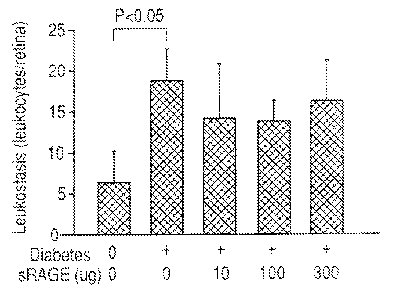
[00151 Figure I is a bar graph showing the effects. of an exemplary RA.GE-
Ig fusion
protein on leuko.stasis in a streptozotocin-induced diabetic mouse model.
[0016] Figures 2A-2D are bar graphs showing .the effects of an exemplary
RAGE-Ig
ilision protein on retinal vascular permeability in various retinal layers in
a
streptozotocin-induced diabetic mouse model.
[0017] Figure 3 is a bar graph Showing the effects of an exemplary RAGE-
Ig fusion
protein on the nitration of retinal proteins in a streptozotoein-induced
diabetic mouse
model.
4
CA 02690056 2009-12-03
WO 2008/157378
PCT/US2008/066956
[0018] Figure 4 is a bar graph showing the effects of an exemplary 'RAGE-
1g fusion
protein on the retinal expression of ICAM in a streptozotocin-induced diabetic
mouse
model.
[0019] Figure 5A is a bar graph showing the effects of an exemplary RA.GE-
Ig fusion
protein on the number of acellular capillaries observed per square mm of
retinal tissue
in diabetic mice after 10 months of diabetes. Figure 58 is a bar graph showing
the
effects of an exemplary RAGE-Ig fusion protein on the number of pericyte
ghosts per
1000 capillary cells observed in diabetic mice after 10 months of diabetes.
[0020] Figure 6 is a bar graph showing the effects of an exemplary R.AGE-
Ig fusion
protein on the 50% response to touch threshold in diabetic mice after 10
months of
diabetes.
[0021] Figure 7 provides a flow chart showing the experimental protocol of
Example
3.
[0022] Figure 8 is a line graph showing. the effects of an exemplary RAGE-
Ig fusion
protein on test animal weights in a type-II collagen induced, arthritis mouse
model.
[0023] Figure 9-1s a bar graph showing the effects of an exemplary RAGE-1g
fusion
protein on the incidence of arthritis in a type-II collagen induced -arthritis
mouse
.model.
[0024] Figure 10 is a bar graph showing the effects of an exemplary RAGE4g
fusion
protein on the onset of arthritis in a type-II collagen induced arthritis-
mouse model.
[0025] Figure 11 is a line graph showing the effects of an exemplary RAGE-
Ig fusion
protein On incidence Of arthritis as a function oftime in a type-II collagen
induced
arthritis mouse model.
[90261 Figure 12 is a line graph Showing the effects of an exemplary RAGE-
Ig fusion
protein on the severity of arthritis as a function of time in a type-11
collagen induced
.arthritis mouse model.
[0027] Figure 13 is a line graph showing the effects of an exemplary RAGE-
Ig fusion
protein on the number of arthritic paws observed as a function of time in a
type-II
collagen induced arthritis mouse model.
CA 02690056 2009-12-03
WO 2008/157378
PCT/US2008/066956
[0028] Figures 14A-14D are photomicrographs showing the effects of an
exemplary
RAGE-Ig fusion protein. on. joint morphology as a function of increasing
amounts of
the fusion protein in a type-II collagen induced arthritis mouse model.
[0029] Figure 15 is a bar graph showing the effects of an exemplary RAGE-
11g fusion
protein on synovitis (black bars) and palms (grey bars) in a type-11 collagen
induced
arthritis mouse model.
[0030] Figure 16 is a bar graph showing the effects of an exemplary RAGE-
Ig fusion
protein on marginal erosion (black bars) and architectural changes (grey bars)
in a
type-ii collagen induced arthritis mouse model.
[0031] Figure 17 is a bar graph showing the effects of an exemplary RAGE-
Ig fusion
protein on overall histological arthritis score in a type-II collagen induced
arthritis
mouse model.
[0032] Figure 18 is a bar graph showing theeffeets of an exemplary RAGE-
Ig fusion
protein on joint matrix protein loss in a type-II-collagen induced arthritis
mouse
model.
[0033] Figures 19.A-19D are photomicrographs of toluidine blue stained
sections
-showing the effects of an exemplary RAGE-Ig fusion protein on joint matrix
protein.
loss in a type-ti collagen induced arthritis mouse model.
DETAILED DESCRITPION OF THE INVENTION
[0034] Definitions
[0035] As used herein the terms "receptor for advanced glycation end
product" or
RAGE refer to proteins having amino acid sequences that are substantially
similar to
the native mammalian RAGE amino acid sequences and function to bind one or
more
RAGE ligands in a ligand-receptor specific manner. The terms "advanced
glyeation
end product" and "AGE" refer to a heterogeneous groupof molecules formed from
the nonenzymatic reaction of reducing sugars with free amino groups of
proteins,
lipids, and nucleic acids as described above.
[0036] As used herein, a "RAGE ligand binding domain" or "RAGE-1.13D"
refers to
any mammalian RAGE protein or any portion of a mammalian RAGE protein that
retains the ability to bind a. RAGE ligand in a ligand-receptor specific
manner.
Specifically, without limitation, a RAGE ligand binding domain includes a
6
CA 02690056 2009-12-03
WO 2008/157378
PCT/US2008/066956
polypeptide having one or more extracellular domains of a .transmembrane RAGE
protein. With reference to Table 6, a suitable RAGE-1,BD may comprise at least
amino acids 1-99, or amino acids 24-99, or amino acids 1-208, or amino acids
24-208,
or amino acids 1-301, or amino acids 24-301, or amino acids 1-344, or amino
acids
24-344 of SEQ IDNO:6.
[00371 The term "isolated," as used in the context of this specification
to define the
purity of the fusion protein, means that the protein is substantially free of
other
proteins with. which it is associated. during production, including without
limitation.
substantially free of other proteins present during expression of the fusion
protein in a
cell culture medium. For example, an isolated protein of the invention may
comprise
I- 25%, 20-25%, -15-20%, 10-15%, 5-10%, 1-5% or less than about 2% by mass of
protein contaminants residual of production processes. Compositions comprising
isolated proteins of the invention, however, can contain other proteins added
as
stabilizers, carriers, excipients or co-therapeutics.
[0038] As used herein, "protein" and "polypeptide" are interchangeable.
[00391 As-Used herein "treating" a disease:or disorder refers to
improving at least one.
sign-or symptom of the subject's disease or disorder.
100401 The term "nucleic acid" refers to polynucicotid.es such as
deoxyribonucleic
acid (DNA), and, where appropriate, ribonucleic acid.(RNA.). The term should
also
be understoodto include,. as equivalents, analogs of either RNA or DNA made
from
nucleotide analogs, and, as applicable to the embodiment being described,
single.
(sense or antisense) and double-stranded polynueleotides.
[0041] The term "or" is used herein to mean, and is used. interchangeably
with, the
term "and/or," unless context clearly indicates otherwise.
[0042) The term "percent identical" refers to sequence identity between
two amino
acid sequences or between two nucleotide sequences. Percent identity can be
determined by comparing a. position in each sequence which may be aligned for
purposes of comparison. Expression as a percentage of identity refers to a
function of
the number of identical amino acids or nucleic acids at positions shared by
the
compared sequences. Various alignment algorithms and/or programs may be used,
including PASTA, BLAST, or ENTRF.Z. PASTA and BLAST are available as a part
of the GCG sequence analysis package (University of Wisconsin, Madison, Wis.),
and
7
CA 02690056 2009-12-03
WO 2008/157378
PCT/US2008/066956
can be used with, e.g., default settings. ENTREZ is available through the
National
Center Ibr Biotechnology 'Information, National Library of Medicine, National
Institutes of Health, Bethesda, Md. In one embodiment, the percent identity of
two
sequences can be determined by the GCG program with a gap weight of 1, e.g..,
each
amino acid gap is weighted as if it were a single amino acid or nucleotide
mismatch
between the two sequences.
[0043] Sequence identity may be determined by comparing a reference
sequence or a
subsequence of the reference sNuence to a test sequence (e.g., a nucleotide
sequence,
an amino acid sequence, etc.). The reference sequence and the test sequence
are
optimally aligned over an arbitrary number of residues termed a. comparison
window.
in order to obtain optimal alignment, additions or deletions, such as gaps,
may be
introduced into the test sequence. The percent sequence identity is determined
by
determining the number of positions at which the same residue is present in
both
sequences and dividing the number of matchingpositions by the total length of
the
sequences in the comparison window and multiplying by 100 to give the
percentage.
In addition to the number of matching positions, the number and. size of gaps
is also
considered in calculating the percentage sequence identity.
[0044] Sequence identity is typically determined using computer programs.
A
representative program is the BLAST (Basic Local Alignment Search Tool)
program
publicly accessible at the National Center for Biotechnology Information
(NCB',
http://www.nebiailmnih.govi). This program compares segments in a test
sequence
to sequences in a database to determine the statistical significance- of the
matches,
then identifies and reports only those matches that are more significant than
a
threshold level. A suitable version of the BLAST program is one that allows
gaps, for
example, version 2.X (Altschul, et al., Nucleic Acids Res 25(17):3389402õ
1997),
Additional suitable programs for identifying proteins with sequence identity
to the
proteins of the invention include, but are not limited to, PHI-BLAST (Pattern
Hit
Initiated BLAST, Mang, et al., Nucleic Acids Res 26(17)3986-90, 1998) and PSI-
BLAST (Position-Specific iterated BLAST, Altschul, et al., Nucleic Acids Res
25(17):3389-402, 1997). The programs are publicly available at the NCBI web
site
listed above and may be used with the default settings in order to determine
sequence
identity according to the invention.
[0045] Fusion Proteins =
8
CA 02690056 2009-12-03
WO 2008/157378
PCT/US2008/066956
[0046] The present invention provides an isolated fusion protein
comprising at least
one poly-peptide comprising: (a) a first amino acid sequence at least 90%,
91%, 92%,
93%, 94%, 95%, 96%, 97%, 98%, or 99% identical to a mammalian receptor for
advanced glycation end product (RAGE) ligand. binding domain, the -first amino
acid
sequence capable of binding a RAGE ligand; and (b) a second amino acid
sequence at
least 90%, 91%, 92%, 93%, 94%, 95%, 96%, 97%, 98%, or 99% identical to a human
heavy chain immtmoglobulin Ig04 constant domain or a fragment thereof, wherein
the first amino acid sequence comprises at least one mutation, or at least two
mutations, or at least three mutations, or 1-4 mutations, or 1-10 mutations
relative, to a
wild type RAGE ligand binding domain. Examples of mutations that may be made
in
the. firstamino acid sequence are those that increase the stability of the
fusion protein,
for example, by making the RAGE ligand binding domain more resistant to
proteolytic degradation, such as those that make the fbsion protein more
resistant to
furin-likeproteases, Suitable fragments of the second amino acid sequence
include
fragments that retain the ability to increase the serum half-life of the
fusion proteins of
Which they are part relative to the serum half life of the same first amino
acid
sequence alOne. Preferably the first amino acid sequence and.the- second amino
acid
sequence are derived from human RAGE ligand binding domain and human IgG4.
[0047] Fusion proteins of the invention may comprise one or more.arnino
acid
sequences in addition to a.. RAGE ligand binding domain, andan IgG4 constant
domain or fragment thereof. For example,. a. fusion -protein of the invention
may
comprise a linker sequence which may be inserted between the RAGE ligand
binding
domain and the IgG sequence. Fusion proteins of the invention may comprise one
or
more tag sequences i for-example, purification, tag sequences such as 6-
Histidines.
Fusion proteins of the invention may comprise one or more epitopes recognized
by
commercially available antibodies, for example, c4nye (EQKLISEEDIõ SEQ ID NO:
9) and hemagglutinin (YPYDVPDYA, SEQ ID NO:10) derived from an epitope tag
of the influenza hemagglutinin protein.
[0048] Any mammalian RAGE protein known to those of skill in the art may
be used
in the practice of the present invention. Preferably the extracellular domain
of the
RAGE protein will be used to identify a ligand binding domain that can be
mutated
and used as the first amino acid sequence of the fusion protein. Suitable
example of
mammalian RAGE proteins include, but are not limited to, primate, human
(e.t.,!õ
9
CA 02690056 2009-12-03
WO 2008/157378
PCT/US2008/066956
GenBank accession no. NP.,_00112,7 and NP 751947), murine(e.g., GenBank
accession no. NP_031451), canine (e.g., GenBank accession no. AAQ81297), rat
(e.g., GenBank accession no. NP 445788). Ix-wine (e.g., GenBank accession
no. AA120128), ovine, equine and porcine (e.g., GenBank accession no.
A.AQ73283)
RAGE domains.
[0049] RAGE amino acid sequences comprising one or more changes or
modifications with respect to the wild type sequence may be used in the
present
invention. Such changes or modifications include, but are not limited to,
point
mutations, deletions from the N-terminal, deletions from the C-terminal,
internal
.deletions, and combinations thereof. Any change or modification may be
introduced
into a RAGE sequence for use in the present invention so long as the resulting
protein
retains biological activity, e.g., the ability to bind one or more RAGE
ligands. The
fusion proteins of the invention also include those with or without endogenous
glycosylation patterns, including without.limitation, fusionproteins in-which
the first
amino- acid sequence is derived from -a mammalian RAGE ligand binding domain
with or without associated native-pattern glycosYlation of the binding domain.
[0050] Any suitable IgG Fe region may be used in the practice-of the
invention,
preferably, from an IgG4. molecule, for example, amino acid residues 149-473
of
GenBank accession no. AM125985. An IgG region -for use in the present
invention
may be an 104 Fe region and may comprise one or more of the CH2 and CI-13
regions Of the1gG4.molecule.
[0051] Examples of suitable fusion proteins are provided in the following
tables.
[0052] Table 1 provides thenueleotide sequence of a human RAGE-IgG4 Pc
fusion
protein gene Secinenee:
CA 02690056 2009-12-03
WO 2008/157378
PCT/US2008/066956
.00531 Table I: Human RAGE-IgG4 Fe Fusion Gene Sequence (SEQ ID NO:!).
AT GGC AGCC GGRACAGCAGTT GGAGCCTGGGT GCTGOTCCTC1MITCTGT GGGGGGCAGTAGTA GarGe C
A
CATCACAGO CCOGA TTGGCCACX:e AC PGGTOC TGAAGIVIMAGGGGC:4CCC:CC AAGAAACCACCCCAGC
TerrAAT GGA WIC TOAACAC GGCCGGACAGAA GCCITXMAGGTCCTGTC TeCCCAGGC:IAGGAGtXfCCC
TGGGACAGTGPGGCWGTG'rCCTTCCCAACGGCTCCCTC'rrCCTTCCGGCTGPCGGGATCCAGGATCAGGG
GATTTTCCGGTGCCAGGCAATGAACACIGAATGGAAAGGAGACCAAGTCC AACTACCGAGTCCGT GTCTACC
AGATTCCTGGC4AGCCAGAAATTGTAGATTCTGCCPCTGAACTCACGOCTGGDOTTCCCAA1AAOGTGOGO
ACATGIVIXITCAGACCGAMIC:TACCCTGCAGCCyACITIVAGCM3GCACTTGGATGGC4.74-
AGCCCCTGGTGCC.
(3..A.AT GAGA-A GGGACTATCTC T GAAGGruNCAGACCAGGAGAC ACCCMAGAC AGGGCTC TCACAC
TCCAGT
CCGAGCTAATGGIVACCCCAGCCCGGOGAGGAGATCCCCCIVCCACC7re'reareiTAWITCAGCCCAGGC
CTTCCCCGACACCGGGCCTTGO.CCACACCCCCCATCCAGICCCCGTGTCTGGGAG7.CTGIVICCTOTGGAGGA
GGITCCAATTGOTGOTGGAWCAGAAGGTGGAGCAGTAGCTCCTOGTGGAACCarAACCCTGACCTGTGAAG
TCCCTGCCCAGCCCTCeire'rCAAATCCAC.TaGATGAAGGATGOTGTGCCCTTGCCCCTTCCCCCCAGCCCT
Cr*KiCTGATC:CTC.TCTGAGATAGC:IGCMCAGGACCAGGGAACCTAC-AGCTGTGIT,a:CACCCATTCC-AC-
CCA.
CCGGCCCC GGAAA GCCGTVCP. GTCAOCATC,I=ATCATCGAACCAC-1CCGAGGAGGi"riCCAACTGCAGGCT
CTGIVGGAGGATCAGGGC`itGGAACTCTACCCCTC:ICCCX7CFMCACCAAGGGCCCATCCGTCTTCCCCCTG
OCGCCOTOCTCCI eN GGAGCnCTCCGPakG.CACAGCCGCCCT(4GGCTGCCM'XrrCAK-cACTAc7TTCCCCG4
ACCGOTGACSGTOrrarsTMaikeTCAGGCGCCCTGACCACCGGCGTCCACACCIMCCMCTGTCCTA.ClaT
CC TC ACCACTCTACTCCCTC,s,ACCAGCCIKX;PCIA CarisOCCMCC ACCACCTTGOCCACCIAA
QACCTACACC
TGCMCGTAGATCACAACCCCAGCAACACCAAGGIVGACAAGAGAGTTGAGTCCANCATGGTCCCCCATG
Cc:CATCATtileCCAWACCTGAGTTCCTGCMGGACCATCAGTC:TTCCTG7reCCCCCAPAAP.,,C.CCAAGGAM
CTCTCATGATCTCCCGOACCCCPCAGGTCA.CGTOCMGGTGG.TC;GACG.TGACCCAGOAAGACCCCGACK3TC
CAGTTCAACTGGTACGTGGA'IvjGCGT(.3GAGGTGCATAATCCCAAGACAAN:4CCG='',X3GGAGGAGCAGTTCA
A
cAf3CACGTACQGTGTCGT.CAGCGTCC TCACCC.:TOCTGCACc4GGACTGOCTGMCWC. AAgGIAGTACAAGT
GC-AAGGTOMCAACAAAGGCCTCCCOTCCTC:s.CAIVGAGAAAACCATOISCCAAACCCAPAGGalkt3CCCCGA
GPMCACAtIlaTGTACACansCCCCCCATCC:!CACGACCIACATGACCAACIA.ACCACICTCAGgCTCACCTGC.CT
ials.MKAAGGC"II.VT A CCCC ACCGAC. A TCGCCCTGGACITGGGAGACCAATCGGCACCCCCAGAAC
ACTACA
ACK CACGCC TCCCGTGC Te-GACTCCGACC-GCTCCTIVIVCCTCTACAGCAGGCTAACCGIV-G.ACMCMGC
AGGTGGCAGGAC4MGAAT.G. T. CTTCTC N7X:TCCGTGAT GC ATGAGGC Te
TGCACAACCACTACACACAGAN
GAG= C CC.TCPCTC TCGC:10
[0.054] Bold text is the coding sequence for the RAGE signal .:sequence,
normal text is
the coding sequence for human RAGE, and underlined text is the coding sequence
fOr
IgG4 Fe region.
II
CA 02690056 2009-12-03
WO 2008/157378
PCT/US2008/066956
100551 Table 2: Amino acid sequence of a human RAGE4gG4Fc fusion protein
(SEQ
ID NO:2).
MAAGTAVGAW VINLSLWGAV VGAQNITARI GEPLVLXCKG APKKPPQRLE 50
TGRTEA. WKVLSPQGGG PWESVARVLP NGSL FL
PAVG IQDEGIFRCQ 100
GKETK SNYRVRVYQI PGKPEI VDSA S ELTAGVPNE
VGTCVSEGSY 150
FAGTL MILD GPLVPN1KG VSVKEQTRRII PETGLFTLQ S
ELVIVTPARGG 200
DPRPrFSCSF S PGL PRIMAL RTAP QPRVW E P V FL EEVQL
VVE PEGGAVA. 250
r-ac-Grvr.urcE vPAQP WMKDGVPLPL F P-SPVL PE
IGPQDQGTY S 300
CVA'IlLISHGP QESRAVS I S E ?GEM P:PA GS VCYjSaL GT
LALAASTKGP 350
SW PIA Pc: SR STSESTAALG CLVKDY P PE P VTVSWNSGAL T
SCATHTFPAV 400
QSSGLIYSLS SIIVTVPS S SL GTKTYTCNVD SIM< VDK
RVESKYGPPC 450
PSCPAPEFLG GP S VFT.:F P OK PKDTLMI SRI! PEVT(.2VV VIM
SQEDPEVQFN 500
NYVDGVEVEN AKTKPRELEce NS T YRWSVL TVINDWING KETKCKVSNK 550
GLPSSI ENT I SKAKGQPRE OPITL PP SQE EllrKINVSLT
CLVKGF P SD 600
I AVERESNGQ PENNYKTTPP VLDSDOSFFL Y SRL TVDK
S R WQRGIVFSC S 650
VMEEEMIINFT TVS', SLSTJG k 671
[0056) Bold
text is the amino acid =pence tbr the RAGE signal sequence, normal
text. is the amino acid sequence for human RAGE, and underlined text is the
amino
acid sequence for IgG4 Fe region.
10057] Table 3.: Human RAGE-Iiinker,Ig64 Fe Fusion Gene Sequence..(SEQ ID
NO:3),
ATGGCAOCCGOAACACCAGTTGOAGCCTOGGTOCTGarCeareAarerGTGOOGGGCAGTACTAGGTOCT.cA
GATCACAGCCCIGGATTGGCGAGCCACTGOTGCTGAAGTGTAAOGV,GGC:X2CCCAia" GAAAcCACCCCAGC.
GCCIKTGAATOGAAACTK1AACACAGGCCGGACAGAAGCCTGGAAGGTcCTLITCTC.CCCACCGAGGAGGCGCC
TGGGACAGTGTGGCTCGTqMCTTGCCAACGGCTC.CCTCTTCCTTcCGGCTGTCGGGATCCAGGATGAGGG
GATTTTCCGCTGCCAGGCMTGAACAGGAATGGAAAGGAGACCAAGTCCAACTACCGAGTC.ICGTGTCTACC
AGATTCCTGGGAAGCCAGAAATTGTAGATTCTGCCTCTGIACTC:ACGGCTC:IGTGTTCCCAATAAGGTGdGG
ACATGTCTGTCAGAWGAAGCTACCCTGCAGGGACTCTTAGCL'GGCACTTCKATGGGAAGCCCCTGGTGCC
GAPITGAGAAGGGAGTATCTGTGAAGGAACAGACCAGGAGACACCCTGAGACAGCMCTCTTCACACTOCAGT
CGGAGCTAATGGTGACCCCAGCCCGGM4.AGGAGATCCCCGTCCCACCITCTCCTGTAGC'rTCACCAGGC
CTTCCCCGACACCC4G'GCCTTGC.GCACAGCCCCCATCCAGCCC.C:GTGTCTGGCIAGCCTGTGCtTCTGGAGGA
GGTCCAATTGGTGOTiCGAGCCAGAAGGTGOACCAGTACCTCCTCGTGGAACCOTAACCCTGACCTGTGAAC
TCCCTCC.C.TACCeCreTeCTCAAATCCACTGGATGAAGGATGGTM(XTCTTGCCOCTTCCCCCCAGC.CCT
GTGCTGATCCTCCCTGI'iGATAGGGCCTCA.GGACCAGGGAACCTACAGCTG'TGTGGCCACCCKrrCCAGCCA
CGGGCCCXAGGAAAGtCGTGC'rGaCAGCATCAaATCATCX;AACCAGGCGAGGAGGGGCCAeNCTGCAGGCT
CTG'TGGGAGGATCAGGGCTGGGAACTCTAGCCCTGGCCGGTAGCCWAIATZZGCTTCCACCAAG
GGCCCATCCGTCTTCCCCCTGGCGCCCTGCTCCAGGAGCACCTCCGAGAGCAC AC47CGCCCTGGGCTGCCT
GGTCAAGGACTACTTCCCC.GAACCGGrGACGGTGTCGTC-GAACTCAGC-CC-CCCTGACCA(XXIGCGTGCACA
CCTTCCC=CTGTCCTACAGTCCTC'AGGACTCTACTCCCTCAGCAGCGTGGTGACCG'rG:-*CCTCCAGCAGC
TTGGGCACGAAGACCTACACCTGCAACGTAGAWACAAGC.:CCAGCAACACCAAGGTGGACAAGAGAGTTGA
GTCCAAATATOGTCCCCCATGCCCATCATOCCCAGCACCTGAGTTCCTWOGalACCATCAGIVITCCTGT
TCCCCCCAAAACCCAAGGACACTCTCATGATCTCCCGGACCCCTC::AGGTCACGTGCGPGGTGGTGGACGI'G
AGCCAGGAAGACCCCGAGGTCCAGTIVAACTGGTACGTGGAIX3GCGTGGAGG.'TGCATAATGCCAAGACAAA
GCCGCGGGAGGAGCA.GTTCIAACAOCACOTACCGTO'GTCAGCGTCC'MACCGTCCIGCACCAGGACTGOC
TGAACCinCAAGGAGTACAAGTGC:AAGMCTCCIIACAAAG3CCTCCCGTCCTCCATCGAGAAAACCRICTCC
AAAGX::CAAAGGGCAGCMCGAGAGCCACAGGTGTACACCCTGCCCCCATCCCAGGAGGAGATGACCAAGI
CCAGOTCAGCCTGACCTGCC.s.TGGTCAAA.C4GCTIVTACCCCA(R7C-ACATCGCCGTGGAGTGGGAGAGCAATG
GGCAGCCGGAOAACAACTACAAGACCAC:GCCTCCCGTGCTGGACTC'CGACGGCTCCTTCTTCCTCTM1AGC
AGGCTAACCGTGGACAAGAGCAGGTGGCAGGAGGGGtAATGTCTTCTCATGCTCCGTGATGCATGAGGCTCT
GCACAACC-:ACTACACACAGAAGAGCGTCTCCCTGTCTCPCGa:AAKMA
[00581 Bold text is the coding sequence for the RAGE signal sequence,
normal text is
the coding sequence for human RAGE, double underline text is the coding
sequence
12
CA 02690056 2009-12-03
WO 2008/157378
PCT/US2008/066956
for the peptide linker, andivingle underlined text is the coding sequence for
4(14 Fe
region.
[00591 Table 4: Amino acid sequence of a ham ;In RAGE-Linker4g04Fc fusion
protein (SEQ ID NO:4).
MAAGTAVGAW VLVMSLWGAV VGAMITARI GEPLVLKCKG APKKPPQRLE 50
wKLNTGRTEA WKVLSPQGOG PWDSVARVLP NGSLFLPAVG IQDEGIFRCQ 100
sNYRVRVYQI PGKPEIVDSA SELTAGVPNK VGTCVSEGSY 150
PAGTLSWELD GKPLVPNEKG VSVKEQTRRH PETGLFTLQS ELMVTPARGG 200
DPRPTFSCSF SPGLPRHRAL PTAPIOPRVW EPVPLEEVQL VVEPEGGAVA 250
PGGTVTLTCE VPAUSPQIH WMKDGVPLPII PPSPVLI1JPE IGPQDQGTYS 300
CVATliSSHGP QMPAVSISI IEPGEEGPTA GSVuGGLGT LALAGplagS. 350
ciASTKGPSVF PTJAPCSRSTS ESTALGCLV KDYFPEPVTV SWNSGALTSG 400
VH=AVLQS SGLYSLSSVV TVPST__,GTK TY.TCNVDHKP SNTKVDKRVE 450
SK.YGP'DrPSC PAPEFLGGPS VFLF-PPY.PKD TY,MISFTPZU TCVVVDVSQE 500
DPEVO-,NsTKYV DGVFNTHmAKT KPREEQFNST vRVw-w-LTVLHODWLNGKFY 550
_ _
KCKVONKGIJP SSIEKTISKA KGQPREPQVY TLPPSQEEMT KNQVSLTCLV 600
KGFYPSDIAV EWESNGQPEN NYKTTPPVLD =SDGSFFLYSR LTVDKSRWQE 650
GNVFSCSVMH EALHNHYTQK SLSLSTJGK 6.78
[00W] Bold text is the.= amino acid scquence for the RAGE signal
sequence, normal
text is the amino acid sequence for human RAGE, double undetline.text is the
albino
acid sequence for the peptide linker, and single underlined text is the amino
acid
sequence for fg04 Fe region.
13
CA 02690056 2009-12-03
WO 2008/157378
PCT/US2008/066956
[0061) Table 5: Human RAGE variant-IgG4 Fc Fusion Gene Sequence (SEQ ID
NO:5).
AT GGCAGCC GGAACAGCAGTTGGAGC CTGGGTCICT GGT C
CTCAGTCTGTOGOGGGCAGTAGTAGGIXICTCAMAC
ATCACAGCCCOGATTCMCGAGCCAMGC.MGC' =TGAAGTGTAAGGGGGCCC C CAAGAAACC CCC ACC GC:1C
TGGAik
TC(.3AAACTGAACACAGGCCGGACAGNACCTTOCIAAGGIV.7.CTGIVTCCCCAGGGAGGAGGCCCCTGGGACACTG
TG
.r:rTCX:21'.17.1r CT 'P. Cee AACGG CeTeTTC C 'MCC GGC TG T C(XX/AT
CCAGGATGAGGGGATITPC CGC,TGCC AG
CCAATGAACAGGAAT.GGAAAGGAGACCAMITCCAAC'rACCGAGTCCGIVIVTACCAGATPCCTGGCAACCCAGAA
A.71'12;TAGATPC T. C4C C TerakikC T. CAC GGCTCGTGTTC C CAM' A ACGTGGO
CACATGTGTGIVAGAGC-(1AAGCTAC
COTGCAGGGACTCPTAGCTGCL'ACPTGGATGGGAAGCCCCTGGT.
G'CCGAAMNGAAGC:XIAGTATC.:TGTGAAGGAA
C AGACCAGGAGACAC CC TGAGACAGGCC TVIVCACAC Tete ACTC GOACKTAATGGTGACC CCACC C
CGOGGNMA
GATCCCCGIACCCACC TTC Tee TGTAGC1".VCAG CC CAGGCCTTCC C CCACQCC GGGC"...0 TT
C:X.7ACACAGC C C C CIATC
C. C Tan?
GGAACCGTAiliCCCIPGACCTCPPGAAGTC:CCTGCCCAGCCCTCTCCTCAAATCCAC:IN1GATGAAGGATGGT
(417GCC CTTC-C C CC TTC CC CC CAGCCC TGPIrre. GATC CTC: CC TO
AGATAGGC4CCTCAGGACC AGGGAAC CTACACIC
TWG'ra.:4Ce ACC C.ATTCCAGC CAC CMGC CCC AQQ11AA(.3C CGT(ile TGTCACC "LTC
ACK:ATC ATC G.AACCAGGCGAG
GAGGGGC CA.ACTGCJAGGCTC T. GTGGGAGGATCAGGCMGGAAC '11CVAGC CC: TGGCC GCTTC CAC
CAAGGGCCC A
TCCGTCTTC CCC C TGGCGCC C TGCTCCAGGAGCACC TC C QAGAGVACAGCC GC CC
TGCCICTGCCTCGTCAACCAC
TACTTCCC CGAAC C.Care:AC GC:rtGiVarGICAACTCAGGC GC C CTGAC CAGCGGCGTGCAC AC C
CCGOC TC; TC
AC C TGCAACOTAGATCACAAGCC -CASCAACIliCCAW3GT,GGACAAGAGAGT. l'CiAGMCAAATA
TCGTCC CC CATC4C
C CAW. ACC PG AC TareTCPC.;GC-1GGAC CATCAGTC 'FTC CTG'17!CC C CCCAMACC
CA.WGACACTC Te
AT.
GhTereCCGGNX:CCTGAGOTCACGTGCX;TGGPC4(1TGGACCreGAGC.CAGGPAGACCC:!CCACCITC.CZA7:1
1PlICAAC
TC,40 AC CaGGATCGCGTGGAGGIvjeATAATGC C AAGAC AAAGCC GCGGGAGGAGCAGTTCAACMICAC
GTACC GT
GPCIGI`CACX:GreCTCACCGTCCTC5CACCAPGACTSGerciAACCKX.7AAGGAGTACAAG.TC-e-
AAGMCPCCAACM13.
GGCC.Te CC GTCCTCCATC GAGAAAAC CATCTC C AAAGCC AA GGGCACCC CC GAGAGC
CACAGGTGTACAC CC TG
CC:CC.C.ATCMAGGAGGAGATGACCAAGAACCACMCAGCCTGACCTGCCTGGIVAMCWPTCTACCC,CAGCSAC
ATCGCCGTGGAG'.1'GGGAGAGCAATGGOCAGCCr4GAGAAC.11-kCTACAAGACCACGCMCCCGTGCT, GC:AC
CGAC
GMTC:CirreTTCCTCTACAC-
CAGGCTAACCYMGACAAGAGC.AGGTGGCAGGAGG3GAATUIVTTC.IVATC4C,TCC
(1.MIITGCATGAGGC TCTC-41-"Ike ATILT A CTAC ACA.CACM GAGC017VrOCCTGar GG
AAMIA
[0062] Bold text is the coding.sequence for the RAGE Signal sequence,
normal text is
the coding sequence for human RAGE. variant, bold wavy underline letters are
sites of
the point mutations introduced into the variant hR.AGE sequence, and
underlined text
is the coding sequence tbr IgG4 Fe region.
14
CA 02690056 2009-12-03
WO 2008/157378
PCT/US2008/066956
[00631 Table 6: Amino acid sequence of a human RAGE variant-IgG4Fc fusion
protein (SEQ. ID NO:6).
1 MAAGTAVGAW VVVLSINGAV VOAQNITARI GEPLVLKCKG APKKPPQRLE
51 WKLNTGRTEA WKVLSPQGGG PWDSVARVLP NGSLFLPAVG TWEGIERCQ
101 AMNRNGKETK SNYRVRVIQI PGKPEIVDSA SELTAGVPNK VGTCVSEGSY
151 PAGTLSWHLD GKPLVPNEKG VSVKEQTRRH PETGLFTLQS ELMVTPARGG
201 DPRPTFSCSP SPGLPRARAL WAPIQPRVW EPVPLEEVQL VVEPEGGAVA
251 PGGTEITLTCE VPAQPSPQIR WMKDGVPLPL PPSPVLILPE IGPQDQGTYS
301 CVATHSSHGP QESRAVSISI IEPGEEGPTA GSVGGSGLGT LALAASTKGP
351 SVFPLAPCSR STSESTAALG CLVKDYFPEP VTVSWNSGAL TSGVHTFPAV
401 LQSSGLYSLS SVVTVPSSSL GTKTYTCNVD RKPSNTKVDK RVESKYGPPC
451 PSCPAPEFLG GPSVFLFETK PRDTLMISRT PEVTCVVVDV SQEDVEVQPN
501 iffrinavroIN AKTKPREEQF NSTYRVVSVL TVLHQDWLNG KEYKCKVSNK
551 GLPSSIEKTI SKAKGQPREP QVYTLPPSQE EVITKNQVSLT CLVKGFYPSD
601 IAVEWESNOQ PENNYKTTPP VLDSDGSFFL YSRLTVDKSR WOGNVPSCS
651 VMHEALHNHY TRKSL512SLG K
[0064] Bold text is the amino acid sequence for the RAGE signal sequence,
normal
text is the amino acid sequence for human RAGE variant, hold wavy underline
letters
are sites of the point mutations introduced into the variant hRAGE, and
underlined.
text is the amino acid sequence for IgG4 Fe region.
CA 02690056 2009-12-03
WO 2008/157378
PCT/US2008/066956
[0065} Table 7: Human RAGE variant-Linker-IgG4 Fe Fusion Gene Sequence
(SEQ
ID NO:7).
ATGGICAGCCGOKACAGCAt3TTGGAGCCTGGGTGCTGGTCCITAGTCTGVGGGGGGCAGTAGTAGGTOCTCAKAsIC
ATCACAGCCCGGAVKTGCGAGCCACTGGTGCTGAAGTGTAAG:MGGCC'CCCAAGAAACCACCCCAGCGGCTGGAA
TGGKAACTG'AACACAGGCCGGACAGAP.4CTTGCsAM3GTCCTGI'CTCCCCAGC4GAGGAGGCCCCTGGGACAGTGr
G
GCTCGTCTCCTIVC:CAACCGCTCCCTCTTCC:71"TCC.GGCTGTCGGGATCCAGGATGAGGGGATTTTCCGG.TGCC
A,G
GCAA':l'GAACAG42:AATGGAAAGGAGACCAAGTCCKACTACCGAGTCCGTCrr.TACCAGAT'I'CCTGWAAGCCA
GAA
ATTGTAGWITCTGCCTCTGAACTCACOGCTGOTG7rCCCAATAACGTGGGGACATararGIVAGAGGGAMICTAC
CCTGCAGGGACTCTTAGCTGGCACI"I"WATGGGAAGCCC.CTGGTGCCGAATGAGAAGGGAGTAT'CTGTG17kAGGA
A
CAGACCAGGAGACACCCTGAGACAGGGCTOrTCACACTGCAGTCGGAGCTAATCGTGACC:::::CAGCCCOMGAGGA
GATCCCCGTCCCACCITCIKTTGTAGCTTJZAGCCCAGGCCTIVCCCGA,CgCCGGGCCTTGCACACAGCCCCCATC
CAGCCCCGTIGTCTGOGAGCCTGrrOCCTCT CrGAGGAGGTCCAATTGC-
TGGTGGAGCCAGIkAGGPGGAGCAGTAGCT
CCTGGI`GGAACCGTAACCCTGACCTGTGAAGTCCCTGCCCAGC:CareTCMCAANIr.C.-
ACTCICATGAAGGATGGT
GTGCCCTTGCCeeTTCCCCCC::AGCCCTGTGCTGATCCTCCCTGAGATAGGLICCTCAGGiICCAGGGAACCTACAGC
tiv..3712TGGCCACCCATT:CCAGCCACGGGCCCCAGGAAAGcCGTGC.TGTCACCATCAc.;CATCATCGMCCAGG
CGAG
GAGGGGCCAACTGCAGGCTCTOTGGGAGGATCAGGGCPCXXIAACTCTAGCCOMGCC.QaTarciaTEgH;Eas.
cific.,:"7µ;'CINTCCACCM.CCGCCCATCUMTTCCCCOMGCGCCCTGCTCCAGGAGCACCTCCGAGAGCACACCC
GCC
GIVCACACCTTCCCGGCTGTCCTACAGTCCTCAGGACTCTACTCCCPCAGCACCGTG3TGACCGTGCCCTCCAM
AGC'Prf-XXX:-
ACGAAGACCTACACCTGCAACGTAG`A.TCACAA.GCCCACCAACACCAAGGTGGACAAGAGAGTTGAG
TCCAAATATGGTCCCCCATGCCCATCATMCCACCACC.PGAGTTC:CliGGGGGGACCATCAGIVisreeliGTTCCCC
:
CCRAAA.CCCAACCACACTCTCATCyATCTCC,CGGACCCCTGAGGIV:ACGTOCGTGarGGTMACOTOAGCONGGAA
GACCCCGAGGTCCAGITCAINCTGGPACGTGOATGGCOTGGAGGMCA'rAATGCCAAGACAAAGCCGCGGOAGGAG
CAGT.T. CAACAGCACCTACCOTGTGGI2CAGCGTCCTCA.CCGTCCTGCACCAGGACTGGCTWXGGCAAGGWTAC
AAGTOCAAGGTCTC.C.:AACAAAGGCCMCCGTCCPCCATCGAGAAAACCAPC.MTCAAAGCCAAAGGMNOCCCXMA
GAGCC4CAGG73TACACCCTGCCCCCATCCCAGGAGGAGATGACC.AAGMCCAGGTCAGCCTGACCTGCCTGGTC
MAGGCPTCTACCCCAGCGACATCCICCGTGOAGAGAGCA.kTGGGCAGCCOGAGAACAACVACAACCACG
CeTeCCGT.C.ICTGGACTCCGACGGCTCCTTCTTCCTCTACAGCAGGCTAACCGTGVACAAGAC-CAG<.i.1 C-
GCAGGAG
.C4GGMT. GTCTTCTCATGCTCCGT.
GATGCATGAG.GCPCTGCACAACCACTACACACAGAAGAGCC`MTCCCTGTCT
CTCGGGAAATGA
[0066] Bold text is the coding sequence for the RAGE signal sequence,
normal text is.
the coding sequence for human RAGE variant, bold wavy underline letters are
sites of
the point mutations introduced into the variant hRAGE sequence, double
underline
text is the sequence encoding a peptide linker, and underlined text is the
coding
sequence for IgG4 Fe region.
16
CA 02690056 2009-12-03
WO 2008/157378
PCT/US2008/066956
[0067] Table 8: Amino acid sequence of human RAGE variant-Linker-IgG4 Fe
(SEQ
ID NO:8).
MAAGTAVGAW VLVLSLWGAV VGAQNITTARI GP:PINT/KM:3 APKKPPQRLE
51 WKLNTGRTEA WKVLSPQGGG PWDSVARVLP NGSLFLPAVG IWEGIPRCQ
101 AMNRIIGKETK SNYRVRVYQI PGEPEIVDSA SELTAGVPNB: VGTCVSEGSY
151 PAGTLSWHLD GKPLVPNEKG VSVKEQTRRH PETGLFTLQS ELMVTPARGG
201 DPRPTFSCSP SPOLPPARAL liTAPIQPRVW EPVPLEEVQL VVEPEGGAVA
251 PGGTVTLTCE VPAQPSPQIH WMKTXNPLPL PPSPVLILPE IGPQDQGTYS
301 CVATRSSHGP QESRAVSISI IEPGEEGPTA GSVGGSGLGT LAT/Alag Sa.,44
351 qp.STKGPSVF PLAPCSRSTS ESTAALGCLV KINFPSPVTV SWNSGALTSG
401 VRTPPAVI42S SGLYSTASVV TVPSSSLGTK TYTCNVDRKP SNTKVEIKKVE
451 SKYGPPCPSC PAPEFLOGPS VFLFPPKPED TLMISRTPEV TCVVVINSQS
501 DPEVUNWYV DGVEVIINAKT KPREEWNST YRVVSVLTVL HWWLNGKEY
551 KCKVSNKGLP SSIEKTISKA IMPREPQVY TLPPSQEEMT KNWSTECIN
601 KGFYPSDIAV EWESEIGQ.PEN NYETTPPVLD SDGSFFLYSR LTVDRSRWQE
651 GNVFSCSVEE EALHNHYWK SLSIISLGE.
[0068] Bold text is the amino acid sequence for the RAGE signal sequence,
normal
text is the amino acid sequence fir human RAGE variant, bold wavy underline
letters
are sites of the point mutafiOnsintroduced into the varianthRAGE, double
underline
text is the amino acid sequence for the peptide linker, and underlined text is
the amino
acid sequence for Ig04 Fe region.
EXPRESSION OF RAGE FUSION PROTEINS
(0069] Fusion proteins of the invention may beprOduced in any protein -
expression
system known to those skilled in the art, for example, eukaryotic: expression
systems,
bacterial expression systems, and viral expression systems. A variety of host
expression vector systems may be utilized to express the fusion protein of the
invention: Such host systems represent vehicles in which the fusion proteins
of the
invention may be produced and from which they may be subsequently purified.
Such
systems include, but are not limited to microorganisms such as bacteria,
yeast, insect
tells, or plant cells. RAGE expressed in yeast or mammalian expression
systems,
e.g.. COS-7 cells, may be similar or slightly different in molecular weight
and
glycosylation pattern than the native molecules, depending upon the expression
system. 'Expression of RAGE DNAs in bacteria such as E. coli provides non-
glycosylated molecules. Different glycosylation patterns may be obtained using
baculoviral expression systems in insect cells. Functional mutant analogs of
mammalian RAGE having inactivated N-glycosylation sites can be produced by
oligonuchxytide synthesis and ligation or by site-specific mutagenesis
techniques.
17
CA 02690056 2009-12-03
WO 2008/157378
PCT/US2008/066956
These analog proteins can be produced in a homogeneous, reduced-carbohydrate
form
in good yield using yeast expression systems.
[0070] Nucleic acid molecules encoding fusion proteins of the invention
may be
obtained, and the nucleotide sequence of the poly-nucleotides determined, by
any
method known in the art. In view of the teachings herein and the known RAGE
polypeptide sequences and their identified or identifiable ligand binding
elements, and
the known sequences for heavy chain 1g0 constant domains, nucleotide sequences
encoding these polypeptides can be determined using methods well known in the
art,
i.e. the nucleotide codons known to encode the particular-amino acids may be
assembled in such a way to generate a nucleic acid that encodes the fusion
protein of
the invention. Nucleotide codons may be selected based upon the expression
system
usixl, for example, by selecting codons that correspond to more abundant tRNA
molecules present in the expression.system, a higher level of fusion protein
may he
expressed. Such a polynucleotide encoding the fusion protein may be assembled
from
chemically synthesized oligonucleotidm(e.g. as described in. Kutmeier et. Al.,
Biotechniques 17:242(.1994), which, briefly, involves the synthesis of
overlapping
oligonucleotides containing portions of the sequence encoding the fusion
protein,
annealing and ligating of those ologonueloetidesõ and then amplification of
the. ligated
oligonucleotides by pal ymerase chain reaction(s) (PCR).
[00711 Recombinant expression of-a fusion protein of the invention
(including other
molecules comprising or. alternatively consisting of fusion protein fragments
or
variants thereof) may require construction of an expression vector(s)
containing a
polynucleotide that encodes the. fusion protein. Once a polynucleotide
encoding the
fusion protein of the invention has been obtained, the vector(s) for the
production of
the fusion protein may be produced by recombinant DNA technology using
techniques well known in. the art. Such expression vectors containing RAGE-.Fe
coding sequences may also contain appropriate transcriptional and
translational.
control signals/sequences, for example, ribosome binding sites (i.e., Kozak
sequences), internal ribosome entry sites (IRES), and polyadenylation sites
etc.
[0072] Nucleic acid molecules encoding fusion proteins of the invention
may be
transferral to mammalian cells utilizing replication-defective retroviral
vectors (e.g.,
vectors derived from. Moloney marine leukemia virus (MIN) or HIV) and
pseudotyped with vesicular stomatitis virus G protein (VSV-G) to stably insert
single
18
CA 02690056 2009-12-03
WO 2008/157378
PCT/US2008/066956
copies of nucleic acid molecules encoding the fusion protein of the invention
into
dividing cells. Retroviral vectors deliver genes coded as RNA that, after
entering the
cell, are reverse transcribed to DNA and integrated stably into the genome of
the host
cell. Multiple gene insertions in a single cell may increase the expression
and
secretion. of the fusion protein. Multiple rounds of infection may also
increase the
number of gene copies integrated and thus the amount of expressed fusion
protein.
The integrated gene(s) encoding the fusion protein are maintained in the cells
through
cell division by virtue of their presence in the genome.
[0073] In some embodiments, the present invention provides a stable cell
line that
expresses fusion proteins of the invention. One suitable method for the rapid
generation of stable, high protein expressing mammalian cell lines is using
the
GParm expression system (Gala -Biotech, a business unit of Catalent Phamia
Solutions,. Middleton, WI.õ Bleck, Gregory T.,.Bioprocessingjournal.com
September/October 2005 p1-7). Such a method may entail producing g replication
defective, pseudotyped retroviral vector based on MMIN and transducing
mammalian cells (for example, CHO cells) with the vector. The vector may
integrate
into the genome of the cells thereby producing a stable cell line.
PURIFICATION OF ISOLATED FUSION PROTEIN
[0074] Isolated. fusion proteins of the invention may be prepared by
culturing suitable
host/vector systems to express the recombinant translationproducts of the
present
DNA sequences, which are then pun fled from culture media or cell extracts
using
techniques well known in the art.
[0075] For example, supernatants from systems which secrete recombinant
protein
into culture media can be first concentrated using a commercially available
protein
concentration filter, for example, an Amicon or Millipore Pellicon ultra
filtration unit.
Following the concentration step, the concentrate can be applied to a suitable
purification matrix. For example, a suitable affinity matrix can comprise, for
example, an AGE or leetin or Protein A or Protein G or antibody molecule bound
to a
suitable support. Alternatively, an anion exchange resin can be employed, for
example, a. matrix or substrate having pendant diethylaminoethyl (DEAE)
groups.
The matrices can be acrylamide, agarose, d.extran, cellulose or other types
commonly
employed in protein purification. Alternatively, a cation exchange step can be
19
CA 02690056 2009-12-03
WO 2008/157378
PCT/US2008/066956
employed. Suitable cation exchangers include various insoluble matrices
comprising
sullopropyl or carboxym ethyl groups. Sulfopropyl groups are preferred.
[0076] Recombinant protein produced in bacterial culture is usually
isolated by initial
extraction from cell pellets, followed by one or more concentration, salting-
out,
aqueous. ion exchange or size exclusion chromatography steps. Finally, high
performance liquid chromatography (HPLC) can be employed for final
purification
steps. Microbial cells employed in expression of recombinant mammalian RAGE
can
be disrupted by any convenient method, including fieeze-thaw cycling,
sonication,
mechanical disruption, or use of cell lysing agents.
[0077] Fermentation of yeast which expresses the fusion protein of the
invention as a
secrete(' protein greatly simplifies purification. Secreted recombinant
protein
resulting from a large-scale fermentation can be purified by methods analogous
to
those disclosed by Urdal et al. (.1. Chromatog. 296:11.71, 1984). This
reference
describes two sequential, reversedThase IIPLC steps for purification of
recombinant
human .GMCSF on a preparative HPLC column.
PHARMACEUTICAL COMPOSITIONS
100781 Fusion proteins of the invention may be formulated in a manner
suitable for
administration to a subject. in need thereof, e.g., may be formulated as
pharmaceutical
compositions. Compositions of the invention may comprise one or more
pharmaceutically-acceptable carrier, excipient or diluent. As used herein
"phannaceutically-acceptable carrier" ineludes, any and all solvents,
dispersion media,
coatings, antibacterial and antifimgal agents, isotonic and absorption
delaying agents,
and the like thane physiologically compatible. In one embodiment, the carrier
is
suitable for parenteral administration. carrier may be suitable -for
administration
into the central nervous system (e.g., intraspinally or intracerebrally).
Alternatively,
the carrier can be suitable for intravenous, subcutaneous, intraperitoneal or
intramuscular administration. In another embodiment, the carrier is suitable
for oral
administration. Pharmaceutically-acceptable, carriers include sterile aqueous
solutions
or dispersions and sterile powders for the extemporaneous preparation of
sterile
injectable solutions or dispersion. The use of such media and agents for
pharmaceutically active substances is well known in the art. Except insofar as
any
conventional, media or agent is incompatible with the fusion proteins of the
invention,
CA 02690056 2009-12-03
WO 2008/157378
PCT/US2008/066956
use. thereof in the pharmaceutical compositions of the invention is
contemplated.
Supplementary active compounds can also be incorporated into the compositions.
[00791 Suitable carriers are typically nontoxic to recipients at the
dosages and
concentrations employed. Ordinarily, the preparation of pharmaceutical
compositions.
of the invention entails combining the fusion protein of the invention with
one or
more of buffers, antioxidants such as ascorbic acid, low molecular weight
(less than
about 10 residues) polypeptides, proteins, amino acids, carbohydrates
including
glucose, trehalose, sucrose or dextrins, chelating agents such as EDTA,
glutathione
and other Stabilizers and excipients. Neutral buffered saline or saline mixed
with
conspecific serum albumin are exemplary appropriate diluents.
THERAPEUTIC ADMINISTRATION OF FUSION PROTEINS OF THE INVENTION
[0080] The present invention contemplates the administration of the
fusion proteins of
the invention in the form of a pharmaceutical composition comprising the.
fusion
protein of the invention and a pharmaceutically- acceptable. diluent or
carrier to a
subject (e.g., a mammal particularly a human) in need thereof. The present
invention
also provides a method for treating human disease with such. compositions.
[0081] Typically, methods -of the invention will comprise administering a
pharmaceutical composition comprising a pharmaceutically effective amount of a
fusion protein of the invention. The pharmaceutically effective amount
employed
may vary according to -actors such as the disease state-, age, sex, and weight
of the
[00821 A pharmaceutically effective amount of a fusion protein of the
invention may
be from about 1 lig fusion protein/1 kg body weight or subject to about 500 mg
fusion
protein/ 1 kg body weight of subject, or from about 10 icg fusion protein/1 kg
body
weight Of subject to about 500 mg fusion protein/1 kg body weight of subject,
or
from about 100 gg fusion protein/1 kg body weight of subject to about 500 mg
fusion
protein/ 1 kg body weight of subject, or from about 1 mg fusion proteitill kg
body
weight of subject to about 500 mg fusion protein/ 1 kg body weight of subject,
or
from about 10 mg fusion proteinil kg body weight of subject to about 500 mg
fusion
protein/ 1 kg body weight of subject, or from about 100 mg fusion protein/I kg
body
weight of subject to about 500 mg fusion protein/ 1 kg body weight of subject.
or
from about 100 lig fusion protein/1 kg body weight of subject to about 25 mg
fusion
21
CA 02690056 2009-12-03
WO 2008/157378
PCT/US2008/066956
protein/ 1. kg body weight of suhject, or from about 1 mg fusion protein/1 kg
body
weight of subject to about 25 mg fusion protein/ 1 kg body weight of subject,
or from
about 5 mg fusion protein/1 kg body weight of subject to about 25 mg fusion
protein/
1 kg body weight of subject, or from about. 10 mg fusion .protein/1 kg body
weight of
subject to about 25 mg fusion protein/ 1 kg body weight of subject, or from
about 15
mg fusion protein/1 kg body weight of subject to about 25 mg fusion protein/ 1
kg
body weight of subject, or from about 100 mg fusion protein/1 kg body weight
of
subject to about 10 mg fusion protein/ 1 kg body weight of subject, or from
about 1.
mg fusion protein/1 kg body weight of subject to about 10 mg fusion protein/ 1
kg
body weight of subject, or from about 2.5 mg fusion protein/I kg body weight
of
subject to about 10 mg fusion protein/ 1 kg body weight of subject, or from
about 5
mg fusion protein/1 kg body weight of subject to about 10 mg fusion protein/ 1
kg
body weight of subject, or from about 7.5 mg fusion protein/I kg body weight
of
subject. to about 10 mg fusion protein/ 1 kg body weight of subject.
[00831 In some embodiments, a pharmaceutically effective.amount of a
fusion protein
of the invention may be 0.5 mg fusion protein/1 kg body weight of subject, 1
mg
fusion proteinli kg body weight of subject, 2 mg fusion protein/1 kg body
weight of
subject, 5 mgfusion protein/I kg body weight of subject, 4 mg fusion protein/1
kg
body weight of subject, 5 mg fusion protein/I kg body weight of subject, 6 mg
fusion
protein/1 kg body weight of subject, 7 mg fusion protein/1 kg body weight of
subject,
8 mg fusion protein/I kg body weight of subject, 9 nig fusion protein/I kg
body
weight of subject, Grit) mg fusion protein/1 kg body weight of subject.
[0084] A unit dosage form refers to physically discrete units suited. as
unitary dosages
for the mammalian subjects to be treated; each unit containing a predetermined
quantity of the fusion protein of the invention calculated to produce the
desired
therapeutic effect in association with the required pharmaceutical carrier. A
unit
dosage form of a fusion protein of the invention may be from about lmg to
about
1000 mg,. from about 25 mg to about 1000 mg, from about 50 mg to about 1000
mg,
from about 100 mg to about 1000 mg, from about 250 mg to about 1000 mg, from
about 500 mg to about 1000 mg, from about 100 mg to about 500 mg, from about
200
mg to about 500 mg, from about 300 to about 500 mg, or from about 400 mg to
about
500 mg. A unit dose of a fusion protein of the invention may be about 100 mg,
200
mg, 300 ingõ 400 tug, 500 mg, 600 tug, or 700 mg.
22
CA 02690056 2009-12-03
WO 2008/157378
PCT/US2008/066956
[0085] Compositions of the invention may comprise fusion proteins of the
invention
at a level of from about 0.1 wt% to about 20 wt%, from about 0.1. wt% to about
18
wt%, from about 0.1 wt% to about. 16 wt%, from about 0.1 wt% to about 14 wt%,
from about 0.1 wt% to about 12 wt%, from about 0.1 wt% to about 10 wt%, from
about 0.1 wt% to about 8 wt%, from about 0.1 wt% to about 6 wt%, from about
0.1
wt% to about 4 wt%, from about 0.1 wt% to about 2 wt%, from. about 0.1 wt% to
about 1 wt%, from about 0.1 wt% to about 0.9 wt%, from about 0.1 wt% to about
0.8
wt%, from about 0.1 wt% to about 0.7 wt%, from about 0.1 wt% to about 0.6 wt%,
from about 0.1 wt% to about 0.5 wt%, from about 0.1 wt% to about 0.4 wt%, from
about 0.1 wt% to about 0.3 wt%, or from about 0.1 wt% to -about 0.2 wt% of the
total
weight of the composition.
[0086] Pharmaceutical compositions of the invention may comprise one or
more
fusion proteins of the invention at a level of from about 1 wt% to about 20
wt%, from
aboutl wt% to about 18 wt%õ from about 1 wt% to about 16 wt%, from about 1 wt%
to about 14 wt%, from about 1 wt% to about 12 wt%, from about 1 wt% to about
10
wt%, from about I wt% to about 9 wt%, from about 1 wt% to about 8 wt%, from
about 1 wt% to about 7 wt%, from about 1 wt% to about 6 wt%, from about I wt%
to
about 5 wt%, from about 1 wt% to about 4 wt%, .from about 1 wt% to about 3
wt%, or
from about -1 wt% to about 2 wt% of the total weight of the composition.
Pharmaceutical compositions of the invention may comprise one or more fusion
proteins of the invention at a level, of about 0.1 wt%, about 0.2 wt%, about
0.3. wt%,
about 0.4 wt%; -about 0.5 wt%, about. 0.6 wt.%, about 01 wt%, about 0.8 wt%,
about
0.9 wt%, about 1 wt%,-.about 2 wt%, about 3 wt%, about 4 wt%, about 5 wt%;
about 6
wt%, about 7 wt%,-aboutS wt%, or about 9 wt% based on the total weight of the.
composition.
[0087] Dosage regimens may be adjusted too-provide the optimum
therapeutic
response. For example, a single bolus may be administered, several divided
doses
may be administered over time or the dose may be proportionally reduced or
increased as indicated by the exigencies of the therapeutic situation. It is
especially
advantageous to fomudate parenteral compositions in dosage unit form for ease
of
administration and uniformity of dosage. Compositions of the invention may be
formulated and administered by intravenous, intramuscular, or subcutaneous
23
CA 02690056 2009-12-03
WO 2008/157378
PCT/US2008/066956
injection. In some embodiments, compositions of the invention may be
administered
subcutaneously or intramuscularly.
[0088] In some embodiments a dosage regimen may entail administering
repeat
doses, for example, administering a weekly dose. Treatment regimens may entail
a
weekly dose for one period of time (for example, for four weeks) followed by a
less
frequent "maintenance" dosage regimen (for example, one monthly or once
bimonthly). Dosage regimens may be adjusted to achieve the desired therapeutic
outcomes.
[0089] Methods of the invention include methods for suppressing AGE-
dependent.
inflammatory responses in humans comprising administering an effective amount
of a
pharmaceutical composition comprising one or more fusion protein of the
invention.
Methods of the invfmtion include methods of inhibiting AGE-mediated
biological activity comprising administering a pharmaceutical composition
comprising one or more fusion proteins of the invention. As discussed above,
AGE
has been implicated in a variety of diseases or conditions such as. autoimmune
diseaaes. Autoimmune disorders diseases or conditionsthat may be treated,
ameliorated, detected, diagnosed, proposed or monitored using the fusion
protein of
the invention include but arc not limited to dermatitis, glomerulonephritis,
multiple
sclerosis,..oveitis ophthalmia, autoimmune pulmonary inflammation, insulin
dependent diabetes mellitus, autoimmune inflammatory-eye, systemic lupus
erythematosus, insulin resistance, rheumatoid arthritis,. diabetic
retinopathy, and
scleroderma.
[0091] Other disorders that may be treated or prevented with the methods
of the
invention may be characterized generally as including any disorder in which an
affected cell exhibits elevated expression of RAGE or of one or more RAGE
ligands,
or any disorder that is treatable (i.e., one or more symptoms may be
eliminated or
ameliorated) by a decrease in RAGE function. For example, RAGE function can he
decreased by administration of an agent that disrupts the interaction between
RAGE
and a RAGE ligand.
[0092] The increased expression of RAGE is associated with several
pathological
states, such as diabetic -vaseulopathy, nephropathy, retinopathy, neuropathy,
and other
disorders, including Alzheimer's disease and immune/inflammatory reactions of
blood
24
CA 02690056 2009-12-03
WO 2008/157378
PCT/US2008/066956
vessel wails. RAGE ligands are produced in tissue affected with many
inflammatory
disorders, including arthritis (such as rheumatoid arthritis). Depositions of
amyloid in
tissues cause a variety of toxic effects on cells and are characteristic of
diseases
termed amyloidoses. RAGE binds to beta-sheet fibrillar material, such as that
found
in amyloid-beta peptide, Meta, amylin, serum amyloid A and prion-derived
peptides.
RAGE is also expressed at increased levels in tissues having amyloid
structures.
Accordingly, RAGE is involved in amyloid disorders. The RAGE-amyloid
interaction
is thought to result in oxidative stress leading to neuronal degeneration.
[0093] A variety of RAGE ligands, and particularly those of the Si
001calgranulin and
A.mphoterin (11MGB) families are produced. in inflamed tissues. This
observation is
true both for acute inflammation, such as that seen. in response to a
lipopolysaccharide
challenge (as in sepsis) and for chronic inflammation, such as that seen in
various
forms of arthritis, ulcerative colitis, inflammatory bowel disease, etc.
Cardiovascular
diseases, and particularly those arising from atherosclerotic plaques,- are
also thought
to have a. substantial inflammatory component. Such diseases include
occlusive,
thrombotic and embolic diseases, such as angina, fragile plaque disorder and
embolic
stroke, respectively. Tumor cells also evince an increased, expression of a
RAGE
ligand, particularly amphoterin, indicating that cancers are also a RAGE-
related
disorder. Furthermore, the oxidative effects and other aspects of Chronic
inflammation may have a contributory effect to the genesis of -certain tumors.
[00941 AGE are a.therapeutic target for rheumatoid arthritis and other
inflammatory
diseases.
[0095] Accordingly, the RAGE-related disorders that may be treated with
an
inventive compositions include, in addition to the autoimmune disorders
discussed
above: amyloidoses (such as Alzheimer's disease), Crohn's disease,. acute
inflammatory diseases (such as sepsis), shock. (e.g.õ septic shock,
hemorrhagic shock),
cardiovascular diseases (e.g., atherosclerosis, stroke, fragile plaque
disorder, angina
and restenosis), diabetes (and particularly cardiovascular diseases in
diabetics),
complications of diabetes, prion-related disorders, cancers, vasculifis and
other
vasculitis syndromes such as necrotizing vasculitides, nephropathies,
retinopathiesõ
and neuropathies.
CA 02690056 2009-12-03
WO 2008/157378
PCT/US2008/066956
[0096] The following examples are provided for illustrative purposes
only, and are in
no way intended to limit the scope of the present invention.
EXAMPLES
[0097] In the following examples, experiments in mice were performed with
a fusion
protein comprising extracellular domains of mouse RAGE (amino acid residues 1 -
342) fused to the hinge, CH2 and 013 domains of the mouse 1gG2a heavy chain FC
region. The construct was expressed in CHO cells using the GPExTM expression
system. The sequence of the mouse RAGE sequence used is provided in the
following table.
[0098.] Table 9 (SEQ ID NO:11) Sequence of mouse RAGE
MPAGTAARAW VINLALIIGAV AGCQNITARI GEPLVLSCKG APKKPPQQLE WICANTGRTEA.
WKVLSPQGGP WDSVARILPN GSLIJLPATGI VDEGTFRCRA. TNRRGKEWS NYRVEVYQIP
GgPhz.VDPAS ELTASVPNKV GTCVSEGSYP AGTLSWHIDG FLLIPDGKET LWEETRRHP
ETCLFTLRSE TATVIPTQGGT HPTF$CSFSL GLPRRRPLNT APIQLRVREP GPPEGIOLLV
EPEGGIVAPG GTVTLTCAIS AOPPPQVITWI KDGAPLPLAP SPVLLLPEVG 1.1EDEGTYSCV
ATEPSEGPQR SPPVSIRVTE TGDEGPAEGS VGESGLGTLA LAEgaalara_PPPCKCPAP
NLLGGPSVFI FRPKIKDVLK ISLSPIVTCV VVDVSEDDPD INISWFVNNV EVETAQTRTE
REDYNSTLRV VSALPNEQD WNSGKEFKCK VNNEDLPAPI ERTISKPKGS VRAPQVYVLY
PPEEEMTKRO VTLTCMYTDP MPEDIYVEWT NNGKTELNYK TITEPVLDSDG SYTMYSKLPV
EKKNNVERNS YSCSVVHEOL HNEETTKSFS TM<
where RAGE signal peptide = plain underline, RAGE ektracelhilar domain =no
underline,
Mouse IgG2a hinge region fiouble unklerline. Mouse IgG2a CH2 region
4.4.fibggigdinP,
and MouselgG2a CH3 region = wavy underline.
EXAMPLE 1
[0099] Effect of RAGE fusion proteins of the invention on streptomtocin
induced
diabetes in mice.
[00100] Streptozotocin induced diabetes in mice is an art recognized model
for
diabetes induced retinal changes (see Obrosova 16, Drel VR, Kumagai AK, Szabo
C.
Pacher P, Stevens MJ. Early diabetes-induced biochemical changes in the
retina:
comparison of rat and mouse models. Diabetologia. 2006 Oct: 49(10):2525-33.)
[0100] The present experiment involved 5 treatment groups containing 15
C5713U6
mice per group: 1) non-diabetic control; 2) diabetic control containing mice
treated
with streptozotocin at 45mg/kg on 5 consecutive days before the study starts
to induce
diabetes; 3). streptozotocin treated mice that also received 10 ggiday mRAGE-
26
CA 02690056 2009-12-03
WO 2008/157378
PCT/US2008/066956
IgG2aFe injected IP, 3 injections/week; 4) streptozotocin treated mice that
also
received 100 rig/day mRAGE-IgG2aFc injected IP, 3 injections/week; and 5)
streptozotocin treated mice that also received 300 rig/day mRAGE-IgG2aFc
injected
IP, 3 injections/week.
[0101] During the study, the mice were assessed tbr body weight, blood
glucose,
glycohemoglobin (GHb), albuminuria, and tactile sensitivity as measure of
sensory
nerve function. The mice were sacrificed at the end of the study and assessed
for
retinal vascular permeability using a -fluorescent probe, leukocyte adherence
to retinal
capillaries, and NF-k13-regulated protein expression (COX-2, ICAM, iNOS).
[0102] Results From Two Month Long Study
[0103] The effects of RAGE-Ig fusion protein on the development of
diabetes-
induced alterations in retinal physiology and metabolism in C57B1/6.I mice
were
studied. The fusion protein was administered intraperitoneally at 3 different
concentrations (10 fig, 100pg, and 300 pg) three times per week. No adverse
effects
of any dose of drug on body weightgain or overall health of the diabetic mice
was
seen. Nonfasted. blood. glucose levels were 155 24 ing/dI (mean . SD), 358
38; 417
36, -376 36, and 370 55 in the Non-diabetic control, Diabetic control;
Diabetic +
pg RAGE-Ig- fusion protein, Diabetic + 100 1.ig RAGE-Ig fusion protein, and
Diabetic + 300 pg RAGE-Ig fusion protein groups, respectively.
[0104] Parameters related to retinopathy measured in the Short-term
studies were (1)
leukostasis, (2) permeability of endogenous albumin from retinal.vessels, (3)
nitration
of retinatproteins, and (4) expression of retinal ICAM and COX-2.
[0105] 1. Leukostasis.
[0106] Methods: At-2 months of diabetes, blood was removed from the
vasculature of
anesthetized animals by complete perfusion with PBS via a heart catheter.
Animals
then were perfused with fluorescein-coupled Concanavalin A lectin (20 pg/m1 in
PBS;
Vector Laboratories, Burlingame, CA) as described previously (see Joussen et
al.,
FASEB J. 2004 Sep;18(12):1450-2). Flat-mounted retinas were imaged via
fluorescence microscopy, .and the number of leukocytes adherent to the
vascular wall
was counted.
[0107] Results: A significant increase in leukostasis was demonstrated in
mice that
had been diabetic for 2 months compared to the nondiabetics (P < 0.05).
Leukostasis
27
CA 02690056 2009-12-03
WO 2008/157378
PCT/US2008/066956
was not. inhibited in any of the groups treated with the RAGE-Ig fusion
protein (see
Figure 1).
[0108] 2. Vascular permeability
[0109] Methods: At 2 months of diabetes, eyes were cryosectioned (10 pm),
fixed in
methanol for 10 min, and washed 4x in PBS. Each section was incubated in sheep
anti-mouse serum albumin (Abeam, Cambridge MA; A88940; 1:2000 dilution) for 2
hrs. Alter washing, sections were incubated in FTIC-labeled secondary antibody
(AB
6743; 1:1000 dilution) for 90 min. Under fluorescence microscopy, the average
amount of fluorescence was determined in 3 different sites for each of 4
retinal layers
(inner plexiform layer, inner nuclear layer, outer plexilonn layer, outer
nuclear lam).
The amount of fluorescence in each site was the average of 10 random
measurements,
and. the amount of fluorescence in each retinal layer was the average of
fluorescence
ineaeh of the..3 different sites within that layer.
[011.0] Results:
[0111] Diabetes resulted in a significant increase in:the fluorescence in
the
nonvaseular retina (id, due to albumin leaking out of the vessels) in each of
the 4
retinal layers studied. The results areshown in Figure 2 (2A inner plexiform
layer,
2B inner nuclear layer, 2C. outer plexiform layer, 2D outer nuclear layer). To
assess
albumin in the inner and outer nuclear layers, we intentionally measured in
the thin
space between nuclei, so these numbers might not be as strong as those from
the.
plexiform layers, Where there were no nuclei to impair our measurements.
[0112] 3. Nitration of retinal proteins
[0113] Methods: At 2 months of diabetes, retinas were isolated and
homogenized.
Dot-blots were made, blotting 50 lig protein homogenate from, each animal onto
nitrocellulose membrane. Membranes were blocked with milk (5%), washed, and
inutunostained using anti-nitrotyrosine (Up-state Biotechnology, Inc. #05-233;
1:500
dilution) for 2 hrs, and then stained with secondary antibody (Bio-Rad goat
anti-
mouse IgG-HRP conjugate; 1:1000 dilution) for 1 hour. After extensive washing,
inununostaining detected by the antibody was visualized by enhanced
chemilumineseence (ECL, Santa Cruz Biotechnology, Santa Cruz, CA).
Immunostain-dependent chemiluminescence was recording on film, and the density
of
28
CA 02690056 2009-12-03
WO 2008/157378
PCT/US2008/066956
the immimostained dots quantitated. Results are expressed as a percent of
values
detected in the nondiabetic controls.
[0114] Results:
[0115] Results are shown in Figure 3. Retinal homogenates from diabetic
mice
showed the expected increase in nitration of proteins. The therapy inhibited
this post-
translational modificationin a dose-dependent manner. Nitration of proteins is
regarded to be a parameter of both oxidative and nitrative stress.
[0116] 4. Expression of retinal ICAM and COX-2
[0117] Methods: Retinas were isolated and sonicated, and the supernatant
used as
whole retinal extract. Samples (50 jig) were fractionated by SDS-PAGE,
electroblotted to nitrocellacose membrane, and membranes blocked in Tris-
buffered
saline containing 0.02% Tween 20 and 5% nonfat milk. Antibodies for ICAM-1
(1:200 dilution; Santa Cruz Biotechnology) and COX-2 were applied, followed by
secondary antibody for 1 hour. After washing, results were visualized by
enhanced
chemihimineseence.
[0118] Results:
[0119] -Results are shown in Figure 4. Since ICAM-1. -expression on
endothelial cells
plays a critical role in adhesion of white blood cells to the vessel wall
(leukostasis),
we measured theeffect of diabetes and the therapy on expression of ICAM-.1 in
retina. Two months of diabete.s malt in a significant increase in
expression of
retinal 1CAM-1. Administration of the RAGE-Ig fusion protein resulted in a
dose-
dependent decrease in expression of the ICAMõ and the highest dose
significantly
inhibited this expression.
[0120] Expression of an immunostained band consistent with the molecular
weight
for COX-2 did not increase in diabetes and did not change in animals getting
the
therapy (not shown).
[0121] The endpoints use in this short term study of the effects of the
RAGE-Ig
fusion protein were selected because all have been found to be associated with
the
development of the early (degenerative) stages of diabetic retinopathy, ie,
various
therapies that have been found to inhibit diabetes-induced degeneration of
retinal
capillaries also have inhibited these defects.
29
CA 02690056 2009-12-03
WO 2008/157378
PCT/US2008/066956
[012.2] :Inhibition of RAGE did inhibit abnormalities related to vascular
permeability
and nitrative stress in the retina. Nitrative stress also is regarded as a
marker of
oxidative stress. The RAGE inhibitor, however, did not inhibit abnormalities
related
to leukostasis.
EXAMPLE 2
[0123] Effect of RAGE fusion proteins of the invention on long-term
streptozotocin
induced diabetes in mice.
[0124] Streptozotocin induced diabetes in mice is an art recognized model
for
diabetes induced retinal changes (see Obtosova 1G, .Drel VR, Kumagai AK, Szabo
C,
Pachsr P. Stevens MJ. Early diabetes-induced biochemical changes in the
retina:
comparison of rat and mouse models. Diabetologia. 2906 Oct: 49(10):2525-33.)
[0125] The long term studies involved 5 treatment groups containing 25
C57BL/6
mice per group: 1) non-diabetic control; .2) diabetic control. containing mice
treated
with streptozotocin at 45mg,/kg on 5 consecutive days before the study starts
to induce
diabetes; 3) streptozotocin treated mice that also received 10 mg/day mRAGE-
IgG2aFc injected :EP, 3 injections/week; 4) streptozotocin treated mice that
also
received 100 .g/day mRAGE-IgG2aFc injected IP, 3 injections/week; and 5)
streptozotocin treated mice that also received 300 Ag/day mitAGE,IgG2aFc
injected
IP, 3 injections/week.
[0126] During the study, the mice were assessed. for body weight, blood-
glucose,
glycohemoglobin alburninuria, and tactile sensitivity as measure-of
sensory
nerve ilinction. The mice weresacrificed at the end of the study and assessed
for
quantitative histopathology and neurodegenerationin-thp-retina.
[0127] Parametersrelated to retinopathy measured in the long-term-study
were (1)
acellular capillaries, (2) pericyte ghosts, and (3) ganglion cells. As a
marker of
peripheral neuropathy, sensitivity of the paw to light touch -was also
measured in the
long-term study:
[0128] Diabetes-induced retinal histopathology
[0129] After 10 mos of diabetes, eyes were fixed in formalin, and one
retina from
each animal was isolated, washed in running water overnight, and digested for
2 hrs-in
crude trypsin solution as we have reported previously. The retinal vasculature
was
CA 02690056 2009-12-03
WO 2008/157378
PCT/US2008/066956
isolated by gently dislodging neural cells with a "brush" made from a single
hair.
When totally cleaned of neural cells, the isolated vasculature was laid. out
on a glass
microscope slide, dried overnight, stained with hematoxylin and periodic acid--
Schiff, dehydrated and .coverslipped. Degenerate (acellular) capillaries were
quantitated in 6-7 field areas corresponding to the mid-retina (200X.
magnification) in
a masked manner. Acellular capillaries were identified as capillary-sized
vessel tubes
having no nuclei anywhere along their length, and were reported per square
millimeter of retinal area. Pericyte ghosts were estimated from the prevalence
of
protruding "bumps" in the capillary basement membranes from which pericytes
had
disappeared. At least 1,000 capillary cells (endothelial cells and pericytes)
in 5 field
areas in the mid-retina (400X magnification) in a masked manner were examined.
Ghosts on any already acellular vessel were excluded.
10130) To study the effects of diabetes on retinal neurodegeneration,
cells in the
ganglion cell layer were counted. Formahn-fixed eyes were embedded in
paraffin,
sectioned sa.gittally through, the retina,, going through the optic nerve, and
stained with
hematoxylin-eosin. The number of cells in the ganglion cell layer were counted
in
two areas (mid-retina and posterior-retina adjacent to optic nerve) on both
sides of the
optic nerve. Comparable areas. from both. sides of the optic nerve were
averaged, and
expressed per unit length.
[0131] Results. As expected base on previous work, long-term diabetes
resulted in a.
significant increase in the number of degenerate, acellular capillariesin the
retina
(Figure 5A). All doses of the RAGE-Ig fusion protein significantly inhibited
this
capillary degeneration, without having any effect on the severity of
hyperglycemia.
Diabetes also tended to increase pericyte degeneration (pericyte ghosts), but
the
results did net achieve statistical significance (Figure 5B). We previously
have found
pericyte loss to be much more difficult to detect in diabetic C57B1/6 mice
compared
to diabetic rats or larger species, and we now regard it as an unreliable
parameter of
vascular disease in this model. Perhaps s a result of the failure to detect
significant
pericyte loss in control diabetics, we did not detect any effect of the RAGE4g
fusion
protein on pericyte loss in these mice.
[0132] Diabetes did not induce a decrease in the number of cells in the
retinal
ganglion cell layer (ie, neurodegeneration) in these C57B1/6 mice. This
finding was
consistent with a prior study of this mouse model. In the absence of an effect
of
31
CA 02690056 2009-12-03
WO 2008/157378
PCT/US2008/066956
diabetes on the retinal neurodegeneration, we are unable to assess whether or
not the
inhibitor would have had an effect on the neurodegeneration.
[0133] Sensitivity to light touch (a marker of peripheral neuropathy).
[0134] Patients with diabetic neuropathy may exhibit a variety of
aberrant sensations
including spontaneous pain, pain evoked by light touch and hyperalgesia. There
is
accumulating data that diabetic rodents reproduce this hyperalgesta, and
develop a
tactile allodynia. In rodents, this is measured as the paw tactile response
threshold.
[0135] Method.s: Mice (8 mos diabetes) were transferred to a testing cage
with a wire
mesh bottom. and allowed to acclimatize for 10 to 15 min. Von Frey .filaments
were
used to determine the 50% mechanical withdrawal threshold for foot withdrawal.
A
series of filaments with logarithmically increasing stiffness, starting with
one that had
a buckling weight of 0.6 g, were applied in sequence to the plantar surface of
the right.
hind paw with. a pressure that caused the filament ta buckle. 'Lifting of the
paw was
recorded as -a positive response and a lighter filament was chosen. for the
next
measurement. If there was no response after 5 seconds, the next heaviest
filament
was used afterwards. This method. was continued until four measurements had
been
made after an initial change in the. behavior oruntil -five consecutive
negative (6. g) or
.ibur consecutive. positive (0-.4 g) responses had occurred. The resulting
sequence of
positive and negative scores was used to calculate the 50% withdrawal response
threshold.
[0136] Results: Diabetes significantly increased the sensitivity of the
paw to light
touch, meaning that it required a lower amount of pressure for diabetic
animals to
withdraw their paw than did nondiabetic animals (Figure 6). This diabetes-
induced
defect was significantly inhibited ateach dose of the sRAGE-Ig fusion protein.
[0131 Retinopathy: The studies. conductedusing the RAGE4g fusion protein
were
conducted for 2 durations of diabetes: (1) long-term (10 mo) studies to assess
the
effect of the therapy on long-term 'histopathology of diabetic retinopathy
that develops
in mice, and (2) 2-3 mo studies to assess physiologic and molecular effects of
the
therapy that presumably underlie the effects on long-term histopathology. The
physiologic and molecular endpoints studied with respect to effects of the
RAGE-Ig
fusion protein were selected because all have been found in other studies to
be
associated with (and likely causally related to) the development of the early
32
CA 02690056 2009-12-03
WO 2008/157378
PCT/US2008/066956
(degenerative) stages of diabetic retinopathy. All three doses of the therapy
clearly
and significantly inhibited the diabetes-induced degeneration of the retinal
vasculature. Likewise, all three doses of the drug seemed to inhibit also
diabetes-
induced increase in retinal permeability in these mice. These findings are of
major
clinical signi ficance,. because the early (nonproliferative) stages of
diabetic
retinopathy.still are defined based on vascular pathology (vascular
nonpeifusion and
degeneration, and increased permeability).
[0138] The effect of the therapy on the measured molecular and
physiologic
endpoints in retinas from diabetic mice was mixed. Inhibition of RAGE did
inhibit
abnormalities related to nitrative stress, a marker of oxidative stress in the
retina. The
RAGE inhibitor, however, did not inhibit abnormalities related to leukostasis.
The
lack of effect of the therapy on leukostasis is surprising in that another
group recently
reported that theirsRAGE did inhibit the increase in leukostasis in diabetes.
Evidence
that we have generated since the start of our studies using the RAGE-1g fusion
protein.
(Diabetes 57:1387-93, 2008), however, indicates that effects of a drug therapy
on
retinal leukostasis in diabetes does not predict the effect of the therapy on
the
degeneration of retinal capillaries in diabetes. Thus, the lack of the therapy
on retinal
leukostasis in no way diminishesthe significance of the observed effects of
the drug.
[0139] Surprisingly, there appeared to be a dose effect of the drug with
respect to
expression of ICAM-1 and nitration of proteins in retinas from the. diabetic
animals,
whereas this dose effect wasnot apparent on retinal capillary permeability and
degeneration. This would seem to suggest that neither KAM nor nitrationare
involved in the retinal vascular defects in diabetes, although we have data
using
knockout animals that argues against-this conclusion.
[0140] It. is clear that the drug did get to the retina, didexert
biologic effects, and did
demonstrate a significant ability of the drug to inhibit at least the early
vascular
lesions of diabetic retinopathy.
[0141] Sensory neuropathy: ft has been postulated by others that advanced
glycation
endproducts (AGEs) and interaction of these AGEs with RAGE induce oxidative
stress, upregulate NF-kB and various NF-kB-mediated proinflammatory genes in
the
nerves, and exaggerate-neurological dysfunction, including altered pain
sensation.
The present data is consistent with evidence that RAGE-mediated signaling
33
CA 02690056 2009-12-03
WO 2008/157378
PCT/US2008/066956
contributes to the development of at least some aspects of diabetic
neuropathy, and
provides evidence that the sRAGE-1g fusion protein inhibits this process in
long-term
studies.
EXAMPLE 3
[0142] Evaluation of a RAGE-1g fusion protein using the Type II Collagen-
Induced
Arthritis Mouse Model.
[0143] Immunization of susceptible strains of mice with type 11 collagen,
the major
component of joint cartilage, induces a progressive, inflammatory arthritis
(Wooley et
al Journal of Experimental Medicine 1981; 154:688-700). Collagen induced
arthritis
(CIA) is characterized clinically by erythema and edema, with affected paw
width
increases of typically 100%. A clinical scoring index has been developed to
assess
disease progression to joint distortion and spondylitis (Wooley, Method v In
Enzymology 1988; 162:36.1-373). Histopathology of affected joints meals
synovitis,
parlous. formation, and cartilage and bone erosion, which may alsO be
represented by
an index. immunological laboratory findings include high antibody levels to
typeH
collagen, and hypenammaglobulinemia. This model is now well established for
testing of bunninOtherapeutio approaches to joint disease (Staines et al,
British
Journal of Rheurnatoiogy-1994; 33(9):798-807), and has been successfully
employed
for the study of both biological and pharmacological agents for the treatment
of
rheumatoid arthritis (RA) (Wooley etal. Arthritis Rheum 1993;36:1.305-13.14,
and
Wooley et aL Journal of Imtnunologp 1995; 151:6602-6607).
[0144] Antagonism of the RAGE receptor is recognized as a potential
therapeutic
target in RA. Blockade of RAGE in mice with collagen-induced-arthritis
resulted in
the suppression of the clinical and histologic evidence-of arthritis, and
disease
amelioration was associated with a reduction in the levels of TIN1Fa, 1L-6,
and matrix
metalloproteinases MMP-3, MMP-9 and MMP-13 in arthritic paw tissue (Hofmann et
al Genes inunun 2002; 3(3):123-135). This indicates that the collagen induced
arthritis is sensitive to RAGE targeted therapy.
[0145] This experiment will evaluate the-influence of RAGE-Ig fusion
protein on
CIA at three doses administered from the time of immunization with type Ill
collagen.
The study design is shown in Figure 7.
34
CA 02690056 2009-12-03
WO 2008/157378
PCT/US2008/066956
[0146] Forty DBAil LacJ mice 810 weeks of age were obtained from Jackson
Labs,
and acclimatized in the facility for a minimum of 10 days prior to
experimentation.
All animals weighed >16 grams at the start of the experiment. Mice were
divided into
one of four treatment groups: 1) 100 IA sterile PBS by i.p. injection daily;
2) 100 gl
sterile PBS containing RAGE-1g fusion protein at lOgg by i.p. injection daily;
3) 100
IA sterile PBS containing RAGE-Ig fusion protein at 100 ug by i.p. injection
daily;
and 4) 1000 sterile PBS containing RAGE-1g fusion protein at 300 pg by i.p.
injection daily.
[0147: Three days after the initial dosing, all mice were injected with
100 pg bovine
type 11 collagen in Freund's complete adjuvant (FCA) intrafdermally at the
base of the
tail. Mice were monitored by daily examination for the onset of disease, which
was
recorded. Mice were weighed weekly, and overall health status noted. Arthritis
affected animals were clinically assessed five times per week until ten weeks
after
immunization, and paw measurements were made.three times per week. Mice
without signs of arthritis ten weeks after immunization were considereddisease
negative.
[0148] RESULTS
[0149] Overall Health and Toxicity. No acute toxic episodes occurred
during the
trial, and all animals survived the duration of the experiment. The treatment
was well
tolerated, and. no adverse signs Such as fur matting or irritation-were
observed. The
mouse weights (Figure $) indicate minor changes in weight over the course of
the
which is typical due to transient weight loss in individual animals
corresponding
to the onset of disease. None of these variations between the groups reached
statistical. significance:
[0150] incidence and. Onset of Arthritis. The terminal incidence of
collagen-arthritis
in the trial is Shown in Figure 9. The control mice reached 100% onset, which
is not
unusual in classic collagen arthritis model, where the typical incidence
ranges from
80%400%. Mice treated with 10 gg day RAGE reached an incidence of 80%, which
was not a significant reduction in incidence. Mice treated with 100 pg day
RAGE
exhibited a 60% incidence of arthritis, which was significantly lower than the
control
group (p<0.05). Surprising the incidence of arthritis in mice treated with 300
lig
RAGE was 100%, and thus similar to the control incidence.
CA 02690056 2009-12-03
WO 2008/157378
PCT/US2008/066956
[0151] The mean (and -SEM) of the day.of disease onset is shown in Figure
10.
Disease onset was typical in the control group, with a meat day with the first
appearance of arthritis of 38.6. Disease onset in mice treated with either 10
pg or 100
1.ig RAGE was nominally, delayed to 42.5, which did not achieved statistical
significance. However, disease onset was significantly delayed (p<0.05) in
mice
treated with RAGE at 300 pg. Therefore, although mice at the high dose did not
exhibit a reduction in disease incidence, the time to the development of
clinically
overt arthritis was markedly increased.
[0152] The modulation of the onset of disease by RAGE treatment may be
readily
assessed by the plot of disease incidence over time (Figure 11). The typical
rapid
disease onset characteristic of CIA is observed in the control group, while
mice
treated with RAGE at either 10 pg or 100 pg resulted in a delay of disease
onset and a
lower terminal incidence of arthritis. For approximately eight weeks, mice
treated
with 300 pg RAGE developed disease in a similar pattern, but a series of late
arthritic
animals resulted in a high disease incidence but delayed disease onset:.
[0153] Disease- Severity and Progression. Analysis of the -
cumulativejoint score in
treated and control animals revealed significant effects of RAGE therapy on
the
severity of collagen-induced arthritis (Figure 12). Control mice developed
the. typical
chronic progressing disease, with a marked increase in the cumulative
arthritis index.
In contrast, mice treated with RAGE atany dose exhibited a marked decrease in
the
arthritis score. The difference between the control and treated groups
achieved a high
level of statistical significance (rØ001) from Day 43 -post immunization,
and this
difference was maintained throughout. the trial. Although RAGE therapy of 100
ngiday achieved the lowest arthritis cumulative score, there were no
significant
differences between the RAGE groups with respect to the.arthritis score,
suggesting
that a 'threshold' effect was achieved, rather than a classic dose dependant
effect.
[0154] The analysis on the influence of RAGE therapy on the number of
arthritic.
paws (Figure 13) does show a significant effect on the progression of the
disease.
Again, a. significant influence was observed on the number of involved paws
from
Day 43 on. The level of significance varied from p<0.00I to p<0.025, Which may
reflect. that the influence of RAGE was more pronounced upon disease severity
than
arthritis progression; however, the maximum number of involved paws (40) is
more
restricted than the maximum cumulative disease score (120). Again, there
Mff.tre no
36
CA 02690056 2009-12-03
WO 2008/157378
PCT/US2008/066956
significant variations between the RAGE treated groups, although the 100 ug
RAGE
group did exhibit the highest level of retardation of arthritis.
[01551 The results suggest that administration of RAGE protein exerted a
marked
effect upon collagen-induced arthritis when administered using a prophylactic
protocol. There were no overt toxic effects of RAGE injection at any dose, and
the.
treatment appeared to be very well tolerated. The overall disease incidence
was
significantly reduced in mice receiving 100 fig daily, and a delay of disease
onset was
observed in mice treated with 300 us/day. However, the most obvious indication
of
clinical activity was observed in the reduction of disease score and.
arthritic paw
count, where a wide separation between RAGE treated mice and control animals
was
detected from Day 43 post immunization. At this point, control animals
underwent
the typical progression of severe arthritis, while RAGE treatment at all doses
retarded
the disease progression.
[0156] HistopatholoiOcal assessment: Limbs from all mice were removed at
the
completion of the clinical assessment study, and stored in neutral. butteral
fortnalin
solution. Joints were decalcified for 18 days in 10% formic acid, dehydrated,
and
embedded in paraffin blocks. Sections were cut along a longitudinal axis,
mounted
and stained with either hematoxylin and eosin or Toluidine Blue. Specimens
were cut
to approximately the mid line, and then sagital central samples. mounted for
evaluation. This allowed a consistent geographic evaluation. Five to ten
samples
were mounted (usually 4 - 6 samples per slide). After staining, the slides
were
permanently bonded with coverslips. A minimum of 3- separate sections per
specimen
were evaluated in a blinded fashion, with the evaluator unaware of the group
assignment. On front limbs, all elbow, wrist, and metacarpal joints were
scored,.
while all knee, ankle, and metatarsal joints Were scored on the rear paws.
Digits were
not evaluated, since the sectioning procedure eliminates most PIP joints.
Slides were
evaluated for the presence of -synovitis, pannus formation, marginal erosions,
architectural changes (mostly subluxation), and destruction. An overall score,
based
on these collective points, was then assigned to each section. The scoring
system was
based as follows:
[0157] Synovitis was judged by the thickness of the synovial membrane, and
scored
as follows: 0 for less than 3 cells thick; 1 for 3 - 5 cells thick; 2 for 6 -
10 cells thick; 3
for 10 - 20 cells thick; and 4 for 20 - 30 cells thiek.
37
CA 02690056 2009-12-03
WO 2008/157378
PCT/US2008/066956
[0158] Pannus formation was scored as follows: 0 for no pannus formation;
1 for
microvillus present;. 2 for clear pannus attachment; 3 for marked pannus
attachment;
and 4 for joint space tilled by pannus.
[0159] Marginal erosions were scored as follows: 0 for no erosions
visible; I for
minor indentation in. area of capsular attachment; 2 for clear erosions of
cartilage; 3
for erosions extend into subchondral bone; and 4 for major erosion of bone and
cartilage.
[0160] Architectural changes were scored as follows: 0 for normal joint
architecture;
I for edematous changes; 2 for minor subluxation of articulating surfaces; 3
for major
subluxation of articulating surfaces; 4 for complete fibrosis and collagen
bridging.
[0161] The overall score reflects: 0 for classical normal joint
appearance; I for minor
changes; consistent with remission; may be clinically normal; 2 for definite
inflammatory arthritis; 3 major inflammatory, erosive disease; and 4 for
destructive,
erosive arthritis.
[0162] Cartilage and Bone Matrix degradation. Serial sections Were
stained for
cartilagematrix components using the histochemical stain Toluidine Blue. The
toluidine blue sections were evaluated tbr proteoglycan loss. The staining at
the.
-articular surface was compared to staining at the. growth plate, and was
scored as
follows: 0 for No proteoglycan loss; Normal Toluidine Blue staining; I for
Minor
proteoglycan loss; Some loss ofstaining from the superficial cartilage; 2 for
Moderateproteoglyean loss; Weak staining.of superficial cartilage; 3 for
Significant
proteoglycan loss; No Toluidine Blue staining of Superficial eartilagep.md 4
for Major
proteoglycan loss; No Toluidine Blue staining of deep cartilage.
[0163] RESULTS
[0164] Histological Findings of arthritis. Sections were
assessed
for the inflammatory and erosive parameters of disease. The appearance of the
arthritis (Figure 14) reveals typical inflammatory erosive disease pathology
for this
time point in the control (PBS treated) group, with the typical arthritic
features of
synovial hypertrophy and hypemlasia, with marked pannus attachment and
marginal
erosions.
[0165] Treatment with the RAGE-Ig fusion protein at 10 pglinl (Figure
14B) resulted
in moderate changes in the inflammatory and erosive parameters, with an
overall
38
CA 02690056 2009-12-03
WO 2008/157378
PCT/US2008/066956
improvement in the appearance of erosions and disrupted cartilage surfaces.
Treatment with the RAGE-Ig fusion protein at 100 pg./nil (Figure 14C) resulted
in a
reduction in pannus formation and erosions compared with the control, and the
overall
difference was quite marked. However, administration of the RAGE-1g fusion
protein
at 300
(Figure. 140) resulted in arthritis that appeared somewhat less Severe
than the pathology seen in the saline control., but was nevertheless quite
severe, with
synovial hypertrophy and hyperplasia, with marked pannus attachment and
marginal
erosions.
[0166] Analysis of the inflammatory scores (Figure 15) revealed a
reduction in the
inflammation in mice treated with the RAGE-1g fusion protein at all doses when
compared with control (saline-treated) animals. However the synovitis was
significantly reduced (p<0.05) only in the 100 ggiml group, and the pannus
formation
showed similar reductions in score (p<0.03). The reductions in the
inflammatory
disease parameters observed using the RAGE-1g fusion protein at either 10
ggiml or
3001.4m1 failed to reach statistical. significance.
[01671 Assessment of changes in the erosive features (erosions and changes
in joint
architecture) Of collagen-induced arthritis showed a similar pattern of
effects. A
significant reduction (P-(0.01) in joint erosions was observed between the
group
treated with the RAGE-1g fusion protein at 100 jag/ml when compared with
control
(saline-treated) animals (Figure 16)* while the reductions observed in mice
treated
with the RAGE-1g fusion protein at 10 pertil and 300 ugiml did not reach
significance.
[0168] The combination of the histopathological parameters into an overall
histological arthritis score (Figure 17) reflected the findings of the
individual
pathology parameters. Significant differences between the control (saline)
treated
animals and micetreated with the RAGE-1g fusion protein at 100
(p<0.02)
were observed, and the overall score in mice treated at 10 p.giml just
achieved
significance (1)=0.05), while no significant reductions in the overall disease
scores
were observed using the RAGE-1g fusion protein at 300 pginil.
[0169] The Toluidine Blue stained sections were examined to determine
Whether the
RAGE-1g fusion protein influenced the loss of matrix proteins from the
arthritic joint
The data (shown in Figures 18 and 19) suggest that the RAGE-1g fusion protein
did
39
CA 02690056 2015-09-18
=
protect against proteoglycan loss, but this effect was only statistically
significant
(p<0.05) at the 100 pg/m1 dose. The PBS control group exhibits major loss of
cartilage matrix (proteoglycans and collagens), and a marked loss of staining
at the
proximal cartilage surface is seen in mice treated with the RAGE-Ig fusion
protein at
300 g/ml. In contrast, there is good preservation of the matrix protein with
administration of the RAGE-Ig fusion protein at 10 g/m1 or 100 g/ml.
[0170] The histological findings confirm the clinical data that indicate
that treatment
of collagen-induced arthritis with the RAG E-Ig fusion protein resulted in an
effect on
the incidence and severity of the disease. The histological parameters reached
high of
levels statistical significance in mice treated with 100 g/ml, and achieved
statistical
significance on the overall pathology in mice treated with 10 g/m1. RAGE-Ig
fusion
protein at 100 g/m1 achieved good preservation of the joint structure, and a
significant
reduction of all the parameters of arthritis under evaluation. The overall
impression is
that the RAGE-Ig fusion protein blocked the erosive phase of arthritis, since
the degree
of inflammatory changes was less influenced than the secondary disease
parameters.
Mice treated with the RAGE-Ig fusion protein at 300 g/m1 were not protected
to the
same degree as the lower doses, again raising the possibility of a suppressive
response
to this level of protein administration. Overall, these findings are in
agreement with the
clinical observations made in the study, and demonstrate that the RAGE-Ig
fusion
protein can exert an anti- arthritic effect.
[0171] While the invention has been described in detail, and with
reference to specific
embodiments thereof, it will be apparent to one of ordinary skill in the art
that various
changes and modifications can be made therein that are consistent with the
description
as a whole and such changes and modifications may be practiced within the
scope of
the appended claims.