Note : Les descriptions sont présentées dans la langue officielle dans laquelle elles ont été soumises.
CA 02700231 2015-04-28
RAPID IN VIVO IDENTIFICATION OF
BIOLOGICALLY ACTIVE NUCLEASES
[0001]
TECHNICAL FIELD
[0002] The present disclosure is in the fields of genome engineering and
nuclease identification.
BACKGROUND
[0003] Nucleases, including zinc finger nucleases and horning
endonucleases
such as Seel, that are engineered to specifically bind to target sites have
been shown
to be useful in genome engineering. For example, zinc finger nucleases (ZFNs)
are
proteins comprising engineered site-specific zinc fingers fused to a nuclease
domain.
Such ZFNs have been successfully used for genome modification in a variety of
different species. See, for example, United States Patent Publications
20030232410;
20050208489; 20050026157; 20050064474; 20060188987; 20060063231; and
International Publication WO 07/014275. These ZFNs can be used to create a
double-
strand break (DSB) in a target nucleotide sequence, which increases the
frequency of
homologous recombination at the targeted locus more than 1000-fold. In
addition, the
inaccurate repair of a site-specific DSB by non-homologous end joining (NHEJ)
can
also result in gene disruption. Creation of two such DSBs results in deletion
of
arbitrarily large regions.
[0004] Currently, ZFNs specific for particular targets are generally
identified
using in vitro assays used to identify engineered zinc finger proteins. See,
e.g., U.S.
Patent Publication No. 20050064474. However, these in vitro assays are time
and
labor intensive. Furthermore, although in vitro methods accurately identify
ZFPs with
the desired binding activity, the architecture of ZFNs and the chromatin
infrastructure
over the target locus in living cells may in some instances hinder the
capacity of these
in vitro assays to accurately predict in vivo ZFN activity.
1
[0005] In vivo screening assays, particularly in yeast host cells,
have been
used to select homing endonucleases that bind to target sites other than their
cognate
binding site. See, e.g., Chames et al. (2005) Nucleic Acids Res 33(20):e178;
Arnould
et al. (2006) J. Mol. Biol. 355:443-458; and U.S. Patent Publication Nos.
20070117128; 20060206949; 20060153826; 20060078552; and 20040002092.
However, such methods have not been broadly applied to any nuclease, including
zinc
finger nucleases. Moreover, previously described in vivo methods do not
identify
biologically active nucleases from a panel of nucleases known to bind to a
specific
target site, nor from a panel of nucleases known to bind to a set of sites
within a
particular genomic region. Rather, these previously-described in vivo
screening
assays utilize a randomly generated library of mutant homing endonucleases to
identify proteins which bind to a particular, specific target site. Thus,
previously-
described assays do not predict in vivo functionality from a collection of
nucleases
known to bind to a particular target, nor from a collection of nucleases known
to bind
to a set of distinct targets within a broader genomic region. Nor do these
assays
accurately determine which nucleases are least toxic to the host cell.
[0006] Thus, there remains a need for additional assays to identify
specific
nucleases, particularly high throughput in vivo assays that identify
functional,
specifically-targeted nucleases.
SUMMARY
[0006a] Certain exemplary embodiments provide a reporter construct for
detecting double-stranded cleavage of a target sequence by a pair of zinc
finger
nucleases, the reporter construct comprising (i) overlapping and non-
functional
sequences encoding a reporter separated by an exogenous sequence target
sequences
recognized by at least two different pairs of zinc finger nucleases; (ii)
regions of
homology to a genome flanking the overlapping and non-functional sequences
encoding the reporter, wherein the reporter is reconstituted when the target
sequences
are cleaved by the pair of zinc finger nucleases.
[0007] The present disclosure relates to development of nucleases, for
example engineered meganucleases and zinc finger nuclease (ZFNs).
Specifically,
described herein are compositions and methods for the efficient screening,
identification, and ranking of biologically active engineered nucleases. In
addition,
2
CA 2700231 2017-08-09
CA 02700231 2015-04-28
the assay systems described herein also allow for rapid toxicity screening of
such
nucleases.
[0008] The rapid identification of highly active and specific lead
nucleases for
a particular target gene as described herein significantly alleviates the
obstacles
associated with repetitive and time-consuming experiments typically performed
in
diverse cell types and organisms.
100091 In one aspect, described herein is a reporter construct for
detecting
double- stranded cleavage of a target sequence by one or more nucleases. The
reporter construct comprises overlapping and non-functional sequences of a
reporter
2a
CA 02700231 2010-03-19
WO 2009/042163
PCT/US2008/011087
gene separated by a target sequence recognized by the nuclease. The 5' region
of the
reporter gene may be operably linked to a constitutive or inducible promoter.
The
reporter gene may encode an enzymatic protein, for example Melt. Expression of
the
reporter construct in a host cell results in a signal that is measurable by
suitable
assays, for example by colorimetric or enzymatic assays performed on intact or
lysed
cells. In certain embodiments, activity of the reporter gene is determined by
assaying
levels of a secreted protein (e.g., the product of the reporter gene itself or
a product
produced directly or indirectly by an active reporter gene product). In
certain
embodiments, the reporter construct also comprises regions of homology
flanking the
discontinuous reporter gene sequences and/or a selectable marker. The regions
of
homology may be to any region of a host cell genome, for example the HO locus
in
yeast. Optionally, a second reporter gene is also included, for example a
reporter that
is transcribed only in the presence of double-stranded breaks. In certain
embodiments, the reporter construct comprises a construct as shown in Fig. 2
or Fig.
9.
[0010] In
another aspect, described herein is a host cell (or population of host
cells) comprising any of the reporter constructs described herein. The host
cell
typically includes the cellular machinery (endogenous or exogenous) for
processing a
double-stranded break to create overlapping single-stranded sequences that are
repaired via single-stranded annealing repair. In certain embodiments, the
host cell is
a yeast cell, for example S. cerevisiae. The reporter construct may be
transiently
expressed in the host cell. Alternatively, the reporter construct is stably
integrated
into the genome of the host cell.
[0011] In yet
another aspect, methods of identifying a nuclease that induce(s)
cleavage at a specific target site are provided. In certain embodiments, the
methods
comprise introducing one or more nuclease and/or one or more nuclease-
expression
constructs encoding a nuclease or a pair of nucleases into a host cell
comprising a
reporter construct as described herein, the reporter construct comprising a
target
sequence recognized by the nuclease(s); incubating the cells under conditions
such
that the nuclease(s) are expressed; and measuring the levels of reporter gene
expression in the cells, wherein increased levels of reporter gene expression
are
correlated with increased nuclease-induced cleavage of the target sequence.
The
nuclease may comprise, for example, a non-naturally occurring DNA-binding
domain
(e.g., an engineered zinc finger protein or an engineered DNA-binding domain
from a
3
CA 02700231 2010-03-19
WO 2009/042163
PCT/US2008/011087
homing endonuclease). In certain embodiments, the nuclease is a zinc finger
nuclease
(ZFN) or pair of ZFNs.
[0012] In yet another aspect, methods of ranking a panel of nucleases
for their
cleavage-inducing activity at a specific target site are provided. The methods
comprise introducing a nuclease of the panel and/or expression constructs
encoding
nuclease of the panel into separate host cells, the host cells each comprising
a reporter
construct as described herein, the reporter construct comprising a target
sequence
recognized by the nuclease(s); incubating the cells under conditions such that
the
nuclease(s) are expressed; measuring the levels of reporter gene expression in
the
cells; and ranking the nuclease(s) according to levels of reporter gene
activity induced
in the host cell. In certain embodiments, the nuclease comprises a ZFN or ZFN
pair.
In other embodiments, the nuclease comprises a homing endonuclease with an
engineered DNA-binding domain and/or a fusion of a DNA-binding domain of a
homing nuclease and a cleavage domain of a heterologous nuclease.
[0013] In another aspect, methods of predicting the in vivo cleavage
activity of
a nuclease are provided. The methods comprise introducing the nuclease and/or
expression constructs encoding a nuclease into a host cell comprising a
reporter
construct as described herein, the reporter construct comprising a target
sequence
recognized by the nuclease; incubating the cells under conditions such that
the
nuclease is expressed; and measuring the levels of reporter gene expression in
the
cells; wherein higher levels or reporter gene expression are predictive of a
nuclease
that will be active in vivo. In certain embodiments, the nuclease comprises a
ZFN or
ZFN pair. In other embodiments, the nuclease comprises a homing endonuclease
with
an engineered DNA-binding domain and/or a fusion of a DNA-binding domain of a
homing nuclease and a cleavage domain of a heterologous nuclease.
[0014] In yet another aspect, methods of determining toxic effects on
a host
cell caused by a nuclease are provided. The methods comprise introducing a
nuclease
and/or one or more expression construct(s) encoding one or more nucleases into
a
host cell; incubating the cells under conditions such that the nuclease(s) are
expressed;
culturing the cells over a period of time; and measuring the growth of cells
in culture
at various time intervals. In certain embodiments, the growth of the cells is
determined by spectrophotometry, for example by determining the optical
density
(OD) of the cultured cells at a suitable wavelength (e.g., Moo nm). The time
intervals at which cell growth is determined may be, for example, hours or
days (e.g.,
4
CA 02700231 2010-03-19
WO 2009/042163
PCT/US2008/011087
2 days, 3 days, 4 days, 5 days, 6, days, 7 days, 8 days, 9 days, 10 days, or
even longer)
after introduction (or induction) of the nuclease expression cassettes. The
nuclease
may comprises a ZFN, a ZFN pair, a meganuclease with an engineered DNA-binding
domain or a fusion of a naturally-occurring or engineered meganuclease DNA-
binding domain and a heterologous cleavage domain. Furthermore, the methods
can
be performed in a host cell comprising the target sequence recognized by the
nuclease
(e.g., a reporter construct as described herein). Alternatively, the methods
may be
performed in a host cell that does not contain the target sequence recognized
by the
nuclease, as a toxic nuclease will delay yeast growth in the presence or
absence of its
target sequence.
[0015] In another aspect, methods of selecting a biologically active
nuclease
(e.g., ZFN, ZFN pair or homing nuclease) are provided. The methods comprise
determining nucleases or that cleaves at a selected target site by any of the
methods
described herein; and determining the toxicity of the nuclease(s) using any of
the
methods described herein, wherein biologically active nuclease(s) exhibiting
cleavage
activity and low toxicity are selected.
[0016] In any of the methods described herein, levels of reporter
gene activity
may be measured directly, for example by directly assaying the levels of the
reporter
gene product (e.g., GFP fluorescence). Alternatively, levels of the reporter
gene can
be assayed by measuring the levels of a downstream product (e.g., enzymatic
product)
of the reaction that requires function of the protein encoded by the reporter
gene. In
addition, in any of these methods, expression of the nuclease(s) may be driven
by a
constitutive or inducible promoter. Furthermore, in any of the methods
described
herein, the nuclease(s) (e.g., ZFN, ZFN pair, engineered homing endonuclease
and/or
fusion or a naturally occurring or engineered homing endonuclease DNA-binding
domain and heterologous cleavage domain) may be known to recognize the target
sequence, for example from results obtained from in vitro assay experiments.
BRIEF DESCRIPTION OF THE DRAWINGS
[0017] Figure 1 is a schematic depicting detection of ZFN activity using a
single stranded annealing (SSA)-based reporter system. "P GALI" refers to a
GAL]
promoter driving expression of a zinc finger nuclease (ZFN1 or ZFN2); "cyc 1
t" refers
to a CYC1 transcription terminator; "HIS3" refers to a wild-type yeast gene
HIS3
which complements specific auxotrophic mutations in yeast (His- phenotype);
5
CA 02700231 2010-03-19
WO 2009/042163
PCT/US2008/011087
"LEU2" refers to a wild-type yeast gene LEU2 which complements specific
auxotrophic mutations in yeast (Leu- phenotype); "HO-L" refers to the left
homology
arm of the reporter construct which targets the reporter to the HO locus; "HO-
R"
refers to the right homology arm of the reporter construct which targets the
reporter to
the HO locus; "Ppm" refers to a portion of a PGK1 promoter; "MEL" and "ELI"
refer to a sequence, which when operably linked, encodes a functional Mel 1
enzyme;
"target" refers to a sequence containing target site(s) for the ZFNs ; "KanMX"
refers
to a sequence encoding kanamycin resistance; "ChrIV" refers to chromosome IV;
"DSB" refers to double stranded break processing; "SSA" refers to single
strand
annealing.
[0018] Figure 2 is a schematic depicting an exemplary SSA MEL1
reporter
construct.
[0019] Figure 3 shows in vitro binding data obtained for various ZFNs
targeted to the NME1 locus.
[0020] Figure 4, panels A and B, are graphs showing results from SSA
annealing assays and toxicity studies for NME-ZFNs. Fig. 4A shows MEL1
activity
of the ZFN pairs shown on the x-axis in yeast cells containing the MEL1
reporter
construct with inserted NME1 target sequence. The left-most bar shows MEL1
activity in the yeast cells prior to induction of the ZFN expression with
galactose with
the indicated ZFN pairs; the bar 2nd from the left shows MEL1 activity in the
yeast
cells 2 hours after induction of expression of the indicated ZFN pairs; the
bar 2' from
the right shows MEL1 activity in the yeast cells 4 hours after induction of
expression
of the indicated ZFN pairs; and the right-most bar shows MEL1 activity in the
yeast
cells 6 hours after induction of expression of the indicated ZFN pairs.
[0021] Fig. 4B depicts growth, as measured by spectrophotometry at ()Dom,
of
yeast host cells containing the MEL1 reporter constructs containing the NME1
target
sequence at various times after introduction of the NME1-targeted ZFN pairs
indicated on the x-axis. The left-most bar shows 0D600 of the yeast cells
prior to
transfection with the indicated ZFN pairs; the bar 2"d from the left shows
0D600 of the
yeast cells 23 hours after introduction of the indicated ZFN pairs; the bar
2"d from the
right shows 0D600 of the yeast cells 27 hours after introduction of the
indicated ZFN
pairs; and the right-most bar shows 0D600 of the yeast cells 30 hours after
introduction
of the indicated ZFN pairs.
6
CA 02700231 2010-03-19
WO 2009/042163
PCT/US2008/011087
[0022] Figure 5 is a blot showing activity of selected NME1-targeted
ZFN
pairs in human K562 cells. The percent of non-homologous end joining (NHEJ) is
shown below each lane.
[0023] Figure 6 is a blot depicting activity of NME-1 targeted ZFN
pair
13674 and 13677 in human K562 cells. "GFP" refers to the green fluorescent
protein
negative control; "D2" refers to activity 2 days after introduction of the ZFN
pair;
"D9" refers to activity 9 days after introduction of the ZFN pair; and "+"
refers to the
positive control. The percent signal is indicated below each lane.
[0024] Figure 7, panels A and B, are graphs showing results from SSA
annealing assays and toxicity studies for PD1-ZFNs. Fig. 7A shows MEL1
activity of
the ZFN pairs shown on the x-axis in yeast cells containing the MEL1 reporter
construct with inserted PD1 target sequence. The left bar shows MEL1 activity
in the
yeast cells prior to induction of expression of the indicated ZFN pairs and
the right
bar shows MEL1 activity in the yeast cells 6 hours after induction of
expression of the
indicated ZFN pairs.
[0025] Fig. 7B depicts growth, as measured by spectrophotometry at
0D600, of
yeast host cells containing the MEL1 reporter constructs containing the PD1
target
sequence at various times after introduction of the NME1-targeted ZFN pairs
indicated on the x-axis. The left bar shows 0D600 of the yeast cells prior to
transfection with the indicated ZFN pairs and the right bar shows 0D600 of the
yeast
cells 30 hours after introduction of the indicated ZFN pairs.
[0026] Fig. 8 is a schematic depicting an exemplary SSA MEL] counter-
selectable SSA reporter construct.
[0027] Fig. 9, panels A and B, are graphs showing results from SSA
annealing
assays and toxicity studies for ZFNs targeted to the golden gene of zebrafish.
Fig. 9A
shows MEL1 activity of the ZFN pairs shown on the x-axis in yeast cells
containing
the MEL1 reporter construct with inserted golden target sequence. The left-
most bar
shows MEL1 activity in the yeast cells prior to induction of the ZFN
expression with
galactose with the indicated ZFN pairs; the bar 2' from the left shows MEL1
activity
in the yeast cells 2 hours after induction of expression of the indicated ZFN
pairs; the
bar 2nd from the right shows MEL1 activity in the yeast cells 4 hours after
induction
of expression of the indicated ZFN pairs; and the right-most bar shows MEL1
activity
in the yeast cells 6 hours after induction of expression of the indicated ZFN
pairs.
7
CA 02700231 2010-03-19
WO 2009/042163
PCT/US2008/011087
[0028] Fig. 9B depicts growth, as measured by spectrophotometry at
0D600, of
yeast host cells containing the MEL1 reporter constructs containing the
zebrafish
golden target sequence at various times after introduction of the golden-
targeted ZFN
pairs indicated on the x-axis. The left-most bar shows 0D600 of the yeast
cells prior to
transfection with the indicated ZFN pairs; the bar 2'd from the left shows
0D600 of the
yeast cells 23 hours after introduction of the indicated ZFN pairs; the bar 2'
from the
right shows 0D600 of the yeast cells 27 hours after introduction of the
indicated ZFN
pairs; and the right-most bar shows 0D600 of the yeast cells 30 hours after
introduction
of the indicated ZFN pairs.
[0029] Fig. 10, panels A and B, are graphs showing results from SSA
annealing assays and toxicity studies for ZFNs targeted to the notail gene of
zebrafish.
Fig. 10A shows MEL1 activity of the ZFN pairs shown on the x-axis in yeast
cells
containing the MEL1 reporter construct with inserted notail target sequence.
The left-
most bar shows MEL1 activity in the yeast cells prior to induction of the ZFN
expression with galactose with the indicated ZFN pairs; the bar 2nd from the
left
shows MEL1 activity in the yeast cells 2 hours after induction of expression
of the
indicated ZFN pairs; the bar 2" from the right shows MEL1 activity in the
yeast cells
4 hours after induction of expression of the indicated ZFN pairs; and the
right-most
bar shows MEL1 activity in the yeast cells 6 hours after induction of
expression of the
indicated ZFN pairs.
[0030] Fig. 10B depicts growth, as measured by spectrophotometry at
0D600,
of yeast host cells containing the MEL1 reporter constructs containing the
zebrafish
notail target sequence at various times after introduction of the notail-
targeted ZFN
pairs indicated on the x-axis. The left-most bar shows 0D600 of the yeast
cells prior to
transfection with the indicated ZFN pairs; the bar 2nd from the left shows
0D600 of the
yeast cells 23 hours after introduction of the indicated ZFN pairs; the bar
2nd from the
right shows 0D600 of the yeast cells 27 hours after introduction of the
indicated ZFN
pairs; and the right-most bar shows 0D600 of the yeast cells 30 hours after
introduction
of the indicated ZFN pairs.
[0031] Figure 11 shows pigmentation of zebrafish embryos upon disruption
of the golden gene. The top panel shows a wild-type organism. The second panel
from the top shows a zebrafish embryo when the golden gene was mutated as
described in Lamason et al. (2005) Science 310(5755):1782-6. The left most
bottom
panel shows eye pigmentation in zebrafish with a golbi+/- background. The 3
right
8
CA 02700231 2010-03-19
WO 2009/042163
PCT/US2008/011087
bottom panels show eye pigmentation in golbl+/- zebrafish injected with 5 ng
of ZFN
mRNA directed against golden gene.
[0032] Figure 12, panels A to D, show tail formation of zebrafish
embryos
upon disruption of the notail/Brachyury (ntl) gene. Fig. 12A shows a wild-type
zebrafish embryo. Fig 12B shows a zebrafish embryo when the notail gene was
mutated as described in Amacher et al. (2002) Development 129(14):3311-23.
Fig.
12C shows a zebrafish embryo with ntl+1" genotype and Fig. 12D shows a
zebrafish
embryo with a nt1+1- genotype injected with 5 ng of ZFN mRNA directed against
notail gene.
[0033] Figure 13 is a graph showing results of growth assays of yeast
reporter
strains expressing various ZFN constructs following selection in
counterselection
medium (5-F0A) and negative selection in ura- media. Yeast cells were
transformed
with either an empty expression vector ("vector"), a ZFN ("8266") or a pool of
the
same ZFN with five different linker sequences ("pool"). The left part of the
graph
shows the growth in the presence of 5-F0A. The right part of the graph shows
growth of the yeast cells in absence of uracil. The bars over each label shows
growth
after the indicated periods of ZFN induction. The left bar (t=0) shows growth
with no
ZFN induction; the middle bar shows growth when ZFNs were induced for 6 hours
(t=6) and the right bar shows when ZFNs were induced for 24 hours (t=24).
DETAILED DESCRIPTION
[0034] Described herein are compositions and methods for high
throughput in
vivo screening systems for identifying functional nucleases. In particular,
the assays
use a reporter system to monitor the ability of a nuclease to induce a double-
stranded
break at their target site. In addition, the assays can be used to determine
the effect of
the nuclease on cell growth (toxicity).
[0035] Engineered nuclease technology is based on the engineering of
naturally occurring DNA-binding proteins. For example, engineering of homing
endonucleases with tailored DNA-binding specificities has been described.
Chames et
al. (2005) Nucleic Acids Res 33(20):e178; Arnould et al. (2006) 1 Mol. Biol.
355:443-
458. In addition, engineering of ZFPs has also been described. See, e.g., U.S.
Patent
Nos. 6,534,261; 6,607,882; 6,824,978; 6,979,539; 6,933,113; 7,163,824; and
7,013,219.
9
CA 02700231 2010-03-19
WO 2009/042163 PCT/US2008/011087
[0036] In addition, ZFPs have been attached to nuclease domains to
create
ZFNs ¨ a functional entity that is able to recognize its intended gene target
through its
engineered (ZFP) DNA binding domain and the nuclease causes the gene to be cut
near the ZFP binding site. See, e.g., Kim et al. (1996) Proc Nat! Acad Sci USA
93(3):1156-1160. More recently, ZFNs have been used for genome modification in
a
variety of organisms. See, for example, United States Patent Publications
20030232410; 20050208489; 20050026157; 20050064474; 20060188987;
20060063231; and International Publication WO 07/014275.
[0037] Although
the rules that allow engineering of ZFPs to bind to specific
DNA sequences are well characterized and accurately identify specific ZFPs,
these
same ZFPs may not bind with equal affinity and/or specificity when
incorporated into
a ZFN. For example, it is likely that the chromosomal substrate can affect the
precise
dimerization of nuclease domains in living cells, consequently diminishing the
cleavage potential, and that the precise chromatin architecture over a given
genomic
locus will differentially affect the ability of ZFNs to bind and cleave their
intended
target sequence. In addition, it is difficult if not impossible for in vitro
assays to
mimic the search parameters that a designed DNA binding domain is subjected to
when presented with a cellular genome in chromatinized form. As a result, it
is
essential to test numerous variants in the relevant organism, or cell lineage,
to identify
a ZFN displaying the optimal characteristics for gene modification.
[0038]
Furthermore, since every in vivo system has its own peculiarities, it is .
necessary to develop specific detection assays to determine ZFN action. Thus,
unlike
previously described in vivo screening methods which screen for homing
endonucleases with binding specificity different from the naturally occurring
homing
endonuclease, the methods described herein provide a rapid and efficient way
of
ranking nucleases already known to bind to a particular target site by
predicting their
in vivo functionality as well as the toxicity of a nuclease to the host cell.
[0039] Thus, the
methods and compositions described herein provide highly
efficient and rapid methods for identifying nucleases that are biologically
active in
vivo. In addition to accurately predicting in vivo nucleases functionality,
the assays
described herein also can be used to determine nuclease toxicity, thereby
allowing
identification of the safest and most functionally active proteins.
CA 02700231 2010-03-19
WO 2009/042163 PCT/US2008/011087
General
[0040] Practice of the methods, as well as preparation and use of the
compositions disclosed herein employ, unless otherwise indicated, conventional
techniques in molecular biology, biochemistry, chromatin structure and
analysis,
computational chemistry, cell culture, recombinant DNA and related fields as
are
within the skill of the art. These techniques are fully explained in the
literature. See,
for example, Sambrook et al. MOLECULAR CLONING: A LABORATORY MANUAL,
Second edition, Cold Spring Harbor Laboratory Press, 1989 and Third edition,
2001;
Ausubel et al., CURRENT PROTOCOLS IN MOLECULAR BIOLOGY, John Wiley & Sons,
New York, 1987 and periodic updates; the series METHODS IN ENZYMOLOGY,
Academic Press, San Diego; Wolffe, CHROMATIN STRUCTURE AND FUNCTION, Third
edition, Academic Press, San Diego, 1998; METHODS IN ENZYMOLOGY, Vol. 304,
"Chromatin" (P.M. Wassarman and A. P. Wolffe, eds.), Academic Press, San
Diego,
1999; and METHODS IN MOLECULAR BIOLOGY, Vol. 119, "Chromatin Protocols"
(P.B. Becker, ed.) Humana Press, Totowa, 1999.
Definitions
[0041] The terms "nucleic acid," "polynucleotide," and
"oligonucleotide" are used
interchangeably and refer to a deoxyribonucleotide or ribonucleotide polymer,
in linear or
circular conformation, and in either single- or double-stranded form. For the
purposes of
the present disclosure, these terms are not to be construed as limiting with
respect to the
length of a polymer. The terms can encompass known analogues of natural
nucleotides, as
well as nucleotides that are modified in the base, sugar and/or phosphate
moieties (e.g.,
phosphorothioate backbones). In general, an analogue of a particular
nucleotide has the
same base-pairing specificity; i.e., an analogue of A will base-pair with T.
[0042] The terms "polypeptide," "peptide" and "protein" are used
interchangeably
to refer to a polymer of amino acid residues. The term also applies to amino
acid polymers
in which one or more amino acids are chemical analogues or modified
derivatives of a
corresponding naturally-occurring amino acids.
[0043] "Binding" refers to a sequence-specific, non-covalent
interaction
between macromolecules (e.g., between a protein and a nucleic acid). Not all
components of a binding interaction need be sequence-specific (e.g., contacts
with
phosphate residues in a DNA backbone), as long as the interaction as a whole
is
11
CA 02700231 2010-03-19
WO 2009/042163 PCT/US2008/011087
sequence-specific. Such interactions are generally characterized by a
dissociation
constant (Kd) of 10-6 M-I or lower. "Affinity" refers to the strength of
binding:
increased binding affinity being correlated with a lower K.
[0044] A "binding protein" is a protein that is able to bind non-
covalently to
another molecule. A binding protein can bind to, for example, a DNA molecule
(a DNA-
binding protein), an RNA molecule (an RNA-binding protein) and/or a protein
molecule (a
protein-binding protein). In the case of a protein-binding protein, it can
bind to itself (to
form homodimers, homotrimers, etc.) and/or it can bind to one or more
molecules of a
different protein or proteins. A binding protein can have more than one type
of binding
activity. For example, zinc finger proteins have DNA-binding, RNA-binding and
protein-
binding activity.
[0045] A "zinc finger DNA binding protein" (or binding domain) is a
protein, or a
domain within a larger protein, that binds DNA in a sequence-specific manner
through one
or more zinc fingers, which are regions of amino acid sequence within the
binding domain
whose structure is stabilized through coordination of a zinc ion. The term
zinc finger
DNA binding protein is often abbreviated as zinc finger protein or ZFP.
[0046] Zinc finger binding domains can be "engineered" to bind to a
predetermined nucleotide sequence. Non-limiting examples of methods for
engineering zinc finger proteins are design and selection. A designed zinc
finger
protein is a protein not occurring in nature whose design/composition results
principally from rational criteria. Rational criteria for design include
application of
substitution rules and computerized algorithms for processing information in a
database storing information of existing ZFP designs and binding data. See,
for
example, US Patents 6,140,081; 6,453,242; and 6,534,261; see also WO 98/53058;
WO 98/53059; WO 98/53060; WO 02/016536 and WO 03/016496.
[0047] A "selected" zinc finger protein is a protein not found in
nature whose
production results primarily from an empirical process such as phage display,
interaction
trap or hybrid selection. See e.g., US 5,789,538; US 5,925,523; US 6,007,988;
US 6,013,453; US 6,200,759; WO 95/19431; WO 96/06166; WO 98/53057;
WO 98/54311; WO 00/27878; WO 01/60970 WO 01/88197 and WO 02/099084.
[0048] "Cleavage" refers to the breakage of the covalent backbone of
a DNA
molecule. Cleavage can be initiated by a variety of methods including, but not
limited
to, enzymatic or chemical hydrolysis of a phosphodiester bond. Both single-
stranded
cleavage and double-stranded cleavage are possible, and double-stranded
cleavage
12
can occur as a result of two distinct single-stranded cleavage events. DNA
cleavage
can result in the production of either blunt ends or staggered ends. In
certain
embodiments, fusion polypeptides are used for targeted double-stranded DNA
cleavage.
[0049] An "cleavage half-domain" is a polypeptide sequence which, in
conjunction with a second polypeptide (either identical or different) forms a
complex
having cleavage activity (preferably double-strand cleavage activity). The
terms "first
and second cleavage half-domains;" "+ and ¨ cleavage half-domains" and "right
and
left cleavage half-domains" are used interchangeably to refer to pairs of
cleavage half-
domains that dimerize.
[0050] An "engineered cleavage half-domain" is a cleavage half-domain
that
has been modified so as to form obligate heterodimers with another cleavage
half-
domain (e.g., another engineered cleavage half-domain). See, also, U.S. Patent
Application Nos. 10/912,932 and 11/304,981 and U.S. Patent No. 8,034,598.
[0051] "Chromatin" is the nucleoprotein structure comprising the cellular
genome. Cellular chromatin comprises nucleic acid, primarily DNA, and protein,
including histones and non-histone chromosomal proteins. The majority of
eukaryotic cellular chromatin exists in the form of nucleosomes, wherein a
nucleosome core comprises approximately 150 base pairs of DNA associated with
an
octamer comprising two each of histones H2A, H2B, H3 and H4; and linker DNA
(of
variable length depending on the organism) extends between nucleosome cores. A
molecule of histone H1 is generally associated with the linker DNA. For the
purposes
of the present disclosure, the term "chromatin" is meant to encompass all
types of
cellular nucleoprotein, both prokaryotic and cukaryotic. Cellular chromatin
includes
both chromosomal and episomal chromatin.
[0052] A "chromosome," is a chromatin complex comprising all or a
portion
of the genome of a cell. The genome of a cell is often characterized by its
karyotype,
which is the collection of all the chromosomes that comprise the genome of the
cell.
The genome of a cell can comprise one or more chromosomes.
[0053] An "episome" is a replicating nucleic acid, nucleoprotein complex or
other structure comprising a nucleic acid that is not part of the chromosomal
karyotype of a cell. Examples of episomes include plasmids and certain viral
genomes.
13
CA 2700231 2017-08-09
CA 02700231 2010-03-19
WO 2009/042163
PCT/US2008/011087
[00541 A "target site" or "target sequence" is a nucleic acid
sequence that
defines a portion of a nucleic acid to which a binding molecule will bind,
provided
sufficient conditions for binding exist. For example, the sequence 5'-GAATTC-
3' is
a target site for the Eco RI restriction endonuclease.
[0055] An "exogenous" molecule is a molecule that is not normally present
in
a cell, but can be introduced into a cell by one or more genetic, biochemical
or other
methods. "Normal presence in the cell" is determined with respect to the
particular
developmental stage and environmental conditions of the cell. Thus, for
example, a
molecule that is present only during embryonic development of muscle is an
exogenous molecule with respect to an adult muscle cell. Similarly, a molecule
induced by heat shock is an exogenous molecule with respect to a non-heat-
shocked
cell. An exogenous molecule can comprise, for example, a functioning version
of a
malfunctioning endogenous molecule or a malfunctioning version of a normally-
functioning endogenous molecule.
[0056] An exogenous molecule can be, among other things, a small molecule,
such as is generated by a combinatorial chemistry process, or a macromolecule
such
as a protein, nucleic acid, carbohydrate, lipid, glycoprotein, lipoprotein,
polysaccharide, any modified derivative of the above molecules, or any complex
comprising one or more of the above molecules. Nucleic acids include DNA and
RNA, can be single- or double-stranded; can be linear, branched or circular;
and can
be of any length. Nucleic acids include those capable of forming duplexes, as
well as
triplex-forming nucleic acids. See, for example, U.S. Patent Nos. 5,176,996
and
5,422,251. Proteins include, but are not limited to, DNA-binding proteins,
transcription factors, chromatin remodeling factors, methylated DNA binding
proteins, polymerases, methylases, demethylases, acetylases, deacetylases,
kinases,
phosphatases, integrases, recombinases, ligases, topoisomerases, gyrases and
helicases.
[0057] An exogenous molecule can be the same type of molecule as an
endogenous molecule, e.g., an exogenous protein or nucleic acid. For example,
an
exogenous nucleic acid can comprise an infecting viral genome, a plasmid or
episome
introduced into a cell, or a chromosome that is not normally present in the
cell.
Methods for the introduction of exogenous molecules into cells are known to
those of
skill in the art and include, but are not limited to, lipid-mediated transfer
(i.e.,
liposomes, including neutral and cationic lipids), electroporation, direct
injection, cell
14
CA 02700231 2010-03-19
WO 2009/042163
PCT/US2008/011087
fusion, particle bombardment, calcium phosphate co-precipitation, DEAE-dextran-
mediated transfer and viral vector-mediated transfer.
[0058] By contrast, an "endogenous" molecule is one that is normally
present
in a particular cell at a particular developmental stage under particular
environmental
conditions. For example, an endogenous nucleic acid can comprise a chromosome,
the genome of a mitochondrion, chloroplast or other organelle, or a naturally-
occurring episomal nucleic acid. Additional endogenous molecules can include
proteins, for example, transcription factors and enzymes.
[0059] A "fusion" molecule is a molecule in which two or more subunit
molecules are linked, preferably covalently. The subunit molecules can be the
same
chemical type of molecule, or can be different chemical types of molecules.
Examples of the first type of fusion molecule include, but are not limited to,
fusion
proteins (for example, a fusion between a ZFP DNA-binding domain and a
cleavage
domain) and fusion nucleic acids (for example, a nucleic acid encoding the
fusion
protein described supra). Examples of the second type of fusion molecule
include,
but are not limited to, a fusion between a triplex-forming nucleic acid and a
polypeptide, and a fusion between a minor groove binder and a nucleic acid.
[0060] Expression of a fusion protein in a cell can result from
delivery of the
fusion protein to the cell or by delivery of a polynucleotide encoding the
fusion
protein to a cell, wherein the polynucleotide is transcribed, and the
transcript is
translated, to generate the fusion protein. Trans-splicing, polypeptide
cleavage and
polypeptide ligation can also be involved in expression of a protein in a
cell. Methods
for polynucleotide and polypeptide delivery to cells are presented elsewhere
in this
disclosure.
[0061] "Eukaryotic" cells include, but are not limited to, fungal cells
(such as
yeast), plant cells, animal cells, mammalian cells and human cells (e.g., T-
cells).
[0062] The terms "operative linkage" and "operatively linked" (or
"operably
linked") are used interchangeably with reference to a juxtaposition of two or
more
components (such as sequence elements), in which the components are arranged
such
that both components function normally and allow the possibility that at least
one of
the components can mediate a function that is exerted upon at least one of the
other
components. By way of illustration, a transcriptional regulatory sequence,
such as a
promoter, is operatively linked to a coding sequence if the transcriptional
regulatory
sequence controls the level of transcription of the coding sequence in
response to the
CA 02700231 2010-03-19
WO 2009/042163
PCT/US2008/011087
presence or absence of one or more transcriptional regulatory factors. A
transcriptional regulatory sequence is generally operatively linked in cis
with a coding
sequence, but need not be directly adjacent to it. For example, an enhancer is
a
transcriptional regulatory sequence that is operatively linked to a coding
sequence,
even though they are not contiguous.
[0063] With respect to fusion polypeptides, the term "operatively
linked" can
refer to the fact that each of the components performs the same function in
linkage to
the other component as it would if it were not so linked. For example, with
respect to
a fusion polypeptide in which a ZFP DNA-binding domain is fused to a cleavage
domain, the ZFP DNA-binding domain and the cleavage domain are in operative
linkage if, in the fusion polypeptide, the ZFP DNA-binding domain portion is
able to
bind its target site and/or its binding site, while the cleavage domain is
able to cleave
DNA in the vicinity of the target site.
[0064] A "vector" is capable of transferring gene sequences to target
cells.
Typically, "vector construct," "expression vector," and "gene transfer
vector," mean
any nucleic acid construct capable of directing the expression of a gene of
interest and
which can transfer gene sequences to target cells. Thus, the term includes
cloning, and
expression vehicles, as well as integrating vectors.
[0065] A "reporter gene" or "reporter sequence" refers to any
sequence that
produces a protein product that is easily measured, preferably in a routine
assay.
Suitable reporter genes include, but are not limited to, Mell, chloramphenicol
acetyl
transferase (CAT), light generating proteins, andp-galactosidase.
Overview
[0066] Described herein are compositions and methods for the identification
of nucleases that cleave their target sites with the highest frequency and are
not toxic
to the host cell. Reporter constructs comprising a target site for the
nucleases to be
tested are described as are host cells comprising these reporter constructs.
In the
methods described herein, the reporter construct comprising the target site
for the
nuclease(s) is introduced into a host cell (e.g., yeast cell) to create a
reporter strain.
When the nuclease(s) are expressed in the cell and induce a double stranded
break
(DSB) at their target site (e.g., induce a double-stranded break), the
reporter gene is
reconstituted by the host cell's single-stranded annealing (SSA) machinery.
Expression of the reporter gene is readily determined by standard techniques
and the
16
CA 02700231 2010-03-19
WO 2009/042163
PCT/US2008/011087
levels of reporter gene expression reflect the ability of the nuclease to
cleave at the
target site. In addition, the host cells can be readily assayed to determine
the effect of
nuclease expression on cell growth.
[0067] Thus, described herein are rapid and efficient high throughput
screening methods for determining the most active and least toxic nucleases
from a
panel of nucleases known to bind to a particular target site. In addition to
allowing
ranking of nucleases according to their activity at the target locus, the
present
disclosure also allows for a determination as to which nucleases display non-
specific
cutting of the genome.
[0068] The reagents and methods described herein that allow for in vivo
characterization of nuclease action can be conducted in budding yeast. The
rapid and
versatile genetics of yeast allows testing of a large panel of nucleases in a
simple
assay in high throughput fashion. The reagents and systems can be used to
screen
nucleases designed against any gene from any organism and the disclosure has
been
validated as correctly identifying the optimally active nuclease pairs using
lower
vertebrate, plant, and human cell cultures.
Reporter Constructs
[0069] The methods and systems described herein make use of a
reporter
constructs comprising a sequence containing a target sequence for the
nucleases to be
tested. The reporter construct is designed so that the reporter gene is
functional only
when the target sequence is cleaved and the reporter reconstituted by single-
strand
annealing (SSA) of the reporter gene sequences. Typically, a reporter
construct is
generated such that any nuclease target sequence(s) can be readily inserted
into the
middle of the reporter gene sequence, for example via a polylinker (see, Figs.
1 and
2).
[0070] One or more target sites for the nuclease(s) to be screened
can be
inserted into the reporter constructs by any suitable methodology, including
PCR or
commercially available cloning systems such as TOPOO and/or Gateway cloning
systems. In certain embodiments, the target site comprises a concatamer of
target
sites. See, also, Example 1. Target sites can be from prokaryotic or
eukaryotic genes,
for example, mammalian (e.g., human), yeast or plant cells.
[0071] Any reporter gene that provides a detectable signal can be
used,
including but not limited, enzymes that catalyze the production of a
detectable
17
CA 02700231 2010-03-19
WO 2009/042163
PCT/US2008/011087
product (e.g. proteases, nucleases, lipases, phosphatases, sugar hydrolases
and
esterases). Non-limiting examples of suitable reporter genes that encode
enzymes
include, for example, MEL1, CAT (chloramphenicol acetyl transferase; Alton and
Vapnek (1979) Nature 282:864 869), luciferase,13-galactosidase, P-
glucuronidase,
lactamase, horseradish peroxidase and alkaline phosphatase (e.g., Toh, et al.
(1980)
Eur. J. Biochem. 182:231 238; and Hall et al. (1983) J. Mol. App!. Gen.
2:101).
Reporter genes that provide a detectable signal directly may also be employed,
for
example, fluorescent proteins such as, for example, GFP (green fluorescent
protein).
Fluorescence is detected using a variety of commercially available fluorescent
detection systems, including a fluorescence-activated cell sorter (FACS)
system for
example.
[0072] In certain embodiments, the reporter gene encodes an enzyme,
for
example MEL1 . The use of the secreted MEL1 reporter gene allows for
convenient
detection of recombination events directly from the growth media without
requirement for cell lysis, as compared to the classic 0-galactosidase yeast
reporter
gene (Aho et al. (1997) Anal Biochem 253:270-272).
[0073] As shown in Figs. 1 and 2, the reporter construct also
typically
comprises sequences flanking the reporter-target-reporter sequences that are
homologous to regions of the host cell genomic DNA. These "homology arms"
allow
for targeted integration of the reporter construct into the host cell to
generate a stable
reporter host cell line. The homology arms can be to any genomic sequence of
the
host cell. Preferably, the homology arms direct insertion of the reporter
construct to
a non-essential site in the host cell genome, for example the HO locus in
yeast. Other
non-limiting examples of suitable insertion sites include auxotrophy markers
such as
URA3, LYS2, and TRP I . Preferably, the reporter construct is inserted into a
locus
whose mutation (or knockout) does not quantitatively affect host cell growth.
The
reporter constructs may be integrated into the host cell genome using standard
techniques. See, e.g., Chames et al., supra and Arnould et al., supra.
Alternatively,
the reporter constructs can be maintained episomally.
[0074] The reporter constructs may also comprise one or more selectable
markers. Positive selection markers are those polynucleotides that encode a
product
that enables only cells that carry and express the gene to survive and/or grow
under
certain conditions. For example, cells that express antibiotic resistance
genes (e.g.
Kai? or Ned) gene are resistant to the antibiotics or their analogs (e.g.
G418), while
18
CA 02700231 2010-03-19
WO 2009/042163
PCT/US2008/011087
cells that do not express these resistance genes are killed in the presence of
antibiotics. Other examples of positive selection markers including hygromycin
resistance, ZeocinTM resistance and the like will be known to those of skill
in the art
(see, Golstein and McCusker (1999) Yeast 15:1541-1553). Negative selection
markers are those polynucleotides that encode a produce that enables only
cells that
carry and express the gene to be killed under certain conditions. For example,
cells
that express thymidine kinase (e.g., herpes simplex virus thymidine kinase,
HSV-TK)
are killed when gancyclovir is added. Other negative selection markers are
known to
those skilled in the art. The selectable marker need not be a transgene and,
additionally, reporters and selectable markers can be used in various
combinations.
[0075] The reporter construct may also include additional reporter
genes, for
example genes that reflect off-target nuclease activity by indicating that the
cell is
undergoing a DNA damage response (DDR). Non-limiting examples of such suitable
additional off-target reporters include genes known to be upregulated by
induction of
even a single DSB, for example RNR2, RNR4, DIN7 , PCL5, DUN]. See, also, Lee
et
al. (2000) Cold Spring Harb Symp Quant Biol. 65:303:314. Additional reporters
can
be independently introduced and may be transiently expressed or stably
integrated
into the host cell.
Host Cells
[0076] Any host cell that reconstitutes a functional reporter upon
cleavage of
the target sequence by the nuclease(s) can be used in the practice of the
present
disclosure. The cell types can be cell lines or natural (e.g., isolated) cells
such as, for
example, primary cells. Cell lines are available, for example from the
American Type
Culture Collection (ATCC), or can be generated by methods known in the art, as
described for example in Freshney et al., Culture of Animal Cells, A Manual of
Basic
Technique, 3rd ed., 1994, and references cited therein. Similarly, cells can
be isolated
by methods known in the art. Other non-limiting examples of cell types include
cells
that have or are subject to pathologies, such as cancerous cells and
transformed cells,
pathogenically infected cells, stem cells, fully differentiated cells,
partially
differentiated cells, immortalized cells and the like. Prokaryotic (e.g.,
bacterial) or
eukaryotic (e.g., yeast, plant, fungal, piscine and mammalian cells such as
feline,
canine, murine, bovine, porcine and human) cells can be used, with eukaryotic
cells
19
CA 02700231 2010-03-19
WO 2009/042163
PCT/US2008/011087
being preferred. Suitable mammalian cell lines include CHO (Chinese hamster
ovary)
cells, HEP-G2 cells, BaF-3 cells, Schneider cells, COS cells (monkey kidney
cells
expressing SV40 T-antigen), CV-1 cells, HuTu80 cells, NTERA2 cells, NB4 cells,
HL-60 cells and HeLa cells, 293 cells (see, e.g., Graham et al. (1977) J. Gen.
Virol.
36:59), and myeloma cells like SP2 or NSO (see, e.g., Galfre and Milstein
(1981)
Meth. Enzymol. 73(B):3 46. Other eukaryotic cells include, for example, insect
(e.g.,
sp. frugiperda), fungal cells, including yeast (e.g., S. cerevisiae, S. pombe,
P. pastoris,
K lactis, H polymorpha), and plant cells (Fleer, R. (1992) Current Opinion in
Biotechnology 3:486 496).
[0077] In a preferred embodiment, the host cell is a yeast cell. Yeast
cells are
advantageously employed because the deletion of the intervening sequences
required
for the reconstitution of the reporter is an efficient process in these cells
and permits
the scanning of large genomic targets. Yeast cells survive the introduction of
a DSB
even if the target is up to 25 kb (Vaze et al. (2002) Mol Cell 10:373-385). In
addition,
as long as 400 base pairs of homologous regions within the reporter construct
are
provided, 100% of yeast cells survive to the break using the SSA repair
pathway
(Sugawara et al. (2000) Mol Cell Biol 20:5300-5309). Any strain of yeast cell
can be
used, including, by way of example, 69-1B or BY4741.
Nucleases
[0078] The methods and compositions described herein are broadly
applicable
and may involve any nuclease of interest. Non-limiting examples of nucleases
include meganucleases and zinc finger nucleases. The nuclease may comprise
heterologous DNA-binding and cleavage domains (e.g., zinc finger nucleases;
meganuclease DNA-binding domains with heterologous cleavage domains) or,
alternatively, the DNA-binding domain of a naturally-occurring nuclease may be
altered to bind to a selected target site (e.g., a meganuclease that has been
engineered
to bind to site different than the cognate binding site).
[0079] In certain embodiment, the nuclease is a meganuclease (homing
endonuclease). Naturally-occurring meganucleases recognize 15-40 base-pair
cleavage sites and are commonly grouped into four families: the LAGLIDADG
family, the GIY-YIG family, the His-Cyst box family and the HNH family.
Exemplary homing endonucleases include I-SceI, I-CeuI, PI-PspI,PI-Sce,I-SceIV
, I-
I-SeeIII, I-CreI,I-TevI, I-TevII and I-TevIII. Their
CA 02700231 2010-03-19
WO 2009/042163
PCT/US2008/011087
recognition sequences are known. See also U.S. Patent No. 5,420,032; U.S.
Patent
No. 6,833,252; Belfort et al. (1997) Nucleic Acids Res. 25:3379-3388; Dujon
etal.
(1989) Gene 82:115-118; Perler et a/. (1994) Nucleic Acids Res. 22, 1125-1127;
Jasin (1996) Trends Genet. 12:224-228; Gimble et al. (1996) J MoL Biol.
263:163-
180; Argast et al. (1998) J. Mol. Biol. 280:345-353 and the New England
Biolabs
catalogue.
[0080] DNA-binding domains from naturally-occurring meganucleases,
primarily from the LAGLIDADG family, have been used to promote site-specific
genome modification in plants, yeast, Drosophila, mammalian cells and mice,
but this
approach has been limited to the modification of either homologous genes that
conserve the meganuclease recognition sequence (Monet et al. (1999), Biochem.
Biophysics. Res. Common. 255: 88-93) or to pre-engineered genomes into which a
recognition sequence has been introduced (Route et al. (1994), Mol. Cell.
Biol. 14:
8096-106; Chilton et al. (2003), Plant Physiology. 133: 956-65; Puchta et al.
(1996),
Proc. Natl. Acad. Sci. USA 93: 5055-60; Rong et al. (2002), Genes Dev. 16:
1568-81;
Gouble et al. (2006), J. Gene Med. 8(5):616-622). Accordingly, attempts have
been
made to engineer meganucleases to exhibit novel binding specificity at
medically or
biotechnologically relevant sites (Porteus et al. (2005), Nat. Biotechnol. 23:
967-73;
Sussman et al. (2004), J. Mol. Biol. 342: 31-41; Epinat et al. (2003), Nucleic
Acids
Res. 31: 2952-62; Chevalier etal. (2002) Molec. Cell 10:895-905; Epinat etal.
(2003)
Nucleic Acids Res. 31:2952-2962; Ashworth et al. (2006) Nature 441:656-659;
Paques et al. (2007) Current Gene Therapy 7:49-66; U.S. Patent Publication
Nos.
20070117128; 20060206949; 20060153826; 20060078552; and 20040002092). In
addition, naturally-occurring or engineered DNA-binding domains from
meganucleases have also been operably linked with a cleavage domain from a
heterologous nuclease (e.g., FokI).
[0081] In other embodiments, the nuclease is a zinc finger nuclease
(ZFN).
ZFNs comprise a zinc finger protein that has been engineered to bind to a
target site in
a gene of choice and cleavage domain or a cleavage half-domain.
[0082] Zinc finger binding domains can be engineered to bind to a sequence
of choice. See, for example, Beerli et al. (2002) Nature Biotechnol. 20:135-
141; Pabo
et al. (2001) Ann. Rev. Biochem. 70:313-340; Isalan etal. (2001) Nature
Biotechnol.
19:656-660; Segal etal. (2001) Curr. Opin. Biotechnol. 12:632-637; Choo et al.
(2000) Curr. Opin. Struct. Biol. 10:411-416. An engineered zinc finger binding
21
domain can have a novel binding specificity, compared to a naturally-occurring
zinc
finger protein. Engineering methods include, but are not limited to, rational
design
and various types of selection. Rational design includes, for example, using
databases
comprising triplet (or quadruplet) nucleotide sequences and individual zinc
finger
amino acid sequences, in which each triplet or quadruplet nucleotide sequence
is
associated with one or more amino acid sequences of zinc fingers which bind
the
particular triplet or quadruplet sequence. See, for example, co-owned U.S.
Patent
Nos. 6,453,242 and 6,534,261.
[0083] Exemplary selection methods, including phage display and two-
hybrid
systems, are disclosed in US Patents 5,789,538; 5,925,523; 6,007,988;
6,013,453;
6,410,248; 6,140,466; 6,200,759; and 6,242,568; as well as WO 98/37186;
WO 98/53057; WO 00/27878; WO 01/88197 and GB 2,338,237. In addition,
enhancement of binding specificity for zinc finger binding domains has been
described, for example, in co-owned WO 02/077227.
[0084] Selection of target sites; ZFNs and methods for design and
construction of fusion proteins (and polynueleotides encoding same) are known
to
those of skill in the art and described in detail in U.S. Patent Application
Publication
Nos. 20050064474 and 20060188987.
[0085] In addition, as disclosed in these and other references, zinc
finger
domains and/or multi-fingered zinc finger proteins may be linked together
using any
suitable linker sequences, including for example, linkers of 5 or more amino
acids in
length. See, e.g., U.S. Patent Nos. 6,479,626; 6,903,185; and 7,153,949 for
exemplary linker sequences 6 or more amino acids in length. The proteins
described
herein may include any combination of suitable linkers between the individual
zinc
fingers of the protein.
[0086] Nucleases such as ZFNs and/or meganucleases also comprise a
nuclease (cleavage domain, cleavage half-domain). As noted above, the cleavage
domain may be heterologous to the DNA-binding domain, for example a zinc
finger
DNA-binding domain and a cleavage domain from a nuclease or a meganuclease
DNA-binding domain and cleavage domain from a different nuclease. Heterologous
cleavage domains can be obtained from any endonuclease or exonuelease.
Exemplary
endonucleases from which a cleavage domain can be derived include, but are not
limited to, restriction endonucleases and homing endonucleases. See, for
example,
22
CA 2700231 2017-08-09
CA 02700231 2010-03-19
WO 2009/042163
PCT/US2008/011087
2002-2003 Catalogue, New England Biolabs, Beverly, MA; and Belfort et al.
(1997)
Nucleic Acids Res. 25:3379-3388. Additional enzymes which cleave DNA are known
(e.g., Si Nuclease; mung bean nuclease; pancreatic DNase I; micrococcal
nuclease;
yeast HO endonuclease; see also Linn et al. (eds.) Nucleases, Cold Spring
Harbor
Laboratory Press,1993). One or more of these enzymes (or functional fragments
thereof) can be used as a source of cleavage domains and cleavage half-
domains.
[0087] Similarly, a cleavage half-domain can be derived from any
nuclease or
portion thereof, as set forth above, that requires dimerization for cleavage
activity. In
general, two fusion proteins are required for cleavage if the fusion proteins
comprise
cleavage half-domains. Alternatively, a single protein comprising two cleavage
half-
domains can be used. The two cleavage half-domains can be derived from the
same
endonuclease (or functional fragments thereof), or each cleavage half-domain
can be
derived from a different endonuclease (or functional fragments thereof). In
addition,
the target sites for the two fusion proteins are preferably disposed, with
respect to
each other, such that binding of the two fusion proteins to their respective
target sites
places the cleavage half-domains in a spatial orientation to each other that
allows the
cleavage half-domains to form a functional cleavage domain, e.g., by
dimerizing.
Thus, in certain embodiments, the near edges of the target sites are separated
by 5-8
nucleotides or by 15-18 nucleotides. However any integral number of
nucleotides or
nucleotide pairs can intervene between two target sites (e.g., from 2 to 50
nucleotide
pairs or more). In general, the site of cleavage lies between the target
sites.
[0088] Restriction endonucleases (restriction enzymes) are present in
many
species and are capable of sequence-specific binding to DNA (at a recognition
site),
and cleaving DNA at or near the site of binding. Certain restriction enzymes
(e.g.,
Type IIS) cleave DNA at sites removed from the recognition site and have
separable
binding and cleavage domains. For example, the Type IIS enzyme Fok I catalyzes
double-stranded cleavage of DNA, at 9 nucleotides from its recognition site on
one
strand and 13 nucleotides from its recognition site on the other. See, for
example, US
Patents 5,356,802; 5,436,150 and 5,487,994; as well as Li etal. (1992) Proc.
Natl.
Acad. Sci. USA 89:4275-4279; Li et al. (1993) Proc. Natl. Acad. Sci. USA
90:2764-
2768; Kim etal. (1994a) Proc. Natl. Acad. Sci. USA 91:883-887; Kim etal.
(1994b)
Biol. Chem. 269:31,978-31,982. Thus, in one embodiment, fusion proteins
comprise the cleavage domain (or cleavage half-domain) from at least one Type
ITS
23
restriction enzyme and one or more zinc finger binding domains, which may or
may
not be engineered.
[0089] An exemplary Type IIS restriction enzyme, whose cleavage domain
is
separable from the binding domain, is Fok I. This particular enzyme is active
as a
dimer. Bitinaite etal. (1998) Proc. Natl. Acad. Sci. USA 95: 10,570-10,575.
Accordingly, for the purposes of the present disclosure, the portion of the
Fok I
enzyme used in the disclosed fusion proteins is considered a cleavage half-
domain.
Thus, for targeted double-stranded cleavage and/or targeted replacement of
cellular
sequences using zinc finger-Fok I fusions, two fusion proteins, each
comprising a
Fokl cleavage half-domain, can be used to reconstitute a catalytically active
cleavage
domain. Alternatively, a single polypeptide molecule containing a zinc finger
binding
domain and two Fok I cleavage half-domains can also be used. Parameters for
targeted cleavage and targeted sequence alteration using zinc finger-Fok I
fusions are
provided elsewhere in this disclosure.
[0090] A cleavage domain or cleavage half-domain can be any portion of a
protein that retains cleavage activity, or that retains the ability to
multimerize (e.g.,
dimerize) to form a functional cleavage domain.
[0091] Exemplary Type ITS restriction enzymes are described in
International
Publication WO 07/014275. Additional restriction enzymes also contain
separable
binding and cleavage domains, and these are contemplated by the present
disclosure.
See, for example, Roberts etal. (2003) Nucleic Acids Res. 31:418-420.
[0092] In certain embodiments, the cleavage domain comprises one or
more
engineered cleavage half-domain (also referred to as dimerization domain
mutants)
that minimize or prevent homodimerization, as described, for example, in U.S.
Patent
Publication Nos. 20050064474 and 20060188987 and in U.S. Application No.
11/805,850 (filed May 23, 2007). Amino acid residues at positions 446, 447,
479,
483, 484, 486, 487, 490, 491, 496, 498, 499, 500, 531, 534, 537, and 538 of
Fok I are
all targets for influencing dimerization of the Fok I cleavage half-domains.
[0093] Exemplary engineered cleavage half-domains of Fok I that form
obligate heterodimers include a pair in which a first cleavage half-domain
includes
mutations at amino acid residues at positions 490 and 538 of Fok I and a
second
cleavage half-domain includes mutations at amino acid residues 486 and 499.
24
CA 2700231 2017-08-09
[0094] Thus, in one embodiment, a mutation at 490 replaces Glu (E)
with Lys
(K); the mutation at 538 replaces Iso (I) with Lys (K); the mutation at 486
replaced
Gin (Q) with Glu (E); and the mutation at position 499 replaces Iso (I) with
Lys (K).
Specifically, the engineered cleavage half-domains described herein were
prepared by
mutating positions 490 (E¨>K) and 538 (I--+K) in one cleavage half-domain to
produce an engineered cleavage half-domain designated "E490K:1538K" and by
mutating positions 486 (Q¨>E) and 499 (I--4,) in another cleavage half-domain
to
produce an engineered cleavage half-domain designated "Q486E:I499L". The
engineered cleavage half-domains described herein are obligate heterodimer
mutants
in which aberrant cleavage is minimized or abolished. See, e.g., Example 1 of
U.S.
Patent No. 8,034,598.
[0095] Engineered cleavage half-domains described herein can be
prepared
using any suitable method, for example, by site-directed mutagenesis of wild-
type
cleavage half-domains (Fok I) as described in Example 5 of U.S. Patent
Publication
No. 20050064474 and Example 38 of U.S. Patent Provisional Application Serial
No.
60/721,054.
[0096] Nuclease expression constructs can be readily designed using
methods
known in the art. See, e.g., United States Patent Publications 20030232410;
20050208489; 20050026157; 20050064474; 20060188987; 20060063231; and
International Publication WO 07/014275. In certain embodiments, expression of
the
nuclease is under the control of an inducible promoter, for example the
galactokinase
promoter which is activated (de-repressed) in the presence of raffinose and/or
galactose and repressed in presence of glucose. In particular, the
galactokinase
promoter is induced and the nuclease(s) expressed upon successive changes in
the
carbon source (e.g., from glucose to raffinose to galactose). Other non-
limiting
examples of inducible promoters include CUP], METI 5, PH05, and tet-responsive
promoters.
Identification of Biologically Active Nucleases
[0097] The host cell containing SSA reporter constructs as described
herein
can be used to identify the most active and the least toxic nucleases from a
panel of
nucleases engineered to bind to a particular target site. The systems of the
present
CA 2700231 2017-08-09
CA 02700231 2010-03-19
WO 2009/042163
PCT/US2008/011087
disclosure take advantage of a particular pathway of homology-directed repair
(HDR)
called single-strand annealing (SSA). If a double-strand break (DSB) occurs
between
two flanking homologous regions, repair of the broken chromosome results in a
deletion containing a single copy of the repeated sequence (Paques and Haber
(1999)
Microbiol Mol Biol Rev 63:349-404). The engineering of a reporter construct
containing two overlapping and non-functional parts of a reporter gene
separated by a
target sequence permits the easy detection of a nuclease-induced DSB.
[0098] As outlined in Fig. 1, identification of nucleases with the
highest in
vivo cleavage activity begins with introduction of a reporter construct. The
reporter
construct can be episomal, for example, using an episome, for example using a
yeast
centromeric plasmid (YCp). Preferably, the reporter construct is integrated
into the
genome of the host cell (e.g., yeast), for example by homologous
recombination.
[0099] After genotyping the strain for the correct integration of the
reporter,
the host strain is transformed with nuclease expression vectors. Preferably,
nuclease
expression is inducible (e.g. galactose-inducible) so that nuclease expression
can be
induced for a selected amount of time by changing the carbon source in the
culture
media. After a recovery period required for the cell machinery to repair the
induced
DSBs, the activity of the reconstituted reporter gene (e.g. Mell enzyme) is
determined
from an aliquot of the media using a suitable (e.g. a colorimetric) assay.
[0100] The activity obtained for each nuclease reflects quantitatively its
capacity to induce a DSB within the chromosomal target sequence. The activity
is
typically normalized to the density of the cells in the culture.
[0101] The in vivo screening systems described herein have the added
benefit
of concomitantly interrogating the entire yeast genome for off-target cleavage
by the
nuclease(s). The host cells also contain the machinery for a second pathway of
DSB
repair, namely non-homologous end joining (NHEJ). In the haploid state, yeast
cells
respond inefficiently to a persistent DSB induced by the continued presence of
an
endonuclease. Only 0.1% of the cells can survive this type of DSB resulting in
a
strong delay in growth of the population (Moore and Haber, 1996). Such off-
target
activity will kill most of the cells and, as cell growth can be easily
monitored by
spectrophotometric determination of cell density within the culture, nucleases
that are
least toxic to cells (presumably by virtue of their specificity and lack of
off-target
cleavage) can be readily identified.
26
CA 02700231 2010-03-19
WO 2009/042163
PCT/US2008/011087
[0102] Furthermore, off-target effects can also be monitored by
utilizing a
"gain of signal" assay that exploits the DNA damage response (DDR) pathway.
Several genes are known to be unregulated by the induction of even a single
DSB
(Lee et al. (2000) Cold Spring Harb Symp Quant Biol 65:303-314). Accordingly,
any
of the compositions or methods described herein may further include a second
reporter gene under the control of a promoter that is transcribed only in the
presence
of DSB. Activity of this second reporter gene can be used to detect broadly
non-
specific nuclease(s).
[0103] A counterselectable gene can also be inserted, for example, in
between
the interrupted MEL1 gene. This would allow for the selection of active
variants
from a population of mutated ZFN. Any counterselectable gene can be used,
including but not limited to URA3 (Fig. 8). A negative selection can be
performed
with this gene based on the specific inhibitor, 5-fluoro-orotic acid (FOA)
that prevents
growth of the prototrophic strains but allows growth of the ura3 mutants. Ura3-
cells
(arising from SSA) can be selected on media containing FOA. The URA3+ cells,
which contain non active ZFN, are killed because FOA is converted to the toxic
compound 5-fluorouracil by the action of decarboxylase, whereas ura3- cells
are
resistant. The negative selection on FOA media is highly discriminating, and
usually
less than 10-2 FOA-resistant colonies are Ura+.
[0104] Thus, reporter strains containing this type of counterselectable
marker
gene are used to eliminate inactive variants, which typically constitute the
vast
majority of mutants generated in such genetic screens. Cells containing active
variants would be resistant to FOA because the URA3 gene will be deleted
during the
repair of the DSB by SSA. This kind of selection diminishes significantly the
work
load since most non-functional colonies/variants are eliminated from the
screen.
[0105] The following Examples relate to exemplary embodiments of the
present disclosure in which the nuclease comprises a ZFN. It will be
appreciated that
this is for purposes of exemplification only and that other nucleases can be
used, for
instance homing endonucleases (meganuclases) with engineered DNA-binding
domains and/or fusions of naturally occurring of engineered homing
endonucleases
(meganuclases) DNA-binding domains and heterologous cleavage domains.
27
CA 02700231 2010-03-19
WO 2009/042163
PCT/US2008/011087
EXAMPLES
Example 1: Engineering of a Yeast Reporter Construct
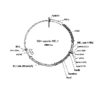
[0104] A SSA reporter construct (see, Fig. 2) targeted to the HO
locus was
generated using the yeast integrating plasmid (Yip) HO-poly-KanMX-HO (Voth et
al.
(2001) Nucleic Acids Res 29:E59-59) as follows. A fragment corresponding to
nucleotides 1 to 750 of the MEL1 gene (Liljestrom (1985) Nucleic Acids Res
13:7257-7268) (relative to the ATG) was cloned into the Sall and BamHI sites
of HO-
poly-KanMX-HO using the following primers: 5'-
aattgtcgacatgtttgetttctactttctcaccgc-
3' (SEQ ID NO:1) and 5'-aattggatccccccattggagctgcc-3' (SEQ 1D NO:2).
Subsequently, a fragment from nucleotides 299 to 2100 were cloned into the Sad
and
EcoRI sites using the following oligos: 5'-aattgagetcagaccacctgcataataacagc-3'
(SEQ
BD NO: 3) and 5'-aattgaattegggcaaaaattggtaccaatgc-3' (SEQ 11D NO:4). Finally,
a 1489
base pair fragment of the PGK1 promoter was cloned into the BsiWI and Sall
sites
using the following oligos: 5'-Aattcgtacgtctaactgatctatccaaaactg-3' (SEQ ID
NO:5)
and 5'-Aattgtcgacttgatcttttggttttatatttgttg-3' (SEQ ID NO:6).
[0105] MEL1 reporter constructs as described above were further
modified to
include a Gateway cassette (Invitrogen), following the manufacturer's
instructions.
In addition, reporter constructs were generated that included shortened
versions of the
PGK1 promoter. Constructs lacking the ampicillin resistance gene were also
generated.
[0106] Reporter constructs including various ZFN target sites for
were also
generated. Briefly, one or more copies of the target sites were generated by
PCR or
by concatamerization and inserted into the Gateway MEL1 reporter construct
using
standard molecular biology techniques. Concatamers were constructed
electronically
using DNAworks (available on the interne which designed overlapping oligos
that
tiled across the target site concatamer. The oligos were used to synthesize
the
synthetic target site concatamer and a two-step cloning process used to
introduce the
concatamer into the reporter construct. First, the concatamer was cloned into
an entry
vector (TOPO , Invitrogen, CA). In the second step, the concatamer was
transferred
from the entry vector to the MEL1 reporter using the Gateway LR ClonaseTM
system (Invitrogen), essentially as described by the manufacturer. Reporter
constructs
containing target sites for CCR5-, IPP2K-, and POU5F1/0ct34-targeted ZFNs were
generated and used for normalization.
28
CA 02700231 2010-03-19
WO 2009/042163
PCT/US2008/011087
Example 2: Integration of the Reporter Construct into Yeast
[0107] The integration of the reporter construct into the 69-1B
strain (S288C
background; MATahis3d200 lys2-1288Ieu2d1) was performed as described (Voth et
al. (2001) Nucleic Acids Res 29:E59-59). The correct integration was confirmed
by
Colony PCR using the following oligos: HO-L: 5'-
TATTAGGTGTGAAACCACGAAAAGT-3' (SEQ ID NO:7); 5'-
ACTGTCATTGGGAATGTCTTATGAT-3' (SEQ ID NO:8); HO-R: 5'-
attacgctcgtcatcaaaatca-3' (SEQ ID NO:9); and 5'-
CATGTCTTCTCGTTAAGACTGCAT-3' (SEQ ID NO:10).
Example 3: ZFN Activity Assay
[0108] To demonstrate that cleavage at the target site of the
reporter construct
restores MEL1 activity, the following experiments were performed. A SSA
reporter
construct was engineered as described above to include a recognition site of
the HO
endonuclease and integrated into host cells as described above. The cells were
then
transfected with expression vectors encoding the HO endonuclease and the cells
cultured in the presence or absence of galactose.
[0109] Results are summarized in Table 1.
Table 1: Cleavage at target site of the reporter construct restores MEL1
activity
Host Cell Expression construct
Galactose MEL1 Activity
Reporter ¨ no HO target empty vector
site
Reporter ¨ no HO target empty vector
site
Reporter ¨ no HO target pGAL HO
site
Reporter ¨ no HO target pGAL HO
site
Reporter with HO target empty vector
site
Reporter with HO target empty vector +
site
29
CA 02700231 2010-03-19
WO 2009/042163
PCT/US2008/011087
Reporter with HO target pGAL HO -/+
site
Reporter with HO target pGAL HO +++
site
[0110] As shown in Table 1, MEL1 activity was only observed in the
presence
of the HO endonuclease, and of its target in the reporter locus. Furthermore,
essentially no MEL1 gene activity was observed when HO expression was not
induced, with the exception of very low-frequency spontaneous MEL1 restoration
events. Induction of HO endonuclease expression converted essentially 100% of
the
cells in the sample to a MEL1 state.
Example 4: Identification of Persistently Biologically Active NME-specific
ZFNs
[0111] ZFNs were designed to recognize sequences within NME and plasmids
comprising sequences encoding these designed NME ZFNs were constructed
essentially as described in Urnov et al. (2005) Nature 435(7042):646-651. The
ZFNs
were tested in in vitro assays (ELISA). Figure 3 shows the information about
the
NME-binding ZFNs and their DNA binding characteristics in vitro.
[0112] For in vivo screening, the coding sequences of the ZFNs were
transferred to galactose inducible expression vectors using standard cloning
procedures (Moehle et al. (2007) Proc Natl Acad Sci USA 104:3055-3060; Mumberg
et al. (1994) Nucleic Acids Res 22:5767-5768; Urnov et al. (2005) Nature
435:646-
651).
[0113] The NME1 cDNA (RZPD 1RAUp969A091D6) target site was
subcloned into the reporter construct and the resulting reporter construct was
integrated into the genome of the yeast strain as described in Example 2.
A. Reporter Activity
[0114] Expression constructs encoding NME1-targeted ZFN pairs were
transformed into the reporter strain in deep well blocks as described in Gietz
and
Woods, (2006) Methods Mol Rio! 313:107-120. In order to eliminate the
cumbersome
manipulation of Petri dishes, pools of transfonnants were selected in liquid
media.
Briefly, the cells were resuspended in 1 ml of SC His-Leu-media and incubated
for 48
CA 02700231 2010-03-19
WO 2009/042163
PCT/US2008/011087
hours at 30 C. To further enrich for transformants, a 1:10 dilution of the
pool was
incubated in fresh media for another 24 hours.
[0115] To de-repress the GAL1 promoter, the pools of transformants
were
diluted 1:10 into 1 mL of SC His-Leu- media containing 2% raffinose as a
source of
carbon and incubated 0/N at 30 C. ZFN expression was induced by diluting the
raffinose cultures 1:10 into lml of SC His-Leu- media containing 2% galactose.
Cells
were then incubated for various amount of time, typically from 2 to 6 hours,
before
addition of 2% glucose to stop expression. Cells were then incubated overnight
to
allow for DSB repair and reporter gene expression.
[0116] In order to normalize the reporter signal to the amount of cells in
the
culture, spectrophotometric readings of the well blocks were taken at 600 nm.
The
deep well block was then centrifuged at 3000g for 5 minutes to pellet yeast
cells and
10 Al of the media is assayed for Mell activity as described in Chen et al.
(2004) Anal
Biochem 335:253-259 and Ryan et al. (1998) Mol Cell Biol 18:1774-1782.
[0117] Results of reporter gene expression are shown in FIG. 4A. The ZFN
pairs are indicated below the bars. For each pair, the left-most bar shows
Mell
activity prior to introduction of the indicated ZFN pairs, the second bar from
the left
shows Mell activity 2 hours after the indicated ZFN pairs are introduced into
the
cells; the third bar from the left shows Mell activity 4 hours after the
indicated ZFN
pairs are introduced into the cells; and the right-most bar shows Mel 1
activity 6 hours
after the indicated ZFN pairs are introduced into the cells. As shown, there
is no
Mell activity in the absence of an active ZFN pair. Furthermore, although all
ZFN
pairs were active to some degree, the assay provided a ranking of the most
active
pairs.
B. Toxicity
[0118] To evaluate toxicity of the various ZFNs, ZFN expression was
induced
by diluting the raffinose cultures 1:100 into lml of SC His-Leu-media
containing 2%
galactose. Cells were then incubated for various amount of time, typically
from 24 to
30 hours. Growth of the populations were then determined by spectrophotometry
reading at 600 nm.
[0119] Yeast cell growth in the presence of ZFNs is shown in Fig. 4B.
The
ZFN pairs are indicated below the bars. For each pair, the left-most bar shows
0D600 prior to introduction of the indicated ZFN pairs, the second bar from
the left
31
CA 02700231 2010-03-19
WO 2009/042163
PCT/US2008/011087
shows 0D600 readings 23 hours after the indicated ZFN pairs are introduced
into the
cells; the third bar from the left shows ()Dom 27 hours after the indicated
ZFN pairs
are introduced into the cells; and the right-most bar shows 0D600 30 hours
after the
indicated ZFN pairs are introduced into the cells. As shown, yeast cells grow
normally in the presence of certain ZFN pairs.
C. Yeast Screen Predicts in vivo Activity
[0120] Various active ZFN pairs identified in the reporter activity
assay (Fig.
4A) were then transformed into human K562 and tested for their ability to
induce
mutations in the NME 1 locus, as described in Miller et al. (2007) Nat
Biotechnol.
25(7):778-85.
[0121] As shown in Fig. 5, the most active ZFN pair against NME1 in a
human cultured cell line is 13674-13677. Furthermore, this ZFN pair was
identified
above as one of the most active proteins (Fig. 4A) that did not affect yeast
cell growth
(Fig. 4B).
[0122] One of the major problems associated with gene modification in
human cell lines is the gradual loss of modified cells over time. With ZFNs,
for
example, after nine days in culture, the percent of gene modified cells often
drops off,
perhaps due in part to the toxicity associated with overexpression of non-
specific
ZFNs. See, in Miller et al. (2007) Nat Biotechnol. 25(7):778-85. Accordingly,
to
further confirm that the ZFN pair identified from the yeast reporter and
toxicity
screens remained nontoxic over time, the percent modification of human
chromosomes in K562 cells was measured 2 days and 9 days after introduction of
the
ZFN pair 13674-13677.
[0123] As shown in Fig. 6, the percent modification of the NME1 locus
observed two days (D2) after transfection of the 13674-13677 ZFN pair was
maintained after nine days (D9) of culture.
[0124] These results demonstrate the yeast system described herein
accurately
predicts in vivo ZFN activity and, in addition, also accurately predicts which
ZFNs
give a persistent signal over time.
Example 5: Identification of Persistently Biologically Active PD1-specific
ZFNs
[0125] ZFNs were assembled against the human PD1 gene and were tested
by
ELISA and CEL1 assays as described in Miller et al. (2007) Nat. Biotechnol.
25:778-
32
CA 02700231 2015-04-28
785 and U.S. Patent Publication No. 20050064474 and International Patent
Publication W02005/014791.
101261 From this initial in vitro screen, two lead ZFN pairs were
identified
and submitted for elaboration in order to try to improve their efficiency.
These pairs
target exons 1 and 5 of this gene, respectively. The elaborated (improved)
proteins
were retested in a time-course experiment, essentially as described in Example
4
above. The results are summarized in Table 2 below.
Table 2: PD1 NHEJ
% NHEJ
Target ZFN pair Day 3 Day 7 Day 9
exon 1 12942/12946 8 7 5
exon 1 12942/12947 10 6 6
exon 5 12934/12971 11 6 1.5
exon 5 12934/12972 11 7.5 2
[01271 As shown in Table 2, treatment of cells with ZFNs against exon
5
causes the loss of a greater proportion of genome-edited cells from the
population,
while the genome-editing signal in cells treated with ZFNs designed against
exon 1 is
much more stable.
101281 These ZFNs were also tested in the yeast system for activity and for
toxicity. Briefly, the PD1 cDNA (NM 005018) was subcloned into the SSA
reporter
construct and the assays performed as described in Example 3.
10129] As shown in Fig. 7, the in vivo assay system clearly confirmed
the
activity of the PD1 ZFNs (Fig. 7A) and also determined the toxicity for all
ZFNs
targeting exon 5 (Fig. 7B). These results correlate with the loss of signal
detected in
human cells. Furthermore, the ZFNs designed against exon 1 show no impairment
of
yeast growth and maintain the signal in human cells.
Example 6: Identification of Biologically Active Zebrafish ZFNs
101301 Panels of ZFNs were assembled against the zebrafish SLC24A5
("golden") and notail ("NTL") genes and screened for activity in a yeast host
cells
comprising an SSA reporter construct with the appropriate target sequence.
33
CA 02700231 2015-04-28
[0131] Results of yeast activity and toxicity screens are shown in
Fig. 9A
(golden ZFN activity); Fig. 9B (golden ZFN toxicity); Fig. 10A (notail ZFN
activity);
and Fig. 10B (notail ZFN toxicity).
101321 ZFNs identified by the yeast screen were then injected into
zebrafish
embryos. Only embryos expressing the golden-ZFNs exhibited somatic mosaicism
for pigmentation (Fig. 11). Similarly, only embryos expressing the NTL-ZFNs
exhibited the forked-tail phenotype (Fig. 12).
[0133] Thus, the in vivo assay system described herein identified ZFNs
that
are biologically active in zebrafish.
Example 7: Identification of active ZFN variants using a selection-based
screen
[01341 Active ZFN variants were selected using a positive and negative
selection reporter yeast strain as follows. The reporter construct (Fig. 8)
was
generated and contained a homodimer recognition site for a well characterized
ZFN
(8266). See, Table 1 of U.S. Patent Publication No. 20080159996 for
recognition
helix and target sequence of 8266. The construct also contained a
counterselectable
gene (URA3) between the interrupted MEL1 gene as described in Example 4. In
yeast cells containing this reporter, cells containing an inactive ZFN will be
killed in
the presence of 5-FOA permitting the selective expansion of cells containing
active
ZFN variants in the population. Similarly, cells containing an active ZFN
cannot
grow in the absence of uracil.
[0135] ZFNs containing five different linker sequences (different in
length
and/or amino acid residues) between the ZFP and the FokI cleavage domain were
prepared and tested in the yeast screen assay described above. ZFNs with the
wild-
type linker sequence (8266) were active. In addition, two moderately active
and two
inactivate variants were identified. A pool of ZFNs containing 8266 as well as
the
two moderately active and two inactive variants was prepared and tested in a
yeast
counterselection assay as follows.
[01361 The ZFN variants were mixed in equimolar ratios and the
reporter
strains was transformed as described in Example 4. ZFN induction was performed
as
in Example 4 for 6 and 24 hours. After a recovery period of 16 hours, the
cells were
diluted 100 fold in media containing the counterselecting agent 5-FOA for 20
hours.
As a verification that the active ZFNs had cleaved the URA3 gene, the cells
were
similarly diluted into media lacking added uracil. As can be seen from the
right
34
CA 02700231 2015-04-28
portion of Figure 13, the cells containing active ZFN (SBS 8266) were not able
to
grow in the absence of uracil. As shown in Figure 13, yeast growth is observed
only
when active ZFN were used to transform the reporter strain.
[0137] the cells were then harvested, total DNA was extracted from the
yeast
population and individual plasmids were obtained by retransforming into E.
coli.
Twelve colonies were picked at random and sequenced. Of the 12, 9 sequences
matched the wild-type linker, 3 sequences corresponded to the moderately
active
variants, while no sequences corresponded to the inactive variants. Thus, it
was
possible to specifically enrich the most active variants within a population
of ZFN
variants.
[0138] These data validate the yeast system for the identification of
biologically active ZFN pairs from a set of biochemically active variants and
also
demonstrate the use of the toxicity assay for the determination of their
relative
specificity.
35