Note : Les descriptions sont présentées dans la langue officielle dans laquelle elles ont été soumises.
CA 02708004 2010-06-04
WO 2008/070117 PCT/US2007/024907
CONJOINT THERAPY FOR TREATING FIBROTIC DISEASES
BACKGROUND OF THE INVENTION
The process of tissue repair as a part of wound healing involves two phases.
The first
phase is the regenerative phase, in which injured cells are replaced by cells
of the same type.
The second phase is the formation of fibrous tissues, also called fibroplasia
or fibrosis, in
which connective tissue replaces normal parenchymal tissues. The tissue repair
process can
become pathogenic if the fibrosis phase continues unchecked, leading to
extensive tissue
remodeling and the formation of permanent scar tissue.
1 o It has been estimated that up to 45% of deaths in the United States can
be attributed to
fibroproliferative diseases, which can affect many tissues and organ systems.
Major organ
fibrotic diseases include interstitial lung disease (ILD), characterized by
pulmonary
inflammation and fibrosis. ILD is known to have a number of causes such as
sarcoidosis,
silicosis, collagen vascular diseases, and systemic scleroderma. However,
idiopathic
pulmonary fibrosis, a common type of ILD, has no known cause. Other organ
fibrotic
disorders include liver cirrhosis, liver fibrosis resulting from chronic
hepatitis B or C
infection, kidney disease, heart disease, and eye diseases including macular
degeneration and
retinal and vitreal retinopathy. Fibroprolifera.tive disorders also include
systemic and local
scleroderma, keloids and hypertrophic scars, atherosclerosis, and restenosis.
Additional
fibroproliferative diseases include excessive scarring resulting from surgery,
chemotherapeutic drug-induced fibrosis, radiation-induced fibrosis, and
injuries and burns.
Currently, treatments are available for fibrotic disorders including general
immunosuppressive drugs such as corticosteroids, and other anti-inflammatory
treatments.
However, the mechanisms involved in regulation of fibrosis appear to be
distinctive from
those of inflammation, and anti-inflammatory therapies are not always
effective in reducing
or preventing fibrosis. Therefore, a need remains for developing treatments to
reduce and
prevent fibrosis and control fibrotic disorders.
Wound healing and the disregulated events leading to fibrosis both involve the
proliferation and differentiation of fibroblasts and the deposition of
extracellular matrix.
Whether these fibroblasts are locally derived or from a circulating precursor
population is
unclear. Fibrocytes are a distinct population of fibroblast-like cells derived
from peripheral
blood monocytes that enter sites of tissue injury to promote angiogenesis and
wound healing.
- 1 -
CA 02708004 2010-06-04
WO 2008/070117 PCT/US2007/024907
Recently, it has been reported that CD14[+] peripheral blood monocytes
cultured in the
absence of serum or plasma differentiate into fibrocytes within 72 hours, but
that serum
amyloid P (SAP) was able to inhibit fibrocyte differentiation at levels
similar to those found
in plasma. In contrast, depleting SAP reduces the ability of plasma to inhibit
fibrocyte
differentiation. Compared with sera from healthy individuals and patients with
rheumatoid
arthritis, sera from patients with scleroderma and mixed connective tissue
disease, two
systemic fibrotic diseases, were less able to inhibit fibrocyte
differentiation in vitro and had
correspondingly lower serum levels of SAP. These results suggest that low
levels of SAP
may thus augment pathological processes leading to fibrosis. These data also
suggest
mechanisms to inhibit fibrosis in chronic inflammatory conditions, or
conversely to promote
wound healing.
As SAP binds to Fc receptors for immunoglobulin G (IgG; FcRs), FcR activation
was
subsequently demonstrated to be an inhibitory signal for fibrocyte
differentiation. FcR are
activated by aggregated IgG, and it has been shown that aggregated but not
monomeric,
human IgG inhibits human fibrocyte differentiation. Monoclonal antibodies that
bind to FcRI
(CD64) or FcRII (CD32) also inhibit fibrocyte differentiation. Aggregated IgG
lacking Fc
domains or aggregated IgA, IgE, or IgM do not inhibit fibrocyte
differentiation. Incubation of
monocytes with aggregated IgG, like SAP, inhibited fibrocyte differentiation.
Using
inhibitors of protein kinase enzymes, it has also been shown that Syk- and Src-
related
tyrosine kinases participate in the inhibition of fibrocyte differentiation.
These observations
suggest that fibrocyte differentiation can occur in situations where SAP and
aggregated IgG
levels are low, such as the resolution phase of inflammation.
BRIEF SUMMARY OF THE INVENTION
The present invention relates to the use of conjoint therapies for treating
fibrotic and
fibroproliferative disorders, involving administering a combination of agents
that suppress
fibrocyte formation ("fibrocyte suppressors") with agents that inhibit
activation of resident
collagen producing cells such as fibroblasts, myofibroblasts, or
myofibrocytes, such as
antagonist of TGF-13 and other profibrotic factors (collectively "profibrotic
factor
antagonists").
In certain embodiments, the subject method and compositions can be practiced
using
such fibrocyte suppressors as serum amyloid P (SAP), IL-12, Laminin-1, anti-
FcyR
- 2 -
CA 02708004 2010-06-04
WO 2008/070117 PCT/US2007/024907
antibodies that are able to cross-link FcyR, aggregated IgG, cross-linked IgG
and/or
combinations thereof. Designations for "SAP", "IL-12", "Laminin-1", IgG and
anti-FcyR
antibodies as used herein also refer to functional fragments of these proteins
unless it is clear
that such fragments are excluded from the usage in a given context. In
certain
embodiments, the fibrocyte suppressor is an agent that induces apoptosis of
monocytes, such
as an IL-15 antagonist.
In certain embodiments, the profibrotic factor antagonists are selected from
antagonists of peptide growth factors, cytokines, chemokines, and the like.
Examples of such
factors that may be antagonized by the subject profibrotic factor antagonists
include
transforming growth factor type beta (TGF-13), VEGF, EGF, PDGF, IGF, RANTES,
members
of the interleukin family (e.g., IL-1, IL-4, IL-5, IL-6, IL-8 and IL-13),
tumor necrosis factor
type alpha (TNF-a), platelet-derived growth factor (PDGF), basic fibroblast
growth factor
(bFGF), monocyte chemoattractant protein type 1 (MCP-1), macrophage
inflammatory
protein (e.g., MIP-la, MIP-2), connective tissue growth factor (CTGF),
endothelin-1,
angiotensin-II, leptin, chemokines (e.g., CCL2, CCL12, CXCL12, CXCR4, CCR3,
CCR5,
CCR7, SLC/CCL21), integrins (e.g., a1131, a2í31 avr36, av133), tissue
inhibitors of matrix
metalloproteinases (e.g., TIMP-1, TIMP-2) and other factors known to promote
or be related
to the formation, growth, or maintenance of fibrotic tissue.
In certain embodiments, the profibrotic factor antagonists can be replaced
with, or
augmented with, a cytokine known to have anti-fibrotic effects itself, such as
IFN-y, BMP-7,
HGF or IL-10.
Such components of the combined treatment may be administered to a target
location
as part of a single formulation, in which the single formulation includes
components for
targeting both events. In other selected embodiments of the present invention,
the
components may be administered as separate formulations.
A decrease in or suppression of both differentiation of fibrocytes and the
formation
and maintenance of fibrotic tissue may alleviate symptoms of numerous
fibrosing diseases or
other disorders caused by fibrosis. For example, it may be used to treat
fibrosis in the liver,
kidney, lung, heart and pericardium, eye, skin, mouth, pancreas,
gastrointestinal tract, brain,
breast, bone marrow, bone, genitourinary, a tumor, or a wound.
The present invention provides methods for modulating fibroblast accumulation
and
collagen deposition in a tissue.
- 3 -
CA 02708004 2010-06-04
WO 2008/070117 PCT/US2007/024907
DETAILED DESCRIPTION OF THE DRAWINGS
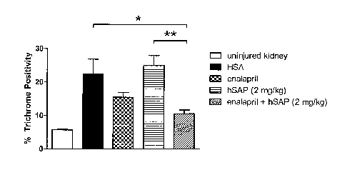
Figure 1 depicts the extent of Trichrome staining in a rat kidney unilateral
ureter obstruction
(UUO) injury model.
DETAILED DESCRIPTION OF CERTAIN EMBODIMENTS
I. Overview
The regulation of events leading to fibrosis involves at least two major
events. One is
the proliferation and differentiation of fibrocytes. Fibrocytes are a distinct
population of
io fibroblast-like cells derived from peripheral blood monocytes that
normally enter sites of
tissue injury to promote angiogenesis and wound healing. Fibrocytes
differentiate from
CD14+ peripheral blood monocytes, and may differentiate from other PBMC cells.
The
presence of SAP, IL-12, Laminin-1, anti-FcyR antibodies, crosslinked IgG
and/or aggregated
IgG may inhibit or at least partially delay this process.
The second major event is the formation and maintenance of fibrotic tissue.
Fibrotic
tissue may be formed and maintained by the recruitment and proliferation of
fibroblast cells,
the formation of new extracellular matrix, and the growth of new vascular
tissue. In
pathologic fibrosis, such as following chronic inflammation, injury, or
idiopathic fibrosis, it is
this excess fibrotic tissue that can lead to tissue damage and destruction.
Since both of the foregoing events are necessary for fibrosis, treatments of
the present
invention include combined compositions and methods in which both of these
events are
targeted. In selected embodiments, the present invention include at least one
composition, or
administration thereof to a target location, that is suitable for the
inhibition or delay of
fibrocyte differentiation, and at least one component, or administration
thereof to the target
location, that is suitable for the inhibition or antagonizing of profibrotic
factors. In selected
embodiments, these components may be formulated or administered as a combined
composition, or may separately and/or independently administered to the target
locations.
The present invention provides methods for treating fibrotic and
fibroproliferative
disorders. The method generally involves administering an effective amount of
a fibrocyte
suppressor in combination with an effective amount of profibrotic factor
antagonist. The
methods provide for treatment of fibrotic diseases, including those affecting
the lung, liver,
heart, kidney and eye. To further illustrate, the subject method can be used
to treat such
- 4 -
CA 02708004 2010-06-04
WO 2008/070117 PCT/US2007/024907
fibroproliferative diseases as glomerulonephritis (GN); diabetic nephropathy;
renal interstitial
fibrosis; renal fibrosis resulting from complications of drug exposure; HIV-
associated
nephropathy; transplant necropathy; liver cirrhosis due to all etiologies;
disorders of the
biliary tree; hepatic dysfunction attributable to infections; pulmonary
fibrosis; adult
respiratory distress syndrome (ARDS); chronic obstructive pulmonary disease
(COPD);
idiopathic pulmonary fibrosis (IPF); acute lung injury (ALI); pulmonary
fibrosis due to
infectious or toxic agents; congestive heart failure; dilated cardiomyopathy;
myocarditis;
vascular stenosis; progressive systemic sclerosis; polymyositis; scleroderma;
Grave's disease;
dermatomyositis; fascists; Raynaud's syndrome, rheumatoid arthritis;
proliferative
i o vitreoretinopathy; fibrosis associated with ocular surgery; acute
macular degeneration, and
excessive or hypertrophic scar or keloid formation in the dermis occurring
during wound
healing resulting from trauma or surgical wounds. Still other exemplary
fibrotic disorders that
can be treated with the subject conjoint therapy are described in further
detail below. The
etiology may be due to any acute or chronic insult including toxic, metabolic,
genetic and
infectious agents.
In some embodiments, an effective amount of fibrocyte suppressor and an
effective
amount of profibrotic factor antagonist are amounts that, when administered in
combination
therapy, are effective to reduce fibrosis by at least about 10%, and more
preferably at least
about 15%, 20%, 25%, 30%, 35%, 40%, 45%, or even at least about 50%, or more,
compared
with the degree of fibrosis in the individual prior to treatment with the
combination therapy.
In other embodiments, the present invention provides methods that involve
administering a synergistic combination of fibrocyte suppressor and
profibrotic cytokine
antagonist. As used herein, a "synergistic combination" of fibrocyte
suppressor and
profibrotic cytokine antagonist is a combined dosage that is more effective in
the therapeutic
or prophylactic treatment than the incremental improvement in treatment
outcome that could
be predicted or expected from a merely additive combination of (i) the
therapeutic or
prophylactic benefit of a fibrocyte suppressor when administered at that same
dosage as a
monotherapy and (ii) the therapeutic or prophylactic benefit of the
profibrotic cytokine
antagonist when administered at the same dosage as a monotherapy.
II. Definitions
- 5 -
CA 02708004 2010-06-04
WO 2008/070117 PCT/US2007/024907
As used herein, the terms "treatment", "treating", and the like, refer to
obtaining a
desired pharmacologic and/or physiologic effect. The effect may be
prophylactic in terms of
completely or partially preventing a disorder or symptom thereof and/or may be
therapeutic
in terms of a partial or complete cure for a fibrotic or fibroproliferative
disorder and/or
adverse affect attributable to the disorder. "Treatment", as used herein,
covers any treatment
of a disease in a mammal, particularly in a human, and includes: (a)
increasing-survival time;
(b) decreasing the risk of death due to the disease; (c) preventing the
disease from occurring
in a subject which may be predisposed to the disease but has not yet been
diagnosed as
having it; (d) inhibiting the disease, i.e., arresting its development (e.g.,
reducing the rate of
disease progression); and (e) relieving the disease, i.e., causing regression
of the disease.
As used herein the term "subject" refers to animals including mammals
including
humans. The term "mammal" includes primates, domesticated animals including
dogs, cats,
sheep, cattle, goats, pigs, mice, rats, rabbits, guinea pigs, captive animals
such as zoo
animals, and wild animals. As used herein the term "tissue" refers to an organ
or set of
specialized cells such as skin tissue, lung tissue, kidney tissue, and other
types of cells.
The term "therapeutically effective amount" is meant an amount of a fibrocyte
suppressor or profibrotic factor antagonist, or a rate of delivery of such
therapeutic agents,
effective to facilitate a desired therapeutic effect. The precise desired
therapeutic effect will
vary according to the fibrotic or fibroproliferative condition to be treated,
the formulation to
be administered, and a variety of other factors that are appreciated by those
of ordinary skill
in the art.
As used herein the terms "fibroproliferative disorder" and "fibrotic disorder"
refer to
conditions involving fibrosis in one or more tissues. As used herein the term
"fibrosis" refers
to the formation of fibrous tissue as a reparative or reactive process, rather
than as a normal
constituent of an organ or tissue. Fibrosis is characterized by fibroblast
accumulation and
collagen deposition in excess of normal deposition in any particular tissue.
As used herein the
term "fibrosis" is used synonymously with "fibroblast accumulation and
collagen deposition".
Fibroblasts are connective tissue cells, which are dispersed in connective
tissue throughout
the body. Fibroblasts secrete a nonrigid extracellular matrix containing type
I and/or type III
collagen. In response to an injury to a tissue, nearby fibroblasts migrate
into the wound,
proliferate, and produce large amounts of collagenous extracellular matrix.
Collagen is a
fibrous protein rich in glycine and proline that is a major component of the
extracellular
- 6 -
,
CA 02708004 2010-06-04
WO 2008/070117
PCT/US2007/024907
matrix and connective tissue, cartilage, and bone. Collagen molecules are
triple-stranded
helical structures called a-chains, which are wound around each other in a
ropelike helix.
Collagen exists in several forms or types; of these, type I, the most common,
is found in skin,
tendon, and bone; and type III is found in skin, blood vessels, and internal
organs.
Fibrotic disorders include, but are not limited to, systemic and local
scleroderma,
keloids and hypertrophic scars, atherosclerosis, restinosis, pulmonary
inflammation and
fibrosis, idiopathic pulmonary fibrosis, liver cirrhosis, fibrosis as a result
of chronic hepatitis
B or C infection, kidney disease, heart disease resulting from scar tissue,
and eye diseases
such as macular degeneration, and retinal and vitreal retinopathy. Additional
fibrotic diseases
0 include fibrosis resulting from chemotherapeutic drugs, radiation-induced
fibrosis, and
injuries and burns.
"Scleroderma" is a fibrotic disorder characterized by a thickening and
induration of
the skin caused by the overproduction of new collagen by fibroblasts in skin
and other
organs. Scleroderma may occur as a local or systemic disease. Systemic
scleroderma may
affect a number of organs. Systemic sclerosis is characterized by formation of
hyalinized and
thickened collagenous fibrous tissue, with thickening of the skin and adhesion
to underlying
tissues, especially of the hands and face. The disease may also be
characterized by dysphagia
due to loss of peristalsis and submucosal fibrosis of the esophagus, dyspnea
due to pulmonary
fibrosis, myocardial fibrosis, and renal vascular changes. Pulmonary fibrosis
affects 30 to
70% of scleroderma patients, often resulting in restrictive lung disease.
"Idiopathic pulmonary fibrosis" is a chronic, progressive and usually lethal
lung
disorder, thought to be a consequence of a chronic inflammatory process.
As used herein the term "profibrotic factors" refers to cytokines, growth
factors or
chemokines which have been observed to promote the accumulation of fibroblasts
and
deposition of collagen in various tissues. A number of cytokines and growth
factors have
been reported to be involved in regulating tissue remodeling and fibrosis.
These include the
"profibrotic cytokines" such as transforming growth factor beta (TGF-0),
interleukin-4 (IL-
4), interleukin-5 (IL-5), and interleukin-13 (IL-13), which have been shown to
stimulate
collagen synthesis and fibrosis in fibrotic tissues (Letterio et al. Ann Rev.
Immunol. 16, 137-
161 (1998), Fertin et al., Cell Mol. Biol. 37, 823-829 (1991), Doucet et al.,
J. Clin. Invest.
101, 2129-2139 (1998). Interleukin-9 (IL-9) has been shown to induce airway
fibrosis in the
lungs of mice (Zhu et al., J. Clin. Invest. 103, 779-788(1999)). In addition
to TGF-P, other
- 7 -
CA 02708004 2010-06-04
WO 2008/070117 PCT/US2007/024907
cytokines or growth factors which have been reported to increase fibrosis in
the fibrotic
disorder idiopathic pulmonary fibrosis (IPF) include granulocyte/macrophage-
colony
stimulating factor (GM-CSF), tumor necrosis factor alpha (TNF-a), interleukin-
1 beta OL-
IN, and connective tissue growth factor (CTGF) (Kelly et al. Curr
Pharmaceutical Des 9: 39-
49 (2003)). Cytokines and growth factors reported to be involved in promoting
pulmonary
fibrosis occurring in scleroderma include TGF-P, interleukin-1 beta (IL-1
interleukin-6
(IL-6), oncostatin M (OSM), platelet derived growth factor (PDGF), the type 2
cytokines IL-
4 and IL-13, IL-9, monocyte chemotactic protein 1 (CCL2/MCP-1), and pulmonary
and
activation-regulated chemokine (CCL18/PARC) (Atamas et al., Cyto Growth Fact
Rev 14:
537-550 (2003)).
As used herein, the terms "antibody" and "antibodies" refer to monoclonal
antibodies,
multispecific antibodies, human antibodies, humanized antibodies, synthetic
antibodies,
chimeric antibodies, camelized antibodies, single-chain antibodies or single-
chain Fvs (scFv),
Fab fragments, F(ab') fragments, disulfide-linked Fvs (sdFv), and intrabodies.
In addition,
unless otherwise indicated (such as in the case of aggregated IgG), the term
includes epitope-
binding fragments of any of the above. In particular, antibodies include
immunoglobulin
molecules and immunologically active fragments of immunoglobulin molecules,
i.e.,
molecules that contain an antigen binding site. Immunoglobulin molecules can
be of any type
(e.g., IgG, IgE, IgM, IgD, IgA and IgY), class or subclass.
As used herein, the terms "single-chain Fv" or "scFv" refer to antibody
fragments
comprise the VH and VL domains of antibody, wherein these domains are present
in a single
polypeptide chain. Generally, the Fv polypeptide further comprises a
polypeptide linker
between the VH and VL domains which enables the scFv to form the desired
structure for
antigen binding. In specific embodiments, scFvs include humanized scFvs.
As used herein, the term "humanized antibody" refers to forms of non-human
(e.g.,
murine) antibodies that are chimeric antibodies which contain minimal sequence
derived
from non-human immunoglobulin. For the most part, humanized antibodies are
human
immunoglobulins (recipient antibody) in which hypervariable region residues of
the recipient
are replaced by hypervariable region residues from a non-human species (donor
antibody)
such as mouse, rat, rabbit or non-human primate having the desired
specificity, affinity, and
capacity. In some instances, Framework Region (FR) residues of the human
immunoglobulin
are replaced by corresponding non-human residues. Furthermore, humanized
antibodies may
- 8 -
CA 02708004 2010-06-04
WO 2008/070117 PCT/US2007/024907
comprise residues which are not found in the recipient antibody or in the
donor antibody.
These modifications are made to further refine antibody performance. In
general, the
humanized antibody will comprise substantially all of at least one, and
typically two, variable
domains, in which all or substantially all of the hypervariable regions
correspond to those of a
non-human immunoglobulin and all or substantially all of the FRs are those of
a human
immunoglobulin sequence. The humanized antibody may comprise at least a
portion of an
immunoglobulin constant region (Fc) that has been altered by the introduction
of amino acid
residue substitutions, deletions or additions (i.e., mutations). In some
embodiments, a
humanized antibody is a derivative. Such a humanized antibody comprises amino
acid
residue substitutions, deletions or additions in one or more non-human CDRs.
The humanized
antibody derivative may have substantially the same binding, better binding,
or worse binding
when compared to a non-derivative humanized antibody. In specific embodiments,
one, two,
three, four, or five amino acid residues of the CDR have been substituted,
deleted or added
(i.e., mutated).
III. Exemplary Embodiments
A. Suppressors of Fibrocyte Proliferation and Differentiation
One component of the conjoint therapies of the instant invention are agents
that
suppress fibrocyte formation. These fibrocyte suppressors, as they are
generically referred to
herein, are agents that may act on CD14[+] peripheral blood monocytes in a
manner that
either suppresses the formation of fibrocytes (e.g., inhibits differentiation
or proliferation), or
in other embodiments, causes the ablation (cell death) of the monocytes.
In certain embodiments, the fibrocyte suppressor is an agent that causes FcyR-
dependent activation of Syk- and Src-related tyrosine kinases in monocytes. In
other
embodiments, the fibrocyte suppresseor can be an agent that works downstream
of the FcyR
complex, causing FcyR-independent activation of a Syk- and Src-related
tyrosine kinase in
monocytes in a manner that suppresses fibrocyte formation. Various small
molecule
activators of the syk-kinase, such as phospho-ITAM peptides and
peptidomimetics thereof,
may be useful for this purpose.
Other exemplary fibrocyte suppressors include:
(0 Serum Amyloid P
- 9 -
CA 02708004 2010-06-04
WO 2008/070117
PCT/US2007/024907
It has been previously identified that fibrocytes may differentiate from CD14+
peripheral blood monocytes, and the presence of human serum dramatically
delays this
process. The factor in human serum that inhibits fibrocyte differentiation is
serum amyloid P
(SAP). SAP, a member of the pentraxin family of proteins that includes C-
reactive protein
(CRP), is produced by the liver, secreted into the blood, and circulates in
the blood as stable
pentamers. SAP binds to receptors for the Fc portion of IgG antibodies (FcyR)
on a variety
of cells and may effectively cross-link FcyR without additional proteins
because SAP is a
pentameric protein with five potential FcyR binding sites per molecule. As SAP
binds to
FcyR, intracellular signaling events consistent with FcyR activation are
initiated.
o In specific embodiments of the present invention, compositions
containing SAP may
be operable to raise SAP concentration in target locations to approximately at
least 0.5 lig/ml.
In humans, 1125 radiolabelled SAP has been previously administered to study
patients with
amyloidosis. In the treatments, approximately 600 [ig of SAP was administered
to an adult
human. Accordingly, administration of approximately 600 lig of SAP
systemically to an
adult human is safe. Higher dosages may also be safe under appropriate
conditions.
SAP supplied in certain compositions of the present invention may include the
entire
SAP protein or a portion thereof, preferably the portion functional in
suppression of fibrocyte
formation. In one embodiment, the functional portion of SAP is selected from
the region that
does not share sequence homology with CRP, which has no effect on fibrocyte
formation.
For instance amino acids 65-89 (KERVGEYSLYIGRHKVTPKVIEKFP-SEQ.ID.N0.1) of
SAP are not homologous to CRP. Amino acids 170-181 (ILSAYAYQGTPLPA-
SEQ.ID.N0.2) and 192-205 (IRGYVIIKPLV-SEQ.ID.N0.3) are also not homologous.
Additionally a number of single amino acid differences between the two
proteins are known
and may result in functional differences.
(ii) IL-12
IL-12 has been previously implicated in fibrosis and fibrosing diseases, but
most
studies have focused on the role of IL-12 in promoting the Thl immune response
or by
triggering the production of interferon-y.
Compositions containing IL-12 may be operable to raise the IL-12 concentration
in
target locations to approximately 0.1 to 10 ng/ml.
(iii) Laminin-1
- 10 -
CA 02708004 2010-06-04
WO 2008/070117 PCT/US2007/024907
Laminins are extracellular matrix proteins involved in movement of monocytes
from
the circulation into tissues. In order for leukocytes to enter tissues, they
must cross through
endothelial cells and the surrounding basement membrane of blood vessel wall.
This process
involves the tethering, rolling and stopping of the leukocytes on the
endothelial cells.
Following adhesion to the endothelial cells, leukocytes then cross between the
endothelial
cells, through the blood vessel wall and into the tissues. The process of
extravasation of cells
through blood vessel walls alters their phenotype and function.
These events are controlled by a series of cell surface adhesion receptors,
including
integrins. Integrins bind to a wide variety of ligands, including
extracellular matrix proteins
(ECM), such as fibronectin, vitronectin, collagen and laminin. Matrix proteins
are present
within the basement of the blood vessel wall, including laminins. Laminins are
a large family
of glycoproteins, with a heterotrimeric structure of a, 13 and y chains. The
use of different a,
13 and y chains leads to the expression of at least 12 different laminin
isoforms. Different
laminins are expressed at different stages of development and at different
sites within the
body.
Compositions containing Laminin-1 may be operable to raise the Laminin-1
concentration in target locations to approximately 1 to 10 [tg/ml.
(iv) Anti-FcyR Antibodies
It has also been identified that Anti-FcyR antibodies may prevent the
differentiation of
peripheral blood monocytes into fibrocytes. Anti-FcyR antibodies are IgG
antibodies that
bind to receptors for the Fc portion of IgG antibodies (FcyR). The anti-FcyR
antibodies bind
through their variable region, and not through their constant (Fc) region.
However, IgG from
the appropriate source (e.g. human IgG for human receptors) may normally bind
to FcyR
through its Fc region. FcyR are found on the surface of a variety of
hematopoietic cells.
There are four distinct classes of FcyR. FcyRI (CD64) is expressed by
peripheral blood
monocytes and binds monomeric IgG with a high affinity. FcyRII (CD32) and
FcyRIII
(CD16) are low affinity receptors for IgG and only efficiently bind aggregated
IgG. FcyRII is
expressed by peripheral blood B cells and monocytes, whereas FcyRIII is
expressed by NK
cells and a subpopulation of monocytes. FcyRIV was recently identified in mice
and is
present on murine peripheral blood monocytes and neutrophils, macrophages and
dendritic
cells and efficiently binds murine IgG2a and IgG2b antibodies. There is a
putative human
- 11 -
CA 02708004 2010-06-04
WO 2008/070117 PCT/US2007/024907
FcyRIV gene, but the biological function of the protein, such as ligand
specificity and cellular
expression is, as yet unknown.
Peripheral blood monocytes express both FcyRI and FcyRII (a subpopulation of
monocytes express FcyRIII), whereas tissue macrophages express all three
classical FcyR.
Clustering of FcyR on monocytes by IgG, either bound to pathogens or as part
of an immune
complex, initiates a wide variety of biochemical events.
FcyR activation and induction of intracellular signaling pathways may occur
when
multiple FcyR are cross-linked or aggregated. This FcyR activation leads to a
cascade of
signaling events initiated by two main kinases. The initial events following
FcyR activation
io involve the phosphorylation of intracellular immunoreceptor tyrosine
activation motifs
(ITAMs) present on the cytoplasmic tail of FcyRII or the FcR-y chain
associated with FcyRI
and FcyRIII, by Src-related tyrosine kinases (SRTK). In monocytes, the main
Src-kinases
associated with FcyRI and FcyRII are hck and lyn. The phosphorylated ITAM then
recruit
cytoplasmic SH2-containing kinases, especially Syk, to the ITAMs and Syk then
activates a
series of downstream signaling molecules.
Anti-FcyR antibodies for FcyRI (anti-FcyRI) and for FcyRII (anti-FcyRII) are
able to
bind to either FcyRI or FcyRII, respectively. These FcyR may then be cross-
linked by the
binding of additional antibodies or other means. This process initiates
intracellular signaling
events consistent with FcyR activation.
Compositions containing anti-FcyRI antibodies and/or anti-FcyRII antibodies,
and/or
cross-linked or aggregated IgG, which may bind to FcyR through the Fc region,
may be used
to suppress the differentiation of fibrocytes in inappropriate locations and
in fibrosing
disorders and chronic inflammatory conditions, inter alia.
In specific embodiments, compositions containing approximately 1 pg/m1 anti-
FcyR
antibodies may be effective to inhibit fibrocyte differentiation by
approximately 50%. In
other embodiments, compositions may contain an amount sufficient to deliver 1
g/ml anti-
FcyR antibodies to the target tissue. In other specific embodiments,
compositions may
contain as little as 0.1 1.1g ml cross-linked or aggregated IgG.
Anti-FcyR antibodies may be administered in a dose of approximately 1.0 pg/mL,
in
an amount sufficient to deliver 1 g/ml anti-FcyR antibodies to the target
tissue, or in another
dose sufficient to inhibit fibrocyte differentiation without causing an
undesirable amount of
cell death in the patient. Aggregated or cross-linked IgG may be administered
in an amount
- 12 -
CA 02708004 2010-06-04
WO 2008/070117 PCT/US2007/024907
sufficient to deliver at least 0.1 [ig/m1 IgG to the target tissue, or in
another dose sufficient to
inhibit fibrocyte differentiation without causing an undesirable amount of
cell death in the
patient.
Anti-FcyR antibodies used in examples of the present disclosure include anti-
FcyRI
antibodies and anti-FcyRII antibodies.
Anti-FcyR antibodies may include any isotype of antibody.
(v) Aggregated Fc domains and Fc-containing antibodies
Cross-linked or aggregated IgG may include any IgG able to bind the target
FcyR
through its Fc region, provided that at least two such IgG antibodies are
physically connected
io to one another.
Antibodies of both types may include whole antibodies or a portion thereof,
preferably the portion functional in suppression of fibrocyte differentiation.
For example,
they may include any antibody portion able to cross-link FcyR. This may
include aggregated
or cross-linked antibodies or fragments thereof, such as aggregated or cross-
linked whole
antibodies, F(ab')2 fragments, and possible even Fc fragments.
Aggregation or cross-linking of antibodies may be accomplished by any known
method, such as heat or chemical aggregation. Any level of aggregation or
cross-linking may
be sufficient, although increased aggregation may result in increased
fibrocyte suppression.
Antibodies may be polyclonal or monoclonal, such as antibodies produced from
hybridoma
cells. Compositions and methods may employ mixtures of antibodies, such as
mixtures of
multiple monoclonal antibodies, which may be cross-linked or aggregated to
like or different
antibodies.
(w) Interleukin-15 antagonists
IL-15 antagonists encompassed by the present invention include a broad variety
of
molecules that antagonize or inhibit IL-15 activity (i.e., IL-15 mediated anti-
apoptosis)
including, but not limited to, anti-IL-15 antibodies, anti-IL-i 5R antibodies,
soluble IL-15Rs,
IL-15 muteins, anti-IL-15 small molecules and anti-IL-15R small molecules.
Other
antagonists, such as binding proteins and peptide mimetics, which are capable
of inhibiting
IL-15 activity, also are included. In a particular embodiment, the antagonist
is capable of
interfering with the assembly of the IL-15Ra, 13, and 7 subunits, e.g., the
antagonist binds to
an epitope located on the 13- or y-chain interacting domain of IL-15. In
another particular
embodiment, the antagonist is an IL-15 mutein, e.g., an IL-15 mutant that is
capable of
- 13 -
CA 02708004 2010-06-04
WO 2008/070117 PCT/US2007/024907
binding to IL-15Ra but is not able to bind to either or both of the 13- and/or
7-subunits of IL-
15R and, therefore, is not able to effect signaling.
B. Profibrotic Factor Antagonists
Another component of the subject conjoint therapies agents that inhibit or
antagonize
of profibrotic factors, such as agents that antagonize one or more growth
factors or cytokines
involved in the formation and maintenance of fibrotic tissue. In this manner,
compositions
and methods of the present invention target both fibrocyte differentiation and
fibrotic tissue
formation and maintenance as part of a combined treatment.
Profibrotic factors that may be targeted with antagonists as part of the
therapies of the
present invention include, without limitation, a growth factor type 11 (TGF-0,
including TGF-
01-5), VEGF, EGF, PDGF, IGF, RANTES, members of the interleukin family (e.g.,
IL-1, IL-
4, IL-5, IL-6, IL-8 and IL-13), tumor necrosis factor type alpha (TNF-a),
platelet-derived
growth factor (PDGF), basic fibroblast growth factor (bFGF), monocyte
chemoattractant
protein type 1 (MCP-1), macrophage inflammatory protein (e.g., MIP-la, MIP-2),
connective
tissue growth factor (CTGF), endothelin-1, angiotensin-II, leptin, chemokines
(e.g., CCL2,
CCL12, CXCL12, CXCR4, CCR3, CCR5, CCR7, SLC/CCL21), integrins (e.g., a1131,
a2131
av[36, ccv[33), tissue inhibitors of matrix metalloproteinases (e.g., TIMP-1,
TIMP-2) and other
factors known to promote or be related to the formation, growth, or
maintenance of fibrotic
tissue. The present invention may include compositions or methods that target
one or more
of the foregoing factors and cytokines.
In certain embodiments, a suitable component of the composition may include
antibodies directed to one or more of the profibrotic factors. Such antibodies
may be
purified, unpurified, or partially purified. The antibodies may be polyclonal
or monoclonal
antibodies, derived from any suitable animal source, such as mouse, rabbit,
rat, human, horse,
goat, bovine, and the like. Such antibodies may include antibody fragments,
single chain
antibodies, polymerized antibodies and/or antibody fragments, and the like.
In certain embodiments, a suitable composition may include antagonists of the
corresponding receptor of one or more of the profibrotic factors. Such
antagonists may
include inactive forms of one or more of the profibrotic factors and/or
cytokines, such as
fragments thereof Such forms in suitable concentrations may compete with its
corresponding profibrotic factors and/or cytokines for binding to its
receptor. Similarly,
- 14 -
CA 02708004 2010-06-04
WO 2008/070117 PCT/US2007/024907
certain antibodies to the receptor may be used to interfere with or prevent
binding thereto of
the corresponding profibrotic factors and/or cytokines.
In other selected embodiments, compositions of the present invention may
include
soluble forms of the receptor of one or more of the profibrotic factors and/or
cytokines, such
that the soluble receptor competes with its corresponding native cellular
receptor for the
target ligand.
In other selected embodiments, suitable components of the composition may
include
compounds that compete with or otherwise interfere with binding of one or more
of the
profibrotic factors and/or cytokines with its receptor. For example, the
proteoglycan decorin
is known to bind to TGF-13, thereby reducing its availability for binding to
its receptor.
Mannose-6-phospate is also known to compete with TGF-13 for binding to its
corresponding
receptor. Other known binding inhibitors of TGF-13 include latent transforming
growth
factor-13 binding protein (LTBP) and latency associated peptide (LAP), both of
which
natively binding to the intracellular precursor of TGF-13.
In certain embodiments, a suitable component of the composition may include
one or
more oligoribonucleotides that contain at least one sequence that is antisense
with respect to
one or more of the profibrotic factors and/or cytokines. Such components may
also include
one or more expression plasmids having suitable transcriptional control
sequences that yield
antisense sequences. In other selected embodiments of the present invention, a
suitable
component may include one or more double-stranded oligoribonucleotides, or
expression
plasmids encoding thereof, that are suitable for degrading transcripts of one
or more of the
profibrotic factors and/or cytokines via RNA-mediated interference.
A suitable profibrotic factor antagonist of the composition may include
components
known to inhibit, attenuate, or interfere with one or more components of the
intracellular
signaling pathways activated by one or more of the profibrotic factors upon
binding to its
corresponding receptor.
For example, a composition of the present invention may include components
that
inhibit or attenuate downstream signal pathway molecules such as SMAD family
members
and SARA.
A suitable component of the composition may include one or more molecules that
are
suitable for inhibiting or interfering with the cellular adhesions require for
fibrosis. For
example, a suitable component may include interfering antibodies to the ICAM-1
and/or
- 15 -
CA 02708004 2010-06-04
WO 2008/070117
PCT/US2007/024907
CD11 molecules, the extracellular matrix and/or a1I31 integrin, the
extracellular matrix
and/or a2I31 integrin, thereby interfering with the adhesion interaction there
between.
In other selected embodiments, a suitable profibrotic factor antagonist may
include
inhibitors of collagen synthesis, such as proline analogs that interfere with
post-translation
processing of collagen precursors. Pirfenidone, for example, is an orally
active small
molecule drug that may inhibit collagen synthesis, down regulate production of
multiple
cytokines and block fibroblast proliferation.
a. TGF-fl antagonists
Cytokines of the transforming growth factor (TGF) beta family play a central
role in
wound healing and in tissue repair, and are found in all tissues. TGF-I3 is
produced by many
parenchymal cell types, as well as infiltrating cells such as lymphocytes,
monocytes/macrophages, and platelets. Following wounding or inflammation, such
cells
such are potential sources of TGF-I3. In general, TGF-f3 stimulates the
production of various
extracellular matrix proteins, inhibits the degradation of these matrix
proteins, and promotes
tissue fibrosis, all of which contribute to the repair and restoration of the
affected tissue. In
many diseases, excessive TGF-I3 contributes to a pathologic excess of tissue
fibrosis that can
compromise normal organ function.
The term "TGF-I3" as used herein includes TGF-I31, TGF132, TGF-I33, TGF-I34
and
TGF-135. Also included are other related proteins with similar properties.
As used herein, a "TGF-13 antagonist" is any molecule that is able to decrease
the
amount or activity of TGF-13, either within a cell or within a physiological
system. Preferably,
the TGF-I3 antagonist acts to decrease the amount or activity of a TGF-I3 1,
2, or 3. For
example, a TGF-I3 antagonist may be a molecule that inhibits expression of TGF-
I3 at the
level of transcription, translation, processing, or transport; it may affect
the stability of TGF-13
or conversion of the precursor molecule to the active, mature form; it may
affect the ability of
TGF-I3 to bind to one or more cellular receptors (e.g., Type I, II or III); or
it may interfere
with TGF-I3 signaling.
A variety of TGF-13 antagonists and methods for their production are known in
the art
and many more are currently under development. The specific TGF-13 antagonist
employed is
not a limiting feature; any effective TGF-I3 antagonist as defined herein may
be useful in the
- 16 -
CA 02708004 2010-06-04
WO 2008/070117 PCT/US2007/024907
methods and compositions of this invention. Preferably, the TGF-P antagonist
is a TGF-131,
TGF-P2, or TGF-03 antagonist. Most preferably the antagonist is a TGF-P1
antagonist.
Examples of TGF-P antagonists include, but are not limited to: monoclonal and
polyclonal antibodies directed against one or more isoforms of TGF-0 (Dasch et
al., U.S. Pat.
No. 5,571,714; see, also, WO 97/13844 and WO 00/66631); TGF-0 receptors,
soluble forms
of such receptors (preferably soluble TGF-P type III receptor), or antibodies
directed against
TGF-P receptors (Segarini et al., U.S. Pat. No. 5,693,607; Lin et al., U.S.
Pat. No. 6,001,969,
U.S. Pat. No. 6,010,872, U.S. Pat. No. 6,086,867, U.S. Pat. No. 6,201,108; WO
98/48024;
WO 95/10610; WO 93/09228; WO 92/00330); latency associated peptide (WO
91/08291);
large latent TGF-P (WO 94/09812); fetuin (U.S. Pat. No. 5,821,227); decorin
and other
proteoglycans such as biglycan, fibromodulin, lumican and endoglin (WO
91/10727;
Ruoslahti et al., U.S. Pat. No. 5,654,270, U.S. Pat. No. 5,705,609, U.S. Pat.
No. 5,726,149;
Border, U.S. Pat. No. 5,824,655; WO 91/04748; Letarte et al., U.S. Pat. No.
5,830,847, U.S.
Pat. No. 6,015,693; WO 91/10727; WO 93/09800; and WO 94/10187); somatostatin
(WO
98/08529); mannose-6-phosphate or mannose-l-phosphate (Ferguson, U.S. Pat. No.
5,520,926); prolactin (WO 97/40848); insulin-like growth factor II (WO
98/17304); IP-10
(WO 97/00691); arg-gly-asp containing peptides (Pfeffer, U.S. Pat. No.
5,958,411; WO
93/10808); extracts of plants, fungi and bacteria (EP-A-813 875; JP 8119984;
and Matsunaga
et al., U.S. Pat. No. 5,693,610); antisense oligonucleotides (Chung, U.S. Pat.
No. 5,683,988;
Fakhrai et al., U.S. Pat. No. 5,772,995; Dzau, U.S. Pat. No. 5,821,234, U.S.
Pat. No.
5,869,462; and WO 94/25588); proteins involved in TGF-P signaling, including
SMADs and
MADs (EP-A-874 046; WO 97/31020; WO 97/38729; WO 98/03663; WO 98/07735; WO
98/07849; WO 98/45467; WO 98/53068; WO 98/55512; WO 98/56913; WO 98/53830; WO
99/50296; Falb, U.S. Pat. No. 5,834,248; Falb et al., U.S. Pat. No. 5,807,708;
and Gimeno et
al., U.S. Pat. No. 5,948,639), Ski and Sno (Vogel, 1999, Science, 286:665; and
Stroschein et
al., 1999, Science, 286:771-774); and any mutants, fragments or derivatives of
the above-
identified molecules that retain the ability to inhibit the activity of TGF-P.
In certain preferred embodiments, the TGF-P antagonist is a human or humanized
monoclonal antibody that blocks TGF-13 binding to its receptor (or fragments
thereof such as
F(ab)2 fragments, Fv fragments, single chain antibodies and other forms or
fragments of
antibodies that retain the ability to bind to TGF-P. A preferred monoclonal
antibody is a
- 17 -
CA 02708004 2010-06-04
WO 2008/070117 PCT/US2007/024907
human or humanized form of the munne monoclonal antibody obtained from
hybridoma
1D11.16 (ATCC Accession No. HB 9849 described in Dasch et al., U.S. Pat. No.
5,783,185).
TGF-f3 receptors and TGF-P-binding fragments of TGF-P receptors, especially
soluble fragments are useful TGF-P antagonists in the methods of the present
invention. In
certain emodiments, the preferred inhibitor of TGF-P function is a soluble TGF-
P receptor,
especially TGF-P type II receptor (TGFBIIR) or TGF-P type III receptor
(TGFBIIIR, or
betaglycan) comprising, e.g., the extracellular domain of TGFBIIR or TGFBIIIR,
most
preferably a recombinant soluble TGF-p receptor (rsTGFBIIR or rsTGFBIIIR). TGF-
P
receptors and TGF-P-binding fragments of TGF-P receptors, especially soluble
fragments are
useful TGF-P antagonists in the methods of the present invention. TGF-P
receptors and the
nucleic acids encoding them are well known in the art. The nucleic acid
sequence encoding
TGF-P type 1 receptor is disclosed in GENBank accession number L15436 and in
U.S. Pat.
No. 5,538,892 of Donahoe et al. The nucleic acid sequence of TGF-P type 2
receptor is
publicly available under GENBank accession numbers AW236001; AI35790;
AI279872;
AI074706; and AA808255. The nucleic acid sequence of TGF-P type 3 receptor is
also
publicly available under GENBank accession numbers NM 003243; AI887852;
AI817295;
and AI681599.
Suitable TGF-P antagonists for use in the present invention will also include
functional mutants, variants, derivatives and analogues of the aforementioned
TGF-P
antagonists, so long as their ability to inhibit TGF-P amount or activity is
retained. As used
herein, "mutants, variants, derivatives and analogues" refer to molecules with
similar shape or
structure to the parent compound and that retain the ability to act as TGF-P
antagonists. For
example, any of the TGF-P antagonists disclosed herein may be crystallized,
and useful
analogues may be rationally designed based on the coordinates responsible for
the shape of
the active site(s). Alternatively, the ordinarily skilled artisan may, without
undue
experimentation, modify the functional groups of a known antagonist and screen
such
modified molecules for increased activity, half-life, bioavailability or other
desirable
characteristics. Where the TGF-P antagonist is a polypeptide, fragments and
modifications of
the polypeptide may be produced to increase the ease of delivery, activity,
half-life, etc (for
example, humanized antibodies or functional antibody fragments, as discussed
above). Given
the level of skill in the art of synthetic and recombinant polypeptide
production, such
- 18 -
CA 02708004 2010-06-04
WO 2008/070117 PCT/US2007/024907
modifications may be achieved without undue experimentation. Persons skilled
in the art may
also design novel inhibitors based on the crystal structure and/or knowledge
of the active sites
of the TGF-13 inhibitors described herein.
Polypeptide inhibitors such as the soluble TGF-P receptors may also be
effectively
introduced via gene transfer. Accordingly, certain embodiment of the present
method involve
the use of a vector suitable for expression of a TGF-I3 receptor or binding
partner, preferably
a soluble receptor or binding partner. In certain preferred embodiments,
administration of a
soluble TGF-I3 antagonist can be effected by gene transfer using a vector
comprising cDNA
encoding the soluble antagonist, most preferably cDNA encoding the
extracellular domain of
TGF-r3 type II (rsTGFBIIR) or type III receptor (rsTGFBIIIR), which vector is
administered,
preferably topically, to a donor organ to cause in situ expression of the
soluble TGF-I3
antagonist in cells of the organ transfected with the vector. Such in situ
expression inhibits
the activity of TGF-13 and curbs TGF-13-mediated fibrogenesis. Any suitable
vector may be
used. Preferred vectors include adenovirus, lenti virus, Epstein Barr virus
(EBV), adeno
associated virus (AAV), and retroviral vectors that have been developed for
the purpose of
gene transfer. Other, non-vector methods of gene transfer may also be used,
for example,
lipid/DNA complexes, protein/DNA conjugates, naked DNA transfer methods, and
the like.
Additional suitable TGF-13 antagonists developed for delivery via adenoviral
gene
transfer include, but are not limited to: a chimeric cDNA encoding an
extracellular domain of
the TGF-f3 type II Receptor fused to the Ig Fc domain (Isaka et al., 1999,
Kidney Int., 55:465-
475), adenovirus gene transfer vector of a dominant-negative mutant of TGF-I3
type II
Receptor (Zhao et al, 1998, Mech. Dev., 72:89-100.), and an adenovirus gene
transfer vector
for decorin, a TGF-I3 binding proteoglycan (Zhao et al., 1999, Am. J.
Physiol., 277:L412-
L422). Adenoviral-mediated gene transfer is very high efficiency compared to
other gene
delivering modalities.
C. Anti-fibrotic Agents
In certain embodiments, the profibrotic factor antagonists can be replaced
with, or
augmented with, a cytokine known to have anti-fibrotic effects, such as as
IFNI', BMP-7,
HGF or IL-10.
- 19 -
CA 02708004 2010-06-04
WO 2008/070117 PCT/US2007/024907
The nucleic acid sequences encoding IFN-y polypeptides may be accessed from
public
databases, e.g. Genbank, journal publications, etc. While various mammalian
IFN-y
polypeptides are of interest, for the treatment of human disease, generally
the human protein
will be used. Human IFN-y coding sequence may be found in Genbank, accession
numbers
X13274; V00543; and NM000619. The corresponding genomic sequence may be found
in
Genbank, accession numbers J00219; M37265; and V00536. See, for example. Gray
et al.
(1982) Nature 295:501 (Genbank X13274); and Rinderknecht et al. (1984) J.
Biol. Chem.
259:6790.
IFN-ylb (Actimmune-rm; human interferon) is a single-chain polypeptide of 140
amino acids. It is made recombinantly in E. coli and is unglycosylated.
Rinderknecht et al.
(1984) J. Biol. Chem. 259:6790-6797.
The IFN-y to be used in the compositions of the present invention may be any
of
natural IFN-ys, recombinant IFN-ys and the derivatives thereof so far as they
have a IFN-y
activity, particularly human IFN-y activity. Although IFN-y is based on the
sequences as
provided above, the production of the protein and proteolytic processing can
result in
processing variants thereof The unprocessed sequence provided by Gray et al.,
supra.
consists of 166 amino acids (aa). Although the recombinant IFN-y produced in
E. coli was
originally believed to be 146 amino acids, (commencing at amino acid 20) it
was
subsequently found that native human IFN-y is cleaved after residue 23, to
produce a 143 aa
protein, or 144 aa if the terminal methionine is present, as required for
expression in bacteria
During purification, the mature protein can additionally be cleaved at the C
terminus after
reside 162 (referring to the Gray et al. sequence), resulting in a protein of
139 amino acids, or
140 amino acids if the initial methionine is present, e.g. if required for
bacterial expression.
The N-terminal methionine is an artifact encoded by the mRNA translational
"start" signal
AUG which, in the particular case of E. coli expression is not processed away.
In other
microbial systems or eukaryotic expression systems, methionine may be removed.
For use in the subject methods, any of the native IFN-y peptides,
modifications and
variants thereof, or a combination of one or more peptides may be used which
may have anti-
fibrotic activity. IFN-y peptides of interest include fragments, and can be
variously truncated
at the carboxy terminal end relative to the full sequence. Such fragments
continue to exhibit
the characteristic properties of human gamma interferon, so long as amino
acids 24 to about
-20-
CA 02708004 2014-07-18
149 (numbering from the residues of the unprocessed polypeptide) are present.
Extraneous
sequences can be substituted for the amino acid sequence following amino acid
155 without
loss of activity. See, for example, U.S. Pat. No. 5,690,925.
Native IFN-y moieties include molecules variously extending from amino acid
residues 24-
150; 24-151, 24-152; 24- 153, 24-155; and 24-157. Any of these variants, and
other variants
known in the art and having IFN-y activity, may be used in the present
methods.
The sequence of the IFN-y polypeptide may be altered in various ways known in
the
art to generate targeted changes in sequence. A variant polypeptide will
usually be
substantially similar to the sequences provided herein, i.e. will differ by at
least one amino
to acid, and may differ by at least two but not more than about ten amino
acids. The sequence
changes may be substitutions, insertions or deletions. Scanning mutations that
systematically
introduce alanine, or other residues, may be used to determine key amino
acids. Specific
amino acid substitutions of interest include conservative and non-conservative
changes.
Conservative amino acid substitutions typically include substitutions within
the following
groups: (glycine, alanine); (valine, isoleucine, leucine); (aspartic acid,
glutamic acid);
(asparagine, glutamine); (serine, threonine); (lysine, arginine); or
(phenylalanine, tyrosine).
Modifications of interest that may or may not alter the primary amino acid
sequence
include chemical derivatization of polypeptides, e.g., acetylation, or
carboxylation; changes
in amino acid sequence that introduce or remove a glycosylation site; changes
in amino acid
sequence that make the protein susceptible to PEGylation; and the like. In one
embodiment,
the invention contemplates the use of IFN-y variants with one or more non-
naturally
occurring glycosylation and/or pegylation sites that are engineered to provide
glycosyl-
and/or PEG-derivatized polypeptides with reduced serum clearance, such as the
IFN-y
polypeptide variants described in International Patent Publication No.
W001/36001. Also
included are modifications of glycosylation, e.g. those made by modifying the
glycosylation
pattems of a polypeptide during its synthesis and processing or in further
processing steps;
e.g. by exposing the polypeptide to enzymes that affect glycosylation, such as
mammalian
glycosylating or deglycosylating enzymes. Also embraced are sequences that
have
phosphorylated amino acid residues, e.g. phosphotyrosine, phosphoserine, or
phosphothreonine.
In other embodiments the anti-fibrotic agent can be an HGF agonists. Examples
include, but are not limited to, Refanalin (Angion Biomedica).
- 21 -
CA 02708004 2010-06-04
WO 2008/070117 PCT/US2007/024907
In still other embodiments, the antifibrotic agent can be a calcium channel
blocker,
such as verapamil. Such agents can have an antifibrotic effect due not only to
their ability to
diminish the synthesis of collagen type I, but also as a consequence to
stimulating the
degradation of collagen type I fibers. In vitro studies of fibroblasts show
that the extracellular
transport of collagen depends on the presence of calcium. Verapamil, a calcium-
channel
blocker, reduces intracellular the calcium concentration and increases
collagenase activity. It
also inhibits the proliferation of fibroblasts.
In still other embodiments, the antifibrotic agent can be an ACE (Angiotensin-
Converting Enzyme) inhibitor such as alacepril, benazepril, captopril,
cilazapril, ceranapril,
delapril, enalapril, enalaprilat, fosinopril, fosinoprilat, imidapril,
lisinopril, moexipril,
perindopril, perindoprilat, quinapril, quinaprilat, ramipril, saralasin
acetate, spirapril,
temocapril, trandolapril, fasidotrilat, beclometasone dipropionate, FPL-66564,
idrapril, MDL-
100240, and S-5590.
In other embodiments, the antifibrotic agent can be an angiotensin receptor
antagonist, such as candesartan, irbesartan, losartan, valsartan, telmisartan,
or eprosartan.
In other embodiments, the antifibrotic agent can be an inhibitor of the VEGF
signaling pathway. Exemplary VEGF receptor antagonists include inhibitors of a
VEGF (e.g.,
VEGF-A, -B, or -C), modulators of VEGF expression (e.g., INGN-241, oral
tetrathiomolybdate, 2-methoxyestradiol, 2-methoxyestradiol nanocrystal
dispersion,
bevasiranib sodium, PTC-299, Veglin), inhibitors of a VEGF receptor (e.g., KDR
or VEGF
receptor III (F1t4), for example anti-KDR antibodies, VEGFR2 antibodies such
as CDP-791,
IMC-1121B), VEGFR3 antibodies such as mF4-31C1 from Imclone Systems,
modulators of
VEGFR expression (e.g., VEGFR1 expression modulator Sirna-027) or inhibitors
of VEGF
receptor downstream signaling.
Exemplary inhibitors of VEGF include bevacizumab, pegaptanib, ranibizumab,
NEOVASTATTm, AE-941, VEGF Trap, and PI-88.
Exemplary VEGF receptor antagonists include inhibitors of VEGF receptor
tyrosine
kinase activity. 4-[4-(1-Amino-1-methylethyl)phenyl]-2-[4-(2-morpholin-4-yl-
ethyl)phenylam- ino]pyrimidine-5-carbonitrile (JNJ-17029259) is one of a
structural class of
5-cyanopyrimidines that are orally available, selective, nanomolar inhibitors
of the vascular
endothelial growth factor receptor-2 (VEGF-R2). Additional examples include:
PTK-
787/ZK222584(Astra-Zeneca), SU5416, SU11248 (Pfizer), and ZD6474 ([N-(4-bromo-
2-
- 22 -
CA 02708004 2014-07-18
fluoropheny1)-6-methoxy-7-[(1-methylpiperidin-4-y1)methoxy- ]quinazolin-4-
amine]),
vandetanib, cediranib, AG-013958, CP-547632, E-7080, XL-184, L-21649, and ZK-
304709.
Other VEGF antagonist agents are broad specificity tyrosine kinase inhibitors,
e.g., SU6668
(see, e.g., Bergers, B. et al., 2003 J. Clin. Invest. 111:1287-95), sorafenib,
sunitinib,
pazopanib, vatalanib, AEE-788, AMG-706, axitinib, BIBF-1120, SU-14813, XL-647,
XL-
999, ABT-869, BAY-57-9352, BAY-73-4506, BMS-582664, CEP-7055, CHIR-265, OSI-
930, and TKI-258. Also useful are agents that down regulate VEGF receptors on
the cell
surface, such as fenretinide, and agents which inhibit VEGF receptor
downstream signaling,
such as squalamine.
In other embodiments, the antifibrotic agent can be a kinase inhibitor.
Examples of
MEK inhibitors include, but are not limited to, PD325901, ARRY-142886, ARRY-
438162
and PD98059. Examples of EGFR inhibitors include, but are not limited to,
Iressa TM
(gefitinib, AstraZeneca), Tarceva TM (erlotinib or OSI-774, OSI
Pharmaceuticals Inc.),
Erbitux TM (cetuximab, Imclone Pharmaceuticals, Inc.), EMD-7200 (Merck AG),
ABX-EGF
(Amgen Inc. and Abgenix Inc.), HR3 (Cuban Government), IgA antibodies
(University of
Erlangen-Nuremberg), TP-38 (IVAX), EGFR fusion protein, EGF-vaccine, anti-EGFr
immunoliposomes (Hermes Biosciences Inc.) and combinations thereof. Examples
of ErbB2
receptor inhibitors include, but are not limited to, CP-724-714, CI-1033
(canertinib),
HerceptinTM (trastuzumab), Omnitare (2C4, petuzumab), TAK-165, GW-572016
(Ionafarnib), GW-282974, EKB-569, PI-166, dHER2 (HER2 Vaccine), APC8024 (HER2
Vaccine), anti-HER/2neu bispecific antibody, B7.her2IgG3, AS HER2
trifunctional bispecfic
antibodies, mAB AR-209 and rnAB 2B-1. Specific IGF1R antibodies that can be
used in the
present invention include those described in International Patent Application
No. WO
2002/053596. Examples of PDGFR
inhibitors include, but are not limited to, SU9518, CP-673,451 and CP-868596.
Examples of
AXL inhibitors include, but are not limited to, SG1-AXL-277 (SuperGen) as well
as
inhibitors disclosed in U.S. Pat. Pub. 20050186571. Examples of FGFR
inhibitors include,
but are not limited to, PD17034, PD166866, and SU5402. Examples of TIE2
inhibitors
include, but are not limited to, those described in Kissau, L. et. al., J Med
Chem, 46:2917-
2931 (2003).
Kinase inhibitors also encompass inhibitors with multiple targets. Pan ERBB
receptor
inhibitors include, but are not limited to, GW572016, CI-1033, EKB-569, and
Omnitarg.
- 23 -
CA 02708004 2010-06-04
WO 2008/070117 PCT/US2007/024907
MP371 (SuperGen) is an inhibitor of c-Kit, Ret, PDGFR, and Lck, as well as the
non-
receptor tyrosine kinase c-src. MP470 (SuperGen) is an inhibitor of c-Kit,
PDGFR, and c-
Met. Imatinib (GleevecTM) is an inhibitor of c-kit, PDGFR, and ROR, as well as
the non-
receptor tyrosine kinase bcl/abl. Lapatinib (Tykerbi) is an epidermal growth
factor receptor
(EGFR) and ERBB2 (Her2/neu) dual tyrosine kinase inhibitor. Inhibitors of
PDGFR and
VEGFR include, but are not limited to, Nexavar TM (sorafenib, BAY43-9006),
SutentTM
(sunitinib, SU11248), and ABT-869. Zactima TM (vandetanib, ZD-6474) is an
inhibitor of
VEGFR and EGFR. In other embodiments the anti-fibrotic agent can be an anti-
oxidant.
Examples include, but are not limited to, Heptax (Hawaii Biotech), N-
acetylcysteine (Pierre
Fabre), tocopherol, silimarin and Sho-saiko-To (H-09).
In other embodiments the anti-fibrotic agent can be inhibitors of collagen
expression.
Examples include, but are not limited to Pirfenidone (InterMune), Halofuginone
(Collgard)
and F351 (Shanghai Genomics).
In other embodiments the anti-fibrotic agent can be an peroxisome
proliferative
activated receptor (PPAR) gamma agonists. Examples include, but are not
limited to,
farglitizar (GSK), pioglitazone (Takeda), rosiglitazone (GSK).
In other embodiments the anti-fibrotic agent can be an Farnesoid X receptor
agonists.
Examples include, but are not limited to, INT-747 (Intercept Pharmaceuticals).
In other embodiments the anti-fibrotic agent can be an caspase inhibitors.
Examples
include, but are not limited to, PF-3491390 (Pfizer, formally IDN-6556), and
LB84318 (LG
Life Sciences).
In other embodiments the anti-fibrotic agent can be an inhibitors of advanced
glycation endproducts (AGES) or their receptors such as RAGE. Examples of AGE
inhibitors
include, but are not limited to, Pyridoxamine (Biostratum). Examples of RAGE
inhibitors
include, but are not limited to, TTP-488 (Transtech Pharma) and TTP-4000
(Transtech
Pharma).
In other embodiments the anti-fibrotic agent can be a LMW heparin or heparin
analog. Examples include, but are not limited to, Sulodexide (Keryx).
In other embodiments the anti-fibrotic agent can be a protein kinase C (PKC)
inhibitor. Examples include, but are not limited to, Ruboxistaurin mesilate
hydrate (Lilly).
In other embodiments the anti-fibrotic agent can be a ADAM-10 inhibitor.
Examples
include, but are not limited to, XL-784 (Exelixis).
- 24 -
CA 02708004 2010-06-04
WO 2008/070117 PCT/US2007/024907
In other embodiments the anti-fibrotic agent can be a copper chelator.
Examples
include, but are not limited to, Trientine (Protemix), Coprexa (Pipex).
In other embodiments the anti-fibrotic agent can be a rho kinase inhibitor.
Examples
include, but are not limited to, SLx-2119 and SLx-3060 (Surface Logix).
D. Exemplary Conditions for Treatment
Fibrosis is generally characterized by the pathologic or excessive
accumulation of
collagenous connective tissue. Fibrotic disorders that may be amenable to
treatment with the
subject method include, but are not limited to, collagen disease, interstitial
lung disease,
human fibrotic lung disease (e.g., obliterative bronchiolitis, idiopathic
pulmonary fibrosis,
pulmonary fibrosis from a known etiology, tumor stroma in lung disease,
systemic sclerosis
affecting the lungs, Hermansky-Pudlak syndrome, coal worker's pneumoconiosis,
asbestosis,
silicosis, chronic pulmonary hypertension, AIDS-associated pulmonary
hypertension,
sarcoidosis, and the like), fibrotic vascular disease, arterial sclerosis,
atherosclerosis, varicose
veins, coronary infarcts, cerebral infarcts, myocardial fibrosis,
musculoskeletal fibrosis, post-
surgical adhesions, human kidney disease (e.g., nephritic syndrome, Alport's
syndrome, HIV-
associated nephropathy, polycystic kidney disease, Fabry's disease, diabetic
nephropathy,
chronic glomerulonephritis, nephritis associated with systemic lupus, and the
like), cutis
keloid formation, progressive systemic sclerosis (PSS), primary sclerosing
cholangitis (PSC),
liver fibrosis, liver cirrhosis, renal fibrosis, pulmonary fibrosis, cystic
fibrosis, chronic graft
versus host disease, scleroderma (local and systemic), Grave's opthalmopathy,
diabetic
retinopathy, glaucoma, Peyronie's disease, penis fibrosis, urethrostenosis
after the test using a
cystoscope, inner accretion after surgery, scarring, myelofibrosis, idiopathic
retroperitoneal
fibrosis, peritoneal fibrosis from a known etiology, drug-induced ergotism,
fibrosis incident
to benign or malignant cancer, fibrosis incident to microbial infection (e.g.,
viral, bacterial,
parasitic, fungal, etc.), Alzheimer's disease, fibrosis incident to
inflammatory bowel disease
(including stricture formation in Crohn's disease and microscopic colitis),
fibrosis induced by
chemical or environmental insult (e.g., cancer chemotherapy, pesticides,
radiation (e.g.,
cancer radiotherapy), and the like), and the like.
Compositions may be applied locally or systemically. The compositions may also
be
supplied in combinations or with cofactors. Compositions may be administered
in an amount
sufficient to restore normal levels, if the composition is normally present in
the target
- 25 -
CA 02708004 2010-06-04
WO 2008/070117 PCT/US2007/024907
location, or they may be administered in an amount to raise levels above
normal levels in the
target location.
The compositions of the present invention may be supplied to a target location
from
an exogenous source, or they may be made in vivo by cells in the target
location or cells in
the same organism as the target location.
Compositions of the present invention may be in any physiologically
appropriate
formulation. They may be administered to an organism by injection, topically,
by inhalation,
orally or by any other effective means.
The same compositions and methodologies described above to suppress or inhibit
excessive fibrosis formation and maintenance may also be used to suppress or
inhibit
inappropriate fibrosis formation. For example, they may treat or prevent a
condition
occurring in the liver, kidney, lung, heart and pericardium, eye, skin, mouth,
pancreas,
gastrointestinal tract, brain, breast, bone marrow, bone, genitourinary, a
tumor, or a wound.
Generally, they may treat or prevent fibrosis resulting from conditions
including but
not limited to rheumatoid arthritis, lupus, pathogenic fibrosis, fibrosing
disease, fibrotic
lesions such as those formed after Schistosoma japonicum infection, radiation
damage,
autoimmune diseases, lyme disease, chemotherapy induced fibrosis, HIV or
infection-
induced focal sclerosis, failed back syndrome due to spinal surgery scarring,
abdominal
adhesion post surgery scarring, and fibrocystic formations.
Specifically, in the liver, they may treat or prevent fibrosis resulting from
conditions
including but not limited to alcohol, drug, and/or chemically induced
cirrhosis, ischemia-
reperfusion, injury after hepatic transplant, necrotizing hepatitis, hepatitis
B, hepatitis C,
primary biliary cirrhosis, and primary sclerosing cholangitis.
Relating to the kidney, they may treat or prevent fibrosis resulting from
conditions
including but not limited to proliferative and sclerosing glomerulonephritis,
nephrogenic
fibrosing dermopathy, diabetic nephropathy, renal tubulointerstitial fibrosis,
and focal
segmental glomerulosclerosis.
Relating to the lung, they may treat or prevent fibrosis resulting from
conditions
including but not limited to pulmonary interstitial fibrosis, drug-induced
sarcoidosis,
pulmonary fibrosis, idiopathic pulmonary fibrosis, asthma, chronic obstructive
pulmonary
disease, diffuse alveolar damage disease, pulmonary hypertension, neonatal
bronchopulmonary dysplasia, chronic asthma, and emphysema.
- 26 -
CA 02708004 2010-06-04
WO 2008/070117 PCT/US2007/024907
Relating to the heart and/or pericardium, they may treat or prevent fibrosis
resulting
from conditions including but not limited to myocardial fibrosis,
atherosclerosis, coronary
artery restenosis, congestive cardiomyopathy, heart failure, and other post-
ischemic
conditions.
Relating to the eye, they may treat or prevent fibrosis resulting from
conditions
including but not limited to exophthalmos of Grave's disease, proliferative
vitroretinopathy,
anterior capsule cataract, acute macular degeneration, corneal fibrosis,
corneal scarring due to
surgery, trabeculectomy-induced fibrosis, and other eye fibrosis.
Relating to the skin, they may treat or prevent fibrosis resulting from
conditions
including but not limited to Depuytren's contracture, scleroderma, keloid
scarring, psoriasis,
hypertrophic scarring due to burns, atherosclerosis, restenosis, and
psuedoscleroderma caused
by spinal cord injury.
Relating to the mouth, they may treat or prevent fibrosis resulting from
conditions
including but not limited to periodontal disease scarring and gingival
hypertrophy secondary
to drugs.
Relating to the pancreas, they may treat or prevent fibrosis resulting from
conditions
including but not limited to pancreatic fibrosis, stromal remodeling
pancreatitis, and stromal
fibrosis.
Relating to the gastrointestinal tract, they may treat or prevent fibrosis
resulting from
conditions including but not limited to collagenous colitis, villous atrophy,
cryp hyperplasia,
polyp formation, fibrosis of Chron's disease, and healing gastric ulcer.
Relating to the brain, they may treat or prevent fibrosis resulting from
conditions
including but not limited to glial scar tissue.
Relating to the breast, they may treat or prevent fibrosis resulting from
conditions
including but not limited to fibrocystic disease and desmoplastic reaction to
breast cancer.
Relating to the bone marrow, they may treat or prevent fibrosis resulting from
conditions including but not limited to fibrosis in myelodysplasia and
neoplastic diseases.
Relating to the bone, they may treat or prevent fibrosis resulting from
conditions
including but not limited to rheumatoid pannus formation.
Relating to the genitourinary system, they may treat or prevent fibrosis
resulting from
conditions including but not limited to endometriosis, uterine fibroids, and
ovarian fibroids.
-27-
CA 02708004 2014-07-18
The following examples are included to demonstrate specific embodiments of the
invention. It should be appreciated by those of skill in the art that the
techniques disclosed in
the examples that follow represent techniques discovered by the inventors to
function well in
the practice of the invention. However, those of skill in the art should, in
light of the present
disclosure, appreciate that many changes can be made in the specific
embodiments that are
disclosed and still obtain a like or similar result without departing from the
scope of
the invention.
(i) Methods of Treating Idiopathic Pulmonary Fibrosis
The present invention provides methods of treating idiopathic pulmonary
fibrosis
io (IPF). The methods generally involve administering to an individual
having IPF a
combination of an effective amount of fibrocyte suppressor and an effective
amount
profibrotic cytokine antagonist.
In some embodiments, the dosing and efficacy of the treatment can be monitored
by
reversal or slowing of progressing of usual interstitial pneumonia (UIP) on
histopathological
evaluation of lung tissue obtained by surgical biopsy. The criteria for a
diagnosis of IPF are
known. Ryu et al. (1998) Mayo Clin. Proc. 73:1085-1101.
In other embodiments, a diagnosis of IPF is a definite or probable IPF made by
high
resolution computer tomography (HRCT). In a diagnosis by HRCT, the presence of
the
following characteristics is noted: (1) presence of reticular abnormality
and/or traction
bronchiectasis with basal and peripheral predominance; (2) presence of
honeycombing with
basal and peripheral predominance; and (3) absence of atypical features such
as
micronodules, peribronchovascular nodules, consolidation, isolated (non-
honeycomb) cysts,
ground glass attenuation (or, if present, is less extensive than reticular
opacity), and
mediastinal adenopathy (or, if present, is not extensive enough to be visible
on chest x-ray).
A diagnosis of definite IPF is made if characteristics (1), (2), and (3) are
met. A diagnosis of
probable IPF is made if characteristics (1) and (3) are met.
In certain preferred embodiments, the subject combination therapy results in
an
increase, such as a statistically significant increase, in pulmonary function.
Pulmonary
function values are well known in the art. The following is an example of
pulmonary function
values that may be used. Other pulmonary function values, or combinations
thereof, are
intended to be within the scope of this invention. The values include, but are
not limited to,
FEV (forced expiratory volume), FVC (forced vital capacity), FEF (forced
expiratory flow),
- 28 -
CA 02708004 2010-06-04
WO 2008/070117 PCT/US2007/024907
Vmax (maximum flow), PEFR (peak expiratory flow rate), FRC (functional
residual
capacity), RV (residual volume), TLC (total lung capacity). FEV measures the
volume of air
exhaled over a pre-determined period of time by a forced expiration
immediately after a full
inspiration. FVC measures the total volume of air exhaled immediately after a
full
inspiration. Forced expiratory flow measures the volume of air exhaled during
a FVC divided
by the time in seconds. Vmax is the maximum flow measured during FVC. PEFR
measures
the maximum flow rate during a forced exhale starting from full inspiration.
RV is the
volume of air remaining in the lungs after a full expiration.
(ii) Methods of Treating Liver Fibrosis
io The present invention provides methods of treating liver fibrosis,
including reducing
clinical liver fibrosis, reducing the likelihood that liver fibrosis will
occur, and reducing a
parameter associated with liver fibrosis. Of particular interest in many
embodiments is
treatment of humans.
Liver fibrosis is a precursor to the complications associated with liver
cirrhosis, such
as portal hypertension, progressive liver insufficiency, and hepatocellular
carcinoma. A
reduction in liver fibrosis thus reduces the incidence of such complications.
Accordingly, the
present invention further provides methods of reducing the likelihood that an
individual will
develop complications associated with cirrhosis of the liver by conjoint
therapy involving the
administration of fibrocyte suppressors and profibrotic cytokine antagonists.
Whether treatment with a combination of fibrocyte suppressor and profibrotic
cytokine antagonist is effective in reducing liver fibrosis is determined by
any of a number of
well-established techniques for measuring liver fibrosis and liver function.
Whether liver
fibrosis is reduced is determined by analyzing a liver biopsy sample. An
analysis of a liver
biopsy comprises assessments of two major components: necroinflammation
assessed by
"grade" as a measure of the severity and ongoing disease activity, and the
lesions of fibrosis
and parenchymal or vascular remodeling as assessed by "stage" as being
reflective of long-
term disease progression. See, e.g., Brunt (2000) Hepatol. 31:241-246; and
METAVIR
(1994) Hepatology 20:15-20. Based on analysis of the liver biopsy, a score is
assigned. A
number of standardized scoring systems exist which provide a quantitative
assessment of the
degree and severity of fibrosis. These include the METAVIR, Knodell, Scheuer,
Ludwig, and
Ishak scoring systems.
-29-
CA 02708004 2010-06-04
WO 2008/070117 PCT/US2007/024907
The METAVIR scoring system is based on an analysis of various features of a
liver
biopsy, including fibrosis (portal fibrosis, centrilobular fibrosis, and
cirrhosis); necrosis
(piecemeal and lobular necrosis, acidophilic retraction, and ballooning
degeneration);
inflammation (portal tract inflammation, portal lymphoid aggregates, and
distribution of
portal inflammation); bile duct changes; and the Knodell index (scores of
periportal necrosis,
lobular necrosis, portal inflammation, fibrosis, and overall disease
activity). The definitions
of each stage in the METAVIR system are as follows: score: 0, no fibrosis;
score: 1, stellate
enlargement of portal tract but without septa formation; score: 2, enlargement
of portal tract
with rare septa formation; score: 3, numerous septa without cirrhosis; and
score: 4, cirrhosis.
o Knodell's scoring system, also called the Hepatitis Activity Index,
classifies
specimens based on scores in four categories of histologic features: I.
Periportal and/or
bridging necrosis; II. Intralobular degeneration and focal necrosis; III.
Portal inflammation;
and IV. Fibrosis. In the Knodell staging system, scores are as follows: score:
0, no fibrosis;
score: 1, mild fibrosis (fibrous portal expansion); score: 2, moderate
fibrosis; score: 3, severe
fibrosis (bridging fibrosis); and score: 4, cirrhosis. The higher the score,
the more severe the
liver tissue damage. Knodell (1981) Hepatol. 1:431.
In the Scheuer scoring system scores are as follows: score: 0, no fibrosis;
score: 1,
enlarged, fibrotic portal tracts; score: 2, periportal or portal-portal septa,
but intact
architecture; score: 3, fibrosis with architectural distortion, but no obvious
cirrhosis; score: 4,
probable or definite cirrhosis. Scheuer (1991) J. Hepatol. 13:372.
The Ishak scoring system is described in Ishak (1995) J. Hepatol. 22:696-699.
Stage
0, No fibrosis; Stage 1, Fibrous expansion of some portal areas, with or
without short fibrous
septa; stage 2, Fibrous expansion of most portal areas, with or without short
fibrous septa;
stage 3, Fibrous expansion of most portal areas with occasional portal to
portal (P--P)
bridging; stage 4, Fibrous expansion of portal areas with marked bridging (P--
P) as well as
portal-central (P--C); stage 5, Marked bridging (P--P and/or P--C) with
occasional nodules
(incomplete cirrhosis); stage 6, Cirrhosis, probable or definite. The benefit
of anti-fibrotic
therapy can also be measured and assessed by using the Child-Pugh scoring
system which
comprises a multicomponent point system based upon abnormalities in serum
bilirubin level,
serum albumin level, prothrombin time, the presence and severity of ascites,
and the presence
and severity of encephalopathy. Based upon the presence and severity of
abnormality of these
-30-
CA 02708004 2010-06-04
WO 2008/070117 PCT/US2007/024907
parameters, patients may be placed in one of three categories of increasing
severity of clinical
disease: A, B, or C.
(iii) Methods of Treating Renal Fibrosis
Renal fibrosis is characterized by the excessive accumulation of extracellular
matrix
(ECM) components. Overproduction of TGF-13 is believed to underly tissue
fibrosis caused
by excess deposition of ECM, resulting in disease. TGF-P's fibrogenic action
results from
simultaneous stimulation of matrix protein synthesis, inhibition of matrix
degradation and
enhanced integrin expression that facilitates ECM assembly.
The present invention provides methods of treating renal fibrosis. The methods
generally involve administering to an individual having renal fibrosis a
combination of
fibrocyte suppressor and profibrotic cytokine antagonist. As used herein, an
"effective
amount" of a fibrocyte suppressor in combination with an "effective amount" of
a profibrotic
cytokine antagonist is a combined dosage that is effective in reducing renal
fibrosis; and/or
that is effective in reducing the likelihood that an individual will develop
renal fibrosis;
and/or that is effective in reducing a parameter associated with renal
fibrosis; and/or that is
effective in reducing a disorder associated with fibrosis of the kidney.
In one embodiment, an effective combination of fibrocyte suppressor and
profibrotic
cytokine antagonist is a combination that is sufficient to reduce renal
fibrosis by at least about
10%, at least about 15%, at least about 20%, at least about 25%, at least
about 30%, at least
about 35%, at least about 40%, at least about 45%, at least about 50%,
compared to the
degree of renal fibrosis in the individual prior to treatment with the
combination therapy of
the present invention.
Whether fibrosis is reduced in the kidney is determined using any known
method. For
example, histochemical analysis of kidney biopsy samples for the extent of ECM
deposition
and/or fibrosis is performed. Other methods are known in the art. See, e.g.,
Masseroli et al.
(1998) Lab. Invest. 78:511-522; U.S. Pat. No. 6,214,542.
In some embodiments, an effective combination of fibrocyte suppressor and
profibrotic cytokine antagonist is that combination that is effective to
increase kidney
function by at least about 10%, at least about 15%, at least about 20%, at
least about 25%, at
least about 30%, at least about 35%, at least about 40%, at least about 45%,
at least about
50%, compared to the basal level of kidney function in the individual prior to
treatment with
the combination therapy of the present invention.
-31 -
CA 02708004 2010-06-04
WO 2008/070117 PCT/US2007/024907
In some embodiments, an effective combination of fibrocyte suppressor and
profibrotic cytokine antagonist is that combination that is effective to slow
the decline in
kidney function by at least about 10%, at least about 15%, at least about 20%,
at least about
25%, at least about 30%, at least about 35%, at least about 40%, at least
about 45%, at least
about 50%, compared to the decline in kidney function that would occur in the
absence of
treatment with the combination therapy of the present invention.
Kidney function can be measured using any known assay, including, but not
limited
to, plasma creatinine level (where normal levels are generally in a range of
from about 0.6 to
about 1.2 mg/dL); creatinine clearance (where the normal range for creatinine
clearance is
from about 97 to about 137 mL/minute in men, and from about 88 to about 128
mL/minute in
women); the glomerular filtration rate (either calculated or obtained from
inulin clearance or
other methods), blood urea nitrogen (where the normal range is from about 7 to
about 20
mg/dL); and urine protein levels.
The invention also provides a method for treatment of renal fibrosis in an
individual
comprising administering to the individual a combination of fibrocyte
suppressor and
profibrotic cytokine antagonist that is effective for prophylaxis or therapy
of renal fibrosis in
the individual, e.g., increasing the time to doubling of serum creatinine
levels, increasing the
time to end-stage renal disease requiring renal replacement therapy (e.g.,
dialysis or
transplant), increasing the probability of survival, reducing the risk of
death, ameliorating the
disease burden or slowing the progression of disease in the individual, while
reducing the
incidence or severity of one or more side effects that would ordinarily arise
from treatment
with an effective amount of the fibrocyte suppressor or profibrotic cytokine
antagonist alone.
(iv) Exemplary Drug Doses
DRUG DOSE INDICATION REFERENCE
Pirfenidone 40 IPF Nagai, S, Hamada, K., et al .,
Intern
mg/kg Med. (2002) 41(12): 1118-1123.
Pirfenidone 50 IPF Raghu, G., Johnson, WC.,
Lockhart, D.,
mg/kg Mageto, Y., Am J. Respir. Crit
Care
Med., (1999) 159(4 Part 1):1061-1069.
Gefitinib (anti 200 Bleomycin lung Ishii, Y,. Fujimoto, S., Fukuda,
T., Am
EGFR antibody) mg/kg model J Respir Crit Care Med.,
(2006)Vol
(mice) 174:550-556.
- 32 -
CA 02708004 2010-06-04
WO 2008/070117 PCT/US2007/024907
AG1478 12 Bleomycin lung Ishii, Y,. Fujimoto, S., Fukuda,
T., Am
(Tyrphostin, EGF TI mg/kg model J Respir Crit Care Med.,
(2006)Vol
inhibitor) 174:550-556.
Imatinib mesylate 50 Bleomycin lung Chaudhary, N., Schnapp, A., and
Park,
(PDGFR/cAblicKit mg/kg model J., Am J Respir Crit Care Med.,
kinase inhibitor) (2006)Vol 173:769-776.
Anti TGF beta 4 nmol Bleomycin lung Wang, Q., Wang, Y., Hyde, DM.,
et al,
receptor antibody model Thorax (1999):54:805-812.
Losartan 27 Bleomycin lung Yao, M., Zhu, J., Zhao, M., and
Lu, Y.,
(angiotensin mg/kg model Respiration (2006):73:236-242.
receptor antagonist)
E. Exemplary Model Systems for Testing Drug Combinations
Bleomycin-Induced Lung Fibrosis
Pulmonary fibrosis is produced in male Sprague-Dawley rats weighing 200-250
grams. An endotracheal dose (via transoral route) of 2.5-6.67 U/kg of
bleomycin dissolved in
0.9% sodium chloride at a volume of 1.5 mL/kg is administered on Day O. On
study Days 1,
3, 5, 7 and 9, rats in the treated group are dosed intravenously via tail vein
with 1.6 mg/kg of
SAP at a dose volume of 1.3 mL/kg. Untreated rats are dosed with 1.3 mL/kg of
saline. On
Day 14 lung function is assessed by measuring blood oxygen saturation (pulse
oximetry)
and/or P02 (blood gas analyzer). The animals are then sacrificed, and the left
lung is
processed for total collagen content (Sircol assay) and the right lung is
fixed in 10% formalin,
sectioned and stained with Sirius Red and hematoxylin and eosin to assess
collagen
deposition. (See Cortijo, et al. Attenuation by oral N-acetylcystein of
bleomycin-induced
lung injury in rats. Eur Respir J 17:1228-1235, 2001)
In a combination treatment study, pulmonary fibrosis is induced in C57BL/6
mice (6-
8weeks of age) by the surgical intratracheal instillation of 0.05 U of
bleomycin (Blenoxane,
sterile bleomycin sulfate; Bristol-Meyers Pharmaceuticals, Evansville, IN)
dissolved in PBS
(approximately 1.7 U/kg) and termed as day 0. Groups are sacrificed and lung
tissues
analyzed at day 21 after bleomycin injection. Control mice will receive
intratracheal PBS.
For both the IFNg and anti-1L13 studies, mice received hSAP in a dosing
schedule (5 or 20
mg/kg, ip. q2d for 5 doses starting on day 11).
- 33 -
CA 02708004 2010-06-04
WO 2008/070117 PCT/US2007/024907
For the IFNg combination study, mice receive bleomycin on day 0 and IFNg on
days -
1, 1 and 2 (im 10,000 U/ mouse; see Table 2). The mice that do not receive
IFNg but did
receive hSAP (groups 4 and 5), were given saline intramuscularly on days -1, 1
and 2.
For theThe hSAP/anti-1L13 study, mice receive anti-1L13 (UMich reagent, 200
ug/dose, pAb, ip; see Table 3) on days 14, 16, 18 and 20 only.
Mice are killed with anesthetic overdose; blood is removed by cardiac puncture
and
collected into EDTA-containing tubes to allow for processing for plasma. Lungs
are perfused
via the left ventricle in situ with sterile PBS (approx 2-3 mL until adequate
perfusion) then
removed en bloc and flash frozen until being processed for protein analysis.
Total soluble
collagen is measured in lung homogenates using the hydroxyproline assay and
analyzed
histologically using Masson trichrome staining.Table 2: Study design for hSAP/
IFNg
combination study in female C57B1/6 mice
Group Group Name Intratracheal hSAP IFNg
Challenge (ip q2d for 5 (im qd -1, 1 and
2)
doses starting
on day 11)
1 Control PBS No No
2 Bleo Control Bleomycin HSA No
3 Bleo + IFN Bleomycin HSA IFNg
Control
4 Bleo + low SAP Bleomycin 5 mg/kg hSAP Saline
5 Bleo + high Bleomycin 20 mg/kg hSAP Saline
SAP
6 Bleo + low IFN Bleomycin 5 mg/kg hSAP IFNg
7 Bleo + high IFN Bleomycin 20 mg/kg hSAP IFNg
Table 3: Study design for hSAP/ anti-1L13 combination study in male C57B1/6
mice
Group Group Name Intratracheal hSAP Anti-1L13
Challenge (ip q2d for 5 (200 ug ip, q2d
- 34 -
CA 02708004 2010-06-04
WO 2008/070117 PCT/US2007/024907
doses starting from day 14)
on day 11)
1 Control PBS No No
2 Bleo Control Bleomycin Saline No
3 Bleo + antiIL13 Bleomycin Saline Yes
Control
4 Bleo + low Bleomycin 5 mg/kg hSAP No
SAP
Bleo + high Bleomycin 20 mg/kg hSAP No
SAP
6 Bleo + low Bleomycin 5 mg/kg hSAP Yes
antiIL13
7 Bleo + high Bleomycin 20 mg/kg hSAP Yes
antiIL13
(h) Liver Fibrosis, Carbon Tetrachloride Administration
Hepatic fibrosis is produced in male Wistar rats weighing 200-225 grams. On
Day 0,
rats receive an intragastric dose of CC14 in olive oil (0.08 mL CC14/mL of
olive oil; initial
5 dose of 412 mg CC14/kg) or olive oil alone (controls). Rats are dosed
with CC14 twice a week
for the duration of the study, with weekly doses adjusted based on body weight
changes to
reduce mortality. Treated rats are dosed IP with 1.6 mg/kg of SAP every other
day beginning
on Day 1; control rats are dosed with equal volumes of vehicle. On Day 24,
rats are
sacrificed, body and liver weights are assessed, and liver tissue is harvested
for analysis.
Total collagen content is measured with the Sircol assay, and collagen
deposition is measured
with Masson trichrome and Sirius red staining. Myrofibroblast activation is
determined by
immunostaining for a-SMA. (See Parsons CJ, et al. Antifibrotic effects of a
tissue inhibitor
of metalloprotein-ase-1 antibody on established liver fibrosis in rats.
Hepatology 40:1106-
1115, 200 and Rivera CA, et al. Attenuation of CCL4-induced hepatic fibrosis
by GdC13
treatment or dietary glycine. Am J Physiol Gastrointest Liver Physiol 281:G200-
G207, 2001)
(iii) Liver Fibrosis, Bile Duct Ligation
Liver injury is induced in adult male rats by ligation of the common bile duct
on Day
0. Treated rats are dosed IP with 1.6 mg/kg of SAP every other day beginning
on Day 1;
- 35 -
CA 02708004 2010-06-04
WO 2008/070117 PCT/US2007/024907
control rats are dosed with equal volumes of vehicle. On Day 14, rats are
sacrificed, body
and liver weights are assessed, and liver tissue is harvested for analysis.
Total collagen
content is measured with the Sircol assay, and collagen deposition is measured
with Masson
trichrome and Sirius red staining. Myrofibroblast activation is determined by
immunostaining for a-SMA. (See Kisseleva T, et al. Bone marrow-derived
fibrocytes
participate in pathogenesis of liver fibrosis. J Hepatology 45:429-438, 2006;
Hellerbrand C,
et al. Expression of intracellular adhesion molecule 1 by activated hepatic
stellate cells.
Hepatology 24:670-676, 1996; and Tramas EG, Symeonidis A. Morphologic and
functional
changes in the livers of rats after ligation and excision of the common bile
duct. Am J Pathol
to 33:13-27, 1957)
(iv) UUO-Induced Renal fibrosis
Unilateral Ureter Obstruction (UUO) in the rat is a suitable model of renal
fibrosis'.
Renal fibrosis was induced in Sprague Dawley rats weighing 200-250 grams. Rats
were
anesthetized with ketamine (100 mg/kg) and xylazine (5 mg/kg). All surgical
procedures
were conducted using aseptic techniques.
The left kidney, renal artery and vein were exposed and the ureter occluded
using
suture. The surgical site was then sutured closed.
Human serum albumin in PBS (group 1) or hSAP in PBS (groups 2-5) was given to
animals by intravenous (iv) injection every other day from day 0 to day 12 as
detailed in
Table 4. Enalapril was be added to the drinking water of animals in groups 2,
5 and 6,
beginning on the day of study and continuing until sacrifice (day 14).
On day 14, animals were sacrificed and both the left occluded and right
contralateral
control kidneys excised.
Table 4. Study design for UUO-induced renal fibrosis
Group ' Number of Treatment Treatment Sacrifice Schedule
Volume
Animals Schedule of
hSAP
1 6 Sprague HSA, ip, q2d Days 0, 2, 4, 6, day 14 Adjust
per
Dawley 8, 10 and 12. body
males weight
- 36 -
CA 02708004 2010-06-04
WO 2008/070117
PCT/US2007/024907
2 6 Sprague Enalapril in drinking Day -1 to 14 day 14
Dawley water at 200 mg/L
males
3 6 Sprague hSAP in PBS, iv, Days 0, 2, 4, 6, day 14 Adjust
per
Dawley q2d, 2 mg/kg 8, 10 and 12. body
males weight
4 6 Sprague hSAP in PBS, iv, q2d, Days 0, 2, 4, 6, day 14
Adjust per
Dawley 2 mg/kg 8, 10 and 12.
body
males Enalapril in drinking Day -1
to 14 weight
water at 200 mg/L
(See M. El Chaar et al., Am J Physiol Renal Physiol 292, F1291 (Apr, 2007) and
M. D.
Burdick et al., Am J Respir Crit Care Med 171, 261 (Feb 1, 2005))
To determine the extent of fibrosis induced by 14 days of UUO, kidney sections
from
all groups of animals were stained with Masson Trichrome and the extent of
Trichrome
positivity determined using image analysis (see Figure 1). In the uninjured
kidney, there was
approximately 5% collagen deposition, whereas the injury mediated by UUO
resulted in an
increase in collagen deposition in the kidney in the HSA control treated
animals
(approximately 22% Trichrome staining). Either enalapril alone (approximately
15%) or 2
mg/kg hSAP alone (approximately 25%) did not statistically inhibit the
increase in collagen
deposition. However, there was a significant inhibition in Trichrome staining
in the animals
treated with the combination of enalapril and hSAP (p<0.05; approximately
10%).
Moreover, the extent of collagen deposition in the animals treated with the
combination of
enalapril and hSAP was statistically attenuated in comparison to hSAP alone
(p<0.01).
Taken together these data indicate that the combination of enalapril + hSAP
provides greater
therapeutic activity in comparison to either enalapril alone or hSAP alone in
a rat model of
UUO-induced renal fibrosis.
- 37 -