Note : Les descriptions sont présentées dans la langue officielle dans laquelle elles ont été soumises.
CA 02708028 2010-06-04
WO 2009/073236 PCT/US2008/013651
-1-
NANO-SCALE CONTRAST AGENTS AND METHODS OF USE
CA 02708028 2010-06-04
WO 2009/073236 PCT/US2008/013651
-2-
GROSS-RETFRENCE E-1-0 R1.',.jA'rFD APPLICATIONS
100011 Pursuant to 35 120, this application claims priority. from U.S.
Provisional Patent Application No. 60/992.481, filed on December 57 2007.
Further, this
application is a continuation-in-part of U.S. Patent Application No.
11/595,808. filed on
November 10. 2006, and U.S. Patent Application No. 11/56:5.936, filed on
December 27, 2007,
both of which are continuations-in-part of U.S. Patent Application No.
10/830,190, filed on April
21, 2004. The above-referenced cases are incorporated herein by reference in
their entireties.
STATEMENT REGARDING f-'FFDD)ERALi,Y SPONSORED
R.l':S1 ARCI i OR I)EVEI_,OPM1 N_T'
100021 This invention was made with United States Government support under
NSF Grant No. 0401627 and NSF ERC Grant No. FEC9731643, both of which were
awarded by
the National Science Foundation- The United Stales Government has certain
rights in the
invention.
BACKGROUND
100031 In compromised vasculature and microvasculature systems, blood vessels
may display increased leakiness through the blood vessel walls. I.)iseases
where vasculature may
be compromised may include cancer, stroke, aneurysm. and internal bleeding.
The development
of compositions and methods to identify leaky vasculaturc would be beneficial
for early
detection and for prognosis of such conditions. Currently, no adequate
clinical tool exists to
transparently and non-invasively identify and characterize leaky and
compromised vasculature.
CA 02708028 2010-06-04
WO 2009/073236 PCT/US2008/013651
-3W
100041 A related need exists for compositions and methods useful far patient
specific, customized tumor characterization and therapy. Nano-systems exist
for the diagnosis
and treatment of many diseases, especially cancer. Nano-systems offer the
possibility of
multifunctionality and are being actively developed for in vivo imaging,
biomolecular profiling
of biomarkers, and targeted drug delivery. Such systems offer the potential to
enhance the
therapeutic index of anti-cancer agents, either by increasing the drug
concentration in the tumor
site, decreasing the exposure of healthy tissue, or both.
100051 Most solid tumors require a complex microvasculature network for their
growth. This blood microvessel network includes a dense immature blood vessel
system with a
high degree of tortuosity and increased leakiness through the vessel wall. The
success of
chemotherapeutic nano-agent therapy for solid tumors is dependent, at least in
part, on the access
that these agents have to tumors via the so-called leaky vasculature of the
tumor. The
development and efleetiveiiess of the above described nano-systems is
currently limited because
no adequate clinical tool exists to transparently and non-invasively
predetermine whether the
blood vessels of the tumor may 1.-~e amenable to nano-carrier-mediated therapy
in an
individualized, patient-specific manner -- that is, to determine whether the
tumor has a leaky
vasculature.
100061 Moreover, no adequate clinical tool exists for co-encapsulation of
therapeutic or anticancer agents with non-radioactive contrast enhancing agent
to allow for direct
X-ray visualization of the biodistribution of the therapeutic or anticancer
agents in the body of a
subject.
CA 02708028 2010-06-04
WO 2009/073236 PCT/US2008/013651
_4_
SUMMARY
100071 In one embodiment, a method for evaluating a subject's vasculature
integrity is provided, the method comprising: introducing a composition into
the subject's
vasculature, the composition comprising: liposomcs, the liposomes
encapsulating one or more
nonradioactive contrast-enhancing agents. and the liposomes comprising:
cholesterol, at least one
phospholipid, and at least one phospholipid which is derivatized with a
polymer chain, wherein
the average diameter of the liposomcs is less than 150 nanometers; generating
images of the
.subject's vasculature; and analyzing the images to detect a leak iii the
Subject,*,, vasoulature.
[00081 In another embodiment, a ,method for dil'fcrcnt.iating between a
malignant
lesion and a benign lesion is provided, the method comprising: introducing a
composition into a
lesion of interest, the composition com])rising: liposomes, a plurality of the
liposomcs
comprising: at least one first lipid or phospholipid; at least one second
lipid or phospholipid
which is derivatized with one or more polymers; and at least one sterically
bulky excipierit
capable of stabilizing the liposomes, wherein the average diameter of the
liposontes is less than
150 nanometcrs, and wherein a plurality of the liposomcs encapsulate at least
one nonradioactive
contrast enhancing agent; generating images ofthe lesion of interest; and
analyzing the images to
determine the extent of accumulation of the composition in the lesion
ofinterest_
1110091 In another embodiment, a method for evaluating the accessibility of
a.tumor
to nano sized therapeutics is provided, the method comprising: introducing a
composition into
the tumor, the composition comprising: liposomcs, each liposom.e comprising:
at least one first
lipid or phospholipid; at least one second lipid or phospholipid which is
dcrivatized with one or
more polymers; and at least one sterically bulky excipient capable of
stabilizing.the liposomes,
CA 02708028 2010-06-04
WO 2009/073236 PCT/US2008/013651
-5-
wherein the average diameter of the liposomes is less than 150 nanorneters,
and wherein each
liposornc encapsulates at least one nenradionctive contrast enhancing agent;
generating images
of the tumor: and analyzing the images to determine the extent of accumulation
of the
composition in the tumor.
100101 In another embodiment, a composition is provided, the composition
comprising: liposomes having an average diameter of less than 150 manometers,
the liposomes
con-Trising: a fit- st lipid or phospholipid: a second lipid or phospholipid
which is derivatized with
a polymer; and a sterically bulky excipient capable of' stabilizing the
lrposornes; wherein the
liposomes co encapsulate a nonradioactive contrast enhancing agent and a hio-
active agent.
1:3R1E T DESCRIPTION OFTTI1 DRAWINGS
100111 The accompanying figures, which are incorporated in and constitute a
part
of the specification, illustrate various example compositions, methods,
results, and so on, and are
used merely to illustrate various example embodiments.
100121 Figure 1 shows a 13762 MA'l B Ill mammary adenocarcinoimi tumor
growth curve in Fischer 344 rats.
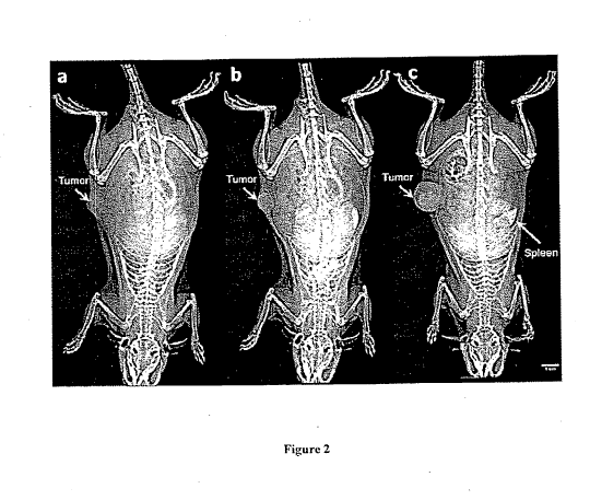
10013J Figure 2 shows whole body images of a rat with a breast tumor in its
right
flank obtained using a clinical digital mammography system before (a) and 1
minute after
administration of a "high' dose (1,244 mgI/kg) of an example nano-probe (b)
resulting in
vasculature visualization of the tumor site as well as normal. tissues. A 72-
hour post-contrast
image of a different rat injected with a "low" does (455 mgl/kg) of the
example nano-probe (c)
CA 02708028 2010-06-04
WO 2009/073236 PCT/US2008/013651
-6-
reveals the accumulation of the nano-probe in the tumor and the splevnr, while
the vasculature 1S
not visible.
100141 Figure 3 shows whole body images of it rat (rat 3 as indicated in
Figure 4)
obtained using; i clinical digital mammography system before and 24. 72, and
120 hours after
administration of a "low" dose (455 m( gl/kg) of the example na.tno-probe.
100151 Figure 4 shows a comparison of (a) the uptake of the example nano-probe
by the breast tumor of seven rats over five clays as imaged by a clinical
mammography system;
and (b) the uptake of the example nano-probe by the normal tissue of the same
seven rats over
the same time frarne.
[00161 Figure 5 shows an example of mammography images of a breast tumor
with high uptake of the example nano-probe (rat 3) and a breast tumor with
moderate uptake of
the example nano-probe (rat 4) over a five day time period.
100171 Figure 6 shows whole body images of a rat obtained u ing a clinical
digital
mammography system injected with saline of equal volume of the volume with the
example
nano-probe. The time points coincide with pre- and post-contrast images of the
nano-probed
rats.
1001.81 Figure 7 shows whole body images of a rat. obtained using a clinical
digital
mammography system before and 10. 32, 63, 125, 182, 315. 430, and 578 seconds
after
administration with a"high" dose (1,341 mgI/kg) (A' a conventional contrast
agent (iohexol).
CA 02708028 2010-06-04
WO 2009/073236 PCT/US2008/013651
-7-
10019) Figure 8 shows example results of the administration of a high dose
(1,344
mgl/kg) of a conventional contrast agent (iohexol) to a rat. which exhibited
negligible tumor
enhancement due to rapid renal clearance.
[00201 Figure 9 shows a comparison of the uptake of the example nano-probe by
the breast tumor of 15 rats within the period of three days as imaged by a
clinical mammography
system.
10021] Figure 10 shows tumor growth curves of a control (untreated) group and
group treated with liposomal doxorubicin.
100221 Figure 11 shows tumor growth curves of a control (untreated) group and
group treated with liposomal doxorubicin.
DETAll.,FF) 1)F,SC'RIPa_'ION
100231 The design, fabrication, characterization, and application of nano-
scale
contrast agents (or "nano-probe"(s)) is provided.
[0024] A typical nano-probe comprises a liposomal composition comprising a
lipid
or phospholipid, a stabilizing exeipient such as cholesterol, and a polymer-
dcrivatized lipid or
phospholipid. Suitable examples of lipids or phospholipids, stabilizing
cxcipicnts, and polymer-
derivatied lipids or phospholipids arc set forth in, for example, U.S. Patent
Application Nos.
10/830,190, 11/595.808, and 11/568., 936.
100251 The liposomal compositions typically encapsulate a contrast enhancing
agent. Suitable contrast enhancing agents include, Itx example, non-
radioactive iodinated
compounds such as iohcxol and iodixariol, as described in U.S. Patent
Application Nos.
CA 02708028 2010-06-04
WO 2009/073236 PCT/US2008/013651
-8-
10/830.190, 11/595,808, and 11/568, 936. The nano-probe may carry high amounts
of iodinated
contrast agent. For example. the nano-probes may carry as much as 130-200 mg
of iodinated
compound per mL of liposomal composition. A typical concentration of iodinated
compound
may be approximately 155 mg /ml...
100261 Other suitable contrast enhancing agents known in the art may be
included,
as necessary or desirable. to effect imaging by other imaging technologies,
such as, for example.
ultrasonagraphy, electron beam (EBT), magnetic resonance imaging (MRI).
magnetic resonance
angiography (MRA), positron emission tomography. and optical imaging,
including fluorescence
and bioluminescence. For example. in certain embodiments, suitable contrast
agents may
include fluorescent dyes. such as, for example, fluorescein iso-thiocynate and
MRl contrast
agents including lanthanide aminocarboxylat.e complexes such as Gadolinium
(111) DTPA and its
variants.
100271 The nano-probes are typically about or approximately 100 nm in average
diameter7 but may range from about 15 to about 150 nm in average diameter.
Thus, a suitable
liposome average diameter may be less than about 150 nm, less than about 120
run, and less than
about 100 nm_ The nano-probes typically have long blood circulation times
(e.g., ti, 18 h in
rats).
100281 The nano-probes may be prepared. for example, by the methods disclosed
in U.S. Patent Application Nos. 10/830.190, 11/595.908. and 1 1 /568, 936, and
in Example 1,
below.
CA 02708028 2010-06-04
WO 2009/073236 PCT/US2008/013651
-9-
10029) Generally speaking, the nano-probe may be detected using at least one
of
the following X-ray diagnostic techniques: computed topography (CT). micro-CT,
mammography, and chest X-ray. In other embodiments, the nano-probe may be
imaged using at
least one of M.RI, magnetic resonance spectroscopy. bioluminescence imaging,
ultriasouaacl,
optical imaging, and optical spectroscopy.
100301 In one embodiment. a method for evaluating a subject's vasculature
integrity is provided- The method, exemplilied in Example 2, below, comprises:
introducing a
composition (a nano-probe) into the subject's vasculature, the composition
comprising:
liposomes, each liposome encapsulating one or more nonradioactive contrast-
enhancing agents.
and each liposome comprising: cholesterol, at least one phospholipid, and at
least one
phospholipid which is dcrivatized with a polymer chain, wherein the average
diameter of the
liposomes is less than 150 nanometers. generating X-ray images of the
subject's vasculature; and
analyzing the X-ray images to detect a leak in the Subject's vasculature.
According to one
embodiment of the method, the nano-probes can interrogate and quantify the
extent of blood
vessel integrity non-invasively using X-ray based imaging techniques.
100311 In one embodiment of the method. analyzing the X-ray images comprises
distinguishing areas having an enhanced X-ray signal from area` having little
or no X-ray signal.
In another embodiment of the method, the composition is characterized in that
the composition
accumulates in an extravascular region of the subject's vasculature when a
leak exists in the
subject's vasculature, in comparison to an intravascular region of the
subject's vasculature,
thereby achieving enhanced X-ray signal in the extravascular region. In one
embodiment of the
method, a low nano-probe dose containing a small amount of non-radioactive
iodinated
CA 02708028 2010-06-04
WO 2009/073236 PCT/US2008/013651
- 10-
compound may achieve X-ray signal enhancement of the extravascular space of a
leaky
vasculature while the low intravascular levels of the iodinated nano-probe
Produce little or no
signal enhancement.
100321 In another embodiment of the method, generirtin- X-ray images comprises
generating X-ray images using at least one of CT, micro-CT, mammography, and
chest. X-ray.
100331 In one embodiment, the leak is indicative of at least one of cancer,
inllarnination, stroke, aneurism. wound healing or other reparative processes,
and trauma. As
such, in one embodiment, the nano-probes may facilitate the detection of
injured. leaky blood
vessels caused by a variety of diseases such as cancer, infla.mrnation,
stroke, aneurism, internal
bleeding due to trauma, and angiogenesis due to regenerative processes such as
wound healing.
100341 In another embodiment. a method is provided for differentiating between
a
malignant lesion and a benign lesion. The method comprises: introducing a
composition (e.g., a
nano-probe) into a lesion of interest, the composition comprising: liposomes.
the liposornes
comprising: at least one first lipid or phospholipid; at least one second
lipid or phospholipid
which is derivatized with one or more polymers; and at least one sterically
bulky excipient
capable of stabilizing the liposornes, wherein the average diameter of the
liposomes is less than
150 nanometers, and wherein the liposornes encapsulate at least one
nonradioactive contrast
enhancing agent. In one embodiment: of the method, the composition may be
characterized in
that the composition accumulates in a malignant lesion to a greater extent.
than in a benign lesion
because malignant tumors have an increased permeability to 5-200 nun sized
particles. The
method further comprises generating X-ray images of the lesion of interest and
analyzing the
X-ray images to determine the extent of accumulation of the composition in the
lesion of interest.
CA 02708028 2010-06-04
WO 2009/073236 PCT/US2008/013651
-11-
[00351 In another embodiment. a method is provided for evaluating the
accessibility of a tumor to nano-sued therapeutics. The method, exemplified in
Example 4,
below, comprises: introducing a composition (a nano-probe) into a tumor of
interest, the
composition comprising: liposomes, a plurality of the liposomcs comprising: at
least one first
lipid or phospholipid: at least one second lipid or phospholipid which is
dcrivatized with one or
more polymers; and at least one sterically bulky excipient capable of
stabilizing the liposomes,
wherein the average diameter of the liposomes is less than 150 nanometers. and
wherein a
plurality of' the liposomes encapsulate. at least one nonradioactive contrast
enhancing agent;
generating X-ray images of the tumor; and analyzing the X-ray images to
determine the extent of
accumulation of the composition in the tumor.
100361 In yet another enibodinient. a composition is provided. The
composition,
an example of which is provided at Example 5, below. may comprise: liposomcs
having an
average diameter of less than 150 nanometers, each liposome comprising: a
first lipid or
phospholipid; a second lipid or phospholipid which is derivatized with a
polymer; and a
sterically bulky excipient capable of stabilizing the liposomes; wherein each
liptisonie en-
encapsulates a nonradioactive contrast enhancing agent and at least one bio-
active agent,
including, but not limited to, a chemotherapeutic, a gene, a protein, a small
molecule, and a
peptide. In one embodiment of the composition, the first lipid or phospholipid
comprises 1,2-
dipatmitoyl-tin-glycero- 3-phosphocholine (DPPC). In another embodiment of the
composition,
the sterically bulky excipient capable of stabilizing the liposomcs comprises
cholesterol- In
another embodiment of the composition, the second lipid or phospholipid which
is derivatized
with a polymer comprises l ,2-diste aruyl-sii-glycero-3 phosphe~ethanolaminc-N-
CA 02708028 2010-06-04
WO 2009/073236 PCT/US2008/013651
-12-
lmethoxy(poly(cthylene glycol))-20001 (rnPEG2000-1)SPE). In another embodiment
of the
composition, the first lipid or phospholipid, the second lipid or phospholipid
which is derivatized
with a polymer, and the sterically bulky excipient capable of stabilizing the
liposomes, are
present in a ratio of 55:40:5. In another embodiment of the composition, the
chemotherapeutic
comprises doxorubicin. In another embodiment of the composition, the liposomes
have an
average diameter of about 1.00 nm.
100371 In one embodiment, the composition may allow flor live or real time
monitoring of the nano-probe biodistribution, thereby allowing for patient-
specific therapies. In
another embodiment, non-invasive pharmacokinetics of" a therapeutic agent may
be achieved
when the therapeutic agent is co-encapsulated with contrast agent within the
nano-probe as-
described with respect to the composition. In another embodiment, the nano-
probe is further
multi-functional in that the nano-probe may be actively targeted via
antibodies and peptides.
100381 One example therapeutic that may be suitable for co-encapsulation is
anthracyclines. f,iposomal anthracyclines have been developed to increase the
therapeutic index
of the anthracycline by maintaining antitumor efficacy while improving the
safety profile.
Anthracyclines. including doxorubicin. are among the most potent
chemotherapeutic agents.
However, this family of chemotherapeutics exemplifies the limitation of many
potent anticancer
drugs in that they are limited by highly problematic system toxicities, which
result in
myelosuppression, acute nausea and vomiting, stomat.itis, and cardiotoxicity.
Polyethylene;
glycol-coated (PEGylated) liposoinal doxorubicin, a 100 nm lipid sac with a
long blood
circulation (t11 55 h). has been approved in the United States For clinical
use for treatment of
refractory Kaposi's sarcoma and ovarian cancer. PEGylated liposomal
doxorubicin has also
CA 02708028 2010-06-04
WO 2009/073236 PCT/US2008/013651
-13-
been investigated liar breast cancer therapy. and has shown similar efficacy
and significantly
lower cardiotoxicity when compared to conventional doxorubiciii. Many other
drugs loaded into
liposomes are approved or undergoing clinical evaluation for cancer therapy,
and may he suitable
for co-encapsulation as described herein, including, but not limited to,
vineristine, lurtotecan, all-
trans retinoic acid, platinum compounds, annamycin, and DNA plasmid encoding 1-
ILA-137 and
02 microglohulin.
100391 In certain embodiments, suitable imaging techniques for the detecting
the
composition may include, for example, at least one ol'the following X-ray
diagnostic techniques:
computed topography (CT), micro-CT, mammography, and chest X-ray. In other
embodiments,
the ntano-probe may be imaged or detected using at least one of MR.1,
ultrasound, and optical
imaging, including fluorescence or bioluminescence imaging.
EXAMPLES
f xaiz3 lwc 1_- 1'rc ration and Cliaracteri7 tion of an Example Nano-Probe
100401 A highly concentrated iodine solution (600 ingl/mL) was prepared by
dissolving iodixanol powder (lyophilized front Visapaquc 320, GlE Healthcare)
in ultrapure water
under continuous stirring and heating at 70 C. A lipid solution in ethanol
comprising 1,2-
dipalnlitoyl-sit-glycero-3-phosphocholine (DPPC), cholesterol, and I.2-
cliste;iroyl-s.n-glycero-3-
phosphoethaiiolalnine-N-[nict.lloxy(poly(cthylcne glycol))-20001 (rnPl'(32000-
DSPE) in the
molar ratio 55:40:5 was hydrated with the iodine solution at 700 C, hollowed
by sequential
extrusion on a Lipex Therinollnc extruder (Northern Lipids, Vancouver,
Canada). This resulted
in encapsulation of the iodine solution within the central aqueous core of
polyethylene
CA 02708028 2010-06-04
WO 2009/073236 PCT/US2008/013651
-14-
glycol-stabilized (Pr Gylated) liposomes. Free7 tin-encapsulated iodixanol was
replaced by a
saline solution (300 mM NaCl) with the same osmolarity as the internal
iodinated phase of the
liposome using a two day dialysis against 300 mM NaCI using a 100,000 MWCO
dialysis
tubing. Following concentration via diafiltration using MicroK.i'os modules
(Spectrum
Laboratories, California) with a 50 run cutoff pore siz.c, the liposonlal
iodine and lipid content
were measured to he 155 nig/mL (all encapsulated) and 165 mM. respectively.
The average
diameter of the liposomes was 96 nm (sd - 8 ntn) as determined by dynamic
light scattering.
The 596 rnOsnt/kg water osmolality of the formulation allowed intravenous
injection, since the
liposomal walls Can Sustain the osmotic pressures expected to occur in
isotonic environments.
Indeed, in vitro leakage experiments against isotonic phosphate buffered
saline exhibited
negligible leakage of the encapsulated iodine (less than 5% of'the initial
payload) over the period
of three days.
Example 2 J1u ins Study Using Nano-Probe cif' 1 xampIc t
100411 The nano-probe of Example I was tested in a ran breast tumor model
developed by inoculation of mammary adenocarcinoma cells (13762 MAT 13 11T
from ATCC)
into the right flank of Fischer fcmalc rats. The imaging studies started on
day seven after tumor
inoculation (tumor volume approximately 440 mmsee Figure 1 for tumor growth
curve).
Tumor volumes (it 15) were obtained by caliper nmeasurements.
10042 Contrast-enhanced mammography was performed with a commercial digital
mammography system (Scnographe 20001.), GE Healthcare) at 49 kVp and 63 niAs
with a
rhodium target and an extra copper filter of 0.3 nln7 thickness. These
settings were used to shape
the x-rays to have optim.tl energies for iodine. Under these conditions, an
optimal X-ray
CA 02708028 2010-06-04
WO 2009/073236 PCT/US2008/013651
-15
spectrum was obtained containing the largest number of X-rays with energies
above the k-edge
of iodine while X-ray dose was significantly reduced when compared to standard
mammography. Initial studies were performed with high doses of the nano-probe
to obtain a
vascular image. A pre-contrast image (Figure 2(a)) and post.-contrast inmates
were acquired 1, 5,
10, and 15 minutes alter tail vein injection of the nano-probe. Blood vessels
were clearly visible
(Figure 2(b)) at a 1,300 mgl/kg, body weight dose of the nano-probe achieving
blood
concentrations of 20 nzgl/ml,. In the case of an adult human, this would
correspond to a dose of
about 654 ml., of the nano-probe (assuming a blood volume of five liters),
which is prohibitively
large for use in humans. However, this high dose was used to clearly visualize
the blood vessels
that the mammography system was capable of detecting.
100431 In monitoring the fate of the nano-probe studies. a pre-contrast image
(indicated as t 0) and post-contrast images were obtained 24. 72, and 120
hours after
administration of the nano-probe at a dose of 455 tngl/kg body weight. "T"his
corresponds to 195
mg lipidlkl; body weight, which is about two times higher than the highest
lipid dose of
liposonia.l drugs when employed in clinical practice. Figure 2(c) shows an
image of an animal
obtained 72 h after the nano-probe injection. No blood vessels are visible in
the normal tissue
(as compared to the enhanced vasculature observed in Figure 2(h)). while the
spleen and the
turner were enhanced. The spleen enhancement was expected since liposornes
within the
ext.ravascular space of the tumor provided for the detection of the nano-probe
(when niaxinium
iodine in The blood circulation was expected), since the iodine levels in the
blood were below the
detectable threshold.
CA 02708028 2010-06-04
WO 2009/073236 PCT/US2008/013651
-16-
10044) Figure 3 displays the timeline of the nano-probe accumulation within
the
tumor lesion for a period of five days. In the same manner, a group of animals
(n 7) was
imaged and monitored at the post-contrast defined time points exhibiting a
similar behavior.
Figure 4(a) summarizes the time course of the tumor enhancement by quantifying
the grey
levels of the lesions using Tmage.T software (NIH, Bethesda, Maryland). A
normalized tumor
enhancement was calculated by subtracting each post-contrast value (t:>=0)
from its tumors that
showed a slow gradual increase of the enhancement during the 120 h time course
(indicated as
rat 2 and 6 in Figure 4(a)), whereas other tumors displayed a faster increase
(indicated as rat 3,
4, and 5 in Figure 4(a)). Another tumor exhibited an initial rapid enhancement
at t - 24 h
followed by a plateau. At t == 120 h. there were lesions with low enhancement
and other lesions
with. much higher enhancement. This discrepancy suggests that. different
amounts of the
nano-probe leaked into each tumor.
100451 The pattern of tumor enhancement due to the nano-probe was plotted in
Figure 4(b) and compared against the normal tissue of the animals in the same
group or the
tumor site of a control group (injected with no agent) exhibiting statistical
differences. No
enhancement was observed in normal tissues. suggesting that the nano-probe
levels in the blood
were below the detectable threshold of the mammography. and implying that no
endogenous
changes of the tumor tissue could contribute such a significant enhancement as
the one seen in
the case of the non-probed lesions. Examples of mirnmography images of a tumor
lesion with
high uptake and a tumor lesion with moderate uptake are shown in Figure 5.
CA 02708028 2010-06-04
WO 2009/073236 PCT/US2008/013651
-17-
Examph: 3 - Comparative Exa.mfllc5 (CO.titrol s.~rou s
[00461 Whole body mammograms of a rat in with no contrast agent (control
group) is shown in Figure 6. Another control croup was injected with a
conventional iodinated
agent (iohexol) at an iodine dose equivalent to the high dose of 1,344
mgI/nil.. ofthe nano-probe.
(See Figure 7). Within the first minute after injection. the normal
vasculature and the tumor
lesion exhibited a slight enhancement, but the iodinated agent was rapidly
cleared via the
kidneys. (See Figure 8).
Example 4 - Non-Invasive Prediction of Nano-Chemotherapy S t cess
100471 The prediction accuracy of the nano-probe was tested in a rat breast
tumor
model developed by inoculation of mammary adenocarcinona cells (13762 MAW 13
111 from
A'1'CC) into the right flank of Tischer female rats. The imaging studies
started on day six after
tumor inoculation. Contrast-enhanced mammography was performed with a
commercial digital
mammography system (Senographe 2000D, Gl. l:lealthcare) at 49 kVp and 63 mAs
with a
rhodium target and an extra copper filter of 0.3 mm thickness. The animals (n
.- 15) were
injected with the example nano-probe in an amount of 455 mgI/kg b.w. Figure 9
summarizes
the time course of the tumor enhancement by quantifying the grey levels of the
lesions using
ltnagci software (t `- 0) from its pre-contrast value (t -~ 0). It is observed
that the enhancement
proliles exhibit dissimilar patterns. Immediately after the imaging session
(at day 9 after tumor
inoculation), the animals were injected with liposomal doaoruhicin at a dose
of 10 mg
doxorubicin per kg b.w.
CA 02708028 2010-06-04
WO 2009/073236 PCT/US2008/013651
- 18-
10048 The response of the tumor to the drug was evaluated by measuring the
sire
of the tumor using a caliper. Figure 10 summarizes the tumor growth curves or
all of the
animals obtained by caliper measurenlctits. The growth curves exhibit high
variability, implying
that each tumor responded differently to the chemotherapeutic. The animals
that displayed
higher uptake of the nano-probe responded better to the treatment (smaller
tumor volumes).
100491 To quantify the relation of the nano-probe prediction to the therapy
response, the tumor growth curves (Figure 10) were fitted to mono-exponential
functions (i.e.,
dVIdt K"111'r gt0"1h * t, where V is tumor volume) to calculate the growth
rate constant, Ktu"
gr""`t'. 1-'he Area Under the Curve (AUCt'r" n01`) of the nano-probe uptake
profiles were calculated
from Figure 9. The K~~niwr of each animal was plotted against the AUCof each
animal
in Figure 11. High AUC'"""' means high ilntrutumoral accumulation of the nano-
probe
predicting high success of the nano-chemotherapeutic, whereas low K11110r
growth means slow
tumor growth or good response to the treatment-
1: 7at~lle 5 - Preparation and in vitro Characterization of an Example Nano-
scale Liposome CO-
Enca solo ng ofatrast Agent and Chemotherapeutic
100501 A lipid solution in ethanol comprising DPP(', cholesterol, and
n1PEC(2000)-DSPI7'in the molar ratio 55:40:5 was hydrated with a 300 mM
ammonium sulfate
iodinatcd solution (iohcxol; 350 mgt/ml.) at 70" C followed by sequential
extrusion on a Lipex
'l"hermoline extruder (Northern Lipids. Vancouver, Canada). This resulted in
encapsulation of
the iodine solution within the central aqueous core of PEGylated liposomes.
Free,
unen caps u lated iodixanol was removed from the external phase of the
liposonie using a two day
dialysis against 300 mM arnmoniurn sulfate using a 100,000 MWCO dialysis
tubing. The
CA 02708028 2010-06-04
WO 2009/073236 PCT/US2008/013651
19-
liposomes were then dialyzed for 12 h with a 100 kL)a M\VC:.O dialysis tubing
against a
phosphate-buffered saline (t'BS) solution to establish an ammonium sulfate
gradient for
doxoruhicin loading.
100511 The liposomal formulation was actively loaded with doxorubicin by an
ammnmoniurn sulfite gradient. Briefly, liposomes and doxoruhicin were mixed at
a ratio of 0.1 nog
of doxorubicin per I mg of DPPC in the liposomes. The liposoiue-doxoruhicin
suspension was
heated at 35" C for 25 ruin. The liposoines were left overnight at room
temperature and dialyzed
twice in 100 kDa MWCO membrane against PBS to remove unencapsulated
doxorubicin.
Following concentration, via diatiltration, using MicroKros modules (Spectrum
Laboratories,
Califbrnia) with a 50 not cutoff pore size, the liposomal iodine and
doxoruhicin content was
measured to be 91 mg/ml., and 1.2 mg/mL (all encapsulated), respectively. The
average diameter
of the liposomes was 102 nm (sd = 6 rim) as determined by dynamic light
scattering.
100521 It is, of course, not possible to describe every conceivable
combination of
components or methodologies for purposes of describing the compositions,
methods, and so on
provided herein. Additional advantages and modifications will readily appear
to those skilled in
the art. Therefore, the invention, in its broader uspecLs. is not limited to
the specific details and
illustrative examples shown and described. Accordingly, departures may be made
from. such
details without departing from the spirit or scope of the applicants' general
inventive concept. A
person of ordinary skill will readily recognize that optimizing or
manipulating any one of these
variables may or will require or make possible the manipulation of one or more
of the other of
these variables, and that any such optimization or manipulation is within the
spirit and scope of
the present embodiments.
CA 02708028 2010-06-04
WO 2009/073236 PCT/US2008/013651
-20-
0 053) Notwithstanding that the numerical ranges and parameters setting forth
the
broad scope of the invention are approximations, the numerical values set
forth in the specific
examples are reported as precisely as possible. Any numerical value, however,
inherently
contains certain errors necessarily resulting from the standard deviation
found in their respective
testing measurements. It should be rioted that the tern "about" may mean up to
and including
_t,10% of the stated value. For example, "about 10" may mean from 9 to 11,
100541 Furthermore, while the compositions, methods. and so on have been
illustrated by describing examples, and while the examples have been described
in considerable
detail. it is not the intention of the applicant to restrict, or in any way.
limit the scope of the
appended claims to such detail. Thus, this application is intended to embrace
alterations,
modifications, and variations that fall within the scope of the appended
claims. The preceding
description is not meant to limit the scope of the invention. Rather, the
scope of the invention is
to be determined by the appended claims and their equivalents.
100551 Finally, to the extent that the term "includes" or "including" is
employed in
the detailed description or the claims, it is intended to be inclusive in a
manner similar to the
Term "comprising," as that term is interpreted when employed as a transitional
word in a claim.
Furthermore, to the extent that the term "or" is employed in the claims (e.g.,
A or 13) it is
intended to mean "A or B or both." When the applicants intend to indicate
"only A or 13, but not
both," then the term "only A or 13 but 'not both" will he employed. Similarly,
when the
applicants intend to indicate "one and only one" of A, B, or C, the applicants
will employ the
phrase "one and only one." Thus. use of the tenet "or" herein is the
inclusive, and not the
exclusive use. See Bryan A. Cramer, A Dictionary of Modern Legal Usage 624
(2d. F.d. 1995).