Note : Les descriptions sont présentées dans la langue officielle dans laquelle elles ont été soumises.
CA 02708957 2015-06-23
IMAGING PROBE HOUSING WITH FLUID FLUSHING
BACKGROUND
[2] The present invention generally relates to an
imaging probe of an imaging catheter. The present invention
more specifically relates to mechanically scanned imaging
probes for use in, for example, an intravascular ultrasound
(IVUS) or intra-cardiac echo-cardiography (ICE) catheter. The
present invention still further relates to such an imaging
probe wherein the imaging probe is configured to assure
efficient and complete fluid flushing from the catheter sheath
to preclude formation of air bubbles in the vicinity of the
ultrasonic transducer of the imaging probe. In addition this
invention relates to imaging probe configuration to ensure the
prevention of air bubbles during rotational operation by
continuously directing fluid across the imaging probes
transmission surface.
[3] IVUS catheters enable the imaging of internal
structures in the body. ICE catheters enable the imaging of
larger internal structures in the body. Coronary IVUS
catheters are used in the small arteries of the heart to
visualize coronary artery disease, for example. Coronary ICE
catheters are used in the cavity of the heart to visualize
structural heart disease, including atrial septal defects
(ASD), patent foramen ovale (PFO) and to guide various
procedures including septal punctures, percutaneous valvular
replacement, and various ablations treatment strategies. To
that end, an IVUS or an ICE catheter will employ at least one
ultrasonic transducer that creates pressure waves to enable
visualization. At least one transducer is usually housed
within a surrounding sheath or catheter member and rotated to
1
CA 02708957 2015-06-23
enable 360 degree visualization. Because air is not an
efficient medium for the transmission of the ultrasonic waves
produced by at least one transducer, a fluid interface between
the transducer and the sheath in which it is disposed is
usually provided. Unfortunately, current imaging probe
configurations do not always prevent the formation of air
bubbles in the fluid in the vicinity of the transducer
resulting in compromised performance of the imaging catheter.
Embodiments of the present invention address this and other
issues.
SUMMARY
[4] Accordingly, there is provided an imaging probe for
use in a catheter for ultrasonic imaging, the catheter
including a sheath having an opening at a distal end for
conducting a fluid there through, the imaging probe
comprising: a distal housing arranged to be received by the
sheath and being coupled to a drive shaft to rotate the distal
housing relative to the sheath; a transducer within the distal
housing for generating and sensing ultrasonic waves, wherein
the transducer is rotatable along with the distal housing
relative to the sheath, and wherein the transducer is
configured at an inclined angle from a proximal portion of a
front side of the transducer to a distal portion of the front
side of the transducer; and a fluid flow promoter carried on
the distal housing that promotes flow of the fluid within the
sheath across the front side of the transducer and out the
opening at the distal end of the sheath.
[5] The imaging probe may further include a wall distal
to the transducer and the fluid flow promoter may include an
opening within the wall and adjacent to the transducer. The
distal housing preferably has a first profile at a proximal
2
CA 02708957 2015-06-23
end of the distal housing, a second profile at the wall distal
to the transducer, and the fluid flow promoter includes the
second profile being greater than the first profile to promote
fluid flow over the transducer and through the opening within
the wall.
[6] The distal housing has a proximal extent and the
fluid flow promoter may include at least one aqua duct within
the proximal extent of the distal housing. The at least one
aqua duct is preferably formed within the proximal extent of
the distal housing at an angle to the center axis. The at
least one aqua duct may comprise at least two aqua ducts. The
at least two aqua ducts may include a first aqua duct that
directs fluid directly onto the front side of the transducer
and a second aqua duct that directs fluid onto the front side
of the transducer from a side of the transducer. The at least
one aqua duct has a proximal side and a distal side and may be
formed so that the proximal side leads the distal side in the
direction of rotation of the distal housing. The at least one
aqua duct may include a radius of curvature.
BRIEF DESCRIPTION OF THE DRAWINGS
[ 9] The features of the present invention which are
believed to be novel are set forth with particularity in the
appended claims. The invention, together with further
features and advantages thereof, may best be understood by
making reference to the following description taken in
conjunction with the accompanying drawings, in the several
figures of which like reference numerals identify identical
elements, and wherein:
3
2664-003-04
CA 02708957 2010-06-10
WO 2009/085849 PCT/US2008/087209
FIG. 1 is a side view, partly in section, of an
ultrasonic imaging catheter in accordance with a first
embodiment of the invention;
FIG. 2 is a partial perspective view of the imaging
probe of the catheter of FIG.1; and
FIG. 3 is a perspective view showing another imaging
probe embodying the invention connected to a drive cable of
an intravascular ultrasound (IVUS) catheter.
DETAILED DESCRIPTION
[10] FIG. 1 shows an imaging catheter 10 with the first
embodiment of the present invention. The imaging catheter 10
is particularly adapted for use as an IVUS catheter, but
those skilled in the art will appreciate that the invention
may be used in many other forms of ultrasound catheters as
well without departing from the present invention. The
catheter 10 generally includes a sheath or catheter member
12 and an imaging probe 14. As shown, the imaging probe 14
is disposed within the sheath 14. The imaging probe 14 is
moveable axially within the sheath 12 to enable the sheath
to remain stationary as the imaging probe is moved to scan
the internal body structures to be visualized. Also, as well
known, the imaging probe 14 is also rotatable to enable 360
degree scanning.
[ 11 ] The imaging probe 14 generally includes a distal
housing 16, a flexible drive shaft 18, and a coaxial cable
20. The distal housing 16 is carried on the distal end of
the flexible drive shaft 18 in a known manner. The drive
shaft 18 may be formed, for example, by winding multiple
strands of metal wire on a mandrel to create a long spring
containing a repeating series of concentric rings, or
windings, of the wire. Two or more springs may be wound, one
4
2664-003-04
CA 02708957 2010-06-10
WO 2009/085849 PCT/US2008/087209
over the other, with adjacent springs being wound in
opposite directions to each other. This provides a drive
shaft that is both flexible and with high torsional
stiffness.
[12] The distal housing 16 generally includes the
ultrasound transducer 22, a distal tip wall 24, and a
proximal cutout surface 26. The transducer 22 is mounted on
a transducer backing 28. The backing 28 and the distal tip
wall 24 are adhered together by a conductive adhesive 27.
The backing 28 is dimensioned and of such a material as to
absorb ultrasonic waves from the backside of the transducer
22 so that only energy from the front side of the transducer
is emitted from the imaging probe 14 in the general
direction indicated by reference character 30 transverse to
the exposed surface of the transducer 22 . The coaxial cable
extends down the drive shaft 18 and includes a center
conductor 32 and a shield lead 34. The center conductor 32
and shield lead 34 are coupled across the transducer 20 as
shown. The coaxial cable 20 couples energy to the transducer
20 to cause the transducer 22 to generate a pressure wave into
the lumen 36 of the sheath 12. The interior of the lumen 36
is preferably filled with a fluid, such as saline. The
saline flows from the proximal end of the catheter 10 to the
distal end of the catheter 10 and serves to efficiently
couple the ultrasonic energy into the sheath and then to the
body. To support the fluid flow, the sheath includes a point
of egress (not shown) for the fluid at its distal end. As
previously mentioned, it is important to prevent air bubbles
from being formed or residing in the vicinity of the
transducer 22.
[13] To assure that air bubble formation in the
vicinity of the transducer 22 is prevented, and with
additional reference to FIG. 2, the distal extent of the
5
2664-003-04
CA 02708957 2010-06-10
WO 2009/085849 PCT/US2008/087209
distal housing 16 includes a distal tip wall 24 distal and
adjacent to the transducer 22. The distal tip wall 24 has an
opening 38 therein adjacent to the transducer 22. Proximal
to the transducer 22, the distal housing 16 has a proximal
cutout forming a tapered surface 26 leading toward the
transducer 22. Fluid flow within the sheath from proximal to
the transducer 22 to distal of the transducer 22 is
conducted down the tapered cutout surface 26, over the
transducer 22, and out the distal tip wall opening 38 in a
continuous manner, without turbulence, to prevent air bubble
formation in the vicinity of the transducer.
[14] The distal housing 16 at the proximal extent of
the tapered cutout surface 26 has or defines a first profile
substantially transverse to the catheter center axis 40 and
the fluid flow. The distal tip wall 24 defines a second
profile also substantially transverse to the catheter center
axis 40 and the fluid flow. The second profile is greater in
dimension than the first profile. Hence, this serves to
promote fluid flow through the distal tip opening 38 and
hence over the transducer 22.
[15] To further promote fluid flow over the transducer
22, the transducer has a surface 22a over which the fluid
flows that is disposed at an angle sloping toward the
catheter center axis in the proximal direction. This
presents a greater surface resistance against the fluid flow
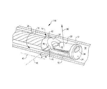
to assure fluid contact therewith.
[16] FIG. 3 shows another imaging catheter 110
according to a further embodiment of the present invention.
The catheter 110 is similar to the catheter 10 of FIGS. 1
and 2 and hence, reference characters for like elements are
repeated in FIG. 3. To further assure that air bubble
formation in the vicinity of the transducer 22 is prevented
during rotational operation, and with additional reference
6
2664-003-04
CA 02708957 2010-06-10
WO 2009/085849 PCT/US2008/087209
to FIG. 3, the proximal extent of the distal housing 26 is
constructed with aqua ducts 41 and 42. As shown in Fig. 3,
one aqua duct directs fluid onto the transducer face 22 from
the top of the proximal portion of the distal housing 26,
while the other aqua duct 41 directs fluid onto the
transducer face 22 from the side. Further, the aqua ducts
are built into the proximal portion 26 of the distal housing
16 at an angle with respect to a line extending along the
catheter drive shaft 43. This is shown in FIG. 3 with the
angle theta being formed with the intersection of a line 43
extending parallel to the catheter drive shaft 13 and a line
44 extending through the center of one of the aqua ducts 41.
Both aqua ducts 41 and 42 are constructed at such an angle
such that the proximal side of each duct leads the distal
side in the direction of rotation. This is shown in FIG. 3
with the clockwise direction of rotation (from the view
looking distally along the catheter drive shaft 13)
indicated by 45. Further, each side of each aqua duct is
constructed with a small radius of curvature shown by 46 in
FIG. 3. One way to achieve the duct side curvatures is to
construct the ducts in a helical spiral with a small pitch,
as, for example, on the order of 0.1 inch. The duct angle
and curvature, coupled with rotation of the distal housing
16 and the fluid flow promoting structure shown in FIG. 1,
act to continuously draw fluid residing within the catheter
sheath 12, proximal to the distal housing 16, onto the face
of the transducer 22. Fluid flow within the sheath from
proximal to the transducer 22 to distal of the transducer 22
is conducted down the tapered cutout surface 26, over the
transducer 22, and out the distal tip wall opening 38 in a
continuous manner, without turbulence, to prevent air bubble
formation in the vicinity of the transducer.
[17] While particular embodiments of the present
invention have been shown and described, modifications may
7
CA 02708957 2015-06-23
be made, and it is therefore intended in the appended claims
to cover all such changes and modifications which fall within
the scope of the invention as defined by those claims.
8