Note : Les descriptions sont présentées dans la langue officielle dans laquelle elles ont été soumises.
CA 02710082 2010-06-18
WO 2009/085209 PCT/US2008/013899
METHODS FOR USING AND IDENTIFYING MODULATORS OF DELTA-LIKE 4
CROSS-REFERENCE TO RELATED APPLICATIONS
This application claims the benefit of priority of U.S. Patent Application
Serial No.
12/005,054, filed on December 20, 2007, which is a continuation-in-part of
U.S. Patent
Application Serial No. 11/514,773, filed September 1, 2006 which claims the
benefit of U.S.
Provisional Application Serial No. 60/713,637, filed September 1, 2005. U.S.
Patent
Application Serial No. 12/005,054 also claims the benefit of U.S. Provisional
Application
Serial No. 60/876,444, filed December 20, 2006 and U.S. Provisional
Application Serial No.
60/901,754, filed February 16, 2007. All the teachings of the above-referenced
applications
are incorporated herein by reference.
BACKGROUND OF THE INVENTION
Angiogenesis, the development of new blood vessels from the endothelium of a
preexisting vasculature, is a critical process in the growth, progression, and
metastasis of
solid tumors within the host. During physiologically normal angiogenesis, the
autocrine,
paracrine, and amphicrine interactions of the vascular endothelium with its
surrounding
stromal components are tightly regulated both spatially and temporally.
Additionally, the
levels and activities of proangiogenic and angiostatic cytokines and growth
factors are
maintained in balance. In contrast, the pathological angiogenesis necessary
for active tumor
growth is sustained and persistent, representing a dysregulation of the normal
angiogenic
system. Solid and hematopoietic tumor types are particularly associated with a
high level of
abnormal angiogenesis. More recently, it has become apparent that certain
types of leukemia
are also influenced by signaling involved in angiogenesis.
Agents that inhibit angiogenesis are useful in treating cancer. AvastinTM
(bevacizumab), a monoclonal antibody that binds to Vascular Endothelial Growth
Factor
(VEGF), has proven to be effective in the treatment of a variety of cancers.
Antagonists of
the SDF/CXCR4 signaling pathway inhibit tumor neovascularization and are
effective against
cancer in mouse models (Guleng et al. Cancer Res. 2005 Jul 1;65(13):5864-71).
The
isocoumarin 2-(8-hydroxy-6-methoxy-l-oxo-1 H-2-benzopyran-3-yl) propionic acid
(NM-3)
has completed phase I clinical evaluation as an orally bioavailable
angiogenesis inhibitor.
NM-3 directly kills both endothelial and tumor cells in vitro and is effective
in the treatment
-1-
CA 02710082 2010-06-18
WO 2009/085209 PCT/US2008/013899
of diverse human tumor xenografts in mice (Agata et al. Cancer Chemother
Pharmacol. 2005
Jun 10; [Epub ahead of print]).
Angiogenesis is a feature of other, non-neoplastic disorders. Various ocular
disorders,
particularly proliferative retinopathies and age-related macular degeneration,
and
inflammatory disorders, such as rheumatoid arthritis and psoriasis, are marked
by increased
vascularization of the affected tissue. Anit-angiogenic agents are effective
for the treatment
of these disorders. MacugenTM, an aptamer that binds to VEGF has proven to be
effective in
the treatment of neovascular (wet) age-related macular degeneration. The
success of TNF-
alpha antagonists in the treatment of rheumatoid arthritis is partially
attributed to anti-
angiogenic effects on the inflamed joint tissue (Feldmann et al. Annu Rev
Immunol.
2001 ;19:163-96).
Arteriogenesis, a process related to but distinct from angiogenesis, occurs
when the
lumen of a pre-existing vessel increases to form a collateral. After
myocardial infarction or
peripheral ischemia (e.g., limb, kidney, etc.) arterioles become more
significant conductance
vessels in order to maintain blood flow after occlusion of the major artery
serving the
affected tissue. Thus, agents that promote arteriogenesis may be used to treat
myocardial
infarction and other ischemic events, and may also be used to prevent an
ischernic event
where a partial arterial occlusion is detected or suspected.
The Notch pathway, and particularly Notch I and Notch4, participates in
angiogenic
processes. Notch signalling is generally involved in the regulation of
processes as diverse as
cellular proliferation, differentiation, specification and survival (Artavanis-
Tsakonas et al.,
1999). Its complexity in vertebrates is illustrated by the existence of
multiple Notch receptor
and ligands, each with distinct patterns of expression. In mammals there are
four Notch
receptors (notch]-4) and five ligands (jagged], 2 and D111, 3 and 4).
Mutations of Notch
receptors and ligands in mice lead to abnormalities in various organs, from
all three genn
lines, including the vascular system (Iso et al., 2003). The Notch pathway
functions through
local cell interactions, the extracellular domain of the ligand, present on
the surface of one
cell, interacts with the extracellular domain of the receptor on an adjacent
cell. This
interaction allows the action of two ADAM proteases on the extracellular
domain of Notch
followed by the action of a y-secretase on the transmembrane domain releasing
the
intracellular domain from the cell membrane and allowing it to be directed to
the nucleus,
where it functions with CSL to activate the expression of transcriptional
repressors of the
enhancer-of-split family (Murnm & Kopan, 2000).
CA 02710082 2010-06-18
WO 2009/085209 PCT/US2008/013899
Arterial versus venous differentiation has long been thought to be mainly
dependent
on physical factors such as blood pressure and oxygen concentration. Recently,
however, the
identification of a number of genes that are specifically expressed in
arterial or venous
endothelial cells well before the onset of circulation, seems to indicate an
important role for
genetic determination of endothelial cells in the primary differentiation
events between
arteries and veins. Among these genes are eph-B4, specifically expressed in
venous
endothelial cells (Adams et al., 1999) and ephrin-B2 (Adams et al., 1999; Gale
et at., 2001),
notch] (Krebs et al., 2000), notch4 (Uyttendaele et al., 1996) and d114
(Shutter et al., 2000),
among others, which are specifically expressed in arterial endothelial cells.
Studies with mutations in zebrafish Notch homologues demonstrate the
importance of
this pathway in regulating the arterial versus venous endothelial
differentiation, downstream
of vascular endothelial growth factor and sonic-hedgehog and upstream of the
ephrin
pathway (Lawson et at., 2002), being the earliest genes expressed in an
endothelial arterial
specific fashion. There is mounting evidence, in both zebrafish and mouse,
that Notch
function is essential in the establishment of the arterial endothelial cell
fate (Lawson et al.,
2002; Fischer et at., 2004; Duarte et al., 2004).
It is a goal of the present disclosure to provide agents and therapeutic
treatments for
modulating angiogenesis, arteriogenesis and vessel identity.
SUMMARY OF THE INVENTION
In certain aspects, the disclosure provides uses for, and methods for
identifying,
agonists and antagonists of the Notch ligand Delta-like 4 (D114).
Surprisingly, as taught
herein, both agonists and antagonists of D114 may be used to treat tumors
undergoing
angiogenesis or in other situations where it is desirable to inhibit or
disrupt angiogenesis.
Furthermore, the disclosure provides methods for stimulating arteriogenesis by
administering
a D114 agonist. Arteriogenesis is the process of collateral artery formation
and growth,
typically in ischemic tissues. Thus D114 agonists may be used to treat
patients suffering from,
or at risk for, an ischemic event, such as a peripheral or coronary ischemia.
The disclosure
further relates to the discovery that upregulation of D114 causes endothelial
cells to adopt an
arterial identity, while inhibition of D114 causes endothelial cells to adopt
a venous identity.
Thus, the disclosure provides methods for altering venous or arterial identity
by using, as
appropriate an agonist or antagonist of D114. Additionally, the disclosure
provides biomarkers
-3-
CA 02710082 2010-06-18
WO 2009/085209 PCT/US2008/013899
that may be used to assess whether an agent of interest is an agonist or
antagonist of D114
signaling.
In one embodiment, the disclosure describes a method for treating cancer
comprising
administering to a subject in need thereof, an effective amount of an
antagonist of D114. This
antagonist may be a polypeptide, particularly a peptide comprising an
extracellular region of
D114. In one aspect, the polypeptide may be a monomer, but may also function
as a dimer.
Merely to illustrate, examples of polypeptides that comprise the extracellular
region of D114
may be selected from the DSL domain, a.a. 27-524, a.a. 1-486, a.a. 27-486,
a.a. 1-442, and
a.a. 27-442, or variants thereof, of SEQ ID NO: 1. Furthermore, a polypeptide
may comprise
at least one of, or a combination of, the following domains of 13114: MNNL,
DSL, EGF5,
EGF5 (see Figure 20A). Additionally, an antagonist of D114 may comprise an
antibody, or a
gragment thereof, that binds to an extracellular region of D114. Such an
antibody may be
monoclonal, human, or humanized. In a particular embodiment, said antibodies
may
comprise at least one of SEQ ID NOs: 4-7. All of the above-mentioned
antagonists of D114
may be covalently joined to a moiety that confers enhanced phanmacokinetic
properties as
disclosed throughout herein. Particularly, the moiety may be selected from an
Fc domain,
His tag, or a polyoxyalkylene (e.g., PEG).
In another embodiment, antagonists of D114 stimulate, in a mammalian
endothelial
cell, at an effective concentration, expression of an arterial phenotype. Such
a phenotype
maybe be selected from, for example, expression of EphrinB2 and connexin37.
Alternatively, antagonists of D114 inhibit, in a mammalian endothelial cell,
at an effective
conventration, expression of a venous phenotype. An example of such a
phenotype may be
the expression of EphB4. In addition, venous phenotypes may include the
inhibition of
Notch-regulated genes, such as Heyl, Hey2, Hesl and Hest.
The disclosure also provides methods for promoting the adoption of arterial
characteristics in a blood vessel such as venous graft or saphenous vein
graft. The method
comprises administering to a subject in need thereof, an effective amount of a
therapeutic
polypeptide comprising an extracellular domain of D114. The polypeptides
comprising an
extracellular domain of D114 may be selected from the DSL domain, a.a. 27-524,
a.a. 1-486,
a.a. 27-486, a.a. 1-442, and a.a. 27-442 of SEQ ID NO: I, all of which may be
covalently
linked to a moiety that confers enhanced pharmacokinetic properties as
disclosed throughout
herein. Particularly, the moiety may be selected from an Fc domain, His tag,
or a
-4-
CA 02710082 2010-06-18
WO 2009/085209 PCT/US2008/013899
polyoxyalkylene (e.g., PEG). Such therapeutic polypeptides may be a monomer or
a dimer as
described above.
In a further embodiment, the disclosure provides a method for inhibiting
angiogenesis, the method comprising administering to a subject in need
thereof, an effective
amount of an antagonist of D114 signaling. However, in some aspects, this
method is also
useful for disrupting angiogenesis. That is, "inhibiting" angiogenesis may be
defined not
only as the prevention of vascular formation, but the prevention of functional
vascular
formation. The antagonist useful for angiogenesis inhibition may be a
polypeptide,
particularly a peptide comprising an extracellular region of D114. In one
aspect, the
polypeptide may be a monomer, but may also function as a dimer. Some examples
of
polypeptides that comprise the extracellular region of D114 may be selected
from the DSL
domain, a.a. 27-524, a.a. 1-486, a.a. 27-486, a.a. 1-442, and a.a. 27-442, or
variants thereof,
of SEQ ID NO: 1. Furthermore, a polypeptide may comprise at least one of, or a
combination of, the following domains of D114: MNNL, DSL, EGFS, EGF5 (see
Figure 20A).
Additionally, an antagonist of D114 may comprise an antibody, or a fragment
thereof, that
binds to an extracellular region of D114. Such an antibody may be monoclonal,
human, or
humanized. In a particular embodiment, said antibodies may comprise at least
one of SEQ
ID NOs: 4-7. All of the above-mentioned antagonists of D114 may be covalently
joined to a
moiety that confers enhanced pharmacokinetic properties as disclosed
throughout herein.
Particularly, the moiety may be selected from an Fc domain, His tag, or a
polyoxyalkylene
(e.g., PEG). Some particular examples of D114-Fc conjugates are illustrated,
by sequence, in
Figure 25.
Furthermore, any of the aforementioned antagonists of D114 signaling inhibit,
in a
mammalian endothelial cell, at an effective concentration, VEGF-stimulated
angiogenesis,
and may be administered to treat angiogenesis-associated disease. Examples of
angiogenesis-
associated diseases include angiogenesis-dependent cancer, benign tumors,
inflammatory
disorders, chronic articular rheumatism and psoriasis, ocular angiogenic
diseases, Osler-
Webber Syndrome, myocardial angiogenesis, plaque neovascularization,
telangiectasia,
hemophiliac joints, angiofibroma, wound granulation, wound healing,
telangiectasia psoriasis
scleroderma, pyogenic granuloma, rubeosis, arthritis and diabetic
neovascularization.
Additionally, angiogenesis may be inhibited by further administering, either
simultaneously
or sequentially, at least one additional anti-angiogenesis agent that inhibits
angiogenesis in an
-5-
CA 02710082 2010-06-18
WO 2009/085209 PCT/US2008/013899
additive or synergistic manner with said antagonist. In a particular
embodiment, said
additional anti-angiogenesis agent may be an inhibitor of a Notch-receptor.
The disclosure further demonstrates that a monomeric polypeptide comprising a
portion of the extracellular domain of D114 promotes angiogenesis at low
concentrations and
inhibits VEGF-mediated angiogenesis at higher concentrations. Soluble D114
polypeptide
promotes arterialization or arteriogenesis at all concentrations. Accordingly,
by selecting the
appropriate dose of monomeric soluble D114 polypeptide, differing effects on
angiogenesis
may be achieved. In certain embodiments, a soluble D114 polypeptide comprises
the DSL
domain of SEQ ID NO:1 (amino acids 173-233) but lacks the transmembrane and
intracellular portions (amino acids 552-685). Optionally, the D114 polypeptide
comprises at
least 200 amino acids in the region of amino acids 27-528 of SEQ ID NO: 1.
Optionally, the
D114 polypeptide comprises amino acids 27-486 of SEQ ID NO:1 and preferably
amino acids
27-524. In certain embodiments, the soluble D114 polypeptide includes a moiety
that confers
desirable pharmacokinetic properties, such as an Fe domain or a
polyoxyalkylene moiety
(e.g., PEG).
In certain embodiments, the disclosure provides methods for stimulating
arteriogenesis. Such methods may comprise administering to a subject in need
thereof, an
effective amount of an agonist of D114 signaling. The subject may have or be
at risk for an
ischemic condition. The subject may have coronary artery disease, including,
for example,
angina or may have had a myocardial infarction. The subject may have a
peripheral artery
disease, such as an ischemic event or partial occlusion in a limb, the brain
or an organ, such
as the kidney. The subject may be diagnosed as being at risk for an ischemic
event.
In certain embodiments, the disclosure provides methods for promoting the
adoption
of arterial characteristics in a blood vessel. Such a method may comprise
administering to a
blood vessel ex vivo or to a subject in need thereof, an effective amount of
an agonist of D114
signaling. The blood vessel may be a venous graft, such as a saphenous vein
graft.
In certain embodiments, the disclosure provides methods for disrupting
angiogenesis.
Such methods may comprise administering to a subject in need thereof, an
effective amount
of an agonist of Dll4 signaling.
In certain embodiments, the disclosure provides methods for disrupting tumor
vasculature. Such methods may comprise administering to a subject in need
thereof, an
effective amount of an agonist of D114 signaling.
-6-
CA 02710082 2010-06-18
WO 2009/085209 PCT/US2008/013899
In certain embodiments, the disclosure provides methods for evaluating the
effects of
a test agent on D114 signaling. A method may comprise (a) contacting a cell of
endothelial
lineage with the test agent; and (b) detecting a phenotype associated with
arterial or venous
phenotype. A test agent that promotes the adoption of an arterial phenotype or
an agent that
inhibits the adoption of a venous phenotype is an agonist of D114 signaling,
while a test agent
that inhibits the adoption of an arterial phenotype or promotes the adoption
of a venous
phenotype is an antagonist of D114 signaling.
The disclosure provides characteristics that may be used to distinguish
agonists and
antagonists of D114 signaling. In general, agonists of D114 signaling
stimulate, in a
mammalian endothelial cell, expression of an arterial phenotype and inhibit
expression of a
venous phenotype. In general, antagonists of D114 signaling inhibit, in a
mammalian
endothelial cell, expression of an arterial phenotype and stimulate expression
of a venous
phenotype. Any known feature that distinguishes arterial and venous
endothelial cells may
be detected for the purpose of assessing arterial and venous phenotypes. For
example,
expression of EphrinB2 and expression of connexin37 may be used as indicators
of arterial
phenotype. As another example, expression of EphB4 may be used as an indicator
of venous
phenotype.
In certain aspects, the disclosure provides methods for inhibiting alpha
smooth
muscle actin (a-SMA) positive cell recruitment to a blood vessel, the method
comprising,
administering to a subject in need thereof, an effective amount of an
inhibitor of D114
signaling. In certain embodiments, the inhibitor is selected from the group
consisting of: an
antibody to D114, a D114-His fusion or a D114-Fc fusion. In certain
embodiments, the a-SMA
positive cell is selected from the group consisting of. a pericyte, a smooth
muscle cell, or a
periendothelial cell. In certain embodiments, the blood vessel is a venous
graft. In certain
embodiments, the venous graft is a saphenous vein graft. In certain
embodiments, the subject
has an angiogenesis-associated disease. In certain embodiments, the
angiogenesis-associated
disease is selected from the group described above. In certain embodiments,
the methods
further include administering at least one additional anti-angiogenesis agent
that inhibits
angiogenesis in an additive or synergistic manner with the inhibitor of D114
signaling.
In further aspects, the disclosure provides compositions of isolated
monoclonal
antibodies or antigen binding portion that binds to an epitope that is
situated in the
extracellular portion of D114. Examples of epitopes situated in the
extracellular portion of
D114 include the MNNL and DSL domains, as well as any one or more of the EGF
repeats as
-7-
CA 02710082 2010-06-18
WO 2009/085209 PCT/US2008/013899
illustrated in Figure 20A. Without limitation, such antibodies may comprise
any one of SEQ
ID NOs: 4-7. Said antibodies may further be humanized antibodies.
BRIEF DESCRIPTION OF THE DRAWINGS
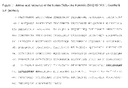
Figure 1 shows the amino acid sequence of the human Delta-like 4 protein (SEQ
ID
NO: I; GenBank NP 061947). The signal sequence, amino acids 1-26, is
underlined. The
transmembrane domain, amino acids 532-552, is bolded. The extracellular domain
of the
mature protein is amino acids 27-531, although imprecision in signal peptide
processing may
result in a protein that is slightly longer or shorter. The intracellular
domain is amino acids
553-685.
Figure 2 shows the nucleic acid sequence (cDNA) encoding the human Delta-like
4
protein (SEQ ID NO:2; GenBank NM_019074). The coding sequence is nucleic acids
321-
2378.
Figure 3 shows pZ/EG-mD114 transgenesis vector and result of Cre activity.
Figure 4. (a) LacZ staining of a ZEG-mD114 embryo at E8.0; (b) EGFP expression
in
the dt embryos at E8.5. (c) haemorrhaging and pericardial edema in dt embryos
at E9Ø
Figure 5. Wholemount PECAMI immunostaining of E9.0 and E9.5 dt and control
embryos. (a) control embryo at E9.0, (b) dt embryo at E9.0 showing a
hypertrophied dorsal
aorta (lower left arrow), ramified ACV (lower right arrow) and an immature
vascular plexus
in the head region (upper arrow) (c) control embryo at E9.5, (d) dt embryo at
E9.5 showing
hypertrophied dorsal aorta and almost no sign of an ACV, immature vascular
plexus in the
head region and hypertrophied sinus venosus and heart ventricle. Half
sectioning the stained
embryos at E9.5 showed that the aorta of the dt embryos (f) atrophies just
posterior to its
connection to the sinus venosus (lower arrow), while in the control embryo (e)
remains with
the same calibre throughout the embryo. The intersomitic vessels (upper arrow)
of the dt
embryos (h) appear slightly dilated and shorter than those of control embryos
(g). In the
dorsal region (lower arrow) of the dt embryos angiogenesis fails to occur. (i)
yolk sac of a
E9.5 control embryo, (j) yolk sac of a dt embryo showing lack of remodelling
of the primary
plexus in contrast to the highly organized structure of the vasculature in the
control embryos.
Figure 6. PECAM I immunostaining in cryosections and microangiography. (a-g)
serial sections of a E9.5 dt embryo (anterior-posterior) showing fusion
between the aorta
(upper right arrow) and the ACV (upper left arrow) just prior to its
connection to the sinus
venosus (lower arrow). In section (a) the ACV consists ofa plexus of small
capillaries (upper
-8-
CA 02710082 2010-06-18
WO 2009/085209 PCT/US2008/013899
left arrow) which join to form a single vessel with a large lumen just prior
to its fusion with
the dorsal aorta. Section (e) shows the aortic atrophy in regions posterior to
the sinus
venosus. (f,g) serial sections of a E9.5 wild type embryo depicting the same
regions stated
above. Microangiography with India ink injection confirmed the existence of
functional
connections between the dorsal aortae and the ACV of dt embryos (i), with ink
flowing
directly from the aortae (left hand arrow) to the sinus venosus (right hand
arrow), in contrast
to the regular flow observed in the control embryos (h).
Figure 7. Venous expression of arterial markers in dt embryos. In situ
hybridization of
cryosections from E9.0 dt embryos with ephrin-B2(a,b,c) and connexin-37
(d,e,f) specific
riboprobes. The mutant embryos show concomitant expression of these arterial
specific
markers in the both the dorsal aortae (AD) and anterior cardinal veins (VCA)
In the control
embryos (c,f), as expected, the expression is restricted to the aortae.
Figure 8. Upregulation of Notch signalling in the venous endothelium of the
mutant
embryos. In situ hybridization of cryosections from E9.0 dt embryos. with heyl
(a,b,c) and
Notchl (d,e,f) specific riboprobes. Both genes appear upregulated in the
anterior cardinal
veins (VCA). In the control embryos (c,f), as expected, the expression is
restricted to the
aortae.
Figure 9. Downregulation of venous specific markers in dt embryos. In situ
hybridization and immunostainings of cryosections from E9.0 dt embryos, (a)
anti-Eph-B4
immunostain, (c) eph-b4 mRNA, and E9.0 control embryos, (b) anti-Eph-B4
immunostain,
(d) eph-b4 mRNA.
Figure 10. Shows a schematic of the human D114 domain structure (top) and an
annotated human D114 amino acid sequence (SEQ ID NO: t) (bottom). The signal
sequence
and DSL domain are underlined and indicated. The eighth EGF8 domain (EGF8) is
shaded.
The AXB (AEGF8) construct contains 19 extra amino acids (RSPSCIYRRSWRSRGAQIL)
(SEQ ID NO:3) at the C-terminus after the CAS residues of the EGF8 repeat. The
P524-His
construct ends at P524, 4 amino acids before the transmembrane domain, with a
6xHis tag at
the C-terminus. Both constructs contain the receptor-binding domain, DSL
domain. Full
length constructs have either a Myc tag or no tag.
Figure I I shows the purified hDll4-P524-6xHis protein (histidine tagged hD114-
P524)
after nickel column purification (SDS-PAGE: CBB-G250 Staining).
Figure 12. hDll4 inhibits tube formation in human arterial endothelial cells
(HUAEC). VEGF was used at 50 ng/ml as a positive control. D114 at lower
concentrations
(30ng/ml or 100 ng/ml) promoted tube formation, while D114 at 500 ng/ml
inhibited tube
-9-
CA 02710082 2010-06-18
WO 2009/085209 PCT/US2008/013899
formation. (data not shown). Quantitative analysis for tube length and the
number of
junctions in sD114-treated HUVECs (Bioquant Image Analysis; mean from
triplicate wells in
2 repetition experiments). Similar results were seen with human arterial
endothelial cell
assay(data not shown).
Figure 13. hD114 inhibits sprouting in human arterial endothelial cells
(HUAEC).
VEGF was used at 20 ng/ml as a positive control. D114 at I OOng/ml or 200
ng/ml promoted
sprouting, while D114 at 500 ng/ml inhibited sprouting.. (not shown).
Quantitative analysis for
vascular area is shown (Bioquant Image Analysis; mean from triplicate wells in
2 repetition
experiments). Similar results were seen with sD114-Fc(data not shown).
Figure 14. hD114 inhibits VEGF-stimulated sprouting in human arterial
endothelial
cells (HUAEC) at high concentrations. VEGF was used at 20 ng/ml. Dll4 at
IOOng/ml had
little effect, while D114 at 200 ng/ml inhibited VEGF-stimulated sprouting.
Figure 15. D114+/- mutant mice show defective increase in vascular
proliferation: (A)
The vasculature of wild type and D114+/- embryos were examined using PECAM
wholemount immunostaining. Dorsal aorta and cardinal vein are labeled a and v.
Absence of
large vessels and an increase in vessel branching and density was seen in
D114+i- embryos at
E 10.5 compared to wild type. (B) Vascular response in D114+/- adult mice was
examined as
in (A) after tumor implantation. Wild type mice showed organized vascular
proliferation in
the tumor (left half), while mutant mice showed markedly increased vascular
response which
lacks organization and vascular hierarchy. (C) Expression of D114 in tumor and
normal
regions in D114+i- mutant mice was examined by B-gal staining. 13114
expression was observed
in a few discrete vessels in the normal tissue, while the tumor region showed
many 8-gal
positive vessels of similar appearance indicative of D114 induction in tumor
vessels. (D)
Pericyte coverage around newly forming vessels was examined by a-SMA
localization. In
wild type mice, the vessels showed co-localization of PECAM and a-SMA (left
panel). In
D114 +/- mice tumor vessels however, the number of a -SMA positive cells
lining the
endothelial cells was profoundly reduced (right panel). (E) D114 is activated
in most but not
all vessels in the tumor. 13-gal (left panel) and PECAM immuno-staining (right
panel) of
adjacent tumor sections show that endothelial specific PECAM staining is more
abundant that
lacZ D/14-reporter expression.
Figure 16. Biochemical properties of sD114: (A) Notch-Fc fusion protein was
coated
directly on ELISA plates. sD114-AP was allowed to bind Notch-Fc and the bound
D114 was
quantitated by the addition of AP substrate. sD114-AP bound efficiently to
Notch I and not
-10-
CA 02710082 2010-06-18
WO 2009/085209 PCT/US2008/013899
Notch 3 (left panel). Binding of sDll-4Fc and sD114-His to Notch 1 was
examined. (B)
HUVEC cells were transfected with expression vectors for sD114-Fc, sD114-His
or vector
alone. Notch responsive Hes-2 gene expression was not induced by sD114
proteins. (C) Notch
activation measured by the induction in Hes-1, Hey-I and Hes-2 when HUVEC
cells were
co-cultivated with choK expressing D114-FL (full length). Addition of
recombinant sDll4-Fc
and sD114-His reduced the induction of Notch responsive genes. Two independent
experiments produced similar results.
Figure 17. sD114 induces vessel response but lack perfusion in murine Matrigel
assay:
(A) Matrigel was injected subcutaneously into Balb/C nu/nu mice. After 6 days,
plugs were
removed and processed in paraffin. Individual sections were stained with H&E
and
representative photographs at x 20 magnification from triplicate plugs in 2
independent
experiments are shown. (B) Matrigel plugs were stained for PECAM.
Photomicrographs were
taken with a Nikon Coolpix 5000 camera on a Nikon Eclipse E400 microscope with
a 4
x/0.13 NA objective and a 10 x eyepiece. Quantitation of vascularized area
averaged (
SEM) from all plugs (Scion Image software) in bar graph. P value <0.01.
Figure 18. sD114 inhibits the tumor growth in a murine tumor xenograft model:
(A)
Mice (n = 6/group) were given implants with I x 106 HT29 cells in a Matrigel
preparation
with PBS or sD114-Fc or sD114-His (5 ug/ml) and tumor volumes measured after 2
weeks,
tumors were harvested and analyzed. Tumor volumes were significantly smaller
in the sD114
arm (Fig 6A). The experiment was repeated twice. (B) In assessing the effect
of endogenous
expression of sD114, HT29 were transfected with expression vector with D114-
FL, sD114-Fc,
sD114-His, or vector alone. Co-expression of truncated CD4 was done to allow
sorting of the
transfected cells. Equal number of the transfected cells were implanted in
mice (n = 6/group).
Tumor volumes were assessed (Fig 6B). Tumor volumes were significantly smaller
in the
sDll4 groups. (C) Microvasculature was assessed by PECAM immuno-staining and
the blood
vessel volume was quantitated as described in method section. (D). Hypoxy
probe was
infused prior to tumor harvest, tumor sections were then probed with MAb and
fluorescent
labeled secondary antibody as described in methods. Hypoxic areas were
quantitated using
lmageJ as described in methods. All values are expressed as mean SEM. *P <
.01 by
Student t test. Photomicrographs were taken using a Nikon Coolpix 5000 camera
and a Nikon
Eclipse E400 microscope with a 10 x eyepiece. Magnification was 20 x/0.5 NA
objectives.
(E). Vascular perfusion was determined by injecting fluorescent labeled lectin
10-15 min
prior to sacrificing mice and harvesting tumors. Lectin was localized to
perfused areas while
blood vessels were delineated with PECAM staining. Lectin and PECAM co-
localized in
- 11 -
CA 02710082 2010-06-18
WO 2009/085209 PCT/US2008/013899
control group, while sD114 group showed marked deficiency of perfusion. (F).
Localization of
a -SMA in tumor vessel. Control group showed co-localization of a-SMA and
PECAM,
while sD114 group had paucity of a-SMA positive cells in the micro-vessels.
Figure 19. Soluble D114 inhibits tumor growth in vivo in a murine zenograft
model.
D114-E6 (residues 1-422 of SEQ ID NO: 1) which lacks EGF-like domains 7 and 8
was
administered intraperitoneally starting at day 5 post implantation at 5 mg/kg,
three times a
week. VEGF-stimulated sprouting in human arterial endothelial cells (HUAEC) at
high
concentrations. The combined effect of D114-E6 with a VEGF neutralizing
antibody
(Avastin) was also examined. Avastin was also administered intraperitoneally
starting at day
5 post implantation at 10 mg/kg, three times a week.
Figure 20. Epitope mapping of anti-D114 antibodies: (A). Illustration of a
complete
set of DIN truncation mutants fused to alkaline phosphatase. Four individual
clones were
identified, each with a specific binding region to D114. (B). Coat 4ug/ml
(100ul) D114
antibodies on ELISA plate overnight in PBS at 4 C. Block the plate with 0.5%
BSA for 2
hours, and then add 20ng of soluble D114 proteins fused with alkaline
phosphatase. After 45
min incubation at room temperature, the plate is washed with PBST and
incubated with
PNPP at 37 C for 20 min. This experiment has been repeated at least three
times.
Figure 21. Protein sequence alignment of human D114 (hD114), mouse DI14
(mD114),
and human DII I (hD111). From N terminus to C terminus, first shaded region
indicates the
epitope for antibody clone #2-6. Second shaded region represents the DSL
region. Third
shaded region, which comprises EGF-like 3 (E3) domain, indicates the epitope
for antibody
clone #6 l B.
Figure 22. Characterization of anti-D114 antibodies: (A). Human D114
antibodies were
raised in mouse by immunization with soluble human D114-His protein (a.a. 1-
524 of SEQ ID
NO: 1). Four antibody clones are summarized. (B). Two antibody clones
designated #2-6
and #61 B efficiently neutralize D114-Notchl interaction. Coat 0.Sug/ml
(I00ul) Notch] -Fc
on ELISA plate overnight in PBS at 4 C. Block the plate with 0.5% BSA for 2
hours, and
then add indicated amount of D114 antibodies premixed with 50ng soluble D114
fused with
alkaline phosphatase. After 45 min incubation at room temperature, the plate
is washed with
PBST and incubated with PNPP at 37 C for 1.5 hours. This experiment has been
repeated at
least three times.
Figure 23. Sequence identifying the variable regions VH (SEQ ID NO: 4) and VL
(SEQ ID NO: 5) of D114 antibody clone designated #61 B.
- 12 -
CA 02710082 2010-06-18
WO 2009/085209 PCT/US2008/013899
Figure 24. Sequence identifying the variable regions VH (SEQ ID NO: 6) and VL
(SEQ ID NO: 7) of D114 antibody clone designated #2-6.
Figure 25. D114-Fc fusion protein linker engineering. The linker region
between
fusion proteins may affect the function of protein of interest. Three
different linkers between
D114 (E6) and human IgGI Fc (starts at EPKS in Fc hinge region) were tested.
There is a
three-fold-difference between LI and L2 fusion proteins. D114-L2-Fc was chosen
for tumor
xenograft study. ELISA plate was coated with 0.5ug/ml (100ul) Notch I-Fe in
PBS at 4 C for
overnight, and then blocked with 0.5% BSA for 2 hours. Indicated amount of
D114-Fc
proteins or BSA were premixed with 50ng D114 (E8)-AP and then added. After
45min
incubation at room temperature, the plate was washed with PBST and incubated
with PNPP
at 37 C for 1 hour.
DETAILED DESCRIPTION OF THE INVENTION
The current invention is based in part on the discovery that Delta-like 4
function is
essential for angiogenesis in vivo, and, moreover, that an increase of Delta-
like 4 activity is
associated with increased proliferation of arterial endothelial cells and an
increased adoption
of an arterial identity by endothelial cells. Applicants generated mouse D114
knockout
mutations that evinced dosage sensitive defects in angiogenesis. Furthermore,
Applicants
generated D114 overexpression models in mouse and demonstrated that increased
expression
of Dll4 causes, in some instances, hypertrophy of arterial tissue and,
moreover, causes venous
tissue to adopt an arterial identity. Based on these results, it is apparent
that angiogenesis, in
which a system of arterial and venous microvessels is generated, is highly
sensitive to D114
activity and may be perturbed (e.g., inhibited or caused to occur in a
disorganized or
ineffective manner) by inhibition or hyperactivation of D114. Thus,
surprisingly, both
agonists and antagonists of D114 may be used to treat tumors undergoing
angiogenesis.
Furthermore, the invention relates to the discovery that overexpression of
D114 can stimulate
arterial growth, and may therefore be used to stimulate arteriogenesis.
Arteriogenesis is the
process of collateral artery formation and growth, typically in ischemic
tissues. Thus D114
agonists may be used to treat patients suffering from, or at risk for, an
ischemic event, such as
a peripheral or coronary ischemia. Furthermore, the disclosure demonstrates
that a soluble
monomeric or dimeric D114 polypeptide can act to inhibit or promote
angiogenesis at low or
high concentrations, respectively.
-13-
CA 02710082 2010-06-18
WO 2009/085209 PCT/US2008/013899
D114 Agonist/Antagonist Determinant
The invention further relates to biomarkers that may be used to assess whether
an
agent of interest is an agonist or antagonist of D114 signaling. The
scientific literature relating
to Delta proteins generally, including D114, provides no clarity as to whether
a particular
agent activates or inhibits D114-mediated signaling. For example, D114 and
Delta extracellular
domains (e.g., soluble monomeric or dimeric forms, forms with deleted
intracellular domains,
and soluble Fc fusions) have been tested in a variety of assays and it remains
unclear whether
any of the observed effects are due to agonist or antagonist activity, or
whether there is any
meaningful activity at all. Moreover, reagents may affect D114 signaling in a
variety of ways.
For example, a reagent may affect Notch I and/or Notch 4 activation, or
activation of
retrograde D114 signaling, possibly mediated by the D114 intracellular domain.
A reagent may
also affect the activity of preseniline protease activity, which may affect
both Notch I and
Notch4. The present disclosure demonstrates that D114 hyperactivation causes
endothelial
cells to adopt an arterial phenotype, typified by expression of EphrinB2 and
connexin37,
while D114 loss of function causes endothelial cells to adopt a venous
identity, typified by
expression of EphB4. This information about the genetically-determined, in
vivo effects of
D114 activity will permit the identification of both known and newly
discovered agents as
agonists or antagonists of D114 signaling.
AGENTS
Accordingly, in certain aspects, the disclosure provides numerous polypeptide
compounds (agents) that may be used to treat cancer as well as angiogenesis
related disorders
and unwanted angiogenesis related processes.
D114 is a Notch ligand and contains a signal sequence, a DSL domain, eight
epidermal
growth factor-like repeats, a transmembrane domain, and an intracellular
region, all of which
are characteristics of members of the Delta protein family. The tissue
distribution of Delta-4
mRNA resembles that previously described for Notch-4 (Int-3) transcripts.
Soluble forms of
the extracellular portion of Delta-4 inhibit the apparent proliferation of
human aortic
endothelial cells, but not human pulmonary arterial endothelial cells. Yoneya
et al. J.
Biochem. Vol. 129, pp. 27-34 (2001).
Members of the Notch family of proteins are transmembrane receptors that
contain
characteristic multiple epidermal growth factor (EGF)-like repeats as well as
conserved
domains such as RAM, ankyrin-like repeat, and PEST sequences. Ligands for
Notch proteins
-14-
CA 02710082 2010-06-18
WO 2009/085209 PCT/US2008/013899
include Delta and Serrate in Drosophila melanogaster, LAG-2 and APX-1 in
Caenorhabditis
elegans, and Delta and Serrate (or Jagged) in vertebrates. These ligands are
also
transmembrane proteins and contain a highly conserved DSL (Delta-Serrate-LAG-
2) motif
upstream of a variable number of EGF-like repeats. The DSL domain is a
characteristic
feature of Notch ligands and is important for protein function; thus, point
mutation of the
DSL domain in LAG-2 results in a loss of activity. Although the Delta and
Jagged (Serrate)
proteins of vertebrates exhibit similar structures, each group of proteins
also possesses
several distinct features. Thus, whereas vertebrate Delta proteins contain
eight EGF-like
repeats, Jagged proteins contain 16 such repeats. Furthermore, the EGF domains
are followed
by a cysteine-rich domain in Jagged proteins but not in Delta proteins.
However, the
consequences of these structural differences remain unclear.
Uyttendaele et at. (1996) cloned cDNAs corresponding to the complete coding
region
of the mouse Notch4 gene. In situ hybridization revealed that Notch4
transcripts are
primarily restricted to endothelial cells in embryonic and adult life,
suggesting a role for
Notch4 during development of vertebrate endothelium.
Li et al. (Genomics. 1998 Jul 1;51(1):45-58) reported that the human NOTCH4
gene
contains 30 exons and spans approximately 30 kb. They isolated cDNAs
corresponding to
6.7-kb NOTCH4(S) and 9.3-kb NOTCH4(L) mRNA isoforms. The predicted protein
encoded
by NOTCH4(S) is 2,003 amino acids long and contains the characteristic Notch
motifs: a
signal peptide, 29 epidermal growth factor (EGF)-like repeats, 3 Notch/lin-12
repeats, a
transmembrane region, 6 cdcl0 (603151)/ankyrin repeats, and the PEST conserved
region at
the C terminus. The sequences of the mouse and human NOTCH4 proteins are 82%
identical.
The incompletely spliced NOTCH4(L) cDNA potentially encodes 2 different
proteins. One
consists of the first 7 EGF repeats. The second contains the transmembrane
domain and
intracellular region and is similar to the mouse int3 protooncoprotein.
Northern blot analysis
revealed that NOTCH4(S) is the major transcript and is expressed in a wide
variety of tissues.
Krebs et at. (2000) generated Notch4-deficient mice by gene targeting. Embryos
homozygous for this mutation developed normally, and homozygous mutant adults
were
viable and fertile. However, the Notch4 mutation displayed genetic
interactions with a
targeted mutation of the related Notch I gene. Both Notch I mutant and Notch 1
/Notch4
double mutant embryos displayed severe defects in angiogenic vascular
remodeling. Analysis
of the expression patterns of genes encoding ligands for Notch family
receptors indicated that
only the D114 gene is expressed in a pattern consistent with that expected for
a gene encoding
-15-
CA 02710082 2010-06-18
WO 2009/085209 PCT/US2008/013899
a ligand for the Notchl and Notch4 receptors in the early embryonic
vasculature. Therefore,
there is an essential role for the Notch signaling pathway in regulating
vascular
morphogenesis and remodeling, and indicate that whereas the Notch4 gene is not
essential
during embryonic development, the Notch4 and Notchl genes have partially
overlapping
roles during embryogenesis in mice.
As noted above, the disclosure provides methods for using and identifying
agonists
and antagonists of D114 signaling. Candidate agonists and antagonists will
generally be any
antibody that binds to, or soluble portions of, proteins involved in the D114
signaling pathway,
including, for example, D114, Notch], Notch4 and presenilin. Candidate
agonists and
antagonists may also be small molecules or other agents that bind to or effect
members of the
pathway. Antisense or RNAi nucleic acids may be used as antagonists of D114,
Notchl,
Notch4 or presenilin or other members of the signaling pathway.
Examples of agents include:
(a) an antibody that binds selectively to D114;
(b) an antibody that binds selectively to Notchl;
(c) an antibody that binds selectively to Notch4;
(d) an antibody that binds to Notchl and Notch4;
(e) a polypeptide monomer comprising a Notch-receptor binding portion of D114;
(f) a polypeptide dimer comprising a Notch-receptor binding portion of D114;
(g) a polypeptide multimer comprising two or more polypeptides comprising a
Notch-
receptor binding portion of D114;
(h) a polypeptide monomer comprising a D114-binding portion of Notch 1 or
Notch4;
(i) a polypeptide multimer comprising two or more polypeptides comprising a
D114-
binding portion of Notch I or Notch4.
Agents that interfere with presenilin activity or other metalloproteinases
(e.g.,
kuzbanian) are expected to modulate D114 signaling. Each of these agents may
be assessed
for agonist or antagonist activity as described herein.
-16-
CA 02710082 2010-06-18
WO 2009/085209 PCT/US2008/013899
D114 Polypeptides
In certain aspects, the agent is a soluble polypeptide comprising an
extracellular
domain of a D114 protein, e.g., as shown in amino acids 27-531 of SEQ ID NO:1.
In a
specific embodiment, the D114 soluble polypeptide comprises a DSL domain of a
D114
protein. In another embodiment, the 13114 soluble polypeptide is a truncate
comprising at
least domains 5 or 6 of the EGF-like domains.
As used herein, the subject soluble polypeptides include fragments, functional
variants, and modified forms of D114 soluble polypeptide. These fragments,
functional
variants, and modified forms of the subject soluble polypeptides may be tested
for activity as
agonists or antagonists of D114 by assessing effects on arterial or venous
phenotype in
endothelial cells.
In certain embodiments, isolated fragments of the subject soluble polypeptides
can be
obtained by screening polypeptides recombinantly produced from the
corresponding
fragment of the nucleic acid encoding an D114. In addition, fragments can be
chemically
synthesized using techniques known in the art such as conventional Merrifield
solid phase f-
Moc or t-Boc chemistry. The fragments can be produced (recombinantly or by
chemical
synthesis) and tested to identify those peptidyl fragments that can modulate
D114 signaling.
In certain embodiments, a functional variant of an D114 soluble polypeptide
comprises
an amino acid sequence that is at least 90%, 95%, 97%, 99% or 100% identical
to residues
27-531 of the amino acid sequence of SEQ ID NO:1.
In certain embodiments, the present invention contemplates making functional
variants by modifying the structure of the subject soluble polypeptide for
such purposes as
enhancing therapeutic or prophylactic efficacy, or stability (e.g., ex vivo
shelf life and
resistance to proteolytic degradation in vivo). Modified soluble polypeptides
can be
produced, for instance, by amino acid substitution, deletion, or addition. For
instance, it is
reasonable to expect, for example, that an isolated replacement of a leucine
with an isoleucine
or valise, an aspartate with a glutamate, a threonine with a serine, or a
similar replacement of
an amino acid with a structurally related amino acid (e.g., conservative
mutations) will not
have a major effect on the biological activity of the resulting molecule.
Conservative
replacements are those that take place within a family of amino acids that are
related in their
side chains.
-17-
CA 02710082 2010-06-18
WO 2009/085209 PCT/US2008/013899
This invention further contemplates a method of generating sets of
combinatorial
mutants of the D114 polypeptides, as well as truncation mutants, and is
especially useful for
identifying functional variant sequences. The purpose of screening such
combinatorial
libraries may be to generate, for example, soluble polypeptide variants which
can act as
agonists or antagonists of Dll4. Combinatorially-derived variants can be
generated which
have a selective potency relative to a naturally occurring soluble
polypeptide. Such variant
proteins, when expressed from recombinant DNA constructs, can be used in gene
therapy
protocols. Likewise, mutagenesis can give rise to variants which have
intracellular half-lives
dramatically different than the corresponding wild-type soluble polypeptide.
For example,
the altered protein can be rendered either more stable or less stable to
proteolytic degradation
or other cellular process which result in destruction of, or otherwise
inactivation of the
protein of interest (e.g., a soluble polypeptide). Such variants, and the
genes which encode
them, can be utilized to alter the subject soluble polypeptide levels by
modulating their half-
life. A short half-life can give rise to more transient biological effects
and, when part of an
inducible expression system, can allow tighter control of recombinant soluble
polypeptide
levels within the cell. As above, such proteins, and particularly their
recombinant nucleic
acid constructs, can be used in gene therapy protocols.
There are many ways by which the library of potential homologs can be
generated
from a degenerate oligonucleotide sequence. Chemical synthesis of a degenerate
gene
sequence can be carried out in an automatic DNA synthesizer, and the synthetic
genes then be
ligated into an appropriate gene for expression. The purpose of a degenerate
set of genes is to
provide, in one mixture, all of the sequences encoding the desired set of
potential soluble
polypeptide sequences. The synthesis of degenerate oligonucleotides is well
known in the art
(see for example, Narang, SA (1983) Tetrahedron 39:3; Itakura et al., (1981)
Recombinant
DNA, Proc. 3rd Cleveland Sympos. Macromolecules, ed. AG Walton, Amsterdam:
Elsevier
pp273-289; Itakura et al., (1984) Annu. Rev. Biochem. 53:323; Itakura et al.,
(1984) Science
198:1056; Ike et al., (1983) Nucleic Acid Res. 11:477). Such techniques have
been employed
in the directed evolution of other proteins (see, for example, Scott et al.,
(1990) Science
249:386-390; Roberts et al., (1992) PNAS USA 89:2429-2433; Devlin et al.,
(1990) Science
249: 404-406; Cwirla et al., (1990) PNAS USA 87: 6378-6382; as well as U.S.
Patent Nos:
5,223,409, 5,198,346, and 5,096,815).
Alternatively, other forms of mutagenesis can be utilized to generate a
combinatorial
library. For example, soluble polypeptide variants (e.g., the antagonist
forms) can be
-18-
CA 02710082 2010-06-18
WO 2009/085209 PCT/US2008/013899
generated and isolated from a library by screening using, for example, alanine
scanning
mutagenesis and the like (Ruf et al., (1994) Biochemistry 33:1565-1572; Wang
et al., (1994)
J. Biol. Chem. 269:3095-3099; Balint et al., (1993) Gene 137:109-118; Grodberg
et al.,
(1993) Eur. J. Biochem. 218:597-601; Nagashima et al., (1993) J. Biol. Chem.
268:2888-
2892; Lowman et al., (1991) Biochemistry 30:10832-10838; and Cunningham et
al., (1989)
Science 244:1081-1085), by linker scanning mutagenesis (Gustin et al., (1993)
Virology
193:653-660; Brown et al., (1992) Mol. Cell Biol. 12:2644-2652; McKnight et
al., (1982)
Science 232:316); by saturation mutagenesis (Meyers et al., (1986) Science
232:613); by
PCR mutagenesis (Leung et al., (1989) Method Cell Mol Biol 1:11-19); or by
random
mutagenesis, including chemical mutagenesis, etc. (Miller et al., (1992) A
Short Course in
Bacterial Genetics, CSHL Press, Cold Spring Harbor, NY; and Greener et al.,
(1994)
Strategies in Mol Biol 7:32-34). Linker scanning mutagenesis, particularly in
a combinatorial
setting, is an attractive method for identifying truncated (bioactive) forms
of the subject
soluble polypeptide.
A wide range of techniques are known in the art for screening gene products of
combinatorial libraries made by point mutations and truncations, and, for that
matter, for
screening cDNA libraries for gene products having a certain property. Such
techniques will
be generally adaptable for rapid screening of the gene libraries generated by
the
combinatorial mutagenesis of the subject soluble polypeptides. The most widely
used
techniques for screening large gene libraries typically comprises cloning the
gene library into
replicable expression vectors, transforming appropriate cells with the
resulting library of
vectors, and expressing the combinatorial genes under conditions in which
detection of a
desired activity facilitates relatively easy isolation of the vector encoding
the gene whose
product was detected. Each of the illustrative assays described below are
amenable to high
through-put analysis as necessary to screen large numbers of degenerate
sequences created by
combinatorial mutagenesis techniques.
In certain embodiments, the soluble polypeptides of the invention may further
comprise post-translational modifications. Such modifications include, but are
not limited to,
acetylation, carboxylation, glycosylation, phosphorylation, lipidation, and
acylation. As a
result, the modified soluble polypeptides may contain non-amino acid elements,
such as
polyethylene glycols, lipids, poly- or mono-saccharide, and phosphates.
Effects of such non-
amino acid elements on the functionality of a soluble polypeptide may be
tested for its
agonist or antagonist effects on D114.
-19-
CA 02710082 2010-06-18
WO 2009/085209 PCT/US2008/013899
In certain aspects, functional variants or modified forms of the subject
soluble
polypeptides include fusion proteins having at least a portion of the soluble
polypeptide and
one or more fusion domains. Well known examples of such fusion domains
include, but are
not limited to, polyhistidine, Glu-Glu, glutathione S transferase (GST),
thioredoxin, protein
A, protein G, and an immunoglobulin heavy chain constant region (Fc), maltose
binding
protein (MBP), which are particularly useful for isolation of the fusion
proteins by affinity
chromatography. For the purpose of affinity purification, relevant matrices
for affinity
chromatography, such as glutathione-, amylase-, and nickel- or cobalt-
conjugated resins are
used. Another fusion domain well known in the art is green fluorescent protein
(GFP).
Fusion domains also include "epitope tags," which are usually short peptide
sequences for
which a specific antibody is available. Well known epitope tags for which
specific
monoclonal antibodies are readily available include FLAG, influenza virus
haemagglutinin
(HA), and c-myc tags. In some cases, the fusion domains have a protease
cleavage site, such
as for Factor Xa or Thrombin, which allows the relevant protease to partially
digest the fusion
proteins and thereby liberate the recombinant proteins therefrom. The
liberated proteins can
then be isolated from the fusion domain by subsequent chromatographic
separation. In
certain embodiments, the soluble polypeptides of the present invention contain
one or more
modifications that are capable of stabilizing the soluble polypeptides. For
example, such
modifications enhance the in vitro half life of the soluble polypeptides,
enhance circulatory
half life of the soluble polypeptides or reducing proteolytic degradation of
the soluble
polypeptides.
In certain embodiments, soluble polypeptides (unmodified or modified) of the
invention can be produced by a variety of art-known techniques. For example,
such soluble
polypeptides can be synthesized using standard protein chemistry techniques
such as those
described in Bodansky, M. Principles of Peptide Synthesis, Springer Verlag,
Berlin (1993)
and Grant G. A. (ed.), Synthetic Peptides: A User's Guide, W. H. Freeman and
Company,
New York (1992). In addition, automated peptide synthesizers are commercially
available
(e.g., Advanced ChemTech Model 396; Milligen/Biosearch 9600). Alternatively,
the soluble
polypeptides, fragments or variants thereof may be recombinantly produced
using various
expression systems as is well known in the art (also see below).
Gene therapy
In certain aspects, the invention relates to isolated and/or recombinant
nucleic acids
encoding a D114 polypeptide. The subject nucleic acids may be single-stranded
or double-
-20-
CA 02710082 2010-06-18
WO 2009/085209 PCT/US2008/013899
stranded, DNA or RNA molecules. These nucleic acids are useful as therapeutic
agents. For
example, these nucleic acids are useful in making recombinant soluble
polypeptides which
are administered to a cell or an individual as therapeutics. Alternative,
these nucleic acids
can be directly administered to a cell or an individual as therapeutics such
as in gene therapy.
In certain embodiments, the invention provides isolated or recombinant nucleic
acid
sequences that are at least 80%, 85%, 90%, 95%, 97%, 98%, 99% or 100%
identical to a
region of the nucleotide sequence depicted in SEQ ID NO:2. One of ordinary
skill in the art
will appreciate that nucleic acid sequences complementary to the subject
nucleic acids, and
variants of the subject nucleic acids are also within the scope of this
invention. In further
embodiments, the nucleic acid sequences of the invention can be isolated,
recombinant,
and/or fused with a heterologous nucleotide sequence, or in a DNA library.
In other embodiments, nucleic acids of the invention also include nucleotide
sequences that hybridize under highly stringent conditions to the nucleotide
sequence
depicted in SEQ ID NO:2, or complement sequences thereof. As discussed above,
one of
ordinary skill in the art will understand readily that appropriate stringency
conditions which
promote DNA hybridization can be varied. One of ordinary skill in the art will
understand
readily that appropriate stringency conditions which promote DNA hybridization
can be
varied. For example, one could perform the hybridization at 6.0 x sodium
chloride/sodium
citrate (SSC) at about 45 C, followed by a wash of 2.0 x SSC at 50 C. For
example, the salt
concentration in the wash step can be selected from a low stringency of about
2.0 x SSC at 50
C to a high stringency of about 0.2 x SSC at 50 C. In addition, the
temperature in the wash
step can be increased from low stringency conditions at room temperature,
about 22 C, to
high stringency conditions at about 65 C. Both temperature and salt may be
varied, or
temperature or salt concentration may be held constant while the other
variable is changed.
In one embodiment, the invention provides nucleic acids which hybridize under
low
stringency conditions of 6 x SSC at room temperature followed by a wash at 2 x
SSC at room
temperature.
Isolated nucleic acids which differ from the subject nucleic acids due to
degeneracy in
the genetic code are also within the scope of the invention. For example, a
number of amino
acids are designated by more than one triplet. Codons that specify the same
amino acid, or
synonyms (for example, CAU and CAC are synonyms for histidine) may result in
"silent"
mutations which do not affect the amino acid sequence of the protein. However,
it is
-21-
CA 02710082 2010-06-18
WO 2009/085209 PCT/US2008/013899
expected that DNA sequence polymorphisms that do lead to changes in the amino
acid
sequences of the subject proteins will exist among mammalian cells. One
skilled in the art
will appreciate that these variations in one or more nucleotides (up to about
3-5% of the
nucleotides) of the nucleic acids encoding a particular protein may exist
among individuals of
a given species due to natural allelic variation. Any and all such nucleotide
variations and
resulting amino acid polymorphisms are within the scope of this invention.
In certain embodiments, the recombinant nucleic acids of the invention may be
operably linked to one or more regulatory nucleotide sequences in an
expression construct.
Regulatory nucleotide sequences will generally be appropriate for a host cell
used for
expression. Numerous types of appropriate expression vectors and suitable
regulatory
sequences are known in the art for a variety of host cells. Typically, said
one or more
regulatory nucleotide sequences may include, but are not limited to, promoter
sequences,
leader or signal sequences, ribosomal binding sites, transcriptional start and
termination
sequences, translational start and termination sequences, and enhancer or
activator sequences.
Constitutive or inducible promoters as known in the art are contemplated by
the invention.
The promoters may be either naturally occurring promoters, or hybrid promoters
that
combine elements of more than one promoter. An expression construct may be
present in a
cell on an episome, such as a plasmid, or the expression construct may be
inserted in a
chromosome. In a preferred embodiment, the expression vector contains a
selectable marker
gene to allow the selection of transformed host cells. Selectable marker genes
are well
known in the art and will vary with the host cell used.
In certain aspect of the invention, the subject nucleic acid is provided in an
expression
vector comprising a nucleotide sequence encoding a D114 polypeptide and
operably linked to
at least one regulatory sequence. Regulatory sequences are art-recognized and
are selected to
direct expression of the soluble polypeptide. Accordingly, the term regulatory
sequence
includes promoters, enhancers, and other expression control elements.
Exemplary regulatory
sequences are described in Goeddel; Gene Expression Technology: Methods in
Enzymology,
Academic Press, San Diego, CA (1990). For instance, any of a wide variety of
expression
control sequences that control the expression of a DNA sequence when
operatively linked to
it may be used in these vectors to express DNA sequences encoding a soluble
polypeptide.
Such useful expression control sequences, include, for example, the early and
late promoters
of SV40, tet promoter, adenovirus or cytomegalovirus immediate early promoter,
the lac
system, the trp system, the TAC or TRC system, T7 promoter whose expression is
directed
- 22 -
CA 02710082 2010-06-18
WO 2009/085209 PCT/US2008/013899
by T7 RNA polymerase, the major operator and promoter regions of phage lambda,
the
control regions for fd coat protein, the promoter for 3-phosphoglycerate
kinase or other
glycolytic enzymes, the promoters of acid phosphatase, e.g., Pho5, the
promoters of the yeast
a-mating factors, the polyhedron promoter of the baculovirus system and other
sequences
known to control the expression of genes of prokaryotic or eukaryotic cells or
their viruses,
and various combinations thereof. It should be understood that the design of
the expression
vector may depend on such factors as the choice of the host cell to be
transformed and/or the
type of protein desired to be expressed. Moreover, the vector's copy number,
the ability to
control that copy number and the expression of any other protein encoded by
the vector, such
as antibiotic markers, should also be considered.
This invention also pertains to a host cell transfected with a recombinant
gene
including a coding sequence for one or more of the subject soluble
polypeptide. The host cell
may be any prokaryotic or eukaryotic cell. For example, a soluble polypeptide
of the
invention may be expressed in bacterial cells such as E. coli, insect cells
(e.g., using a
baculovirus expression system), yeast, or mammalian cells. Other suitable host
cells are
known to those skilled in the art.
Method of producing soluble polypeptide
Accordingly, the present invention further pertains to methods of producing
the
subject soluble polypeptides. For example, a host cell transfected with an
expression vector
encoding a D114 soluble polypeptide can be cultured under appropriate
conditions to allow
expression of the 13114 soluble polypeptide to occur. The D114 soluble
polypeptide may be
secreted and isolated from a mixture of cells and medium containing the
soluble
polypeptides. Alternatively, the soluble polypeptides may be retained
cytoplasmically or in a
membrane fraction and the cells harvested, lysed and the protein isolated. A
cell culture
includes host cells, media and other byproducts. Suitable media for cell
culture are well
known in the art. The soluble polypeptides can be isolated from cell culture
medium, host
cells, or both using techniques known in the art for purifying proteins,
including ion-
exchange chromatography, gel filtration chromatography, ultrafiltration,
electrophoresis, and
immunoaffinity purification with antibodies specific for particular epitopes
of the soluble
polypeptides. In a preferred embodiment, the soluble polypeptide is a fusion
protein
containing a domain which facilitates its purification.
- 23 -
CA 02710082 2010-06-18
WO 2009/085209 PCT/US2008/013899
A recombinant nucleic acid of the invention can be produced by ligating the
cloned
gene, or a portion thereof, into a vector suitable for expression in either
prokaryotic cells,
eukaryotic cells (yeast, avian, insect or mammalian), or both. Expression
vehicles for
production of a recombinant soluble polypeptide include plasmids and other
vectors. For
instance, suitable vectors include plasmids of the types: pBR322-derived
plasmids, pEMBL-
derived plasmids, pEX-derived plasmids, pBTac-derived plasmids and pUC-derived
plasmids
for expression in prokaryotic cells, such as E. coli.
The preferred mammalian expression vectors contain both prokaryotic sequences
to
facilitate the propagation of the vector in bacteria, and one or more
eukaryotic transcription
units that are expressed in eukaryotic cells. The pcDNAI/amp, pcDNAI/neo,
pRc/CMV,
pSV2gpt, pSV2neo, pSV2-dhfr, pTk2, pRSVneo, pMSG, pSVT7, pko-neo and pHyg
derived
vectors are examples of mammalian expression vectors suitable for transfection
of eukaryotic
cells. Some of these vectors are modified with sequences from bacterial
plasmids, such as
pBR322, to facilitate replication and drug resistance selection in both
prokaryotic and
eukaryotic cells. Alternatively, derivatives of viruses such as the bovine
papilloma virus
(BPV-1), or Epstein-Barr virus (pHEBo, pREP-derived and p205) can be used for
transient
expression of proteins in eukaryotic cells. Examples of other viral (including
retroviral)
expression systems can be found below in the description of gene therapy
delivery systems.
The various methods employed in the preparation of the plasmids and
transformation of host
organisms are well known in the art. For other suitable expression systems for
both
prokaryotic and eukaryotic cells, as well as general recombinant procedures,
see Molecular
Cloning A Laboratory Manual, 2nd Ed., ed. by Sambrook, Fritsch and Maniatis
(Cold Spring
Harbor Laboratory Press, 1989) Chapters 16 and 17. In some instances, it may
be desirable
to express the recombinant SLC5A8 polypeptide by the use of a baculovirus
expression
system. Examples of such baculovirus expression systems include pVL-derived
vectors
(such as pVL1392, pVL 1393 and pVL941), pAcUW-derived vectors (such as
pAcUWI), and
pBlueBac-derived vectors (such as the 13-gal containing pBlueBac Ill).
Techniques for making fusion genes are well known. Essentially, the joining of
various DNA fragments coding for different polypeptide sequences is performed
in
accordance with conventional techniques, employing blunt-ended or stagger-
ended termini
for ligation, restriction enzyme digestion to provide for appropriate termini,
filling-in of
cohesive ends as appropriate, alkaline phosphatase treatment to avoid
undesirable joining,
and enzymatic ligation. In another eimbodiment, the fusion gene can be
synthesized by
_24_
CA 02710082 2010-06-18
WO 2009/085209 PCT/US2008/013899
conventional techniques including automated DNA synthesizers. Alternatively,
PCR
amplification of gene fragments can be carried out using anchor primers which
give rise to
complementary overhangs between two consecutive gene fragments which can
subsequently
be annealed to generate a chimeric gene sequence (see, for example, Current
Protocols in
Molecular Biology, eds. Ausubel et al., John Wiley & Sons: 1992).
Antibody
In certain aspects, the present invention provides antibodies that have
agonist or
antagonist effects on D114 signaling. Such antibodies may bind to antigens
such as D114,
Notchl or Notch4. Preferably, the antibody binds to an extracellular domain of
such
antigens. It is understood that antibodies may be polyclonal or monoclonal;
intact or
truncated, e.g., F(ab')2, Fab, Fv; xenogeneic, allogeneic, syngeneic, fully
human or modified
forms thereof, e.g., humanized, chimeric. Fully human antibodies may be
selected from
transgenic animals that express human immunoglobulin genes or assembled from
recombinant libraries expressing antibody fragments.
For example, by using immunogens derived from D114, Notchl or Notch4, anti-
protein/anti-peptide antisera or monoclonal antibodies can be made by standard
protocols
(see, for example, Antibodies: A Laboratory Manual ed. by Harlow and Lane
(Cold Spring
Harbor Press: 1988)). A mammal, such as a mouse, a hamster or rabbit can be
immunized
with an immunogenic form of the peptide. (e.g., a polypeptide or an antigenic
fragment which
is capable of eliciting an antibody response, or a fusion protein). Techniques
for conferring
immunogenicity on a protein or peptide include conjugation to carriers or
other techniques
well known in the art. An immunogenic portion of an antigen can be
administered in the
presence of adjuvant. The progress of immunization can be monitored by
detection of
antibody titers in plasma or serum. Standard ELISA or other immunoassays can
be used with
the immunogen as antigen to assess the levels of antibodies.
Following immunization of an animal with an antigenic preparation, antisera
can be
obtained and, if desired, polyclonal antibodies can be isolated from the
serum. To produce
monoclonal antibodies, antibody-producing cells (lymphocytes) can be harvested
from an
immunized animal and fused by standard somatic cell fusion procedures with
immortalizing
cells such as myeloma cells to yield hybridoma cells. Such techniques are well
known in the
art, and include, for example, the hybridoma technique (originally developed
by Kohler and
Milstein, (1975) Nature, 256: 495-497); the human B cell hybridoma technique
(Kozbar et
- 25 -
CA 02710082 2010-06-18
WO 2009/085209 PCT/US2008/013899
al., (1983) Immunology Today, 4: 72), and the EBV-hybridoma technique to
produce human
monoclonal antibodies (Cole et al., (1985) Monoclonal Antibodies and Cancer
Therapy, Alan
R. Liss, Inc. pp. 77-96). Hybridoma cells can be screened immunochemically for
production
of antibodies specifically reactive with D114, Notchl, Notch4 or other target
polypeptide and
monoclonal antibodies isolated from a culture comprising such hybridoma cells.
The term "antibody" as used herein is intended to include fragments thereof
which are
also specifically reactive with antigen. Antibodies can be fragmented using
conventional
techniques and the fragments screened for utility in the same manner as
described above for
whole antibodies. For example, F(ab)2 fragments can be generated by treating
antibody with
pepsin. The resulting F(ab)2 fragment can be treated to reduce disulfide
bridges to produce
Fab fragments. The antibody of the present invention is further intended to
include
bispecific, single-chain, and chimeric and humanized molecules having affinity
for antigen
conferred by at least one CDR region of the antibody. Techniques for the
production of
single chain antibodies (US Patent No. 4,946,778) can also be adapted to
produce single
chain antibodies. Also, transgenic mice or other organisms including other
mammals, may be
used to express humanized antibodies. In preferred embodiments, the antibodies
further
comprise a label attached thereto and able to be detected (e.g., the label can
be a radioisotope,
fluorescent compound, enzyme or enzyme co-factor).
In certain preferred embodiments, an antibody of the invention is a monoclonal
antibody, and in certain embodiments the invention makes available methods for
generating
novel antibodies. For example, a method for generating a monoclonal antibody
that binds
specifically to D114, Notchl or Notch4 may comprise administering to a mouse
an amount of
an immunogenic composition comprising the antigen polypeptide effective to
stimulate a
detectable immune response, obtaining antibody-producing cells (e.g., cells
from the spleen)
from the mouse and fusing the antibody-producing cells with myelorna cells to
obtain
antibody-producing hybridomas, and testing the antibody-producing hybridornas
to identify a
hybridoma that produces a monocolonal antibody that binds specifically to the
antigen. Once
obtained, a hybridoma can be propagated in a cell culture, optionally in
culture conditions
where the hybridoma-derived cells produce the monoclonal antibody. The
monoclonal
antibody may be purified from the cell culture.
In certain embodiments, the disclosure provides humanized versions of any of
the
antibodies disclosed herein, as well as antibodies and antigen binding
portions thereof that
comprise at least one CDR portion derived from an antibody disclosed herein,
particularly the
-26-
CA 02710082 2010-06-18
WO 2009/085209 PCT/US2008/013899
CDR3. In preferred embodiments, the antibody is a monoclonal antibody that is
immunocompatible with the subject to which it is to be administered, and
preferably is
clinically acceptable for administration to a human.
In certain embodiments, single chain antibodies, and chimeric, humanized or
primatized (CDR-grafted) antibodies, as well as chimeric or CDR-grafted single
chain
antibodies, comprising portions derived from different species, are also
encompassed by the
present invention as antigen binding portions of an antibody. The various
portions of these
antibodies can be joined together chemically by conventional techniques, or
can be prepared
as a contiguous protein using genetic engineering techniques. For example,
nucleic acids
encoding a chimeric or humanized chain can be expressed to produce a
contiguous protein.
See, e.g., Cabilly et al., U.S. Pat. No. 4,816,567; Cabilly et al., European
Patent No.
0,125,023 BI; Boss et al., U.S. Pat. No. 4,816,397; Boss et al., European
Patent No.
0,120,694 B1; Neuberger, M. S. et al., WO 86/01533; Neuberger, M. S. et al.,
European
Patent No. 0,194,276 131; Winter, U.S. Pat. No. 5,225,539; and Winter,
European Patent No.
0,239,400 B1. See also, Newman, R. et al., BioTechnology, 10: 1455-1460
(1992), regarding
primatized antibody. See, e.g., Ladner et al., U.S. Pat. No. 4,946,778; and
Bird, R. E. et al.,
Science, 242: 423-426 (1988)), regarding single chain antibodies.
In addition, functional fragments of antibodies, including fragments of
chimeric,
humanized, primatized or single chain antibodies, can also be produced.
Functional
fragments of the subject antibodies retain at least one binding function
and/or modulation
function of the full-length antibody from which they are derived. Preferred
functional
fragments retain an antigen binding function of a corresponding full-length
antibody (e.g.,
specificity for D114). Certain preferred functional fragments retain the
ability to inhibit one or
more functions characteristic of D114, such as a binding activity, a signaling
activity, and/or
stimulation of a cellular response.
As shown in the Examples below, Applicants have generated monoclonal
antibodies
against D114 as well as hybridoma cell lines producing D114 monoclonal
antibodies. These
antibodies were further characterized in many ways, such as, their ability to
inhibit
interaction between D114 and Notch and their cross-reactivity. Further,
epitope mapping
studies reveals that these D114 antibodies may specifically bind to one or
more regions of
13114. For example, as illustrated in Figure 21, the antibody clone designated
#2-6 binds to a
region that spans an area that includes the MNNL domain, while a clone
designated #61 B
- 27 -
CA 02710082 2010-06-18
WO 2009/085209 PCT/US2008/013899
binds to a region that includes the EGF-like 3 domain. Other antibody clones
that have been
identified bind to other EGF-like domains, as shown in Figure 20A.
In addition, the techniques used to screen antibodies in order to identify a
desirable
antibody may influence the properties of the antibody obtained. For example,
an antibody to
be used for certain therapeutic purposes will preferably be able to target a
particular cell type.
Accordingly, to obtain antibodies of this type, it may be desirable to screen
for antibodies that
bind to cells that express the antigen of interest (e.g., by fluorescence
activated cell sorting).
Likewise, if an antibody is to be used for binding an antigen in solution, it
may be desirable
to test solution binding. A variety of different techniques are available for
testing
antibody: antigen interactions to identify particularly desirable antibodies.
Such techniques
include ELISAs, surface plasmon resonance binding assays (e.g. the Biacore
binding assay,
Bia-core AB, Uppsala, Sweden), sandwich assays (e.g. the paramagnetic bead
system of
IGEN International, Inc., Gaithersburg, Maryland), western blots,
immunoprecipitation
assays and immunohistochemistry.
Antisense and RNAi
In certain aspects, the disclosure provides isolated nucleic acid compounds
comprising at least a portion that hybridizes to a D114 transcript under
physiological
conditions and decreases the expression of D114 in a cell. Such nucleic acids
may be used as
D114 antagonists, as described herein. The D114 transcript may be any pre-
splicing transcript
(i.e., including introns), post-splicing transcript, as well as any splice
variant. In certain
embodiments, the D114 transcript has a sequence corresponding to the cDNA set
forth in SEQ
ID NO:2, and particularly the coding portion thereof. In certain aspects, the
disclosure
provides isolated nucleic acid compounds comprising at least a portion that
hybridizes to a
Notchl or Notch4 transcript under physiological conditions and decreases the
expression of
Notchl or Notch4 in a cell. These may be used as D114 antagonists also. The
Notchl or
Notch4 transcript may be any pre-splicing transcript (i.e., including
introns), post-splicing
transcript, as well as any splice variant.
Examples of categories of nucleic acid compounds include antisense nucleic
acids,
RNAi constructs and catalytic nucleic acid constructs. A nucleic acid compound
may be
single or double stranded. A double stranded compound may also include regions
of
overhang or non-complementarity, where one or the other of the strands is
single stranded. A
single stranded compound may include regions of self complementarity, meaning
that the
- 28 -
CA 02710082 2010-06-18
WO 2009/085209 PCT/US2008/013899
compound forms a so-called "hairpin" or "stem-loop" structure, with a region
of double
helical structure. A nucleic acid compound may comprise a nucleotide sequence
that is
complementary to a region consisting of no more than 1000, no more than 500,
no more than
250, no more than 100 or no more than 50 nucleotides of the D114, Notchl or
Notch4 nucleic
acid sequence. The region of complementarity will preferably be at least 8
nucleotides, and
optionally at least 10 or at least 15 nucleotides. A region of complementarity
may fall within
an intron, a coding sequence or a noncoding sequence of the target transcript,
such as the
coding sequence portion. Generally, a nucleic acid compound will have a length
of about 8
to about 500 nucleotides or base pairs in length, and optionally the length
will be about 14 to
about 50 nucleotides. A nucleic acid may be a DNA (particularly for use as an
antisense),
RNA or RNA:DNA hybrid. Any one strand may include a mixture of DNA and RNA, as
well as modified forms that cannot readily be classified as either DNA or RNA.
Likewise, a
double stranded compound may be DNA:DNA, DNA:RNA or RNA:RNA, and any one
strand may also include a mixture of DNA and RNA, as well as modified forms
that cannot
readily be classified as either DNA or RNA. A nucleic acid compound may
include any of a
variety of modifications, including one or modifications to the backbone (the
sugar-phosphate
portion in a natural nucleic acid, including internucleotide linkages) or the
base portion (the
purine or pyrimidine portion of a natural nucleic acid). An antisense nucleic
acid compound
will preferably have a length of about 15 to about 30 nucleotides and will
often contain one
or more modifications to improve characteristics such as stability in the
serum, in a cell or in
a place where the compound is likely to be delivered, such as the stomach in
the case of
orally delivered compounds and the lung for inhaled compounds. In the case of
an RNAi
construct, the strand complementary to the target transcript will generally be
RNA or
modifications thereof. The other strand may be RNA, DNA or any other
variation. The
duplex portion of double stranded or single stranded "hairpin" RNAi construct
will preferably
have a length of 18 to 40 nucleotides in length and optionally about 21 to 23
nucleotides in
length, so long as it serves as a Dicer substrate. Catalytic or enzymatic
nucleic acids may be
ribozymes or DNA enzymes and may also contain modified forms. Nucleic acid
compounds
may inhibit expression of the target by about 50%, 75%, 90% or more when
contacted with
cells under physiological conditions and at a concentration where a nonsense
or sense control
has little or no effect. Preferred concentrations for testing the effect of
nucleic acid
compounds are 1, 5 and 10 micromolar. Nucleic acid compounds may also be
tested for
effects on cellular phenotypes, such as arterial or venous identity.
-29-
CA 02710082 2010-06-18
WO 2009/085209 PCT/US2008/013899
Methods of screening/assays
There are numerous approaches to screening for candidate agents that act as
agonists
or antagonists of D114 signaling. The disclosure provides characteristics that
may be used to
distinguish agonists and antagonists of D114 signaling. In general, agonists
of D114 signaling
stimulate, in a mammalian endothelial cell, expression of an arterial
phenotype and inhibit
expression of a venous phenotype. In general, antagonists of D114 signaling
inhibit, in a
mammalian endothelial cell, expression of an arterial phenotype and stimulate
expression of a
venous phenotype. Any known feature that distinguishes arterial and venous
endothelial cells
may be detected for the purpose of assessing arterial and venous phenotypes.
For example,
expression of EphrinB2 and expression of connexin37 may be used as indicators
of arterial
phenotype. As another example, expression of EphB4 may be used as an indicator
of venous
phenotype.
Agents may also be screened for binding activity to D114, Notchl or Notch4, or
for
the ability to stimulate or inhibit the production of the active intracellular
domain of Notchl
(NICD), Notch4 or D114. Expression from hairy/enhancer of split (HES)
sensitive promoters
may also be useful in determining whether Notch signaling is activated. NICD
stimulates
expression of HES and HES-driven promoters.
Compounds identified through any screening system can then be tested in
animals to
assess their effects on angiogenesis, arteriogenesis, or anti-tumor activity
in vivo, as well as
effects on arterial or venous identity in vivo
High-throughput screening of compounds or molecules can be carried out to
identify
agents or drugs which inhibit angiogenesis or inhibit tumor growth. Test
agents can be any
chemical (element, molecule, compound, drug), made synthetically, made by
recombinant
techniques or isolated from a natural source. For example, test agents can be
peptides,
polypeptides, peptodds, sugars, hormones, or nucleic acid molecules. In
addition, test agents
can be small molecules or molecules of greater complexity made by
combinatorial chemistry,
for example, and compiled into libraries. These libraries can comprise, for
example,
alcohols, alkyl halides, amines, amides, esters, aldehydes, ethers and other
classes of organic
compounds. Test agents can also be natural or genetically engineered products
isolated from
lysates or growth media of cells -- bacterial, animal or plant -- or can be
the cell lysates or
growth media themselves. Presentation of test compounds to the test system can
be in either
an isolated form or as mixtures of compounds, especially in initial screening
steps.
-30-
CA 02710082 2010-06-18
WO 2009/085209 PCT/US2008/013899
For example, an assay can be carried out to screen for compounds that
specifically
inhibit binding of D114 (ligand) to Notchl/Notch4(receptor), or vice-versa,
e.g., by inhibition
of binding of labeled ligand- or receptor-Fc fusion proteins to immortalized
cells.
In one embodiment of an assay to identify a substance that interferes with
interaction
of two cell surface molecules (e.g., Notchl and D114), samples of cells
expressing one type of
cell surface molecule are contacted with either labeled ligand or labeled
ligand plus a test
compound (or group of test compounds). The amount of labeled ligand which has
bound to
the cells is determined. A lesser amount of label (where the label can be, for
example, a
radioactive isotope, a fluorescent or colormetric label) in the sample
contacted with the test
compound(s) is an indication that the test compound(s) interferes with
binding. The
reciprocal assay using cells expressing a ligand can be used to test for a
substance that
interferes with the binding of an Eph receptor or soluble portion thereof.
An assay to identify a substance which interferes with interaction between
D114 and
Notchl/Notch4 can be performed with the component (e.g., cells, purified
protein, including
fusion proteins and portions having binding activity) which is not to be in
competition with a
test compound, linked to a solid support. The solid support can be any
suitable solid phase or
matrix, such as a bead, the wall of a plate or other suitable surface (e.g., a
well of a microtiter
plate), column pore glass (CPG) or a pin that can be submerged into a
solution, such as in a
well. Linkage of cells or purified protein to the solid support can be either
direct or through
one or more linker molecules.
In one embodiment, an isolated or purified protein can be immobilized on a
suitable
affinity matrix by standard techniques, such as chemical cross-linking, or via
an antibody
raised against the isolated or purified protein, and bound to a solid support.
The matrix can
be packed in a column or other suitable container and is contacted with one or
more
compounds (e.g., a mixture) to be tested under conditions suitable for binding
of the
compound to the protein. For example, a solution containing compounds can be
made to
flow through the matrix. The matrix can be washed with a suitable wash buffer
to remove
unbound compounds and non-specifically bound compounds. Compounds which remain
bound can be released by a suitable elution buffer. For example, a change in
the ionic
strength or pH of the elution buffer can lead to a release of compounds.
Alternatively, the
elution buffer can comprise a release component or components designed to
disrupt binding
of compounds (e.g., one or more ligands or receptors, as appropriate, or
analogs thereof
which can disrupt binding or competitively inhibit binding of test compound to
the protein).
-31-
CA 02710082 2010-06-18
WO 2009/085209 PCT/US2008/013899
Fusion proteins comprising all, or a portion of, a protein linked to a second
moiety not
occurring in that protein as found in nature can be prepared for use in
another embodiment of
the method. Suitable fusion proteins for this purpose include those in which
the second
moiety comprises an affinity ligand (e.g., an enzyme, antigen, epitope). The
fusion proteins
can be produced by inserting the protein or a portion thereof into a suitable
expression vector
which encodes an affinity ligand. The expression vector can be introduced into
a suitable
host cell for expression. Host cells are disrupted and the cell material,
containing fusion
protein, can be bound to a suitable affinity matrix by contacting the cell
material with an
affinity matrix under conditions sufficient for binding of the affinity ligand
portion of the
fusion protein to the affinity matrix.
In one aspect of this embodiment, a fusion protein can be immobilized on a
suitable
affinity matrix under conditions sufficient to bind the affinity ligand
portion of the fusion
protein to the matrix, and is contacted with one or more compounds (e.g., a
mixture) to be
tested, under conditions suitable for binding of compounds to the receptor or
ligand protein
portion of the bound fusion protein. Next, the affinity matrix with bound
fusion protein can
be washed with a suitable wash buffer to remove unbound compounds and non-
specifically
bound compounds without significantly disrupting binding of specifically bound
compounds.
Compounds which remain bound can be released by contacting the affinity matrix
having
fusion protein bound thereto with a suitable elution buffer (a compound
elution buffer). In
this aspect, compound elution buffer can be formulated to permit retention of
the fusion
protein by the affinity matrix, but can be formulated to interfere with
binding of the
compound(s) tested to the receptor or ligand protein portion of the fusion
protein. For
example, a change in the ionic strength or pH of the elution buffer can lead
to release of
compounds, or the elution buffer can comprise a release component or
components designed
to disrupt binding of compounds to the receptor or ligand protein portion of
the fusion protein
(e.g., one or more ligands or receptors or analogs thereof which can disrupt
binding of
compounds to the receptor or ligand protein portion of the fusion protein).
immobilization
can be performed prior to, simultaneous with, or after contacting the fusion
protein with
compound, as appropriate. Various permutations of the method are possible,
depending upon
factors such as the compounds tested, the affinity matrix selected, and
elution buffer
formulation. For example, after the wash step, fusion protein with compound
bound thereto
can be eluted from the affinity matrix with a suitable elution buffer (a
matrix elution buffer).
Where the fusion protein comprises a cleavable linker, such as a thrombin
cleavage site,
- 32 -
CA 02710082 2010-06-18
WO 2009/085209 PCT/US2008/013899
cleavage from the affinity ligand can release a portion of the fusion with
compound bound
thereto. Bound compound can then be released from the fusion protein or its
cleavage
product by an appropriate method, such as extraction.
Therapeutic Applications
In certain embodiments, the present invention provides methods of inhibiting
angiogenesis and methods of treating angiogenesis-associated diseases. In
other
embodiments, the present invention provides methods of inhibiting or reducing
tumor
growth and methods of treating an individual suffering from cancer. These
methods involve
administering to the individual a therapeutically effective amount of one or
more modulators
of D114 signaling as described above. These methods are particularly aimed at
therapeutic
and prophylactic treatments of animals, and more particularly, humans.
As described herein, angiogenesis-associated diseases include, but are not
limited to,
angiogenesis-dependent cancer, including, for example, solid tumors, blood
born tumors such
as leukemias, and tumor metastases; benign tumors, for example hemangiomas,
acoustic
neuromas, neurofibromas, trachomas, and pyogenic granulomas; inflammatory
disorders such
as immune and non-immune inflammation; chronic articular rheumatism and
psoriasis; ocular
angiogenic diseases, for example, diabetic retinopathy, retinopathy of
prematurity, macular
degeneration, corneal graft rejection, neovascular glaucoma, retrolental
fibroplasia, rubeosis;
Osler-Webber Syndrome, corneal diseases, rubeosis, arthritis, diabetic
neovascularization.
In particular, therapeutic agents of the present invention are useful for
treating or
preventing a cancer (tumor), including, but not limited to, colon carcinoma,
breast cancer,
mesothelioma, prostate cancer, bladder cancer, squamous cell carcinoma of the
head and
neck (HNSCC), Kaposi sarcoma, and leukemia. In certain embodiments, the
subject methods
of the invention can be used alone. Alternatively, the subject methods may be
used in
combination with other conventional anti-cancer therapeutic approaches
directed to treatment
or prevention of proliferative disorders (e.g., tumor). For example, such
methods can be used
in prophylactic cancer prevention, prevention of cancer recurrence and
metastases after
surgery, and as an adjuvant of other conventional cancer therapy. The present
invention
recognizes that the effectiveness of conventional cancer therapies (e.g.,
chemotherapy,
radiation therapy, phototherapy, immunotherapy, and surgery) can be enhanced
through the
use of a subject polypeptide therapeutic agent. In certain embodiments of such
methods, one
or more therapeutic agents of the disclosure can be administered, together
(simultaneously) or
- 33 -
CA 02710082 2010-06-18
WO 2009/085209 PCT/US2008/013899
at different times (sequentially). In addition, therapeutic agents can be
administered with
another type of compounds for treating cancer or for inhibiting angiogenesis.
A wide array of conventional compounds have been shown to have anti-neoplastic
activities. These compounds have been used as pharmaceutical agents in
chemotherapy to
shrink solid tumors, prevent metastases and further growth, or decrease the
number of
malignant cells in leukemic or bone marrow malignancies. Although chemotherapy
has been
effective in treating various types of malignancies, many anti-neoplastic
compounds induce
undesirable side effects. It has been shown that when two or more different
treatments are
combined, the treatments may work synergistically and allow reduction of
dosage of each of
the treatments, thereby reducing the detrimental side effects exerted by each
compound at
higher dosages. In other instances, malignancies that are refractory to a
treatment may
respond to a combination therapy of two or more different treatments.
When a therapeutic agent of the present invention is administered in
combination with
another conventional anti-neoplastic agent, either concomitantly or
sequentially, such
therapeutic agent is shown to enhance the therapeutic effect of the anti-
neoplastic agent or
overcome cellular resistance to such anti-neoplastic agent. This allows
decrease of dosage of
an anti-neoplastic agent, thereby reducing the undesirable side effects, or
restores the
effectiveness of an anti-neoplastic agent in resistant cells.
Pharmaceutical compounds that may be used for combinatory anti-tumor therapy
include, merely to illustrate: aminoglutethimide, amsacrine, anastrozole,
asparaginase, beg,
bicalutamide, bleomycin, buserelin, busulfan, campothecin, capecitabine,
carboplatin,
carmustine, chlorambucil, cisplatin, cladribine, clodronate, colchicine,
cyclophosphamide,
cyproterone, cytarabine, dacarbazine, dactinomycin, daunorubicin, dienestrol,
diethylstilbestrol, docetaxel, doxorubicin, epirubicin, estradiol,
estramustine, etoposide,
exemestane, filgrastim, fludarabine, fludrocortisone, fluorouracil,
fluoxyrnesterone,
flutamide, gemcitabine, genistein, goserelin, hydroxyurea, idarubicin,
ifosfamide, imatinib,
interferon, irinotecan, ironotecan, letrozole, leucovorin, leuprolide,
levamisole, lomustine,
mechlorethamine, medroxyprogesterone, megestrol, melphalan, mercaptopurine,
mesna,
methotrexate, mitomycin, mitotane, mitoxantrone, nilutamide, nocodazole,
octreotide,
oxaliplatin, paclitaxel pamidronate, pentostatin, plicamycin, porfmer,
procarbazine,
raltitrexed, rituximab, streptozocin, suramin, tarnoxifen, temozolomide,
teniposide,
-34-
CA 02710082 2010-06-18
WO 2009/085209 PCT/US2008/013899
testosterone, thioguanine, thiotepa, titanocene dichloride, topotecan,
trastuzumab, tretinoin,
vinblastine, vincristine, vindesine, and vinorelbine.
These chemotherapeutic anti-tumor compounds may be categorized by their
mechanism of action into, for example, following groups: anti-metabolites/anti-
cancer agents,
such as pyrimidine analogs (5-fluorouracil, floxuridine, capecitabine,
gemcitabine and
cytarabine) and purine analogs, folate antagonists and related inhibitors
(mercaptopurine,
thioguanine, pentostatin and 2-chlorodeoxyadenosine (cladribine));
antiproliferative/antimitotic agents including natural products such as vinca
alkaloids
(vinblastine, vincristine, and vinorelbine), microtubule disruptors such as
taxane (paclitaxel,
docetaxel), vincristin, vinblastin, nocodazole, epothilones and navelbine,
epidipodophyllotoxins (etoposide, teniposide), DNA damaging agents
(actinomycin,
amsacrine, anthracyclines, bleomycin, busulfan, camptothecin, carboplatin,
chlorambucil,
cisplatin, cyclophosphamide, cytoxan, dactinomycin, daunorubicin, doxorubicin,
epirubicin,
hexamethylmelamineoxaliplatin, iphosphamide, melphalan, merchlorehtamine,
mitomycin,
mitoxantrone, nitrosourea, plicamycin, procarbazine, taxol, taxotere,
teniposide,
tri ethyl enethiophosphoram1de and etoposide (VP 16)); antibiotics such as
dactinomycin
(actinomycin D), daunorubicin, doxorubicin (adriamycin), idarubicin,
anthracyclines,
mitoxantrone, bleomycins, plicamycin (mithramycin) and mitomycin;,enzymes (L-
asparaginase which systemically metabolizes L-asparagine and deprives cells
which do not
have the capacity to synthesize their own asparagine); antiplatelet agents;
antiproliferative/antimitotic alkylating agents such as nitrogen mustards
(mechlorethamine,
cyclophosphamide and analogs, melphalan, chlorambucil), ethylenimines and
methylmelamines (hexamethylmelamine and thiotepa), alkyl sulfonates-busulfan,
nitrosoureas (carmustine (BCNU) and analogs, streptozocin), trazenes -
dacarbazinine
(DTIC); antiproliferative/antimitotic antimetabolites such as folic acid
analogs
(methotrexate); platinum coordination complexes (cisplatin, carboplatin),
procarbazine,
hydroxyurea, mitotane, aminoglutethimide; hormones, hormone analogs (estrogen,
tamoxifen, goserelin, bicalutamide, nilutamide) and aromatase inhibitors
(letrozole,
anastrozole); anticoagulants (heparin, synthetic heparin salts and other
inhibitors of
thrombin); fibrinolytic agents (such as tissue plasminogen activator,
streptokinase and
urokinase), aspirin, dipyridamole, ticlopidine, clopidogrel, abciximab;
antimigratory agents;
antisecretory agents (breveldin); immunosuppressives (cyclosporine, tacrolimus
(FK-506),
sirolimus (rapamycin), azathioprine, mycophenolate mofetil); anti-angiogenic
compounds
- 35 -
CA 02710082 2010-06-18
WO 2009/085209 PCT/US2008/013899
(TNP-470, genistein) and growth factor inhibitors (vascular endothelial growth
factor
(VEGF) inhibitors, fibroblast growth factor (FGF) inhibitors); angiotensin
receptor blocker;
nitric oxide donors; anti-sense oligonucleotides; antibodies (trastuzumab);
cell cycle
inhibitors and differentiation inducers (tretinoin); mTOR inhibitors,
topoisomerase inhibitors
(doxorubicin (adriamycin), amsacrine, camptothecin, daunorubicin,
dactinomycin, eniposide,
epirubicin, etoposide, idarubicin and mitoxantrone, topotecan, irinotecan),
corticosteroids
(cortisone, dexamethasone, hydrocortisone, methylpednisolone, prednisone, and
prenisolone); growth factor signal transduction kinase inhibitors;
mitochondria] dysfunction
inducers and caspase activators; and chromatin disruptors.
In certain embodiments, pharmaceutical compounds that may be used for
combinatory anti-angiogenesis therapy include: (1) inhibitors of release
of''angiogenic
molecules," such as bFGF (basic fibroblast growth factor); (2) neutralizers of
angiogenic
molecules, such as an anti-(3bFGF antibodies; and (3) inhibitors of
endothelial cell response
to angiogenic stimuli, including collagenase inhibitor, basement membrane
turnover
inhibitors, angiostatic steroids, fungal-derived angiogenesis inhibitors,
platelet factor 4,
thrombospondin, arthritis drugs such as D-penicillamine and gold thiomalate,
vitamin D3
analogs, alpha-interferon, and the like. For additional proposed inhibitors of
angiogenesis,
see Blood et al., Bloch. Biophys. Acta., 1032:89-118 (1990), Moses et al.,
Science, 248:1408-
1410 (1990), Ingber et al., Lab. Invest., 59:44-51 (1988), and U.S. Pat. Nos.
5,092,885,
5,112,946, 5,192,744, 5,202,352, and 6573256. In addition, there are a wide
variety of
compounds that can be used to inhibit angiogenesis, for example, peptides or
agents that
block the VEGF-mediated angiogenesis pathway, endostatin protein or
derivatives, lysine
binding fragments of angiostatin, melanin or melanin-promoting compounds,
plasminogen
fragments (e.g., Kringles 1-3 of plasminogen), tropoin subunits, antagonists
of vitronectin
av133, peptides derived from Saposin B, antibiotics or analogs (e.g.,
tetracycline, or
neomycin), dienogest-containing compositions, compounds comprising a MetAP-2
inhibitory
core coupled to a peptide, the compound EM-138, chalcone and its analogs, and
naaladase
inhibitors. See, for example, U.S. Pat. Nos. 6,395,718, 6,462,075, 6,465,431,
6,475,784,
6,482,802, 6,482,810, 6,500,431, 6,500,924, 6,518,298, 6,521,439, 6,525,019,
6,538,103,
6,544,758, 6,544,947, 6,548,477, 6,559,126, and 6,569,845.
Depending on the nature of the combinatory therapy, administration of the
therapeutic
agents of the invention may be continued while the other therapy is being
administered and/or
-36-
CA 02710082 2010-06-18
WO 2009/085209 PCT/US2008/013899
thereafter. Administration of the polypeptide therapeutic agents may be made
in a single
dose, or in multiple doses. In some instances, administration of the
therapeutic agents is
commenced at least several days prior to the conventional therapy, while in
other instances,
administration is begun either immediately before or at the time of the
administration of the
conventional therapy.
In certain embodiments, the disclosure provides methods for stimulating
arteriogenesis. Such methods may comprise administering to a subject in need
thereof, an
effective amount of an agonist of D114 signaling. The subject may have or be
at risk for an
ischemic condition. The subject may have coronary artery disease, including,
for example,
angina or may have had a myocardial infarction. The subject may have a
peripheral artery
disease, such as an ischemic event or partial occlusion in a limb, the brain
or an organ, such
as the kidney. The subject may be diagnosed as being at risk for an ischemic
event.
In certain embodiments, the disclosure provides methods for promoting the
adoption
of arterial characteristics in a blood vessel. Such a method may comprise
administering to a
blood vessel ex vivo or to a subject in need thereof, an effective amount of
an agonist of D114
signaling. The blood vessel may be a venous graft, such as a saphenous vein
graft, such as
may be used in a coronary bypass surgery.
Formulation
In certain embodiments, the subject therapeutic agents of the present
invention are
formulated with a pharmaceutically acceptable carrier. Such therapeutic agents
can be
administered alone or as a component of a pharmaceutical formulation
(composition). The
compounds may be formulated for administration in any convenient way for use
in human or
veterinary medicine. Wetting agents, emulsifiers and lubricants, such as
sodium lauryl
sulfate and magnesium stearate, as well as coloring agents, release agents,
coating agents,
sweetening, flavoring and perfuming agents, preservatives and antioxidants can
also be
present in the compositions.
In certain aspects, the disclosure provides pharmaceutical compositions
comprising
any of the various nucleic acid compounds targeted to Dll4, Notch 1, Notch4 or
other
members of the pathway. A pharmaceutical composition will generally include a
pharmaceutically acceptable carrier.
Pharmaceutical compositions suitable for parenteral administration may
comprise one
or more polypeptide therapeutic agents in combination with one or more
pharmaceutically
-37-
CA 02710082 2010-06-18
WO 2009/085209 PCT/US2008/013899
acceptable sterile isotonic aqueous or nonaqueous solutions, dispersions,
suspensions or
emulsions, or sterile powders which may be reconstituted into sterile
injectable solutions or
dispersions just prior to use, which may contain antioxidants, buffers,
bacteriostats, solutes
which render the formulation isotonic with the blood of the intended recipient
or suspending
or thickening agents. Examples of suitable aqueous and nonaqueous carriers
which may be
employed in the pharmaceutical compositions of the invention include water,
ethanol, polyols
(such as glycerol, propylene glycol, polyethylene glycol, and the like), and
suitable mixtures
thereof, vegetable oils, such as olive oil, and injectable organic esters,
such as ethyl oleate.
Proper fluidity can be maintained, for example, by the use of coating
materials, such as
lecithin, by the maintenance of the required particle size in the case of
dispersions, and by the
use of surfactants.
These compositions may also contain adjuvants, such as preservatives, wetting
agents,
emulsifying agents and dispersing agents. Prevention of the action of
microorganisms may
be ensured by the inclusion of various antibacterial and antifungal agents,
for example,
paraben, chlorobutanol, phenol sorbic acid, and the like. It may also be
desirable to include
isotonic agents, such as sugars, sodium chloride, and the like into the
compositions. In
addition, prolonged absorption of the injectable pharmaceutical form may be
brought about
by the inclusion of agents which delay absorption, such as aluminum
monostearate and
gelatin.
Injectable depot forms are made by forming microencapsule matrices of one or
more
polypeptide therapeutic agents in biodegradable polymers such as polylactide-
polyglycolide.
Depending on the ratio of drug to polymer, and the nature of the particular
polymer
employed, the rate of drug release can be controlled. Examples of other
biodegradable
polymers include poly(orthoesters) and poly(anhydrides). Depot injectable
formulations are
also prepared by entrapping the drug in liposomes or microemulsions which are
compatible
with body tissue.
Formulations of the subject polypeptide therapeutic agents include those
suitable for
oral/ nasal, topical, parenteral, rectal, and/or intravaginal administration.
The formulations
may conveniently be presented in unit dosage form and may be prepared by any
methods
well known in the art of pharmacy. The amount of active ingredient which can
be combined
with a carrier material to produce a single dosage form will vary depending
upon the host
being treated, the particular mode of administration. The amount of active
ingredient which
-33-
CA 02710082 2010-06-18
WO 2009/085209 PCT/US2008/013899
can be combined with a carrier material to produce a single dosage form will
generally be
that amount of the compound which produces a therapeutic effect.
In certain embodiments, methods of preparing these formulations or
compositions
include combining another type of anti-tumor or anti-angiogenesis therapeutic
agent and a
carrier and, optionally, one or more accessory ingredients. In general, the
formulations can
be prepared with a liquid carrier, or a finely divided solid carrier, or both,
and then, if
necessary, shaping the product.
Formulations for oral administration may be in the form of capsules, cachets,
pills,
tablets, lozenges (using a flavored basis, usually sucrose and acacia or
tragacanth), powders,
granules, or as a solution or a suspension in an aqueous or non-aqueous
liquid, or as an oil-in-
water or water-in-oil liquid emulsion, or as an elixir or syrup, or as
pastilles (using an inert
base, such as gelatin and glycerin, or sucrose and acacia) and/or as mouth
washes and the
like, each containing a predetermined amount of a subject therapeutic agent as
an active
ingredient.
In solid dosage forms for oral administration (capsules, tablets, pills,
dragees,
powders, granules, and the like), one or more polypeptide therapeutic agents
of the present
invention may be mixed with one or more pharmaceutically acceptable carriers,
such as
sodium citrate or dicalcium phosphate, and/or any of the following: (1)
fillers or extenders,
such as starches, lactose, sucrose, glucose, mannitol, and/or silicic acid;
(2) binders, such as,
for example, carboxymethylcellulose, alginates, gelatin, polyvinyl
pyrrolidone, sucrose,
and/or acacia; (3) humectants, such as glycerol; (4) disintegrating agents,
such as agar-agar,
calcium carbonate, potato or tapioca starch, alginic acid, certain silicates,
and sodium
carbonate; (5) solution retarding agents, such as paraffin; (6) absorption
accelerators, such as
quaternary ammonium compounds; (7) wetting agents, such as, for example, cetyl
alcohol
and glycerol monostearate; (8) absorbents, such as kaolin and bentonite clay;
(9) lubricants,
such a talc, calcium stearate, magnesium stearate, solid polyethylene glycols,
sodium lauryl
sulfate, and mixtures thereof; and (10) coloring agents. In the case of
capsules, tablets and
pills, the pharmaceutical compositions may also comprise buffering agents.
Solid
compositions of a similar type may also be employed as fillers in soft and
hard-filled gelatin
capsules using such excipients as lactose or milk sugars, as well as high
molecular weight
polyethylene glycols and the like.
39-
CA 02710082 2010-06-18
WO 2009/085209 PCT/US2008/013899
Liquid dosage forms for oral administration include pharmaceutically
acceptable
emulsions, microemulsions, solutions, suspensions, syrups, and elixirs. In
addition to the
active ingredient, the liquid dosage forms may contain inert diluents commonly
used in the
art, such as water or other solvents, solubilizing agents and emulsifiers,
such as ethyl alcohol,
isopropyl alcohol, ethyl carbonate, ethyl acetate, benzyl alcohol, benzyl
benzoate, propylene
glycol, 1,3-butylene glycol, oils (in particular, cottonseed, groundnut, corn,
germ, olive,
castor, and sesame oils), glycerol, tetrahydrofuryl alcohol, polyethylene
glycols and fatty acid
esters of sorbitan, and mixtures thereof. Besides inert diluents, the oral
compositions can also
include adjuvants such as wetting agents, emulsifying and suspending agents,
sweetening,
flavoring, coloring, perfuming, and preservative agents.
Suspensions, in addition to the active compounds, may contain suspending
agents
such as ethoxylated isostearyl alcohols, polyoxyethylene sorbitol, and
sorbitan esters,
microcrystalline cellulose, aluminum metahydroxide, bentonite, agar-agar and
tragacanth,
and mixtures thereof.
Therapeutic agents of the invention can be administered topically, either to
skin or to
mucosal membranes such as those on the cervix and vagina. The topical
formulations may
further include one or more of the wide variety of agents known to be
effective as skin or
stratum corneum penetration enhancers. Examples of these are 2-pyrrolidone, N-
methyl-2-
pyrrolidone, dimethylacetamide, dimethylformamide, propylene glycol, methyl or
isopropyl
alcohol, dimethyl sulfoxide, and azone. Additional agents may further be
included to make
the formulation cosmetically acceptable. Examples of these are fats, waxes,
oils, dyes,
fragrances, preservatives, stabilizers, and surface active agents. Keratolytic
agents such as
those known in the art may also be included. Examples are salicylic acid and
sulfur.
Dosage forms for the topical or transdermal administration include powders,
sprays,
ointments, pastes, creams, lotions, gels, solutions, patches, and inhalants.
The subject
polypeptide therapeutic agents may be mixed under sterile conditions with a
pharmaceutically acceptable carrier, and with any preservatives, buffers, or
propellants which
may be required. The ointments, pastes, creams and gels may contain, in
addition to a subject
polypeptide agent, excipients, such as animal and vegetable fats, oils, waxes,
paraffins,
starch, tragacanth, cellulose derivatives, polyethylene glycols, silicones,
bentonites, silicic
acid, talc and zinc oxide, or mixtures thereof.
-40-
CA 02710082 2010-06-18
WO 2009/085209 PCT/US2008/013899
Powders and sprays can contain, in addition to a subject polypeptide
therapeutic
agent, excipients such as lactose, talc, silicic acid, aluminum hydroxide,
calcium silicates, and
polyamide powder, or mixtures of these substances. Sprays can additionally
contain
customary propellants, such as chlorofluorohydrocarbons and volatile
unsubstituted
hydrocarbons, such as butane and propane.
Formulations for intravaginal or rectal administration may be presented as a
suppository, which may be prepared by mixing one or more compounds of the
invention with
one or more suitable nonirritating excipients or carriers comprising, for
example, cocoa
butter, polyethylene glycol, a suppository wax or a salicylate, and which is
solid at room
temperature, but liquid at body temperature and, therefore, will melt in the
rectum or vaginal
cavity and release the active compound.
In other embodiments, polypeptide therapeutic agents of the instant invention
can be
expressed within cells from eukaryotic promoters. For example, a soluble
polypeptide of
D114 or Notchl/Notch4 can be expressed in eukaryotic cells from an appropriate
vector. The
vectors are preferably DNA plasmids or viral vectors. Viral vectors can be
constructed based
on, but not limited to, adeno-associated virus, retrovirus, adenovirus, or
alphavirus.
Preferably, the vectors stably introduced in and persist in target cells.
Alternatively, viral
vectors can be used that provide for transient expression. Such vectors can be
repeatedly
administered as necessary. Delivery of vectors encoding the subject
polypeptide therapeutic
agent can be systemic, such as by intravenous or intramuscular administration,
by
administration to target cells ex-planted from the patient followed by
reintroduction into the
patient, or by any other means that would allow for introduction into the
desired target cell
(for a review see Couture et al., 1996, TIG., 12, 510).
EXEMPLIFICATION
The invention now being generally described, it will be more readily
understood by
reference to the following examples, which are included merely for purposes of
illustration of
certain aspects and embodiments of the present invention, and are not intended
to limit the
invention.
Example 1: Dosage-sensitive requirement for mouse D114 in artery development.
Duarte et al. (Genes Dev. 2004 Oct 15;18(20):2474-8) demonstrated that loss-of-
function mutations in mouse D114 cause defects in vasculogenesis and
angiogenesis, and that
these defects are dosage dependent, with D114+'_ mice showing a less severe
phenotype that
-41 -
CA 02710082 2010-06-18
WO 2009/085209 PCT/US2008/013899
homozygous D114-/- mice. Additionally, the loss of 13114 function causes a
loss of arterial
vessel identity. These results demonstrate a level of sensitivity to D114
signaling that is
unprecedented in the Notch pathway. The sensitivity of vascular development to
D114 dosage
indicates that antagonists of the D114-Notchl/4 signaling pathway will be
highly effective in
inhibiting angiogenesis.
Example 2: mDLL4 overexpression causes arterial hypertrophy and loss of venous
identity in
developing mouse embryos.
In this study Applicants set out to further investigate the role of mDll4 in
mammalian
vascular development by producing and characterizing murine gain-of- function
mutants. To
achieve generalized overexpression of mDll4, conditional transgenic mouse
lines, ZEG-
mDll4, were produced. When crossed with a constitutive ere line, CAG-Cre mice
(Sakai et
al., 1997), these mice express the native form of mDll4 under the control of
the chick beta
actin promoter and CMV enhancer. What follows is the description of the gain-
of function
phenotype observed.
The mD114 cDNA was cloned in pCALL2-MigR (Lobe et al., 1999) to produce the
pZ/EG-mD114 transgenesis vector (Fig.7), which was electroporated into RI
mouse
embryonic stem (ES) cells. The transgenic mouse lines, derived from
electroporated ES cells
by standard methods (Nagy & Rossant, 2000), were crossed to the constitutive
ere line,
CAG-Cre. Resulting embryos were analysed for EGFP fluorescence, the secondary
reporter,
which is co-expressed with mD114 in those cells where the Cre recombination
has taken place.
EGFP expression was found to be strong and generalized in double transgenic
embryos (dt)
(Fig.8), which occurred in normal Mendelian ratios at E8.5 through E9.5. These
embryos
displayed severe haemorrhaging in the head, heart, branchial arches and
posterior ventral
region, pericardial edema and incomplete turning at E9.0 (Fig.8 c). After E
10.5 no double-
transgenic embryos were recovered.
Immunostaining with PECAMI antisera in wholemount and cryosections of double
transgenic embryos revealed arteriovenous malformations, in particular fusions
between the
dorsal aortae and the anterior cardinal veins (Fig.l O), as early as E8.5. At
this stage there was
also an evident degree of aortic hypertrophy, which became progressively more
pronounced
until E9.5, suggesting a role for the Notch pathway in the regulation of
endothelial cell
proliferation (Fig.9). In line with this hypothesis, BrdU incorporation
studies showed a 40%
average increase in endothelial cell proliferation at the anterior dorsal
aortae of the double
transgenic embryos (not shown). The anterior cardinal veins (ACV), on the
other hand,
-42-
CA 02710082 2010-06-18
WO 2009/085209 PCT/US2008/013899
appeared ramified as if they had not undergone correct angiogenic remodelling
(Fig.9) or
nearly absent, except for the region directly connecting to the sinus venosus
(Fig. 10).
Angiogenic remodelling has also failed to occur in the yolk sac and in the
head region, the
primary capillary plexus persisting in both cases (Fig.9). The intersomitic
vessels were
slightly enlarged and shorter than normal, probably as a consequence of
fusions between the
arterial and venous vessels and failure to ramify at the dorsal-most extremity
(Fig.9). They
were also occasionally seen invading somites, which suggests a disruption of
their growth
path orientation (not shown). At E9.5, the aortae of dt embryos were seen
connecting directly
to the sinus venosus region (Fig.10), creating an arterial microcirculation
between the
extremities of the heart. Presumably as a consequence of insufficient blood
flow, the aorta is
severely atrophied posteriorly to its connection to the sinus venosus (Fig.9
and 10).
Microangiography studies with India ink injections show that in these mutants
the blood
flows from the aortic arches to the aortae and then directly into the sinus
venosus, with almost
no blood reaching the posterior dorsal aortae (Fig. 10).
The characterization of arterial- and venous-specific marker expression
revealed a
striking mutant phenotype, in which all blood vessels in the embryo presented
exclusively
arterial markers. The veins of the double transgenic embryos showed ectopic
expression of
arterial-specific markers such as ephrin-B2, connexin37, heyland notch]
(Fig.11 and 12).
Expression of venous markers (such as EphB4), on the-other hand, could not be
detected in
these mutants (Fig.13). Furthermore, hematopoietic clusters, which are
normally specific of
major arteries in the aorta-gonad-mesonephros region, were detected in the
sinus venosus
region of the mutant embryos at E9.0 (not shown), providing evidence for
functional
arteriolization of venous structures.
mD114 overexpression causes aortic hypertrophy and arteriovenous shunting,
localized
angiogenic arrest, ectopic expression of endothelial arterial identity markers
in venous vessels
and downregulation of endothelial venous identity markers.
These results demonstrate a role for mD114 in the establishment of the
endothelial
arterial cell identity, and suggest its involvement in the regulation of
angioblast/endothelial
cell proliferation and angiogenesis.
-43-
CA 02710082 2010-06-18
WO 2009/085209 PCT/US2008/013899
Example 3: Human Delta-like (hd114) Regulates Endothelial Cell Tube Formation
and
Endothelial Cell Sprouting.
The extracellular domain of human d114 (amino acids 26-524 of SEQ ID NO:1) was
cloned in mammalian expression vector with His tag or Fc tag, and expressed as
secreted
protein in 293 cell and CHO-K cells. Purified d114 was first shown to bind
Notch I and then
tested for its in vitro activity on endothelial cell when cultivated on
matrigel. See Figures 10
and 11. Endothelial cells organized to make tube like structures within 8-24
hr, and the tube
formation was minimal in growth factor deprived condition. Addition of VEGF at
20ng/inl
induces tube formation. D114 when tested in growth factor deficient
conditions, induced tube
formation at lower dose levels (100 and 200 ng/ml) while the higher dose level
of 500ng/ml
failed to induce tube formation (Figure 12). D114 however did not induce tube
formation
when added to VEGF containing cultures..
Human d114 was also tested in sprouting assay where endothelial cell spheroids
were
treated with varying dose levels of d114 (Figures 13 and 14). Sprouting was
induced at the
lower dose levels and while higher dose level failed to promote sprouting
(Figure 13). In
contrast, VEGF-treated endothelial cell sprouts were minimally affected by
lower levels of
d114, but showed marked reduction in sprouting at higher dose levels (Figure
14).
Methods and Materials for Example 3:
Generation of endothelial cell, smooth muscle cell, and coculture spheroids
Endothelial cell and smooth muscle cell spheroids of defined cell number were
generated. In brief, SM or HUVE cells were suspended in corresponding culture
medium
containing 0.25% (w/v) carboxymethylcellulose and seeded in nonadherent round-
bottom 96-
well plates. Under these conditions, all suspended cells contribute to the
formation of a single
spheroid per well of defined size and cell number (standard size: 2250
cells/spheroid; in vitro
angiogenesis: 750-1000 cells/spheroid). To generate coculture spheroids, equal
amounts of
suspended SM and HUVE cells (standard size: 1 125 SMC and 1 125 HUVEC per
spheroid; in
vitro angiogenesis: 500 SMC and 500 HUVEC per spheroid) were mixed and seeded
in
nonadherent round-bottom 96-well plates as described above. Spheroids were
cultured for at
least 24 h and used for the corresponding experiments.
In vitro angiogenesis assay
In vitro angiogenesis in collagen gels was quantitated using endothelial cell,
smooth
muscle cell, and coculture spheroids as described previously. In brief;
spheroids containing
-44-
CA 02710082 2010-06-18
WO 2009/085209 PCT/US2008/013899
750-1000 cells were generated overnight, after which they were embedded into
collagen gels.
A collagen stock solution was prepared prior to use by mixing 8 vol acidic
collagen extract of
rat tails (equilibrated to 2 mg/ml, 4 C) with 1 vol l Ox EBSS (Gibco BRL,
Eggenstein,
Germany); 1 vol 0.1 N NaOH to adjust the pH to 7.4. This stock solution (0.5
ml) was mixed
with 0.5 ml room temperature medium (ECGM basal medium [PromoCell] with 40%
FCS
containing 0.5% (w/v) carboxymethylcellulose to prevent sedimentation of
spheroids prior to
polymerization of the collagen gel, 50 spheroids, and the corresponding test
substance. The
spheroid containing gel was rapidly transferred into prewarmed 24-well plates
and allowed to
polymerize (1 min), after which 0.1 ml ECGM basal medium was pipetted on top
of the gel.
The gels were incubated at 37 C, 5% CO2, and 100% humidity. After 24 h, in
vitro
angiogenesis was digitally quantitated by measuring the length of the sprouts
that had grown
out of each spheroid (ocular grid at l 00x magnification) using the digital
imaging software
DP-Soft (Olympus) analyzing at least 10 spheroids per experimental group and
experiment.
Fluorescent cell labeling
SMC and HUVEC were labeled using the fluorescent dyes PKH26 (red fluorescence)
and PKH67 (green fluorescence) following manufacturer's instructions. After
trypsinization,
suspended cells were washed once with HBSS, membrane labeled with PKH26 or
PKH67 for
5 min, and washed three times using corresponding culture medium. Quality of
cell labeling
was examined using fluorescence microscopy.
Ultrastructural analysis
Spheroids were fixed in Karnovsky's fixative, postfixed in 1.0% osmium
tetroxide,
dehydrated in a graded series of ethanol, and embedded in Epon. Sections of
0.5 m were cut
and stained with azure 11 methylene blue for light microscopic evaluation.
Ultrathin sections
(50-80 nm) were cut, collected on copper grids, and automatically stained with
uranyl acetate
and lead citrate for observation with a Zeiss EM 10 electron microscope.
For quantitation of interendothelial junctional complexes in surface spheroid
endothelial cells, all junctional complexes of 20 randomly selected spheroids
per
experimental group in two independent preparations were counted. Results were
expressed as
the number of junctional complexes per 100 surface monolayer endothelial cells
(analysis of
at least 200 EC per experimental group).
-45-
CA 02710082 2010-06-18
WO 2009/085209 PCT/US2008/013899
Morphological and immunohistochemical analysis
Spheroids were harvested and centrifuged for 3 min at 200 g. Cultured
monolayer
cells were harvested by trypsinization and collected by centrifugation.
Spheroids and pelleted
monolayer cells were fixed in HBSS containing 4% paraformaldehyde and
processed for
paraffin embedding; after dehydration (graded series of ethanol and
isopropanol, I h each),
the specimens were first immersed with paraffin I (melting temperature 42 C)
for 12 h at
60 C. Spheroids and monolayer cells were again collected by centrifugation and
immersed
with paraffin II (melting temperature 56 C) for 12 h at 70 C. Finally, the
resulting paraffin
block was cooled to room temperature and trimmed for sectioning. For
histochemical
analyses, paraffin sections (4 m) were cut, deparaffinized, and rehydrated.
Sections were
then incubated with 3% H202 in H2O to inhibit endogenous peroxidase. After
washings in
phosphate-buffered saline, the sections were incubated for 30 min with
blocking solution
(10% normal goat serum), followed by incubation with the corresponding primary
antibody
in a humid chamber at 4 C overnight. Then they were incubated with secondary
antibody
(biotinylated goat anti-rabbit immunoglobulin or biotinylated goat anti-mouse
immunoglobulin antibody; Zymed, San Francisco, Calif.), exposed to
streptavidin peroxidase,
developed with diaminobenzidine as substrate, and weakly counterstained with
hematoxylin.
Detection of apoptotic cells in spheroids
Apoptotic cells were visualized by histochemical detection of nucleosomal
fragmentation products (TUNEL) applying the In Situ Cell Death Detection Kit,
following
the manufacturer's instructions. In brief, nucleosomal fragmentation products
in sections of
paraffin-embedded spheroids were detected after deparaffination and proteinase
K digestion
by 3' end labeling with fluorescein-dUTP using terminal deoxynucleotidyl
transferase.
Labeling was visualized either directly by fluorescence microscopy or
indirectly after
incubating the sections with peroxidase-labeled anti-fluorescein antibody and
developing
with diaminobenzidine as substrate.
DNA fragmentation enzyme-linked immunoassay (ELISA)
Quantitation of fragmented DNA was performed by ELISA (Cell Death Detection
ELISA Kit). Fragmented DNA of 10 spheroids was extracted by lysis for 60 min
at room
temperature with vigorous shaking. The extracts were centrifuged for 10 Amin
at 13,000 g and
300 p1 of the supernatant was incubated with peroxidase-labeled anti-DNA
antibody and
biotinylated anti-histone antibody in streptavidin-coated microtiter plates
following the
-46-
CA 02710082 2010-06-18
WO 2009/085209 PCT/US2008/013899
manufacturer's instructions. After washing, binding of mono- and
oligonucleosomal DNA
was visualized by developing with the peroxidase substrate ABTS (2,2'-azino-
di[3-
ethylbenzthiazolin-sulfonate]). Plates were analyzed at 405 nm using an
automated microtiter
plate reader .
Statistical analysis
All results are expressed as mean SD. Differences between experimental
groups
were analyzed by unpaired Student's t test. P values <0.05 were considered
statistically
significant
Example 4: Vascular proliferation in embryonic and adult 13114 +/- mutant
mice.
Dll4-/- and most of D114+i_ mice die in utero due to defective vascular
development.
Close examination of the D114+i_ embryos showed normal vasculogenesis until
E8.75, when
the first vascular defect became apparent. There was increased vascular
proliferation
appearing like honeycomb and lacking hierarchical arterial branching and
maturation (Fig
15A). We next studied vascular response and remodeling in adult D114+'- mutant
and wild
type mice. Mice (six week old) were implanted with 5180 tumor cells. Tumor and
adjacent
tissue harvested after 10 days was examined for vascular response by PECAM,
and a-SMA
immuno-localization. Wild type mice showed increased vascular response in the
tumor
(Fig15B) and the vessels had organized network. In comparison DI14+i- mice
showed an even
greater increase in the vascular response (1.5 fold increase, P<0,05).
Furthermore the vessels
showed lack of architecture and loss of hierarchy. Thus vascular response was
increased but
maturation was lacking. Maturation of newly forming vessels accompanies the
recruitment of
pericytes. We hypothesized that newly forming vessels in D114+i- mice may be
defective in
pericyte recruitment. Thus localization of pericytes with a-SMA antibodies
showed abundant
signal in tumor vessels in wild type mice, whereas tumor vessels in DI14+i_
mice showed a
profound deficiency in pericyte coverage (Fig 15D). Reduced recruitment of
pericytes may
contribute to the lack of vascular hierarchy in D114+i- mice tumor vessels.
Furthermore these
findings reveal a novel function of D114 in the recruitment of pericytes to
newly forming
vessels. We next wished to determine if defective vascular response in adult
mice lead to
alteration in gene expression, in particular 04. To this end we utilized the
LacZ reporter
included in the targeting vector used to generate mutant mice to observe D114
promoter
activity. Dll4+i- mutant mice showed highly structured LacZ expressing vessels
in the normal
tissue adjacent to the tumor (Fig 15C), whereas LacZ activity was markedly
increased in
-47-
CA 02710082 2010-06-18
WO 2009/085209 PCT/US2008/013899
vessels within the tumor vessels (Fig 15C), indicative of D114 activation in
the tumor
vasculature. PECAM localization in serial sections of the tumor vessels was
done to
determine the extent of D114 activation in tumor vasculature. D114 is
expressed in the majority
but not all tumor vessels (Fig 15E).
Example 5: Soluble D114 inhibits D114-Notch signaling.
We next wished to determine if soluble D114 could antagonize Notch activation.
Extracellular domain of human D114 fused either to AP, Fc or His tag were
expressed in
mammalian cells, purified and determined to bind Notch] -Fc ( Fig 16A) and
Notch4-Fc (data
not shown) but not Notch3-Fc (Fig 16A) or Fc alone (data not shown). Notch
activation is
dependent on the expression of D114 in the cellular context. To test that the
soluble forms of
D114 do not induce Notch activation, we introduced various D114 constructs in
endothelial
cells and examined the induction of downstream Notch responsive genes (Heyl,
Hey2, Hesl,
and Hest) induction by RT-PCR. Representative data for the absence of Notch
activation is
shown by the lack of Hes2 induction by Dll4-Fc or D114-His (Figl6B). Hes2 is
downstream
of Notch and induced by full length D114 when presented in the cellular
context (Fig16C). We
next determined if sD114 can inhibit the activity of cellular D114 in inducing
Notch signaling.
To this end full length-D114 (D114-FL) was introduced into choK cells and co-
cultured with
HUVEC cells expressing target Notchl and Notch 4. D114-FL induces Notch
regulated genes,
Hey], Hey2, Hes] and Hes2 (Fig 16C) in human endothelial cells using human
gene specific
primer pairs. In identical experiments, addition of human sD114-Fc and sDll4-
His blocked
D114-FL induced activation of both Hey], Hey2, Hes] and Hes2 (Fig 16C). Thus
soluble 13114
functions as an antagonist of D114-Notch signaling. Quantitation of gene
expression showed
that D114-Fc inhibited Heyl, Hey2, Hesl and Hes2 to 69, 26, 29 and 46% of
control, while
sD114-His reduced their expression to 48,3, 10 and 28 % respectively.
Example 6: sD114 induces vascularization of Matrigel plugs in vivo.
To further demonstrate that sD114 can directly inhibit angiogenesis in vivo,
we
performed a murine Matrigel plug experiment. Matrigel was supplemented with
VEGF,
sD114-Fc, sDll4-His or various combinations, and injected into the ventral
abdominal
subcutaneous tissue of Balb/C nu/nu mice. Matrigel plugs without growth
factors had
virtually no vascularization after 6 days (Figure 17A), while VEGF recruited
endothelial cells
and formed various stages of vascular structures including those with open
lumen containing
red blood cells throughout the plug. sD114 in the context of VEGF showed a
marked increase
-48-
CA 02710082 2010-06-18
WO 2009/085209 PCT/US2008/013899
in vascular structures (Fig 17) which appeared like thin strings, and mostly
lacked lumen.
Similarly, sD114 alone, in the absence of VEGF was also capable of inducing
angiogenesis.
Immuno-chemical examination with PECAM further demonstrates the contrast
between
VEGF induced large vessel filled with red blood cells and sD114 induced
vessels which
express PECAM but lack lumen and lack perfusion defined by the presence of red
blood cells
(Fig 17B). Hemoglobin was also quantitated specifically by the Drabkin method
using a
Drabkin reagent kit. and fold change was compared with control measurement
represented as
1. Median hemoglobin levels were thus were 1, 9.7, and 2.5, and 3 in control,
VEGF, D114-
Fc, and D114-His groups. There was a near four fold decrease in hemoglobin
concentration in
sDll4 containing Matrigel plugs, compared to VEGF alone.
Example 7: sD114 inhibits the growth of human tumors in athymic mice.
Tumor vessels have distinctive gene expression profile over resting vessels.
D114 is
one of the genes induced in tumor vessels in certain human and murine tumors
(Fig 15C).
D114 induction may be a generalized feature of tumor vessels, one which could
be beneficial
for tumor growth. D114 expression is seen predominantly in the tumor
vasculature. To
determine the effect of sD114 on tumor cells, tumor cell viability in vitro
with various
concentrations of sD114 was tested (HT29, MCF-7, SCC-15, B16, PC3 and KS-SLK
cell
lines), and no effect was observed (data not shown). Given the ability of D114
to profoundly
affect angiogenesis in vivo, and the observed sD114 alteration of the vascular
response, we
speculated that sD114 may modulate tumor growth in vivo. We therefore examined
the activity
of sD114 in vivo in tumor xenograft models. HT29 (human colon carcinoma cell
line) and KS-
SLK (human Kaposi's sarcoma cell line) cells were premixed with Matrigel-
containing
vehicle or sD114 and implanted subcutaneously. Compared to control tumors,
xenografts
supplemented with sD114 exhibited a significantly reduced tumor growth over
two weeks
(Figure 18A). Similar results were obtained in KS-SLK. Median tumor volume of
control
tumors at 2 weeks was 585 mm3 while that of tumors in Matrigel containing
sD114-His was
267 mrn3 respectively.
We next studied the effect of sD114 when produced by tumor cells. HT29 and KS-
SLK cells were transfected with expression vectors to produce D114-FL, sD114-
Fc, sD114-His
and expression of each protein was confirmed in Western blot assays (data not
shown). Co-
expression of truncated CD4 allowed sorting of transfected cells to over 90%
purity. Equal
numbers of cells (I W'' per injection site) were implanted in athymic mice (6-
8 tumors per
group) and tumor volume was measured for two weeks. Tumor volume was similar
in vector
-49-
CA 02710082 2010-06-18
WO 2009/085209 PCT/US2008/013899
alone and D114-FL, while sD114-Fc and sDll4-His had markedly reduced tumor
volume (over
70% reduction with sD114-His) (Fig 18B). Harvested tumors harvested at the
time were
examined for vascular density using PECAM immuno-staining. Tumors expressing
D114-FL
or vector alone showed highly structured vessels (Fig 18C). In contrast sD114
expressing
tumors showed marked changes in the vessel architecture. There were many more
branching
points in sD114 expressing tumor vasculature compared to vector alone or D114-
FL (Fig 18).
Similar results were obtained in KS-SLK tumor xenografts. Remarkably the
vessels appeared
thin and often lacking apparent lumen. These characteristics were reminiscent
of blood vessel
branching in Dll4+1- mice, and in Matrigel plugs impregnated with sDll4.
Consistent with
poorly forming thin vessel lacking lumen, we examined the areas of hypoxia.
Analysis of
hypoxia focused on viable tumor regions only. There were large and wide areas
of hypoxia in
sD114-Fc and sDll4-His. Quantitation of these areas showed marked increase in
hypoxic
regions of sD114 expressing tumors compared to both D114-FL and vector
expressing tumors
(Fig 18D). Tumor perfusion was also measured using fluorescent labeled lectin
which binds
to the luminal surface of the blood vessels. There was very limited perfusion
in the sD114
expressing tumors as compared to D114-FL and vector transfected cells (Fig
18E). In addition
we determined the presence of pericytes on newly forming tumor vessels by
localizing a-
SMA expression. Tumor vessels in wild type mice showed normal pericyte
coverage whereas
tumor vessels in D114+1 mice present a dramatic reduction of pericyte coverage
as determined
by the number of a -SMA positive cells lining the endothelial cells . sDll4
similarly reduced
the number of pericytes in tumor vessels in athymic mice bearing human tumors.
Materials and Methods
The materials and methods used in examples 4-7 are set forth below:
Analysis of D114 germ-line mutant mice in embryos and adults: D114 knock out
mice were
generated in CDI background and described previously 16. D114-1 and most
Dll4+i- mouse
embryos have a lethal phenotype. The vasculature of D114+1 embryos was
visualized with
platelet endothelial cell adhesion molecule (PECAM) and alpha smooth muscle
actin (a -
SMA) staining. Dl/4+i- mice that survived to adulthood were studied for
alterations in the
vasculature. D//4+1- CDI male mice and wild type mice (6-8 weeks old) were
transplanted
with 5X 10⁶ tumor cells (S180 mouse sarcoma cell line). Tumors were harvested
after 2
weeks for analysis. D114 expression was studied in Dll4+'- mice in the tumor
tissue and
-50-
CA 02710082 2010-06-18
WO 2009/085209 PCT/US2008/013899
adjacent normal tissue by the use of a LacZ reporter included in the targeting
vector. Whole-
mount embryo immunohistochemistry (PECAM antibody was from Pharmingen) and
lacZ
staining were carried out by standard techniques
RT-PCR analysis: First-strand cDNA was synthesized from total RNA using a
SuperScript
Preamplification System kit (GIBCOBRL) and used (0.1 g) for PCR with specific
primers
for D114, GAPDH, P -actin, Heyl, Hey2, Hesl, Hest (primer pairs used in this
study are
available on request), PCR products were visualized by ethidium bromide
staining.
Antibodies and other reagents: Anti-PECAM (M20) from Santa Cruz Biotechnology
(Santa
Cruz, CA), anti- a -SMA (Dako, Carpinteria, CA), IgG-Fc fragment and antihuman
Fc from
Jackson Laboratories (Bar Harbor, ME), Notchl-Fc and Notch3-Fc from R&D (R&D
systems), Hypoxyprobe-1 from Chemicon (Chemicon International, Temecula, CA),
Rhodamine labeled Ricinus Communis Agglutinin I (RCA) from Vector labs (Vector
labs,
Burlingame, CAsi), and alkaline phosphatase substrate PNPP was purchased from
Sigma
(Sigma Chemicals, St. Louis, MO).
Cell culture: Normal human umbilical vein endothelial cells (HUVECs) and human
umbilical arterial endothelial cells (HUAECs) were obtained from Cambrex
(Walkersville,
MD) and maintained in EGM2-supplemented medium (Invitrogen). For all
experiments,
HUVECs and HUAECs were used at passages 4 or below and collected from a
confluent
dish. ChoK cell line was obtained from American Type Culture Collection
(Manassas, VA)
and cultured under recommended conditions.
D114 constructs: Full length human D114 gene was cloned by PCR amplification
from human
cDNA (Clonetech) made from fetal lung tissues. Both full length (amino acid
residues 1-685)
and C-terminally His-tagged extracellular domain (amino acid residues 1-486)
proteins were
expressed from pcDNA3.l expression vector (Invitrogen). Fc fusion protein was
expressed
from pCXFc vector (Invitrogen). AP fusion protein was expressed from pAPtag-2
vector
(GeneHunter Corp.). All proteins were transiently expressed in ChoK cells
(ATCC) using
Lipofectamine 2000 (Invitrogen). His-tagged and Fe-fusion D114 proteins were
purified
through Nickel-NTA column and Protein A-Sepharose column19.
Notch receptor binding and activation pathway: Five pg/ml Notch l -Fc and
Notch3-Fc
were coated overnight at 4 C in PBS on 96-well plates. D114-AP was diluted in
PBS and 0. 1%
-51-
CA 02710082 2010-06-18
WO 2009/085209 PCT/US2008/013899
Tween-20 (PBST), 50 l of each dilution was incubated with Notch-Fe and
blocked with 5%
milk in PBS for one hour. Wells were washed three times with PBST, developed
with PNPP
and read at OD405. Typically, human umbilical vein endothelial cells (HUVECs)
were grown
in 100-mm dishes until 80% confluence and were co-cultured with choK cells
transiently
expressing full length D114 (1:lration) or choK cells transfected with vector
alone. Co-
cultures were treated with either rD114-His or rD114-Fc for a period of 24 hr,
cells were
harvested and total RNA was isolated for further analysis20.
Cell sorting: For sorting transfected cells, the MACSelect 4.1 transfected
cell selection kit
was used as per manufacturer's instructions. In brief, cells were
cotransfected with expression
vector containing the plasmid of interest and pMACS 4.1 plasmid. After 36 hr,
cells were
harvested with 5 mM EDTA and incubated with MACSelect 4 Microbeads for 15 min
at 4 C.
The cell suspension was then passed via an MS+ column in a magnetic field.
After 3 washes,
the column was removed from the field and selected cells eluted in culture
medium. Selection
efficiency was confirmed by FACS analysis of sorted cells with fluorescent
D114 monoclonal
antibody (data not shown).
EC tube formation assay: Matrigel (250 L; BD Biosciences, Palo Alto, CA) was
placed in
each well of an ice-cold 24-well plate. The plate was allowed to sit at room
temperature for
15 minutes, and 37 C for 30 minutes for Matrigel to polymerize. HUVECs in EGM2
medium
were plated at a concentration of 1x10⁴ cells/well with test material at
various concentrations
in triplicates. After 6-hour and 24-hour incubations, pictures were taken for
each
concentration using a Bioquant Image Analysis system (Bioquant, Nashville,
TN). Length of
cords formed and number of junctions were compared among various groups using
ImageJ
software (NIH, Bethesda, MD). Experiments were repeated twice'9.
Vessel sprouting: Endothelial cell spheroids were generated by suspending
equal number of
endothelial cells (1000 cells/well) in culture medium containing 0.25% (w/v)
carboxymethylcellulose and seeded in nonadherent round-bottom 96-well plates.
Endothelial
cells were suspended to form a single spheroid per well. Spheroids were
embedded into
collagen gels and cultured for at least 24 h. Sprouting was recorded digitally
(ocular grid at
100x magnification) using the digital imaging DP-Soft (Olympus) analyzing at
least 10
spheroids per experimental group and experiment. Sprouting was also
quantitated by
measuring the length of the sprouts by lmageJ 19.
-52-
CA 02710082 2010-06-18
WO 2009/085209 PCT/US2008/013899
Murine Matrigel plug angiogenesis assay: In vivo angiogenesis was assayed
using the
Matrigel plug assay. Matrigel rapidly forms a solid gel at body temperature,
trapping the
factors to allow slow release and prolonged exposure to surrounding tissues.
Matrigel (8.13
mg/mL, 0.5 mL) in liquid form at 4 C was mixed with vehicle alone (PBS
containing 0.25%
BSA) or VEGF, or sD114, or VEGF and sD114 together. Matrigel (0.5 mL) was
injected into
the abdominal subcutaneous tissue of female Balb/C nu/nu mice (6 weeks old, 5
mice per
group) along the peritoneal midline. On day 6, mice were humanely killed and
plugs were
recovered' weighed, and divided for hemoglobin measurement and immuno-
histochemical
analysis. Vascular identity of the infiltrating cells was established with
PECAM immuno-
staining. The experiment was repeated three times. The vascularized area in
each section was
calculated using ImageJ. Hemoglobin in one-half of the Matrigel plug was
measured using
the Drabkin method (Drabkin reagent kit 525; Sigma, St. Louis, MO) using the
manufacturer's recommended protocol.
Immunohistochemistry and immunofluorescence: Sections (5 m) of formalin-fixed
paraffin-
embedded tissues were processed using standard methods16'19. Sections were
incubated with
primary antibody overnight at 4 C and appropriate secondary antibody for 1
hour at room
temperature. Antibody binding was localized with ABC staining kit form Vector
Laboratories
(Burlingame, CA) according to the manufacturer's instructions and peroxidase
activity
detected using DAB substrate solution (Vector Laboratories).. Routine negative
controls were
exclusion of primary and secondary antibody and substitution of normal IgG
isotope for
primary antibody. The positive staining area was estimated using ImageJ and
analyzed by
Student t test.
Fluorescent immunostaining was performed in a similar fashion to detect the
expression level
of EC-specific markers including PECAM. Appropriate fluorescein-conjugated
secondary
antibodies (Sigma-Aldrich, St Louis, MO) were used and nuclei were
counterstained with 4',
6-diamidino-2-phenylindole dihydrochloride hydrate (DAP 1). Slides were
mounted with
Vectashield antifade mounting solution (Vector Laboratories) and images
obtained using an
Olympus AX70 fluorescence microscope and Spot v2.2.2 (Diagnostic Instruments,
Sterling
Heights, MI) digital imaging system.
Murine tumor xenografts: Tumor cells (1.5 x 10⁶) HT29 (human colon cancer cell
line) or
KS-IMM (human Kaposi's sarcoma cancer cell line) were implanted subcutaneously
in flanks
of male athymic BalbC nu/nu nice (6-8 weeks old, 6 mice/group and repeated
twice). For
assessing local effects of sD114, tumor cells were mixed with Matrigel (1 :1
vol/vol; BD
- 53 -
CA 02710082 2010-06-18
WO 2009/085209 PCT/US2008/013899
Biosciences) with or without 5 g/ml of sDll4. Tumor volume was measured on
day 14
estimated as 0.52 x a x b2, where a and b are the largest and smallest lengths
of the palpable
tumor. The Student t test was used to compare tumor volumes, with P < .05
being considered
significant. Animals were humanely killed and tumor and adjacent normal
tissues were
harvested. Harvested tissues were divided to either fixed in formalin or
frozen in OCT for
analysis. Distribution and intensity of hypoxia was studied using hypoxyprobe-
1 (HP1-100,
Chemicon) infused intraperitoneally at a dose of 60mg/kg one hour prior to the
tumor harvest
and localized using recommended protocol. Vessel perfusion was studied using
rhodamine
labeled Ricinus Communis Agglutinin I (Vector Labs) infused 10-15 minutes
prior to the
tumor harvest and analyzed using the manufacturer recommended protocol. All
procedures
were approved by our Institutional Animal Care and Use Committees and
performed in
accordance with the Animal Welfare Act regulations.
-54-
CA 02710082 2010-06-18
WO 2009/085209 PCT/US2008/013899
References
Adams, R. H., Wilkinson, G. A., Weiss, C., Diella, F., Gale, N. W., Deutsch,
U., Risau, W.
and Klein, R. (1999). Genes Dev 13, 295-306.
Artavanis-Tsakonas, S., Rand, M. D. and Lake, R. J. (1999). Science 284, 770-
6.
Duarte, A., Hirashima, M., Benedito, R., Trindade, A., Diniz, P., Bekman, E.,
Costa, L.,
Henrique, D., Rossant, J. (2004) Genes Dev18, 2474-8.
Fischer, A., Schumacher, N., Maier, M., Sendtner, M. and Gessler, M.
(2004).Genes Dev 18,
901-11.
Gale, N. W., Baluk, P., Pan, L., Kwan, M., Holash, J., DeChiara, T. M.,
McDonald, D. M.
and Yancopoulos, G. D. (2001). Dev Biol 230, 151-60.
Iso, T., Hamamori, Y. and Kedes, L. (2003) Arterioscler Thromb Vasc Biol 23,
543-53.
Krebs, L. T., Xue, Y., Norton, C. R., Shutter, J. R., Maguire, M., Sundberg,
J. P., Gallahan,
D., Closson, V., Kitajewski, J., Callahan, R. et al. (2000) Genes Del' 14,
1343-52.
Lawson, N. D., Vogel, A. M. and Weinstein, B. M. (2002) Dev Cell 3, 127-36.
Nagy, A. and Rossant, J. (2000). In Gene targeting: a practical approach, (ed.
A. L. Joyner),
pp. 177-206. New York: Oxford University Press.
Mumm, J. S. and Kopan, R. (2000) Dev Biol 228, 151-65.
Sakai, K. and Miyazaki, J. (1997) Biochem Biophys Res Commun 237, 318-24.
Shutter, J. R., Scully, S., Fan, W., Richards, W. G., Kitajewski, J.,
Deblandre, G. A., Kintner,
C. R. and Stark, K. L. (2000) Genes Dev 14, 1313-8.
Uyttendaele, H., Marazzi, G., Wu, G., Yan, Q., Sassoon, D. and Kitajewski, J.
(1996)
Development 122, 2251-9.
Yoneya et al. J. Biochem. Vol. 129, pp. 27-34 (2001)
Li et al. Genomics. 1998 Jul 1;51(1):45-58.
Lobe et al., Dev Biol. 1999 Apr 15;208(2):281-92.
Risau W. Mechanisms of angiogenesis. Nature. 1997;386:671-674.
Rossant J and Hirashima M. Vascular development and patterning: making the
right choices.
Curr Opin Genet Dev. 2003;13:408-412.
Folkman J. Fundamental concepts of the angiogenic process. Curr Mol Med.
2003;3:643-651.
Carmeliet P. Angiogenesis in life, disease and medicine. Nature. 2005;438:932-
936.
Ferrara N, Carver-Moore K, Chen H, et al. Heterozygous embryonic lethality
induced by
- 55 -
CA 02710082 2010-06-18
WO 2009/085209 PCT/US2008/013899
targeted inactivation of the VEGF gene. Nature. 1996;380:439-442.
Bray SJ. Notch signalling: a simple pathway becomes complex. Nat Rev Mol Cell
Biol.
2006;7:678-689.
Iso T, Hamamori Y, Kedes L. Notch signaling in vascular development.
Arterioscler Thromb
Vasc Biol. 2003;23:543-553.
Hainaud P, Contreres JO, Villemain A, et al. The Role of the Vascular
Endothelial Growth
Factor-Delta-like 4 Ligand/Notch4-Ephrin B2 Cascade in Tumor Vessel Remodeling
and
Endothelial Cell Functions. Cancer Res. 2006;66:8501-8510.
Krebs LT, Xue Y, Norton CR, et al. Notch signaling is essential for vascular
morphogenesis
in mice. Genes Dev. 2000;14:1343-1352.
Uyttendaele H, Marazzi G, Wu G, Yan Q, Sassoon D, Kitajewski J. Notch4/int-3,
a
mammary proto-oncogene, is an endothelial cell-specific mammalian Notch gene.
Development. 1996;122:2251-2259.
Shutter JR, Scully S, Fan W, et al. D114, a novel Notch ligand expressed in
arterial
endothelium. Genes Dev. 2000;14:1313-1318.
Wang HU, Chen ZF, Anderson DJ. Molecular distinction and angiogenic
interaction between
embryonic arteries and veins revealed by ephrin-B2 and its receptor Eph-B4.
Cell.
1998;93:741-753.
Gerety SS, Wang HU, Chen ZF, Anderson DJ. Symmetrical mutant phenotypes of the
receptor EphB4 and its specific transmembrane ligand ephrin-B2 in
cardiovascular
development. Mol Cell. 1999;4:403-414.
Lawson ND, Vogel AM, Weinstein BM. sonic hedgehog and vascular endothelial
growth
factor act upstream of the Notch pathway during arterial endothelial
differentiation. Dev Cell.
2002;3:127-136.
Fischer A, Schumacher N, Maier M, Sendtner M, Gessler M. The Notch target
genes Heyl
and Hey2 are required for embryonic vascular development. Genes Dev.
2004;18:901-911.
Duarte A, Hirashima M, Benedito R, et al. Dosage-sensitive requirement for
mouse D114 in
artery development. Genes Dev. 2004;18:2474-2478.
Krebs LT, Shutter JR, Tanigaki K, Honjo T, Stark KL, Gridley T.
Haploinsufficient lethality
and formation of arteriovenous malformations in Notch pathway mutants. Genes
Dev.
2004; 18:2469-2473.
Hogan B, Beddington R, Constantini F, and Lacy E. Manipulating the mouse
embryo. Cold
Spring Harbor, NY: Laboratory Press; 1994.
Kertesz N, Krasnoperov V, Reddy R, et al. The soluble extracellular domain of
EphB4
(sEphB4) antagonizes EphB4-EphrinB2 interaction, modulates angiogenesis, and
inhibits
tumor growth. Blood. 2006;107:2330-2338.
Xia G, Kumar SR, Hawes D, et al. Expression and significance of vascular
endothelial
-56-
CA 02710082 2010-06-18
WO 2009/085209 PCT/US2008/013899
growth factor receptor 2 in bladder cancer. J Urol. 2006;175:1245-1252.
Carmeliet P, Ferreira V, Breier G, et al. Abnormal blood vessel development
and lethality in
embryos lacking a single VEGF allele. Nature. 1996;380:435-439.
Shweiki D, Itin A, Soffer D, Keshet E. Vascular endothelial growth factor
induced by
hypoxia may mediate hypoxia-initiated angiogenesis. Nature. 1992;359:843-845.
Mailhos C, Modlich U, Lewis J, Harris A, Bicknell R, Ish-Horowicz D. Delta4,
an
endothelial specific notch ligand expressed at sites of physiological and
tumor angiogenesis.
Differentiation. 2001;69:135-144.
Diez H, Fischer A, Winkler A, et al. Hypoxia-mediated activation of D114-Notch-
Hey2
signaling in endothelial progenitor cells and adoption of arterial cell fate.
Exp Cell Res. 2006.
INCORPORATION BY REFERENCE
All publications and patents mentioned herein are hereby incorporated by
reference in
their entirety as if each individual publication or patent was specifically
and individually
indicated to be incorporated by reference.
While specific embodiments of the subject invention have been discussed, the
above
specification is illustrative and not restrictive. Many variations of the
invention will become
apparent to those skilled in the art upon review of this specification and the
claims below. The
full scope of the invention should be determined by reference to the claims,
along with their
full scope of equivalents, and the specification, along with such variations
- 57 -