Note : Les descriptions sont présentées dans la langue officielle dans laquelle elles ont été soumises.
CA 02720863 2010-10-07
WO 2009/124404 PCT/CA2009/000516
METHODS OF DIAGNOSING ACUTE CARDIAC ALLOGRAFT REJECTION
[0001] This application claims priority benefit of U.S. Provisional
applications 61/071,038, filed
April 9, 2008; US /071,037, filed April 9, 2008; US 61/071,07 filed April 10,
2008; and US
61/157,161, filed March 3, 2009, all of which are herein incorporated by
reference.
FIELD OF INVENTION
[00021 The present invention relates to methods of diagnosing acute rejection
of a cardiac
allograft using genomic expression profiling, proteomic expression profiling,
metabolite
profiling, or alloreactive T-cell genomic expression profiling.
BACKGROUND OF THE INVENTION
[0003] Transplantation is considered the primary therapy for patients with end-
stage vital organ
failure. While the availability of immunosuppressants such as cyclosporine and
Tacrolimus has
improved allograft recipient survival and wellbeing, identification of
rejection of the allograft as
early and as accurately as possible, and effective monitoring and adjusting
immunosuppressive
medication doses is still of primary importance to the continuing survival of
the allograft
recipient.
100041 Rejection of an allograft may be generally described as the result of
recipient's immune
response to nonself antigens expressed by the donor tissues. Acute rejection
may occur within
days or weeks of the transplant, while chronic rejection maybe a slower
process, occurring
months or years following the transplant.
[0005] At present, invasive biopsies, such as endomyocardial, liver core, and
renal fine-needle
aspiration biopsies, are widely regarded as the gold standard for the
surveillance and diagnosis of
allograft rejections, but are invasive procedures which carry risks of their
own (e.g. Mehra MR,
et al. Curr.Opin.Cardiol. 2002 Mar; 17(2):131-136.). Biopsy results may also
be subject to
reproducibility and interpretation issues due to sampling errors and inter-
observer variabilities,
despite the availability of international guidelines such as the Banff schema
for grading liver
allograft rejection (Ormonde et al 1999. Liver Transplantation 5:261-268) or
the Revised ISHLT
transplantation scale (Stewart et al. 2005. J Heart Lung Transplant, 2005; 24:
1710-20).
Although less invasive (imaging) techniques have been developed such as
angiography and 1VUS
for monitoring chronic heart rejection, these alternatives are also
susceptible to limitations
similar to those associated with biopsies.
-1-
CA 02720863 2010-10-07
WO 2009/124404 PCT/CA2009/000516
[0006] The severity of allograft rejection as determined by biopsy may be
graded to provide
standardized reference indicia. The International Society for Heart and Lung
Transplantation
scale (ISHLT) provides a means of grading biopsy samples for heart transplant
subjects (Table
1).
Table 1: International Society for Heart and Lung Transplantation grading of
heart transplant
rejection for histopathologic biopsy analysis
Grade Comment
OR No acute cellular rejection: No evidence of mononuclear inflammation or
myocyte damage.. or necrosis.
....... ........ . ......................._.
.. .........._.... --. _._...............
......._..............................__..._...__._....,.........
IR Mild, low-grade, acute cellular rejection: Mononuclear cells are present
...... _.._ ............................_. _. and there maybe limited myocyte
damage and necrosis._...._.....__._......._._...._........_.
2R Moderate, intermediate-grade, acute cellular rejection: Two or more foci
of mononuclear cells with associated myocyte damage and necrosis are
present. The damage may be found in the same biopsy, or two separate
biopsies. Eosinophils maybe present.
............................................
...........__...................._......._....._.. ...---......... ......
......... .... ............. .
3R Severe, high-grade, acute cellular rejection: Widespread, diffuse myocyte
damage and necrosis, and disruption of normal architecture across several
biopsies. Edema, interstitial hemorrhage and vasculitis may be present.
The infiltrate may be polymorphous.
[0007] Indicators of allograft rejection may include a heightened and
localized immune response
as indicated by one or more of localized or systemic inflammation, tissue
injury, allograft
infiltration of immune cells, altered composition and concentration of tissue-
and blood- derived
proteins, differential oxygenation of allograft tissue, edema, thickening of
the endothelium,
increased collagen content, altered intramyocardial blood flow, infection,
necrosis of the allograft
and/or surrounding tissue, and the like.
[0008] Allograft rejection may be described as `acute' or `chronic'. Acute
rejection is generally
considered to be rejection of a tissue or organ allograft within -6 months of
the subject receiving
the allograft. Acute rejection may be characterized by cellular and humoral
insults on the donor
tissue, leading to rapid graft dysfunction and failure of the tissue or organ.
Chronic rejection is
generally considered to be reject of a tissue or organ allograft beyond 6
months, and may be
several years after receiving the allograft. Chronic rejection may be
characterized by progressive
tissue remodeling triggered by the alloimmune response may lead to gradual
neointimal
formation within arteries, contributing to obliterative vasculopathy,
parenchymal fibrosis and
consequently, failure and loss of the graft. Depending on the nature and
severity of the rejection,
-2-
CA 02720863 2010-10-07
WO 2009/124404 PCT/CA2009/000516
there may be overlap in the indicators or clinical variables observed in a
subject undergoing, or
suspected of undergoing, allograft rejection - either chronic or acute.
[0009] Attempts have been made to reduce the number of biopsies per patient,
but may be
generally unsuccessful, due in part to the difficulty in pinpointing the sites
where rejection starts
or progresses, and also to the difficulty in assessing tissue without
performing the actual biopsy.
Noninvasive surveillance techniques have been investigated, and may provide a
reasonable
negative prediction of allograft rejection, but may be of less practical
utility in a clinical setting
(Mehra et al., supra).
[0010] The scientific and patent literature is blessed with reports of this
marker or that being
important for identification/diagnosis/prediction/treatment of every medical
condition that can be
named. Even within the field of alograft rejection, a myriad of markers are
recited (frequently
singly), and conflicting results may be presented. This conflict in the
literature, added to the
complexity of the genome (estimates range upwards of 30,000 transcriptional
units), the variety
of cell types (estimates range upwards of 200), organs and tissues, and
expressed proteins or
polypeptides (estimates range upwards of 80,000) in the human body, renders
the number of
possible nucleic acid sequences, genes, proteins, metabolites or combinations
thereof useful for
diagnosing acute organ rejection is staggering. Variation between individuals
presents
additional obstacles, as well as the dynamic range of protein concentration in
plasma (ranging
from 10-6 to 103 g/ mL) with many of the proteins of potential interest
existing at very low
concentrations) and the overwhelming quantities of the few, most abundant
plasma proteins
(constituting - 99% of the total protein mass.
[0011] The CARGO study (Cardiac Allograft Rejection Gene Expression
Observation) (Deng et
al., 2006. Am J. Transplantation 6:150-160) used custom microarray analysis of
7300 genes and
RT-PCR to examine gene expression profile in subjects exhibiting an ISHLT
score of 3A or
greater in samples taken 6 months or more post-transplant.
[0012] Metabolite profiling has been suggested as a tool for assessing organ
function, disease
states and the like (Wishart 2005. 5:2814-2820). Numerous publications are
found relating
generally to this field, and recently a database of the human `metabolome' has
been published
(Wishart et al, 2007. Nucleic Acids Research 35:D521-D526), however
identification of
particular metabolite profiles or signatures useful in assessing or diagnosing
allograft rejection
remains to be determined.
-3-
CA 02720863 2010-10-07
WO 2009/124404 PCT/CA2009/000516
[0013] Immune cells that have a role in recognizing may be useful as
indicators of allograft
rejection. WO 2005/05721 describes methods for distinguishing immunoreactive T-
lymphocytes
that bind specifically to donor antigen presenting cells, providing a
population of T-lymphocytes
that are specifically immunoreactive to the donor antigens. Again however,
particular markers
that may be useful in assessing or diagnosing allograft rejection remain to be
determined.
[0014] Traum et al., 2005 (Pediatr. Transplant 9(6):700-711) provides a
general overview of
transplantation proteomics. Exploration of biomarkers directly in the plasma
proteome presents
two main challenges - the dynamic range of protein concentrations extends from
10-6 to 103 g/
mL (Anderson et al. 2004. Mol Cell Proteomics 3:311-326), with many of the
proteins of
potential interest existing at very low concentrations and the most abundant
plasma proteins
comprising as much as 99% of the total protein mass.
[0015] Maintenance or measurement of B2M serum levels in heart transplant
patients was
suggested as helpful in managing long-term immunosuppressive therapy (Erez et
al., 1998. J
Heart Lung Transplant 17:538-541). PCT Publication WO 2009/003142 disclose
that B2M,
along with another protein may be useful as biomarkers for peripheral artery
disease.
[0016] Borozdenkova et al. 2004 (J. Proteome Research 3:282-288) discloses
that alpha B-
crystallin and tropmyosin were elevated in a set of cardiac transplant
subjects.
[0017] Ishihara, 2008 (J. Mol Cell Cardiology 45:S33) discloses that ADIPOQ
may have a role
in cardiac transplantation, and Nakano (Transplant Immunology 2007 17:130-136)
suggests that
upregulation of ADIPOQ may be necessary for overcoming rejection in liver
transplant subjects.
[0018] Antibodies that bind SHBG (PCT Publication WO 2007/024715) and F 10
(PCT
Publication WO 2005/020927) are suggested as being useful in preventing graft
rejection.
[0019] SERPINF1 and C1Q are disclosed as biomarkers associated with an
increased risk of a
cardiovascular event; the biomarkers may be detected in a sample of an
atherosclerotic plaque
from a subject (PCT Publication WO 2009/017405); sequences for SERPINF1 may
also be
useful in an assay to select optimal blood vessel graft (US Publication
2006/0003338).
[0020] Complement is also known to have a role in rejection of allografts -
Csencits et al., 2008
(Am J. Transplantation 8:1622-1630) summarizes past studies on various
complement
-4-
CA 02720863 2010-10-07
WO 2009/124404 PCT/CA2009/000516
components and observes an accelerated humoral immune response in C I Q-/-
mice allograft
recipients.
[0021] PCT Publications W02006/083986, W0206/122407, US Publications
2008/0153092,
2006/0141493 and US7235358 disclose methods for using panels of biomarkers
(proteomic or
genomic) for diagnosing or detecting various disease states ranging from
cancer to organ
transplantation
[0022] Alakulppi et al, 2007 (Transplantation 83:791-798) discloses the
diagnosis of acute renal
allograft rejection using RT-PCR for eight markers.
[0023] A review by Fildes et al 2008 (Transplant Immunology 19:1-11) discusses
the role of cell
types in immune processes following lung transplantation, and discloses that
AICL (CLEC2B)
interaction with NK cell proteins may have a role in acute and chronic
rejection
[0024] Integration of multiple platforms (proteomics, genomics) has been
suggested for
diagnosis and monitoring of various cancers, however discordance between
protein and mRNA
expression is identified in the field (Chen et al., 2002.Mol Cell Proteomicsl
:304-313; Nishizuka
et al., 2003 Cancer Research 63:5243-5250). Previous studies have reported low
correlations
between genomic and proteomic data (Gygi SP et al. 1999. Mol Cell Biol.19:1720-
1730; Huber
et al., 2004 Mol Cell Proteomics 3:43-55).
[0025] Methods of assessing or diagnosing allograft rejection that are less
invasive, repeatable
and more robust (less susceptible to sampling and interpretation errors) are
greatly desirable.
SUMMARY OF THE INVENTION
[0026] The present invention relates to methods of diagnosing acute rejection
of a cardiac
allograft using one or more of genomic expression profiling, proteomic
expression profiling,
metabolite profiling, or alloreactive T-cell genomic expression profiling,
[0027] The complex pathobiology of acute cardiac allograft rejection is
reflected in the
heterogeneity of markers identified herein. Markers identified herein
distribute over a range of
biological processes: cellular and humoral immune responses, acute phase
inflammatory
pathways, matrix remodeling effects, lipid metabolism, stress response and the
like.
-5-
CA 02720863 2010-10-07
WO 2009/124404 PCT/CA2009/000516
[0028] In accordance with one aspect of the invention, there is provided a
method of diagnosing
acute allograft rejection in a subject using genomic expression profiling, the
method comprising
:a) determining the expression profile of one or more than one genomic markers
in a biological
sample from the subject, the markers selected from the group comprising TRF2,
SRGAP2PI,
KLF4, YLPM1, BID, MARCKS, CLEC2B, ARHGEF7, LYPLALI, WRB, FGFRIOP2, MBD4;
b) comparing the expression profile of the one or more than one markers to a
control profile; and
c) determining whether the expression level of the one or more than one
genomic markers is
increased or decreased relative to the control profile, wherein increase or
decrease of the at least
nine markers is indicative of the acute rejection status.
[0029] In accordance with another aspect of the invention, the method further
comprises
obtaining a value for one or more clinical variables and comparing the one or
more clinical
variables to a control.
[0030] In accordance with another aspect of the invention, the method may
further comprise
determining the genomic expression profile of one or more markers listed in
Table 6.
[0031 ] In accordance with another aspect of the invention, TRF2 and FGFRI OP2
may be
increased relative to a control, and SRGAP2Pl, KLF4, YLPM1, BID, MARCKS,
CLEC2B,
ARHGEF7, LYPLALI, WRB, MBD4 may be decreased relative to a control.
[0032] In accordance with another aspect of the invention, the control is a
non-rejection, allograft
recipient subject or a non-allograft recipient subject.
[0033] In accordance with another aspect of the invention, the control is an
autologous control.
[0034] In accordance with another aspect of the invention, there is provided a
kit for assessing,
predicting or diagnosing acute allograft rejection in a subject using genomic
expression profiling,
the kit comprising reagents for specific and quantitative detection of one or
more than one of
TRF2, SRGAP2P1, KLF4, YLPM1, BID, MARCKS, CLEC2B, ARHGEF7, LYPLALI, WRB,
FGFRIOP2, MBD4, along with instructions for the use of such reagents and
methods for
analyzing the resulting data. The kit may further comprise one or more
oligonucleotides for
selective hybridization to one or more than one gene or transcript encoding
TRF2, SRGAP2P1,
KLF4, YLPM1, BID, MARCKS, CLEC2B, ARHGEF7, LYPLALI, WRB, FGFRIOP2, MBD4.
Instructions or other information useful to combine the kit results with those
of other assays to
-6-
CA 02720863 2010-10-07
WO 2009/124404 PCT/CA2009/000516
provide a non-rejection cutoff index or control for the diagnosis of a
subject's rejection status
may also be provided in the kit.
[0035] In accordance with one aspect of the invention, there is provided a
method of diagnosing
acute allograft rejection in a subject, the method comprising :a) determining
the expression
profile of five or more than five markers in a biological sample from the
subject, the markers
selected from the group comprising a polypeptide encoded by B2M, F 10, CP,
CST3, ECMP 1,
CFH, C1QC, CFI, APCS, C1R, SERPINFI, PLTP, ADIPOQ and SHBG; b) comparing the
expression profile of the one or more than one markers to a control profile;
and c) determining
whether the expression level of the one or more than one markers is increased
or decreased
relative to the control profile, wherein increase or decrease of the one or
more than one markers
is indicative of the acute rejection status.
[0036] In accordance with another aspect of the invention, the five or more
than five markers
include PLTP, ADIPOQ, B2M, F10 and CP.
[0037] In accordance with another aspect of the invention, the five or more
than five markers
include PLTP, ADIPOQ, B2M, F 10 and CP, and one or more than one of ECMP 1, C
1 QC, C 1 R
and SERPINFI.
[0038] In accordance with another aspect of the invention, the method further
comprises
obtaining a value for one or more clinical variables and comparing the one or
more clinical
variables to a control.
[0039] In accordance with another aspect of the invention, B2M, F10, CP, CST3,
ECMP1, CFH,
C1 QC, CFI, APCS, CIR and/or SERPINFI may be increased relative to a control,
and PLTP,
ADIPOQ and/or SHBG may be decreased relative to a control.
[0040] In accordance with another aspect of the invention, the control is a
non-rejection, allograft
recipient subject or a non-allograft recipient subject
[0041] In accordance with another aspect of the invention, the control is an
autologous control.
[0042] In accordance with another aspect of the invention, there is provided a
kit for assessing,
predicting or diagnosing acute allograft rejection in a subject, the kit
comprising reagents for
specific and quantitative detection of five or more than five of comprising a
polypeptide encoded
by B2M, Flo, CP, CST3, ECMP1, CFH, C1QC, CFI, APCS, C1R, SERPINF1, PLTP,
ADIPOQ
-7-
CA 02720863 2010-10-07
WO 2009/124404 PCT/CA2009/000516
and SHBG, along with instructions for the use of such reagents and methods for
analyzing the
resulting data. Instructions or other information useful to combine the kit
results with those of
other assays to provide a non-rejection cutoff index or control for the
diagnosis of a subject's
rejection status may also be provided in the kit.
[0043] In accordance with another aspect of the invention, the five or more
than five markers
include a polypeptide encoded by PLTP, ADIPOQ, 132M, F 10 and CP.
[0044] In accordance with another aspect of the invention, the five or more
than five markers
include PLTP, ADIPOQ, B2M, F 10 and CP, and one or more than one of ECMP 1, C
1 QC, C 1 R
and SERPINFI.
[0045] In accordance with one aspect of the invention, there is provided a
method of diagnosing
acute allograft rejection in a subject, the method comprising :a) determining
the expression
profile of one or more than one markers in a biological sample comprising
alloreactive T-cells
from the subject, the one or more than one markers selected from the group
comprising KLF12,
TTLL5, 239901_at, 241732 at, OFD1, MIRHI, WDR21A, EFCAB2, TNRC15, LENG10,
MYSM1, 237060at, C19orf59, MCLI, ANKRD25, MYL4; b) comparing the expression
profile of the one or more than one markers to a non-rejector alloreactive T-
cell control profile;
and c) determining whether the expression level of the markers is increased or
decreased relative
to the control profile, wherein up-regulation or down-regulation of the
markers is indicative of
the acute rejection status.
[0046] In accordance with another aspect of the invention, KLF 12, TTLL5,
239901 at,
241732_at, OFD1, MIRHI, WDR21A, EFCAB2, TNRC15, LENG10 and MYSM1 maybe
decreased relative to a control, and 237060_at, C19orf59, MCLI, ANKRD25 and
MYL4 may be
increased relative to a control.
[0047] In accordance with another aspect of the invention, there is provided a
kit for diagnosing
acute allograft rejection in a subject, the kit comprising reagents for
isolation of alloreactive T-
cells, reagents for specific and quantitative detection of KLF12, TTLL5,
239901_at, 241732_at,
OFD1, MIRH1, WDR21A, EFCAB2, TNRC15, LENG10, MYSM1, 237060_at, C19orf59,
MCL1, ANKRD25, MYL4, along with instructions for the use of such reagents and
methods for
analyzing the resulting data. The kit may further comprise one or more
oligonucleotides for
selective hybridization to one or more than one of a gene or transcript
encoding some or part of
-8-
CA 02720863 2010-10-07
WO 2009/124404 PCT/CA2009/000516
KLF 12, TTLL5, 239901 _at, 241732_at, OFD I , MIRH 1, WDR21A, EFCAB2, TNRC15,
LENG10, MYSMI, 237060 at, Cl9orf59, MCLI, ANKRD25, MYL4. Instructions or other
information useful to combine the kit results with those of other assays to
provide a non-rejection
cutoff index or control for the diagnosis of a subject's rejection status may
also be provided in
the kit.
[0048] In accordance with one aspect of the invention, there is provided a
method of diagnosing
acute allograft rejection in a subject, the method comprising : a) determining
the expression
profile of one or more than one markers in a biological sample from the
subject, the one or more
than one markers selected from the group comprising KLF12, TTLL5, 239901_at,
241732_at,
OFDI, MIRH1, WDR21A, EFCAB2, TNRC15, LENGIO, MYSMI, 237060 at, C19orf59,
MCL1, ANKRD25, MYL4; b) comparing the expression profile of the one or more
than one
markers to a control profile; and c) determining whether the expression level
of the markers is
increased or decreased relative to the control profile, wherein increase or
decrease of the markers
is indicative of the acute rejection status.
[0049] In accordance with another aspect of the invention, the method further
comprises
obtaining a value for one or more clinical variables and comparing the one or
more clinical
variables to a control.
[0050] In accordance with another aspect of the invention, the control is a
non-rejection, allograft
recipient subject or a non-allograft recipient subject.
[0051 ] In accordance with another aspect of the invention, the control is an
autologous control.
[0052] In accordance with another aspect of the invention, there is provided a
method of
diagnosing cardiac allograft rejection using a metabolite profile in a
subject, the method
comprising the following steps: measuring the concentration of at least three
markers in a
biological sample from the subject, the markers selected from the group
comprising creatine,
taurine, serine, carnitine and glycine; comparing the concentration of each of
the at least three
markers to a non-rejector metabolite profile cutoff index, and determining a
rejection status of
the subject; whereby the rejection status of the subject is indicated by the
concentration of each
of the at least three markers being above or below the control metabolite
profile cutoff index.
[0053] In accordance with another aspect of the invention, at least three
markers are taurine,
serine and glycine, the concentration of the markers is an absolute
comparison, and each of
-9-
CA 02720863 2010-10-07
WO 2009/124404 PCT/CA2009/000516
taurine, serine and glycine markers are decreased relative to a non-rejection
metabolite cutoff
index.
[0054] In accordance with another aspect of the invention, the at least three
markers are glycine,
creatine and carnitine; the concentration of the markers is relative to a
metabolite baseline
comparison; and each of creatine and carnitine markers are increased relative
to a non-rejection
metabolite profile cutoff index, and glycine marker is decreased relative to a
non-rejection
metabolite profile cutoff index.
[0055] In accordance with another aspect of the invention, the method of
diagnosing cardiac
allograft rejection using a metabolite profile further comprises obtaining a
value for one or more
clinical variables.
[0056] It is therefore an advantage of some aspects of the present invention
to provide a method
of diagnosing acute allograft rejection without a biopsy of the transplanted
tissue or organ.
[0057] This summary of the invention does not necessarily describe all
features of the invention.
Other aspects, features and advantages of the present invention will become
apparent to those of
ordinary skill in the art upon review of the following description of specific
embodiments of the
invention.
BRIEF DESCRIPTION OF THE DRAWINGS
[0058] These and other features of the invention will become more apparent
from the following
description in which reference is made to the appended drawings wherein:
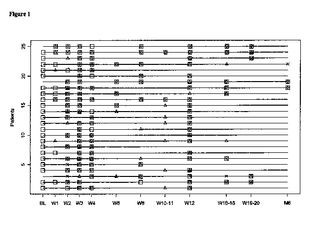
[0059] Figure 1 shows a sample map of the subject in the study. Squares
indicate the time points
for which a sample for microarray data was available. Circles designate
diagnosis of a related
tissue biopsy with >2R rejection versus the triangles which illustrate I R
rejection in the related
tissue biopsy. Xs are the samples linked to a tissue biopsy with no rejection.
[0060] Figure 2 shows the results of subject classification using a biomarker
panel of 12 genes.
Subjects were previously determined to have acute rejection (?2R) or no
rejection (OR). The list
of genes for this biomarker panel include: Transferrin receptor 2 (TFR2), SLIT-
ROBO Rho
GTPase activating protein 2 Pseudogene 1 (SRGAP2P 1), Kruppel-like factor 4
(KLF4), YLP
motif containing 1 (YLPM1), BH3 interacting domain death agonist (BID),
Myristoylated
alanine-rich protein kinase C substrate (MARCKS), C-type lectin domain family
2, member B
-10-
CA 02720863 2010-10-07
WO 2009/124404 PCT/CA2009/000516
(CLEC2B), Rho guanine nucleotide exchange factor (GEF) 7, (ARHGEF7 / BETA-
PIX),
Lysophospholipase-like 1 (LYPLALI), Tryptophan rich basic protein (WRB), FGFR1
oncogene
partner 2 (FGRI OP2), Methyl-CpG binding domain protein 4 (MBD4). Diamond -
acute
rejector (AR); Circle - non rejector (NR)
[0061 ] Figure 3 shows a proposed relationship between the biomarkers ARHGEF7,
TRF2, BID,
MARCKS, KLF4, CLEC2B and MBD4.
[0062] Figure 4 shows a summary of subject classification using clinical
variable profiling.
Diamond - acute rejector (AR); Circle - non rejector (NR)
[0063] Figure 5. Proportion of protein group codes (PGC's) identified using
different peptide
counts (p). Average peptide counts across iTRAQ runs were used for PGC's
identified in
multiple runs. "Total" (horizontal slash bar), "Analyzed" (diagonal slash bar)
and "Panel"
(vertical slash bar) represent the sets of PGC's detected in at least one of
the 18 samples included
in the discovery, detected in at least 2/3 of the AR (acute rejection )and NR
(non-rejection)
groups, and identified with significant differential relative concentrations,
respectively.
;Figure 6. Plasma protein panel A proteomic markers. A. Average of the score
generated by LDA based on panel A for all available AR samples (solid line)
and NR samples
(dashed or stippled line) at each timepoint. B. Score when patients
transitioned between NR and
AR episodes. The first consecutive AR time points were considered and averaged
(AR) from AR
patients (solid line). Consecutive timepoints of NR before (NR before AR) and
after (NR after
AR) AR were considered and averaged from the same patients. A control curve
(dashed or
stippled line) was constructed for NR patients matched as closely as possible
to AR patients by
available timepoints. Standard deviations within each group are represented
using vertical bars.
[0065] Figure 7: Internal validation of proteomic markers. Classification of
13 new subject
samples using panel A (FDR<25%) and panel B (selected by SDA). Scores
generated by both
classifiers were re-centered to set both the cut-off lines for classification
at zero. Average scores
for each AR (open star) and NR (solid star) samples in the training set are
displayed using red
and black asterisks, respectively. Scores for each AR (solid triangle) and NR
(solid square)
samples in the test set are shown. Samples with positive values were
classified as AR and those
with negative values were classified as NR by LDA.
-11-
CA 02720863 2010-10-07
WO 2009/124404 PCT/CA2009/000516
[0066] Figure 8: Technical validation of proteomic markers. iTRAQ versus ELISA
relative
protein levels (relative to pooled control) of 5 validated proteins from the
18 subject samples
used in the discovery. AR samples = open circles; NR samples = solid circle.
Spearman's
correlation coefficients (Cor) and p-values from a test of positive
correlation are displayed for
each protein in the bottom-right of each plot.
[0067] Figure 9 shows a sample map of the subjects whose samples were included
in the
metabolomics study. Square indicates the time points for which a sample for
metabolomic data
was available. Circle indicates diagnosis of a related tissue biopsy with >2R
rejection versus the
triangles which illustrate 1R rejection in the related tissue biopsy. X are
the samples linked to a
tissue biopsy with no rejection.
[0068] Figure 10 shows the grouping of subjects in metabolomics study,
exhibiting OR or >2R
rejection of a cardiac allograft when metabolite concentrations were analyzed
using a moderated
t-test. When the absolution concentration of the post-transplant sample was
analyzed, three
metabolites were statistically significant using a moderated t-test. The
horizontal line illustrates
the mean of each group. The total sample population included six samples from
acute rejector
(AR) subjects and 21 from non-rejector (NR) subjects. Diamond - acute rejector
(AR); Circle -
non rejector (NR)
[0069] Figure 11 shows the grouping of subjects exhibiting OR or >2R rejection
when
metabolite concentrations were analyzed using a moderated t-test. When the
concentration of the
post-transplant sample was compared to the baseline concentration, three
metabolites were
statistically significant using a moderated t-test. The line illustrates the
mean of each group. The
total sample population included six samples from AR subjects and 21 from NR
subjects.
Diamond - acute rejector (AR); Circle - non rejector (NR)
[0070] Figure 12 shows a sample map of the subjects in the alloreactive T-cell
subject
population. Squares indicate the time points for which a sample for microarray
data was
available. Circles designate diagnosis of a related tissue biopsy with >2R
rejection versus the
triangles which illustrate 1R rejection in the related tissue biopsy. Xs are
the samples linked to a
tissue biopsy with no rejection.
[0071 ] Figure 13: Alloreactive T cell gene biomarkers enhance the
classification ability of whole
blood gene biomarkers to discriminate acute from no rejection. A panel of
genes from whole
-12-
CA 02720863 2010-10-07
WO 2009/124404 PCT/CA2009/000516
blood are used as a biomarker panel (A) to differentiate acute from no
rejection. When 2 genes
from the Alloreactive T cell list are added, the classification is even more
separated (B).
Diamond - acute rejector (AR); Circle - non rejector (NR)
[0072] Figure 14 shows examples of Protein Coverage Maps for proteins in
panels A and B
(Table 10) for iTRAQ experiment (this run was used to process B-314-W12, B-314-
W6 and B -
415-W 12. Proteins in each group (with a common Protein Group Code, PGC) are
shown, and
aligned where two or more proteins share a PGC. Double underline, no bold =
peptides
identified with a confidence interval (confidence of identification) > 95%;
Single underline, no
bold = 50% _<CI < 95%; No underline, bold = 0% <CI < 50%; and Plain text (no
underline, no
1o bold) for no detected peptides. A: PGC 151: Phospholipid transfer protein
precursor -
IP100643034.2 (PLTP) Isoform 1 of Phospholipid transfer protein precursor (SEQ
ID NO: 1);
IP100217778.1 (PLTP) Isoform 2 of Phospholipid transfer protein precursor (SEQ
ID NO: 2);
IP1000227333 (PLTP) 45 kDa protein (SEQ ID NO: 3). B: B: PGC 92: Adiponectin
precursor
IPI00020019.1 (SEQ ID NO: 4). C: PGC 61: Pigment epithelium-derived factor
precursor
IPI00006114.4 (SEQ ID NO: 14). D: PGC 188: Beta-2-microglobulin -
IP100868938.1 (-) Beta-
2-microglobulin (SEQ ID NO: 5); IPI00796379.1 (B2M) B2M protein (SEQ ID NO:
6);
IPI00004656.2 (B2M) Beta-2-microglobulin (SEQ ID NO: 7). E: PGC 84:
Coagulation factor X
precursor IPI00019576.1 (SEQ ID NO: 8). F: PGC 6: Ceruloplasmin (IPI00017601.1
(SEQ ID
NO: 9). G: PGC 76: Complement C 1 q subcomponent subunit C precursor
IP100022394.2 (SEQ
ID NO: 12). H: PGC 26: Complement Cir subcomponent precursor IP100296165.5
(SEQ ID
NO: 13). I: PGC 62: Extracellular matrix protein - IPI00645849.1 Extracellular
matrix protein 1
(SEQ ID NO: 10); IP100003351.2 Extracellular matrix protein 1 precursor (SEQ
ID NO: 11).
Peptides that were identified in the iTRAQ experiments are listed in Figure
17.
[0073] Figure 15 shows examples of Protein CoverageMaps for additional
identified proteomic
markers (Table 10) for iTRAQ experiment (this run was used to process B-314-
W12, B-314-W6
and B-415-W12. Proteins in each group (with a common Protein Group Code, PGC)
are shown,
and aligned where two or more proteins share a PGC. Double underline, no bold
= peptides
identified with a confidence interval (confidence of identification) ? 95%;
Single underline, no
bold = 50% <CI < 95%; No underline, bold = 0% <CI < 50%; and Plain text (no
underline, no
bold) for no detected peptides. These proteins were outside of Panels A and B,
but
demonstrated differential expression between AR and NR subjects (pval<0.05) A:
PGC 110:
Cystatin -C precursor (CST3) IP100032293.1 (SEQ ID NO: 15). B: PGC138: Sex
hormone-
-13-
CA 02720863 2010-10-07
WO 2009/124404 PCT/CA2009/000516
binding globulin (SHBG) isoform 2 IPI00219583.1 ( SEQ ID NO: 16); SHBG isoform
1
IPI00023019.1 (SEQ ID NO: 17). C: PGC 8: CFH isoform 1 IP100029739.5 (SEQ ID
NO: 18).
D: PGC 50: Complement factor I (CFI) precursor IP100291867.3 (SEQ ID NO: 19);
IP100872555.2 (encoded by cDNA FLJ76262) (SEQ ID NO: 20). E: PGC 48: Serum
amyloid P-
component precursor 1P100022391.1 (SEQ ID NO: 21).
[0074] Figure 16A-L shows target sequences of 12 nucleic acid markers useful
for diagnosis of
acute cardiac allograft rejection, listed in Table 6 (SEQ ID NOs: 25-36).
[0075] Figure 17 shows exemplary peptides identified in iTRAQ assays according
to some
embodiments of the present invention. The list further includes their assigned
protein group
codes and SEQ ID NOs 37-307.
[0076] Figure 18 A-P shows target sequences of 16 nucleic acid markers useful
for diagnosis of
acute cardiac allograft rejection in alloreactive T-cells (listed in Table 9)
(SEQ ID NOs: 345-
360).
[0077] Figure 19 A-Z, AA-KK shows target sequences of 37 nucleic acid markers
useful for
diagnosis of acute cardiac allograft rejection (listed in Table 10) (SEQ ID
NOs: 361-397).
DETAILED DESCRIPTION
[0078] In the description that follows, a number of terms are used
extensively, the following
definitions are provided to facilitate understanding of various aspects of the
invention. Use of
examples in the specification, including examples of terms, is for
illustrative purposes only and is
not intended to limit the scope and meaning of the embodiments of the
invention herein.
Numeric ranges are inclusive of the numbers defining the range. In the
specification, the word
"comprising" is used as an open-ended term, substantially equivalent to the
phrase "including,
but not limited to," and the word "comprises" has a corresponding meaning.
[0079] The present invention provides for methods of diagnosing rejection in a
subject that has
received a tissue or organ allograft, specifically a cardiac allograft.
[0080] The present invention provides genomic, T-cell, nucleic acid, proteomic
expression
profiles or metabolite profiles related to the assessment, prediction or
diagnosis of allograft
rejection in a subject. While several of the elements in the genomic or T-cell
expression profiles,
proteomic expression profiles or metabolite profiles may be individually known
in the existing
-14-
CA 02720863 2010-10-07
WO 2009/124404 PCT/CA2009/000516
art, the specific combination of the altered expression levels (increased or
decreased relative to a
control) of specific sets of genomic, T-cell, proteomic or metabolite markers
comprise a novel
combination useful for assessment, prediction or diagnosis or allograft
rejection in a subject.
[0081] An allograft is an organ or tissue transplanted between two genetically
different subjects
of the same species. The subject receiving the allograft is the `recipient',
while the subject
providing the allograft is the `donor'. A tissue or organ allograft may
alternately be referred to as
a `transplant', a `graft', an `allograft', a `donor tissue' or `donor organ',
or similar terms. A
transplant between two subjects of different species is a xenograft.
[0082] Subjects may present with a variety of symptoms or clinical variables
well-known in the
literature, however none of these of itself is a predictive or diagnostic of
allograft rejection. A
myriad of clinical variables maybe used in assessing a subject having, or
suspected of having,
allograft rejection, in addition to biopsy of the allograft. The information
gleaned from these
clinical variables is then used by a clinician, physician, veterinarian or
other practitioner in a
clinical field in attempts to determine if rejection is occurring, and how
rapidly it progresses, to
allow for modification of the immunosuppressive drug therapy of the subject.
Examples of
clinical variables are described in Table 2.
[0083] Clinical variables (optionally accompanied by biopsy), while currently
the only practical
tools available to a clinician in mainstream medical practice, are not always
able to cleanly
differentiate between an AR (an "acute rejector") and an NR (a "non-rejector")
subject, as is
illustrated in Figure 4. While the extreme left and right subjects are
correctly classified as AR or
NR, the bulk of the subjects are represented in the middle range and their
status is unclear. This
does not negate the value of the clinical variables in the assessment of
allograft rejection, but
instead indicates their limitation when used in the absence of other methods.
[0084] Table 2: Clinical variables for possible use in assessment of allograft
rejection.
Clinical Variable Name Renal/Heart/Liver/ Variable Explanation
All
Primary Diagnosis All Diagnosis leading to transplant
Secondary Diagnosis All Diagnosis leading to transplant
"Transplant Procedure - Living
related, Living unrelated, or
cadaveric"
-15-
CA 02720863 2010-10-07
WO 2009/124404 PCT/CA2009/000516
Blood Type All Blood Type
Blood Rh All Blood Rh
Height (cm) All Height (cm)
Weight (kg) All Weight (kg)
BMI All Calculation: Weight/ (Height)2
Liver Ascites All
HLA Al All
HLA A2 All
HLA BI All
HLA B2 All
HLA DR1 All
HLA DR2 All
CMV All Viral Status
CMV Date All Date of viral status
HIV All Viral Status
HBV All Viral Status
HBV Date All Date of viral status
HbsAb All Viral Status
HbcAb (Total) All Viral Status
HBvDNA All Viral Status
HCV All Viral Status
HCV Genotype All Hepatitis C genotype
HCV Genotype Sub All "Hepatitis C genotype, subtype"
EBV All Viral Status
Zoster All Viral Status
Dialysis Start Date All Dialysis Start Date
Dialysis Type All Dialysis Type
Cytoxicity Current Level All
Cytoxicity Current Date All
Cytoxicity Peak Level All
Cytoxicity Peak Date All
Flush Soln All Type of Flush Solution used at transplant
Cold Time 1 All
Cold Time 2 All
Re-Warm Time 1 All
Re-Warm Time 2 All
HTLV 1 All
HTLV 2 All
HCV RNA All
-16-
CA 02720863 2010-10-07
WO 2009/124404 PCT/CA2009/000516
24hr Urine All 24 Hour urine output
Systolic Blood Pressure All Blood Pressure reading
Diastolic Blood Pressure All Blood Pressure reading
24 Hr Urine All 24 hour urine
Sodium All Blood test
Potassium All Blood test
Chloride All Blood test
Total CO2 All Blood test
Albumin All Blood test
Protein All Blood test
Calcium All Blood test
Inorganic Phosphate All Blood test
Magnesium All Blood test
Uric Acid All Blood test
Glucose All Blood test
Hemoglobin AIC All Blood test
CPK All Blood test
Parathyroid Hormone All Blood test
Homocysteine All Blood test
Urine Protein All Urine test
Creatinine All Blood test
BUN All Blood test
Hemaglobin All Blood test
Platelet Count All Blood test
WBC Count All Blood test
Prothrombin Time All Blood test
Partial Thromboplastin Time All Blood test
INR All Blood test
Gamma GT All Blood test
AST All Blood test
Alkaline Phosphatase All Blood test
Amylase All Blood test
Total Bilirubin All Blood test
Direct Bilirubin All Blood test
LDH All Blood test
ALT All Blood test
Triglycerides All Blood test
Cholesterol All Blood test
HDL Cholesterol All Blood test
-17-
CA 02720863 2010-10-07
WO 2009/124404 PCT/CA2009/000516
LDL Cholesterol All Blood test
FEV1 All Lung function test
FVC All Lung function test
Total Ferritin All Blood test
TIBC All Blood test
Transferrin Saturated All Blood test
Ferritin All Blood test
Angiography Heart Heart function test
Intravascular ultrasound Heart Heart function test
Dobutamine Stress Heart Heart function test
Echocardiography
Cyclosporine WB All Immunosuppressive levels
Cyclosporine 2 hr All Immunosuppressive levels
Tacrolimus WB All Immunosuppressive levels
Sirolimus WB All Immunosuppressive total daily dose
Solumedrol All Immunosuppressive total daily dose
Prednisone All Immunosuppressive total daily dose
Prednisone ALT All Immunosuppressive total daily dose
Tacrolimus All Immunosuppressive total daily dose
Cyclosporine All Immunosuppressive total daily dose
Imuran All Immunosuppressive total daily dose
Mycophonelate Mofetil All Immunosuppressive total daily dose
Sirolimus All Immunosuppressive total daily dose
OKT3 All Immunosuppressive total daily dose
ATG All Immunosuppressive total daily dose
ALG All Immunosuppressive total daily dose
Basiliximab All Immunosuppressive total daily dose
Daclizumab All Immunosuppressive total daily dose
Ganciclovir All Anti-viral total daily dose
Lamivudine All Anti-viral total daily dose
Riboviron All Anti-viral total daily dose
Interferon All Anti-viral total daily dose
Hepatisis C Virus RNA All test for presence of HCV values Q
CMV Antigenemia All Antiviral and Virus
Valganciclovir All Anti-viral total daily dose
Neutrophil Number All Blood test
C Peptide All Blood test
Peg Interferon All Anti-viral total daily dose
GFR All Glomerular Filtration Rate
-18-
CA 02720863 2010-10-07
WO 2009/124404 PCT/CA2009/000516
Complication Events All Complication Type
Biopsy Scores Renal Borderline, 1A, 1B, 2A, 2B, 3,
Hyperacute
Biopsy Scores Liver Portal inflammation, Bile duct
inflammation damage, Venous
endothelial inflammation each scored
from 1 to 3
Donor Blood Type All Donor Blood Type
Donor Blood Rh All Donor Rh
Donor HLA Al All Donor HLA Al
Donor HLA A2 All Donor HLA A2
Donor HLA B1 All Donor HLA 131
Donor HLA B2 All Donor HLA B2
Donor HLA DR1 All Donor HLA DR1
Donor HLA DR2 All Donor HLA DR2
Donor CMV All Donor CMV
Donor HIV All Donor HIV
Donor HBV All Donor HBV
Donor HbsAb All Donor HbsAb
Donor HbcAb (total) All Donor HbcAb (total)
Donor Hbdna All Donor Hbdna
Donor HCV All Donor HCV
Donor EBV All Donor EBV
[0085] The multifactorial nature of allograft rejection prediction, diagnosis
and assessment is
considered in the art to exclude the possibility of a single biomarker that
meets even one of the
needs of prediction, diagnosis or assessment of allograft rejection.
Strategies involving a
plurality of markers may take into account this multifactorial nature.
Alternately, a plurality of
markers may be assessed in combination with clinical variables that are less
invasive (e.g. a
biopsy not required) to tailor the prediction, diagnosis and/or assessment of
allograft rejection in
a subject.
[0086] Regardless of the methods used for prediction, diagnosis and assessment
of allograft
rejection, earlier is better- from the viewpoint of preserving organ or tissue
function and
preventing more systemic detrimental effects. There is no `cure' for allograft
rejection, only
maintenance of the subject at a suitably immunosuppressed state, or in some
cases, replacement
-19-
CA 02720863 2010-10-07
WO 2009/124404 PCT/CA2009/000516
of the organ if rejection has progressed too rapidly or is too severe to
correct with
immunosuppressive drug intervention therapy.
[0087] Applying a plurality of mathematical and/or statistical analytical
methods to a protein or
polypeptide dataset, metabolite concentration data set, or nucleic acid
expression dataset may
indicate varying subsets of significant markers, leading to uncertainty as to
which method is
`best' or `more accurate'. Regardless of the mathematics, the underlying
biology is the same in a
dataset. By applying a plurality of mathematical and/or statistical methods to
a microarray
dataset and assessing the statistically significant subsets of each for common
markers, uncertainty
may be reduced, and clinically relevant core group of markers may be
identified.
[0088] "Markers", "biological markers" or "biomarkers" may be used
interchangeably and refer
generally to detectable (and in some cases quantifiable) molecules or
compounds in a biological
sample. A marker may be down-regulated (decreased), up-regulated (increased)
or effectively
unchanged in a subject following transplantation of an allograft. Markers may
include nucleic
acids (DNA or RNA), a gene, or a transcript, or a portion or fragment of a
transcript in reference
to `genomic' markers (alternately referred to as "nucleic acid markers");
polypeptides, peptides,
proteins, isoforms, or fragments or portions thereof for `proteomic' markers,
or selected
molecules, their precursors, intermediates or breakdown products (e.g. fatty
acid, amino acid,
sugars, hormones, or fragments or subunits thereof) ("metabolite markers" or
"metabolomic
markers"). In some usages, these terms may reference the level or quantity of
a particular
protein, peptide, nucleic acid or polynucleotide, or metabolite (in absolute
terms or relative to
another sample or standard value) or the ratio between the levels of two
proteins,
polynucleotides, peptides or metabolites, in a subject's biological sample.
The level may be
expressed as a concentration, for example micrograms per milliliter; as a
colorimetric intensity,
for example 0.0 being transparent and 1.0 being opaque at a particular
wavelength of light, with
the experimental sample ranked accordingly and receiving a numerical score
based on
transmission or absorption of light at a particular wavelength; or as relevant
for other means for
quantifying a marker, such as are known in the art. In some examples, a ratio
may be expressed
as a unitless value. A "marker" may also reference to a ratio, or a net value
following subtraction
of a baseline value. A marker may also be represented as a `fold-change', with
or without an
indicator of directionality (increase or decrease/ up or down). The increase
or decrease in
expression of a marker may also be referred to as `down-regulation' or `up-
regulation', or similar
indicators of an increase or decrease in response to a stimulus, physiological
event, or condition
-20-
CA 02720863 2010-10-07
WO 2009/124404 PCT/CA2009/000516
of the subject. A marker may be present in a first biological sample, and
absent in a second
biological sample; alternately the marker may be present in both, with a
statistically significant
difference between the two. Expression of the presence, absence or relative
levels of a marker in
a biological sample maybe dependent on the nature of the assay used to
quantify or assess the
marker, and the manner of such expression will be familiar to those skilled in
the art.
[0089] A marker may be described as being differentially expressed when the
level of expression
in a subject who is rejecting an allograft is significantly different from
that of a subject or sample
taken from a non-rejecting subject. A differentially expressed marker may be
overexpressed or
underexpressed as compared to the expression level of a normal or control
sample.
[0090] A "profile" is a set of one or more markers and their presence,
absence, relative level or
abundance (relative to one or more controls). For example, a metabolite
profile is a dataset of the
presence, absence, relative level or abundance of metabolic markers. A
proteomic profile is a
dataset of the presence, absence, relative level or abundance of proteomic
markers. A genomic or
nucleic acid profile a dataset of the presence, absence, relative level or
abundance of expressed
nucleic acids (e.g. transcripts, mRNA, EST or the like). A profile may
alternately be referred to
as an expression profile.
[0091 ] The increase or decrease, or quantification of the markers in the
biological sample may be
determined by any of several methods known in the art for measuring the
presence and/or relative
abundance of a gene product or transcript, or a nucleic acid molecule
comprising a particular
sequence, polypeptide or protein, metabolite or the like. The level of the
markers may be
determined as an absolute value, or relative to a baseline value, and the
level of the subject's
markers compared to a cutoff index (e.g. a non-rejection cutoff index).
Alternately the relative
abundance of the marker may be determined relative to a control. The control
may be a clinically
normal subject (e.g. one who has not received an allograft) or may be an
allograft recipient that
has not previously demonstrated rejection.
[0092] In some embodiments, the control may be an autologous control, for
example a sample or
profile obtained from the subject before undergoing allograft transplantation.
In some
embodiments, the profile obtained at one time point (before, after or before
and after
transplantation) may be compared to one or more than one profiles obtained
previously from the
same subject. By repeatedly sampling the same biological sample from the same
subject over
time, a composite profile, illustrating marker level or expression over time
may be provided.
-21-
CA 02720863 2010-10-07
WO 2009/124404 PCT/CA2009/000516
Sequential samples can also be obtained from the subject and a profile
obtained for each, to
allow the course of increase or decrease in one or more markers to be followed
over time For
example, an initial sample or samples may be taken before the transplantation,
with subsequent
samples being taken weekly, biweekly, monthly, bimonthly or at another
suitable, regular interval
and compared with profiles from samples taken previously. Samples may also be
taken before,
during and after administration of a course of a drug, for example an
immunosuppressive drug.
[0093] Techniques, methods, tools, algorithms, reagents and other necessary
aspects of assays
that may be employed to detect and/or quantify a particular marker or set of
markers are varied.
Of significance is not so much the particular method used to detect the marker
or set of markers,
but what markers to detect. As is reflected in the literature, tremendous
variation is possible.
Once the marker or set of markers to be detected or quantified is identified,
any of several
techniques may be well suited, with the provision of appropriate reagents. One
of skill in the art,
when provided with the set of markers to be identified, will be capable of
selecting the
appropriate assay (for example, a PCR based or a microarray based assay for
nucleic acid
markers, an ELISA, protein or antibody microarray or similar immunologic
assay, or in some
examples, use of an iTRAQ, iCAT or SELDI proteomic mass spectrometric based
method) for
performing the methods disclosed herein.
[0094] The present invention provides nucleic acid expression profiles (both
genomic and T-cell)
proteomic expression profiles and metabolite profiles related to the
assessment, prediction or
diagnosis of allograft rejection in a subject. While several of the elements
in the genomic or T-
cell expression profiles, proteomic expression profiles or metabolite profiles
may be individually
known in the existing art, the specific combination of the altered expression
levels (increased or
decreased relative to a control) of specific sets of genomic, T-cell,
proteomic or metabolite
markers comprise a novel combination useful for assessment, prediction or
diagnosis of allograft
rejection in a subject.
[0095] For example, detection or determination, and in some cases
quantification, of a nucleic
acid may be accomplished by any one of a number methods or assays employing
recombinant
DNA technologies known in the art, including but not limited to, as sequence-
specific
hybridization, polymerase chain reaction (PCR), RT-PCR, microarrays and the
like. Such assays
may include sequence-specific hybridization, primer extension, or invasive
cleavage.
Furthermore, there are numerous methods for analyzing/detecting the products
of each type of
-22-
CA 02720863 2010-10-07
WO 2009/124404 PCT/CA2009/000516
reaction (for example, fluorescence, luminescence, mass measurement,
electrophoresis, etc.).
Furthermore, reactions can occur in solution or on a solid support such as a
glass slide, a chip, a
bead, or the like.
[0096] Methods of designing and selecting probes for use in microarrays or
biochips, or for
selecting or designing primers for use in PCR-based assays are known in the
art. Once the
marker or markers are identified and the sequence of the nucleic acid
determined by, for
example, querying a database comprising such sequences, or by having an
appropriate sequence
provided (for example, a sequence listing as provided herein), one of skill in
the art will be able
to use such information to select appropriate probes or primers and perform
the selected assay.
[0097] Standard reference works setting forth the general principles of
recombinant DNA
technologies known to those of skill in the art include, for example: Ausubel
et al, Current
Protocols In Molecular Biology, John Wiley & Sons, New York (1998 and
Supplements to
2001); Sambrook et al, Molecular Cloning: A Laboratory Manual, 2d Ed., Cold
Spring Harbor
Laboratory Press, Plainview, New York (1989); Kaufman et al , Eds., Handbook
Of Molecular
And Cellular Methods In Biology And Medicine, CRC Press, Boca Raton ( 1995);
McPherson,
Ed., Directed Mutagenesis: A Practical Approach, IRL Press, Oxford (1991).
[0098] Proteins, protein complexes or proteomic markers may be specifically
identified and/or
quantified by a variety of methods known in the art and may be used alone or
in combination.
Immunologic- or antibody-based techniques include enzyme-linked immunosorbent
assay
(ELISA), radioimmunoassay (RIA), western blotting, immunofluorescence,
microarrays, some
chromatographic techniques (i.e. immunoaffinity chromatography), flow
cytometry,
immunoprecipitation and the like. Such methods are based on the specificity of
an antibody or
antibodies for a particular epitope or combination of epitopes associated with
the protein or
protein complex of interest. Non-immunologic methods include those based on
physical
characteristics of the protein or protein complex itself. Examples of such
methods include
electrophoresis, some chromatographic techniques (e.g. high performance liquid
chromatography
(HPLC), fast protein liquid chromatography (FPLC), affinity chromatography,
ion exchange
chromatography, size exclusion chromatography and the like), mass
spectrometry, sequencing,
protease digests, and the like. Such methods are based on the mass, charge,
hydrophobicity or
hydrophilicity, which is derived from the amino acid complement of the protein
or protein
complex, and the specific sequence of the amino acids. Examples of methods
employing mass
-23-
CA 02720863 2010-10-07
WO 2009/124404 PCT/CA2009/000516
spectrometry include those described in, for example, PCT Publication WO
2004/019000, WO
2000/00208, US 6670194. Immunologic and non-immunologic methods may be
combined to
identify or characterize a protein or protein complex. Furthermore, there are
numerous methods
for analyzing/detecting the products of each type of reaction (for example,
fluorescence,
luminescence, mass measurement, electrophoresis, etc.). Furthermore, reactions
can occur in
solution or on a solid support such as a glass slide, a chip, a bead, or the
like.
[0099] Methods of producing antibodies for use in protein or antibody arrays,
or other
immunology based assays are known in the art. Once the marker or markers are
identified and the
amino acid sequence of the protein or polypeptide is identified, either by
querying of a database
or by having an appropriate sequence provided (for example, a sequence listing
as provide
herein), one of skill in the art will be able to use such information to
prepare one or more
appropriate antibodies and perform the selected assay.
[00100] For preparation of monoclonal antibodies directed towards a biomarker,
any
technique that provides for the production of antibody molecules by continuous
cell lines in
culture may be used. Such techniques include, but are not limited to, the
hybridoma technique
originally developed by Kohler and Milstein (1975, Nature 256:495-497), the
trioma technique
(Gustafsson et al., 1991, Hum. Antibodies Hybridomas 2:26-32), the human B-
cell hybridoma
technique (Kozbor et al., 1983, Immunology Today 4:72), and the EBV hybridoma
technique to
produce human monoclonal antibodies (Cole et al., 1985, In: Monoclonal
Antibodies and Cancer
Therapy, Alan R. Liss, Inc., pp. 77-96). Human antibodies may be used and can
be obtained by
using human hybridomas (Cote et al., 1983, Proc. Natl. Acad. Sci. USA 80:2026-
2030) or by
transforming human B cells with EBV virus in vitro (Cole et al., 1985, In:
Monoclonal
Antibodies and Cancer Therapy, Alan R. Liss, Inc., pp. 77-96).Techniques
developed for the
production of "chimeric antibodies" (Morrison et al, 1984, Proc. Natl. Acad.
Sci. USA 81:6851-
6855; Neuberger et al, 1984, Nature 312:604-608; Takeda et al, 1985, Nature
314:452-454) by
splicing the genes from a mouse antibody molecule specific for a biomarker
together with genes
from a human antibody molecule of appropriate biological activity can be used;
such antibodies
are within the scope of this invention. Techniques described for the
production of single chain
antibodies (U.S. Patent 4,946,778) can be adapted to produce a biomarker -
specific antibodies.
An additional embodiment of the invention utilizes the techniques described
for) the
construction of Fab expression libraries (Huse et al, 1989, Science 246:1275-
1281) to allow rapid
and easy identification of monoclonal Fab fragments with the desired
specificity for a biomarker
-24-
CA 02720863 2010-10-07
WO 2009/124404 PCT/CA2009/000516
proteins. Non-human antibodies can be "humanized" by known methods (e.g., U.S.
Patent No.
5,225,539).
[001011 Antibody fragments that contain the idiotypes of a biomarker can be
generated by
techniques known in the art. For example, such fragments include, but are not
limited to, the
F(ab')2 fragment which can be produced by pepsin digestion of the antibody
molecule; the Fab'
fragment that can be generated by reducing the disulfide bridges of the
F(ab')2 fragment; the Fab
fragment that can be generated by treating the antibody molecular with papain
and a reducing
agent; and Fv fragments. Synthetic antibodies, e.g., antibodies produced by
chemical synthesis,
are useful in the present invention
[00102] Standard reference works described herein and known to those skilled
in the
relevant art describe both immunologic and non-immunologic techniques, their
suitability for
particular sample types, antibodies, proteins or analyses. Standard reference
works setting forth
the general principles of immunology and assays employing immunologic methods
known to
those of skill in the art include, for example: Harlow and Lane, Antibodies: A
Laboratory
Manual, 2d Ed., Cold Spring Harbor Laboratory Press, Cold Spring Harbor, N. Y.
(1999);
Harlow and Lane, Using Antibodies: A Laboratory Manual. Cold Spring Harbor
Laboratory
Press, New York; Coligan et al. eds. Current Protocols in Immunology, John
Wiley & Sons, New
York, NY (1992-2006); and Roitt et al., Immunology, 3d Ed., Mosby-Year Book
Europe
Limited, London (1993).
[00103] Standard reference works setting forth the general principles of
peptide synthesis
technology and methods known to those of skill in the art include, for
example: Chan et al.,
Fmoc Solid Phase Peptide Synthesis, Oxford University Press, Oxford, United
Kingdom, 2005;
Peptide and Protein Drug Analysis, ed. Reid, R., Marcel Dekker, Inc., 2000;
Epitope Mapping,
ed. Westwood et al., Oxford University Press, Oxford, United Kingdom, 2000;
Sambrook et al.,
Molecular Cloning: A Laboratory Manual, 3rd ed., Cold Spring Harbor Press,
Cold Spring
Harbor, NY 2001; and Ausubel et al., Current Protocols in Molecular Biology,
Greene
Publishing Associates and John Wiley & Sons, NY, 1994).
[00104] A subject's rejection status may be described as an "acute rejector"
(AR) or as a
"non-rejector" (NR) and is determined by comparison of the concentration of
the markers to that
of a non-rejector cutoff index. A "non-rejector cutoff index" is a numerical
value or score,
beyond or outside of which a subject is categorized as having an AR rejection
status. The non-
-25-
CA 02720863 2010-10-07
WO 2009/124404 PCT/CA2009/000516
rejector cutoff index may be alternately referred to as a `control value', a
`control index', or
simply as a `control'. A non-rejector cutoff-index may be the concentration of
individual markers
in a control subject population and considered separately for each marker
measured; alternately
the non-rejector cutoff index may be a combination of the concentration of the
markers, and
compared to a combination of the concentration of the markers in the subject's
sample provided
for diagnosing. The control subject population may be a normal or healthy
control population, or
may be an allograft recipient population that has not, or is not, rejecting
the allograft. The
control maybe a single subject, and for some embodiments, maybe an autologous
control. A
control, or pool of controls, may be constant e.g. represented by a static
value, or may be
cumulative, in that the sample population used to obtain it may change from
site to site, or over
time and incorporate additional data points. For example, a central data
repository, such as a
centralized healthcare information system, may receive and store data obtained
at various sites
(hospitals, clinical laboratories or the like) and provide this cumulative
data set for use with the
methods of the invention at a single hospital, community clinic, for access by
an end user (i.e. an
individual medical practitioner, medical clinic or center, or the like).
[00105] The non-rejector cutoff index may be alternately referred to as a
`control value', a
`control index' or simply as a `control'. In some embodiments the cutoff index
maybe further
characterized as being a metabolite cutoff index (for metabolite profiling of
subjects), a genomic
cutoff index (for genomic expression profiling of subjects), a proteomic
cutoff index (for
proteomic profiling of subjects), or the like.
[00106] A "biological sample" refers generally to body fluid or tissue or
organ sample
from a subject. For example, the biological sample may a body fluid such as
blood, plasma,
lymph fluid, serum, urine or saliva. A tissue or organ sample, such as a non-
liquid tissue sample
may be digested, extracted or otherwise rendered to a liquid form - examples
of such tissues or
organs include cultured cells, blood cells, skin, liver, heart, kidney,
pancreas, islets of
Langerhans, bone marrow, blood, blood vessels, heart valve, lung, intestine,
bowel, spleen,
bladder, penis, face, hand, bone, muscle, fat, cornea or the like. A plurality
of biological samples
may be collected at any one time. A biological sample or samples may be taken
from a subject at
any time, including before allograft transplantation, at the time of
translation or at anytime
following transplantation. A biological sample may comprise nucleic acid, such
as
deoxyribonucleic acid or ribonucleic acid, or a combination thereof, in either
single or double-
stranded form. When an organ is removed from a donor, the spleen of the donor
or a part of it
-26-
CA 02720863 2010-10-07
WO 2009/124404 PCT/CA2009/000516
may be kept as a biological sample from which to obtain donor T-cells. When an
organ is
removed from a living donor, a blood sample may be taken, from which donor T-
cells may be
obtained. Alloreactive T-cells may be isolated by exploiting their specific
interaction with
antigens (including the MHC complexes) of the allograft. Methods to enable
specific isolation of
alloreactive T-cells are described in, for example PCT Publication WO
2005/05721, herein
incorporated by reference.
[00107] A lymphocyte is nucleated or `white' blood cell (leukocyte) of
lymphoid stem cell
origin. Lymphocytes include T-cells, B-cells natural killer cells and the
like, and other immune
regulatory cells. A "T-cell" is a class of lymphocyte responsible for cell-
mediated immunity, and
for stimulating B-cells. A stimulated B-cell produces antibodies for specific
antigens. Both B-
cells and T-cells function to recognize non-self antigens in a subject. Non-
self antigens include
those of viruses, bacteria and other infectious agents as well as allografts.
[00108] An alloreactive T-cell is a T-cell that is activated in response to an
alloantigen. A
T-cell that is reactive to a xenoantigen is a xenoreactive T-cell. A
xenoantigen is an antigen from
another species or species' tissue, such as a xenograft. Alloreactive T cells
are the front-line of
the graft rejection immune response. They are a subset (-0.1-1%) of the
peripheral blood
mononuclear cells (PBMC) which recognize allogeneic antigens present on the
foreign graft.
They may infiltrate the foreign graft, to initiate a cascade of anti-graft
immune response, which,
if unchecked, will lead to rejection and failure of the graft. Alloreactive T
cells, therefore
provide specificity compared to other sources of markers, or may function as a
complementary
source of markers that differentiate between stages of organ rejection.
[00109] The term "subject" or "patient" generally refers to mammals and other
animals
including humans and other primates, companion animals, zoo, and farm animals,
including, but
not limited to, cats, dogs, rodents, rats, mice, hamsters, rabbits, horses,
cows, sheep, pigs, goats,
poultry, etc. A subject includes one who is to be tested, or has been tested
for prediction,
assessment or diagnosis of allograft rejection. The subject may have been
previously assessed or
diagnosed using other methods, such as those described herein or those in
current clinical
practice, or may be selected as part of a general population (a control
subject).
[00110] A fold-change of a marker in a subject, relative to a control maybe at
least 0.1,
0.2, 0.3, 0.4, 0.5, 0.6, 0.7, 0.8, 0.9, 1.0, 1.1, 1.2, 1.3, 1.4, 1.5, 1.6,
1.7, 1.8, 1.9, 2.0, 2.1, 2.2, 2.3,
2.4, 2.5, 2.6, 2.7, 2.8, 2.9, 3.0, 3.1, 3.2, 3.3, 3.4, 3.5, 3.6, 3.7, 3.8,
3.9, 4.0, 4.1, 4.2, 4.3, 4.4, 4.5,
-27-
CA 02720863 2010-10-07
WO 2009/124404 PCT/CA2009/000516
4.6, 4.7, 4.8, 4.9, 5.0 or more, or any amount therebetween. The fold change
may represent a
decrease, or an increase, compared to the control value.
[00111] One or more than one includes 1, 2,3, 4, 5, 6, 7, 8, 9, 10, 11, 12,
13, 14, 15, 16 or
more.
[00112] "Down-regulation" or `down-regulated may be used interchangeably and
refer to a
decrease in the level of a marker, such as a gene, nucleic acid, metabolite,
transcript, protein or
polypeptide. "Up-regulation" or "up-regulated" may be used interchangeably and
refer to an
increase in the level of a marker, such as a gene, nucleic acid, metabolite,
transcript, protein or
polypeptide. Also, a pathway, such as a signal transduction or metabolic
pathway may be up- or
down-regulated.
[00113] Once a subject is identified as an acute rejector, or at risk for
becoming an acute
rejector by any method (genomic, proteomic, metabolomic or a combination
thereof), therapeutic
measures may be implemented to alter the subject's immune response to the
allograft. The
subject may undergo additional monitoring of clinical values more frequently,
or using more
sensitive monitoring methods. Additionally the subject may be administered
immunosuppressive
medicaments to decrease or increase the subject's immune response. Even though
a subject's
immune response needs to be suppressed to prevent rejection of the allograft,
a suitable level of
immune function is also needed to protect against opportunistic infection.
Various medicaments
that may be administered to a subject are known; see for example, Goodman and
Gilman's The
Pharmacological Basis of Therapeutics I1 th edition. Ch 52, pp 1405-1431 and
references therein;
LL Brunton, JS Lazo, KL Parker editors. Standard reference works setting forth
the general
principles of medical physiology and pharmacology known to those of skill in
the art include:
Fauci et al. , Eds., Harrison's Principles Of Internal Medicine, 14th Ed.,
McGraw-Hill
Companies, Inc. (1998). Other preventative and therapeutic strategies are
reviewed in the
medical literature- see, for example Kobashigawa et al. 2006. Nature Clinical
Practice.
Cardiovascular Medicine 3:203-21.
[00114] Genomic nucleic acid expression profiling
[00115] A method of diagnosing acute allograft rejection in a subject as
provided by the
present invention comprises 1) determining the expression profile of one or
more than one
nucleic acid markers in a biological sample from the subject, the nucleic acid
markers selected
-28-
CA 02720863 2010-10-07
WO 2009/124404 PCT/CA2009/000516
from the group comprising TRF2, SRGAP2P1, KLF4, YLPM1, BID, MARCKS, CLEC2B,
ARHGEF7, LYPLALI, WRB, FGFRIOP2, MBD4; 2) comparing the expression profile of
the
one or more than one nucleic acid markers to a non-rejector profile; and 3)
determining whether
the expression level of the one or more than one nucleic acid markers is up-
regulated or down-
regulated relative to the control profile, wherein up-regulation or down-
regulation of the one or
more than one nucleic acid markers is indicative of the rejection status.
[00116] Therefore, the invention also provides for a method of predicting,
assessing or
diagnosing allograft rejection in a subject as provided by the present
invention comprises 1)
measuring the increase or decrease of one or more than one nucleic acid
markers selected from
the group comprising TRF2, SRGAP2P1, KLF4, YLPMI, BID, MARCKS, CLEC2B,
ARHGEF7, LYPLALI, WRB, FGFRIOP2, MBD4; and 2) determining the `rejection
status' of
the subject, wherein the determination of `rejection status' of the subject is
based on comparison
of the subject's nucleic acid marker expression profile to a control nucleic
acid marker
expression profile.
[00117] The phrase "gene expression data", "gene expression profile" "nucleic
acid
expression profile" or "marker expression profile" as used herein refers to
information regarding
the relative or absolute level of expression of a gene or set of genes in a
biological sample. The
level of expression of a gene may be determined based on the level of a
nucleic acid such as
RNA including mRNA, transcribed from or encoded by the gene.
[00118] A "polynucleotide", "oligonucleotide", "nucleic acid" or "nucleotide
polymer" as
used herein may include synthetic or mixed polymers of nucleic acids,
including RNA, DNA or
both RNA and DNA, both sense and antisense strands, and may be chemically or
biochemically
modified or may contain non- natural or derivatized nucleotide bases, as will
be readily
appreciated by those skilled in the art. Such modifications include, for
example, labels,
methylation, substitution of one or more of the naturally occurring
nucleotides with an analog,
internucleotide modifications such as uncharged linkages (e.g., methyl
phosphonates,
phosphotriesters, phosphoamidates, carbamates, etc.), charged linkages (e. g.,
phosphorothioates,
phosphorodithioates, etc.), pendent moieties (e.g., polypeptides), and
modified linkages (e.g.,
alpha anomeric polynucleotides, etc.). Also included are synthetic molecules
that mimic
polynucleotides in their ability to bind to a designated sequence via hydrogen
bonding and other
chemical interactions.
-29-
CA 02720863 2010-10-07
WO 2009/124404 PCT/CA2009/000516
[00119] An oligonucleotide includes variable length nucleic acids, which may
be useful as
probes, primers and in the manufacture of microarrays (arrays) for the
detection and/or
amplification of specific nucleic acids. Oligonucleotides may comprise DNA,
RNA, PNA or
other polynucleotide moieties as described in, for example, US 5,948,902. Such
DNA, RNA or
oligonucleotide strands may be synthesized by the sequential addition (5'-3'
or 3'-5') of activated
monomers to a growing chain which may be linked to an insoluble support.
Numerous methods
are known in the art for synthesizing oligonucleotides for subsequent
individual use or as a part
of the insoluble support, for example in arrays (BERNFIELD MR. and ROTTMAN FM.
J. Biol.
Chem. (1967) 242(18):4134-43; SULSTON J. et al. PNAS (1968) 60(2):409-415;
GILLAM S. et
al. Nucleic Acid Res.(1975) 2(5):613-624; BONORA GM. et al. Nucleic Acid
Res.(1990)
18(11):3155-9; LASHKARI DA. et al. PNAS (1995) 92(17):7912-5; MCGALL G. et al.
PNAS
(1996) 93(24):13555-60; ALBERT TJ. et al. Nucleic Acid Res.(2003) 31(7):e35;
GAO X. et al.
Biopolymers (2004) 73(5):579-96; and MOORCROFT MJ. et al. Nucleic Acid
Res.(2005)
33(8):e75). In general, oligonucleotides are synthesized through the stepwise
addition of
activated and protected monomers under a variety of conditions depending on
the method being
used. Subsequently, specific protecting groups may be removed to allow for
further elongation
and subsequently and once synthesis is complete all the protecting groups may
be removed and
the oligonucleotides removed from their solid supports for purification of the
complete chains if
so desired.
[00120] A "gene" is an ordered sequence of nucleotides located in a particular
position on
a particular chromosome that encodes a specific functional product and may
include untranslated
and untranscribed sequences in proximity to the coding regions (5' and 3' to
the coding
sequence). Such non-coding sequences may contain regulatory sequences needed
for
transcription and translation of the sequence or splicing of introns, for
example, or may as yet to
have any function attributed to them beyond the occurrence of the mutation of
interest. A gene
may also include one or more promoters, enhancers, transcription factor
binding sites,
termination signals or other regulatory elements. A gene may be generally
referred to as `nucleic
acid'.
[00121] The term "microarray," "array," or "chip" refers to a plurality of
defined nucleic
acid probes coupled to the surface of a substrate in defined locations. The
substrate may be
preferably solid. Microarrays, their methods of manufacture, use and analysis
have been
generally described in the art in, for example, U.S. Patent Nos. 5,143,854
(Pirrung), 5,424,186
-30-
CA 02720863 2010-10-07
WO 2009/124404 PCT/CA2009/000516
(Fodor), 5,445,934 (Fodor), 5,677,195 (Winkler), 5,744,305 (Fodor), 5,800,992
(Fodor),
6,040,193 (Winkler), and Fodor et al. 1991. Science, 251 :767-777.
[00122] `Hybridization" includes a reaction in which one or more
polynucleotides and/or
oligonucleotides interact in an ordered manner (sequence-specific) to form a
complex that is
stabilized by hydrogen bonding - also referred to as `Watson-Crick' base
pairing. Variant base-
pairing may also occur through non-canonical hydrogen bonding includes
Hoogsteen base
pairing. Under some thermodynamic, ionic or pH conditions, triple helices may
occur,
particularly with ribonucleic acids. These and other variant hydrogen bonding
or base-pairing are
known in the art, and may be found in, for example, Lehninger - Principles of
Biochemistry, 3`d
edition (Nelson and Cox, eds. Worth Publishers, New York.).
[00123] Hybridization reactions can be performed under conditions of different
"stringency". The stringency of a hybridization reaction includes the
difficulty with which any
two nucleic acid molecules will hybridize to one another. Stringency may be
increased, for
example, by increasing the temperature at which hybridization occurs, by
decreasing the ionic
concentration at which hybridization occurs, or a combination thereof. Under
stringent
conditions, nucleic acid molecules at least 60%, 65%, 70%, 75% or more
identical to each other
remain hybridized to each other, whereas molecules with low percent identity
cannot remain
hybridized. An example of stringent hybridization conditions are hybridization
in 6x sodium
chloride/sodium citrate (SSC) at about 44-45 C, followed by one or more washes
in 0.2xSSC,
0.1 % SDS at 50 C, 55 C, 60 C, 65 C, or at a temperature therebetween.
[00124] Hybridization between two nucleic acids may occur in an antiparallel
configuration - this is referred to as `annealing', and the paired nucleic
acids are described as
complementary. A double-stranded polynucleotide may be "complementary", if
hybridization can
occur between one of the strands of the first polynucleotide and the second.
The degree of which
one polynucleotide is complementary with another is referred to as homology,
and is
quantifiable in terms of the proportion of bases in opposing strands that are
expected to hydrogen
bond with each other, according to generally accepted base-pairing rules.
[00125] In general, sequence-specific hybridization involves a hybridization
probe, which
is capable of specifically hybridizing to a defined sequence. Such probes may
be designed to
differentiate between sequences varying in only one or a few nucleotides, thus
providing a high
degree of specificity. A strategy which couples detection and sequence
discrimination is the use
-31-
CA 02720863 2010-10-07
WO 2009/124404 PCT/CA2009/000516
of a "molecular beacon", whereby the hybridization probe (molecular beacon)
has 3' and 5'
reporter and quencher molecules and 3' and 5' sequences which are
complementary such that
absent an adequate binding target for the intervening sequence the probe will
form a hairpin loop.
The hairpin loop keeps the reporter and quencher in close proximity resulting
in quenching of
the fluorophor (reporter) which reduces fluorescence emissions. However, when
the molecular
beacon hybridizes to the target the fluorophor and the quencher are
sufficiently separated to
allow fluorescence to be emitted from the fluorophor.
[00126] Probes used in hybridization may include double-stranded DNA, single-
stranded
DNA and RNA oligonucleotides, and peptide nucleic acids. Hybridization
conditions and
methods for identifying markers that hybridize to a specific probe are
described in the art - see,
for example, Brown, T. "Hybridization Analysis of DNA Blots" in Current
Protocols in
Molecular Biology. FM Ausubel et al, editors. Wiley & Sons, 2003. doi:
10.1002/0471142727.mbO210s21. Suitable hybridization probes for use in
accordance with the
invention include oligonucleotides, polynucleotides or modified nucleic acids
from about 10 to
about 400 nucleotides, alternatively from about 20 to about 200 nucleotides,
or from about 30 to
about 100 nucleotides in length.
[00127] Specific sequences may be identified by hybridization with a primer or
a probe,
and this hybridization subsequently detected.
[00128] A "primer" includes a short polynucleotide, generally with a free 3'-
OH group that
binds to a target or "template" present in a sample of interest by hybridizing
with the target, and
thereafter promoting polymerization of a polynucleotide complementary to the
target. A
"polymerase chain reaction" ("PCR") is a reaction in which replicate copies
are made of a target
polynucleotide using a "pair of primers" or "set of primers" consisting of
"upstream" and a
"downstream" primer, and a catalyst of polymerization, such as a DNA
polymerase, and typically
a thermally-stable polymerase enzyme. Methods for PCR are well known in the
art, and are
taught, for example, in Beverly, SM. Enzymatic Amplification of RNA by PCR (RT-
PCR) in
Current Protocols in Molecular Biology. FM Ausubel et al, editors. Wiley &
Sons, 2003. doi:
10.1002/0471142727.mb 1505s56. Synthesis of the replicate copies may include
incorporation
of a nucleotide having a label or tag, for example, a fluorescent molecule,
biotin, or a radioactive
molecule. The replicate copies may subsequently be detected via these tags,
using conventional
methods.
-32-
CA 02720863 2010-10-07
WO 2009/124404 PCT/CA2009/000516
[00129] A primer may also be used as a probe in hybridization reactions, such
as Southern or
Northern blot analyses (see, e.g., Sambrook, J., Fritsh, E. F., and Maniatis,
T. Molecular Cloning:
A Laboratory Manual. 2nd, ed., Cold Spring Harbor Laboratory, Cold Spring
Harbor Laboratory
Press, Cold Spring Harbor, N.Y., 1989).
[00130] A "probe set" (or `primer set') as used herein refers to a group of
oligonucleotides
that may be used to detect one or more expressed nucleic acids, or expressed
genes. Detection
may be, for example, through amplification as in PCR and RT-PCR, or through
hybridization, as
on a microarray, or through selective destruction and protection, as in assays
based on the
selective enzymatic degradation of single or double stranded nucleic acids.
Probes in a probe set
may be labeled with one or more fluorescent, radioactive or other detectable
moieties (including
enzymes). Probes may be any size so long as the probe is sufficiently large to
selectively detect
the desired gene - generally a size range from about 15 to about 25, or to
about 30 nucleotides is
of sufficient size. A probe set may be in solution, e.g. for use in multiplex
PCR. Alternately, a
probe set may be adhered to a solid surface, as in an array or microarray.
[00131] In some embodiments of the invention, a probe set for detection of
nucleic acids
expressed by a set of genomic markers comprising one or more of TRF2,
SRGAP2P1, KLF4,
YLPM1, BID, MARCKS, CLEC2B, ARHGEF7, LYPLALI, WRB, FGFRIOP2, and MBD4 is
provided. Such a probe set may be useful for determining the rejection status
of a subject. The
probe set may comprise one or more pairs of primers for specific amplification
(e.g. PCR or RT-
PCR) of nucleic acid sequences corresponding to one or more of TRF2, SRGAP2P
1, KLF4,
YLPMI, BID, MARCKS, CLEC2B, ARHGEF7, LYPLALI, WRB, FGFRIOP2 and MBD4. In
another embodiment of the invention, the probe set is part of a microarray.
[00132] It will be appreciated that numerous other methods for sequence
discrimination and
detection are known in the art and some of which are described in further
detail below. It will
also be appreciated that reactions such as arrayed primer extension mini
sequencing, tag
microarrays and sequence-specific extension could be performed on a
microarray. One such
array based genotyping platform is the microsphere based tag-it high
throughput array
(BORTOLIN S. et al. 2004 Clinical Chemistry 50: 2028-36). This method
amplifies genomic
DNA by PCR followed by sequence-specific primer extension with universally
tagged primers.
The products are then sorted on a Tag-It array and detected using the Luminex
xMAP system.
-33-
CA 02720863 2010-10-07
WO 2009/124404 PCT/CA2009/000516
[00133] It will be appreciated by a person of skill in the art that any
numerical designations
of nucleotides or amino acids within a sequence are relative to the specific
sequence. Also, the
same positions may be assigned different numerical designations depending on
the way in which
the sequence is numbered and the sequence chosen. Furthermore, sequence
variations such as
insertions or deletions, may change the relative position and subsequently the
numerical
designations of particular nucleotides or amino acids at or around a
mutational site. For
example, the sequences represented by accession numbers AO006825.13,
AC016026.15,
AY309933.2, AY4771193.1, CQ786436.1, AF042083.1, AF087891.1, AK094795.1,
AY005151.1, BO009197.2, BM842561.1, BQ068464. 1, CR407603.1, CR600736.1,
NM_00196.2 all represent human BID nucleotide sequences, but may have some
sequence
differences, and numbering differences between them. As another example, the
sequences
represented by accession numbers NP_932070.1, NP 932071.1, NP001187.1,
EAW57770.1,
CAG17894.1, AAC34365.1, AAP97190.1, AAQ15216.1, AAH36364.1, CAG28531.1,
P55957.1
all represent human BID polypeptide sequences, but may have some sequence
differences, and
numbering differences between them.
[00134] Selection and/or design of probes, primers or probe sets for specific
detection of
expression of any gene of interest, including any of the above genes is within
the ability of one of
skill in the relevant art, when provided with one or more nucleic acid
sequences of the gene of
interest. Further, any of several probes, primers or probe sets, or a
plurality of probes, primers or
probe sets may be used to detect a gene of interest, for example, an array may
include multiple
probes for a single gene transcript - the aspects of the invention as
described herein are not
limited to any specific probes exemplified.
[00135] Sequence identity or sequence similarity may be determined using a
nucleotide
sequence comparison program (for DNA or RNA sequences, or fragments or
portions thereof) or
an amino acid sequence comparison program (for protein, polypeptide or peptide
sequences, or
fragments or portions thereof), such as that provided within DNASIS (for
example, but not
limited to, using the following parameters: GAP penalty 5, #of top diagonals
5, fixed GAP
penalty 10, k-tuple 2, floating gap 10, and window size 5). However, other
methods of alignment
of sequences for comparison are well-known in the art for example the
algorithms of Smith &
Waterman (1981, Adv. Appl. Math. 2:482), Needleman & Wunsch Q. Mol. Biol.
48:443, 1970),
Pearson & Lipman (1988, Proc. Nat'l. Acad. Sci. USA 85:2444), and by
computerized
-34-
CA 02720863 2010-10-07
WO 2009/124404 PCT/CA2009/000516
implementations of these algorithms (e.g. GAP, BESTFIT, FASTA, and BLAST), or
by manual
alignment and visual inspection.
[00136] If a nucleic acid or gene, polypeptide or sequence of interest is
identified and a
portion or fragment of the sequence (or sequence of the gene polypeptide or
the like) is provided,
other sequences that are similar, or substantially similar may be identified
using the programs
exemplified above. For example, when constructing a microarray or probe
sequences, the
sequence and location are known, such that if a microarray experiment
identifies a `hit' (the
probe at a particular location hybridizes with one or more nucleic acids in a
sample, the sequence
of the probe will be known (either by the manufacturer or producer of the
microarray, or from a
database provided by the manufacturer - for example the NetAffx databases of
Affymetrix, the
manufacturer of the Human Genome U133 Plus 2.0 Array). If the identity of the
sequence source
is not provided, it may be determined by using the sequence of the probe in a
sequence-based
search of one or more databases. For peptide or peptide fragments identified
by proteomics
assays, for example iTRAQ, the sequence of the peptide or fragment may be used
to query
databases of amino acid sequences as described above. Examples of such a
database include
those maintained by the National Centre for Biotechnology Information, or
those maintained by
the European Bioinformatics Institute.
[00137] A protein or polypeptide, nucleic acid or fragment or portion thereof
may be
considered to be specifically identified when its sequence may be
differentiated from others
found in the same phylogenetic Species, Genus, Family or Order. Such
differentiation may be
identified by comparison of sequences. Comparisons of a sequence or sequences
maybe done
using a BLAST algorithm (Altschul et al. 1009. J. Mol Biol 215:403-410). A
BLAST search
allows for comparison of a query sequence with a specific sequence or group of
sequences, or
with a larger library or database (e.g. GenBank or GenPept) of sequences, and
identify not only
sequences that exhibit 100% identity, but also those with lesser degrees of
identity. For
example, regarding a protein with multiple isoforms (either resulting from,
for example, separate
genes or variant splicing of the nucleic acid transcript from the gene, or
post translational
processing), an isoform may be specifically identified when it is
differentiated from other
isoforms from the same or a different species, by specific detection of a
structure, sequence or
motif that is present on one isoform and is absent, or not detectable on one
or more other
isoforms.
-35-
CA 02720863 2010-10-07
WO 2009/124404 PCT/CA2009/000516
[00138] Access to the methods of the invention may be provided to an end user
by, for
example, a clinical laboratory or other testing facility performing the
individual marker tests -
the biological samples are provided to the facility where the individual tests
and analyses are
performed and the predictive method applied; alternately, a medical
practitioner may receive the
marker values from a clinical laboratory and use a local implementation or an
internet-based
implementation to access the predictive methods of the invention.
[00139] Determination of statistical parameters such as multiples of the
median, standard
error, standard deviation and the like, as well as other statistical analyses
as described herein are
known and within the skill of one versed in the relevant art. Use of a
particular coefficient, value
or index is exemplary only and is not intended to constrain the limits of the
various aspects of the
invention as disclosed herein.
[00140] Interpretation of the large body of gene expression data obtained
from, for
example, microarray experiments, or complex RT-PCR experiments may be a
formidable task,
but is greatly facilitated through use of algorithms and statistical tools
designed to organize the
data in a way that highlights systematic features. Visualization tools are
also of value to
represent differential expression by, for example, varying intensity and hue
of colour (Eisen et al.
1998. Proc Nat! Acad Sci 95:14863-14868). The algorithm and statistical tools
available have
increased in sophistication with the increase in complexity of arrays and the
resulting datasets,
and with the increase in processing speed, computer memory, and the relative
decrease in cost of
these.
[00141] Mathematical and statistical analysis of nucleic acid or protein
expression
profiles, or metabolite profiles may accomplish several things -
identification of groups of genes
that demonstrate coordinate regulation in a pathway or a domain of a
biological system,
identification of similarities and differences between two or more biological
samples,
identification of features of a gene expression profile that differentiate
between specific events or
processes in a subject, or the like. This may include assessing the efficacy
of a therapeutic
regimen or a change in a therapeutic regimen, monitoring or detecting the
development of a
particular pathology, differentiating between two otherwise clinically similar
(or almost identical)
pathologies, or the like.
[00142] Clustering methods are known and have been applied to microarray
datasets, for
example, hierarchical clustering, self-organizing maps, k-means or
deterministic annealing.
-36-
CA 02720863 2010-10-07
WO 2009/124404 PCT/CA2009/000516
(Eisen et al, 1998 Proc Natl Acad Sci USA 95:14863- 14868; Tamayo, P., et al.
1999. Proc Natl
Acad Sci USA 96:2907-2912; Tavazoie, S., et at. 1999. Nat Genet 22:281-285;
Alon, U., et al.
1999. Proc Natl Acad Sci USA 96:6745-6750). Such methods may be useful to
identify groups
of genes in a gene expression profile that demonstrate coordinate regulation,
and also useful for
the identification of novel genes of otherwise unknown function that are
likely to participate in
the same pathway or system as the others demonstrating coordinate regulation.
[00143] The pattern of nucleic acid or protein expression in a biological
sample may also
provide a distinctive and accessible molecular picture of its functional state
and identity (DeRisi
1997; Cho 1998; Chu 1998; Holstege 1998; Spellman 1998). Two different samples
that have
related gene expression patterns are therefore likely to be biologically and
functionally similar to
one another, conversely two samples that demonstrate significant differences
may not only be
differentiated by the complex expression pattern displayed, but may indicate a
diagnostic subset
of gene products or transcripts that are indicative of a specific pathological
state or other
physiological condition, such as allograft rejection.
[00144] Applying a plurality of mathematical and/or statistical analytical
methods to a
microarray dataset may indicate varying subsets of significant markers,
leading to uncertainty as
to which method is `best' or `more accurate'. Regardless of the mathematics,
the underlying
biology is the same in a dataset. By applying a plurality of mathematical
and/or statistical
methods to a microarray dataset and assessing the statistically significant
subsets of each for
common markers to all, the uncertainty is reduced, and clinically relevant
core group of markers
is identified.
100145] Genomic expression profiling markers ("gnomic markers")
[00146] The present invention provides for a core group of markers useful for
the
assessment, prediction or diagnosis of allograft rejection, including acute
allograft rejection,
comprising TRF2, SRGAP2PI, KLF4, YLPM1, BID, MARCKS, CLEC2B, ARHGEF7,
LYPLALI, WRB, FGFRIOP2, MBD4.
[00147] Of the 39 genes or transcripts (Table 6) that were detected,
quantified and found
to demonstrate a statistically significant fold change in the AR samples
relative to non-rejecting
transplant (NR) controls for at least one of the three modified t-tests
applied, 12 markers are in
the union set (statistically significant for all three tests). The fold change
for each marker in the
-37-
CA 02720863 2010-10-07
WO 2009/124404 PCT/CA2009/000516
larger set of 39 was at least two-fold, and may represent an increase/up-
regulation or
decrease/down-regulation of the gene or transcript in question.
(00148] The product of the Transferrin receptor 2 (TFR2) gene mediates
cellular uptake of
transferrin-bound iron in a non-iron dependent manner. TFR2 may be involved in
iron
metabolism, hepatocyte function and erythrocyte development and
differentiation. Nucleotide
sequences of human TFR2 are known (e.g. GenBank Accession No. AF053356,
AK022002,
AK000421).
[00149] SLIT-ROBO Rho GTPase activating protein 2 Pseudogene I (SRGAP2P1) is a
pseudogene demonstrating sequence similarity to SRGAP2. Nucleotide sequences
of human
SRGAP2P1 are known (e.g. GenBank Accession No. AL358175.18, BC017972.1,
BC036880.1,
BC 112927. 1, DQ786311.1).
[00150] The product of the Kruppel-like factor 4 (KLF4) gene may function as
an activator
or repressor of transcription. Nucleotide sequences of human KLF4 are known
(e.g. GenBank
Accession No. CH410015.1, DQ658241.1, AF022184.1, AK095134.1).
[00151] The product of the YLP motif containing I (YLPM 1) gene may have a
role in
modulation of telomerate activity and cell division. Nucleotide sequences of
human YLPM 1 are
known (e.g. GenBank Accession No. AK095760.1, A0006530.4, A0007956.5,
AL832365.1,
B0007792.1).
[00152] The BH3 interacting domain death agonist (BID) gene encodes a death
agonist
that heterodimerizes with either agonist BAX or antagonist BCL2. The encoded
protein is a
member of the BCL-2 family of cell death regulators. It is a mediator of
mitochondrial damage
induced by caspase-8. Nucleotide sequences of human BID are known (e.g.
GenBank
Accession No. A0006825.13, AF042083.1, AF087891.1, AK094795.1).
[00153] The product of the myristoylated alanine-rich protein kinase C
substrate
(MARCKS) gene is an actin filament crosslinking protein and a substrate for
protein kinase C.
Phosphorylation by protein kinase C or binding to calcium-calmodulin inhibits
its association
with actin and with the plasma membrane, leading to its presence in the
cytoplasm. The protein is
thought to be involved in cell motility, phagocytosis, membrane trafficking
and mitogenesis.
Nucleotide sequences of human MARCKS are known (e.g. GenBank Accession No.
AL132660.14, CH471051.2, All 42997.1,BCO13004.2).
-38-
CA 02720863 2010-10-07
WO 2009/124404 PCT/CA2009/000516
[00154] The C-type lectin domain family 2, member B (CLEC2B) gene encodes a
member
of the C-type lectin/C-type lectin-like domain (CTL/CTLD) superfamily. Members
of this family
share a common protein fold and have diverse functions, such as cell adhesion,
cell-cell
signalling, glycoprotein turnover, and roles in inflammation and immune
response. The encoded
type 2 transmembrane protein may function as a cell activation antigen.
Nucleotide sequences of
human CLEC2B are known (e.g. GenBank Accession No. CH471094.1, AC007068.17,
AY142147.1, B0005254.1).
[00155] The Rho guanine nucleotide exchange factor (GEF, ARHGEF7, BETA-PIX)
gene encodes a member of the Rho guanine nucleotide exchange factor family.
Nucleotide
sequences of human BETA-PIX are known (e.g. GenBank Accession No. BC050521.1,
NM_003899.3).
[00156] Lysophospholipase-like 1 (LYPLALI) - nucleotide sequences of human
LYPLALI are known (e.g. GenBank Accession No. CH471100.2, AK291542.1,
AY341430.1,
BC016711.1)
[00157] The Tryptophan rich basic protein (WRB) gene encodes a basic nuclear
protein of
unknown function, widely expressed in adult and fetal tissues. Nucleotide
sequences of human
WRB are known (e.g. GenBank Accession No. AL163279.2, CH471079.2, AK293113.1,
BC012415.1).
[00158] FGFR1 oncogene partner 2 (FGFRIOP2) is a fusion gene involving a
chromosome 12 x 8 translocation, identified in an 8; 11 myleoproliferative
syndrome patient.
Nucleotide sequences of human FGR1 OP2 are known (e.g. GenBank Accession No.
CH471094.1, AF161472.1, AK001534.1, AL117608.1).
[00159] The product of the methyl-CpG binding domain protein 4 (MBD4) gene
encodes a
nuclear protein having a methyl-CpG binding domain, and capable of binding
specifically to
methylated DNA. Sequence similarities suggest a role in DNA repair. Nucleotide
sequences of
human MBD4 are known (e.g. GenBank Accession No. AF120999.1, CH471052.2,
AF072250.1,
AF532602.1)
-39-
CA 02720863 2010-10-07
WO 2009/124404 PCT/CA2009/000516
Biological Pathways associated with genomic biomarkers of the invention
[00160] Biomarkers of the present invention are associated with biological
pathways that
may be particularly affected by the immune processes and a subject's response
to an allograft
rejection. Figure 3 illustrates a pathway-based relationship between the
biomarkers ARHGEF7,
TRF2, BID, MARCKS, KLF4, CLEC2B and MBD4. Examples of pathways include:
1. BETAPIX -) Racl 4 STAT1 - KLF4
2. KLF4 - (c-MYC 4 CREB 1) - CLECSF2
3. STATI 4 BID
4. KLF - Beta-catenin 4 HDAC1 4 MBD4
5. BETA-PIX 4 CDC42 4 PKC-zeta 4 MARCKS
6. KLF4 4 SP1 4 HLA-H 4 TfR2
[001611 ARHGEF7, TRF2, BID, MARCKS, KLF4, CLEC2B and MBD4 may, therefore,
have a biological role in the allograft rejection process, and represent a
therapeutic target.
[00162] Large scale gene expression analysis methods, such as microarrays have
indicated
that groups of genes that have an interaction (often with two or more degrees
of separation) are
expressed together and may have common regulatory elements. Other examples of
such
coordinate regulation are known in the art, see, for example, the diauxic
shift of yeast (DiRisi et
al 1997 Science 278:680-686; Eisen et al. 1998. Proc Natl Acad Sci 95:14863-
14868).
[00163] BID is one of the gene products whose transcript demonstrates a
statistically
significant difference between an AR and NR subject. It is known that BID is
cleaved into active
fragments during ischemia/reperfusion in an animal model (Chen et al 2001. J.
Biol Chem
276:30724-8). The decrease in BID transcripts observed in AR subjects compared
to NR subjects
suggests that BID may have a key effect in the cellular events occurring
during organ rejection,
but the pathways through which BID exerts its effect may be unexpected. Other
markers
exhibiting differential expression between AR and NR subjects that may
interact with BID, or
interact with an interactor of BID and thus participate in the pathway or
pathways triggered by
allograft rejection include, but are not limited to, FasR (CD95), FLASH,
Caspase-8, HGK
-40-
CA 02720863 2010-10-07
WO 2009/124404 PCT/CA2009/000516
(MAP4K4), MEKKI (MAP3K1) and Myosin Va. BID may, therefore, have a biological
role in
the allograft rejection process, and represent a therapeutic target.
[00164] BETA-PIX is another of the gene products whose transcript demonstrates
a
statistically significant difference between an AR and NR subject. It is known
that a variety of
signaling molecules are affected by, or affect, the cyclic AMP-dependent
protein kinase (PKA)
pathway to regulate cellular behaviors, including intermediary metabolism, ion
channel
conductivity, and transcription. PKA plays a central role in cytoskeletal
regulation and cell
migration. Other markers that may interact with BETA-PIX, or interact with an
interactor of
BETA-PIX and thus participate in the pathway or pathways triggered by
allograft rejection
include, but are not limited to, ITGA4 (Integrin alpha 4), ITGBI (Integrin
beta 1), ADCY7
(Adenylate cyclase), PRKACB (PKA catalytic subunit), PRKARIA (PKA regulatory
subunit),
RAC1, RhoA, PPPIR12A (MLCP(regulatory subunit)), MYL4 (MELC). BETA-PIX may,
therefore, have a biological role in the allograft rejection process, and
represent a therapeutic
target.
15ithout wishing to be bound by theory, other genes or transcript described
herein,
for example TRF2, SRGAP2P1, KLF4, YLPM1, BID, MARCKS, CLEC2B, ARHGEF7,
LYPLALI, WRB, FGFR1 OP2 or MBD4 may have a biological role in the allograft
rejection
process, and represent a therapeutic target
[00166] The invention also provides for a kit for use in predicting or
diagnosing a
subject's rejection status. The kit may comprise reagents for specific and
quantitative detection
of TRF2, SRGAP2P1, KLF4, YLPM1, BID, MARCKS, CLEC2B, ARHGEF7, LYPLALI,
WRB, FGFRIOP2, MBD4, along with instructions for the use of such reagents and
methods for
analyzing the resulting data. The kit may be used alone for predicting or
diagnosing a subject's
rejection status, or it may be used in conjunction with other methods for
determining clinical
variables, or other assays that may be deemed appropriate. The kit may
include, for example, one
or more labelled oligonucleotides capable of selectively hybridizing to the
marker. The kit may
further include, for example, one or more oligonucleotides operable to amplify
a region of the
marker (e.g. by PCR). Instructions or other information useful to combine the
kit results with
those of other assays to provide a non-rejection cutoff index for the
prediction or diagnosis of a
subject's rejection status may also be provided.
-41-
CA 02720863 2010-10-07
WO 2009/124404 PCT/CA2009/000516
Alloreactive T-cell profiling
[00167] Profiling of the nucleic acids expressedin alloreactive lymphocytes,
such as T-
cells or T-lymphocytes ("alloreactive T-cell profiling") may also be used for
diagnosing allograft
rejection. Alloreactive T-cell profiling may be used alone, or in combination
with genomic
expression profiling, proteomic profiling or metabolomic profiling.
[00168] Alloreactive T cells are the front-line of the graft rejection immune
response.
They are a subset (-0.1-1%) of the peripheral blood mononuclear cells (PBMC)
which recognize
allogeneic antigens present on the foreign graft. They may infiltrate the
foreign graft, to initiate a
cascade of anti-graft immune response, which, if unchecked, will lead to
rejection and failure of
the graft. Alloreactive T cells, therefore, provide specificity compared to
other sources of
markers, or may function as a complementary source of markers that
differentiate between stages
of organ rejection. Gene expression profiles from an alloreactive T cell
population may further
be used across different organ transplants, and may also serve to better
distinguish between organ
rejection and immune activation due to other reasons (allergies, viral
infection and the like).
[00169] Alloreactive T-cell profiling may also be used in combination with
metabolite
("metabolomics"), genomic or proteomic profiling. Minor alterations in a
subject's genome, such
as a single base change or polymorphism, or expression of the genome (e.g.
differential gene
expression) may result in rapid response in the subject's small molecule
metabolite profile.
Small molecule metabolites may also be rapidly responsive to environmental
alterations, with
significant metabolite changes becoming evident within seconds to minutes of
the environmental
alteration - in contrast, protein or gene expression alterations may take
hours or days to become
evident. The list of clinical variables indicates several metabolites that may
be used to monitor,
for example, cardiovascular disease, obesity or metabolic syndrome - examples
include
cholesterol, homocysteine, glucose, uric acid, malondialdehyde and ketone
bodies. Other non-
limiting examples of small molecule metabolites are listed in Table 3.
[00170] Markers from alloreactive T-cells may be used alone for the diagnosis
of allograft
rejection, or may be used in combination with markers from whole blood.
[00171] The present invention also provides for a core group of markers useful
for the
assessment, prediction or diagnosis of allograft rejection, including acute
allograft rejection,
-42-
CA 02720863 2010-10-07
WO 2009/124404 PCT/CA2009/000516
comprising KLF12, TTLL5, 239901_at, 241732at, OFD1, MIRHI, WDR21A, EFCAB2,
TNRC15, LENG10, MYSMI, 237060 at, C19orf59, MCL1, ANKRD25, MYL4.
[00172] The 16 genes or transcripts (Table 9) that were detected, quantified
and found to
demonstrate a statistically significant fold change in the alloreactive T-
cells of AR subjects
relative to non-rejecting transplant (NR) controls were statistically
significant in each of the
moderated t-tests applied. The fold change for each marker was at least 1.6-
fold, and may
represent an increase/up-regulation or decrease/down-regulation of the gene or
transcript in
question.
[00173] A method of diagnosing acute allograft rejection in a subject as
provided by the
present invention comprises 1) determining the expression profile of one or
more than one
markers in a biological sample from the subject, the one or more than one
markers selected from
the group comprising KLFI2, TTLL5, 239901_at, 241732at, OFD1, MIRH1, WDR21A,
EFCAB2, TNRC15, LENG10, MYSM1, 237060_at, C19orf59, MCLI, ANKRD25, MYL4; 2)
comparing the expression profile of the one or more than one markers to a non-
rejector allograft
T-cell control profile; and 3) determining whether the expression level of the
one or more than
one markers is up-regulated or down-regulated relative to the control profile,
wherein up-
regulation or down-regulation of the markers is indicative of the rejection
status.
[00174] Alloreactive T-cell genomic expression profiling markers
("alloreactive T-cell
markers")
[00175] The Kruppel-like factor 12 (KLF 12) gene encodes an developmentally
regulated
transcription factor and has a role in vertebrate development and
carcinogenesis. Nucleotide
sequences of human KLF12 are known (e.g. GenBank Accession No. CH471093.1,
CQ834616,1,
AJ243274.1, AK291397.1).
[00176] The tubulin tyrosine ligase-like family, member 5 (TTLL5) gene encodes
a protein
that may have a role in catalysis of the ATP-dependent post translational
modification of alpha-
tubulin. Nucleotide sequences of human TTLL5 are known (e.g. GenBank Accession
No.
AC009399.5, AB023215.1, AK024259.1, AY237126.1).
[00177] The OFDI (oral-facial-digital syndrome 1, 71-7A; SGBS2; CXorf5;
MGC117039;
MGC117040) gene is located on the X chromosome and encodes a centrosomal
protein.
-43-
CA 02720863 2010-10-07
WO 2009/124404 PCT/CA2009/000516
Nucleotide sequences of human OFD1 are known (e.g. GenBank Accession No. NT-01
1757,
NM_003611).
[00178] MIRH1 (microRNA host gene (non-protein coding) 1, MIRH1, C13orf25,
FLJ 14178, MGC 126270) encodes a a microRNA. Nucleotide sequences of human
MIRH1 are
known (e.g. GenBank Accession No. BC109081, NW-00 1838084).
[00179] The WDR21 A (WD repeat domain 21 A, DKFZp434KI 14, MGC20547,
MGC46524, WDR21) gene encodes a WD repeat-containing protein. Nucleotide
sequences of
human WDR21A are known (e.g. GenBank Accession No. NW-001 83 8113, NW 925561n
NM_181340, NM_181341).
[00180] The EFCAB2 gene (EF-hand calcium binding domain 2, FLJ33608, MGC12458,
RP1I-290P14.1) encodes a calcium ion binding protein. Nucleotide sequences of
human
EFCAB2 are known (e.g. GenBank Accession No. NM 032328, and B0005357).
[00181] The TNRC15 (GIGYF2, GRB10 interacting GYF protein 2, PERQ2; PERQ3;
FLJ23368; KIAA0642; DKFZp686I15154; DKFZp686J17223) gene encodes a product
that may
interact with Grb 10. Nucleotide sequences of human TNRC 15 are known (e.g.
GenBank
Accession No. NW-00 183 8867, NW 921618, and NT 005403).
[00182] LENG10 is a leukocyte receptor cluster (LRC), member 10. Nucleotide
sequences
of human LENG10 is known, for example GenBank Accession No.: AF211977.
[00183] The gene for MYSM1 (myb-like, SWIRM and MPN domains 1, 2A-DUB;
KIAA1915; RP4-592A1.1; DKFZp779J1554; DKFZp779J1721) encodes a deubiquitinase
with
a role in regulation of transcription via coordination of histone acetylation
and deubiquitination.
Nucleotide sequences of human MYSMI are known, for example GenBank Accession
No.:
NM-00 1085487, and NW-001 83 8579.
[00184] C 19orf59 (chromosome 19 open reading frame 59, MCEMP 1, MGC 132456)
encodes a single-pass transmembrane protein, and may have a role in regulating
mast cell
differentiation or immune responses.. Nucleotide sequences of human C19orf59
are known, for
example GenBank Accession No.: NC_000019.8., and NM 174918. This gene encodes
[00185] MCL1 (myeloid cell leukemia sequence 1 (BCL2-related), EAT, MCLIL,
MCL1 S, MGC104264, MGC 1839, TM). The product encoded by this gene may be
involved in
-44-
CA 02720863 2010-10-07
WO 2009/124404 PCT/CA2009/000516
regulation of apoptosis. Nucleotide sequences of human MCLI are known, for
example:
GenBank Accession No.: NM 021960, and NM 182763.
[00186] ANKRD25 also known as KANK2 (KN motif and ankyrin repeat domains 2),
DKFZp434N161, FLJ20004, KIAA1518, MGC119707, MXRA3, SIP. Nucleotide sequences
of
human MCL1 are known, for example: GenBank Accession No.: NM 015493, AB284125,
and
DJ053242. The product of the ANKRD25 gene may be an SRC interacting protein
(SIP) and
have a role in sequestering SRC coactivators in the cytoplasm and buffer the
availability of these
coactivators, thus providing a mechanism for the regulation of the
transcription regulators.
[00187] MYL4 (myosin, light chain 4, alkali; atrial, embryonic), also known as
ALC1,
AMLC, GTI, and PRO1957. Nucleotide sequences of human MYL4 are known, for
example:
GenBank Accession No.: NM 000258, NW_001838448, NW 926883, NM_001002841 and
NM_002476. The product encoded by this gene encodes a myosin alkali light
chain that is found
in embryonic muscle and adult atria.
[00188] The invention also provides for a kit for use in predicting or
diagnosing a
subject's rejection status. The kit may comprise reagents for specific and
quantitative detection
of KLF12, TTLL5, 239901_at, 241732_at, OFD1, MIRH1, WDR21A, EFCAB2, TNRC15,
LENGIO, MYSM1, 237060_at, C19orf59, MCL1, ANKRD25, MYL4, along with
instructions
for the use of such reagents and methods for analyzing the resulting data. The
kit may be used
alone for predicting or diagnosing a subject's rejection status, or it maybe
used in conjunction
with other methods for determining clinical variables, or other assays that
may be deemed
appropriate. The kit may include, for example, one or more labelled
oligonucleotides capable of
selectively hybridizing to the marker. The kit may further include, for
example, one or more
oligonucleotides operable to amplify a region of the marker (e.g. by PCR).
Instructions or other
information useful to combine the kit results with those of other assays to
provide a non-rejection
cutoff index for the prediction or diagnosis of a subject's rejection status
may also be provided.
Methods for selecting and manufacturing such oligonucleotides, as well as
their inclusion on a
`chip' or an array, and methods of using such chips or arrays are referenced
or described herein.
-45-
CA 02720863 2010-10-07
WO 2009/124404 PCT/CA2009/000516
Proteomic profiling for diagnosing allograft rejection
[00189] Proteomic profiling may also be used for diagnosing allograft
rejection. Proteomic
profiling may be used alone, or in combination with genomic expression
profiling, metabolite
profiling, or alloreactive T-cell profiling.
[00190] In some embodiments, the invention provides for a method of diagnosing
acute
allograft rejection in a subject comprising 1) determining the expression
profile of one or more
than one proteomic markers in a biological sample from the subject, the
proteomic markers
selected from the group comprising a polypeptide encoded by B2M, F10, CP,
CST3, ECMP1,
CFH, CIQC, CFI, APCS, CIR, SERPINFI, PLTP, ADIPOQ and SHBG; 2) comparing the
expression profile of the one or more than one proteomic markers to a non-
rejector profile; and
3) determining whether the expression level of the one or more than one
proteomic markers is
increased or decreased relative to the control profile, wherein increase or
decrease of the one or
more than one proteomic markers is indicative of the acute rejection status.
[00191] The invention also provides for a method of predicting, assessing or
diagnosing
allograft rejection in a subject as provided by the present invention
comprises 1) measuring the
increase or decrease of five or more than five proteomic markers selected from
the group
comprising a polypeptide encoded by B2M, Flo, CP, CST3, ECMP1, CFH, C1QC, CFI,
APCS,
C 1 R, SERPINF 1, PLTP, ADIPOQ and SHBG; and 2) determining the `rejection
status' of the
subject, wherein the determination of `rejection status' of the subject is
based on comparison of
the subject's proteomic marker expression profile to a control proteomic
marker expression
profile. The five or more than five markers may include a polypeptide encoded
by PLTP,
ADIPOQ, B2M, F 10 and CP. In some embodiments of the invention, the five or
more than five
markers include a polypeptide encoded byPLTP, ADIPOQ, B2M, F10 and CP, and one
or more
than one of ECMP1, C1QC, CIR and SERPINFI.
[00192] A myriad of non-labelling methods for protein identification and
quantitation are
currently available, such as glycopeptide capture (Zhang et al., 2005. Mol
Cell Proteomics 4:144-
155), multidimensional protein identification technology (Mud-PIT)Washburn et
al., 2001
Nature Biotechnology (19:242-247), and surface-enhanced laser desorption
ionization (SELDI-
TOF) (Hutches et al., 1993. Rapid Commun Mass Spec 7:576-580). In addition,
several isotope
labelling methods which allow quantification of multiple protein samples, such
as isobaric tags
for relative and absolute protein quantification (iTRAQ) (Ross et al, 2004 Mol
Cell Proteomics
-46-
CA 02720863 2010-10-07
WO 2009/124404 PCT/CA2009/000516
3:1154-1169); isotope coded affinity tags (ICAT) (Gygi et al., 1999 Nature
Biotecnology 17:994-
999), isotope coded protein labelling (ICPL) (Schmidt et al., 2004. Proteomics
5:4-15), and N-
terminal isotope tagging (NIT) (Fedjaev et al., 2007 Rapid Commun Mass
Spectrom 21:2671-
2679; Nam et al., 2005. J Chromatogr B Analyt Technol Biomed Life Sci. 826:91-
107), have
become increasingly popular due to their high-throughput performance, a trait
particular useful in
biomarker screening/identification studies.
[00193] A multiplexed iTRAQ methodology was employed for identification of
plasma
proteomic markers in allograft recipients. iTRAQ was first described by Ross
et al, 2004 (Mol
Cell Proteomics 3:1154-1169). Briefly, subject plasma samples (control and
allograft recipient)
were depleted of the 14 most abundant proteins and quantitatively analyzed by
iTRAQ-MALDI-
TOF/TOF. resulted in the identification of about 200 medium-to-low abundant
proteins per
iTRAQ run and 1000 proteins cumulatively. Of these, 129 of proteins were
detected in at least
2/3 of samples within AR and NR groups, and were considered for statistical
analyses. Fourteen
candidate plasma proteins with differential relative concentrations between AR
and NR were
identified. Two classifiers were constructed using LDA, a multivariate
analysis that seeks for the
linear combination of markers that best discriminates both groups. Results
were validated further
using additional samples (test set) from an extended cohort of patients. (A
technical validation
using ELISA was also performed and corroborated the results from iTRAQ. The
ELISA results
on their own demonstrated differential protein levels in AR versus NR samples.
[00194] Thus, although single candidate biomarkers may not clearly
differentiate groups
(with some fold-changes being relatively small), together, the identified
markers achieved a
satisfactory classification (100% sensitivity and >91% specificity).
[00195] Exemplary peptide sequences comprising one or more proteomic markers
that
may be detected in a sample are provided in Figure 17. These peptides were
produced by a tryptic
digest (as described herein) and identified in the iTRAQ experiments.
Detection of one or more
than one peptide in a sample is indicative of the proteomic marker being
present in the sample.
While iTRAQ was one exemplary method used to detect the peptides, other
methods described
herein, for example immunological based methods such as ELISA may also be
useful.
Alternately, specific antibodies may be raised against the one or more
proteins, isoforms,
precursors, polypeptides, peptides,or portions or fragments thereof, and the
specific antibody
used to detect the presence of the one or more proteomic marker in the sample.
Methods of
-47-
CA 02720863 2010-10-07
WO 2009/124404 PCT/CA2009/000516
selecting suitable peptides, immunizing animals (e.g. mice, rabbits or the
like) for the production
of antisera and/or production and screening of hybridomas for production of
monoclonal
antibodies are known in the art, and described in the references disclosed
herein.
[00196] Proteomic expression profiling markers ("proteomic markers")
[00197] One or more precursors, splice variants, isoforms may be encoded by a
single
gene Examples of genes and the isoforms, precursors and variants encoded are
provided in Table
8, under the respective Protein Group Code (PGC).
[00198] A polypeptide encoded by PLTP (isoform 1) (Phospholipid Transfer
Protein ;
alternately referred to as Lipid transfer protein II, HDLCQ9) is a lipid
transfer protein in human
serum, and may have a role in high density lipoprotein (HDL) remodeling and
cholesterol
metabolism. Nucleotide sequences encoding PLTP are known (e.g. GenBank
Accession Nos.
AY509570, NM006227, NM_I82676). Amino acid sequences for PLTP are known (e.g.
GenPept Accession Nos AAA36443, NP_872617, NP_006218, P55058).
[00199] A polypeptide encoded by ADIPOQ (Adiponectin; alternately referred to
as
APM1, ADPN, Adipocyte, Clq-, and collagen domain-containing, ACRP30) is a
hormone
secretedby adipocytes that regulates energy homeostasis and glucose and lipid
metabolism.
Nucleotide sequences encoding ADIPOQ are known (e.g. GenBank Accession
No.EU420013,
BC096308, NM_004797). Amino acid sequences for ADOPOQ are known (e.g. GenPept
Accession No. NP 004788, CAB52413, Q60994, Q15848, BAA08227).
[00200] A polypeptide encoded by B2M (Beta-2-Microglobulin) is a serum protein
found
in association with the major histocompatibility complex (MHC) class 1 heavy
chain on the
surface of most nucleated cells. Nucleotide sequences encoding B2M are known
(e.g. GenBank
Accession No. NM 004048, BU658737.1, BC032589.1 and A1686916.1.). Amino acid
sequences for B2M are known (e.g. GenPept Accession No. P61769, AAA51811,
CAA23830).
[00201] A polypeptide encoded byF 10 (Coagulation Faxtor X, Factor X) is the
zymogen of
factor Xa, a serine protease that occupies a pivotal position in the clotting
process. It is activated
either by the contact-activated (intrinsic) pathway or by the tissue factor
(extrinsic) pathway.
Factor Xa, in combination with factor V, then activates prothrombin to form
the effector enzyme
of the coagulation cascade Nucleotide sequences encoding F 10 are known (e.g.
GenBank
-48-
CA 02720863 2010-10-07
WO 2009/124404 PCT/CA2009/000516
Accession No.NG 009258, NM_000504, CB 158437.1, CR607773.1 and BC046125.1.).
Amino
acid sequences for F10 are known (e.g. AAA52490, AAA527644, AAA52486, P00742).
[00202] A polypeptide encoded by CP (Ceruloplasmin, also known as ferroxidase;
iron
(II):oxygen oxidoreductase, EC 1.16.3.1) is a blue alpha-2-glycoprotein that
binds 90 to 95% of
plasma copper and has 6 or 7 cupric ions per molecule. It is involved in
peroxidation of Fe(II)
transferrin to form Fe(III) transferrin. CP is a plasma metalloprotein.
Nucleotide sequences
encoding CP are known (e.g. GenBank Accession No. NG 001106, NM_000096,
DC334592.1,
BC142714.1 and BC146801.1). Amino acid sequences for CP are known (e.g.
GenPept
Accession No. NP 000087, DC334592.1, BC 142714.1 and BC146801.1).
[00203] A polypeptide encoded by ECMP 1 (ECM 1, Extracellular Matrix Protein
1) is
expressed in many tissue types and associates with connective tissue proteins
and has been
demonstrated to promote angiogenesis and play a role in endothelial cell
proliferation, wound
repair and matrix remodeling. ECM1 is involved in the wnt//3-catenin signaling
pathway.
Nucleotide sequences encoding ECMP 1 are known (e.g. GenBank Accession No.
NM_022664,
NM 004425, DA963826.1, U68186.1, CR593353.1 and CA413352.1.). Amino acid
sequences
for ECMP 1 are known (e.g. GenPept Accession No. NP_073155, NP_004416,
AAB88082,
AAB88081).
[00204] A polypeptide encoded by C1QC (Complement component CIq, C chain) is a
component of complement Cl, an initiator of the classical complement pathway.
Nucleotide
sequences encoding CIQC are known (e.g. GenBank Accession No. NM_172369,
NM_001114101, CB995661.1, DA849505.1, B0009016.1 and BG060138.1). Amino acid
sequences for C1QC are known (e.g. GenPept Accession No. NP_001107573,
NP_758957,
P02747).
[00205] A polypeptide encoded by C1R (Complement component 1, r subcomponent)
is
part of a complex including Cl q, Cl r and C I s to form the complement
protein C 1. Nucleotide
sequences encoding C1R are known (e.g. GenBank Accession No. NM_001733,
BC035220.1.).
Amino acid sequences for C1R are known (e.g. GenPept Accession No. P00736,
NP_001724,
AAA58151, CAA28407).
[00206] A polypeptide encoded by SERPINFI (PEDF, Pigment Epithelium-derived
factor)
is a serine protease inhibitor. Nucleotide sequences encoding SERPINF1 are
known (e.g.
-49-
CA 02720863 2010-10-07
WO 2009/124404 PCT/CA2009/000516
GenBank Accession No. NM_002615, AA351026.1, CA405781.1, BU154385.1,
BM981180.1,
BQ773314.1,W22661.1 and AA658568.1.). Amino acid sequences for SERPINFI are
known
(e.g. GenPept Accession No. NP_002606, P36955, AAA60058).
[00207] A polypeptide encoded by CST3 (Cystatin 3, cystatin C, Gamma-trace) is
an
inhibitor of lysosomal proteinases. Nucleotide sequences encoding CST3 are
known (e.g.
GenBank Accession No. NM_000099, BC13083.1 ). Amino acid sequences for CST3
are known
(e.g. GenPept Accession No. NP_000090, CAG46785.1, CAA29096.1).
[00208] A polypeptide encoded by SHBG (Sex-hormone binding globulin, androgen-
binding protein, ABP, testosterone-binding beta-globulin, TEBG) is a plasma
glycoprotein that
to binds sex steroids. Nucleotide sequences encoding SHBG are known (e.g.
GenBank Accession
No. AK302603.1, NM_001040.2). Amino acid sequences for SHBG are known (e.g.
GenPept
Accession No. P04728.2, CAA34400. 1, NP001031.2).
[00209] A polypeptide encoded by CFH (Complement factor H, FH) is secreted
into the
bloodstream and has an essential role in the regulation of complement
activation. Nucleotide
sequences encoding CFH are known (e.g. GenBank Accession No. NM_000186.3,
NM001014975.2, BM842566.1, Y00716.1,AL049744.8, BP324193.1 and BC142699.1.).
Amino
acid sequences for CFH are known (e.g. GenPept Accession No. NP-000 177.2,
NP_001014975.1, P08603.4, Q14006, Q5TFM2).
[00210] A polypeptide encoded by CFI (Complement component I ("eye"),
Complement
factor I, C3b inactivator) is a serine proteinase in the complement pathway
responsible for
cleaving and inactivating the activities of C4b and C3b. Nucleotide sequences
encoding CFI are
known (e.g. GenBank Accession No. NM_000204, DC392360.1, J02770.1, AK290625.1,
N63668.1 and BM955734.1.). Amino acid sequences for CFI are known (e.g.
GenPept Accession
No. NP_000195, P05156, AAA52466).
[00211] A polypeptide encoded by APCS (Amyloid P component,serum; Serum
amyloid
P, SAP) is a member of the pentraxin family, and a constituent of amyloid
protein deposits
Nucleotide sequences encoding APCS are known (e.g. GenBank Accession No.
NM_001639,
CR450313, BC070178). Amino acid sequences for APCS are known (e.g. GenPept
Accession
No. NP001630, P02743, AAA60302, BAA00060).
-50-
CA 02720863 2010-10-07
WO 2009/124404 PCT/CA2009/000516
[00212] Interpretation of the large body of expression data obtained from, for
example,
iTRAQ protein or proteomic experiments, but is greatly facilitated through use
of algorithms and
statistical tools designed to organize the data in a way that highlights
systematic features.
Visualization tools are also of value to represent differential expression by,
for example, varying
intensity and hue of colour. The algorithm and statistical tools available
have increased in
sophistication with the increase in complexity of arrays and the resulting
datasets, and with the
increase in processing speed, computer memory, and the relative decrease in
cost of these.
[00213] Mathematical and statistical analysis of protein or polypeptide
expression profiles
may accomplish several things - identification of groups of genes that
demonstrate coordinate
regulation in a pathway or a domain of a biological system, identification of
similarities and
differences between two or more biological samples, identification of features
of a gene
expression profile that differentiate between specific events or processes in
a subject, or the like.
This may include assessing the efficacy of a therapeutic regimen or a change
in a therapeutic
regimen, monitoring or detecting the development of a particular pathology,
differentiating
between two otherwise clinically similar (or almost identical) pathologies, or
the like.
[00214] The pattern of protein or polypeptide expression in a biological
sample may also
provide a distinctive and accessible molecular picture of its functional state
and identity (DeRisi
1997; Cho 1998; Chu 1998; Holstege 1998; Spellman 1998). Two different samples
that have
related gene expression patterns are therefore likely to be biologically and
functionally similar to
one another, conversely two samples that demonstrate significant differences
may not only be
differentiated by the complex expression pattern displayed, but may indicate a
diagnostic subset
of gene products or transcripts that are indicative of a specific pathological
state or other
physiological condition, such as allograft rejection.
[00215] The present invention provides for a core group of markers useful for
the
assessment, prediction or diagnosis of allograft rejection, including acute
allograft rejection,
comprising five or more than five of B2M, F10, CP, CST3, ECMP1, CFH, C1QC,
CFI, APCS,
C1R, SERPINFI, PLTP, ADIPOQ and SHBG.
[00216] The invention also provides for a kit for use in predicting or
diagnosing a
subject's rejection status. The kit may comprise reagents for specific and
quantitative detection
of five or more than five of B2M, F I O, CP, CST3, ECMP 1, CFH, C 1 QC, CFI,
APCS, C 1 R,
SERPINF1, PLTP, ADIPOQ and SHBG, along with instructions for the use of such
reagents and
-51-
CA 02720863 2010-10-07
WO 2009/124404 PCT/CA2009/000516
methods for analyzing the resulting data. For example, the kit may comprise
antibodies or
fragments thereof, specific for the proteomic markers (primary antibodies),
along with one or
more secondary antibodies that may incorporate a detectable label; such
antibodies may be used
in an assay such as an ELISA. Alternately, the antibodies or fragments thereof
may be fixed to a
solid surface, e.g. an antibody array. The kit may be used alone for
predicting or diagnosing a
subject's rejection status, or it maybe used in conjunction with other methods
for determining
clinical variables, or other assays that may be deemed appropriate.
Instructions or other
information useful to combine the kit results with those of other assays to
provide a non-rejection
cutoff index for the prediction or diagnosis of a subject's rejection status
may also be provided.
[00217] Methods for selecting and manufacturing such antibodies, as well as
their
inclusion on a `chip' or an array, or in an assay, and methods of using such
chips, arrays or assays
are referenced or described herein.
Metabolite profiling for diagnosing allograft rejection
[00218] Metabolite profiling ("metabolomics" or "metabolomic profiling") may
also be
used for diagnosing allograft rejection. Metabolite profiling may be used
alone, or in
combination with genomic expression profiling, proteomic profiling or
alloreactive T-cell
profiling. Minor alterations in a subject's genome, such as a single base
change or
polymorphism, or expression of the genome (e.g. differential gene expression)
may result in
rapid response in the subject's small molecule metabolite profile. Small
molecule metabolites
may also be rapidly responsive to environmental alterations, with significant
metabolite changes
becoming evident within seconds to minutes of the environmental alteration -
in contrast, protein
or gene expression alterations may take hours or days to become evident. The
list of clinical
variables indicates several metabolites that may be used to monitor, for
example, cardiovascular
disease, obesity or metabolic syndrome - examples include cholesterol,
homocysteine, glucose,
uric acid, malondialdehyde and ketone bodies.
[00219] Of a set of 33 metabolites (Table 3) that were detected and quantified
in a
population of AR subjects and NR subjects, 5 demonstrated a statistically
significant change in
the AR subjects compared to NR subjects. The fold-change varied depending on
the marker and
the comparison method used - a fold-change of at least 0.44 for taurine
(decrease), 0.59 for
serine (decrease) and 0.75 for glycine (decrease) using an absolute
concentration based analysis;
or a fold change of at least 0.65 for glycine (decrease), 2.9 for creatine
(increase) and 1.89
-52-
CA 02720863 2010-10-07
WO 2009/124404 PCT/CA2009/000516
(increase) for carnitine. The balance of the metabolites did not exhibit a
statistically significant
change compared to the NR subject population.
[002201 Metabolomic expression profiling markers ("metabolomic markers" or
"metabolic markers")
[00221 ] Creatine (2-(carbamimidoyl-methyl-amino)acetic acid; CAS Registry No.
57-00-1) is an
amino acid found in various tissues - in muscle tissue it is found in a
phosphorylated form
(phosphocreatine). Creatine is involved in ATP metabolism for cellular energy,
and is excreted
in the urine as creatinine. The high energy phosphate group of ATP is
transferred to creatine to
form phosphocreatine - this is reversibly catalyzed by creatine kinase.
to [00222] Taurine (2-amino-Ethanesulfonic acid; CAS Registry No. 107-35-7) is
a sulfur-
containing amino acid. It is an essential amino acid in pre-term and newborns
in humans and
other species. Taurine has multiple roles in the body, including
neurotransmitter, cell membrane
stabilization and ion transport. Decreased myocardial taurine level has been
previously found to
be associated with ischemic heart failure (Kramer et al 1981 Am. J. Physiol.
240:H238-46).
[00223] Carnitine ((L-)carnitine; (3R)-3-hydroxy-4-trimethylammonio-butanoate;
CAS Registry
No. 541-15-1) is a nitrogen-containing amino acid, and can be synthesized by
most healthy
organisms. It also has a key role in energy metabolism (specifically fatty
acid transport in the
mitochondria) in muscles.
[00224] Glycine (2-amioacetic acid; CAS Registry No. 56-40-6) is a
nonessential amino
acid involved in production of various important biopolymers (protein, nucleic
acid, collagen,
phospholipids) and also in energy release.
[00225] Serine ((L-) serine; 2-amino-3-hydroxy-propanoic acid; CAS Registry
No. 56-45-
1) is a nonessential amino acid derived from glycine. Serine may exhibit
concentration in cell
membranes, and products of its metabolism may be essential for cell
proliferation and also for
specific functions in the CNS - L-serine is a carbon source for de novo
synthesis of purine
nucleotides, and deoxythymidine monophosphate. In recent years, L-serine and
the products of
its metabolism have been recognized not only to be essential for cell
proliferation, but also to be
necessary for specific functions in the central nervous system (e.g. De Konig
et al. 2003.
Biochem J. 371:653-61). Without wishing to be bound by theory, given that
serine maybe
-53-
CA 02720863 2010-10-07
WO 2009/124404 PCT/CA2009/000516
derived from glycine, the relative lower level of serine observed in AR
patients may be in line
with the experimental results observed for glycine.
[00226] Table 3: Metabolites identified and quantified in NMR spectra of serum
samples
obtained from subject population.
Compound Name
Glucose Lactate
Glutamine Alanine
Glycine Proline
Glycerol Valine
Taurine Lysine
Citrate Serine
Leucine Ornithine
Creatinine Tyrosine
Phenylalanine Pyruvate
Histidine Carnitine
Glutamate Acetate
Isoleucine As ara 'ne
Betaine 3-H drox bu ate
Creatine Propylene glycol
2-H drox bu ate Formate
Methionine Choline
Acetone
[00227] Therefore, a method for diagnosing allograft rejection in a subject as
provided by
the present invention comprises 1) measuring the concentration of at least
three markers selected
from the group comprising serine, glycine, taurine, creatine or carnitine; 2)
comparing the
concentration of each of the at least three markers to a non-rejector cutoff
index, and 3)
determining the `rejection status' of the subject; whereby the rejection
status of the subject is
indicated by the concentration of each of the at least three markers being
above or below the non-
rejector cutoff index.
[00228] Various techniques and methods maybe used for obtaining a metabolite
profile of
a subject. The particulars of sample preparation may vary with the method
used, and also on the
metabolites of interest - for example, to obtain a metabolite profile of amino
acids and small,
generally water soluble molecules in the sample may involve filtration of the
sample with a low
molecular weight cutoff of 2-10 kDa, while obtaining a metabolite profile of
lipids, fatty acids
and other generally poorly-water soluble molecules may involve one or more
steps of extraction
-54-
CA 02720863 2010-10-07
WO 2009/124404 PCT/CA2009/000516
with an organic solvent and/or drying and resolubilization of the residues.
While some
exemplary methods of detecting and/or quantifying markers have been indicated
herein, others
will be known to those skilled in the art and readily usable in the methods
and uses described in
this application.
[00229] Some examples of techniques and methods that may be used (either
singly or in
combination) to obtain a metabolite profile of a subject include, but are not
limited to, nuclear
magnetic resonance (NMR), gas chromatography (GC), gas chromatography in
combination with
mass spectroscopy (GC-MS), mass spectroscopy, Fourier transform MS (FT-MS),
high
performance liquid chromatography or the like. Exemplary methods for sample
preparation and
techniques for obtaining a metabolite profile may be found at, for example,
the Human
Metabolome Project website (Wishart DS et al., 2007. Nucleic Acids Research
35:D521-6).
[00230] Standard reference works setting forth the general principles of such
methods
useful in metabolite profiling as would be known to those of skill in the art
include, for example,
Handbook of Pharmaceutical Biotechnology, (ed. SC Gad) John Wiley & Sons,
Inc., Hoboken,
NJ, (2007), Chromatographic Methods in Clinical Chemistry and Toxicology (R
Bertholf and R.
Winecker, eds.) John Wiley & Sons, Inc., Hoboken, NJ, (2007), Basic One- and
Ttivo-
Dimensional NMR Spectroscopy by H., Friebolin. Wiley-VCH 4"' Edition (2005).
[002311 In one example, at least three markers are selected from the group
comprising
creatine, taurine, serine, carnitine, glycine. Quantification of the markers
in the biological
sample may be determined by any of several methods known in the art.
Concentration of the
markers may be determined as an absolute value, or relative to a baseline
value, and the
concentration of the subject's markers compared to a cutoff index (e.g. a non-
rejection cutoff
index).
[00232] Access to the methods of the invention may be provided to an end user
by, for
example, a clinical laboratory or other testing facility performing the
individual marker tests -
the biological samples are provided to the facility where the individual tests
and analyses are
performed and the predictive method applied; alternately, a medical
practitioner may receive the
marker values from a clinical laboratory and use a local implementation or an
internet-based
implementation to access the predictive methods of the invention.
-55-
CA 02720863 2010-10-07
WO 2009/124404 PCT/CA2009/000516
[00233] The invention also provides for a kit for use in predicting or
diagnosing a
subject's rejection status. The kit may comprise reagents for specific and
quantitative detection
of taurine, glycine, carnitine, creatine or serine, along with instructions
for the use of such
reagents and methods for analyzing the resulting data. The kit may be used
alone for predicting
or diagnosing a subject's rejection status, or it may be used in conjunction
with other methods for
determining clinical variables, or other assays that may be deemed
appropriate. Instructions or
other information useful to combine the kit results with those of other assays
to provide a non-
rejection cutoff index for the prediction or diagnosis of a subject's
rejection status may also be
provided.
Methods
Subjects and Specimens for genomic, metabolomic and Alloreactive T-cell
genomic studies
[00234] Subjects were enrolled according to Biomarkers in Transplantation
standard
operating procedures. Subjects waiting for a cardiac transplant at the St.
Paul's Hospital,
Vancouver, BC were approached by the research coordinators and 39 subjects who
consented
were enrolled in the study. All cardiac transplants are overseen by the
British Columbia
Transplant (BCT) and all subjects receive standard immunosuppressive therapy.
Age, gender,
ethnicity and primary disease of the subjects are summarized in Table 4,
below. Blood samples
from consented subjects were taken before transplant (baseline) and at weeks
1, 2, 3, 4, 8, 12, 26
and every 6 months up to 3 years post-transplant. Blood was collected in
PAXGeneTM tubes,
placed in an ice bath for delivery, frozen at -20 C for one day and then
stored at -80 C until RNA
extraction.
-56-
CA 02720863 2010-10-07
WO 2009/124404 PCT/CA2009/000516
[00235] Table 4: Cardiac transplant subject demographics.
Subjects Subjects
with AR without AR
(n=6) (n=12)
Mean Age (standard deviation 48.73 (16.64) 54.32 14.83
Gender (n, % male) 4 (66.6%) 10(83.4%
Ethnicity (n,%)
Caucasian 6(100%) 10(83.4%)
Filipino - 1(8.3%)
Other - 1(8.3%)
Primary Disease (n,
Cardiom o ath - Ischemic (coronary artery disease) 4(66.6%) 5(41.7%)
Cardiom o ath - Idiopathic dilated 1(16.7%) 2 (16.7%)
Cardiom o ath - Dilated 1(16.7%) 1(8.3%)
Cardiornyopathy - Unspecified - 2 16.7%)
Congenital heart disease - 1 (8.3%)
Cardiogenic shock - 1(8.3%)
[00236] Heart transplant subject data was reviewed and 25 subjects with no
serious
complications were selected. PAXGeneTM blood from time series samples at
baseline and weeks
1, 2, 3, 4, 8, and 12 post-transplant was selected for RNA extraction and
microarray analysis
(Figure 1).
[00237] Subjects and methods for Proteomic expression studies
Patients
[00238] A longitudinal study, approved by the Human Research Ethics Board of
the
University of British Columbia, was conducted on a series of subjects, with
signed consent, who
received a cardiac transplant at St. Paul's Hospital, Vancouver, British
Columbia between March
2005 and February 2008. Transplant subjects received standard triple
immunosuppressive
therapy consisting of cyclosporine, prednisone and mycophenolate mofetil.
Cyclosporine was
replaced by tacrolimus for women and by sirolimus in cases of renal
impairment. Basilimax
induction was used as a standard protocol. Blood samples were collected prior
to transplant and
serially for up to 3 years post-transplant, and at times of suspected
rejection. Pre-transplant and
protocol heart tissue biopsies were collected and placed directly into
RNAlateim Tissue Protect
Tubes and stored at -80 C. The biopsies were blindedly reviewed by multiple
experienced
cardiac pathologists and classified according to the current ISHLT grading
scale. Patients with
-57-
CA 02720863 2010-10-07
WO 2009/124404 PCT/CA2009/000516
rejection grade > 2R were identified as having AR for purposes of this
investigation. Such
patients received appropriate treatments for acute rejection.
[00239] The present proteomic study was based on 23 adult cardiac transplant
patients
with ages ranging from 26 to 70 years, 77% male. Most of these patients were
Caucasian (92%);
52% presenting with ischemic heart disease as the primary disease before
transplant. Seven
patients had at least one acute rejection (AR) with ISHLT Grade > 2R during
the first 5 months
post-transplant (AR patients). The other 16 patients did not have an AR
episode during same
period (NR patients). Samples collected from these 23 patients at different
time points resulted in
a study cohort of 10 AR samples and 10 NR samples (ISHLT Grade=OR) from AR
patients, and
40 NR samples from NR patients.
[00240] A potential panel of plasma proteomic markers of cardiac acute
rejection was
identified using the first timepoint of AR from 6 AR patients and matching
timepoints from 12
NR patients. In the internal validation, a test set of samples was constructed
using single samples
per patient that were randomly selected from the remaining set of samples,
resulting in a test set
with 11 NR samples from NR patients, and 2 AR samples. Samples available at
additional
timepoints were used to visualize the properties of the proteomic classifier
panel.
Sample Processing
[00241] Blood samples were collected in EDTA tubes, immediately stored on ice.
Plasma
was obtained within 2 hours from each whole blood sample by centrifugation,
aliquoted and
stored at -80 C. Peripheral blood plasma drawn from 16 healthy individuals was
pooled,
aliquoted and stored at -70 T. Heart transplant patient samples were
immediately stored on ice
before processing and storage at -70 C within 2 hours. All blood was drawn
into tubes with
EDTA as an anti-coagulant (BD Biosciences; Franklin Lakes, NJ). Each plasma
sample was then
thawed to room temperature, diluted 5 times with 10 mM phosphate buffered
saline (PBS) at pH
7.6, and filtered with spin-X centrifuge tube filters (Millipore). Diluted
plasma was injected via
a 325 .L sample loop onto a 5 mL avian antibody affinity column (Genway
Biotech; San Diego,
CA) to remove the 14 most abundant plasma proteins: albumin, fibrinogen,
transferin, IgG, IgA,
IgM, haptoglobin, a2-macroglobulin, al-acid glycoprotein, al-antitrypsin,
Apoliprotein-I,
Apoliprotein-II, complement C3 and Apoliprotein B). Flow-through fractions
were collected and
precipitated by adding TCA to a final concentration of 10% and incubated at 4
C for 16-18
hours. The protein precipitate was recovered by centrifugation 3200 g at 4 C
for 1 hour, washed
-58-
CA 02720863 2010-10-07
WO 2009/124404 PCT/CA2009/000516
three times with ice cold acetone (EMD; Gibbstown, NJ) and re-hydrated with
200-300 L
iTRAQ buffer consisting of 45:45:10 saturated urea (J.T. Baker; Phillipsburg,
NJ), 0.05 M TEAB
buffer (Sigma-Aldrich; St Louis, MO), and 0.5% SDS (Sigma-Aldrich; St Louis,
MO). Each
sample was then stored at -70 C. Samples of depleted plasma protein, 100 mg,
were digested
with sequencing grade modified trypsin (Promega; Madison, WI) and labeled with
iTRAQ
reagents according to manufacturer's protocol (Applied Biosystems; Foster
City, CA). To ensure
interpretable results across different runs, a common reference sample was
processed together
with 3 patient samples in all runs. The reference sample consisted of a pool
of plasma from 16
healthy individuals and was consistently labeled with iTRAQ reagent 114.
Patient samples were
randomly labeled with iTRAQ reagents 115, 116 and 117. iTRAQ labeled peptides
were then
pooled and acidified to pH 2.5-3Ø and separated first by strong cation
exchange chromatography
(PolyLC Inc., Columbia, MD USA), followed by reverse phase chromatography
(Michrom
Bioresources Inc., Auburn, CA USA) and spotted directly onto 384 spot MALDI
ABI 4800
plates, 4 plates per experiment, using a Probot microfraction collector (LC
Packings, Amsterdam,
Netherlands).
[00242] Trypsin Digest and iTRAQ labeling
[00243] Total protein concentration was determined using the bicinchoninic
acid assay
(BCA) (Sigma-Aldrich, St Louis, MO USA) and 100 g of total protein from each
sample was
precipitated by the addition of 10 volumes of HPLC grade acetone at -20 C
(Sigma-Aldrich,
Seelze, Germany) and incubated for 16-18 hours at -20 T. The protein
precipitate was recovered
by centrifugation at 16 110 xg for 10 min and solubilized in 50 mM TEAB buffer
(Sigma-
Aldrich; St Louis, MO) and 0.2% electrophoresis grade SDS (Fisher Scientific;
Fair Lawn, NJ).
Proteins in each sample were reduced with TCEP (Sigma-Aldrich; St Louis, MO)
at 3.3 mM and
incubated at 60 C for 60 min. Cysteines were blocked with methyl methane
thiosulfonate at a
final concentration of 6.7 mM and incubated at room temperature for 10 min.
[00244] Reduced and blocked samples were digested with sequencing grade
modified
trypsin (Promega; Madison, WI) and incubated at 37 C for 16-18 hours. Trypsin
digested
peptide samples were dried in a speed vacuum (Thermo Savant; Holbrook, NY) and
labeled with
iTRAQ reagent according to the manufacturer's protocol (Applied Biosystems;
Foster City, CA).
Labeled samples were pooled and acidified to pH 2.5-3.0 with concentrated
phosphoric acid
(ACP Chemicals Inc; Montreal, QC, Canada).
-59-
CA 02720863 2010-10-07
WO 2009/124404 PCT/CA2009/000516
[00245] 2D-LC Chromatography
[00246] iTRAQ labeled peptide were separated by strong cation exchange
chromatography
(SCX) using a 4.6 mm internal diameter (ID) and 100 mm in length
Polysulphoethyl A column
packed with 5 m beads with 300 A pores (Po1yLC Inc., Columbia, MD USA) on a
VISION
workstation (Applied Biosystems; Foster City, CA). Mobile phases used were
Buffer A
composed of 10 mM monobasic potassium phosphate (Sigma-Aldrich; St Louis, MO)
and 25%
acetonitrile (EMD Chemicals; Gibbstown, NJ) pH 2.7, and Buffer B that was the
same as A
except for the addition of 0.5 M potassium chloride (Sigma-Aldrich St Louis,
MO, USA).
Fractions of 500 L were collected over an 80 minute gradient divided into two
linear profiles: 1)
0-30 min with 5% to 35% of Buffer B, and 2) 30-80 min with 35% to 100% of
Buffer B. The 20
to 30 fractions with the highest level of peptides, detected by UV trace, were
selected and the
volume reduced to 150 L pre fraction. Peptides were desalted by loading
fractions onto a C18
PepMap guard column (300 m ID x 5 mm, 5 m, 100 A, LC Packings, Amsterdam)
and
washing for 15 min at 50 pL/min with mobile phase A consisting of
water/acetonitrile/TFA
98:2:0.1 (v/v). The trapping column was then switched into the nano flow
stream at 200 nL/min
where peptides were loaded onto a Magic C18 nano LC column (15 cm, 5 m pore
size, 100 A,
Michrom Bioresources Inc., Auburn CA, USA) for high resolution chromatography.
Peptides
were eluted by the following gradient: 0-45 min with 5% to 15% B
(acetonitrile/waterlTFA
98:2:0.1, v/v); 45-100 min with 15% to 40% B, and 100-105 min with 40% to 75%
B. The eluent
was spotted directly onto 96 spot MALDI ABI 4800 plates, 4 plates per
experiment, using a
Probot microfration collector (LC Packings, Amsterdam, Netherlands). Matrix
solution, 3
mg/mL a-cyano-4-hydroxycinnamic acid (Sigma-Aldrich, St Louis, MO USA) in 50%
ACN,
0.1 % TFA, was then added at 0.75 L per spot.
1002471 Mass Spectrometry and Data Processing
[00248] Peptides spotted onto MALDI plates were analyzed by a 4800 MALDI
TOF/TOF
analyzer (Applied Biosystems; Foster City, CA) controlled using 4000 series
Explorer version
3.5 software. The mass spectrometer was set in the positive ion mode with an
MS/MS collision
energy of 1 keV. A maximum of 1400 shots/spectrum were collected for each
MS/MS run
causing the total mass time to range from 35 to 40 hours. Peptide
identification and quantitation
was carried out by ProteinPilotTM Software v2.0 (Applied Biosystems/MDS Sciex,
Foster City,
CA USA) with the integrated new ParagonTM Search Algorithm (Applied
Biosystems) (Shilov et
-60-
CA 02720863 2010-10-07
WO 2009/124404 PCT/CA2009/000516
al., 2007) and Pro GroupTM Algorithm. Database searching was performed against
the
international protein index (IPI HUMAN v3.39) (Kersey et al, 2004). The
precursor tolerance
was set to 150 ppm and the iTRAQ fragment tolerance was set to 0.2 Da.
Identification
parameters were set for trypsin cleavages, cysteine alkylation by MMTS, with
special factors set
at urea denaturation and an ID focus on biological modifications. The detected
protein threshold
was set at 85% confidence interval.
[00249] Pro GroupTM Algorithm (Applied Biosystems) assembled the peptide
evidence
from the ParagonTM Algorithm into a comprehensive summary of the proteins in
the sample and
organized the set of identified proteins in protein groups to maintain minimal
lists of protein
identities within each iTRAQ run. The relative protein levels (protein ratios
of concentrations of
labels 115, 116 and 117 relative to label 114, respectively) were estimated by
Protein Pilot using
the corresponding peptide ratios (including singleton peaks). The average
protein ratios were
calculated by ProteinPilot based on a weighted average of the log ratios of
the individual peptides
for each protein. The weight of each log ratio was the inverse of the Error
Factor, an estimate of
the error in the quantitation, calculated by Pro Group Algorithm. This
weighted average were
then converted back into the linear space and corrected for experimental bias
using the Auto Bias
correction option in Pro Group Algorithm. Peptide ratios coming from the
following cases were
excluded from the calculation of the corresponding average protein ratios:
shared peptides (i.e.,
the same peptide sequence was claimed by more than one protein), peptides with
a precursor
overlap (i.e., the spectrum yielding the identified peptide was also claimed
by a different protein
but with an unrelated peptide sequence), peptides with a low confidence (i.e.,
peptide ID
confidence < 1.0%), peptides that did not have an iTRAQ modification, peptides
with only one
member of the reagent pair identified, and peptide ratios where the sum of the
signal-to-noise
ratio for all of the peak pairs was less than 9. Further information on these
and other quantitative
measures assigned to each protein and on the bias correction are given in
ProteinPilot Software
documentation.
[00250] In this study, plasma proteins, depleted of the 14 most abundant
proteins and
constituting less than 5% of the total plasma protein mass were analyzed to
identify plasma
proteomic markers of cardiac acute rejection. As in other shotgun proteomic
methods, peptide
and protein identification in iTRAQ methodology is based on MS/MS peptide
spectra and
database searching. Given the ambiguities usually encountered in the protein
identification
process, many software tools, like ProteinPilot, organize the data by protein
groups containing
-61-
CA 02720863 2010-10-07
WO 2009/124404 PCT/CA2009/000516
proteins with similar sequences within each experimental run (Nesvizhiskii and
Aebersold,
2005). In general, an individual reference name (identifier) is selected as
the most likely present
protein to represent each group and to be transferred into the protein summary
table with
corresponding average iTRAQ ratios. However, in some cases, there is no way to
differentiate
among the different proteins in the group, and in general there is no
conclusive evidence about
the absence of the non-top proteins in the group. This problem imposes some
challenges when
matching different replicates as some proteins may appear to be undetected in
some replicates
when they are truly present but represented by another member of its group. To
address this
problem and to maximize the number of proteins analyzed a novel algorithm,
called Protein
Group Code Algorithm (PGCA), was developed. PGCA assigns an identification
code to all the
proteins in the same protein group within a run and a common code to "similar"
protein groups
across runs. The assigned protein group code (PGC) was then used to match
proteins across
different replicates of the experiment. This procedure maintains common
identifier nomenclature
for related proteins and protein families across all experimental runs.
Statistical Analysis
[00251] A one-protein at a time evaluation of differential relative levels was
performed
using a robust moderated t-test (eBayes - Smyth GK. Linear models and
empirical Bayes
methods for assessing differential expression in microarray experiments. Stat
Appl Genet Mol
Biol. 2004;3:Article3 (Berkeley Electronic Press) on a set of proteins that,
designated by the
protein group code assigned by PGCA, had been detected in at least 4 out of 6
AR samples and 8
out of 12 NR samples (i.e., at least 2/3 detection within each analyzed
group). eBayes, originally
designed for gene expression analysis, decreases the number of false positives
caused by
artificially low sample variance estimates when the sample size in the study
is small. In addition,
the robust version of eBayes is less sensitive to observations deviating from
the bulk of the data
than classical, non-robust tests. Protein group codes with mean relative
concentrations (relative
to pooled control level) differing significantly between AR and NR (with p-
value < 0.05) were
considered for further analysis. Different criteria were used to identify two
potential plasma
protein panels : A) false discovery rate (FDR) below 25%, and B) forward
selection stepwise
discriminant analysis (SDA) maximizing the ability to separate the AR and the
NR groups (using
R package klaR In R: A language and environment for statistical computing. R
Foundation for
Statistical Computing, Vienna, Austria).
-62-
CA 02720863 2010-10-07
WO 2009/124404 PCT/CA2009/000516
[002521 Linear Discriminant Analysis (LDA) was performed to estimate the
ability of the
proteomic panels to classify new samples. Two classifiers were built using
panels A and B,
respectively. The same training (18 subject samples) and test (13 new subject
samples) sets were
used in both cases (see section Patients). In SDA and LDA, the relative
concentration for each
protein undetected in patient sample(s) and/or pooled control was imputed
using the average
relative concentration calculated from training samples in each group (AR and
NR means). All of
the statistical analyses were implemented using R version 2.6Ø
[00253] From the panel of 14 markers, 5 proteins were validated by Enzyme-
Linked
ImmunoSorbent Assay (ELISA) using commercially available kits and following
manufacturer's
directions: adiponectin, beta-2 microglobulin, cystatin C (all from R&D
Systems, Minneapolis,
MN), factor X (Diapharma, West Chester, OH), and sex hormone-binding globulin
(Alpco,
Salem, NH). Patient samples and the same pooled control used in the iTRAQ
experiments were
assayed in duplicate by ELISA and analyzed on a VersaMax Tunable Microplate
Reader
(Molecular Devices, Sunnyvale, CA).
Alloreactive T-cell Isolation
Table 5: Cardiac transplant subject demographics for alloreactive T-cell gene
expression
profiling.
Subjects Subjects
with AR without AR
n=4 (n=5
Mean Age (standard deviation 47.38 (15.95) 59.48 (3.38)
Gender (n, % male) 2(50%) 3 60%)
Ethnicity (n,%)
Caucasian 4 (100%) 5(10001,)
Primary Disease (n,
Cardiom o ath - Ischemic (coronary artery disease 4(66.6%) 3(60%)
-
Cardiomyopathy - Idiopathic dilated 1(25%)
Cardiom o ath - Dilated - 1(20%)
Congenital heart disease - 1(20%)
-
Arrh hmo enic (R) Ventricular D s lasia 1 (25%)
[00254] For acute whole blood RNA extraction and microarray analysis, heart
transplant
subject data was reviewed and 25 subjects with no serious complications were
selected.
PAXGeneTM blood from time series samples at baseline and weeks 1, 2, 3, 4, 8,
and 12 post-
-63-
CA 02720863 2010-10-07
WO 2009/124404 PCT/CA2009/000516
transplant was selected for RNA extraction and microarray analysis (Figure 1).
For Alloreactive
T-cell isolation, RNA extraction and microarray analysis, Blood or spleen
samples were collected
from consented donors before, at the time, or shortly after transplant. Nine
heart transplant
subjects were selected for the study based on consent from the donor. This
subject distribution
and timeline of sampling is illustrated in Figure 12, subject demographics are
indicated in Table
5.
Biotinylation of APC membranes
[00255] To create biotin coated antigen presenting cell (APC) membranes, white
blood
cells were first isolated from either donor spleen or donor sodium heparin
blood. The cells were
then pelleted via centrifugation at 1500 RPM for 5 minutes. A buffer
containing 0.2 mg/mL of
NHS-biotin (biotin) in PBS was then prepared. The supernatant was removed and
the APCs
resuspended in biotin solution added at a ratio of 1 L of buffer per 3000
cells. The tube was
inverted a few times for good mixing and incubated at 4 C for 30 minutes. The
tube was then
filled with FACS buffer and centrifuged at 1500 RPM for 5 minutes to pellet
the cells. The cells
were resuspended in FACS buffer and an aliquot removed to determine the extent
of
biotinylation by staining with SA-PE. The remaining APCs were prepped into
membranes as
follows. The APC suspension was centrifuged in the 15 mL tubes at 1500 RPM for
5 minutes to
pellet the cells. The supernatant was aspirated and the pellet was resuspended
in 1 mL of lysis
buffer per 2 x 10⁷ cells. A minimum of 2 mL of lysis buffer was used to make
the subsequent
homogenization step more efficient. The lysate was allowed to sit on ice for 5
minutes. The
cells were then lysed using the Polytron PT 3000 automated homogenizer
(Brinkmann). Care
was taken to ensure that the generator was fully inserted inside the tube. The
RPM were then
gradually increased on the homogenizer until a speed is reached at which not
much froth is being
generated (>10,000 RPM) and the sample was homogenized for 2 minutes at this
speed. The
contents of the tube were then centrifuged at 2000 RPM for 5 minutes at 4 C to
pellet the
remaining non-homogenized cells and unwanted debris. One mL aliquots of
supernatant were
then transferred into separate 1.5 mL microcentrifuge tubes. These tubes were
then centrifuged
at 14,000 RPM for 15 minutes at 4 C to pellet the plasma membranes. The
supernatant was
aspirated and the pellets were resuspended in 100 L of a resuspension buffer.
Next, a protein
determination was performed to quantify the amount of membrane in the solution-
an
absorbance reading was taken at A280 using a spectrophotometer using I% BSA as
the
-64-
CA 02720863 2010-10-07
WO 2009/124404 PCT/CA2009/000516
reference. Resuspension buffer was then used to generate 100 L aliquots of a
cell membrane
suspension containing 2 g of protein per L.
[00256] To ensure adequate biotinylation, an aliquot containing 100,000 cells
in 100 L of
FACS buffer was removed and 5 gL of SA-PE added. After mixing by pipetting,
the cells were
placed on the nutator at 4 C for 30 minutes in the dark. The tube was then
filled with FACS
buffer and centrifuged at 1500 RPM for 5 minutes to pellet the cells. The
supernatant was then
removed and this wash step was repeated twice more to remove any excess SA-PE.
Finally, the
cells were resuspended in 300 L of FACS buffer for flow cytometric analysis.
Binding of biotinylated APC membranes to responder cells
[00257] 10 g of biotinylated membranes were added to each well containing >
1.5 x 10⁵
cells (either PBMCs, PBMCs stained with a fluorochrome conjugated anti-CD3
antibody, or
purified CD3+ T cells). The volume of membranes added was usually 5 gL as the
membrane
preparations were usually stored in aliquots of 200 .tg in 100 L of FACS
buffer. The cells were
incubated on the nutator for 60 minutes at 4 C in the dark. The wells were
then filled with
FACS buffer and the samples centrifuged at 1500 RPM for 5 minutes. The
supernatant was
removed and more FACS buffer added. This wash step was performed a total of
three times.
The supernatant was again removed and the cells resuspended in 100 gL of FACS
buffer. 2 gL
of SA conjugated to a fluorochrome was then added (if the PBMCs were
previously stained with
a fluorochrome conjugated anti-CD3 antibody, we ensured that the SA conjugated
fluorochrome
was unique). The samples were incubated on the nutator for 60 minutes at 4 C
in the dark. The
wells were then filled with FACS buffer and the samples centrifuged at 1500
RPM for 5 minutes.
The supernatant was removed and more FACS buffer added. This wash step was
performed a
total of three times. The samples were then transferred to the appropriate
tube for flow
cytometric analysis in 300 pL of FACS buffer.
Extraction of alloreactive T cells (cells that have bound biotinylated APC
membranes)
[00258] Responder PBMCs that have bound allogeneic biotinylated APC membranes
can
be isolated using the EasySep Biotin Selection Kit (StemCell Technologies,
Vancouver). This
enabled the study of three different subpopulations of responder cells:
unmanipulated PBMCs,
PBMCs that have bound allogeneic APC membranes (i.e. alloreactive T cells),
and PBMCs that
have not bound allogeneic APC membranes (i.e. whole PBMCs depleted of
alloreactive T cells).
In a 15 mL FalconTM polystyrene round-bottom tube, 1 x 10⁶ PBMCs were
incubated with 300
-65-
CA 02720863 2010-10-07
WO 2009/124404 PCT/CA2009/000516
g of APC membranes [either from syngeneic (control) or allogeneic
(experimental) sources] in
3mL of staining buffer supplemented with 5 M of Mgt+. on the nutator for 1
hour at 4'C. The
tube was then filled with FACS buffer and centrifuged at 1500 RPM for 5 minute
and the
supernatant was aspirated. This wash step was then repeated again and the cell
pellet
resuspended in 1 mL of FACS buffer and transferred to a 5 mL FalconTM
polystyrene round-
bottom tube. 100 gL of EasySep Biotin Selection Cocktail (which includes the
tetrameric
antibody complexes) was then added and the cells incubated at room temperature
for 15 minutes.
50 L of well mixed EasySep magnetic nanoparticles were then added to the
cells and the tube
incubated at room temperature for 10 minutes. The tube was then filled to 2.5
mL with FACS
buffer and placed inside the EasySep magnetic for 5 minutes. The tube and
magnet were
picked up together and the contents of the tube (PBMCs that had not bound
biotinylated APC
membranes) inverted into a fresh 5 mL tube-this inverted position was held for
3 minutes. This
negative fraction contains PBMCs that have not bound the biotinylated APC
membranes. The
cells bound to the bead comprised the portion of the biological sample
enriched for alloreactive T
cells, which were then subjected to RNA extraction.
RNA Extraction and Microarray Analysis
[00259] RNA extraction was performed on thawed samples using the PAXgeneTM
Blood
RNA Kit [Cat #762134] to isolate total RNA. Between 4 and 10 g of RNA was
routinely
isolated from 2.5 ml whole blood and the RNA quality confirmed using the
Agilent BioAnalyzer.
Samples with 1.5 gg of RNA, an RIN number >5, and A240/A280 >1.9 were packaged
on dry
ice and shipped by Federal Express to the Microarray Core (MAC) Laboratory,
Children's
Hospital, Los Angeles, CA for Affymetrix microarray analysis. The microarray
analysis was
performed by a single technician at the CAP/CLIA accredited MAC laboratory.
Nascent RNA
was used for double stranded cDNA synthesis. The cDNA was then labeled with
biotin,
fragmented, mixed with hybridization cocktail and hybridized onto GeneChip
Human Genome
U133 Plus 2.0 Arrays. The arrays were scanned with the Affymetrix System in
batches of 48 with
an internal RNA control made from pooled normal whole blood. Microarrays were
checked for
quality issues using Affymetrix version 1.16.0 and affyPLM version 1.14.0
BioConductor
packages (Bolstad, B., Low Level Analysis of High-density Oligonucleotide
Array Data:
Background, Normalization and Summarization. 2004, University of California,
Berkeley;
Irizarry et al. 2003. Biostatistics 4(2): 249-64). The arrays with lower
quality were repeated with
a different RNA aliquot from the same time point. The AffymetrixTM NetAffxTM
Annotation
-66-
CA 02720863 2010-10-07
WO 2009/124404 PCT/CA2009/000516
database Update Release 25 (March 2008) was used for identification and
analysis of microarray
results.
NMR Compound Identification (for metabolite studies)
[00260] Ultrafiltration of selected serum samples was carried out using a 3
kDa MW 500
pL maximum volume cutoff filter (Pall Life Sciences) in order to separate
higher molecular
weight components from the metabolites of interest. NMR-ready serum samples
were prepared
by transferring a 300 pL aliquot of the ultrafiltered fluid to a 1.5 mL
Eppendorf tube followed by
the addition of 35 pL D20 and 15 .tL of a standard solution (3.73 mM DSS
(disodium-2,2-
dimethyl-2-silapentane-5-sulphonate), 233 mM imidazole, and 0.47% NaN3 in H2O,
Sigma-
Aldrich, Mississauga, ON). Each serum sample prepared in this manner contained
0.16 mM
DSS, 10 mM imidazole, and 0.02% NaN3 at a pH of 7.3-7.7. The sample (350 L)
was then
transferred to a standard SHIGEMI microcell NMR tube for NMR spectra analysis.
[00261] All 'H-NMR spectra were collected on a 500 MHz Inova (Varian Inc.,
Palo Alto,
CA) spectrometer equipped with either a 5 mm HCN Z-gradient pulsed-field
gradient (PFG)
room-temperature probe or a Z-gradient PFG Varian cold-probe. 'H-NMR spectra
were acquired
at 25 C using the first transient of the tnnoesy-presaturation pulse
sequence, which was chosen
for its high degree of high quantitative accuracy (E.J. Saude, C.M. Slupsky,
B.D. Sykes,
Metabolomics 2 (2006) 113.). Spectra were collected with 64 transients using a
4 s acquisition
time and a 1 s recycle delay. For certain confirmatory experiments, higher
field (800 MHz
Varian Inova) instruments and larger numbers of transients (256) were used.
[00262] Prior to spectral analysis, all FIDs were zero-filled to 64k data
points, and a line
broadening of 0.5 Hz was applied. The methyl singlet of the buffer constituent
DSS served as an
internal standard for chemical shift referencing (set to 0 ppm) and for
quantification. All 1H-
NMR spectra were processed and analyzed using the Chenomx NMR Suite
Professional software
package version 4.6 (Chenomx Inc., Edmonton, AB). The Chenomx NMR Suite
software allows
for qualitative and quantitative analysis of an NMR spectrum by "fitting"
spectral signatures
from an internal database of reference spectra to the full NMR spectrum (A.M.
Weljie, J.
Newton, P. Mercier, E. Carlson, C.M. Slupsky, Anal. Chem. 78 (2006) 4430).
Spectral fitting
for each metabolite was done using the standard Chenomx 500 MHz (pH 6-8)
metabolite library.
Concentration data was corrected for bandpass filter attenuation as previously
described (E.J.
Saude, B.D. Sykes, Metabolomics 3 (2007) 19). In addition to these checks,
sample spiking was
-67-
CA 02720863 2010-10-07
WO 2009/124404 PCT/CA2009/000516
used to confirm the identity of many spectral signatures seen in the NMR
spectra. Sample spiking
was performed by adding 20-200 pM of the presumptive compound to selected
serum samples
and checking to see if the corresponding 1H NMR signals changed as expected.
Statistical Analysis
[00263] The statistical analysis was performed using SAS version 9.1, R
version 2.6.1 and
BioConductor version 2.1 (Gentleman, R., et al., Genome Biology, 2004. 5: p.
R80).
[00264] For analysis of genomic and T-Cell microarray data, Robust Multi-array
Average
(RMA) (Bolstad, et al. Bioinformatics, 2003. 19(2): p. 185-93) technique was
used for
background correction, normalization and summarization as available in the
Affymetrix
BioConductor package. A noise minimization was then performed; probe sets with
expression
values consistently lower than 50 across at least 3 samples were considered as
noise and
eliminated from further analysis. The remaining probe sets were analyzed using
three different
moderated T-tests. Two of the methods are available in the Linear Models for
Microarray data
(limma) BioConductor package - robust fit combined with eBayes and least
square fit combined
with eBayes. The third statistical analysis method, Statistical Analysis of
Microarrays (SAM), is
available in the same BioConductor package. A gene was considered
statistically significant if it
had a false discovery rate (FDR) <0.05 in all three methods and a fold change
>2 in all three
moderated T-tests (Smyth, G., Limma: linear models for microarray data, in
Bioinformatics and
Computational Biology Solutions using R and Bioconductor, R. Gentleman, et
al., Editors. 2005,
Springer: New York). The biomarker panel genes were identified by applying
Stepwise
Discriminant Analysis (SDA) with forward selection on the statistically
significant probe sets.
Linear Discriminant Analysis (LDA) was used to train and test the biomarker
panel as a
classifier.
[00265] The metabolite data was analyzed in two different ways. First, the
absolute
concentration of the acute rejection (AR) sample (ISHLT grading ?2R) was
compared to the non-
rejection (NR) samples (ISHLT grade OR). Second, the relative to baseline
concentration of AR
samples was compared to the relative to baseline concentration of NR samples.
The relative
concentration is calculated for each subject by dividing the post-transplant
sample's
concentration value by the baseline sample's concentration level. For each
analysis two different
moderated T-test was used and in both analyses, metabolites with an FDR (false
discovery rate)
<0.05 were considered statistically significant. The two different t-tests
were Significance
-68-
CA 02720863 2010-10-07
WO 2009/124404 PCT/CA2009/000516
Analysis of Microarrays (SAM) and robust eBayes. Metabolites were deemed to be
significant
from either t-test. SAM identified the metabolites significant for the
relative to baseline
concentration data, and robust eBayes t-test identified the metabolites
significant for the absolute
concentration data.
Example 1 Genomic expression profiling
[00266] 39 differentially expressed probe sets were identified, each of which
demonstrated
at a least 2-fold difference between samples from acute rejection patients
(AR) and those from
non-rejection patients (NR) (Table 6). A subset of twelve markers was
identified which
consistently differentiated AR and NR subjects (indicated in Table 6 with
"++"). As per Figure
2, the increase or decrease in the TRF2, SRGAP2P1, KLF4, YLPMI, BID, MARCKS,
CLEC2B,
ARHGEF7, LYPLALI, WRB, FGFRI OP2 and MBD4 markers allowed for categorization
of
each sample as an AR or NR.
-69-
CA 02720863 2010-10-07
WO 2009/124404 PCT/CA2009/000516
i Q
,~ a)
E
{r ++
Vl D ~
a 0 o,
:c^ o y 0
U a ~~
M 00 Q% 0
C y VJ O y N N N N N M M
tmd ~ U
' 3 A 18 b v
ar a U p O M N O O 00 N
H C fs~ C7 N M C' C4 N N N
N Ow 00
G lo: O O v~ N
o f .I I
p eny M 000 1~ CO COG ~ vii N
dA N lei N ptp~ N .-I
10-4 m 0% G> in
O N O r `'+O O W O O
y
.D y N et
P+ bD G v
42
of CV
N t~G O ~
bb N _
0 (D
rh El
1-8 5
a .c C7 ~n C7 a.
c
v =y C N
v+3 $ ~~ N
c w ` w a. U a
a~ >
N [+ Cr Ci N C/1 M l~l -+I 0% Mk O\' 0
N 000 0 Q 0 0 0 % b
O G" $' Q d N
u O\ -y IRr in
ran V] U ~,' as N hl N N N N
Vn
CA 02720863 2010-10-07
WO 2009/124404 PCT/CA2009/000516
00 N M It N1 '0 O O OI" -4 -
M M M M en en M M en M M M
0 0 o a o 0 0 0 0 0 0 0
'v ~ b o b ~ -d v ~ -o v ~
- O 4 fl %0 c1l O M een dCN en - cY N
N C' t l N N N ell C41 C4 tV eV tV
eq C14 --+ -I O O N N M r-+ N -i
d N M tn to %0 N 00 c% 'o d -~ N 00 00 00
lC!
01 N M N 00 %D %0 'D \0 N '" en N N M g N '0 0
t+ \0 ON 00 Cl %0 %0 %0 \0 en -= r` d [F N V1 %0 ON
to 00 N' n M O Nenenenentn.=+NNN NNt- .--~ \0
M 0 .-, O O 0 M M M en en 0 0- -l 00000 0
I OI 0I OI OI OI 0I 001001 OI - 0 001 NI NI OI.-++I Cl OI 0
Z Z`
d) ~V, bt ti U
b0 m o N 41 ~, y Q
-07 = N y M ai .~ b4 M
44
oW o, a aw'edA 8.0,
oA~: o ~' o '~3a 0 El
avi
ell
0
--4 d u4
O pap...~ w A v .~ ~, ~ U d
en e~ (01 f i
cyV en: yl f ~I en m) yl Nr tol 00
0
1 i I I 1 O I I
N .+ 00 O M =-~ N
v1 a N 00 00 h O el 00 et
00 N %0 Vl 00 en t-
v'S ~O Cl 01 V1 N M O N h 'O
M N 0 V'1 O O 'n O O O in %n
N N Cl Cl .-n N N N - ^
71
CA 02720863 2010-10-07
WO 2009/124404 PCT/CA2009/000516
N Co 0 0 -. N
-- -+ 1-4 --4 r+ N N N
en m en en en en m Cn
0 0
-~ N ~D N N N O O
N N O O =--O O ~t
N N N N N N M N
en 00
M - O 00 'a N
00 00 00 00 00 v
O Ot O 00 N %0 ~y 'O NI 'O N 'O en en ~t h '0 N r'1
O+N o0 CO CN OLD Ole OCR [-t~NNNNN
oo 00 eF 00 0 et 0 d= 0 0 d= 0 00 00 00 00 00 00 00
0000 N %0 -N --~N -N -N ONNNNNNN
=-+O~ O O 00 00 00 00 Ovy%n%A%n V1 'a 'a
01 r, l OI OI 001 O OI OI 0I 00 0 .-..-. '. '. - 0
cc %v
S. =g
a a
L eo c st ~ ==' c ~c . :~ -
sl
- owA D``¾.;',Nr3~etd'si~et~i~~rN~t~'F-sn
N
t~ ~ 'et
a U ~ ~ ~ ~ w w
yi rnl KI kl kl kl kl k
1 1 I I 1 'Tr
N
00 00 O M 0 N N N
0 0
N N N N N N N 141
72
CA 02720863 2010-10-07
WO 2009/124404 PCT/CA2009/000516
N N N N N N NOn
M M M M M M M M
'L3 'CI b b b b b 'L7
-+ N et tp O - .-~
00 O .-r O N M N
cv cV tV cv M nl tV ni
f'1 O r+ 00
- N d
N_ N_ N
NMet %n~ot- It. 0000 In '000 tt'0NN-'000'00% N000s0 t- 0m
t- t` NNNN t- .-. 0000Ntntntnen 0000NN'0 to vA'0 ON N '0'0
00 00 00 00 00 00 00 .-+ 0 0 N M M M 00 %0 1O ON - O N N 0 0 0 N C' N
NNNNNNNN 1-4 .- -MMMe} In In In -+N 0p NO~a1O' N
V11nIn V1v1410O0enMMOd O0~t 0 0M MM 0p N00
0' 0OOOO0 0l- .. 000 0 OI..yl O1 Ol' 4
~r~ vOy~ vimy! ~
X C' v N 0
U j y j Odd j ^ .~ C
U W U F U y O
00 -OZ. Cw
00 00 ~+ 4
C/] ran 0 ~' ra
U U O
H1 ml cv c0 cad cd CaI Enl
O '0
~1 ~1 N 0 ~1 00
a 00 00 ~o \0 OA
eq \0 t-- r- cq
N O N N 0 N
N C11 N N N N
73
CA 02720863 2010-10-07
WO 2009/124404 PCT/CA2009/000516
M N M M m M M M 00 M
M M M M M M M M M
0
pC 'Zp - b -8 _9 b T3
b b
O =~+ V:V' O N d 00 H h - N
M fV N eV hl CV M M n1 lV fV
.-~ N N M N N
.-+ M 00 DD K) 'd;
N
O M 00 M %0 N 00 et 00 l C- ~O =-+ d d M
%0 %0 M 'D O\ -- N m 0 0 N M +n O N ~O N
01 N Q\ V) .-~ .-. M .-CO 0000000000 00 =-+ 00 - t7
N V) V) . i M \0 - .-r ,-4 - - V) M N V) 00
N 00 If 0 N OOO~en Mfn nen en en O O O N M
OI.-.I Oi OI OI OO0IO ON 0% 0%{0'{o,~ O% 0% 0 0 0 O ~1
~+ i~+ ! ! ! vS VG PS ~i iS VG ~+ F~ !~ ~C+ rG
9
i- 0 U b ~O4 ~i ~/ ~-. O C O
y y p Q cy _ ~yy ~w U N p D ti '~
Olt
00 0
ca A
~~. N I y 00 N O+ v! 9,7, M
47 p
p ra y O b p K) N O N A .OD O N u O
4. p U c ++
~Uw,aW a ab ma' vs
c)
00
w
Olt
.. +-+ rn a MGM ON
U A Q
IL4 I &a 2 1 5~
00 N ^ + N Ct f I`- ~-+ N %0 00
00 N en '.D 0' %0 N ,V1õ M
C In =~+ O N N N O C VVl)
N N N N N N N N N N
74
CA 02720863 2010-10-07
WO 2009/124404 PCT/CA2009/000516
V
M M M
M
N
N
N c j M
- - vt
OO
l~ M Ct /n V1 ON
4:) in
NN N h_
M 00 00 00
.-" - p
O O
00000 0
$ Go C"
S.
Eno m
w C, 'n
y-. o 0 0
U
w
ct)
ON
xj U
ca
iCll ~
~ ~ C) yl
00
Ifl N
^N
CA 02720863 2010-10-07
WO 2009/124404 PCT/CA2009/000516
Example 2 - Biological pathways based on genomic expression profiling
[00268] Using a combination of bioinformatics and literature-based approaches,
various
pathways have been identified based on selected differentially expressed
genes. Interactions
between them have also been elucidated in our current results. Figure 3
illustrates a pathway-
based relationship between the biomarkers ARHGEF7, TRF2, BID, MARCKS, KLF4,
CLEC2B
and MBD4.
[00269] Interactions between the biomarker genes and/or gene products:
1. BETAPIX - Racl - STATI 4 KLF4
BETA-PIX - Rac 1 (Park et al, 2004. Mol Cell Biol 24:4384-94)
Racl - STATI - KLF4 (Uddin et al, 2000 J.Biol Chem 275:27634-40;Feinberg et
al 2005. J. Biol.Chem 280:38247-58 )
2. KLF4 4 (c-MYC 4 CREB1) 4 CLECSF2
KLF4 4 c-MYC (Kharas et al 2007. Blood. 109:747-55)
c-MYC 4 CREB1 (Tamura et al 2005 EMBO J. 24:2590-601)
CREBI- CLECSF2 (Zhang et al 2005. Proc Natl Acad Sci. 102:4459-64)
3. STATI 4 BID
STATI - KLF4 (Uddin et al, 2000 J.Biol Chem 275:27634-40;Feinberg et al 2005.
J. Biol.Chem 280:38247-58 )
STATI 4 BID (Hartmann et al 2005. Genes & Development 19:2953-2968)
4. KLF 4 Beta-catenin 3 HDAC 1 3 MBD4
KLF 4 beta catenin (Zhang et al, 2006. Mol. Cell Biol. 26:2055-64)
beta-catenin 4 HDAC1 (Baek et al 2003. Proc Natl Acad Sci 100:3245-50)
HDAC1 4 MBD4 (Kondo et al 2005. mo. Cell Biol 25:4388-96)
5. BETA-PIX 4 CDC42 4 PKC-zeta 3 MARCKS
-76-
CA 02720863 2010-10-07
WO 2009/124404 PCT/CA2009/000516
BETA-PIX 4 CDC42 (Feng et al 2002. J Biol Chem 277:5644-50)
CDC42 - PKC-zeta (Slater et al 2001. Biochemistry 40:4437-45)
PKC-zeta -* MARCKS (Hartwig et al 1992. Nature 356:618-22)
6. KLF4 4 SP 1 4 HLA-H - TfR2
KLF44 SP1 (Kanai et al 2006. Clin Cancer Res 12:6395-402)
SP1 -* HLA-H (Mura et al 2004. FASEB J. 18:1922-4)
HLA-H - TFR2 (Goswami et al 2006. J. Biol Chem. 281:28494-8)
Example 3: Metabolite profiling
[00270] Metabolite profiles of subjects were obtained as described. 33
metabolites (Table
3) were identified and quantified in 53 serum samples obtained from the
subject population.
Comparisons between AR and NR subject samples. Subject samples were identified
as AR or
NR based on ISHLT biopsy score (>_ 2R for AR, OR for NR). ISHLT biopsy scores
are
determined by a pathologist's assessment of an endomyocardial biopsy (Stewart
et al 2005,
supra.)
[00271] Metabolites exhibiting a statistically significant change are listed
in Tables 7a-d.
[00272] As illustrated in Figure 10, the absolute concentration for each of
taurine, serine
and glycine allowed for determination of the rejection status of each of the
subjects in the
population tested. All subjects having an ISHLT biopsy score > 2R were
correctly assigned a
rejection status of AR; while all subjects having an ISHLT biopsy score OR
were correctly
assigned a rejection status of NR by metabolite profiling.
[00273] When the concentration of the post-transplant sample was compared to
the
baseline concentration, three metabolites were statistically significant using
a moderated t-test.
The line illustrates the mean of each group. The total sample population
included six samples
from AR subjects and 21 from NR subjects.
-77-
CA 02720863 2010-10-07
WO 2009/124404 PCT/CA2009/000516
Table 7a : Absolute concentration values for taurine, serine and glycine in AR
and NR subjects.
Absolute concentration (micromolar)
Metabolite Taurine Serine Glycine
AR1 6.727920455 5.321928095 7.118941073
AR2 -4.321928095 5.426264755 6.87036472
AR3 6.714245518 5.754887502 6.727920455
AR4 -4.321928095 -4.321928095 6.87036472
AR5 4.95419631 5.321928095 6.988684687
AR6 -4.321928095 5.95419631 7.17990909
NRI -4.321928095 5.169925001 7.247927513
NR2 7.17990909 6.321928095 7.169925001
NR3 7.17990909 6.321928095 7.499845887
NR4 -4.321928095 6.108524457 7.108524457
NR5 6.06608919 6.189824559 7.321928095
NR6 7.199672345 6.894817763 7.864186145
NR7 6.475733431 6.672425342 7.392317423
NR8 6.459431619 7.247927513 8.154818109
NR9 7.294620749 6.375039431 7.813781191
NR10 6.727920455 6.189824559 7.64385619
NR11 6.392317423 -4.321928095 6.988684687
NR12 6.614709844 5.906890596 7.169925001
NR13 6.87036472 -4.321928095 7.169925001
NR14 8.184875343 6.169925001 7.169925001
NR15 4.321928095 -4.321928095 6.62935662
NR16 7.022367813 6.375039431 7.276124405
NR17 5.882643049 5.781359714 7
NR18 -4.321928095 6.209453366 7.409390936
NR19 6.247927513 6.044394119 7.098032083
NR20 5.977279923 5.209453366 7.247927513
NR21 6.857980995 6.189824559 7.294620749
NR22 -4.321928095 6.475733431 7.499845887
[00274] Table 7b. Heart metabolite markers - Absolute Concentration : mean,
std dev for
AR and NR subject data of Table 7a.
Metabolite mean(AR) SD(AR) mean(NR) SD(NR)
Taurine 0.905 5.762 4.621 4.375
Serine 3.909 4.040 4.767 3.724
Glycine 6.959 0.169 7.325 0.331
-78-
CA 02720863 2010-10-07
WO 2009/124404 PCT/CA2009/000516
[00275] Table 7c . Relative to baseline concentration values for glycine,
creatine and
carnitine in AR and NR subjects
Relative concentration
Metabolite Glycine Creatine Carnitine
AR1 0.391020618 0.584962501 0.494764692
AR2 -0.01227833 2.887525271 1.600392541
AR3 -0.154722595 2.263034406 1.293731203
AR4 -0.01227833 0.093109404 -1.632268215
AR5 -2.613086102 -0.900464326 1.070389328
AR6 -2.421861698 -0.637429921 1.263034406
NR1 0.520007059 -0.415037499 0.125530882
NR2 0.442004547 1 0.750021747
NR3 0.617202838 0 -0.494764692
NR4 -2.493246332 -0.559427409 1.070389328
NR5 -2.279842694 -1.807354922 0.765534746
NR6 0.588061739 -1.125530882 0.649502753
NR7 0.116193018 -0.702319451 0.349942471
NR8 0.878693704 -0.803602787 -0.718229032
NR9 0.537656786 -2.263034406 -0.628031223
NR10 0.192645078 -1.280107919 -1.371968777
NR11 -0.181240315 -1.137503524 0.061400545
NR12 0.455679484 0 -0.134301092
NR13 0.455679484 0.099535674 0.263034406
NR14 0.455679484 1.618909833 -0.032421478
NR15 -0.084888898 0.099535674 -1.584962501
NR16 0.116253068 -0.308122295 -0.359081093
NR17 -0.159871337 -2.115477217 -0.928446739
NR18 0.249519599 -0.176877762 -1.560714954
NR19 -0.031250934 1.365649472 0.584962501
NR20 0.118644496 -0.378511623 -0.308122295
NR21 0.165337732 -0.378511623 -0.378511623
NR22 0.37056287 -0.893084796 0.791413378
[00276] As illustrated in Figure 11, the relative to baseline concentration
for each of
glycine, creatine and carnitine allowed for determination of the rejection
status of each of the
subjects in the population tested. All subjects having an ISHLT biopsy score >
2R were correctly
assigned a rejection status of AR; while all subjects having an ISHLT biopsy
score OR were
correctly assigned a rejection status of NR.
-79-
CA 02720863 2010-10-07
WO 2009/124404 PCT/CA2009/000516
[00277] Table 7d Heart metabolite markers - Relative to baseline Concentration
:mean,
std dev for AR and NR subject data of Table 7c.
Metabolite mean(AR) SD AR) mean(NR) SD R)
Glycine -0.803 1.341 0.0477 0.834
Creatine 0.715 1.546 -0.461 0.993
Carnitine 0.681 1.191 -0.140 0.777
[00278] Table 8. Magnitude and direction of fold-change
Fold Change Direction
AR and NR comparision method Metabolite (AR versus (AR versus
NR) NR)
Absolute concentration based analysis Taurine 0.444 Down
Absolute concentration based analysis Serine 0.593 down
Absolute concentration based analysis Glycine 0.759 down
Relative to baseline concentration based analysis Glycine 0.657 down
Relative to baseline concentration based analysis Creatine 2.890 up
Relative to baseline concentration based analysis Carnitine 1.893 u
[00279] "Absolute concentration" is a comparison between AR and NR samples.
"Relative to baseline concentration is a ratio of AR/BL or NR/BL, followed by
a comparison of
the resulting ratios. When assessed using the absolute concentration method,
creatine and
carnitine do not exhibit a significant change (data not shown). When
metabolites are assessed
using the relative to baseline method, taurine and serine do not exhibit a
significant change (data
not shown).
[00280] Higher level of creatine was found in ARs as compared to NR (Table 8) -
this
may be a reflection of the creatine kinase (CK) level in the AR patients.
Upregulation of CK has
been used clinically to indicate injury to the skeletal or heart muscle (i.e.
in myocardial
infarction). Since acute rejection would involve immune-mediated insults to
the transplanted
organ, it is possible that like CK, creatine is also increased in ARs
(relative to NRs) as another
indication of allograft damage.
[00281] Taurine levels were found to be lower in AR subjects (relative to NRs)
(Table 8).
Given that low level of taurine has been found in condition such as
hypertension, it may be
-80-
CA 02720863 2010-10-07
WO 2009/124404 PCT/CA2009/000516
possible that taurine can serve, rather as a specific indicator of increased
pressure in the heart, a
general biomarker for heart under stress.
[00282] It may be possible that the increased level of carnitine seen in
rejection patients is
partly due to the (compensatory) response of the allograft - to upregulate the
fat utilization and
thus generating more energy for the heart to counteract the negative effects
ischemia/reperfusion,
oxygen radical generation and alloimmune response can have on the myocardial
energy
metabolism.
[00283] The above results provide further evidence that differentially
expressed level of
taurine may serve as a biomarker of allograft rejection (especially
considering higher levels of
taurine were observed in NRs in our data). Based on the aforementioned study
by Rashke et al. ,
it is biologically plausible that the NRs benefited from increased level of
taurine which ultimately
protects the heart from PMN-induced reperfusion injury and oxidative stress.
[00284] Without wishing to be bound by theory, the above results may suggest
that, given
the role of glycine in production of biopolymers, a subject may exhibit
additional demand for
glycine to support or upregulate the production of DNA and phospholipids (e.g.
for cell
membranes) to meet the requirements of the immune cells (e.g. CD4+ and 8+
cells, NK cells and
the like) involved in an allograft rejection response. Alternatively, glycine
level is lower in AR
than NR, possibly because the allograft rejection response and damage to the
allograft have
disrupted the normal cellular metabolism and energy production of the
surrounding recipient
cells and tissues.
Example 4: Alloreactive T-Cell profiling
[00285] 200 probe sets corresponding to 196 genes were differentially
expressed between
alloreactive T cell samples belonging to AR and NR samples (p>0.01). Based on
the expression
values of these probe sets, the AR subject samples clustered together
separately from the NR
subject samples (data not shown). 239901_at 241732_at and 237060 at may
represent previously
unidentified transcripts or genes specific to alloreactive T cells, or
otherwise present in
sufficiently low copy number so as to be masked using conventional techniques.
[00286] As discussed above, each of the differentially expressed probe sets
demonstrated
at a least 1.6-fold difference between samples from acute rejection patients
(AR) and those from
non-rejection patients (NR), and a subset of twelve genomic markers
identified, which
-81-
CA 02720863 2010-10-07
WO 2009/124404 PCT/CA2009/000516
consistently differentiated AR and NR subjects. When Alloreactive T-cells were
isolated from
subject samples, and subjected to microarray analysis for identification of
alloreactive T-cell
genomic markers. Table 9 lists the markers demonstrating at least a 1.6 fold
change. The
increase or decrease in the KLF12, TTLL5, 239901 _at, 241732_at, OFD1, MIRH1,
WDR21A,
EFCAB2, TNRC15, LENGIO, MYSMI, 237060 at, C19orf59, MCL1, ANKRD25, MYL4
allowed for categorization of each sample as AR or NR (illustrated in Figure
13A),. Figure 13b
shows that the increase or decrease in alloreactive T-cell markers KLF12,
TTLL5, 239901 at,
241732at, OFDl, MIRH1, WDR21A, EFCAB2, TNRC15, LENG10, MYSM1, 237060 at,
C19orf59, MCLI, ANKRD25, MYL4, when considered in combination with the
increase or
decrease in genomic markers TRF2, SRGAP2P1, KLF4, YLPM1, BID, MARCKS, CLEC2B,
ARHGEF7, LYPLALI, WRB, FGFRI OP2 and MBD4 markers allowed for a greater
delination
and better defined categorization of each sample as an AR or NR.
[00287] The above results demonstrate that specific sets of genomic markers or
alloreactive T-cell genomic markers, taken alone or together, provide for a
useful and consistent
differentiation between subjects who are acute rejectors, or non-rejectors.
-82-
CA 02720863 2010-10-07
WO 2009/124404 PCT/CA2009/000516
ctt ~
c
4- 00
O N v
V
'~ O y~j 41 tO N 00 0\ 0 .-e N M
O Zi y M m e~+1 M M M M e+1 M
3 00
r G.
> c 0 F ~ O
N ~ A~~a ~5 ~ 333 ~ 3
- Oa A 0 AA A A A A
M' y VV 00 N 1-4 0\ N 00 O 000 N
-4 N
y 'b ^ ON O vdi 000 O 0 0 0 0
4 . b0 O_ O +~O ^r O VOy
N ~p v~j cl~ 0 N N 4
0 O U .~ .~ .-~ .-. .. ..
cdI j~~
00 %0 m 00
M N O '0 b 't z
h
'o en CSI co m m - C14
0 OD 00
M O en en N. 000 00
N 'L7 V1 N N ONO e! ~O en
~-i p O Q\ N N '0 ' M r-r
O fs. ~-+ 0 N N fV N N N N N
U Q"
O
4
-4 .-~
V 0 --4 00 en
V O 7i N
N O '0cnneenne 00
O pip 'in $ '' O -000otri 00
w p ~ cl o o.~ o 00
be AR
-L 0
> b w
ea
to lk~
as
Q N
01 cd C/! N ^.,~ N =Ua
W O O .~ .-= U
a
cc co r-
00
a 0 en IA rn 'A
M 0% 00 ON tN t- N t- N 0
O 0O O in O\ .-. .~ N q1
N ('4
N N 0 -
N+
83
CA 02720863 2010-10-07
WO 2009/124404 PCT/CA2009/000516
00
M M M M M M M
O N 00 '0 00 N
%0 fn
en C% cn W 0
oo
00
00
.r
N O '0 N
M '0
t- CD in \0 S;
M N to N M
00 r- 00
N
00 in
N M oooo O N
N M M
00 Ch
N ~ M
r fl in 00 0~ f N'0
M ~ 00 o' rn -ESN
OI O, ~-+I O~ o~ol 0 0
O 0 0
00 3 _ 0!
eo
vi.~
.a < no 30, t3 0 ia
-~wAN~ a44 a - ' El -
0 In
o\ N
16
`3 cal eat
00 000 00
00
d O N N 00
t- kn N N N N N N
84
CA 02720863 2010-10-07
WO 2009/124404 PCT/CA2009/000516
Example 5: Proteomic profiling
[00289] A total of 906 protein group codes (PGC's) were detected in at least
one of the 18
samples included in the discovery analysis and processed in 17 different iTRAQ
experiments. Of
these PGC's, 129 were detected in at least 2/3 of the 6 AR and 12 NR samples.
From these two
sets of PGC's, 56% and 2% were identified based on a single peptide identifier
(Figure 5). Thus,
the majority of the proteins identified based on only one peptide were not
identified in most of
iTRAQ runs and were not further analyzed. Moreover, 57% and 40% of the 129
analyzed PGC's
were identified based on >5 and >10 distinct peptides, respectively (Figure
5).
Discovery Analysis: Identification of Plasma Protein Markers
[00290] Statistical analysis identified 14 of the 129 analyzed PGC's whose
relative
concentrations differed significantly (p-value < 0.05) between AR and NR
samples (Table 10).
Of the 14 identified PGC's, 11 were up-regulated in AR versus NR samples: B2M,
F10, CP,
CST3, ECMP1, CFH, CIQC, CFI, APCS, C1R and SERPINF1. The other 3 PGC's, PLTP,
ADIPOQ and SHBG, were down-regulated. All PGC's were identified based on >2
distinct
peptide sequences (in accordance with Paris Consensus, as per the Publication
Guidelines for the
Journal "Molecular and Cellular Proteomics" as of April 2007). Exemplary
peptides identified in
the iTRAQ experiments, the protein group code assigned and the SEQ ID NO: are
listed in
Figure 17.
[00291] Panel of plasma proteins with differential relative levels between AR
and NR
samples (p < 0.05). "PGC" contains the Protein Group Code assigned by PGCA.
Accession
numbers and protein names within each group, corresponding genes, p-values
calculated by the
robust moderated t-test (eBayes), fold changes with directions (plus and minus
signs for up- or
down-regulated, respectively) in AR relative to NR are given in the next five
columns. Two
panels were selected by a false discovery rate (FDR) criterion (A) and SDA (B)
and are indicated
in the last column. Panel A was selected by applying a FDR cut-off of 25%,
which is equivalent
to a p<0.01, on the PGCs and panel B was identified by SDA as the set of PGCs
that provide the
best separation between acute rejection and non-rejection samples (Table 10).
[00292] The forward selection SDA algorithm incorporates one protein group
code at a
time from the list of potential markers. In the first step it identifies the
protein group code with
the best performance based on leave-one-out cross validation. In the second
step it identifies the
-85-
CA 02720863 2010-10-07
WO 2009/124404 PCT/CA2009/000516
second protein group code that, together with the previously identified code,
best performs in a
leave-one-out cross validation. This procedure is repeated until the
improvement in the
performance can not be significantly improved. In each cross-validation,
performance is
measured with the ability of the model to separate between acute rejection and
non-rejection
groups.
-86-
CA 02720863 2010-10-07
WO 2009/124404 PCT/CA2009/000516
W O .-N M ' kn %D e- cc Os C.
-+
Cn .-+ N M It h N 1.0 00 as N N
a ¾ ~ ~ 4~ <C aq tx1 fYl CQ
U
ty~ d h ti C4 r) NCV of
CT ~' ~}' v'> OO N Vl m M N CT '0 - 00
O M si' ~ 00 M 00 00 M Vl ON M
O CCCC O O N M V' 't N N cal et
O O O O O O O 0 0 0 O O C O
C C C C 0 C C O 0 0 O O O
O
C7a ¾aNa wUW CJ Qv,O E
o
a a ~' to
o g
O b u
y N a u e~ a7 ^' u p .0 M
o
9 . oxi obi o N w p.
U U cn rn U
o c oq~ o as o o 0,
1. 1. ~~ C p CV N +~~r. '"' O v v u C p
p .a .~. A at
en 'b u u N o O o 0 U rn
+ Cy .r M .... ~. C4 ^" .-. .-. .-. N N N en v .-. \O .-. .r rr Vl e"'1 N
It 00 M CT oo '.O CT NO - O~ - Vl 4 M 00 vi M CT O~ C` vi -
.... 3k m N M .-r m eA P- N O Ul 0% 4,O - CT - 00 co - M eD h CT
O [ N O O'. 'O M W1 8 00 en en .+e .M N %0 00 h O N 00 eA M
p M N O CIO Z5 %00% O~ N vl m CT N 'O %O N -+ 'O 0% M O'. - N CSI
NN~00-+-. OC^ O
N
0 CTOMOo0 N N ch t` N
+-~ y O 00 0 00000 pp e} NOOMM N O O No0 O
p 0 0 0 0 0 00 O O O 0 0 O O
O u O_ O_ O_ _O O O_ O_ O O O_ _O _O O O O O O_ O_ O_ O O O O_
NlElEle7 fiel~3ai E1 E7
U7 00
N1 0-0 00 '-o ~ N N
O '.o O - 00 %
87
CA 02720863 2010-10-07
WO 2009/124404 PCT/CA2009/000516
[00293] Two potential protein panels were identified based on a false
discovery rate
threshold (panel A) and a SDA (panel B). To visualize results across time, a
single score was
generated by a classifier built based on panel A using LDA (Figure 6-A).
Medians of this score
for all AR (solid line) and NR (stippled line) samples available at each
timepoint are displayed,
and standard deviations are displayed using vertical bars (Figures 6A, B).
Panel A clearly
discriminated AR from NR at all timepoints with stronger separations after 4
weeks post-
transplant. Figure 6-B shows the score when patients transitioned between NR
and AR episodes.
The first consecutive timepoints of AR were considered and averaged from AR
patients (solid
line). Similarly, consecutive timepoints of NR before and after AR were
considered and averaged
from the same patients. A control curve was constructed for NR patients
matched as closely as
possible to AR patients by available timepoints (dashed line). Interestingly,
the score for AR
patients was differentially elevated at the timepoint(s) of AR compared to non-
rejection states
before or after acute rejection episode(s). On the contrary, NR patients
presented a fairly constant
pattern across matched timepoints. Similar results were obtained for the
classifier built using
panel B.
Internal Validation
[00294] Results of an internal validation using an additional 13 patient
samples using
classifier A (built by LDA using panel A), and classifier B (built using panel
B) are illustrated in
Figure 7. For visualization, the scores generated by both classifiers were re-
centered to set the
cut-off lines for classification at zero. Average scores for each of the AR
and NR samples in the
training set are displayed using red and black asterisks, respectively. Scores
for each AR and NR
samples in the test set are displayed using red triangles and black dots,
respectively, showing a
clear discrimination between AR and NR groups. Samples with positive values
were classified as
AR and those with negative values were classified as NR by LDA. Classifier A
correctly
classified all samples (100% sensitivity and specificity). Classifier B
improved on the ability to
separate the groups, but misclassified one NR sample (100% sensitivity and 91
% specificity).
Example 6: Validation of proteomic expression profile by ELISA
[00295] From the panel of proteins in Table 10, 5 were validated by ELISA:
adiponectin,
beta-2 microglobulin, cystatin C, factor X, and sex hormone-binding globulin.
Although ELISA
values are essentially absolute measures of protein levels, to ease
comparability to the iTRAQ
results, protein levels were reported relative to those of the pooled control
(Figure 8). Two
-88-
CA 02720863 2010-10-07
WO 2009/124404 PCT/CA2009/000516
important points were observed from the acquired data. First, differential
protein levels between
the AR and NR groups were validated. The robust moderated t-test (eBayes) was
again used
adjusting the correlation structure for the availability of technical
duplicates in the data. Second,
the correlations between ELISA and iTRAQ relative protein levels were
examined. As outliers in
the data can either lower the estimate of a strong correlation or inflate the
estimate of a weak
correlation, the Spearman correlation coefficient was used instead of the
Pearson correlation
coefficient.
[00296] A total of 4 out of 5 validated markers demonstrated differential
protein levels in
AR versus NR with p-values <0.055 (Table 11). In addition, the levels of all
validated proteins
were found to be in the same direction (up- and down-regulated) for AR versus
NR samples in
both iTRAQ and ELISA, thus corroborating the results found by iTRAQ. Figure 8
demonstrates
the correlation of protein level determined by iTRAQ (x-axis) and ELISA (y-
axis) for the 18
samples used in the discovery analysis. Results provided evidence of a strong
correlation
between the measurements of both platforms (correlation coefficients above 0.6
and p-values
from a test of positive correlation smaller than 0.006 for 4 out of 5
validated proteins). Together
these results show that measurements from both platforms are well correlated.
[00297] Table 11: ELISA technical validation. P-values calculated by the
robust
moderated t-test (eBayes), fold changes and their directions (plus and minus
signs for up- or
down-regulated, respectively) in AR relative to NR are given for each
validated protein.
Protein Name P value Fold change
SHBG 0.0002 -1.83
ADIPOQ 0.0014 -2.60
Cystatin-C 0.0333 +1.21
B2M 0.0534 +1.64
Coagulation factor X 0.0846 +1.05
[00298] All citations are herein incorporated by reference, as if each
individual publication
was specifically and individually indicated to be incorporated by reference
herein and as though
-89-
CA 02720863 2010-10-07
WO 2009/124404 PCT/CA2009/000516
it were fully set forth herein. Citation of references herein is not to be
construed nor considered
as an admission that such references are prior art to the present invention.
[00299] One or more currently preferred embodiments of the invention have been
described by way of example. The invention includes all embodiments,
modifications and
variations substantially as hereinbefore described and with reference to the
examples and figures.
It will be apparent to persons skilled in the art that a number of variations
and modifications can
be made without departing from the scope of the invention as defined in the
claims. Examples
of such modifications include the substitution of known equivalents for any
aspect of the
invention in order to achieve the same result in substantially the same way.
-90-