Note : Les descriptions sont présentées dans la langue officielle dans laquelle elles ont été soumises.
CA 02748009 2011-06-21
WO 2010/075575 PCT/US2009/069518
COMPOSITIONS AND METHODS FOR RE-PROGRAMMING CELLS
WITHOUT GENETIC MODIFICATION
CROSS-REFERENCE TO RELATED APPLICATIONS
[0001] The present application claims priority to U.S. Provisional Application
61/203,438, filed 12/23/2008; U.S. Provisional Application 61/210,586, filed
March 19,
2009; U.S. Provisional Application 61/216,511, filed May 18, 2009; and U.S.
Provisional
Application 61/226,659, filed July 17, 2009, all of which are incorporated
herein by
reference in their entirety.
BACKGROUND OF THE INVENTION
[0002] Embryonic stem cells are capable of differentiating into many types of
cells
of human body. The majority of somatic cells are terminally differentiated and
were
believed to lack the capability of changing to other types of somatic cells.
Recent
advances in induced pluripotent stem cell (iPSC) and transdifferentiation
fields have
changed this paradigm. Somatic cells can be reprogrammed to induced
pluripotent stem
cell (iPSC), i.e. via ectopic expression of four transcription factors, i.e.
Oct4 (e.g. SEQ ID
NO: 1), Sox2 (e.g. SEQ ID NO: 2), K1f4 (SEQ ID NO: 3), and cMyc (e.g. SEQ ID
NO: 4)
via viral transduction (Okita et al., 2007; Takahashi and Yamanaka, 2006). A
number of
modified genetic approaches were further developed to produce iPSCs with
potentially
reduced risks, including using non-integrating adenoviruses to deliver
reprogramming
genes (Stadtfeld et al., 2008), transient transfection of reprogramming
plasmids (Okita et
al., 2008), apiggyBac transposition system and Cre-excisable viruses (Soldner
et al.,
2009; Woltjen et al., 2009). Furthermore, strategies of exploiting endogenous
gene
expression in certain cell types also allowed easier reprogramming and/or with
less
required exogenous genes (Aasen et al., 2008; Kim et al., 2008; Shi et al.,
2008b).
Moreover, small molecules have been identified that enhance reprogramming
efficiency
and replace certain reprogramming factors (Huangfu et al., 2008a; Huangfu et
al., 2008b;
Li et al., 2009; Shi et al., 2008a; Shi et al., 2008b). However, all of those
methods to date
still involve the use of genetic materials with drawbacks of introducing
unknown,
unwanted, or even harmful genome modifications by exogenous sequences in
target cells
and having inadequate control over expression levels of transgenes. To address
these
drawbacks, there are needs in the field to reprogram cells without relying
upon or
1
CA 02748009 2011-06-21
WO 2010/075575 PCT/US2009/069518
introducing exogenous genetic materials such as exogenous genes or DNA
fragments or
vector containing exogenous DNA or genes.
BRIEF SUMMARY OF THE INVENTION
[0003] One aspect of the present disclosure relates to a transducible material
comprising an effector domain. The effector domain is capable of exerting
reprogramming changes of a biological sample once transduced into a biological
sample.
In certain embodiments, the effector domain is inherently capable of
transducing into the
biological sample.
[0004] In certain embodiments, the transducible material further comprises a
transduction domain which is covalently or non-covalently associated with or
linked to
the effector domain. In certain embodiments, the transduction domain is
covalently
linked to the effector domain through a linker.
[0005] In certain embodiments, the transducible material is capable of
selectively
transducing into one or more specific biological samples or becoming
transducible in a
specific environment surrounding the biological sample.
[0006] Another aspect of the present disclosure relates to a composition
comprising
a biological sample and a transducible material, wherein the transducible
material has
transduced into the biological material.
[0007] Another aspect of the present disclosure relates to a method of
reprogramming a biological sample by exposing the biological sample to a
composition
comprising a transducible material.
[0008] Another aspect of the present disclosure relates to a method of
treating a
disease or condition in a biological organism comprising administering a
pharmaceutical
composition comprising a transducible material into the biological organism.
[0009] Another aspect of the present disclosure relates to a method of
developing
cell-based therapies for various diseases or conditions comprising the step of
reprogramming an iPSC, an embryonic stem cell, or a progenitor cell to a
transplantable
somatic cell or a transplantable progenitor cell using a transducible
material.
[0010] Another aspect of the present disclosure relates to a method of
developing
disease models comprising the step of reprogramming an iPSC, an embryonic stem
cell,
2
CA 02748009 2011-06-21
WO 2010/075575 PCT/US2009/069518
or a progenitor cell to a transplantable somatic cell or a transplantable
progenitor cell
using a transducible material.
[0011] Another aspect of the present disclosure relates to a method of
identifying an
effector domain comprising covalently or non-covalently associating a test
effector
domain to a transduction domain to form a test transducible molecule, exposing
the test
molecule to a biological sample, and measuring a reprogramming level of the
biological
sample.
BRIEF DESCRIPTION OF THE DRAWINGS
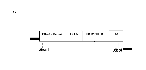
[0012] Figure 1: Characterization of transducible materials (I). (A) Schematic
of
protein expression vector for transducible materials Oct4-11R (SEQ ID NO: 12),
Sox2-
11R (SEQ ID NO: 13), K1f4-11R (SEQ ID NO: 14), and cMyc-11R (SEQ ID NO: 15),
Linker: SEQ ID NO: 55; Effector Domain: Oct4 (SEQ ID NO: 1), Sox2 (SEQ ID NO:
2),
K1f4 (SEQ ID NO: 3), or cMyc (SEQ ID NO: 4). (B) Stability of the four
transducible
materials (Oct4-llR , Sox2-11R, K1f4-11R, and cMyc-11R) under the cell culture
condition examined by Western blot analysis.
[0013] Figure 2: Characterization of transducible materials (II). Protein
transduction
of 11R-tagged transducible materials into OG2-MEF cells examined by
immunocytochemistry. Oct4: MEF cells transuded with Oct4-11R (green), Sox2:
MEF
cells transuded with Sox2-11R (red), K1f4: MEF cells transuded with K1f4-11R
(red) and
cMyc: MEF cells transuded with cMyc-11R (green). DAPI: Cells stained with DAPI
to
visualize the nuclei (blue) and the images were merged.
[0014] Figure 3: Characterization of transducible materials (III). Protein
induced
pluripotent stem (piPS) cells clonally expanded and self-renewed in chemical
defined
media and feeder free condition.
[0015] Figure 4: Generation of piPS cells by transducible materials Oct4-11R,
Sox2-
11R, K1f4-11R, and cMyc-11R. (A) Timeline of piPS cell generation. (B) Oct4-
GFP+
piPS cell colonies initially observed around day 30-35. Phase: representative
phase
contrast image; and GFP: fluorescence image. (C) Oct4-GFP+ piPS cells
sustained long
term self-renewal under conventional mESC growth condition. (D) The long-term
expanded piPS cells grew as compact and domed colonies that expressed strong
ALP, a
typical pluripotency marker. (E) piPS cells expressed other typical
pluripotency markers,
3
CA 02748009 2011-06-21
WO 2010/075575 PCT/US2009/069518
examined by immunofluorescence: SEA-1 (red), Sox2 (red), Oct4 (ed) and Nanog
(red).
DAPI: DAPI staining for visualization of the nuclei (blue), and the images
were merged.
(F) RT-PCR analysis of endogenous pluripotency gene expression in piPS cells.
(G)
Oct4 promoter methylation analysis by bisulfite genomic sequencing. Open and
closed
circles indicate unmethylated and methylated CpGs, respectively.
[0016] Figure 5: In vitro and in vivo pluripotency of piPS cells (I). piPS
cells
effectively differentiated in vitro into cells in the three germ layers: Tuj
1: characteristic
TUJ1+ neuronal cells-ectoderm (red): Bryt: Brachyury+ mesoderm cells (red);
and Sox17:
Soxl7+ definitive endoderm cells. Images were merged with DAPI (blue)
staining.
[0017] Figure 6: In vitro and in vivo pluripotency of piPS cells (II) (A) RT-
PCR
analysis of in vitro differentiation of piPS cells. (B) Chimeric embryos (13.5
dpc, 2 out
of 7, left) obtained after transfer of the piPS cell aggregated embryos into a
pseudo-
pregnant mouse (top). Such piPS cells contributed to the germline cells (Oct4-
GFP
positive) in isolated genital ridge tissue from chimeric embryos (bottom).
[0018] Figure 7: Schematic of protein expression vectors for transducible
materials.
His6: SEQ ID NO: 59; Effector Domain: Ngn3 (SEQ ID NO: 8), PDX1 (SEQ ID NO:
9);
MafA (SEQ ID NO: 10), or Foxp3 (SEQ ID NO: 11); Linker: SEQ ID NO: 55.
[0019] Figure 8: Reprogramming of liver and pancreatic exocrine cells to
insulin-
producing beta cells by transducible materials His6-Ngn3 -11 R (SEQ ID NO:
30), His6-
PDX1-11R (SEQ ID NO: 31) and His6-MafA-11R (SEQ ID NO: 32) in mouse (I).
Mouse-1, Mouse-2 and Mouse-3 were treated with bovine serum albumin (BSA)
protein
(control group). Mouse-4, Mouse-5 and Mouse-6 were treated with His6-Ngn3 -11
R,
His6-PDX1-11R and His6-MafA-11R (treatment group). A) Immunofluorescent
analysis
(IFA) of Mouse-1 liver; B) IFA of Mouse-2 liver; and C) IFA of Mouse-3 liver.
[0020] Figure 9: Reprogramming of liver and pancreatic exocrine cells to
insulin-
producing beta cells by transducible materials His6-Ngn3-11R, His6-PDX1-11R
and
His6-MafA-11R in mouse (II). Mouse-1, Mouse-2 and Mouse-3 were treated with
bovine serum albumin (BSA) protein (control group). Mouse-4, Mouse-5 and Mouse-
6
were treated with His6-Ngn3-11R, His6-PDX1-11R and His6-MafA-11R (treatment
group). A) IFA of Mouse-4 liver (1); B) IFA of Mouse-4 liver (2); C) IFA of
Mouse-5
4
CA 02748009 2011-06-21
WO 2010/075575 PCT/US2009/069518
liver (1); D) IFA of Mouse-5 liver (2); E) IFA of Mouse-6 liver (1); and F)
IFA of
Mouse-6 liver (2).
[0021] Figure 10: Reprogramming of liver and pancreatic exocrine cells to
insulin-
producing beta cells by transducible materials His6-Ngn3-11R, His6-PDX1-11R
and
His6-MafA-11R in mouse (III). Mouse-1, Mouse-2 and Mouse-3 were treated with
bovine serum albumin (BSA) protein (control group). Mouse-4, Mouse-5 and Mouse-
6
were treated with His6-Ngn3-11R, His6-PDX1-11R and His6-MafA-11R (treatment
group). A) IFA of Mouse-1 pancrease; B) IFA of Mouse-2 pancrease (1); C) IFA
of
Mouse-2 pancrease (2); and D) IFA of Mouse-3 pancrease.
[0022] Figure 11: Reprogramming of liver and pancreatic exocrine cells to
insulin-
producing beta cells by transducible materials His6-Ngn3-11R, His6-PDX1-11R
and
His6-MafA-11R in mouse (IV). Mouse-1, Mouse-2 and Mouse-3 were treated with
bovine serum albumin (BSA) protein (control group). Mouse-4, Mouse-5 and Mouse-
6
were treated with His6-Ngn3-11R, His6-PDX1-11R and His6-MafA-11R (treatment
group). A) IFA of Mouse-4 pancrease (1); B) IFA of Mouse-4 pancrease (2); C)
IFA of
Mouse-5 pancrease (1); D) IFA of Mouse-5 pancrease (2); and E) IFA of Mouse-6
pancrease.
[0023] Figure 12: Reprogramming of T cells to Treg cells by transducible
material
His6-Foxp3-11R (SEQ ID NO: 33) (IA). Flow cytometric analysis of CD4 and CD25
protein expression in PBMC with lacking of CD14 monocytes: isotope control,
PBS
control, sample buffer control and protein (BSA 100 g/ml) control.
[0024] Figure 13: Reprogramming of T cells to Treg cells by transducible
material
His6-Foxp3-11R (IB). Flow cytometric analysis of CD4 and CD25 protein
expression in
PBMC with lacking of CD14 monocytes treated with His16-Foxp3-11R of l0 g/ml,
20
gg/ml, or 50 gg/ml.
[0025] Figure 14: Reprogramming of T cells to Treg cells by transducible
material
His6-Foxp3-11R (IIA). Flow cytometric analysis of CD4 and CD25 protein
expression
in PBMC: isotope control, and PBS control.
[0026] Figure 15: Reprogramming of T cells to Treg cells by transducible
material
His6-Foxp3-11R (IIB). Flow cytometric analysis of CD4 and CD25 protein
expression
in PBMC: sample buffer control and protein (BSA 100 g/ml) control.
CA 02748009 2011-06-21
WO 2010/075575 PCT/US2009/069518
[0027] Figure 16: Reprogramming of T cells to Treg cells by transducible
material
His6-Foxp3-11R (IIC). Flow cytometric analysis of CD4 and CD25 protein
expression
in PBMC treated with Hisl6-Foxp3-11R of 10 g/ml, or 50 gg/ml.
[0028] Figure 17: Reprogramming of T cells to Treg cells by transducible
material
His6-Foxp3-11R (IID). Flow cytometric analysis of CD4 and CD25 protein
expression
in PBMC treated with Hisl6-Foxp3-11R of 100 gg/ml.
DETAILED DESCRIPTION OF THE INVENTION
[0029] One aspect of the present disclosure relates to a transducible material
comprising an effector domain.
[0030] In certain embodiments, a transducible material used herein refers to a
material or a molecule which is not DNA or derived from DNA but is capable of
crossing
or transducing or being crossed through a membrane of a biological sample
(e.g., a cell
membrane) so that the transducible material can enter or be brought into the
inside of the
biological sample from the outside of the biological sample and exerts
reprogramming
efforts. For example, the transducible material may interact with cell-surface
receptors
which facilitate the entry of the material into cells through receptor
mediated endocytosis.
[0031] In certain embodiments, a transducible material is a selective
transducible
material which is more likely to transduce into a specific type of biological
samples (e.g.
cancer or tumor cells) or becomes transducible in a specific microenvironment
in or
around a biological sample (e.g. micro environment around cancer or tumor)
than other
biological samples. For example, the selective transducible material comprises
a
transduction domain (e.g. a cell-targeting peptide or an activatable cell
penetrating
peptide) that preferably delivers the selective transducible material into a
specific type of
biological sample or become transducible in a microenvironment around a
biological
sample.
[0032] Without being bounded to any theories, it is contemplated that the
transducible materials may cross a cell membrane and enter into cytoplasm to
reprogram
cytoplasm activities such as translation, post-translation modification,
signaling pathway,
apoptosis pathway. It is further contemplated that the transducible material
may cross the
nucleus membrane and reprogram or modulate DNA or chromosomal replication,
gene
transcription, and RNA splicing.
6
CA 02748009 2011-06-21
WO 2010/075575 PCT/US2009/069518
[0033] An effector domain is a motif or a molecule which, once inside a
biological
sample, is capable of exerting reprogramming changes of the biological sample.
The
effector domain may interact with molecules (e.g., proteins, DNA, RNA, sugars,
and
lipids) in the biological sample (e.g., in cytoplasm or nuclei) and lead to
changes such as
proliferation, differentiation, dedifferentiation, transdifferentiation,
retrodifferentiation,
transdertermination, apoptosis, and morphogenesis. The effector domain can be
1) a
polypeptide, or a fragment or a mimic thereof; 2) a polynucleotide which
cannot be gene
expressed once transduced or incorporated into the genome of the biological
sample or
cause genetic modification but nevertheless interacts with molecules in the
biological
sample (e.g., a ribozyme, an antisense molecule, a siRNA or miRNA, an
oligonucleotide,
and the like); and 3) a small molecule or other chemical compound (e.g.
chemotherapy
drugs).
[0034] In certain embodiments, an effector domain is inherently transducible,
e.g.
PDX1 (e.g. SEQ ID NO: 9) and NeuroD (e.g. SEQ ID NO: 7).
[0035] One example of the effector domain is a polypeptide such as a
transcription
factor, a chromosome remodeling protein, an antibody, or a fragment or mimic
thereof.
Another example of the effector domain is a small molecule which is not a
polymer and
binds with a biolpolymer such as protein, nucleic acid, or polysaccharide and
alters the
activity or function of the biopolymer. Examples of small molecules include,
without
limitation, acetylation inhibitors, transcription activators, signal pathway
activators,
signal pathway inhibitors, and methylation inhibitors.
[0036] In another embodiment, an effector domain can be at least one
polypeptide
that reprograms a somatic cell into a stem cell or change a cell state from
one to another.
For example, the effector domain can be 1) a polypeptide selected from the
group
consisting of K1f4 (e.g. SEQ ID NO: 3), Sox2 (e.g. SEQ ID NO: 2), Lin28 (e.g.
SEQ ID
NO: 5), Oct4 (e.g. SEQ ID NO: 1), cMyc (e.g. SEQ ID NO: 4), Nanog (e.g. SEQ ID
NO:
6), and any combination thereof; 2) a polypeptide selected from the group
consisting of
K1f4, Sox2, Oct4, cMyc, and any combination thereof; 3) a polypeptide selected
from the
group consisting of Sox2, Oct4, Lin28, Nanog, and any combination thereof, 4)
a
polypeptide selected from the group consisting of Ngn3 (e.g. SEQ ID NO: 8),
PDX1 (e.g.
SEQ ID NO: 9), MafA (e.g. SEQ ID NO: 10), NeuroD (e.g. SEQ ID NO: 7), and any
7
CA 02748009 2011-06-21
WO 2010/075575 PCT/US2009/069518
combination thereof; 5) a polypeptide comprising Foxp3 (e.g. SEQ ID NO: 11);
6) a
polypeptide selected from the group consisting of Oct4, Sox2, K1f4, Lin28,
Nanog, cMyc,
Ngn3, PDX1, MafA, NeuroD, Foxp3, and any combination thereof; 7) a combination
of
polypeptides Oct4, Sox2, K1f4 and cMyc; and 8) a combination of polypeptides
Ngn3,
PDX1 and MafA.
[0037] In certain embodiments, a transducible material further comprises a
transduction domain. A transduction domain is a motif that is capable of
facilitating the
entry of the transducible material into a biological sample (e.g., a cell).
The transducible
domain is associated with the effector domain covalently, noncovalently or via
a linker.
In certain embodiments, the transduction domain is covalently linked to the
effector
domain through a linker. In certain embodiments, the linker is a glycine-rich
linker that
comprises one or more glycine residues (e.g. esggggspg (SEQ ID NO: 55)).
[0038] Examples of a transduction domain include, without limitation, polymers
such as cationic lipid polymers and nanoparticles, protein transduction
domains (PTD),
cell penetrating peptides (CPP1), cell permeating peptides (CPP2), activatable
cell
penetrating peptides or conjugates (ACPP), and cell-targeting peptides (CTP).
[0039] CPP1, CPP2, and PTD are peptides known to facilitate delivery of a
molecular cargo associated thereof into cells. The association between a CPP
1, CPP2 or
PTD and the molecular cargo can be through covalent bond or non-covalent
interactions.
The molecular cargo can be small chemical molecules, peptides, protein,
fragment of
DNA, RNA such as siRNA and miRNA, or nanosize particles. For example, CPP 1
and
PTD include 5 to 20 amino acid peptide motifs that are capable of penetrating
cells
independent of surface transporters and of cell cycle phase. CPP1 and PTD can
also be
capable of penetrating through blood-brain barriers. CPP1 and PTD can deliver
proteins
and peptides in vitro and in vivo with uniform distribution throughout the
organism after
parenteral administration. Cationic PTDs can act as nuclear localization
signals and carry
an associated molecular cargo to cell nuclei. Examples of protein transduction
domains
include, without limitation, TAT (e.g. YGRKKRRQRRR, SEQ ID NO: 34), poly-
arginine (e.g. poly-arginine having 7-11 arginine residues such as RRRRRRR,
RRRRRRRR, RRRRRRRRR, RRRRRRRRRR (SEQ ID NO: 35)and RRRRRRRRRRR
(SEQ ID NO: 36)), Penetratin (Antennapedia, e.g. RQIKIWFQNRRMKWKK (SEQ ID
8
CA 02748009 2011-06-21
WO 2010/075575 PCT/US2009/069518
NO: 38)), VP22 (e.g. DAATATRGRSAASRPTQRPRAPARSASRPRRPVQ (SEQ ID
NO: 39)), Transportan (e.g. GWTLNSAGYLLGKINLKALAALAKKIL (SEQ ID NO:
40)), MAP (e.g. KLALKLALKALKAALKLA (SEQ ID NO: 41)), MTS (e.g.
AAVALLPAVLLALLP (SEQ ID NO: 42)), PEP-1 (e.g.
KETWWETWWTEWSQPKKKRKV (SEQ ID NO: 43)), Arg/Trp analogue (e.g.
RRWRRWWRRWWRRW (SEQ ID NO: 44)), polyguanidine peptoids (e.g.
polyguanidine peptoids with a 6-methylene spacer between backbone and
guanidine
group such as N-arg 5, 7 or 9 peptoids), HIV-1 Rev (e.g. SEQ ID NO: 60), Flock
house
virus coat peptide (e.g. SEQ ID NO: 61), and DNA-binding peptides such as c-
Fos (e.g.
SEQ ID NO: 62), c-Jun (e.g. SEQ ID NO: 63)and yeast GCN4 (e.g. SEQ ID NO: 64).
[0040] Cell-targeting peptides are proteins or peptides that bind to cell-
surface
receptors and enter cells through endocytosis. In certain embodiments, a cell-
target
peptide targets specific tissues or cell types, for example, GnRH peptides
(e.g. SEQ ID
NO: 58) target biological samples that express GnRH receptors (e.g. solid
tumors and
hormone-responsive cancer cell lines). More examples of cell-targeting
peptides and the
specific biological samples targeted are listed in Table 1.
Table 1. Examples of cell-targeting peptides and the specific biological
samples
targeted.
Sequence Peptide sequence Length Targeted tissue or cells
ID No. and the cellular targets
SEQ ID TSPLNIHNGQKL 12 Human head and neck solid
NO: 45 tumors
SEQ ID CGKRK 5 Tumor neovasculature,
NO: 46 Heparan sulfate
SEQ ID CGNKRTRGC 7 breast carcinoma
NO: 47
SEQ ID SMSIARL 7 Prostate vasculature
NO: 48
SEQ ID FQHPSFI 7 hepatocellular carcinoma cell
NO: 49 line
RGD 3 Integrin receptor', aV(33
9
CA 02748009 2011-06-21
WO 2010/075575 PCT/US2009/069518
NGR 3 Tumor neovasculature,
Amino-peptidase N
SEQ ID VHSPNKK 7 Endothelial VCAM-1
NO: 50 expressing cells;
VCAM-1
SEQ ID RRPYIL 6 Adenocarcinoma cells;
NO: 51 Neurotensin receptor
SEQ ID EDYELMDLLAYL 12 Various carcinoma
NO: 52
SEQ ID LTVSPWY 7 breast carcinoma; erbB2
NO: 53
SEQ ID ATWLPPR 7 Tumor neovasculature;
NO: 54 VEGF receptor
[0041] An activatable cell penetrating peptide or conjugate (ACPP) comprises a
cationic CPP1, CPP2 or PTD and a neutralizing anionic counterpart. In certain
embodiments, the cationic CPP1, CPP2 or PTD and the anionic counterpart are
associated via noncovalent interactions (e.g. charge-charge interaction)
and/or covalent
cleavable linker (e.g. matrix metaloprotease (MMP) cleavable sequence).
Transduction
of an ACPP into cells are inhibited until the noncovalent interactions are
disrupted and/or
the cleavable linker is cleaved. For example, without being bound to any
theory, the
anionic counterparts comprise one or more pH sensitive groups such as
sulfonamide
groups, which are protonated at pH 7.4 (the pH in the blood stream) and become
neutral
at slightly acidic pH (e.g. pH 6.8). Therefore, charge-charge interactions
between
cationic CPP 1, CPP2 or PTD and the anionic counterpart can be interrupted in
slightly
acidic microenvironment (e.g. in or around tumor or cancer). MMP concentration
in
blood stream is lower than that in a microenvironment around tumor or cancer.
Therefore, MMP cleavable sequence, which is not cleaved in the bloodstream, is
cleaved
in environment around tumor or cancer. The cationic CPP 1, CPP2 or PTD is no
longer
neutralized by the anionic counterpart, and therefore is exposed to facilitate
the
translocation into cells (e.g. tumor or cancer cells). In certain embodiments,
the CPP1,
CPP2 or PTD is TAT. In certain embodiments, the anionic counterparts comprise
pH-
CA 02748009 2011-06-21
WO 2010/075575 PCT/US2009/069518
sensitive polymer (e.g. di-block copolymer) comprising pH-sensitive groups
(e.g.
sulfonamide groups).
[0042] For another example, an activatable cell penetrating conjugate
comprises a
conventional hydrophobic core made of a polymer into which an effective domain
is
incorporated, a peripheral hydrophilic layer composed of poly-ethylene glycol
and one or
more cationic CPPls, CPP2s or PTDs, and one or more anionic counterpart that
neutralize the cationic CPP12, CPP2s or PTDs through charge-charge
interactions. Such
charge-charge interactions are expected to shield the cationic charges during
delivery
until the transduction material reaches a slightly acidic microenvironment
(e.g. tumor or
cancer), which triggers protonation of the anionic counterparts and disrupts
the charge-
charge association. Subsequently the cationic CPPls, CPP2s, or PTDs,
previously
quenched by the anionic counterpart, are now capable of facilitating delivery
of the
effector domain into the surrounding cells (e.g. tumor or cancer cells).
[0043] In certain embodiments, a selective transducible material comprises a
transduction domain selected from the group consisting of cell-targeting
peptides and
activatable cell penetrating peptides and activatable cell penetrating
conjugates.
[0044] A transduction domain is associated to an effector domain via covalent
bond,
non-covalent interactions or through a linker. Thus a transducible material
can be made
by obtaining the transduction domain and the effector domain separately and
associate
together through a covalent bond or non-covalent interactions (e.g., repulsive
interactions,
dipole interactions, hydrogen bonding interactions, dispersive interactions,
charge-charge
interactions, solvent, counter ion and entropic effects, and water and
hydrophobic effects).
In certain embodiments, the transduction material is prepared by mixing the
effector
domain and transducible domain. Alternatively, a transducible material can be
produced
by isolating the material from natural resources or recombinantly. In the case
when both
domains are peptides or polypeptides, the effector domain can be linked to the
N-
terminus or C-terminus of the transduction domain and the transducible
polypeptide can
be made synthetically through chemical synthesis or recombinantly through
recombinant
technology.
11
CA 02748009 2011-06-21
WO 2010/075575 PCT/US2009/069518
[0045] In certain embodiments, a transducible material comprises an effector
domain
that is inherently transducible, and a transduction domain associated with the
effector
domain via covalent or non-covalent interactions.
[0046] In certain embodiments, a transducible material further comprises one
or
more motifs that do not interrupt the function of the effector domain or the
transduction
domain. In certain embodiments, these motifs are linked covalently, non-
covalently or
through a linker to the effector domain and/or the transduction domain. In
certain
embodiments, these motifs facilitate the preparation and/or purification of
the
transducible material. One example of such motif is a polyhistidine-tag to
facilitate
protein purification in preparation of the transducible material. In certain
embodiments,
the polyhistidine-tag comprises at least six histidine residues (e.g.
MGSSHHHHHHSSGLVPRGSH ("His6," SEQ ID NO: 59)).
[0047] In certain embodiments, a transducible material includes, for example,
Oct4-
11R (SEQ ID NO: 12), Sox2-11R (SEQ ID NO: 13), K1f4-11R (SEQ ID NO: 14), Lin28-
11R (SEQ ID NO: 16), Nanog-11R (SEQ ID NO: 17), cMyc-11R (SEQ ID NO: 15),
Ngn3-11R (SEQ ID NO: 19), PDX1-11R (SEQ ID NO: 20), MafA-11R (SEQ ID NO:
21), NeuroD-11R (SEQ ID NO: 18), and Foxp3-11R (SEQ ID NO: 22), wherein 11R
(SEQ ID NO: 37) stands for a polyarginine sequence of 11 arginine residues
linking to a
linker through which the polyarginie sequence is covalently linked to the
effector domain.
In certain embodiments, a transducible material includes, for example, His6-
Oct4-11R
(SEQ ID NO: 23), His6-Sox2-11R (SEQ ID NO: 24), His6-K1f4-11R (SEQ ID NO: 25),
His6-Lin28-11R (SEQ ID NO: 27), His6-Nanog-11R (SEQ ID NO: 28), His6-cMyc-11R
(SEQ ID NO: 26), His6-Ngn3-11R (SEQ ID NO: 30), His6-PDX1-11R (SEQ ID NO: 31),
His6-MafA-11R (SEQ ID NO: 32), His6-NeuroD-11R (SEQ ID NO: 29), and His6-
Foxp3-11R (SEQ ID NO: 33).
[0048] In certain embodiments, a transducible material can be combined with
one or
more adjuvants such as growth factors to stimulate cellular growth. For
example, islet
growth factor (e.g. betacellulin) is used as an adjuvant in reprogramming
liver and/or
pancreatic exocrine cells to insulin producing cells (e.g. 0 cells).
[0049] Another aspect of the present disclosure relates to a composition
comprising
a biological sample and at least one transducible material, wherein the
transducible
12
CA 02748009 2011-06-21
WO 2010/075575 PCT/US2009/069518
material has transduced into the biological sample. For example, the
composition
includes a transducible material comprising Foxp3 (e.g. Foxp3-11R and His6-
Foxp3-11R)
and a T cell wherein the transducible material has transduced into the T cell;
a
composition includes a piPS cell and one or more transducible materials
comprising a
polypeptide selected from the group consisting of Oct4, K1f4, Sox2 and cMyc,
and any
combination thereof (e.g. Oct4-11R, KIP-11R, Sox2-11R, cMyc-11R, His6-Oct4-
11R,
His6-K1f4-11R, His6-Sox2-11R and His6-cMyc-11R); and a composition including a
liver or pancreatic exocrine cell and one or more transducible materials
comprising a
polypeptide selected from the group consisting of Ngn3, PDX1, MafA, NeuroD,
and any
combination thereof (e.g. Ngn3-11R, PDX1-11R, MafA-11R, His6-Ngn3-11R, His6-
PDX1-11R and His6-MafA-11R) wherein the transducible materials have transduced
into
the liver or pancreatic exocrine cell.
[0050] Another aspect of the present disclosure relates to a method of
reprogramming a biological sample by exposing the biological sample to a
composition
comprising a transducible material. In certain embodiments, the method
preferably
reprograms a specific type of biological sample (e.g. cancer or tumor cells)
or biological
samples in or around a specific micro environment within a biological organism
(e.g.
microenvironment around cancer or tumor) than other biological samples by
exposing
biological samples to a composition comprising a selective transducible
material.
[0051] In one embodiment, a biological sample includes a cell, a cluster of
cells, a
tissue, an organ, a biological body from a biological organism. The biological
sample
can be normal, healthy sample or abnormal, diseased sample (e.g., cancer or
tumor).
[0052] A biological organism includes, for example, a microorganism (e.g.,
bacteria),
a fungus, a plant and an animal (e.g., a human).
[0053] An organ from an animal biological organism (e.g., human) includes, for
example, a circulatory organ (e.g., heart, blood and blood vessels), a
digestive organ (e.g.,
salivary glands, esophagus, stomach, liver, gallbladder, pancreas, intestines,
rectum and
anus), an endocrine organ (e.g., endocrine glands such as the hypothalamus,
pituitary or
pituitary gland, pineal body or pineal gland, thyroid, parathyroids and
adrenals, i.e.,
adrenal glands), an integumentary organ (e.g., skin, hair and nails), a
lymphatic organ
(e.g., lymph nodes and vessels, tonsils, adenoids, thymus and spleen), a
muscular organ
13
CA 02748009 2011-06-21
WO 2010/075575 PCT/US2009/069518
(e.g., muscles), a nervous organ (e.g., brain, spinal cord, peripheral nerves
and nerves), a
reproductive organ (e.g., ovaries, fallopian tubes, uterus, vagina, mammary
glands, testes,
vas deferens, seminal vesicles, prostate and penis), a respiratory organ
(e.g., the pharynx,
larynx, trachea, bronchi, lungs and diaphragm), a skeletal organ (e.g., bones,
cartilage,
ligaments and tendons), a urinary system (e.g., kidneys, ureters, bladder and
urethra). An
organ can be normal or healthy, and alternatively, abnormal or unhealthy
(e.g.,
cancerous).
[0054] An organ from a plant biological organism includes, for example, root,
stem,
leaf, flower, seed and fruit.
[0055] A tissue from a biological sample (e.g. an animal) includes a
connective
tissue, a muscle tissue, a nervous tissue, and an epithelial tissue. A tissue
can be normal
or healthy, and alternatively, abnormal or unhealthy (e.g., cancerous). A
tissue from a
biological sample (e.g. a plant) includes an epidermis, a vascular tissue and
a ground
tissue.
[0056] A cell can be prokaryotic or eukaryotic. A prokaryotic cell includes,
for
example, bacteria. A eukaryotic cell includes, for example, a fungus, a plant
cell, and an
animal cell. The types of an animal cell (e.g., a mammalian cell or a human
cell)
includes, for example, a cell from circulatory/immune system or organ (e.g., a
B cell, a T
cell (cytotoxic T cell, natural killer T cell, regulatory T cell, T helper
cell), a natural killer
cell, a granulocyte (e.g., basophil granulocyte, an eosinophil granulocyte, a
neutrophil
granulocyte and a hypersegmented neutrophil), a monocyte or macrophage, a red
blood
cell (e.g., reticulocyte), a mast cell, a thrombocyte or megakaryocyte, and a
dendritic cell);
a cell from an endocrine system or organ (e.g., a thyroid cell (e.g., thyroid
epithelial cell,
parafollicular cell), a parathyroid cell (e.g., parathyroid chief cell,
oxyphil cell), an
adrenal cell (e.g., chromaffin cell), and a pineal cell (e.g., pinealocyte));
a cell from a
nervous system or organ (e.g., a glioblast (e.g., astrocyte and
oligodendrocyte), a
microglia, a magnocellular neurosecretory cell, a stellate cell, a boettcher
cell, and a
pituitary cell (e.g., gonadotrope, corticotrope, thyrotrope, somatotrope, and
lactotroph ));
a cell from a respiratory system or organ (e.g., a pneumocyte (a type I
pneumocyte and a
type II pneumocyte), a clara cell, a goblet cell, an alveolar macrophage); a
cell from
circular system or organ (e.g., myocardiocyte and pericyte); a cell from
digestive system
14
CA 02748009 2011-06-21
WO 2010/075575 PCT/US2009/069518
or organ (e.g., a gastric chief cell, a parietal cell, a goblet cell, a paneth
cell, a G cell, a D
cell, an ECL cell, an I cell, a K cell, an S cell, an enteroendocrine cell, an
enterochromaffin cell, an APUD cell, a liver cell (e.g., a hepatocyte and
Kupffer cell)); a
cell from integumentary system or organ (e.g., a bone cell (e.g., an
osteoblast, an
osteocyte, and an osteoclast), a teeth cell (e.g., a cementoblast, and an
ameloblast), a
cartilage cell (e.g., a chondroblast and a chondrocyte), a skin/hair cell
(e.g., a trichocyte, a
keratinocyte, and a melanocyte (Nevus cell)), a muscle cell (e.g., myocyte),
an adipocyte,
a fibroblast, and a tendon cell), a cell from urinary system or organ (e.g., a
podocyte, a
juxtaglomerular cell, an intraglomerular mesangial cell, an extraglomerular
mesangial
cell, a kidney proximal tubule brush border cell, and a macula densa cell),
and a cell from
reproductive system or organ (e.g., a spermatozoon, a sertoli cell, a leydig
cell, an ovum,
an oocyte). A cell can be normal, healthy cell; or a diseased or unhealthy
cell (e.g., a
cancer cell).
[0057] A cell further includes a mammalian stem cell which include an
embryonic
stem cell, a fetal stem cell, an induced pluripotent stem cell, and an adult
stem cell. A
stem cell is a cell that is capable of undergoing cycles of cell division
while maintaining
an undifferentiated state and differentiating into specialized cell types. A
stem cell can
be an omnipotent stem cell, a pluripotent stem cell, a multipotent stem cell,
an
oligopotent stem cell and an unipotent stem cell (See, Hans R. Scholer (2007).
"The
Potential of Stem Cells: An Inventory" in Nikolaus Knoepffler, Dagmar
Schipanski, and
Stefan Lorenz Sorgner. Humanbiotechnology as Social Challenge. Ashgate
Publishing,
Ltd. pp. 28), any of which may be induced from a somatic cell. A stem cell may
also
include a cancer stem cell.
[0058] In another embodiment, "reprogramming a biological sample" used herein
is
exchangeable with or refers to modulating, altering, or changing the
biological activities
of the biological sample (e.g., cell) or modulating, altering, or changing the
state or status
of the biological sample from one to another. For example, by exposing a
biological
sample (e.g., a cell) to a transducible material, the biological activities of
the cell (e.g.,
cell growth, cell division, cell metabolism, cell cycle, cell signaling, DNA
replication,
transcription, RNA splicing, protein synthesis, post-translation modification)
are
modulated or altered so as to lead to cell proliferation, differentiation
(e.g., from
CA 02748009 2011-06-21
WO 2010/075575 PCT/US2009/069518
progenitor cells to terminally differentiated cells), dedifferentiation (e.g.,
from terminally
differentiated cells to pluripotent stem cells), transdifferentiation (e.g.,
from one type of
terminally differentiated cells to another type of terminally differentiated
cells),
retrodifferentiation (e.g., from terminally differentiated cells to progenitor
cells),
transdertermination (e.g., from one type of progenitor cells to a type of
terminally
differentiated cells that are usually derived from another type of progenitor
cells under
natural conditions), apoptosis (e.g., cell death of cells or cancer cells),
morphogenesis,
and changes in the cell fate. For another example, the state of a biological
sample can be
altered or changed from abnormal or diseased state to normal or healthy state
(e.g., from
cancer cells to noncancer cells); from one cell type to another cell type
(e.g., from
undifferentiated stem cells to differentiated stem cells or specialized
cells), from
differentiated or specialized cells to undifferentiated cells or stem cells
(e.g., an
omnipotent stem cell, a pluripotent stem cell, a multipotent stem cell, an
oligopotent stem
cell and an unipotent stem cell)(e.g., from fibroblast cells to induced
pluripotent stem
cells (iPSCs)), from somatic cells to stem cells or induced stem cells, from
one state of
stem cells to another state of stem cells (e.g., from ominipotent stem cells
to pluripotent
stem cells), from one type of differentiated cells to another type of
differentiated cells
(e.g., T-cells to regulatory T cells, pancreatic exocrine cells to insulin-
producing beta
cells).
[0059] In another embodiment, a biological sample is exposed to a transducible
material and reprogrammed. The biological sample can be exposed in vitro, in
vivo or ex
vivo. For example, the biological sample is exposed in vitro through
contacting the
sample with the transducible material in an environment outside of a living
biological
organism (e.g., in a cell culture system or a test tube). The biological
sample is exposed
in vivo through contacting the material with a biological organism containing
the sample
or introducing (e.g., through administration) the material into the organism.
The
transducible materials can be administered via any known administration route
such as
for example parenteral (e.g., subcutaneous, intraperitoneal, intravenous,
including
intravenous infusion, intramuscular, or intradermal injection) or non-
parenteral (e.g., oral,
intranasal, intraocular, sublingual, rectal, or topical) route. The biological
sample is
exposed ex vivo when the biological sample (e.g., a cell, a tissue or an
organ) is taken
16
CA 02748009 2011-06-21
WO 2010/075575 PCT/US2009/069518
outside the biological organism, contacted with the transducible material, and
placed
back to the same or different biological organisms. Examples of ex vivo
exposures
comprise removing a biological sample from the biological organism, exposing
the
biological sample to a transducible material, and transplanting the biological
sample
transduced with the transducible material back to the biological organism.
[0060] In certain embodiments, OG2-MEF cells are exposed to a composition
comprising protein Oct4-11R, Sox2-11R, KIP-l1R and cMyc-11R and reprogrammed
to
induced pluripotent stem cells (iPSCs).
[0061] In certain embodiments, T cells are exposed to a composition comprising
protein Foxp3-11R or His6-Foxp3-11R and programmed to regulatory T cells (Treg
cells).
[0062] In certain embodiments liver and/or pancreatic exocrine cells are
exposed to a
composition comprising one or more proteins selected from the group consisting
of
Ngn3-11R, PDX1-11R, MafA-11R, NeuroD-11R, His6-Ngn3-11R, His6-PDX1-11R,
His6-MafA-11R, and His6-NeuroD-11R and reprogrammed into insulin producing
cells
(e.g. 0 cells). In certain embodiments, the composition further comprises one
or more
adjuvant such as Islet growth factor (e.g. betacellulin). In certain
embodiments, the
composition comprises His6-Ngn3-11R, His6-PDX1-11R, and His6-MafA-11R. In
certain embodiments, the composition comprises His6-Ngn3-11R, His6-PDX1-11R,
His6-MafA-11R and betacellulin. Without bond to the mechanism, it is further
contemplated that such reprogramming is through transdetermination and/or
transdifferentiation.
[0063] Another aspect of the present disclosure relates to a method of
treating,
preventing or reducing a disease or condition in a biological organism by
administering a
composition comprising a transducible material into the organism. In certain
embodiments, the composition is a pharmaceutical composition comprising a
transducible material. In certain embodiments, the composition comprises a
selective
transducible material. The treatment, prevention or reduction of a disease or
condition is
associated with the change or reprogramming of a biological sample (e.g., a
cell, a tissue
or an organ) in the organism.
17
CA 02748009 2011-06-21
WO 2010/075575 PCT/US2009/069518
[0064] In certain embodiments, the disease or condition treatable by the
method
include, without limitations, tumor, cancer, metabolic diseases or conditions
(e.g. type I
and type II diabetes and obesity), inflammatory conditions, cardiac diseases,
neurogenerative diseases (e.g. anemia, amyotrophic lateral sclerosis, spinal
cord injury,
bums, or arthritis), autoimmune diseases or conditions (e.g. acute
disseminated
encephalomyelitis (ADEM), Addison's disease, alopecia areata, ankylosing
spondylitis,
antiphospholipid antibody syndrome (APS), anemia (e.g. autoimmune hemolytic
anemia
and pernicious anaemia), arthritis, psoriatic arthritis, rheumatoid arthritis,
diabetes
mellitus type 1, autoimmune hepatitis, autoimmune inner ear disease, bullous
pemphigoid,
coeliac disease, Chagas disease, chronic obstructive pulmonary disease, Crohns
disease,
dermatomyositis, endometriosis, Goodpasture's syndrome, Graves' disease,
Guillain-
Barre syndrome (GBS), Hashimoto's disease, hidradenitis suppurativa, Kawasaki
disease,
IgA nephropathy, idiopathic thrombocytopenic purpura, interstitial cyctitis,
lupus
erythematosus, mixed connective tissue disease, morphea, multiple sclerosis
(MS),
myasthenia gravis, narcolepsy, neuromyotonia, pemphigus vulgaris, psoriasis,
polymyositis, primary billiary cirrhosis, schizophrenia, scleroderma,
Sjogren's syndrome,
stiff person syndrome, temporal arteritis ("Giant cell arteritis"), ulcerative
colitis,
vasculitis, vitiligo, and Wegener's granulomatosis).
[0065] For example, it is contemplated that a transducible material can be
administered to a biological organism having a tumor to activate the apoptosis
of the
tumor cells or make tumor cells more sensitive to chemotherapy, radiotherapy,
or cancer
drugs.
[0066] In certain embodiments, a transducible material can be administered to
a
biological organism to enhance or attenuate immune system and thus treat or
prevent
immune-related diseases or inflammatory diseases. For example, protein Foxp3-
11R or
His6-Foxp3-11R is transduced to T cells and programs them to Treg cells, which
suppress the overactive immune system and thus is a treatment for auto-immune
diseases
[0067] In certain embodiments, a transducible material can be administered to
a
biological organism to treat metabolic diseases or conditions such as type I
diabetes, type
II diabetes, or obesity. For example, to treat diabetes, a composition
comprising a protein
selected from the group consisting of Ngn3-11R, PDX1-11R, MafA-11R, NeuroD-
11R,
18
CA 02748009 2011-06-21
WO 2010/075575 PCT/US2009/069518
His6-Ngn3 -11 R, His6-PDX1-11R, His6-MafA-11R, His6-NeuroD-11R and any
combination thereof can be transduced into liver and/or pancreatic exocrine
cells and
programs them to insulin producing cells (e.g. 0 cells). In certain
embodiments, one or
more adjuvant such as Islet growth factor (e.g. betacellulin) is/are also
administered to
the biological organism. In certain embodiments, the composition comprises
His6-Ngn3-
11R, His6-PDX1-11R, and His6-MafA-11R. In certain embodiments, the composition
comprises His6-Ngn3-11R, His6-PDX1-11R, His6-MafA-11R and betacellulin.
Without
bond to the mechanism, it is further contemplated that such reprogramming is
through
transdetermination and/or transdifferentiation.
[0068] It is further contemplated that a transducible material can be
administered to
a biological organism to treat cardiac diseases such as myocardial infarction
or ischemia.
[0069] Another aspect of the present disclosure relates to a method of
reprogramming iPSCs, embryonic stem cells, or other types of stem or
progenitor cells to
certain types of somatic cells or progenitor cells, which can be developed as
cell-based
therapies for various diseases or conditions, including anemia,
neurodegenerative
diseases, cancer, amyotrophic lateral sclerosis, spinal cord injury, bums,
heart diseases,
diabetes, and arthritis. The stem cells or progenitor cells may be patient-
specific or non-
patient-specific, repaired to rid of molecular defects or not, before they are
exposed to
transducible materials for controlled differentiation or reprogramming. The
reprogrammed cells may be enriched, purified, or manipulated before
transplanted back
to patients.
[0070] Another aspect of the present disclosure relates to a method of
reprogramming iPSCs, embryonic stem cells, or other types of stem or
progenitor cells to
certain types of somatic cells or progenitor cells, which can be used as
disease models for
drug screening, mechanism study, toxicity assay, or other research and drug
discovery
and development tools. For example, the method comprises exposing an iPSC, an
embryonic stem cell, or a progenitor cell to a composition comprising a
transducible
material to reprogram the iPSC, embryonic stem cell, or progenitor cell to a
transplantable somatic cell or a transplantable progenitor cell; transplanting
the
transplantable somatic cell or transplantable progenitor cell into a
biological sample or a
biological organism; developing the biological sample or biological organism
to become
19
CA 02748009 2011-06-21
WO 2010/075575 PCT/US2009/069518
a disease model. For another example, the method comprises reprogramming
patient-
specific cells to iPSCs using a transducible materials; further generating
different type of
cells from patient specific iPSCs with or without tranducible materials; and
developing a
disease model using patient-specific iPSCs or iPSC-derived cells. For another
example,
the method of developing drug screening or toxicity models comprises
reprogramming
somatic cells, progenitor cells, or multipotent cells to iPSCs using a
transducible material;
further generating different type of cells from iPSCs with or without exposing
to
transducible materials; and using iPSCs and/or iPSC-derived cells to screen
the effects
and/or toxicities of different compounds.
[0071] Another aspect of the present disclosure relates to a method of
developing
cell-based therapies for various diseases or conditions comprising the step of
reprogramming an iPSC, an embryonic stem cell, or a progenitor cell to a
transplantable
somatic or progenitor cell using a transducible material; transplanting the
transplantable
somatic or progenitor cell into a biological sample or biological organism;
assessing the
therapeutic effect of the transplantable somatic or progenitor cell.
[0072] Another aspect of the present disclosure relates to a method of
identifying a
effector domain, wherein the method comprises the steps of covalently linking
a test
effector domain to a know transduction domain to form a test transducible
molecule;
exposing the test molecule to a biological sample, and measuring the
reprogramming of
the biological sample to indicate whether the test effector domain can exerts
a change in
the biological sample. It is also contemplated that another aspect of the
present
disclosure relates to a method of identifying a transducible domain, wherein
the method
comprises the steps of covalently linking a known effector domain to a test
transduction
domain to form a test transducible molecule; exposing the test molecule to a
biological
sample, and measuring the location of the test molecule in or the
reprogramming effect of
the biological sample to indicate whether the test transduction domain can
transduce the
effector domain into the biological sample.
EXAMPLES
[0073] The following examples are provided to better illustrate the claimed
invention and are not to be interpreted in any way as limiting the scope of
the invention.
All specific compositions, materials, and methods described below, in whole or
in part,
CA 02748009 2011-06-21
WO 2010/075575 PCT/US2009/069518
fall within the scope of the invention. These specific compositions,
materials, and
methods are not intended to limit the invention, but merely to illustrate
specific
embodiments falling within the scope of the invention. One skilled in the art
may
develop equivalent compositions, materials, and methods without the exercise
of
inventive capacity and without departing from the scope of the invention. It
will be
understood that many variations can be made in the procedures herein described
while
still remaining within the bounds of the invention. It is the intention of the
inventors that
such variations are included within the scope of the invention.
[0074] Example 1. Reprogramming somatic cells to induced pluripotent stem
cells
(iPSCs)
[0075] l.a. Preparation of transducible material Oct4-11R, Sox2-11R, K1f4-11R,
and
cMyc-11 R.
[0076] A poly-arginine protein transduction domain was fused to the C-terminal
of
each reprogramming proteins Oct4, Sox2, K1f4 and cMyc through a linker SEQ ID
NO.
55 to form a fused protein Oct4-11R, Sox2-11R, K1f4-11R and cMyc-l iR
respectively
(Figure IA). These poly-arginine fused proteins were expressed in E. Coli in
inclusion
body form, which were then solubilized, refolded, and further purified to
render
transducible materials Oct4-11R, Sox2-11R, K1f4-11R and cMyc-11R. The protein
identities were confirmed by mass spectrometry and Western blot analysis
(Figure 1 B).
[0077] 1.b. Cell permeability and stability of transducible material Oct4-11R,
Sox2-
11 R, K1f4-11 R, and cMyc-11 R
[0078] A transducible material (Oct4-11R, Sox2-11R, K1f4-11R, or cMyc-l 1R)
was
added to mouse embryonic fibroblast (MEF) cells at various concentrations for
6-72
hours. Cell morphology and protein presence were examined by
immunocytochemistry.
The transducible materials were found to enter cells at concentrations of 0.5-
8 g/ml
within 6 hours, and translocated into nucleus (Figure 2). In addition, the
transduced
proteins were fairly stable inside of cells for up to 48 hours (Figure 3).
[0079] l.c. Reprogramming OG2/Oct4-GFP reporter MEF cells.
[0080] The protein transduction condition described in paragraph 0047 was used
to
reprogram OG2/Oct4-GFP reporter MEF cells. Cells were treated in 4 cycles. In
each
cycle the fibroblasts (initially seeded at the density of 5x10⁴ cells/well in
a six-well plate)
21
CA 02748009 2011-06-21
WO 2010/075575 PCT/US2009/069518
were first treated with transducible materials Oct4-11R, Sox2-11R, K1f4-11R
and cMyc-
11R at 8 g/ml in the mESC growth media supplemented with or without 1 mM
valproic
acid (VPA, a inhibitor of the enzyme histone deacetylase 1 (HDAC 1)) for
overnight,
followed by changing to the same media without the transducible material and
VPA, and
culturing for additional 36 hours before the next cycle of the treatment.
After completing
repeated protein transduction of a transducible material, the treated cells
were transferred
onto irradiated MEF feeder cells and kept in mESC growth media until colonies
emerged
around day 30-35 (Figure 4A). 3 GFP+ colonies per 5x10⁴ cells were obtained
when the
cells were transduced with Oct4-11R, Sox2-11R, K1f4-11R, and cMyc-11R and
treated
with VPA, and 1 GFP+ colony per 5x10⁴ cells were obtained when the cells were
transduced with Oct4-11R, Sox2-11R, or K1f4-11R respectively and treated with
VPA.
Those initial GFP+ colonies were subsequently passaged under conventional mESC
growth conditions to yield piPS cells, and were further characterized.
[0081] The generated murine piPS cells have been stably expanded for over
twenty
passages, and were morphologically indistinguishable to classic mES cells,
forming
compact domed small colonies (Figures 4B and 4C). They expressed typical
pluripotency markers examined by immunocytochemistry and staining, including
ALP
(Figure 4D), Oct4, Nanog, Sox2, and SSEAl (Figure 4E). RT-PCR analysis
confirmed
endogenous gene expression of these pluripotency markers and additional
pluripotency
genes (Figure 4F). A single cell survival assay also demonstrated that piPS
cells clonally
expanded efficiently as Oct4-positive colonies in feeder-free and N2/B27-
chemically
defined conditions. Furthermore, bisulphite genomic sequencing analyses of the
Oct4
promoter revealed that it was demethylated in piPS cells similarly to the mES
cells, while
the MEFs' Oct4 promoter was hypermethylated (Figure 4G). This result further
suggests
a reactivation of the pluripotency transcription program in these piPS cells.
[0082] To examine the developmental potential of piPS cells, standard in vitro
differentiation using embryoid bodies (EB) or monolayer chemically defined
step-wise
differentiation, as well as in vivo chimerism assays were performed. piPS
cells
efficiently formed EB in suspension, and differentiated into cells in the
three primary
germ layers, including primitive endoderm (AFP, Soxl7), foregut endoderm
(FoxA2),
pancreatic cells endoderm (PDX1, Pax6), mesoderm (Brachyury), and neural
(Sox1) and
22
CA 02748009 2011-06-21
WO 2010/075575 PCT/US2009/069518
neuronal cells (0111-tubulin)-ectoderm (Figures 5 and 6 A). These piPS cells
efficiently
incorporated into the inner cell mass of a blastocyst following aggregation
with an 8-cell
embryo, and led to chimerism with germline contribution (Figure 6B) in vivo
after the
aggregated embryos were transplanted into mice, as suggested by observation of
Oct4-
GFP+ cells in the gonad tissue in 2 out of 7 embryos (Figure 6B bottom). These
in vitro
and in vivo characterizations collectively confirm that the purified
transduction material
Oct4-11R, Sox2-11R, K1f4-11R, and cMyc-11R are able to reprogram MEFs to piPS
cells, which are morphologically and functionally similar to conventional mES
cells.
[0083] Example 2. Reprogramming of liver and pancreatic exocrine cells to
insulin-
producing beta cells by transducible materials His6-Ngn3-11R, His6-PDX1-11R
and
His6-MafA-11R in mouse.
[0084] A poly-arginine protein transduction domain was fused respectively to
the C-
terminal of each reprogramming protein (Ngn3, PDX1 and MafA) through a linker
(SEQ
ID NO: 55) to form His6-Ngn3-11R, His6-PDX1-l 1R and His6-MafA-l 1R
respectively
(Figure 7). His6 (SEQ ID NO: 59) was included to facilitate protein
purification. These
poly-arginine fused proteins were expressed in E. Coli in inclusion body form,
which
were then solubilized, refolded, and further purified to prepare transducible
materials
His6-Ngn3-11R, His6-PDX1-11R and His6-MafA-11R. The protein identities were
confirmed by mass spectrometry and Western blot analysis.
[0085] Six CD-1 mice (Charles River Laboratory) were divided into two groups:
the
treatment group and the control group. Transducible material His6-Ngn3-11R
(lmg/kg),
His6-PDX 1-11 R (Img/kg), and His6-MafA-11 R (Img/kg) were injected into each
mouse
by intraperitoneal (IP) in treatment group (Mouse-4, Mouse-5 and Mouse-6) and
BSA
(Img/kg) was injected into each mouse in the control group (Mouse-1, Mouse-2
and
Mouse-3). There was no Greenish-brown or Yellow aspirate when needle
penetrated into
each mouse peritonea. Injections were repeated every day for 7 days. Mice of
both
treatment and control group were sacrificed on the 3rd day after the
completion of all
injections. The mouse liver and pancreas were washed with 1X PBS and fixed by
4%
paraformaldehyde for overnight. Then the liver and pancreatic tissues were
processed by
standard Paraffin Embedding protocol. The Tissue sections, 5-micro in
thickness, were
prepared routinely with histology microtomes and mounted on standard histology
glass
23
CA 02748009 2011-06-21
WO 2010/075575 PCT/US2009/069518
slides. The wax in tissues was dissolved by xylene during processing of tissue
sections.
Tissue sectioning and histologic and immunohistochemical staining were
performed
using routine methods. For indirect fluorescent-antibody (IFA) assay, the
slides were
blocked with 0.05% Tween-20 (TBST) and 3% BSA for 1 hour at RT and were
incubated
with mouse anti-insulin antibody (Invitrogen) at 4 C overnight. The slides
were washed
three times with PBS for 15 minutes at RT and incubated with fluorescein
isothiocyanate
(FITC) conjugated swine anti-mouse antibody (KPL) for 2 hours at RT. Same
concentration of Mouse IgG was used as isotype control. Anti-DAPI antibody was
added
to slides as a nuclear marker. The slides were washed as before and mounted
with
aqueous mounting media (Biomeda, Foster City, CA). Endothelial markers were
identified under the microscope (Olympus BX5 1, San Diego, CA) and merged
cells were
analyzed by Microsuite Biological Suite program (Olympus BX5 1, San Diego, CA)
(Figures 8-11). The results showed that the treatment group had more insulin-
producing
cells (Figure 9) in livers comparing to the control group (Figure 8). The
pancreas of the
control group showed a cluster of insulin-producing cells (Figure 10), while
the pancreas
of the treatment group showed insulin-producing cells in bigger area (Figure
11).
Therefore, the results showed that treatment of transducible materials His6-
Ngn3-11R,
His6-PDX1-11R, and His6-MafA-11R converted liver and/or pancreas cells to
insulin-
producing cells.
[0086] Example 3. Reprogramming of T cells and programs them to Treg cells
using transducible material Foxp3.
[0087] A poly-arginine protein transduction domain was fused to the C-terminal
of
each reprogramming protein Foxp3 through a linker (SEQ ID NO: 55) to form His6-
Foxp3-11R (Figure 7). His6 (SEQ ID NO: 59) was included to facilitate protein
purification. The poly-arginine fused protein was expressed in E. Coli in
inclusion body
form, which were then solubilized, refolded, and further purified to prepare
transducible
materials His6-Foxp3-11R. The protein identities were confirmed by Western
blot
analysis.
[0088] 100ml of healthy human blood was collected from a donor and the
peripheral
blood mononuclear cells (PBMCs) were isolated by density-gradient
centrifugation using
Histopaque-1077 (Sigma-Aldrich, St Louis, MO). CD 14+ monocytes were removed
by
24
CA 02748009 2011-06-21
WO 2010/075575 PCT/US2009/069518
magnetic bead selection (Miltenyi Biotec, Auburn, CA).Briefly, 108 PBMCs were
incubated with 200 gL anti-CD 14 microbeads (Miltenyi Biotec) in ice for 30
minutes.
The cells were washed with cold 1X PBS with 2% FCS and centrifuged at 300g for
10
minutes and then resuspended in 1X PBS with 2% FCS. The cell suspension was
applied
to the magnetic column and unbinding cells were passed through by washing 3
times with
1X PBS with 2% FCS. The PBMC/mono- were harvested by centrifuged at 300g for
10
minutes.
[0089] The PBMC/mono- were cultured in 6-well plates (Becton Dickinson,
Gaithersburg, MD) supplemented with 10% FBS, nonessential amino acids, 2 mM
glutamine, 1 mM sodium pyruvate, 25 mM HEPES, 200 units/ml penicillin, and
streptomycin at 37 C and 5% CO2. After 1 hour of culture, His6-Foxp3-11R (10
g/ml,
20 gg/ml, or 50 gg/ml) was added to the cells. BSA (100gg/ml) was added to
another
well as control. Same concentration of His6-Foxp3-11R or BSA was added after
cultured
for two days. After 5 days of culture, the cells were washed with PBS twice.
The cells
were re-suspended in 100 gL diluted and added rabbit anti-human CD25 for 90
minutes.
The cells were washed three time with cold 1XPBS supplied 2% FBS and then the
conjugated-PE mouse anti-human CD4 as well as conjugated-FITC goat anti-rabbit
IgG
were added to the cells for 60 minutes in ice. Conjugated-PE mouse IgG and
rabbit IgG
were incubated with another group cells as Isotype control. The cells were
washed with
PBS for flow cytometric analysis using a Beckman Coulter FC500 cytometer with
Cytomics CXP software (Beckman Coulter, Fullerton, CA) (Figures 12 and 13).
The
results showed that the CD4+CD25+T cells (Treg cells) have dramatically
increased with
treatment of transducible material His6-Foxp3-11 R, and the increase is
protein-dose
dependent.
[0090] 100ml of healthy human blood was collected from a donor and the
peripheral
blood mononuclear cells (PBMCs) were isolated by density-gradient
centrifugation using
Histopaque- 1077 (Sigma-Aldrich, St Louis, MO). The PBMC/mono-were cultured in
6-
well plates (Becton Dickinson, Gaithersburg, MD) supplemented with 10% FBS,
nonessential amino acids, 2 mM glutamine, 1 mM sodium pyruvate, 25 mM HEPES,
200
units/ml penicillin, and streptomycin at 37 C and 5% C02. After 1 hour of
culture,
Foxp3 (10 g/ml, 50 gg/ml, 100 gg/ml) were added to the cells. BSA (100 g/ml )
was
CA 02748009 2011-06-21
WO 2010/075575 PCT/US2009/069518
added to another well as control. Same concentration of the Foxp3 or BSA was
added
after cultured two days. Following 5 days of culture, the cells were washed
with PBS
twice. The cells were re-suspended in 100 gL diluted and added rabbit anti-
human CD25
for 90 minutes. The cells were washed three time with cold 1XPBS supplied 2%
FBS and
then the conjugated-PE mouse anti-human CD4 as well as conjugated-FITC goat
anti-
rabbit IgG were added to the cells for 60minutes in ice. Conjugated-PE mouse
IgG and
rabbit IgG were incubated with another group cells as Isotype control. The
cells were
washed with PBS for flow cytometric analysis using a Beckman Coulter FC500
cytometer with Cytomics CXP software (Beckman Coulter, Fullerton, CA) (Figures
14-
17). The results showed that the CD4+CD25+T cells (Treg cells) have
dramatically
increased with treatment of transducible material His6-Foxp3 -11 R, and the
increase is
protein-dose dependent.
26