Note : Les descriptions sont présentées dans la langue officielle dans laquelle elles ont été soumises.
CA 02766595 2012-02-02
TITLE OF INVENTION
SURGEON'S AID FOR MEDICAL DISPLAY
FIELD OF THE INVENTION
[0001] The present invention relates to a system for displaying images
from a surgical procedure. Specifically, the present invention relates to a
method
and apparatus for generating an overlay aid on images from a live surgical
procedure.
BACKGROUND OF THE INVENTION
[0002] In modern medicine, treatments are being carried out more and
more using technical imaging methods. By way of example, miniaturized cameras
are inserted into the body of a patient, and the image taken by the camera is
displayed to the physician on a monitor installed in his/her working area. In
this way,
the physician can, for example, examine an internal organ or a joint for
diagnostic
purposes and he/she can also carry out surgical operations in a minimally
invasive
fashion. By arranging a monitor in the working area of the physician, i.e. in
the sterile
area, the physician may track all the operations that he or she undertakes on
the
patient live on the monitor, the corresponding monitor image being picked up
by the
medical imaging system. Accordingly, during various types of minimally
invasive
surgeries, such as, endoscopic, arthroscopic and laparoscopic procedures, a
surgeon is able to visibly examine the interior of an organ, joint or other
anatomical
structure while the surgeon is conducting the surgery.
[0003] Recent developments have resulted in systems incorporating
various audiovisual devices to allow others in the surgical suite or located
remotely
therefrom who may be assisting or observing, to better monitor the surgical
procedure. Accordingly, both still images and live video being acquired during
the
CA 02766595 2012-02-02
-2-
surgery can be output to various different monitors or recording devices both
within,
and outside of the surgical suite. Additionally, various devices have been
incorporated into these systems to allow the surgeon, or other individuals
assisting
or observing, to utilize the imaging capabilities of the system in different
ways,
simultaneously or at different times, for a variety of different objectives.
[0004] Moreover, when there are multiple persons assisting in or observing
a surgery, it is often necessary to call attention to or identify certain
areas of interest
within the patient's body shown on a live surgical monitor. For example, an
instructor may wish to call attention to certain internal organs or
structures,
pathologies or procedures to students while observing a surgery. In addition,
a
supervising surgeon may direct the main practitioner to add more sutures in an
area
of interest.
[0005] In order to further improve communication during these surgical
procedures, it is desired to have a method or device for calling attention to
or
identifying certain areas of interest displayed on the live surgical monitor.
This would
facilitate efficient and clear communication regarding a particular area of
interest and
diminish confusion, misunderstandings and misinterpretations.
[0006) Certain methods and devices have been tried to identify regions of
interest on a live surgical monitor, including, use of a laser pointer or
cursor or
"circling" or annotating on a touch screen by the surgeon or assistants, or
others
assisting in or observing the surgery. These known methods have many
disadvantages. First, the surgeon cannot operate a laser pointer or make
indications
on a touch screen while also safely performing the surgical procedure. Second,
these known methods, including the use of a cursor, require the use of an
additional
hand, which the surgeon often cannot spare.
CA 02766595 2012-02-02
-3-
SUMMARY OF THE INVENTION
[0007] Against this background, it is an object of the present invention to
provide a method and an apparatus for identifying regions of interest on
images
displayed on a live surgical monitor.
[0008] It is another object of the invention to provide such a method and
apparatus in a simple and cost effective way.
[0009] It is another object of the invention that the image properties of the
method and/or apparatus for identifying regions of interest be configurable
and
adjustable.
[0010] It is yet another object of the invention to enable a physician to
configure and adjust the properties of image in and around an identified
region of
interest.
[0011] In accordance with one aspect of the invention, a configurable
overlay pattern for identifying regions of interest on a surgical monitor. In
another
aspect, the areas of interest defined by the overlay pattern may be labelled
with
coordinates, such as numbers and/or letters, for ease of reference. If, for
example,
the overlay pattern is a grid, the rows and columns of the grid may be
labelled with
Cartesian coordinates. In accordance with another aspect of the invention, the
properties of the surgical image in and/or around an identified region of
interest may
be adjusted. In accordance with a further aspect of the invention, the overlay
pattern
may be applied to displayed images recalled from an image archive. The applied
overlay pattern may also be maintained on captured images that are
subsequently
saved to an archive.
[0012] Moreover, the novel method and apparatus have the advantage
that the surgical image including the overlay pattern is directly available
for further
CA 02766595 2012-02-02
-4-
processing outside the sterile area. This further processing can include, for
example,
displaying on a remote training monitor and/or archiving in an electronic
patient card
file. The novel system therefore offers an extended field of application.
[0013] In one aspect, a system for identifying an area of interest on a
surgical image, comprising a camera for generating surgical image data; a
camera
control unit receiving and processing said surgical image data from said
camera;
software executing on said camera control unit for applying an overlay pattern
to said
surgical image data; and a display controlled by said camera control unit for
displaying said surgical image data and said overlay pattern, is provided. The
system may also include a storage device for saving the surgical image data
and the
overlay pattern. The surgical image data may be video data, still frame data
or
combinations thereof. The overlay pattern itself may comprise a grid,
crosshairs,
quadrants, one or more hash marks, a circle or an oval and the pattern may be
applied centered on the image as displayed or at the edges. A key for
identifying
one or more regions of the overlay pattern may also be provided. At least one
property of the overlay pattern may also be adjustable, including brightness,
contrast, opacity, resolution and color. The properties of the overlay may be
adjusted via one or more a buttons located on said camera, via a touchscreen
or via
a voice recognition software executing on the camera control unit.
[0014] In another aspect, a system for identifying an area of interest on a
surgical image, comprising a source of surgical image data; an image
processing
unit in communication with said source, the surgical image processing unit
being
configured to receive the surgical image data and combine it with an overlay
pattern
for identifying an area of interest; and a destination in communication with
said
image processing unit for receiving said surgical image data combined with
said
overlay pattern, is provided. The system may further include software
executing on
said image processing unit for combining said surgical image data with said
overlay
pattern. The source of image data, which may be video data, still frame data
and
combinations thereof, may be a camera, a storage medium, or a camera control
unit.
CA 02766595 2012-02-02
-5-
The destination may be a display, which may be configured to simultaneously
display surgical image data from more than one source in combination with an
overlay pattern, or a storage medium.
[0015] In yet another aspect, a method for identifying an area of interest on
a surgical image, comprising providing a source of surgical image data;
transmitting
the surgical image data to a camera control unit from the source; combining
said
surgical image data with an overlay pattern in said camera control unit;
transmitting
said surgical image data combined with said overlay pattern to a display;
displaying
said surgical image data combined with said overlay pattern on said display,
is
provided. Software executing on said camera control unit for combining said
surgical
image data with said overlay pattern may also be provided. The method may also
include the step of saving the surgical image data combined with said overlay
pattern
to a storage medium in communication with said camera control unit. The method
may further comprise the steps of selecting a desired pattern, adjusting the
source of
image data such that an area of interest is located near a region of said
overlay
pattern, and identifying an area of interest in said surgical image data by
referencing
said overlay pattern.
[0016] It goes without saying that the features mentioned above and those
yet to be explained below can be used not only in the combination respectively
specified, but also in other combinations or on their own without departing
from the
scope of the present invention.
BRIEF DESCRIPTION OF THE DRAWINGS
[0017] FIG. 1 is a schematic illustration of one embodiment of a system for
identifying an area of interest on a surgical image.
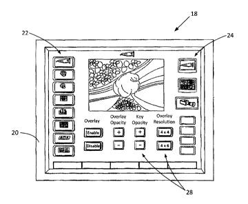
[0018] FIG. 2 is a view of an embodiment of a input for use with the system
for identifying an area of interest on a surgical image as shown in FIG. 1.
CA 02766595 2012-02-02
-6-
[0019] FIG. 3 is a view of an overlay pattern in the form of a grid with a
key, both at 100% opacity, combined with surgical image data, for use with the
system for identifying an area of interest on a surgical image as shown in
FIG. 1.
[0020] FIG. 4 is a view of an overlay pattern in the form of a grid with a
key, both at 50% opacity, combined with surgical image data, for use with the
system
for identifying an area of interest on a surgical image as shown in FIG. 1.
(0021] FIG. 5 is a view of an overlay pattern combined in the form of a grid
at 50% opacity and a key at 100% opacity, combined with surgical image data,
for
use with the system for identifying an area of interest on a surgical image as
shown
in FIG. 1.
[0022] FIG. 6 is a view of an overlay pattern in the form of a grid with a
key, both at 100% opacity, combined with surgical image data that has been
zoomed
in, for use with the system for identifying an area of interest on a surgical
image as
shown in FIG. 1.
[0023] FIG. 7 is a view of an overlay pattern in the form of a centered
crosshairs, combined with surgical image data, for use with the system for
identifying
an area of interest on a surgical image as shown in FIG. 1.
[0024] FIG. 8 is a view of an overlay pattern for use with the system for
identifying an area of interest on a surgical image as shown in FIG. 1.
[0025] FIG. 9 is a view of an overlay pattern for use with the system for
identifying an area of interest on a surgical image as shown in FIG. 1.
[0026] FIG. 10 is a view of an overlay pattern for use with the system for
identifying an area of interest on a surgical image as shown in FIG. 1.
CA 02766595 2012-02-02
-7-
[0027] FIG. 11 is a view of an overlay pattern for use with the system for
identifying an area of interest on a surgical image as shown in FIG. 1.
[0028] FIG. 12 is a view of an overlay pattern for use with the system for
identifying an area of interest on a surgical image as shown in FIG. 1.
[0029] FIG. 13 is a view of an overlay pattern for use with the system for
identifying an area of interest on a surgical image as shown in FIG. 1.
[0030] FIG. 14 is a view of an overlay pattern for use with the system for
identifying an area of interest on a surgical image as shown in FIG. 1.
[0031] FIG. 15 is a view of an overlay pattern for use with the system for
identifying an area of interest on a surgical image as shown in FIG. 1.
[0032] FIG. 16 is a view of an overlay pattern for use with the system for
identifying an area of interest on a surgical image as shown in FIG. 1.
[0033] FIG. 17 is a view of an overlay pattern for use with the system for
identifying an area of interest on a surgical image as shown in FIG. 1.
[0034] FIG. 18 is a view of an overlay pattern for use with the system for
identifying an area of interest on a surgical image as shown in FIG. 1.
[0035] FIG. 19 is a view of an overlay pattern for use with the system for
identifying an area of interest on a surgical image as shown in FIG. 1.
[0036] FIG. 20 is a view of an overlay pattern for use with the system for
identifying an area of interest on a surgical image as shown in FIG. 1.
[0037] FIG. 21 is a view of an overlay pattern for use with the system for
identifying an area of interest on a surgical image as shown in FIG. 1.
CA 02766595 2012-02-02
-8-
DETAILED DESCRIPTION OF THE INVENTION
[0038] The present invention provides a system 10 for identifying certain
areas of interest in surgical image data by applying an overlay pattern, such
as a
Cartesian grid, crosshairs, quadrants, etc., on the surgical image. The
overlay
pattern allows a surgeon to then refer or call attention to areas of interest
in the
surgical image data by referencing the overlay pattern or a portion thereof.
As will
be discussed in detail below, the overlay may also include an key, which may
include
alphanumeric labels or coordinates, which may assist the surgeon in
identifying the
area or portion of the overlay to which he/she is referring.
[0039] Referring to FIG. 1, the system 10 includes at least one source 12
of surgical image data in communication with at least one processing unit 14
and at
least one destination 16 for the surgical image data. The at least one source
12 of
surgical image data connected to the processing unit 14 may include any
device,
system, or network that generates, acquires, stores, monitors, or controls
surgical
image data for use in generating medical images, such as still frame images or
video. For example, the at least one source 12 may include an image
acquisition
device, such as endoscopic cameras, video endoscopes, room cameras, light
cameras, and boom cameras. Likewise, the at least one source 12 may include
any
recording, storage, and/or archival device or system, such as traditional
video
cassette recorders or digital video recording devices (such as a linear tape
deck or
DVD recording device), image capture devices, a PACS (Picture Archiving and
Communication System) computer, or a Hospital Information System. Finally, the
at
least one source 12 may include any other device from which surgical image
data
may be received, such as a patient monitor or a central computer for
controlling
various devices, or may simply be auxiliary inputs for connecting external
devices
that may supply surgical image data to the system.
[0040] Additionally, a source 12 may be a source of surgical image data
that receives surgical image data from yet another source. For example, a
source
CA 02766595 2012-02-02
-9-
may be a linear tape deck that is recording live video as it supplies the
video to the
computer. The linear tape deck, in turn, may receive the live video from an
endoscopic camera presently being used on a patient, as is further described
below.
As another example, a source 12 may be a processor for routing images from
multiple other sources to the processing unit (i.e., a screen splitter), such
as a quad
image processor. The source 12 connected to the processing unit may also be a
camera control unit (CCU).
[0041] The at least one processing unit 14 may include any device,
system, or network that processes images generated from surgical image data.
For
example, the processing unit 14 may be a general processor, a computer, or a
CCU,
which may be integrated in a camera or may be a modular CCU external to the
camera.
[0042] The at least one destination 16 for the surgical image data supplied
by the at least one source 12 may include any device, system, or network that
displays surgical images generated from the image data, or otherwise
communicates
the image data to viewers, or stores the image data. For example, the at least
one
destination may include any of various displays, such as, for example, a flat
panel
display, a plasma screen, or a computer monitor. Additionally, the at least
one
destination may include a recording device or a storage medium.
[0043] Further, the at least one destination 16 for the surgical image data
may be located within the operating room, or it may be at a location remote
from the
operating room. One object of the invention is to assist all those viewing or
analyzing surgical image data to identify areas of interest in the surgical
image data.
For example, an overlay pattern applied to surgical image data may be used by
a
surgeon performing the surgery to communicate with an assisting surgeon that
is not
present in the operating room, but who is able to view the surgical image data
with
the overlay pattern on a destination 16, such as a monitor, at some other
remote
CA 02766595 2012-02-02
-10-
location. Further, the overlay pattern may be applied to surgical image data
displayed on a monitor located in a lecture hall or classroom for teaching
purposes.
[0044] Moreover, the destination 16 may be capable of displaying surgical
image data from more than one source. For example, the destination 16 may be a
monitor with picture-in-picture (PIP) capabilities. In this embodiment, the
user may
choose to apply (or presets may set) an overlay pattern to all or some sets of
surgical image data displayed on the monitor. Similarly, if there are several
destinations 16 for surgical image data from several sources 12, then user may
choose to apply (or presets may set) an overlay pattern to all or some sets of
surgical image data sent to the destinations 16.
[0045] As illustrated in FIG 1, the system 10 may also include at least one
input 18 in communication with the at least on source 12 and/or the processing
unit
14. The at least one input 18 may include any interface whereby a user to
enable/disable and/or adjust the properties of the overlay pattern. In one
embodiment, the input 18 may be a button or menu located on the source 12 of
surgical image data, such as an endoscopic camera, itself. Alternatively, the
input
18 may be a user interface that may include physical buttons for a surgeon to
press,
or may also include a touch-screen monitor.
[0046] In another embodiment, shown in FIG. 2, the input 18 may include
one or more icons on a touchscreen 20. In this embodiment, the system 10 may
include software executing on the processing unit 14 that causes the
touchscreen 20
to simultaneously display several icons. The icons are sensitive to the touch
of the
user and will cause a command to be sent to the processing unit 14. By
pressing
certain source icons 22 or destination icons 24, the user can select a
particular
source and destination by pressing the touchscreen 20 at the locations of the
icon.
The user can also manipulate or alter the surgical images being displayed in
the
display window 26 on the touch screen in order to affect the surgical images
ultimately being communicated to the destinations. For example, the
touchscreen 20
CA 02766595 2012-02-02
-11-
may also include at least one icon 28 which allows the user to enable/disable
the
overlay pattern, adjust the properties of the overlay pattern, and select
which surgical
image data to which the overlay pattern will be applied and to which
destination 16
the combined surgical image will be transmitted.
[0047] In some embodiments, the system 10 may also be configured to
accept voice commands, allowing the user to vocally enable or disable the
overlay
pattern and adjust properties of the overlay pattern itself without having to
touch the
imaging device or user interface. In this embodiment, the at least one input
18 may
include voice recognition software executing on said processing unit 14 for
accepting
voice commands, allowing the surgeon to vocally enable or disable the overlay
and
adjust properties of the overlay itself without having to physically touch the
source
12, processing unit 14 or input 18 themselves.
[0048] In some further embodiments, the input 18 may include
accelerometer data from the camera head or image motion vector detection. The
overlay pattern may be automatically enabled or disabled or the properties of
the
overlay pattern may be adjusted in response to the input of this data.
[0049] The input 18 may also include preset data saved by the user that
will act on the processing unit 14 to enable/disable the overlay pattern at
certain
times as preset by the user. The preset data may also include the preferred
type of
overlay pattern and/or the properties of the overlay pattern the user desires
to be
sent to the destination 16.
[0050] As shown in FIGS. 3 and 7-21, the overlay pattern 30 may be
provided in any number of designs, which may be set by the user. For example,
as
shown in FIGS. 3-6, the overlay pattern 30 may be a grid. In addition, as
shown in
FIG. 7, the overlay pattern 30 may include a single crosshairs placed at the
center of
the surgical image as displayed. In other embodiments, the overlay pattern may
be
one or more hash marks or crosshairs overlaid across a portion of the surgical
CA 02766595 2012-02-02
-12-
image, the entire surgical image, or at the edges of the image. The overlay
may also
be separated into quadrants, with any number of circles, ovals hash marks or
any
combination thereof within the quadrants. The overlay may also be one or more
circles, ovals or other shapes.
[0051] The desired overlay pattern 30 may be chosen by the user through
an input 18, some examples of which are described above. For example, the
source
12, such as an endoscopic camera, may include buttons for selecting and
setting a
desired overlay pattern 30. The user may also chose to apply the overlay
pattern 30
to one or all of the sources 12 of surgical image data 34. Once the overlay
pattern
30 is selected, the surgical image data 34 from the one or more selected
sources 12
is combined with the overlay pattern 30 in the processing unit 14 and the
combined
surgical image is transmitted to the one or more selected destinations 18.
[0052] In one embodiment, the overlay pattern is applied to live surgical
images, in real time. For example, as shown in FIG. 3, the combined surgical
image
data 34 and overlay pattern 30 may be transmitted to a display 32. The display
may
be located in the operating room and/or it may be located somewhere remote
from
the operating room for viewing by other surgeons assisting in the surgery or
by
students observing the surgery for educational purposes.
[0053] Further, the overlay pattern 30 may be applied to surgical image
data 34 that has been recalled from an image archive, such as on a storage
medium.
The applied overlay pattern 30 may also be maintained on captured surgical
image
data 34 that is subsequently saved to an archive and may be recalled later for
viewing on a display.
[0054] The overlay pattern 30 may be applied to the surgical image data at
a "fixed" position, meaning that the overlay 30 will be applied at a fixed
position with
respect to the displayed image, i.e., centered on the surgical image. However,
the
user may adjust the surgical image data 34 separately with respect to the
overlay
CA 02766595 2012-02-02
-13-
pattern 30. In operation, the user views the surgical image data with the
overlay
pattern 30 on a display 32 and adjusts the image captured by the source 12
(i.e., a
camera) until the particular area of interest is located at or near an
identifiable region
of the overlay pattern 30. Using the embodiment shown in FIG. 7, the surgeon
will
adjust the field of view of the camera until the area of interest is centered
at the
crosshairs. This allows the surgeon to unequivocally "point" to the area of
interest,
simply by adjusting the camera.
[0055] The overlay pattern may also include a key 36 for assisting the user
in identifying and discussing areas or portions of the overlay pattern 30,
and, in turn,
identifying and discussing an area of interest on the underlying surgical
image data
34. In one example, the key 36 may include alphanumeric labels or coordinates.
For example, as shown in FIG. 3 the rows and columns of a grid overlay may be
labeled with letters and numbers-the vertical axis labeled with letters and
the
horizontal axis labeled with numbers (or vice versa) allowing reference to an
area of
the surgical image with a simple letter-number combination (e.g. "C3" or "D2",
etc.).
In another embodiment, if the overlay pattern 30 comprises hash-marks as shown
in
FIG. 11, the hash marks may be labeled with coordinates. As shown in FIGS. 16
and 18, the quadrants or other defining shapes may be individually labeled
with an
alphanumeric key 36.
[0056] Certain properties of the overlay 30 and key 36 may be adjustable,
including, but not limited to, the resolution (i.e., number of rows by number
of
columns, number of circles, etc.) of the overlay, the opacity of the overlay
pattern 30
and/or key 36, the distribution of the opacity of the overlay pattern 30
and/or key 36,
the color of the overlay 30 and/or key 36, the brightness of the overlay 30
and/or key
36, the thickness of the lines of the overlay pattern 30, the size of the font
of the key
36, etc. The user may choose to enable the overlay pattern and set its
properties
prior to the start of the surgical procedure, or the overlay may be
enabled/disabled
and the properties may be adjusted at any time during the surgical procedure.
CA 02766595 2012-02-02
-14-
[0057] In one example, the overlay pattern 30 and key 36 can be applied
to the surgical image in varying levels of opacity. The overlay may also
include an
indicator 38, which may display certain properties of the overlay 30 as set by
the
user. For example, FIG. 3 illustrates the overlay 30 as a grid and key 36
applied at
100% opacity. FIG. 4 illustrates the overlay 30 and the key 36 both applied at
50%
opacity. The properties of the overlay 30 can be constant or can vary across
the
display 32. For example, the overlay pattern 30 can be more opaque at the
edges of
the display 32 and gradually become more transparent toward the center of the
display 32.
[0058] In a further embodiment, the adjustable properties of the overlay
pattern and coordinates may be adjusted independently of one another. For
example, as shown in FIG. 5, the overlay may be set to 50% opacity whereas the
key 36 may be maintained at 100% opacity.
[0059] Various properties of the camera control unit (CCU) may also be
changed so as to effect a change in the surgical image data 34 at and/or
around
certain coordinates or a region of the overlay 30 identified by the user. For
example,
the brightness, contrast, color, or zoom of the surgical image data 34 may be
adjusted at and/or around the coordinates or region identified. The
coordinates or
region of the overlay 30 may be identified via an input 18, for example by
button
press on the source 12 or by touching an icon or the display window 26 of a
touchscreen 20. The system 10 may also be configured to include voice
recognition
of certain regions or coordinates of the overlay pattern 30 to change the
properties of
the CCU.
[0060] Moreover, the zoom level of the surgical image data 34 itself may
be adjusted independent of the overlay pattern 30. The resolution of the
overlay
pattern 30 will remain the same, while the surgical image data 34 is zoomed in
or
out. For example, FIG. 6 illustrates a zoomed-in version of the surgical image
data
CA 02766595 2012-02-02
-15-
34 of FIG. 3, where the resolution of the grid overlay pattern remains
constant (4
rows by 6 columns).
[0061] The user may set or adjust the properties of the overlay pattern 30
and/or the key 36 at the beginning of, or during, a surgical procedure. For
example,
the user may select a grid overlay, choose the number of columns and rows, and
set
the color all at prior to commencing a surgical procedure. The user may also
establish presets to which the overlay 30 will default. In one embodiment
shown in
FIGS. 1-7, the resolution of the grid overlay is four rows by six columns.
However,
other grid overlay resolutions are contemplated, such as 4x4. The overlay can
be of
a varying number or a fixed number of columns, rows, quadrants, etc.
[0062] Additionally, the overlay pattern and/or resolution may be preset or
chosen by the surgeon in accordance with the aspect ratio of the display
monitors.
For example, the surgeon may chose a standard definition (SD) display having a
4x3
aspect ratio and the overlay pattern would be chosen or adjusted accordingly.
The
surgeon may also chose a high definition (HD) display having a 16x9 aspect
ratio
and the overlay pattern would be chose or adjusted accordingly. Overlay
patterns
incorporating ovals, such as the pattern shown in FIG. 15, are well suited for
HD
displays whereas overlay patterns incorporating circles, such as the pattern
shown in
FIG. 14, are well suited for SD displays.
[0063] In a further embodiment, the system 10 may automatically enable
the overlay if a motion vector detector (as an input 18) senses a still image
for a
certain period of time. Conversely, the system 10 may automatically disable
the
overlay if a motion vector detector senses a moving image for a certain period
of
time. Further, the system 10 may automatically "time out" after the overlay
has been
enabled for a preset period of time, or "time-out" if the surgical image has
been still
for a certain period of time.
CA 02766595 2012-02-02
-16-
[0064] When the overlay is enabled or disabled, either by automatic
sensing, "time-out" or by direct input from the user, the overlay could be
programmed
to either immediately appear at 100% opacity or immediately disappear.
Alternatively, the overlay could be programmed to gradually appear or
disappear by
gradual increase or decrease in opacity. These properties will be discussed
further
below.
[0065] The scope of the claims should not be limited by the preferred
embodiments set forth in the foregoing description, but should be given the
broadest
interpretation consistent with the description as a whole.