Note : Les descriptions sont présentées dans la langue officielle dans laquelle elles ont été soumises.
CA 02768282 2012-01-16
-1-
Apparatus for ophthalmic laser surgery
The invention relates to an apparatus for ophthalmic laser surgery.
Pulsed laser radiation is used in numerous techniques in the treatment of the
human eye. In some of these techniques, the eye to be treated is pressed
against a transparent contact element, which, with its contact surface that
faces
towards the eye, forms a reference surface for the positioning of the beam
focus
along the z direction (this, according to a usual notation, means the
direction of
io propagation of the laser beam). In particular, treatment techniques used to
produce cuts (incisions) in the eye tissue by means of focussed femtosecond
laser radiation frequently employ such contact elements as a z reference for
the
laser focus. Owing to the contact element being pressed against the eye in
such
a way that the eye comes into close-fitting, flat bearing contact with the
contact
surface of the contact element that faces towards the eye, the contact element
defines the z position of the front surface of the eye. Through referencing of
the
beam focus along the z direction in relation to this contact surface of the
contact
element, it is then ensured that the incision, or the individual
photodisruption
(the creation of an incision in the human eye by means of pulsed femtosecond
laser radiation is normally based on the effect of so-called laser-induced
optical
breakdown, which results in a photodisruption) is located at the required
position
in the depth of the eye tissue.
Incisions made by a laser occur, for example, in the case of so-called fs-
LASIK,
in which an anterior cover disc of the cornea, referred to as a flap in the
art, is
cut free by means of femtosecond laser radiation. As in the case of the
classic
LASIK technique (LASIK: laser in-situ keratomileusis), this flap, still
hanging to
the rest of the corneal tissue in a hinge region, can be folded aside in order
to
treat ablatively the underlying tissue by means of UV laser radiation. Another
3o application for the making of intratissue incisions in the eye is that of
so-called
corneal lenticule extraction, in which, within the corneal tissue, a lens-
shaped
disk is cut out all round by means of femtosecond laser radiation. This disk
is
then removed through an additional incision extending to the eye surface (the
additional incision is made either by means of a scalpel or likewise by means
of
femtosecond laser radiation). In the case of corneal transplants
(keratoplasty),
likewise, an incision can be made in the cornea by means of focussed, pulsed
laser radiation.
CA 02768282 2012-01-16
-2-
For hygiene reasons, the contact element carrying the contact surface is often
a
disposable article, which has to be exchanged before each treatment. In the
production of the contact elements, certain manufacturing tolerances cannot be
precluded in general, even in the case of very high precision manufacturing.
After an exchange of the contact element, therefore, the z position of the con-
tact surface facing towards the eye can differ - even if only slightly - from
that in
the case of the previously used contact element. In the case of laser
treatments
by means of focussed femtosecond laser radiation, focus diameters are prefera-
bly as small as possible, in order to restrict the photodisruption as local as
possi-
io ble. Modern devices operate, for example, with focus diameters in the low
one-
digit pm range. A corresponding precision is desirable for incision guidance
in
the z direction. This requires a correspondingly precise manufacturing of the
contact element, but this precision cannot always be ensured. In the case of
reduced manufacturing precision of the contact element, this may result in an
imprecise incision guidance along the z direction in the corneal tissue.
The object of the invention is to provide an apparatus for ophthalmic laser
sur-
gery that makes high-precision laser treatment of an eye possible.
To achieve this object, according to the invention, an apparatus for
ophthalmic
laser surgery is proposed, comprising a contact surface for formative bearing
contact of an eye to be treated, components for providing focussed, pulsed
treatment laser radiation and for directing the same through the contact
surface
onto the eye, a measuring device for measuring the position of the contact sur-
face along the direction of propagation of the treatment laser radiation, the
measuring device providing position data representing the measured position of
the contact surface at at least one location of the contact surface, and an
elec-
tronic process and control unit, which is connected to the measuring device
and
which is adapted to control the focus position of the treatment laser
radiation in
3o dependence on the position data.
The invention makes it possible to determine and/or verify the position of the
contact surface along the z direction (according to the direction of
propagation
of the treatment laser radiation) and to correct appropriate control
parameters of
the laser apparatus in dependence on the measured position of the contact
surface. The z position of the contact surface is measured, for example, with
reference to a given reference point in a fixed coordinate system of the laser-
CA 02768282 2012-01-16
-3-
surgery apparatus. A differing z position of the contact surface in the
coordinate
system can be obtained for differing contact elements, depending on manufac-
turing precision. The process and control unit takes account of these
variations
in its control of the focus of the treatment laser radiation, such that an
incision
pattern or pattern of photodisruptions to be realized in the eye is actually
located
at the required location in the depth of the eye (i.e. at the required
location in
the z direction). In this way, highly precise incision depths are possible,
for ex-
ample, in the production of a LASIK flap, in the case of corneal lenticule
extrac-
tions or in the case of keratoplasty procedures.
According to a development of the invention, the measuring device can be
adapted to measure the position of the contact surface at a plurality of
differing
locations of the same. Through sampling of the contact surface at a plurality
of
locations of the same, it is possible, in addition to the determination of the
z
position of the contact surface, to acquire its angular orientation in space
(angu-
larity relative to the beam axis). This is because it cannot be precluded that
the
manufacturing tolerances mentioned also affect the relative angular
orientation
of the contact surface facing towards the eye relative to a predefined
mounting
surface of the contact element. Moreover, the manufacturing tolerances do not
have to be equal all over in an x-y plane orthogonal to the z direction, for
which
reason multi-point sampling of the contact surface makes individual correction
of
the z position of the focus position possible for differing locations within
the x-y
plane.
The measuring device is preferably an optical coherence interferometric measur-
ing device and for this purpose comprises an optical interferometer.
The contact surface is frequently part of an exchangeably arranged disposable
component. It must be emphasized, of course, that the invention does not re-
3o quire any disposable nature of the element carrying the contact surface.
The
invention is equally applicable in the case of designs having a fixedly built-
in, or
at least multiple-use, contact surface.
The contact surface is preferably formed by a transparent applanation plate or
a
transparent contact glass. Applanation plates, at least on their plate side
that
faces towards the eye, have a planar applanation surface, by means of which
levelling of the front side of the eye is achieved. The use of applanation
plates
CA 02768282 2012-01-16
-4-
for the purpose of referencing the eye to be treated may be advantageous in
terms of a high beam quality of the laser radiation. Nevertheless, it is
equally
possible, within the scope of the invention, to use as a contact element a
contact
glass having a lens surface, facing towards the eye, that is typically concave
or
convex in form. The advantage of such contact glasses is, for example, a
lesser
increase of the pressure inside the eye upon pressing on the eye.
The contact surface is preferably formed by a transparent contact element that
is part of a patient adapter, in particular exchangeably coupled to a
focussing
objective of the apparatus.
According to the invention there is further provided a method for laser
treatment
of an eye, comprising the steps:
- producing a formative bearing contact between the eye and a contact sur-
face,
- providing focussed, pulsed treatment laser radiation and directing the
same through the contact surface onto the eye,
- generating position data representing a measured position of the contact
surface at at least one location of the contact surface along the direction
of propagation of the treatment laser radiation, and
- controlling the focus position of the treatment laser radiation in depend-
ence on the generated position data.
In the case of the method, likewise, the position data can be representative
of a
measured position of the contact surface at a plurality of differing locations
of
the same.
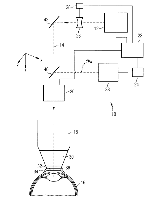
In the following, the invention is explained in further detail with reference
to the
single appended drawing. Fig. 1 shows, in a highly schematic form, an embodi-
ment of an apparatus for ophthalmic laser surgery. The laser-surgery apparatus
is denoted generally by 10. It comprises an fs laser 12, which emits pulsed
laser
radiation having pulse durations in the range of femtoseconds. The laser radia-
tion propagates along an optical beam path 14, and finally reaches an eye 16
to
be treated. Various components for guiding and shaping the laser radiation are
arranged in the beam path 14. In particular, these components include a focus-
sing objective 18 (for example, an F-Theta objective) and a scanner 20, which
is
CA 02768282 2012-01-16
-5-
connected upstream from the objective 18 and by means of which the laser
radiation provided by the laser 12 can be deflected in a plane (x-y plane) or-
thogonal to the beam path 14. A coordinate system drawn in the figure
indicates
this plane, and also a z axis defined by the direction of the beam path 14.
The
scanner 20 is constructed, for example, in a manner known per se, from a pair
of galvanometrically controlled deflection mirrors, which are each responsible
for
deflecting the beam in the direction of one of the axes spanning the x-y
plane. A
central process and control unit 22 controls the scanner 20 in accordance with
a
control program that is stored in a memory 24 and that implements an incision
io profile to be generated in the eye 16 (the incision profile represented by
a three-
dimensional pattern of sampling points, at each of which a photodisruption is
to
be effected).
Furthermore, the mentioned components for guiding and shaping the laser ra-
diation include at least one controllable optical element 26 for z adjustment
of
the beam focus of the laser radiation. In the example shown, this optical ele-
ment is formed by a lens. An appropriate actuator 28, which is controlled by
the
process and control unit 22, serves to control the lens 26. For example, the
lens
26 can be mechanically movable along the optical beam path 14. Alternatively,
it
is conceivable to use a controllable liquid lens of variable refractive power.
In the
case of an unchanged z position and also otherwise unchanged setting of the
focussing objective 18, a z displacement of the beam focus can be achieved by
moving of a longitudinally displaceable lens or by refractive index variation
of a
liquid lens. It is understood that other components, for instance a deformable
mirror, are also conceivable for the purpose of z displacement of the beam fo-
cus. Owing to its comparatively higher inertia, it is expedient to set beam
focus
by the focussing objective 18 coarsely (i.e. focussing on a predefined z
reference
position) and to effect the z displacements of the beam focus that are prede-
fined by the incision profile by a component arranged outside the focussing
objective 18 and having a shorter reaction speed.
On the beam exit side, the focussing objective 18 is coupled to a patient
adapter
30, which serves to produce a mechanical coupling between the eye 16 and the
focussing objective 18. Usually, in the case of treatments of the type
considered
here, a suction ring, which is not represented in greater detail in the
drawing but
which is known per se, is placed onto the eye and fixed there by suction
force.
The suction ring and the patient adapter 30 form a defined mechanical
interface
CA 02768282 2012-01-16
-6-
that couples the patient adapter 30 to the suction ring. In this respect,
reference
can be made, for example, to the international patent application
PCT/EP2008/006962, the entirety of which is hereby included by reference.
The patient adapter 30 serves as a carrier for a transparent contact element
32,
which, in the example shown, is realized as a plane-parallel applanation
plate.
The patient adapter 30 comprises, for example, a taper sleeve body, the ap-
planation plate 32 being arranged at its narrower (in the drawing, lower)
sleeve
end. In the region of the wider (in the drawing, upper) sleeve end, on the
other
io hand, the patient adapter 30 is mounted on the focussing objective 18,
where it
has appropriate formations that, if required, enable the patient adapter 30 to
be
detachably fixed to the focussing objective 18.
Since it is in contact with the eye 16 during the treatment, the applanation
plate
32 is an article that is critical from the aspect of hygiene, and which
therefore,
expediently, is to be exchanged after each treatment. For this purpose, the
applanation plate 32 can be exchangeably mounted on the patient adapter 30.
Alternatively, the patient adapter 30, together with the applanation plate 32,
can
form a disposable unit, for which purpose the applanation plate 32 can be non-
2o detachably connected to the patient adapter 30.
In any case, the underside of the applanation plate 32 that faces towards the
eye forms a planar contact surface 34, against which the eye 16 is pressed for
the purpose of preparation of the treatment. This effects a levelling of the
front
surface of the eye while, at the same time, deforming the cornea of the eye
16,
which is denoted by 36.
To enable the contact surface 34 to be used as a reference for the z control
of
the beam focus, it is necessary to know its z position in the coordinate
system of
the laser-surgery apparatus. Owing to unavoidable manufacturing tolerances, it
cannot be precluded that, in the case of fitting of differing applanation
plates or
differing patient adapters 30 that are each equipped with an applanation plate
32, the z position and possibly also the angular orientation of the contact
surface
34 exhibits variations of greater or lesser significance. Insofar as these
variations
are not taken into account in the z control of the beam focus, unwanted errors
are obtained in the actual position of the incisions produced in the eye 16.
CA 02768282 2012-01-16
-]-
Consequently, the laser-surgery apparatus 10 includes an optical coherence
interferometric measuring device 38, for example an OLCR measuring device
(OLCR: optical low coherence reflectometry) that emits a measuring beam
which, by means of an immovably arranged, semi-transparent deflection mirror
s 40, is coupled into the beam path 14 of the treatment laser radiation of the
laser
12. The measuring device 38 brings the generated measuring beam into inter-
ference with a reflection beam coming back from the eye 16. The z position of
the contact surface 34 can be determined with reference to the coordinate sys-
tem of the laser-surgery apparatus from the interference measurement data
lo obtained in this respect. For this reason, the interference measurement
data can
also be termed positional measurement data. The process and control unit 22
obtains the interference measurement data from the measuring device 38 and,
from this data, calculates the z position of that location of the contact
surface 34
at which the measuring beam impinged or through which the measuring beam
15 passed. In the following laser treatment of the eye 16, the process and
control
unit 22 takes account of the thus determined actual z position of the contact
surface 34 in the z control of the beam focus, this being in such a way that
the
incision is actually made at the intended position in the depth of the cornea
36.
For this purpose, the z position of the beam focus that is to be set is
referenced
20 to the measured z position of the contact surface 34 by the process and
control
unit 22.
In the example shown, the measuring beam emitted by the measuring device 38
passes through the scanner 20. This enables the deflection function of the
scan-
25 ner 20 to be used also for the measuring beam. The scanner module 20 could
also include a second, separate scanner, solely for the OLCR, which, being
equipped with smaller mirrors, operates significantly more rapidly. However,
the
actual scanner mirror of the measuring device 38 can also be arranged sepa-
rately in the first beam path 14a of the OLCR (not indicated in Fig. 1). Thus,
a
30 sampling of the contact surface 34 by the measuring beam and, consequently,
a
z measuring of the contact surface 34 at differing locations is possible. In
this
way, it is possible to generate a table or other suitable data structure that,
for
differing positions in the x-y plane, gives the z position of the contact
surface 34
measured there in each case or gives a z correction value, which is calculated
in
35 dependence on the locally measured z position of the contact surface 34 and
which is taken into account by the process and control unit 22 in the z
control of
the beam focus. If, for example, the incision profile is defined by a table
that, for
CA 02768282 2012-01-16
-8-
each photodisruption to be made, gives its z position with reference to a
known,
predetermined point in the coordinate system of the laser surgery apparatus,
the
table for the incision profile can be appropriately corrected by the process
and
control unit 22 on the basis of such z correction values.
In one embodiment, the scanner can include a pair of mirrors or a deflection
unit
operating according to another deflection technique, which is used jointly for
the
x-y deflection of the laser radiation and of the measuring beam. In another
embodiment, the scanner 20 can include separate pairs of mirrors or,
generally,
io separate deflection units, of which the one is used for the x-y deflection
of the
laser radiation and the other is used for the x-y deflection of the measuring
beam. The deflection unit for the measuring beam could be equipped, for exam-
ple, with smaller, more rapidly movable mirrors than the deflection unit for
the
laser radiation. In yet another embodiment, a deflection unit for the
measuring
beam can be arranged in that portion of the beam path of the measuring beam
that is located in front of the deflection mirror 40. This portion is denoted
by 14a
in Fig. 1.
It is understood that, in yet an alternative embodiment, the scanner 20 can be
located in front of the deflection mirror 40 in the direction of propagation
of the
laser radiation and, accordingly, a z measurement of the contact surface 34 at
only a single location is possible. In this case, the process and control unit
22
can calculate a global z correction quantity which, in the z control of the
beam
focus, is applied equally for all sites in the x-y plane.
The reference 42 denotes a further immovable deflection mirror that serves to
guide the treatment laser radiation.