Note : Les descriptions sont présentées dans la langue officielle dans laquelle elles ont été soumises.
CA 02769308 2012-01-26
WO 2011/014750 PCT/US2010/043872
INHIBITION OF TUMOR METASTASIS USING BV8- OR G-CSF-ANTAGONISTS
RELATED APPLICATIONS
This application is a non-provisional application filed under 37 CFR
1.53(b)(l),
claiming priority under 35 USC 119(e) to provisional application number
61/230,571
filed July 31, 2009, and provisional application number 61/350,558 filed June
2, 2010, the
contents of which are incorporated herein by reference.
FIELD OF THE INVENTION
The present invention relates generally to compositions and methods that are
useful for
treatment of conditions and diseases associated with angiogenesis and tumor
metastasis. In
particular, the invention concerns the prevention or treatment of tumor
metastasis using G-CSF
antagonists and/or Bv8 antagonists.
BACKGROUND OF THE INVENTION
It is well established that angiogenesis plays an important role in tumor
progression and
metastasis and anti-angiogenesis represents a clinically validated anti-cancer
strategy (Folkman,
Nat Med 1, 27-31 (1995); Ferrara and Kerbel, Nature 438, 967-974 (2005);
Carmeliet, Nat Med
9, 653-660 (2003)).
Over the last several years, the contribution of various bone marrow (BM)-
derived cell
types to tumor angiogenesis has been the object of intense investigation
(Coussens and Werb,
Nature 420, 860-867 (2002); Rafii et at., Nat Rev Cancer 2:826-35 (2002); De
Palma et at.,
Trends Immunol 28:519-524 (2007); Shojaei et at., Trends Cell Biol 18:372-378
2008)).
Among these cell types, CDl 1b+Grl+ cells are frequently increased in the
tumors and in the
peripheral blood (PB) of tumor-bearing animals, and have been shown to promote
tumor
angiogenesis (Yang et at., Cancer Cell 6, 409-421 (2004)), and to suppress
immune functions,
hence, the denomination of myeloid-derived suppressor cells (MDSC) (Bronte et
at., Blood
96:3838-3846 (200)). However, the initiating mechanisms responsible for
peripheral
mobilization, tumor-homing, and acquisition of proangiogenic properties in
CD11b+Grl+ cells
remain to be elucidated.
1
CA 02769308 2012-01-26
WO 2011/014750 PCT/US2010/043872
CD1 lb+Grl+ cells produce several angiogenic factors, including Bv8, a
secreted protein
previously characterized as a mitogen for specific endothelial cell type, as a
growth factor for
hematopoietic progenitors (LeCouter et at., Proc Natl Acad Sci USA 100, 2685-
2690 (2003);
LeCouter et at., Proc Natl Acad Sci USA 101, 16813-16818 (2004)), and as a
neuromodulator
(Cheng et at., Nature 417, 405-410 (2002); Matsumoto et at., Proc Natl Acad
Sci USA
103:4140-4145 (2006)). Analysis of several xenografts, as well as of a
transgenic cancer
model (RIP-Tag), suggested that Bv8 promotes tumor angiogenesis through
increased
peripheral mobilization of myeloid cells and local stimulation of angiogenesis
(Shojaei et at.,
Nature 450:825-831 (2007); Shojaei et at., Proc Natl Acad Sci USA 105:2640-
2645 (2008)). It
has been reported that granulocyte colony stimulating factor (G-CSF) as a
strong inducer of
Bv8 expression, both in vitro and in vivo (Shojaei et at., Proc Natl Acad Sci
USA 106(16):6742-
7 (2009)). Physiologically, G-CSF has an important role in mobilization of
hematopoietic stem
cells, progenitors, and mature cells, particularly neutrophils, into the blood
circulation
(Rapoport et at, Blood Rev 6:43-57 (1992); Lieschke et at., Blood 84:1737-1746
(1994)). G-
CSF is also necessary for differentiation of progenitors to cells of
granulocytic lineage, such as
neutrophils, eosinophils and basophils. Although a few reports have suggested
that G-CSF
administration enhances tumor angiogenesis and growth (Natori et. at., Biochem
Biophys Res
Commun 297:1058-1061 (2002); Okazaki, T. et al., Intlmmunol 18, 1-9 (2006)),
the evidence
implicating this factor in tumorigenesis is far from conclusive.
Metastasis is a complex series of steps in which cancer cells leave the
original tumor
site and migrate to other parts of the body via the bloodstream or the
lymphatic system.
Metastatic tumors are very common in the late stages of cancer. Metastasis is
a major cause of
death from solid tumors. Unfortunately, the treatment options currently
available are rarely
able to cure metastatic cancer.
Thus, there is a need to discover and understand how angiogenesis and tumor
metastatic
progression can be effectively inhibited, prevented and treated. The present
invention
addresses these and other needs, as will be apparent upon review of the
following disclosure.
SUMMARY OF THE INVENTION
The present invention is based, at least in part, on the discovery that G-CSF
and Bv8 are
involved in angiogenesis and tumor metastasis.
In one aspect, the invention provides methods of inhibiting or reducing tumor
metastasis
comprising administering to a subject an effective amount of a G-CSF
antagonist. In certain
2
CA 02769308 2012-01-26
WO 2011/014750 PCT/US2010/043872
embodiments, the method further comprises administering to the subject an
effective amount of
Bv8 antagonist. In certain embodiments, the G-CSF antagonist inhibits or
reduces the spread of
a primary tumor to a pre-metastatic organ of the subject. In certain
embodiments, reducing
tumor metastasis comprises reducing size or number of lung metastases. In
certain
embodiments, inhibiting or reducing tumor metastasis comprises reducing one or
more
expression levels of the following molecules: G-CSF, Bv8, PKR1, MMP-9, S100A8
or
S100A9. In certain embodiments, inhibiting or reducing tumor metastasis
comprises reducing
expression levels of G-CSF and PKR1. In certain embodiments, inhibiting or
reducing tumor
metastasis comprises reducing expression levels of MMP-9, S l 00A8 and S 1
OOA9. In certain
embodiments, inhibiting or reducing tumor metastasis comprises reducing
expression levels of
Bv8, MMP-9, S l 00A8 and S 1 OOA9. In certain embodiments, inhibiting or
reducing tumor
metastasis comprises reducing expression levels of G-CSF, Bv8 and PKR1. In
certain
embodiments, the expression levels are mRNA expression levels. In certain
embodiments, the
expression levels are protein expression levels. In certain embodiments,
expression levels of
one or more of these molecules are reduced in a pre-metastatic organ of the
subject. In certain
embodiments, the pre-metastatic organ is lung. In certain embodiments, the pre-
metastatic
organ is liver.
In certain embodiments, the methods further comprise administering to the
subject an
effective amount of VEGF antagonist. In certain embodiments, the subject has
been previously
treated with VEGF antagonist. In certain embodiments, the VEGF antagonist is
an anti-VEGF
antibody or a fragment thereof. In certain embodiments, the anti-VEGF antibody
is
bevacizumab or a fragment or variant thereof. In certain embodiments, the
methods further
comprise administering to the subject an effective amount of a
chemotherapeutic agent.
In one aspect, the invention provides methods of reducing expression level of
G-CSF,
Bv8, PKR1, MMP-9, S l 00A8 or S l 00A9 in a pre-metastatic organ comprising
administering to
the subject an effective amount of a G-CSF antagonist and/or Bv8 antagonist.
In another
aspect, the invention provides methods of reducing expression level of one or
more of the
following molecules, G-CSF, Bv8, PKR1, MMP-9, S100A8 or SiOOA9, in a pre-
metastatic
organ comprising administering to the subject an effective amount of a G-CSF
antagonist
and/or Bv8 antagonist. In certain embodiments, expression levels of PKR1, MMP-
9, SiOOA8
and/or S100A9 are reduced in the pre-metastatic organ of the subject when G-
CSF antagonist is
administered to the subject. In certain embodiments, expression levels of
PKR1, MMP-9,
S l 00A8 and/or S l 00A9 are reduced in the pre-metastatic organ of the
subject when Bv8
3
CA 02769308 2012-01-26
WO 2011/014750 PCT/US2010/043872
antagonist is administered to the subject. In certain embodiments, expression
levels of Bv8,
MMP-9, S 100A8 and/or S 100A9 are reduced in the pre-metastatic organ of the
subject when G-
CSF antagonist is administered to the subject. In certain embodiments,
expression levels of
Bv8, MMP-9, S 100A8 and/or S 100A9 are reduced in the pre-metastatic organ of
the subject
when Bv8 antagonist is administered to the subject. In certain embodiments,
expression levels
of MMP-9, S 100A8 and S 100A9 are reduced in the pre-metastatic organ of the
subject when G-
CSF antagonist is administered to the subject. In certain embodiments,
expression levels of
MMP-9, S 100A8 and S 100A9 are reduced in the pre-metastatic organ of the
subject when Bv8
antagonist is administered to the subject. In certain embodiments, expression
levels of G-CSF
and PKR1 are reduced in the pre-metastatic organ of the subject when G-CSF
antagonist is
administered to the subject. In certain embodiments, expression levels of G-
CSF, PKR1 and
MMP-9 are reduced in the pre-metastatic organ of the subject when G-CSF
antagonist is
administered to the subject.
In one aspect, the invention provides methods of inhibiting migration of
metastatic
tumor cells to a pre-metastatic organ comprising administering to the subject
an effective
amount of a G-CSF antagonist or Bv8 antagonist. In another aspect, the
invention provides
methods of inhibiting migration of neutrophils to a pre-metastatic organ
comprising
administering to the subject an effective amount of a G-CSF antagonist or Bv8
antagonist. In
certain embodiments, the methods comprise administering to the subject an
effective amount of
the G-CSF antagonist and an effective amount of the Bv8 antagonist.
In certain embodiments, the pre-metastatic organ of the subject is lung,
liver, brain,
bone or lymph node. In certain embodiments, the pre-metastatic organ of the
subject is lung or
liver. In certain embodiments, the pre-metastatic organ of the subject is
lung.
In certain embodiments, the G-CSF antagonist is an anti-G-CSF antibody. In
certain
embodiments, the anti-G-CSF antibody is a monoclonal antibody. In certain
embodiments, the
anti-G-CSF antibody is a humanized antibody. In certain embodiments, the anti-
G-CSF
antibody is a human antibody.
In certain embodiments, the Bv8 antagonist is an anti-Bv8 antibody. In certain
embodiments, the Bv8 antagonist is an anti-PKR1 antibody. In certain
embodiments, the anti-
Bv8 antibody or anti-PKR1 antibody is a monoclonal antibody. In certain
embodiments, the
anti-Bv8 antibody or anti-PKR1 antibody is a humanized antibody. In certain
embodiments,
the anti-Bv8 antibody or anti-PKR1 antibody is a human antibody.
In another aspect, the invention provides methods of predicting whether a
tumor in a
4
CA 02769308 2012-01-26
WO 2011/014750 PCT/US2010/043872
subject will respond effectively to treatment with a G-CSF antagonist and/or
Bv8 antagonist
comprising determining whether a sample from the subject comprises a cell that
expresses Bv8,
PKR1 and/or G-CSF at a level greater than the expression level in a reference
sample, wherein
detection of said cell indicates that the subject has the tumor that will
respond effectively to
treatment with said G-CSF antagonist and/or Bv8 antagonist. In certain
embodiments, the
detection of a cell that expresses Bv8, PKR1 and G-CSF at levels greater than
the expression
levels in a reference sample indicates that the subject has the tumor cells
that will respond
effectively to treatment with said G-CSF antagonist and/or Bv8 antagonist. In
certain
embodiments, the mRNA expression levels of Bv8, PKR1 and/or G-CSF are
measured. In
certain embodiments, the protein expression levels of Bv8, PKR1 and/or G-CSF
are measured.
In certain embodiments, the tumor is metastatic tumor.
In another aspect, the invention provides methods of predicting whether a
tumor in a
subject will respond effectively to treatment with a G-CSF antagonist and/or
Bv8 antagonist
comprising determining whether a sample from the subject comprises a
functional human
counterpart of CD11b+Grl+ cell that expresses Bv8 at a level greater than the
expression level
in a reference sample, wherein detection of said functional human counterpart
of CD 11b+Gr1+
cell indicates that the subject has the tumor that will respond effectively to
treatment with said
G-CSF antagonist and/or Bv8 antagonist. In certain embodiments, the mRNA
expression level
of Bv8 is measured. In certain embodiments, the protein expression level of
Bv8, is measured.
In certain embodiments, the human counterpart cells are human immature myeloid
cells. In
certain embodiments, the human counterpart cells are human myeloid derived
suppressor cells.
In certain embodiments, the human counterpart cells are precursors of human
neutrophils,
monocytes or macrophages. In certain embodiments, the human counterpart cells
are human
neutrophils, monocytes or macrophages. In certain embodiments, the tumor is
metastatic
tumor.
In yet another aspect, the invention provides methods of predicting whether a
tumor in a
subject will respond effectively to treatment with a G-CSF antagonist and/or
Bv8 antagonist
comprising determining whether a sample from the subject has increased number
or frequency
of functional human counterpart of CD11b+Grl+ cells compared to a reference
sample,
wherein the increased number or frequency of functional human counterpart of
CD11b+Grl+
cells indicates that the subject has the tumor that will respond effectively
to treatment with said
G-CSF antagonist and/or Bv8 antagonist. In certain embodiments, the human
counterpart cells
are human immature myeloid cells. In certain embodiments, the human
counterpart cells are
5
CA 02769308 2012-01-26
WO 2011/014750 PCT/US2010/043872
human myeloid derived suppressor cells. In certain embodiments, the human
counterpart cells
are precursors of human neutrophils, monocytes or macrophages. In certain
embodiments, the
human counterpart cells are neutrophils, monocytes or macrophages. In certain
embodiments,
the tumor is metastatic tumor.
In one aspect, the invention provides methods of treating a tumor in a subject
with a G-
CSF antagonist and/or Bv8 antagonist comprising (a) determining whether a
sample from the
subject comprises a cell that expresses Bv8, PKR1 and/or G-CSF at a level
greater than the
expression level in a reference sample, and (b) if the sample comprises a cell
that expresses
Bv8, PKR1 and/or G-CSF at a level greater than the expression level in the
reference sample,
administering to the subject an effective amount of said G-CSF antagonist
and/or Bv8
antagonist. In certain embodiments, the cell is a functional human counterpart
of CD1 lb+Grl+
cell. In certain embodiments, the human counterpart cells are human immature
myeloid cells.
In certain embodiments, the human counterpart cells are human myeloid derived
suppressor
cells. In certain embodiments, the human counterpart cells are precursors of
human
neutrophils, monocytes or macrophages. In certain embodiments, the human
counterpart cell is
neutrophils, monocytes or macrophages. In certain embodiments, the cell is
metastatic tumor
cell. In certain embodiments, the cells are from a pre-metastatic organ of the
subject. In certain
embodiments, if the sample comprises a cell that expresses Bv8, PKR1 and G-CSF
at levels
greater than the expression levels in the reference sample, then an effective
amount of said G-
CSF antagonist and/or Bv8 antagonist is administered to the subject.
In certain embodiments, the mRNA expression levels of Bv8, PKR1 and/or G-CSF
are
measured. In certain embodiments, the protein expression levels of Bv8, PKR1
and/or G-CSF
are measured.
In another aspect, the invention provides methods of treating a tumor in a
subject with a
G-CSF antagonist and/or Bv8 antagonist comprising (a) determining whether a
sample from the
subject has increased number or frequency of functional human counterpart of
CDl1b+Grl+
cells compared to a reference sample, and (b) if the sample has the increased
number or
frequency of functional human counterpart of CD11b+Grl+ cells compared to the
reference
sample, administering to the subject an effective amount of said G-CSF
antagonist and/or Bv8
antagonist. In certain embodiments, the human counterpart cells are human
immature myeloid
cells. In certain embodiments, the human counterpart cells are human myeloid
derived
suppressor cells. In certain embodiments, the human counterpart cells are
precursors of human
neutrophils, monocytes or macrophages. In certain embodiments, the human
counterpart cells
6
CA 02769308 2012-01-26
WO 2011/014750 PCT/US2010/043872
are neutrophils, monocytes or macrophages. In certain embodiments, the tumor
is metastatic
tumor.
In another aspect, the invention provides methods of treating tumor,
comprising (a)
administering to a tumor-bearing subject an effective amount of a G-CSF
antagonist, and (b)
monitoring the efficacy of said treatment by determining the number or
frequency of functional
human counterpart of CD11b+Grl+ cells in a sample obtained from the subject
after the
treatment, relative to the number or frequency of functional human counterpart
of
CD1 lb+Grl+ cells in a sample obtained from the subject before the treatment,
wherein a
reduced number or frequency of functional human counterpart of CD11b+Grl+
cells in the
sample obtained from the subject after the treatment indicates that the
treatment is effective. In
certain embodiments, the human counterpart cells are human immature myeloid
cells. In
certain embodiments, the human counterpart cells are human myeloid derived
suppressor cells.
In certain embodiments, the human counterpart cells are precursors of human
neutrophils,
monocytes or macrophages. In certain embodiments, the human counterpart cells
are
neutrophils, monocytes or macrophages. In certain embodiments, the tumor is
metastatic
tumor.
In another aspect, the invention provides methods of treating tumor,
comprising (a)
administering to a tumor-bearing subject an effective amount of a G-CSF
antagonist and/or Bv8
antagonist, and (b) monitoring the efficacy of said treatment by determining
the expression
level of Bv8, MMP-9, S 100A8 or S 100A9 in a sample obtained from the subject
after the
treatment, relative to the expression level of Bv8, MMP-9, S 100A8 or S 100A9
in a sample
obtained from the subject before the treatment, wherein a reduced expression
level of Bv8,
MMP-9, S 100A8 or S 100A9 in the sample obtained from the subject after the
treatment
indicates that the treatment is effective.
In certain embodiments, expression levels of at least two of the following
four
molecules, Bv8, MMP-9, S 100A8 and S 100A9, are reduced in the sample obtained
from the
subject after the treatment compared to the sample obtained from the subject
before the
treatment. In certain embodiments, expression levels of at least three of the
following four
molecules, Bv8, MMP-9, S 100A8 and S 100A9, are reduced in the sample obtained
from the
subject after the treatment compared to the sample obtained from the subject
before the
treatment. In certain embodiments, the mRNA expression levels of Bv8, MMP-9,
S100A8
and/or Si 00A9 are measured. In certain embodiments, the protein expression
levels of Bv8,
MMP-9, Si 00A8 and/or Si 00A9 are measured. In certain embodiments, the tumor
is
7
CA 02769308 2012-01-26
WO 2011/014750 PCT/US2010/043872
metastatic tumor.
In certain embodiments, the mRNA expression levels of Bv8, MMP-9, S100A8
and/or
S100A9 are increased in pre-metastatic lung and/or metastatic lung. In certain
embodiments,
the protein expression levels of Bv8, MMP-9, S 10OA8 and/or S 10OA9 are
increased in pre-
metastatic lung and/or metastatic lung. In certain embodiments, the mRNA
expression levels of
Bv8, MMP-9, S 10OA8 and/or S 10OA9 are increased in pre-metastatic tissue or
tumor tissue. In
certain embodiments, the protein expression levels of Bv8, MMP-9, S 10OA8
and/or S 10OA9
are increased in pre-metastatic tissue or tumor tissue. In certain
embodiments, the treatment
with anti-Bv8 antibody is efficacious if the mRNA expression level of Bv8
and/or MMP-9 is
decreased after the treatment with anti-Bv8 antibody. In certain embodiments,
the treatment
with anti-Bv8 antibody decreases the mRNA expression level of Bv8 and/or MMP-9
in pre-
metastatic lungs. In certain embodiments, the treatment with anti-Bv8 antibody
is efficacious if
the protein expression level of Bv8 and/or MMP-9 is decreased after the
treatment with anti-
Bv8 antibody. In certain embodiments, the treatment with anti-Bv8 antibody
decreases the
protein expression level of Bv8 and/or MMP-9 in pre-metastatic lungs. In
certain
embodiments, the treatment with anti-G-CSF antibody is efficacious if the mRNA
expression
level of Bv8 and/or MMP-9 is decreased after the treatment with anti-G-CSF
antibody. In
certain embodiments, the treatment with anti-G-CSF antibody decreases the mRNA
expression
level of Bv8 and/or MMP-9 in pre-metastatic lungs and/or metastatic lungs. In
certain
embodiments, the treatment with anti-G-CSF antibody is efficacious if the
protein expression
level of Bv8 and/or MMP-9 is decreased after the treatment with anti-G-CSF
antibody. In
certain embodiments, the treatment with anti-G-CSF antibody decreases the
protein expression
level of Bv8 and/or MMP-9 in pre-metastatic lungs and/or metastatic lungs. In
certain
embodiments, the treatment with anti-PKRi antibody is efficacious if the mRNA
expression
level of Bv8 and/or MMP-9 is decreased after the treatment with anti-PKRi
antibody. In
certain embodiments, the treatment with anti-PKRi antibody decreases the mRNA
expression
level of Bv8 and/or MMP-9 in pre-metastatic lungs and/or metastatic lungs. In
certain
embodiments, the treatment with anti-PKRi antibody is efficacious if the
protein expression
level of Bv8 and/or MMP-9 is decreased after the treatment with anti-PKRi
antibody. In
certain embodiments, the treatment with anti-PKRi antibody decreases the
protein expression
level of Bv8 and/or MMP-9 in pre-metastatic lungs and/or metastatic lungs.
In certain embodiments, the tumor is a metastatic tumor. In certain
embodiments, the
treatment with G-CSF antagonists and/or Bv8 antagonists prevents the
metastatic tumor from
8
CA 02769308 2012-01-26
WO 2011/014750 PCT/US2010/043872
metastasizing to pre-metastatic tissues or pre-metastatic organs elsewhere in
the body. In
certain embodiments, the sample from the subject is a tissue, plasma, serum,
or any
combinations thereof. In certain embodiments, the samples used in these
methods are from a
pre-metastatic or metastatic organ. In certain embodiments, the pre-metastatic
or metastatic
organ is lung, liver, brain, bone, lymph node or ovary. In certain
embodiments, the samples are
from a pre-metastatic or metastatic tissue. In certain embodiments, the pre-
metastatic or
metastatic tissue is from subject's lung, liver, brain, ovary, lymph node,
bone or bone marrow.
In certain embodiments, the sample is a tissue sample from a pre-metastatic
organ. In certain
embodiments, if the sample is from pre-metastatic organ or pre-metastatic
tissue, the sample
does not contain any tumor cells. In certain embodiments, the sample is
primary tumor tissue.
In certain embodiments, the mRNA expression level of a gene is measured using
qRT-
PCR or qPCR. In certain embodiments, the mRNA expression level is measured
using
microarrary. In certain embodiments, the mRNA expression level is measured
using ISH (in
situ hybridization). In certain embodiments, the protein expression level of a
gene is measured
using IHC assay.
In another aspect, the invention provides methods of inhibiting mobilization
of
functional human counterpart of CD11b+Grl+ cells from the bone marrow to a pre-
metastatic
or metastatic organ comprising administering to a subject an effective amount
of a G-CSF
antagonist and/or Bv8 antagonist. In certain embodiments, the human
counterpart cells are
human immature myeloid cells. In certain embodiments, the human counterpart
cells are
human myeloid derived suppressor cells. In certain embodiments, the human
counterpart cells
are precursors of human neutrophils, monocytes or macrophages. In certain
embodiments, the
human counterpart cell is neutrophils, monocytes or macrophages.
In one aspect, the invention provides methods of treating tumor in a subject
relapsed
from or refractory to anti-cancer therapy comprising administering to a
subject an effective
amount of a G-CSF antagonist. In certain embodiments, the anti-cancer therapy
comprises a
VEGF antagonist. In certain embodiments, the VEGF antagonist is an anti-VEGF
antibody or
fragment thereof. In certain embodiments, the anti-VEGF antibody is
bevacizumab or a
fragment or variant thereof.
In one aspect, the invention provides methods of treating a benign, pre-
cancerous, or
non-metastatic cancer in a subject, comprising administering to the subject an
effective amount
of a G-CSF antagonist and/or Bv8 antagonist. In certain embodiments,
administering of the G-
CSF antagonist and/or Bv8 antagonist prevents the benign, pre-cancerous, or
non-metastatic
9
CA 02769308 2012-01-26
WO 2011/014750 PCT/US2010/043872
cancer from becoming an invasive or metastatic cancer. In certain embodiments,
the benign,
pre-cancerous, or non-metastatic cancer is a stage 0, stage I, or stage II
cancer. In certain
embodiments, administering of the G-CSF antagonist and/or Bv8 antagonist
prevents the
benign, pre-cancerous or non-metastatic cancer from progressing to a stage III
or stage IV
cancer. In certain embodiments, administering of the G-CSF antagonist and/or
Bv8 antagonist
reduces tumor size.
In another aspect, the invention provides methods of treating a subject with
operable
cancer comprising administering to the subject an effective amount of a G-CSF
antagonist
and/or Bv8 antagonist and performing surgery whereby the cancer is resected.
In certain
embodiments, the G-CSF antagonist and/or Bv8 antagonist is administered to the
subject prior
to surgery. In certain embodiments, the methods further comprise the step of
administering to
the subject an effective amount of the G-CSF antagonist and/or Bv8 antagonist
after surgery to
prevent recurrence of the cancer. In certain embodiments, administering of the
G-CSF
antagonist and/or Bv8 antagonist prevents proliferation of micrometastases.
In another aspect, the invention provides methods of neoadjuvant therapy in a
subject
with operable cancer comprising administering to the subject a G-CSF
antagonist and/or Bv8
antagonist.
In another aspect, the invention provides methods of preventing recurrence of
cancer in
a subject, comprising administering to the subject a G-CSF antagonist and/or
Bv8 antagonist,
wherein said administering prevents cancer recurrence in the subject. In yet
another aspect, the
invention provides methods of reducing the likelihood of cancer recurrence in
a subject,
comprising administering to the subject a G-CSF antagonist and/or Bv8
antagonist, wherein
said administering reduces the likelihood of cancer recurrence in the subject.
In certain
embodiments, the administering of the G-CSF antagonist and/or Bv8 antagonist
prevents or
reduces the likelihood of reoccurrence of a clinically detectable tumor, or
metastasis thereof. In
certain embodiments, the subject has had definitive surgery prior to the
administering the G-
CSF antagonist and/or Bv8 antagonist.
In another aspect, the invention provides methods of preventing the regrowth
of a tumor
in a subject comprising the steps of removing the tumor and thereafter
administering to the
subject a G-CSF antagonist and/or Bv8 antagonist. In yet another aspect, the
invention
provides methods of preventing the recurrence of cancer in a subject having a
tumor comprising
the steps of removing the tumor and thereafter administering to the subject a
G-CSF antagonist
and/or Bv8 antagonist. In certain embodiments, the methods further comprise a
period of time
CA 02769308 2012-01-26
WO 2011/014750 PCT/US2010/043872
between removal of the tumor and administering the G-CSF antagonist and/or Bv8
antagonist,
wherein the period of time is sufficient for the surgical incision to be fully
healed or to reduce
the risk of wound dehiscence.
In certain embodiments, the G-CSF antagonist administered is an anti-G-CSF
antibody
or a fragment thereof. In certain embodiments, the Bv8 antagonist administered
is an anti-Bv8
antibody or a fragment thereof. In certain embodiments, the Bv8 antagonist
administered is an
anti-PKR1 antibody or a fragment thereof.
In certain embodiments, the antagonists and antibodies of the present
invention are
administered sequentially. In certain embodiments, the antagonists and
antibodies of the
present invention are administered concurrently.
In one aspect, the invention provides methods of inhibiting lung metastasis,
liver
metastasis, brain metastasis, ovary metastasis, lymph node metastasis and/or
bone metastasis
comprising administered to a subject an effective amount of an anti-G-CSF
antibody and/or
anti-Bv8 antibody. In another aspect, the invention provides methods of
inhibiting lung
metastasis comprising administered to a subject an effective amount of an anti-
G-CSF
antibody, wherein the anti-G-CSF antibody inhibits tumor from metastasizing to
a pre-
metastatic lung. In another aspect, the invention provides methods of
inhibiting lung metastasis
comprising administered to a subject an effective amount of an anti-Bv8
antibody, wherein the
anti-Bv8 antibody inhibits tumor from metastasizing to a pre-metastatic lung.
In another
aspect, the invention provides methods of inhibiting lung metastasis
comprising administered to
a subject an effective amount of an anti-PKR1 antibody, wherein the anti-PKR1
antibody
inhibits tumor from metastasizing to a pre-metastatic lung. In another aspect,
the invention
provides methods of inhibiting lung metastasis comprising administered to a
subject an
effective amount of an anti-G-CSF antibody and an effective amount of an anti-
Bv8 antibody.
In another aspect, the invention provides methods of inhibiting lung
metastasis comprising
administered to a subject an effective amount of an anti-G-CSF antibody and an
effective
amount of an anti-PKR1 antibody. In another aspect, the invention provides
methods of
inhibiting lung metastasis comprising administered to a subject an effective
amount of an anti-
Bv8 antibody and an effective amount of an anti-PKR1 antibody.
In another aspect, the invention provides methods of inhibiting liver
metastasis
comprising administered to a subject an effective amount of an anti-G-CSF
antibody, wherein
the anti-G-CSF antibody inhibits tumor from metastasizing to a pre-metastatic
liver. In another
aspect, the invention provides methods of inhibiting liver metastasis
comprising administered to
11
CA 02769308 2012-01-26
WO 2011/014750 PCT/US2010/043872
a subject an effective amount of an anti-Bv8 antibody, wherein the anti-Bv8
antibody inhibits
tumor from metastasizing to a pre-metastatic liver. In another aspect, the
invention provides
methods of inhibiting liver metastasis comprising administered to a subject an
effective amount
of an anti-PKRI antibody, wherein the anti-PKRI antibody inhibits tumor from
metastasizing
to a pre-metastatic liver.
In certain embodiments, the antibodies or antibody fragments can be chimeric,
humanized or human.
In certain embodiments, the methods of the present invention further comprise
administering to the subject an effective amount of a VEGF antagonist. In
certain
embodiments, the subject has a tumor previously treated with a VEGF
antagonist. In certain
embodiments, the subject is relapsed from or refractory to a VEGF antagonist.
In certain
embodiments, the VEGF antagonist is an anti-VEGF antibody or a fragment
thereof. In certain
embodiments, the anti-VEGF antibody is bevacizumab or a fragment or variant
thereof. In
certain embodiments, the methods of the present invention further comprise
administering to
the subject an effective amount of a chemotherapeutic agent. In certain
embodiments, the
subject being treated is subjected to chemotherapy and/or radiation therapy,
where the
chemotherapy may, for example, comprise the administration of a cytotoxic
agent. In certain
embodiments, the additional treatment is a treatment known as "standard of
care" for the
treatment of the particular tumor targeted. In certain embodiments, the G-CSF
antagonist is
administered in combination with a different anti-tumor agent and/or treatment
regiment, such
as chemotherapy and/or radiation therapy. In certain embodiments, the Bv8
antagonist is
administered in combination with a different anti-tumor agent and/or treatment
regiment, such
as chemotherapy and/or radiation therapy. In certain embodiments, the methods
of the present
invention further comprise administration of an additional inhibitor of
angiogenesis, such as,
for example, an antibody to an angiogenic factor.
In certain embodiments, the subject is mammal. In certain embodiments, the
subject is
human. In certain embodiments, the subject is diagnosed with cancer. In
certain embodiments,
the tumor is cancer. In certain embodiments, the metastatic tumor is cancer.
In certain
embodiments, the cancer is colon cancer, lung cancer, breast cancer, renal
cell cancer, ovarian
cancer, prostate cancer, bladder cancer, melanoma or glioblastoma.
Any embodiment described above or any combination thereof applies to any and
all
methods of the inventions described herein.
12
CA 02769308 2012-01-26
WO 2011/014750 PCT/US2010/043872
BRIEF DESCRIPTION OF THE DRAWINGS
Figure 1. Bv8 is strongly up-regulated in pre-metastatic lungs of mice
bearing metastatic tumors. A. Design and results of the microarray study
comparing
gene expression in lungs from Balb/c naive mice, mice bearing non-metastatic
tumors
67NR and metastatic 4T1 tumors. The "metastatic" gene expression profile is
clearly
separated from both naive from "non-metastatic" profiles. Each profile column
represents one individual mouse. B. Schematic representation of the top 24 up-
and
down-regulated genes in the pre-metastatic lungs from mice bearing 4T1 tumors
compared to naive or 67NR-bearing mice. C. Bv8 protein concentrations in the
pre-
metastatic lungs of mice bearing various tumors (n=3 per group). D. FACS
analysis of
Cdl lb+Grl+ cells in the pre-metastatic lungs of Balb/c mice bearing various
tumors.
Graphs present Means SEM.
Figure 2. Increased levels of G-CSF and Bv8 are associated with a metastatic
phenotype. A. Plasma levels of SDF1a, P1GF, VEGF-A, M-CSF, GM-CSF, G-CSF and
Bv8 two weeks after inoculation of non-metastatic or metastatic tumor cells.
Asterisk (*)
indicates significant difference when compared to naive group. B. Analysis of
total
numbers of Cdl lb+Grl+ cells in lungs from mice bearing 4T1 tumors and treated
with
anti-Bv8, anti-G-CSF antibodies, or combination (n=10 per group). 4T1 cells
were
orthotopically inoculated into the 4th mammary fat pad of female CB6F1 mice.
Treatment
with antibodies began 2 days after inoculation of the cells, and was performed
as
described in Materials and Methods section in Example 1. Lungs were perfused
with
PBS and harvested 1 week (pre-metastatic phase) or 5.5 weeks (metastatic
phase) after
inoculation of cells. Asterisk (*) indicates significant difference when
compared to
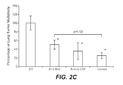
corresponding ISO group. C. Numbers of tumors in lungs of mice bearing 4T1
tumors
and treated as in panel B. Analysis was performed 5.5 weeks after tumor
inoculation.
ISO - isotype control antibody (n=5 per group). Asterisk (*) indicates
significant
difference relative to Naive group. D. Numbers of lung tumors analyzed by
micro-CT in
Balb-c mice bearing 66c14 tumors and treated with control (ISO) or anti-Bv8
antibody
(n=8 for ISO group, and n=10 for anti-Bv8 group). Renderings (2 per group) of
representative micro-CT scanned lungs demonstrating metastatic nodules (red)
in ISO and
anti-Bv8 groups are shown below the graph. Data shown are Means SEM.
13
CA 02769308 2012-01-26
WO 2011/014750 PCT/US2010/043872
Figure 3. G-CSF initiates the pre-metastatic "niche" and enhances the
metastatic potential of several tumors. A. FACS analysis of Cdl lb+Grl+ cells
in
tissues isolated from mice treated with vehicle or human rG-CSF (for 5 days,
g/mouse). Tissues were harvested twenty-four hours after the last dose of rG-
CSF.
5 Mice were perfused with PBS prior to the tissue harvest (n=5 per group).
Asterisk (*)
indicates significant difference when compared to Vehicle group. Data are
represented as
a percentage of Cdl lb+Grl+ (or Cdl lb+Grl-) cells among live cells. B. Bv8
levels
measured by ELISA in tissues matching those in panel A. Values were normalized
to the
total protein content and are plotted on a logarithmic scale. Asterisk (*)
indicates
10 significant difference when compared to Vehicle group (n=5 per group). C.
Numbers of
tumors in lungs of mice pre-treated for 5 days with human rG-CSF (10 g/mouse,
grey
bars) or vehicle (white bars). Following G-CSF treatment, mice (5 per group)
were
injected intravenously (through tail-vein) with tumor cells (10,000 cells per
mouse).
66c14 and 4T1 cells were injected into Balb/c mice, whereas B16F10 cells into
C57BL/6
mice. rG-CSF treatment followed for another 5 days. Lungs were analyzed for
the
presence of visible tumors 3 weeks after cell inoculation. Asterisk (*)
indicates
significant difference between vehicle and G-CSF treated mice (n=5 per group).
D. Representative images of lungs from mice injected with B 16F 10 cells and
pre-treated
with either vehicle or rG-CSF as in panel C. The lung from a mouse treated
with rG-CSF
has the higher number of tumors (black spots). E. Number of lung tumors in
Balb/c mice
intravenously injected with non-metastatic 67NR cells and treated with human
rG-CSF as
in panel C. Mice were analyzed for the presence of visible lung tumors 3 weeks
after cell
inoculation. Asterisk (*) indicates significant difference. Frequency denotes
numbers of
mice with detectable tumors in lungs (n=5 per group). F. Numbers of tumors in
lungs of
mice treated daily with mouse rG-CSF (2.5 g/mouse) and then treated with
control (ISO),
anti-Bv8, anti-Grl or anti-G-CSF antibody and injected intravenously with
66c14 cells as
described in Materials and Methods section in Example 1. Lungs were analyzed
for the
presence of tumors 3 weeks after cells inoculation. Data shown are Means
SEM.
Figure 4. Bv8 mediates G-CSF induced metastasis through enhancement of
cancer cell migration. A. qRT-PCR analysis of PKRI and PKR2 expression by non-
and
metastatic cancer cells in vitro. Data are presented as values normalized to
Hprtl
expression (n=6 per group). Asterisk (*) indicates significant difference when
compared
to 67NR cells. B. Migration of non-metastatic and metastatic cells in response
to
14
CA 02769308 2012-01-26
WO 2011/014750 PCT/US2010/043872
increasing concentrations of human Bv8. 1% FBS served as positive control.
Cells that
had migrated through the collagen-coated 8 m pore well were counted 16 hours
after
assay initiation (n=5 per group). Asterisk (*) indicates significant
difference when
compared to untreated group. C. In vivo extravasation of 4T1 cells assessed 36
hours
after intravenous inoculation (tail-vein) of cells labeled with CellTracker
Green. Balb/c
Nude mice were pre-treated with mouse rG-CSF (2.5 g/mouse, daily) for 5
consecutive
days and then were treated with control (ISO), anti-Bv8, anti-Grl or anti-G-
CSF
antibodies. "No label" denotes a group of mice which was inoculated with cells
that had
not been labeled with CellTracker Green. Images show 4T1 tumor cells
(indicated with
white arrow heads) in representative lung sections. Scale bar = 50 m. D.
Quantification
of the in vivo extravasation assay from panel C. On average, three mice per
group were
used and 15 images (5 random sections per mouse) were included into the
analysis. E.
Kaplan-Meier curves representing the probability of overall survival of breast
cancer
patients from Pawitan Breast dataset stratified based on the expression of G-
CSF. Curves
shows a strong correlation between high levels of G-CSF and reduced survival.
F.
Schematic model of the role of G-CSF and Bv8 in metastasis. LU - lung, T -
primary
tumor, BM - bone marrow. Data are Means SEM.
Figure 5. Pre-metastatic lungs are tumor-free at the time of tissue harvest.
Additional analysis of the pre-metastatic lungs from mice bearing tumors. A.
Schematic representation of 4T 1-related cell lines used in the study, with
known
metastatic sites. B. qRT-PCR expression analysis of Hygromycin gene in lungs
or
primary tumors showing absence of Hygromycin signal in lungs from mice bearing
4T1-
Luc-zsGreen-Hygro tumors. Note positive signal from the primary tumor. Lungs
were
harvested 6 days after tumor cell inoculation. C. BLI imaging of mice bearing
4T1-Luc-
zsG-Hygro tumors. Note the lack of signal in the lung area and positive signal
in the
primary tumor. D. Bv8 qRT-PCR analysis of lung tissues used for microarray
analysis.
E. qRT-PCR analysis of Bv8 expression in the pre-metastatic lungs of mice
bearing non-
and metastatic tumors. F. qRT-PCR analysis of Bv8 transcript levels in Cdl
lb+Grl+ and
Cdl lb+Grl- cells isolated from lung tissue from Naive mice or mice with 4T1
tumors.
G. Example of Bv8 IHC staining in the pre-metastatic lungs from mice bearing
67NR or
4T1 tumors. Dark staining in the pre-metastatic lung tissue bearing 4T1 tumors
indicates
Bv8-positive cells. 67NR or 4T1 cells were orthotopically inoculated in the
4th mammary
fat pad and lungs were harvested 1 week later and Bv8 was detected by IHC as
described
CA 02769308 2012-01-26
WO 2011/014750 PCT/US2010/043872
in Materials and Methods section in Example 1. Scale bar represents 50 m. H.
FACS
analysis of Cdl lb+Grl+ in pre-metastatic lungs from mice bearing B16F10 and
LLC
tumors (Matrigel-injected mice served as control group), as well as in MMTV-
PyMT
mice (PyMT-negative siblings served as control group) (n=5 per group). I. Bv8
expression levels in the pre-metastatic lungs from mice with LLC (Bv8 protein
measured
by ELISA), B16F10 and MMTV-PyMT (Bv8 RNA measured by qRT-PCR) tumors.
Data are shown as expression relative to Naive tissue. Values shown are Means
SEM.
Figure 6. Additional analysis of G-CSF and Bv8 expression in tumor-bearing
mice. Additional analysis of tumor burden. A. G-CSF concentrations in cell
culture
supernatants after 48h-incubation, as described in Materials ands Methods
section in
Example 1 (n=3 per group). Asterisk (*) indicates significant difference when
compared
to Naive group. B. Bv8 levels in lungs and plasma in mice bearing 4T1 tumors
and
treated with ISO control or anti-G-CSF antibody (n=5 per group). Asterisk (*)
indicates
significant difference when compared to Naive group. C. Plasma and tumor G-CSF
levels in mice bearing 4T 1 tumors in the pre-metastatic and metastatic phases
(n=5 per
group). Asterisk (*) indicates significant difference when compared to Naive
group. D.
4T1 primary tumor growth in mice treated with control (ISO), anti-Bv8, anti-G-
CSF or
both antibodies (n=10 per group). 4T1 cells were orthotopically inoculated
into the right
4th mammary fat pad of female CB6F1 mice. Asterisk (*) next to anti-Bv8
treatment
group indicates significant difference when compared to ISO group. E. 66c14
primary
tumor growth in mice treated with control (ISO), anti-Bv8, anti-G-CSF and both
antibodies (n=10 per group). 66c14 cells were orthotopically inoculated into
the right 4th
mammary fat pad of female Balb/c mice. Asterisk (*) next to anti-Bv8 treatment
group
indicates significant difference when compared to ISO group. F. Lung tumor
multiplicity
in mice bearing 66c 14 tumors 6 weeks after tumor inoculation. Tumors were
implanted
and treatment performed as in panel E (n=10 per group). Asterisk (*) indicates
significant difference when compared to ISO group. Graphs show Means SEM.
Figure 7. Analysis of G-CSF induced metastasis in mice intravenously (tail-
vein) injected with cancer cells. A. Increases in lung mass (compared to lungs
from
Naive mice) following inoculation of tumor cells and treatment with human rG-
CSF or
Vehicle. Data correspond to the groups in Fig. 3C. Asterisk (*) indicates
significant
difference (n=5 per group). B. H&E staining of lungs from mice injected with
67NR
cells and treated with Vehicle or G-CSF. Note presence of tumors in the G-CSF-
treated
16
CA 02769308 2012-01-26
WO 2011/014750 PCT/US2010/043872
group. Scale bar in magnified images = 50 m. T - tumor area, L - normal lung
area. C.
FACS analysis of Cdl lb+Grl+ cells in lungs of mice treated daily (for 3 days)
with
mouse rG-CSF (1 g/mouse) or vehicle and treated with control antibody (ISO),
anti-Bv8,
anti-Grl or anti-G-CSF antibody as described in Materials and Methods section
in
Example 1 (n=5 per group). D. Number of lung tumors in mice 3 weeks after
inoculation
of 4T1 cells. Mice were pre-treated (daily) with vehicle or human rG-CSF and
treated
with control (ISO) or anti-Bv8 or anti-Grl antibody. Treatment with both, rG-
CSF and
antibodies, continued for additional 5 days after inoculation of cancer cells.
E. Number
of lung tumors in mice inoculated with 4T1 cells. Lungs were analyzed 3 weeks
later.
Animals were also treated with control (ISO), anti-Bv8, anti-VEGF, anti-Grl,
or anti-G-
CSF antibody. F. FACS analysis of VEGFR1+ cells in lungs in mice treated daily
with
mouse rG-CSF or vehicle for 3 consecutive days. G. FACS analysis of Cdl
lb+Grl+
(and Grl-) cells in lung in mice treated daily with mouse rG-CSF or vehicle as
in panel F.
H. Distribution of VEGFRI+ cells among all Cdl lb+Grl+ (or Grl-) cells in mice
dosed
with vehicle or mouse rG-CSF as in panels F and G. For panels F, G, and H
asterisk (*)
indicates significant difference when compared to Vehicle group, NS - not
significant
(n=5 per group). Values shown are Means SEM.
Figure 8. Mechanisms underlying the G-CSF-initiated pre-metastatic
microenvironment. A. qRT-PCR gene expression analysis of MMP-9, S10OA8 and
S10OA9 in total lung tissue from mice pre-treated with vehicle or mouse rG-CSF
(1 g/mouse, daily, for 3 days), and then treated with control (ISO), anti-Bv8,
anti-Grl or
anti-G-CSF antibodies. Asterisk (*) indicates significant difference when
compared to G-
CSF+ISO group (n=5 per group). B. Bv8, S10OA8, S10OA9 and MMP9 gene expression
measured by qRT-PCR in Cdl lb+Grl+ (or Grl-) cells sorted from lungs from mice
pre-
treated with vehicle or rG-CSF. Single cell suspensions were generated from
the tissues,
cells were stained with specific anti-Cdl lb and anti-Grl antibodies, and
double-positive
cells (Cdl lb+Grl+), Cdl lb+Grl-, and double-negative cells (Cdl lb-Grl-)
cells
populations were separated by FACS. Total RNA was isolated from the cells and
gene
expression was measured by qRT-PCR. Data are presented as relative expression
to
Hprtl (n=5 per group). Asterisk (*) indicates significant difference when
compared to
Vehicle group. C. MMP-9 concentrations measured by ELISA in the pre-metastatic
or
metastatic lungs from mice bearing orthotopically inoculated 4T1 tumors and
treated with
control (ISO), anti-Bv8, anti-G-CSF or combination of both anti-Bv8 and anti-G-
CSF
17
CA 02769308 2012-01-26
WO 2011/014750 PCT/US2010/043872
antibodies (n=5 per group). Naive - lungs from mice without tumor. Samples are
from
the corresponding experiment in Fig. 2B. Asterisk (*) indicates significant
difference
when compared to ISO group. D. Immunoblot analysis showing phosphorylation of
ERK1/2 (p-ERKl/2) upon stimulation with Bv8 in 67NR and 66c14 cells. Cells
were
stimulated with Bv8 (5ng/ml) or with 1% FBS for the indicated times. Duplicate
samples
are shown. E. Quantification of in vivo extravasation of 66c14 cells assessed
36 hours
after intravenous inoculation (tail-vein) of cells labeled with CellTracker
Green. Mice
were pre-treated with mouse rG-CSF (2.5 g/mouse, daily) for 5 consecutive days
and
then treated with control (ISO), anti-Bv8, anti-Grl or anti-G-CSF antibodies.
"No label"
denotes a group of mice which was inoculated with cells that had not been
labeled with
CellTracker Green. Values shown are Means SEM.
Figure 9. High G-CSF expression in cancer patients is associated with reduced
survival. A. Kaplan-Meier curves representing the probability of overall
survival of breast
cancer patients from Chin Breast group stratified based on the expression of G-
CSF. Graph
shows strong correlation between high levels of G-CSF and reduced survival. B.
Kaplan-Meier
curve representing the probability of overall survival of breast cancer
patients from Blaveri
Bladder group stratified on the basis of G-CSF. Graph shows significant
correlation between
high levels of G-CSF and shorter survival. C. Summary of datasets used in this
study to
analyze clinical significance of G-CSF.
Figure 10. Pre-metastatic lungs are tumor-free at the time of tissue harvest.
Additional analysis of the pre-metastatic lungs from tumor-bearing mice.
Timing of
development of lung metastasis in 4T1 tumor bearing mice, qRT-PCR expression
analysis of
Hygromycin gene in lungs or primary tumors showing absence of Hygromycin
signal in lungs
up to 2 weeks after inoculation of 4Tl-Luc-zsGreen-Hygro tumors. Note positive
signal from
the primary tumor. Lungs were harvested every 7 days after tumor inoculation
and analyzed
for the presence of Hygromycin that correlates with the presence of cancer
cells in the tissue.
Figure 11. FACS analysis of Cdllb+Grl+ cells in pre-metastatic tissues.
Additional analysis of Bv8 and G-CSF expression. A. FACS analysis of different
cell
populations in pre-metastatic lungs from mice bearing 4T1 tumors, 2 weeks
after tumor
inoculation. B. G-CSF concentrations in cell culture supernatants after 48h-
incubation (n=3 per
group). Asterisk (*) indicates significant difference when compared to Naive
and non-
metastatic groups. C. Number of Ly6G+Ly6C+ cells in peripheral blood of Naive
or PyMT-
tumor bearing FVB mice treated with isotype IgG (ISO), anti-Bv8 (2D3) or anti-
GCSF
18
CA 02769308 2012-01-26
WO 2011/014750 PCT/US2010/043872
antibody (upper panel). FACS analysis of Ly6G+Ly6C+ cells in lung, primary
tumor and bone
marrow of Naive or PyMT-tumor bearing FVB mice (lower panel). Asterisk (*)
indicates
significant difference when compared to Naive group, while double asterisk (*
*) indicates
significant difference when compared to ISO group.
Figure 12. Increased levels of G-CSF and Bv8 are associated with a metastatic
phenotype. A. Numbers of tumor foci in lungs of SCID/bg mice bearing MDA-MB-
231-X1.1
tumors and treated with anti-Bv8 (2D3) or anti-G-CSF for 6 weeks (n=10 per
group). Asterisk
(*) indicates significant difference relative to ISO group. B. MDA-MB-231-X1.1
primary
tumor growth in SCID/bg mice treated with control (ISO), anti-Bv8 (2D3), or
anti-G-CSF
(n=10 per group). C. Numbers of lung tumors in Balb-c Nude mice bearing 66c14
tumors and
treated with indicated antibody (n=10 per group). Asterisk (*) indicates
significant difference
relative to ISO group. Data shown are Means SEM.
Figure 13. Analysis of lung tumor burden and primary tumor growth in mice
bearing orthotopically inoculated tumors. A and B. Number of lung metastases
in mice with
Bv8 WT or KO BMMNCs obtained by fetal liver transplantation. Mice were
inocluated with
66c14, panel A, or 4T1, panel B, tumors (orthotopic model) and treated with
isotype control
IgG (ISO) or anti-Bv8 (2B9+3F1) antibody. Asterisk (*) indicates significant
difference when
compared to WT-ISO group. C. Summary of statistical analysis of Bv8 WT and KO
expereiments from panels A and B. Note that comparison is significant only if
linear function
values are less than 0. Statistical significance was achieved only for the
following
comparisons: WT anti-Bv8 vs WT ISO, KO ISO vs WT ISO, KO anti-Bv8 vs WT ISO
and
ISO vs all other groups. D. Number of metastases per lung in FVB mice bearing
MMTV-
PyMT tumors 7 weeks after tumor inoculation. Tumors were implanted and
treatment
performed (n=6 per group). Asterisk (*) indicates significant difference when
compared to ISO
group. E. MMTV-PyMT primary tumor growth in FVB mice treated with control
(ISO), anti-
Bv8 (2D3), or anti-G-CSF (n=10 per group). F. Expression level of mouse and
human G-CSF
(mG-CSF and hG-CSF) in plasma and primary tumor in mice bearing MDA-MB-231-
X1.1
tumors. Asterisk (*) indicates significant difference when compared to Naive
group. Data
shown are Means SEM.
Figure 14. G-CSF initiates the pre-metastatic "niche" and enhances the
metastatic
potential of several tumors. A. Numbers of tumors in lungs of mice treated
daily with vehicle
or mouse rG-CSF (2.5 g/mouse) and treated with control (ISO), anti-Bv8 (2D3),
or anti-G-
CSF antibody and injected intravenously with MDA-MB-231-L1.1 cells (n=5 per
group). B.
19
CA 02769308 2012-01-26
WO 2011/014750 PCT/US2010/043872
Numbers of tumors in lungs of mice treated daily with vehicle or rG-CSF (2.5
g/mouse) and
then treated with control (ISO), anti-Bv8 (2D3), anti-Ly6G, anti-Grl antibody
or anti-G-CSF
antibody and injected intravenously with 66c14 cells. Lungs were analyzed for
the presence of
tumors 3 weeks after cells inoculation (n=10 per group). In panels A and B,
asterisk (*)
indicates significant difference when compared to Vehicle-ISO group, whereas
double asterisk
(**) indicates significant difference when compared to G-CSF-ISO group. Data
shown are
Means SEM.
Figure 15. Bv8 mediates G-CSF induced metastasis through enhancement of
cancer cell migration. A. Number of lung tumors in Balb/c mice intravenously
injected with
67NR or 67NR-PKRI cells. Mice were pre-treated with vehicle or human rG-CSF.
Mice were
analyzed for the presence of visible lung tumors 3 weeks after cell
inoculation. Asterisk (*)
indicates significant difference between vehicle and G-CSF treated mice.
Frequency denotes
numbers of mice with detectable tumors in lungs (n=10 per group). B. Gene
expression
analysis of MDA-MB-231 cells that were isolated either from lung metastases
(Lung) or from
prmary tumors (Tumor). Three independent cell lines were analyzed per each
group (for Lung:
cell lines L1.1, L2.1, and L3.1; for Tumor: cell lines T1.1, T2.1, and T3.1).
Expression was
compared to the parental MDA-MB-231-D3H1 cell line. Asterisk (*) indicates
significant
difference when compared to parental cell line, whereas double asterisk (* *)
indicates
significant difference when compared to Tumor or parental cell lines. Data are
Means SEM.
C. Schematic presentation of MDA-MB-231 clones used for gene expression
analysis in Fig.
15B. MDA-MB-231-D3H1 were injected into 4th mammary fat pad or through tail-
vein of
SCID/bg mice. Subsequently, cancer cells from established tumors (either in
breast or lung)
were isolated and expanded in vitro. From each tissue, three independent cell
lines (L1.1, L2.1,
and L3.1 from three seperate lungs and T1.1, T2. 1, and T3.1 from three
independent primary
tumor) were established.
Figure 16. Mechanisms underlying the G-CSF-initiated pre-metastatic
microenvironment. A. qRT-PCR analysis of PKRJ expression in 67NR cells over-
expressing
PKRI I. Note that the expression level is comparable to 66c14 cells. B. qRT-
PCR analysis of
PKRJ and G-CSF expression in 66c14 cells expressing shRNA targeting PKRJ
(shPKR1).
Note that G-CSF is not affected by shPKRI I. sh(Control) is a scrambled shRNA
used as control.
Asterisk (*) indicates significant difference when compared to sh(Control)
group. C. Number
of tumors in lungs of mice that were pre-treated with vehicle or G-CSF and
injected with 66c14
or 66c14-shPKR1 cells. Analysis was performed 3 weeks after cell inoculation.
Asterisk (*)
CA 02769308 2012-01-26
WO 2011/014750 PCT/US2010/043872
indicates significant difference when compared to 66c14-WT ISO group, while
double asterisk
(* *) indicates significant difference when compared to 66c14-WT G-CSF group
(n=10 per
group). Data shown are Means SEM.
DETAILED DESCRIPTION OF THE INVENTION
Before describing the present invention in detail, it is to be understood that
this
invention is not limited to particular compositions or biological systems,
which can, of course,
vary. It is also to be understood that the terminology used herein is for the
purpose of
describing particular embodiments only, and is not intended to be limiting.
Unless defined otherwise, technical and scientific terms used herein have the
same
meaning as commonly understood by one of ordinary skill in the art to which
this
invention belongs. Singleton et at., Dictionary of Microbiology and Molecular
Biology
2nd ed., J. Wiley & Sons (New York, N.Y. 1994), and March, Advanced Organic
Chemistry Reactions, Mechanisms and Structure 4th ed., John Wiley & Sons (New
York,
N.Y. 1992), provide one skilled in the art with a general guide to many of the
terms used
in the present application. All references cited herein, including patent
applications and
publications, are incorporated by reference in their entirety.
A. Definitions
For purposes of interpreting this specification, the following definitions
will apply
and whenever appropriate, terms used in the singular will also include the
plural and vice
versa. In the event that any definition set forth below conflicts with any
document
incorporated herein by reference, the definition set forth below shall
control.
The terms "granulocyte colony-stimulating factor", "G-CSF", "colony-
stimulating
factor 3" and "CSF3" are used herein interchangeably, and refer to the full-
length polypeptide
and/or the active fragments of the full-length polypeptide.
"Native sequence G-CSF" comprises a polypeptide having the same amino acid
sequence as G-CSF derived from nature, regardless of its mode of preparation.
Thus, native
sequence G-CSF can have the amino acid sequence of naturally occurring human G-
CSF,
murine G-CSF, or G-CSF from any other mammalian species. Examples of native
sequence
human G-CSF amino acid sequences are shown in SEQ ID NOs: 8, 10, 12 and 14.
Human and
murine G-CSF sequences are also disclosed, for example, in Nagata et at.
Nature 319, 415-
418(1986); Nagata et at. EMBO J. 5, 575-581 (1986), and Tsuchiya et at. Proc
Natl Acad Sci
21
CA 02769308 2012-01-26
WO 2011/014750 PCT/US2010/043872
83(20), 7633-7637 (1986). Such native sequence G-CSF can be isolated from
nature or can be
produced by recombinant and/or synthetic means. The term "native sequence G-
CSF"
specifically encompasses naturally occurring prepro, pro and mature forms and
truncated forms
of G-CSF, naturally occurring variant forms and naturally occurring allelic
variants.
"G-CSF variants" are biologically active G-CSF polypeptides having an amino
acid
sequence which differs from the sequence of a native sequence G-CSF
polypeptide for human
and murine G-CSF, by virtue of an insertion, deletion, modification and/or
substitution of one
or more amino acid residues within the native sequence. G-CSF variants
generally have less
than 100% sequence identity with a native sequence G-CSF, such as the human G-
CSF shown
in SEQ ID NOs: 8, 10, 12 and 14. Ordinarily, however, a biologically active G-
CSF variant
will have an amino acid sequence with at least about 70% amino acid sequence
identity with
the amino acid sequence of a naturally occurring G-CSF. In certain
embodiments, a
biologically active G-CSF variant will have an amino acid sequence with at
least about 75%,
about 80%, about 85%, about 90%, about 95%, or at least about 99% amino acid
sequence
identity, in 1% increments, with the amino acid sequence of a naturally
occurring G-CSF. In
certain embodiments, the G-CSF variants include peptide fragments of at least
5 amino acids
that retain a biological activity of the corresponding native sequence G-CSF
polypeptide. G-
CSF variants also include G-CSF polypeptides wherein one or more amino acid
residues are
added at the N- or C-terminus of, or within, a native G-CSF sequence. G-CSF
variants also
include G-CSF polypeptides where a number of amino acid residues are deleted
and optionally
substituted by one or more amino acid residues. G-CSF variants also may be
covalently
modified, for example by substitution with a moiety other than a naturally
occurring amino acid
or by modifying an amino acid residue to produce a non-naturally occurring
amino acid.
The term "G-CSF antagonist" when used herein refers to a molecule which binds
to G-
CSF and inhibits or substantially reduces a biological activity of G-CSF. Non-
limiting
examples of G-CSF antagonists include antibodies or antigen-binding fragments
thereof,
proteins, peptides, glycoproteins, glycopeptides, glycolipids,
polysaccharides, oligosaccharides,
nucleic acids, bioorganic molecules, peptidomimetics, pharmacological agents
and their
metabolites, transcriptional and translation control sequences, and the like.
Antagonists also
include small molecule inhibitors of G-CSF, antisense molecules directed to G-
CSF, RNA
aptamers, and ribozymes against G-CSF. In one embodiment of the invention, the
G-CSF
antagonist is an antibody, especially an anti- G-CSF antibody or fragment
thereof which binds
human G-CSF.
22
CA 02769308 2012-01-26
WO 2011/014750 PCT/US2010/043872
By "G-CSF antagonist antibody" is meant an antibody that is a G-CSF
antagonist, as
hereinabove defined, and thus partially or fully blocks, inhibits, or
neutralizes the ability to
modulate Cdl lb+Grl+ cell mobilization or functional human counterpart of CD1
lb+Grl+ cell
mobilization, modulate expression of Bv8, promote tumor angiogenesis and/or
promote tumor
metastasis. In certain embodiments, the human counterpart cells are human
immature myeloid
cells. In certain embodiments, the human counterpart cells are human myeloid
derived
suppressor cells. In certain embodiments, the human counterpart cells are
precursors of human
neutrophils, monocytes or macrophages. In certain embodiments, the human
counterpart cell is
neutrophils, monocytes or macrophages.
The terms "Bv8," "Bv8 homologue," "prokineticin-2," (also known as "PK2,"
KAL4,"
and "MITI") are used herein interchangeably, the full-length polypeptide
and/or the active
fragments of the full-length polypeptide.
"Native sequence Bv8" comprises a polypeptide having the same amino acid
sequence
as Bv8 derived from nature, regardless of its mode of preparation. Thus,
native sequence Bv8
can have the amino acid sequence of naturally occurring human Bv8, murine Bv8,
or Bv8 from
any other mammalian species. For example a full-length native sequence human
Bv8 amino
acid sequence is shown in SEQ ID NO: 2. A second full-length native sequence
human Bv8 is
shown in SEQ ID NO: 4. These two sequences are the result of the alternative
splicing of an
exon that encodes a canonical heparin binding domain. Thus the native sequence
human Bv8
whose amino acid sequence is shown in SEQ ID NO: 2 comprises a heparin binding
domain,
while the native sequence Bv8 depicted in SEQ ID NO: 4 does not. A native
sequence mouse
Bv8 amino acid sequence is shown in SEQ ID NO: 6. Human and murine Bv8
sequences are
also disclosed, for example, in Wechselberger et al. (FEBS Lett. 462:177-181
(1999)) and Li et
al. (Mol. Pharm. 59:692-698 (2001)). Such native sequence Bv8 can be isolated
from nature or
can be produced by recombinant and/or synthetic means. The term "native
sequence Bv8"
specifically encompasses naturally occurring prepro, pro and mature forms and
truncated forms
of Bv8, naturally occurring variant forms (e.g. alternatively spliced forms,
such as that shown
in SEQ ID NO: 4, and naturally occurring allelic variants. In certain
embodiments, native
sequence Bv8 is a full-length native sequence human Bv8 as shown in SEQ ID NO:
2. In
certain embodiments, native sequence Bv8 is a full-length native sequence
human Bv8 as
shown in SEQ ID NO: 4.
"Bv8 variants" are biologically active Bv8 polypeptides having an amino acid
sequence
which differs from the sequence of a native sequence Bv8 polypeptide, such as
those shown in
23
CA 02769308 2012-01-26
WO 2011/014750 PCT/US2010/043872
(SEQ ID NOs: 2, 4 and 6 for human and murine Bv8, by virtue of an insertion,
deletion,
modification and/or substitution of one or more amino acid residues within the
native sequence.
Bv8 variants generally have less than 100% sequence identity with a native
sequence Bv8, such
as the human Bv8 of SEQ ID NO: 2 or SEQ ID NO:4. Ordinarily, however, a
biologically
active Bv8 variant will have an amino acid sequence with at least about 70%
amino acid
sequence identity with the amino acid sequence of a naturally occurring Bv8.
In certain
embodiments, a biologically active Bv8 variant will have an amino acid
sequence with at least
about 75%, about 80%, about 85%, about 90%, about 95%, and to at least about
99% amino
acid sequence identity, in I% increments, with the amino acid sequence of a
naturally occurring
Bv8. The Bv8 variants include peptide fragments of at least 5 amino acids that
retain a
biological activity of the corresponding native sequence Bv8 polypeptide. Bv8
variants also
include Bv8 polypeptides wherein one or more amino acid residues are added at
the N- or C-
terminus of, or within, a native Bv8 sequence. Bv8 variants also include Bv8
polypeptides
where a number of amino acid residues are deleted and optionally substituted
by one or more
amino acid residues. Bv8 variants also may be covalently modified, for example
by
substitution with a moiety other than a naturally occurring amino acid or by
modifying an
amino acid residue to produce a non-naturally occurring amino acid. Bv8
variants may
comprise a heparin binding domain.
The term "Bv8 antagonist," as used herein, refers to any molecule that
partially or fully
blocks, inhibits, or neutralizes the ability of a native sequence Bv8 to
modulate mobilization of
Cdl lb+Grl+ cells or functional human counterpart of CD1 lb+Grl+ cells, to
promote
angiogenesis during tumor development and/or to promote tumor metastasis.
Suitable
antagonist molecules specifically include antagonist antibodies or antigen-
binding fragments
thereof, proteins, peptides, glycoproteins, glycopeptides, glycolipids,
polysaccharides,
oligosaccharides, nucleic acids, bioorganic molecules, peptidomimetics,
pharmacological
agents and their metabolites, transcriptional and translation control
sequences, and the like.
Antagonists also include small molecule inhibitors of Bv8, and fusions
proteins, receptor
molecules and derivatives which bind specifically to Bv8 thereby sequestering
its binding to its
target, antagonist variants of Bv8, antisense molecules directed to Bv8, RNA
aptamers, and
ribozymes against Bv8.
In particular, Bv8 antagonists include, without limitation, antibodies and
antibody
fragments specifically binding to a native sequence Bv8 polypeptide, or a
native sequence Bv8
receptor (PKR1/EG-VEGFR1 or PKR2/EG-VEGFR2) polypeptide. In certain
embodiments,
24
CA 02769308 2012-01-26
WO 2011/014750 PCT/US2010/043872
Bv8 antagonist is PKR1 antagonist. In certain embodiments, PKR1 antagonist is
anti-PKR1
antibody. Methods for identifying antagonists of a Bv8 polypeptide may
comprise contacting a
Bv8 polypeptide with a candidate antagonist molecule and measuring a
detectable change in the
ability of Bv8 to modulate Cdl lb+Grl+ cell mobilization or functional human
counterpart of
CD1 lb+Grl+ cell mobilization, promote tumor angiogenesis and/or promote tumor
metastasis.
By "Bv8 antagonist antibody" is meant an antibody that is a Bv8 antagonist, as
hereinabove defined, and thus partially or fully blocks, inhibits, or
neutralizes the ability to
modulate Cdl lb+Grl+ cell mobilization or functional human counterpart of CD1
lb+Grl+ cell
mobilization, promote tumor angiogenesis and/or promote tumor metastasis.
"Bv8 receptor" is a molecule to which Bv8 binds and which mediates the
biological
properties of Bv8. Therefore, the term "Bv8 receptor" includes within its
meaning
PKR1/GPR73/EG-VEGF receptor-1/ PROKR1 and PKR2/ GPR73L1/EG-VEGF receptor-
2/PROKR2 (LeCouter et al., 2003, Proc. Natl. Acad. Sci. USA, 100:2685-2690;
Lin et al., 2002,
J. Biol. Chem., 277:19276-19280; Masuda et al., 2002, Biochem. Biophys. Res.
Commun.,
293:396-402).
The term "biological activity" and "biologically active" with regard to a
polypeptide
refer to the ability of a molecule to specifically bind to and regulate
cellular responses, e.g.,
proliferation, migration, etc. Cellular responses also include those mediated
through a receptor,
including, but not limited to, migration, and/or proliferation. In this
context, the term
"modulate" includes both promotion and inhibition.
"Active" or "activity," in connection with Bv8 or G-CSF, for the purposes
herein refers
to form(s) of Bv8 or G-CSF which retain a biological and/or an immunological
activity of
native or naturally-occurring Bv8 or G-CSF, wherein "biological" activity
refers to a biological
function (either inhibitory or stimulatory) caused by a native or naturally-
occurring Bv8 or G-
CSF, other than the ability to induce the production of an antibody against an
antigenic epitope,
possessed by a native or naturally-occurring Bv8 or G-CSF, and an
"immunological" activity
refers to the ability to induce the production of an antibody against an
antigenic epitope
possessed by a native or naturally-occurring Bv8 or G-CSF. In certain
embodiments, the
biological activity of G-CSF is the ability to modulate Cdl lb+Grl+ cell (or
functional human
counterpart of CD1 lb+Grl+ cell) mobilization, modulate expression of Bv8,
promote tumor
angiogenesis and/or promote tumor metastasis. In certain embodiments, the
biological activity
of Bv8 is the ability to modulate Cdl lb+Grl+ cell (or functional human
counterpart of
CD 11b+Gr1+ cell) mobilization, promote tumor angiogenesis and/or promote
tumor metastasis.
CA 02769308 2012-01-26
WO 2011/014750 PCT/US2010/043872
In certain embodiments, the human counterpart cells are human immature myeloid
cells. In
certain embodiments, the human counterpart cells are human myeloid derived
suppressor cells.
In certain embodiments, the human counterpart cells are precursors of human
neutrophils,
monocytes or macrophages. In certain embodiments, the human counterpart cells
are human
neutrophils, monocytes or macrophages.
A "hematopoietic stem/progenitor cell" or "primitive hematopoietic cell" is
one which
is able to differentiate to form a more committed or mature blood cell type.
"Lymphoid blood
cell lineages" are those hematopoietic precursor cells which are able to
differentiate to form
lymphocytes (B-cells or T-cells). Likewise, "lymphopoeisis" is the formation
of lymphocytes.
"Erythroid blood cell lineages" are those hematopoietic precursor cells which
are able to
differentiate to form erythrocytes (red blood cells) and "erythropoeisis" is
the formation of
erythrocytes.
The phrase "myeloid blood cell lineages", for the purposes herein, encompasses
all
hematopoietic progenitor cells, other than lymphoid and erythroid blood cell
lineages as defined
above, and "myelopoiesis" involves the formation of blood cells (other than
lymphocytes and
erythrocytes).
In certain embodiments, a myeloid cell population can be enriched in myeloid
immune
cells that are Grl+/CDl lb+ (or CDl lb+Grl+) or Grl+/Mac-l+. In certain
embodiments, a
human myeloid cell population can be enriched in myeloid immune cells that are
functional
human counterpart of Grl+/CDl lb+ (or functional human counterpart of CDl
lb+Grl+). See
e.g., Almand, B. et al. Jlmmunol 166, 678-689 (2001); Young, M. R. & Lathers,
D. M. IntJ
Immunopharmacol 21, 241-252 (1999). In certain embodiments, the human
counterpart cells
are human immature myeloid cells. In certain embodiments, the human
counterpart cells are
human myeloid derived suppressor cells. In certain embodiments, the human
counterpart cells
are precursors of human neutrophils, monocytes or macrophages. In certain
embodiments, the
human counterpart cells are neutrophils, monocytes or macrophages. In certain
embodiments,
the myeloid immune cells express a marker for myeloid cells of the macrophage
lineage,
CD1 lb, and a marker for granulocytes, Grl. A Grl+/CD1 lb+ can be selected by
immunoadherent panning, for example, with an antibody to Grl+.
The term "Grl antagonist" when used herein refers to a molecule which binds to
Grl
and inhibits or substantially reduces a biological activity of Grl. Non-
limiting examples of Grl
antagonists include antibodies, proteins, peptides, glycoproteins,
glycopeptides, glycolipids,
polysaccharides, oligosaccharides, nucleic acids, bioorganic molecules,
peptidomimetics,
26
CA 02769308 2012-01-26
WO 2011/014750 PCT/US2010/043872
pharmacological agents and their metabolites, transcriptional and translation
control sequences,
and the like. Antagonists also include small molecule inhibitors of Grl,
antisense molecules
directed to Grl, RNA aptamers, and ribozymes against Grl. In one embodiment of
the
invention, the Grl antagonist is an antibody, especially an anti-Grl antibody
or fragment
thereof which binds human Grl. In certain embodiments, an anti-Grl antibody is
capable of
inhibiting the biological activity of G-CSF.
The term "VEGF" or "VEGF-A" as used herein refers to the 165-amino acid human
vascular endothelial cell growth factor and related 121-, 189-, and 206- amino
acid human
vascular endothelial cell growth factors, as described by Leung et al. (1989)
Science 246:1306,
and Houck et al. (1991) Mol. Endocrin, 5:1806, together with the naturally
occurring allelic and
processed forms thereof. The term "VEGF" also refers to VEGFs from non-human
species
such as mouse, rat or primate. Sometimes the VEGF from a specific species are
indicated by
terms such as hVEGF for human VEGF, mVEGF for murine VEGF, and etc. The term
"VEGF" is also used to refer to truncated forms of the polypeptide comprising
amino acids 8 to
109 or 1 to 109 of the 165-amino acid human vascular endothelial cell growth
factor.
Reference to any such forms of VEGF may be identified in the present
application, e.g., by
"VEGF (8-109)," "VEGF (1-109)" or "VEGF165." The amino acid positions for a
"truncated"
native VEGF are numbered as indicated in the native VEGF sequence. For
example, amino
acid position 17 (methionine) in truncated native VEGF is also position 17
(methionine) in
native VEGF. The truncated native VEGF has binding affinity for the KDR and
Flt-1 receptors
comparable to native VEGF.
"VEGF biological activity" includes binding to any VEGF receptor or any VEGF
signaling activity such as regulation of both normal and abnormal angiogenesis
and
vasculogenesis (Ferrara and Davis-Smyth (1997) Endocrine Rev. 18:4-25; Ferrara
(1999) J.
Mol. Med. 77:527-543); promoting embryonic vasculogenesis and angiogenesis
(Carmeliet et
at. (1996) Nature 380:435-439; Ferrara et at. (1996) Nature 380:439-442); and
modulating the
cyclical blood vessel proliferation in the female reproductive tract and for
bone growth and
cartilage formation (Ferrara et at. (1998) Nature Med. 4:336-340; Gerber et
at. (1999) Nature
Med. 5:623-628). In addition to being an angiogenic factor in angiogenesis and
vasculogenesis,
VEGF, as a pleiotropic growth factor, exhibits multiple biological effects in
other physiological
processes, such as endothelial cell survival, vessel permeability and
vasodilation, monocyte
chemotaxis and calcium influx (Ferrara and Davis-Smyth (1997), supra and Cebe-
Suarez et at.
Cell. Mol. Life Sci. 63:601-615 (2006)). Moreover, studies have reported
mitogenic effects of
27
CA 02769308 2012-01-26
WO 2011/014750 PCT/US2010/043872
VEGF on a few non-endothelial cell types, such as retinal pigment epithelial
cells, pancreatic
duct cells, and Schwann cells. Guerrin et al. (1995) J. Cell Physiol. 164:385-
394; Oberg-Welsh
et al. (1997) Mol. Cell. Endocrinol. 126:125-132; Sondell et al. (1999) J.
Neurosci. 19:5731-
5740.
A "VEGF antagonist" refers to a molecule capable of neutralizing, blocking,
inhibiting,
abrogating, reducing or interfering with VEGF activities including, but not
limited to, its
binding to one or more VEGF receptors. VEGF antagonists include, without
limitation, anti-
VEGF antibodies and antigen-binding fragments thereof, receptor molecules and
derivatives
which bind specifically to VEGF thereby sequestering its binding to one or
more receptors,
anti-VEGF receptor antibodies and VEGF receptor antagonists such as small
molecule
inhibitors of the VEGFR tyrosine kinases. The term "VEGF antagonist," as used
herein,
specifically includes molecules, including antibodies, antibody fragments,
other binding
polypeptides, peptides, and non-peptide small molecules, that bind to VEGF and
are capable of
neutralizing, blocking, inhibiting, abrogating, reducing or interfering with
VEGF activities.
Thus, the term "VEGF activities" specifically includes VEGF mediated
biological activities of
VEGF.
An "anti-VEGF antibody" is an antibody that binds to VEGF with sufficient
affinity and
specificity. In one embodiment, the anti-VEGF antibody can be used as a
therapeutic agent in
targeting and interfering with diseases or conditions wherein the VEGF
activity is involved.
An anti-VEGF antibody will usually not bind to other VEGF homologues such as
VEGF-B or
VEGF-C, nor other growth factors such as P1GF, PDGF or bFGF. In one
embodiment, anti-
VEGF antibodies include a monoclonal antibody that binds to the same epitope
as the
monoclonal anti-VEGF antibody A4.6.1 produced by hybridoma ATCC HB 10709; a
recombinant humanized anti-VEGF monoclonal antibody (see Presta et al. (1997)
Cancer Res.
57:4593-4599), including but not limited to the antibody known as "bevacizumab
(BV)," also
known as "rhuMAb VEGF" or "AVASTIN ." AVASTIN is presently commercially
available.
Bevacizumab comprises mutated human IgG1 framework regions and antigen-binding
complementarity-determining regions from the murine antibody A.4.6.1 that
blocks binding of
human VEGF to its receptors. Approximately 93% of the amino acid sequence of
bevacizumab, including most of the framework regions, is derived from human
IgG1, and about
7% of the sequence is derived from A4.6.1. Bevacizumab has a molecular mass of
about
149,000 daltons and is glycosylated. Bevacizumab and other humanized anti-VEGF
antibodies
are further described in U.S. Pat. No. 6,884,879, issued February 26, 2005.
Additional anti-
28
CA 02769308 2012-01-26
WO 2011/014750 PCT/US2010/043872
VEGF antibodies include the G6 or B20 series antibodies (e.g., G6-23, G6-31,
B20-4.1), as
described in PCT Application Publication No. W02005/012359. For additional
antibodies see
U.S. Pat. Nos. 7,060,269, 6,582,959, 6,703,020; 6,054,297; W098/45332; WO
96/30046;
W094/10202; EP 0666868B1; U.S. Patent Application Publication Nos. 2006009360,
20050186208, 20030206899, 20030190317, 20030203409, and 20050112126; and
Popkov et
at., Journal of Immunological Methods 288:149-164 (2004).
The term "B20 series polypeptide" as used herein refers to a polypeptide,
including an
antibody that binds to VEGF. B20 series polypeptides includes, but not limited
to, antibodies
derived from a sequence of the B20 antibody or a B20-derived antibody
described in US
Publication No. 20060280747, US Publication No. 20070141065 and/or US
Publication No.
20070020267, the content of these patent applications are expressly
incorporated herein by
reference. In one embodiment, B20 series polypeptide is B20-4.1 as described
in US
Publication No. 20060280747, US Publication No. 20070141065 and/or US
Publication No.
20070020267. In another embodiment, B20 series polypeptide is B20-4. 1.1
described in US
Application No. 12/315,220, the entire disclosure of which is expressly
incorporated herein by
reference.
The term "G6 series polypeptide" as used herein refers to a polypeptide,
including an
antibody that binds to VEGF. G6 series polypeptides includes, but not limited
to, antibodies
derived from a sequence of the G6 antibody or a G6-derived antibody described
in US
Publication No. 20060280747, US Publication No. 20070141065 and/or US
Publication No.
20070020267. G6 series polypeptides, as described in US Publication No.
20060280747, US
Publication No. 20070141065 and/or US Publication No. 20070020267 include, but
not limited
to, G6-8, G6-23 and G6-31.
By "metastasis" is meant the spread of cancer from its primary site to other
places in the
body. Cancer cells can break away from a primary tumor, penetrate into
lymphatic and blood
vessels, circulate through the bloodstream, and grow in a distant focus
(metastasize) in normal
tissues elsewhere in the body. Metastasis can be local or distant. Metastasis
is a sequential
process, contingent on tumor cells breaking off from the primary tumor,
traveling through the
bloodstream or lymphatics, and stopping at a distant site. After the tumor
cells come to rest at
another site, they re-penetrate through the blood vessels or lymphatic walls,
continue to
multiply, and eventually another tumor is formed. At the new site, the cells
establish a blood
supply and can grow to form a life-threatening mass. In certain embodiments,
this new tumor
is referred to as a metastatic (or secondary) tumor. In certain embodiments,
the term metastatic
29
CA 02769308 2012-01-26
WO 2011/014750 PCT/US2010/043872
tumor refers to a tumor that is capable of metastasizing, but has not yet
metastasized to tissues
or organs elsewhere in the body. In certain embodiments, the term metastatic
tumor refers to a
tumor that has metastasized to tissues or organs elsewhere in the body. In
certain embodiments,
metastatic tumors are comprised of metastatic tumor cells.
The "metastatic organ" or "metastatic tissue" is used in the broadest sense,
refers to an
organ or a tissue in which the cancer cells from a primary tumor or the cancer
cells from
another part of the body have spread. Examples of metastatic organ and
metastatic tissue
include, but not limited to, lung, liver, brain, ovary, bone, bone marrow and
lymph node.
The "pre-metastatic organ" or "pre-metastatic tissue" as used herein, refers
to an organ
or a tissue in which no cancer cells from a primary tumor or from another part
of the body have
been detected. In certain embodiments, the pre-metastatic organ or pre-
metastatic tissue as
used herein, refers to an organ or tissue that is in the phase before the
spread of cancer cells
from a primary tumor or from another part of the body to this organ or tissue
have occurred.
Examples of pre-metastatic organ or pre-metastatic tissue include, but not
limited to, lung, liver,
brain, ovary, bone, bone marrow and lymph node.
By "micrometastasis" is meant a small number of cells that have spread from
the
primary tumor to other parts of the body. Micrometastasis may or may not be
detected in a
screening or diagnostic test.
By "non-metastatic" is meant a cancer that is benign or that remains at the
primary site
and has not penetrated into the lymphatic or blood vessel system or to tissues
other than the
primary site. In certain embodiments, a non-metastatic cancer is any cancer
that is a Stage 0, I,
or II cancer, and occasionally a Stage III cancer.
Reference to a tumor or cancer as a "Stage 0," "Stage I," "Stage II," "Stage
III," or
"Stage IV" indicates classification of the tumor or cancer using the Overall
Stage Grouping or
Roman Numeral Staging methods known in the art. Although the actual stage of
the cancer is
dependent on the type of cancer, in general, a Stage 0 cancer is an in situ
lesion, a Stage I
cancer is small localized tumor, a Stage II and III cancer is a local advanced
tumor which
exhibits involvement of the local lymph nodes, and a Stage IV cancer
represents metastatic
cancer. The specific stages for each type of tumor is known to the skilled
clinician.
By "primary tumor" or "primary cancer" is meant the original cancer and not a
metastatic lesion located in another tissue, organ, or location in the
subject's body. In certain
embodiments, primary tumor is comprised of primary tumor cells.
By "benign tumor" or "benign cancer" is meant a tumor that remains localized
at the
CA 02769308 2012-01-26
WO 2011/014750 PCT/US2010/043872
site of origin and does not have the capacity to infiltrate, invade, or
metastasize to a distant site.
"Cancer recurrence" herein refers to a return of cancer following treatment,
and includes
return of cancer in the primary organ, as well as distant recurrence, where
the cancer returns
outside of the primary organ.
By "tumor dormancy" is meant a prolonged quiescent state in which tumor cells
are
present but tumor progression is not clinically apparent. A dormant tumor may
or may not be
detected in a screening or diagnostic test.
By "tumor burden" is meant the number of cancer cells, the size of a tumor, or
the
amount of cancer in the body. Tumor burden is also referred to as tumor load.
By "tumor number" is meant the number of tumors.
A "URMCNAs" refers to nucleic acids that are upregulated in tumors, pre-
metastatic
organs and/or metastatic organs. URMCNAs include, but are not limited to,
Stfa3, CAMP,
Bv8, TNFSF14, MGAM, Ngp, S100A8, S100A9, LRPIB, CEACAM4, HAO1, CIOorf46,
CLECSF9, Mcpt8, PRTN3, Slfn3 and MMP-9. In certain embodiment, the URMCNAs
refer to
Bv8, S 100A8, S 100A9 and MMP-9.
A "URMCPs" refers to proteins that are upregulated in tumors, pre-metastatic
organs
and/or metastatic organs. URMCPs include, but are not limited to, Stfa3, CAMP,
Bv8,
TNFSF14, MGAM, Ngp, S100A8, S100A9, LRP1B, CEACAM4, HAO1, ClOorf46,
CLECSF9, Mcpt8, PRTN3, Slfn3 and MMP-9. In certain embodiment, the URMCPs
refer to
Bv8, S 100A8, S 100A9 and/or MMP-9.
A "DRMCNAs" refers to nucleic acids that are downregulated in tumors, pre-
metastatic
organs and/or metastatic organs. DRMCNAs include, but are not limited to TYMS,
CRIPTO
CR-1, MPHOSPHI, CDH6, WWP2, DMC1, BCHE, NSLE16484, F9, GDAP1, LOC375188,
DNTT, PPIL3, KCND2, ZNF597, IGSF4D and GTF3C4.
A "DRMCPs" refers to proteins that are downregulated in tumors, pre-metastatic
organs
and/or metastatic organs. DRMCPs include, but are not limited to TYMS, CRIPTO
CR-1,
MPHOSPHI, CDH6, WWP2, DMC1, BCHE, NSLE16484, F9, GDAP1, LOC375188, DNTT,
PPIL3, KCND2, ZNF597, IGSF4D and GTF3C4.
The term "sample" as used herein, refers to a composition that is obtained or
derived
from a subject of interest that contains a cellular and/or other molecular
entity that is to be
characterized and/or identified, for example based on physical, biochemical,
chemical and/or
physiological characteristics. In certain embodiments, the definition
encompasses blood and
other liquid samples of biological origin and tissue samples such as a biopsy
specimen or tissue
31
CA 02769308 2012-01-26
WO 2011/014750 PCT/US2010/043872
cultures or cells derived therefrom. The source of the tissue sample may be
solid tissue as from
a fresh, frozen and/or preserved organ or tissue sample or biopsy or aspirate;
blood or any blood
constituents; bodily fluids; and cells from any time in gestation or
development of the subject
or plasma.
In another embodiment, the definition includes biological samples that have
been
manipulated in any way after their procurement, such as by treatment with
reagents,
solubilization, or enrichment for certain components, such as proteins or
polynucleotides, or
embedding in a semi-solid or solid matrix for sectioning purposes. In certain
embodiments, a
"section" of a tissue sample is meant a single part or piece of a tissue
sample, e.g. a thin slice of
tissue or cells cut from a tissue sample.
Samples include, but not limited to, primary or cultured cells or cell lines,
cell
supernatants, cell lysates, platelets, serum, plasma, vitreous fluid, lymph
fluid, synovial fluid,
follicular fluid, seminal fluid, amniotic fluid, milk, whole blood, urine,
cerebro-spinal fluid,
saliva, sputum, tears, perspiration, mucus, tumor lysates, and tissue culture
medium, as well as
tissue extracts such as homogenized tissue, tumor tissue, and cellular
extracts.
In one embodiment, the sample is a clinical sample. In another embodiment, the
sample
is used in a diagnostic assay. In certain embodiments, the sample is obtained
from a pre-
metastatic organ or a pre-metastatic tissue. In certain embodiments, the
sample is obtained
from a primary tumor or metastatic tumor. Tissue biopsy is often used to
obtain a
representative piece of tumor tissue. Alternatively, tumor cells can be
obtained indirectly in the
form of tissues or fluids that are known or thought to contain the tumor cells
of interest. For
instance, samples of lung cancer lesions may be obtained by resection,
bronchoscopy, fine
needle aspiration, bronchial brushings, or from sputum, pleural fluid or
blood.
In certain embodiments, a sample is obtained from a subject or patient prior
to treatment
with G-CSF antagonist and/or Bv8 antagonist. In certain embodiments, a sample
is obtained
from a subject or patient after the treatment with G-CSF antagonist and/or Bv8
antagonist. In
certain embodiments, a sample is obtained from a subject or patient prior to
VEGF antagonist
therapy. In certain embodiments, a sample is obtained from a subject or
patient after VEGF
antagonist therapy. In certain embodiments, a sample is obtained from a
subject or patient prior
to anti-VEGF antibody therapy. In certain embodiments, a sample is obtained
from a subject or
patient after anti-VEGF antibody therapy. In certain embodiments, a sample is
obtained before
a cancer has metastasized. In certain embodiments, a sample is obtained after
a cancer has
metastasized.
32
CA 02769308 2012-01-26
WO 2011/014750 PCT/US2010/043872
A "reference sample" as used herein, refers to any sample, standard, or level
that is used
for comparison purposes. In one embodiment, a reference sample is obtained
from a healthy
and/or non-diseased part of the body of the same subject or patient. In
another embodiment, a
reference sample is obtained from an untreated tissue and/or cell of the body
of the same
subject or patient.
In certain embodiments, a reference sample is a single sample or combined
multiple
samples from the same subject or patient that are obtained at one or more
different time points
than when a sample is obtained. For example, a reference sample is obtained at
an earlier time
point from the same subject or patient than when a sample is obtained. Such
reference sample
may be useful if the reference sample is obtained during initial diagnosis of
cancer and the
sample is later obtained when the cancer becomes metastatic.
In one embodiment, a reference sample is obtained from a healthy and/or non-
diseased
part of the body of an individual who is not the subject or patient. In
another embodiment, a
reference sample is obtained from an untreated tissue and/or cell part of the
body of an
individual who is not the subject or patient.
In certain embodiments, a reference sample includes all types of biological
samples as
defined above under the term "sample" that is obtained from one or more
individuals who is not
the subject or patient. In certain embodiments, a reference sample is obtained
from one or more
individuals with cancer who is not the subject or patient.
In certain embodiments, a reference sample is a combined multiple samples from
one or
more healthy individuals who are not the subject or patient. In certain
embodiments, a
reference sample is a combined multiple samples from one or more individuals
with cancer
who are not the subject or patient. In certain embodiments, a reference sample
is pooled RNA
samples from pre-metastatic organs or pre-metastatic tissues from one or more
individuals who
are not the subject or patient. In certain embodiments, a reference sample is
pooled RNA
samples from pre-metastatic organs or pre-metastatic tissues from one or more
individuals with
cancer who are not the subject or patient.
Expression level/amount of a gene, protein or biomarker in a first sample is
increased as
compared to expression level/amount in a second sample if the expression
level/amount of the
gene, gene product, e.g., protein or biomarker in the first sample is greater
than the expression
level/amount of the gene, gene product, e.g., protein or biomarker in the
second sample.
Expression levels/amount can be determined based on any suitable criterion
known in the art,
including but not limited to mRNA, cDNA, proteins, protein fragments and/or
gene copy.
33
CA 02769308 2012-01-26
WO 2011/014750 PCT/US2010/043872
Expression levels/amounts can be determined qualitatively and/or
quantitatively.
In certain embodiments, the increase in expression level/amount of the gene,
gene
product, e.g., protein or biomarker in first sample is at least about 5%, 10%,
20%, 25%, 30%,
40%, 50%, 60%, 70%, 80%, 85%, 90% or 100% the expression level/amount of the
respective
gene, gene product, e.g., protein or biomarker, detected by standard art known
methods such as
those described herein, as compared to second sample. In certain embodiments,
the increase in
expression level/amount of the gene, gene product, e.g., protein or biomarker
in first sample is
at least about 1.5X, 1.75X, 2X, 3X, 4X, 5X, 6X, 7X, 8X, 9X, I OX, 25X, 50X,
75X, or 100X the
expression level/amount of the respective gene, gene product, e.g., protein or
biomarker in
second sample. In certain embodiments, the first sample is the sample obtained
from the
subject and the second sample is the reference sample.
In certain embodiments, the term "decrease" or "reduce" herein refers to an
overall
reduction in expression level/amount of the gene, gene product, e.g., protein
or biomarker in
first sample of about 5%, 10%, 20%, 25%, 30%, 40%, 50%, 60%, 70%, 80%, 85%,
90%, 95%,
100% compared to the expression level/amount of the respective gene, gene
product, e.g.,
protein or biomarker, detected by standard art known methods such as those
described herein,
as compared to second sample. In certain embodiments, the term reduce refers
to the decrease
in expression level/amount of a gene or biomarker in first wherein the
decrease is at least about
0.9X, 0.8X, 0.7X, 0.6X, 0.5X, 0.4X, 0.3X, 0.2X, O.1X, 0.05X, or O.O1X the
expression
level/amount of the respective gene or biomarker in second sample. In certain
embodiments,
the first sample is the sample obtained from the subject and the second sample
is the reference
sample.
In certain embodiments, the samples are normalized for both differences in the
amount
of RNA or protein assayed and variability in the quality of the RNA or protein
samples used.
Such normalization may be accomplished by measuring and incorporating the
expression of
certain normalizing genes, including well known housekeeping genes, such as
GAPDH.
Alternatively, normalization can be based on the mean or median signal of all
of the assayed
genes or a large subset thereof (global normalization approach). On a gene-by-
gene basis,
measured normalized amount of a patient tumor mRNA or protein is compared to
the amount
found in a reference set. Normalized expression levels for each mRNA or
protein per tested
tumor per patient can be expressed as a percentage of the expression level
measured in the
reference set. The expression level measured in a particular patient sample to
be analyzed will
fall at some percentile within this range, which can be determined by methods
well known in
34
CA 02769308 2012-01-26
WO 2011/014750 PCT/US2010/043872
the art.
The term "detecting" or "detection" is used in the broadest sense to include
both
qualitative and quantitative measurements of a target molecule. Detection
includes any
means of detecting, including direct and indirect detection.
"Polynucleotide," or "nucleic acid," as used interchangeably herein, refer to
polymers of
nucleotides of any length, and include DNA and RNA. The nucleotides can be
deoxyribonucleotides, ribonucleotides, modified nucleotides or bases, and/or
their analogs, or
any substrate that can be incorporated into a polymer by DNA or RNA polymerase
or by a
synthetic reaction. A polynucleotide may comprise modified nucleotides, such
as methylated
nucleotides and their analogs.
"Oligonucleotide," as used herein, generally refers to short, generally single-
stranded,
generally synthetic polynucleotides that are generally, but not necessarily,
less than about 200
nucleotides in length. The terms "oligonucleotide" and "polynucleotide" are
not mutually
exclusive. The description above for polynucleotides is equally and fully
applicable to
oligonucleotides.
In certain embodiments, polynucleotides are capable of specifically
hybridizing to a
gene under various stringency conditions. "Stringency" of hybridization
reactions is readily
determinable by one of ordinary skill in the art, and generally is an
empirical calculation
dependent upon probe length, washing temperature, and salt concentration. In
general, longer
probes require higher temperatures for proper annealing, while shorter probes
need lower
temperatures. Hybridization generally depends on the ability of denatured DNA
to reanneal
when complementary strands are present in an environment below their melting
temperature.
The higher the degree of desired homology between the probe and hybridizable
sequence, the
higher the relative temperature which can be used. As a result, it follows
that higher relative
temperatures would tend to make the reaction conditions more stringent, while
lower
temperatures less so. For additional details and explanation of stringency of
hybridization
reactions, see Ausubel et at., Current Protocols in Molecular Biology, Wiley
Interscience
Publishers, (1995).
Stringent conditions or high stringency conditions may be identified by those
that: (1)
employ low ionic strength and high temperature for washing, for example 0.015
M sodium
chloride/0.0015 M sodium citrate/0.1% sodium dodecyl sulfate at 50 C; (2)
employ during
hybridization a denaturing agent, such as formamide, for example, 50% (v/v)
formamide with
0.1% bovine serum albumin/0.1 % Ficoll/0.1 % polyvinylpyrrolidone/50mM sodium
phosphate
CA 02769308 2012-01-26
WO 2011/014750 PCT/US2010/043872
buffer at pH 6.5 with 750 mM sodium chloride, 75 mM sodium citrate at 42 C; or
(3) employ
50% formamide, 5 x SSC (0.75 M NaCl, 0.075 M sodium citrate), 50 mM sodium
phosphate
(pH 6.8), 0.1 % sodium pyrophosphate, 5 x Denhardt's solution, sonicated
salmon sperm DNA
(50 g/ml), 0.1% SDS, and 10% dextran sulfate at 42 C, with washes at 42 C in
0.2 x SSC
(sodium chloride/sodium citrate) and 50% formamide at 55 C, followed by a high-
stringency
wash consisting of 0.1 x SSC containing EDTA at 55 C.
Moderately stringent conditions may be identified as described by Sambrook et
al.,
Molecular Cloning: A Laboratory Manual, New York: Cold Spring Harbor Press,
1989, and
include the use of washing solution and hybridization conditions (e.g.,
temperature, ionic
strength and %SDS) less stringent that those described above. An example of
moderately
stringent conditions is overnight incubation at 37 C in a solution comprising:
20% formamide,
5 x SSC (150 mM NaCl, 15 mM trisodium citrate), 50 mM sodium phosphate (pH
7.6), 5 x
Denhardt's solution, 10% dextran sulfate, and 20 mg/ml denatured sheared
salmon sperm
DNA, followed by washing the filters in 1 x SSC at about 37-50 C. The skilled
artisan will
recognize how to adjust the temperature, ionic strength, etc. as necessary to
accommodate
factors such as probe length and the like.
An "isolated" nucleic acid molecule is a nucleic acid molecule that is
identified and
separated from at least one contaminant nucleic acid molecule with which it is
ordinarily
associated in the natural source of the polypeptide nucleic acid. An isolated
nucleic acid
molecule is other than in the form or setting in which it is found in nature.
Isolated nucleic acid
molecules therefore are distinguished from the nucleic acid molecule as it
exists in natural cells.
However, an isolated nucleic acid molecule includes a nucleic acid molecule
contained in cells
that ordinarily express the polypeptide where, for example, the nucleic acid
molecule is in a
chromosomal location different from that of natural cells.
A "primer" is generally a short single stranded polynucleotide, generally with
a free 3'-
OH group, that binds to a target potentially present in a sample of interest
by hybridizing with a
target sequence, and thereafter promotes polymerization of a polynucleotide
complementary to
the target.
The term "housekeeping gene" refers to a group of genes that codes for
proteins whose
activities are essential for the maintenance of cell function. These genes are
typically similarly
expressed in all cell types.
The term "biomarker" as used herein refers generally to a molecule, including
a gene,
protein, carbohydrate structure, or glycolipid, the expression of which in or
on a mammalian
36
CA 02769308 2012-01-26
WO 2011/014750 PCT/US2010/043872
tissue or cell can be detected by standard methods (or methods disclosed
herein) and is
predictive, diagnostic and/or prognostic for a mammalian cell's or tissue's
sensitivity to
treatment regimes based on inhibition of angiogenesis e.g., an anti-angiogenic
agent such as a
VEGF-specific inhibitor, Bv8-specific inhibitor, G-CSF-specific inhibitor. In
certain
embodiments, the expression of such a biomarker is determined to be higher or
lower than that
observed for a reference sample. Expression of such biomarkers can be
determined using a
high-throughput multiplexed immunoassay such as those commercially available
from Rules
Based Medicine, Inc. or Meso Scale Discovery. Expression of the biomarkers may
also be
determined using, e.g., PCR or FACS assay, an immunohistochemical assay or a
gene chip-
based assay.
The term "array" or "microarray," as used herein refers to an ordered
arrangement of
hybridizable array elements, preferably polynucleotide probes (e.g.,
oligonucleotides), on a
substrate. The substrate can be a solid substrate, such as a glass slide, or a
semi-solid substrate,
such as nitrocellulose membrane. The nucleotide sequences can be DNA, RNA, or
any
permutations thereof.
A "target gene," "target biomarker," "target sequence," "target nucleic acid"
or "target
protein," as used herein, is a polynucleotide or protein of interest, the
detection of which is
desired. Generally, a "template," as used herein, is a polynucleotide that
contains the target
nucleotide sequence. In some instances, the terms "target sequence," "template
DNA,"
"template polynucleotide," "target nucleic acid," "target polynucleotide," and
variations
thereof, are used interchangeably.
"Amplification," as used herein, generally refers to the process of producing
multiple
copies of a desired sequence. "Multiple copies" mean at least 2 copies. A
"copy" does not
necessarily mean perfect sequence complementarity or identity to the template
sequence. For
example, copies can include nucleotide analogs such as deoxyinosine,
intentional sequence
alterations (such as sequence alterations introduced through a primer
comprising a sequence
that is hybridizable, but not complementary, to the template), and/or sequence
errors that occur
during amplification.
A "native sequence" polypeptide comprises a polypeptide having the same amino
acid
sequence as a polypeptide derived from nature. Thus, a native sequence
polypeptide can have
the amino acid sequence of naturally occurring polypeptide from any mammal.
Such native
sequence polypeptide can be isolated from nature or can be produced by
recombinant or
synthetic means. The term "native sequence" polypeptide specifically
encompasses naturally
37
CA 02769308 2012-01-26
WO 2011/014750 PCT/US2010/043872
occurring truncated or secreted forms of the polypeptide (e.g., an
extracellular domain
sequence), naturally occurring variant forms (e.g., alternatively spliced
forms) and naturally
occurring allelic variants of the polypeptide.
An "isolated" polypeptide or "isolated" antibody is one that has been
identified and
separated and/or recovered from a component of its natural environment.
Contaminant
components of its natural environment are materials that would interfere with
diagnostic or
therapeutic uses for the polypeptide, and may include enzymes, hormones, and
other
proteinaceous or nonproteinaceous solutes. In certain embodiments, the
polypeptide will be
purified (1) to greater than 95% by weight of polypeptide as determined by the
Lowry method,
or more than 99% by weight, (2) to a degree sufficient to obtain at least 15
residues of N-
terminal or internal amino acid sequence by use of a spinning cup sequenator,
or (3) to
homogeneity by SDS-PAGE under reducing or nonreducing conditions using
Coomassie blue,
or silver stain. Isolated polypeptide includes the polypeptide in situ within
recombinant cells
since at least one component of the polypeptide's natural environment will not
be present.
Ordinarily, however, isolated polypeptide will be prepared by at least one
purification step.
A "polypeptide chain" is a polypeptide wherein each of the domains thereof is
joined to
other domain(s) by peptide bond(s), as opposed to non-covalent interactions or
disulfide bonds.
[0001] A polypeptide "variant" means a biologically active polypeptide having
at
least about 80% amino acid sequence identity with the corresponding native
sequence
polypeptide. Such variants include, for instance, polypeptides wherein one or
more
amino acid (naturally occurring amino acid and/or a non-naturally occurring
amino acid)
residues are added, or deleted, at the N- and/or C-terminus of the
polypeptide. Ordinarily,
a variant will have at least about 80% amino acid sequence identity, or at
least about 90%
amino acid sequence identity, or at least about 95% or more amino acid
sequence identity
with the native sequence polypeptide. Variants also include polypeptide
fragments (e.g.,
subsequences, truncations, etc.), typically biologically active, of the native
sequence.
The term "protein variant" as used herein refers to a variant as described
above and/or a
protein which includes one or more amino acid mutations in the native protein
sequence.
Optionally, the one or more amino acid mutations include amino acid
substitution(s). Protein
and variants thereof for use in the invention can be prepared by a variety of
methods well
known in the art. Amino acid sequence variants of a protein can be prepared by
mutations in
the protein DNA. Such variants include, for example, deletions from,
insertions into or
substitutions of residues within the amino acid sequence of protein. Any
combination of
38
CA 02769308 2012-01-26
WO 2011/014750 PCT/US2010/043872
deletion, insertion, and substitution may be made to arrive at the final
construct having the
desired activity. The mutations that will be made in the DNA encoding the
variant must not
place the sequence out of reading frame and preferably will not create
complementary regions
that could produce secondary mRNA structure. EP 75,444A.
In certain embodiments, a polypeptide "variant" (i.e. a variant of any
polypeptide
disclosed herein) means a biologically active polypeptide having at least
about 80% amino acid
sequence identity with the corresponding native sequence polypeptide. Such
variants include,
for instance, polypeptides wherein one or more amino acid (naturally occurring
amino acid
and/or a non-naturally occurring amino acid) residues are added, or deleted,
at the N- and/or C-
terminus of the polypeptide. Ordinarily, a variant will have at least about
80% amino acid
sequence identity, or at least about 90% amino acid sequence identity, or at
least about 95% or
more amino acid sequence identity with the native sequence polypeptide.
Variants also include
polypeptide fragments (e.g., subsequences, truncations, etc.), typically
biologically active, of
the native sequence.
"Percent (%) amino acid sequence identity" herein is defined as the percentage
of amino
acid residues in a candidate sequence that are identical with the amino acid
residues in a
selected sequence, after aligning the sequences and introducing gaps, if
necessary, to achieve
the maximum percent sequence identity, and not considering any conservative
substitutions as
part of the sequence identity. Alignment for purposes of determining percent
amino acid
sequence identity can be achieved in various ways that are within the skill in
the art, for
instance, using publicly available computer software such as BLAST, BLAST-2,
ALIGN,
ALIGN-2 or Megalign (DNASTAR) software. Those skilled in the art can determine
appropriate parameters for measuring alignment, including any algorithms
needed to achieve
maximal alignment over the full-length of the sequences being compared. For
purposes herein,
however, % amino acid sequence identity values are obtained as described below
by using the
sequence comparison computer program ALIGN-2. The ALIGN-2 sequence comparison
computer program was authored by Genentech, Inc. has been filed with user
documentation in
the U.S. Copyright Office, Washington D.C., 20559, where it is registered
under U.S.
Copyright Registration No. TXU510087, and is publicly available through
Genentech, Inc.,
South San Francisco, California. The ALIGN-2 program should be compiled for
use on a
UNIX operating system, e.g., digital UNIX V4.OD. All sequence comparison
parameters are
set by the ALIGN-2 program and do not vary.
For purposes herein, the % amino acid sequence identity of a given amino acid
sequence
39
CA 02769308 2012-01-26
WO 2011/014750 PCT/US2010/043872
A to, with, or against a given amino acid sequence B (which can alternatively
be phrased as a
given amino acid sequence A that has or comprises a certain % amino acid
sequence identity to,
with, or against a given amino acid sequence B) is calculated as follows:
100 times the fraction X/Y
where X is the number of amino acid residues scored as identical matches by
the
sequence alignment program ALIGN-2 in that program's alignment of A and B, and
where Y is
the total number of amino acid residues in B. It will be appreciated that
where the length of
amino acid sequence A is not equal to the length of amino acid sequence B, the
% amino acid
sequence identity of A to B will not equal the % amino acid sequence identity
of B to A.
The term "antagonist" when used herein refers to a molecule capable of
neutralizing,
blocking, inhibiting, abrogating, reducing or interfering with the activities
of a protein
including its binding to one or more receptors in the case of a ligand or
binding to one or more
ligands in case of a receptor. Antagonists include antibodies and antigen-
binding fragments
thereof, proteins, peptides, glycoproteins, glycopeptides, glycolipids,
polysaccharides,
oligosaccharides, nucleic acids, bioorganic molecules, peptidomimetics,
pharmacological
agents and their metabolites, transcriptional and translation control
sequences, and the like.
Antagonists also include small molecule inhibitors, and fusions proteins,
receptor molecules
and derivatives which bind specifically to a protein thereby sequestering its
binding to its target,
antagonist variants of the protein, antisense molecules directed to a protein,
RNA aptamers, and
ribozymes against a protein.
The term "antibody" is used in the broadest sense and specifically covers
monoclonal
antibodies (including full length or intact monoclonal antibodies), polyclonal
antibodies,
multivalent antibodies, multispecific antibodies (e.g., bispecific antibodies)
formed from at
least two intact antibodies, and antibody fragments (see below) so long as
they exhibit the
desired biological activity.
Unless indicated otherwise, the expression "multivalent antibody" is used
throughout
this specification to denote an antibody comprising three or more antigen
binding sites. The
multivalent antibody is typically engineered to have the three or more antigen
binding sites and
is generally not a native sequence IgM or IgA antibody.
A "blocking" antibody or an "antagonist" antibody is one which inhibits or
reduces
biological activity of the antigen it binds. Certain blocking antibodies or
antagonist antibodies
substantially or completely inhibit the biological activity of the antigen.
An "agonist antibody," as used herein, is an antibody which partially or fully
mimics at
CA 02769308 2012-01-26
WO 2011/014750 PCT/US2010/043872
least one of the functional activities of a polypeptide of interest.
"Antibody fragments" comprise only a portion of an intact antibody, generally
including
an antigen binding site of the intact antibody and thus retaining the ability
to bind antigen.
Examples of antibody fragments encompassed by the present definition include:
(i) the Fab
fragment, having VL, CL, VH and CH1 domains; (ii) the Fab' fragment, which is
a Fab
fragment having one or more cysteine residues at the C-terminus of the CH 1
domain; (iii) the
Fd fragment having VH and CH1 domains; (iv) the Fd' fragment having VH and CH1
domains
and one or more cysteine residues at the C-terminus of the CH 1 domain; (v)
the Fv fragment
having the VL and VH domains of a single arm of an antibody; (vi) the dAb
fragment (Ward et
al., Nature 341, 544-546 (1989)) which consists of a VH domain; (vii) isolated
CDR regions;
(viii) F(ab')2 fragments, a bivalent fragment including two Fab' fragments
linked by a
disulphide bridge at the hinge region; (ix) single chain antibody molecules
(e.g. single chain Fv;
scFv) (Bird et al., Science 242:423-426 (1988); and Huston et al., PNAS (USA)
85:5879-5883
(1988)); (x) "diabodies" with two antigen binding sites, comprising a heavy
chain variable
domain (VH) connected to a light chain variable domain (VL) in the same
polypeptide chain
(see, e.g., EP 404,097; WO 93/11161; and Hollinger et al., Proc. Natl. Acad.
Sci. USA,
90:6444-6448 (1993)); (xi) "linear antibodies" comprising a pair of tandem Fd
segments (VH-
CH1-VH-CH1) which, together with complementary light chain polypeptides, form
a pair of
antigen binding regions (Zapata et al. Protein Eng. 8(10):1057 1062 (1995);
and US Patent No.
5,641,870).
The term "monoclonal antibody" as used herein refers to an antibody obtained
from a
population of substantially homogeneous antibodies, i.e., the individual
antibodies comprising
the population are identical except for possible mutations, e.g., naturally
occurring mutations,
that may be present in minor amounts. Thus, the modifier "monoclonal"
indicates the character
of the antibody as not being a mixture of discrete antibodies. Monoclonal
antibodies are highly
specific, being directed against a single antigen. In certain embodiments, a
monoclonal
antibody typically includes an antibody comprising a polypeptide sequence that
binds a target,
wherein the target-binding polypeptide sequence was obtained by a process that
includes the
selection of a single target binding polypeptide sequence from a plurality of
polypeptide
sequences. For example, the selection process can be the selection of a unique
clone from a
plurality of clones, such as a pool of hybridoma clones, phage clones, or
recombinant DNA
clones. It should be understood that a selected target binding sequence can be
further altered,
for example, to improve affinity for the target, to humanize the target
binding sequence, to
41
CA 02769308 2012-01-26
WO 2011/014750 PCT/US2010/043872
improve its production in cell culture, to reduce its immunogenicity in vivo,
to create a
multispecific antibody, etc., and that an antibody comprising the altered
target binding sequence
is also a monoclonal antibody of this invention. In contrast to polyclonal
antibody preparations
that typically include different antibodies directed against different
determinants (epitopes),
each monoclonal antibody is directed against a single determinant on the
antigen. In addition to
their specificity, monoclonal antibody preparations are advantageous in that
they are typically
uncontaminated by other immunoglobulins.
The modifier "monoclonal" indicates the character of the antibody as being
obtained
from a substantially homogeneous population of antibodies, and is not to be
construed as
requiring production of the antibody by any particular method. For example,
the monoclonal
antibodies to be used in accordance with the present invention may be made by
a variety of
techniques, including, for example, the hybridoma method (e.g., Kohler and
Milstein, Nature,
256:495-97 (1975); Hongo et at., Hybridoma, 14 (3): 253-260 (1995), Harlow et
at.,
Antibodies: A Laboratory Manual, (Cold Spring Harbor Laboratory Press, 2nd ed.
1988);
Hammerling et at., in: Monoclonal Antibodies and T-Cell Hybridomas 563-681
(Elsevier, N.Y.,
1981)), recombinant DNA methods (see, e.g., U.S. Patent No. 4,816,567), phage-
display
technologies (see, e.g., Clackson et at., Nature, 352: 624-628 (1991); Marks
et at., J. Mol. Biol.
222: 581-597 (1991); Sidhu et at., J. Mol. Biol. 338(2): 299-310 (2004); Lee
et at., J. Mol. Biol.
340(5): 1073-1093 (2004); Fellouse, Proc. Natl. Acad. Sci. USA 101(34): 12467-
12472 (2004);
and Lee et at., J. Immunol. Methods 284(1-2): 119-132(2004), and technologies
for producing
human or human-like antibodies in animals that have parts or all of the human
immunoglobulin
loci or genes encoding human immunoglobulin sequences (see, e.g., WO
1998/24893; WO
1996/34096; WO 1996/33735; WO 1991/10741; Jakobovits et at., Proc. Natl. Acad.
Sci. USA
90: 2551 (1993); Jakobovits et al., Nature 362: 255-258 (1993); Bruggemann et
al., Year in
Immunol. 7:33 (1993); U.S. Patent Nos. 5,545,807; 5,545,806; 5,569,825;
5,625,126;
5,633,425; and 5,661,016; Marks et at., Bio/Technology 10: 779-783 (1992);
Lonberg et al.,
Nature 368: 856-859 (1994); Morrison, Nature 368: 812-813 (1994); Fishwild et
at., Nature
Biotechnol. 14: 845-851 (1996); Neuberger, Nature Biotechnol. 14: 826 (1996);
and Lonberg
and Huszar, Intern. Rev. Immunol. 13: 65-93 (1995).
The monoclonal antibodies herein specifically include "chimeric" antibodies in
which a
portion of the heavy and/or light chain is identical with or homologous to
corresponding
sequences in antibodies derived from a particular species or belonging to a
particular antibody
class or subclass, while the remainder of the chain(s) is identical with or
homologous to
42
CA 02769308 2012-01-26
WO 2011/014750 PCT/US2010/043872
corresponding sequences in antibodies derived from another species or
belonging to another
antibody class or subclass, as well as fragments of such antibodies, so long
as they exhibit the
desired biological activity (U.S. Patent No. 4,816,567; and Morrison et al.,
Proc. Natl. Acad.
Sci. USA 81:6851-6855 (1984)).
"Humanized" forms of non-human (e.g., murine) antibodies are chimeric
antibodies that
contain minimal sequence derived from non-human immunoglobulin. For the most
part,
humanized antibodies are human immunoglobulins (recipient antibody) in which
residues from
a hypervariable region of the recipient are replaced by residues from a
hypervariable region of a
non-human species (donor antibody) such as mouse, rat, rabbit or nonhuman
primate having the
desired specificity, affinity, and capacity. In some instances, framework
region (FR) residues
of the human immunoglobulin are replaced by corresponding non-human residues.
Furthermore, humanized antibodies may comprise residues that are not found in
the recipient
antibody or in the donor antibody. These modifications are made to further
refine antibody
performance. In general, the humanized antibody will comprise substantially
all of at least one,
and typically two, variable domains, in which all or substantially all of the
hypervariable loops
correspond to those of a non-human immunoglobulin and all or substantially all
of the FRs are
those of a human immunoglobulin sequence. The humanized antibody optionally
will also
comprise at least a portion of an immunoglobulin constant region (Fc),
typically that of a
human immunoglobulin. For further details, see Jones et al., Nature 321:522-
525 (1986);
Riechmann et al., Nature 332:323-329 (1988); and Presta, Curr. Op. Struct.
Biol. 2:593-596
(1992). See also, e.g., Vaswani and Hamilton, Ann. Allergy, Asthma & Immunol.
1:105-115
(1998); Harris, Biochem. Soc. Transactions 23:1035-1038 (1995); Hurle and
Gross, Curr. Op.
Biotech. 5:428-433 (1994); and U.S. Pat. Nos. 6,982,321 and 7,087,409. See
also van Dijk and
van de Winkel, Curr. Opin. Pharmacol., 5: 368-74 (2001). Human antibodies can
be prepared
by administering the antigen to a transgenic animal that has been modified to
produce such
antibodies in response to antigenic challenge, but whose endogenous loci have
been disabled,
C.O., immunized xe.nomice ( see, e.,., [1.S. Pat. Nos. 6,075,181 and 6,1+0,584
regarding
XENOMOUSETM technology). See also, for exaimmple, Li et al- Proc. 'crd. Accul.
&,";. USA,
103:355'7-3562 (2006) regarding human antibodies generated via a human cell
hybridonia
technology,
A "human antibody" is one which possesses an amino acid sequence which
corresponds
to that of an antibody produced by a human and/or has been made using any of
the techniques
for making human antibodies as disclosed herein. This definition of a human
antibody
43
CA 02769308 2012-01-26
WO 2011/014750 PCT/US2010/043872
specifically excludes a humanized antibody comprising non-human antigen-
binding residues.
Human antibodies can be produced using various techniques known in the art. In
one
embodiment, the human antibody is selected from a phage library, where that
phage library
expresses human antibodies (Vaughan et al. Nature Biotechnology 14:309-314
(1996): Sheets
et al. PNAS (USA) 95:6157-6162 (1998)); Hoogenboom and Winter, J. Mol. Biol.,
227:381
(1991); Marks et al., J. Mol. Biol., 222:581 (1991)). Human antibodies can
also be made by
introducing human immunoglobulin loci into transgenic animals, e.g., mice in
which the
endogenous immunoglobulin genes have been partially or completely inactivated.
Upon
challenge, human antibody production is observed, which closely resembles that
seen in
humans in all respects, including gene rearrangement, assembly, and antibody
repertoire. This
approach is described, for example, in U.S. Patent Nos. 5,545,807; 5,545,806;
5,569,825;
5,625,126; 5,633,425; 5,661,016, and in the following scientific publications:
Marks et al.,
Bio/Technology 10: 779-783 (1992); Lonberg et al., Nature 368: 856-859 (1994);
Morrison,
Nature 368:812-13 (1994); Fishwild et al., Nature Biotechnology 14: 845-51
(1996);
Neuberger, Nature Biotechnology 14: 826 (1996); Lonberg and Huszar, Intern.
Rev. Immunol.
13:65-93 (1995). Alternatively, the human antibody may be prepared via
immortalization of
human B lymphocytes producing an antibody directed against a target antigen
(such B
lymphocytes may be recovered from an individual or may have been immunized in
vitro). See,
e.g., Cole et al., Monoclonal Antibodies and Cancer Therapy, Alan R. Liss, p.
77 (1985);
Boerner et al., J. Immunol., 147 (1):86-95 (1991); and US Pat No. 5,750,373.
The term "variable" refers to the fact that certain portions of the variable
domains differ
extensively in sequence among antibodies and are used in the binding and
specificity of each
particular antibody for its particular antigen. However, the variability is
not evenly distributed
throughout the variable domains of antibodies. It is concentrated in three
segments called
hypervariable regions both in the light chain and the heavy chain variable
domains. The more
highly conserved portions of variable domains are called the framework regions
(FRs). The
variable domains of native heavy and light chains each comprise four FRs,
largely adopting a
beta-sheet configuration, connected by three hypervariable regions, which form
loops
connecting, and in some cases forming part of, the beta-sheet structure. The
hypervariable
regions in each chain are held together in close proximity by the FRs and,
with the
hypervariable regions from the other chain, contribute to the formation of the
antigen-binding
site of antibodies (see Kabat et al., Sequences of Proteins of Immunological
Interest, 5th Ed.
Public Health Service, National Institutes of Health, Bethesda, MD. (1991)).
The constant
44
CA 02769308 2012-01-26
WO 2011/014750 PCT/US2010/043872
domains are not involved directly in binding an antibody to an antigen, but
exhibit various
effector functions, such as participation of the antibody in antibody-
dependent cellular toxicity.
The term "hypervariable region," "HVR," or "HV," when used herein refers to
the
amino acid residues of an antibody which are responsible for antigen-binding.
For example, the
term hypervariable region refers to the regions of an antibody variable domain
which are
hypervariable in sequence and/or form structurally defined loops. Generally,
antibodies
comprise six HVRs; three in the VH (H1, H2, H3), and three in the VL (L1, L2,
L3). In native
antibodies, H3 and L3 display the most diversity of the six HVRs, and H3 in
particular is
believed to play a unique role in conferring fine specificity to antibodies.
See, e.g., Xu et al.,
Immunity 13:37-45 (2000); Johnson and Wu, in Methods in Molecular Biology
248:1-25 (Lo,
ed., Human Press, Totowa, NJ, 2003). Indeed, naturally occurring camelid
antibodies
consisting of a heavy chain only are functional and stable in the absence of
light chain. See,
e.g., Hamers-Casterman et al., Nature 363:446-448 (1993); Sheriff et al.,
Nature Struct. Biol.
3:733-736 (1996).
A number of HVR delineations are in use and are encompassed herein. The Kabat
Complementarity Determining Regions (CDRs) are based on sequence variability
and are the
most commonly used (Kabat et al., Sequences of Proteins of Immunological
Interest, 5th Ed.
Public Health Service, National Institutes of Health, Bethesda, MD. (1991)).
Chothia refers
instead to the location of the structural loops (Chothia and Lesk J. Mol.
Biol. 196:901-917
(1987)). The AbM HVRs represent a compromise between the Kabat HVRs and
Chothia
structural loops, and are used by Oxford Molecular's AbM antibody modeling
software. The
"contact" HVRs are based on an analysis of the available complex crystal
structures. The
residues from each of these HVRs are noted below.
Loop Kabat AbM Chothia Contact
L1 L24-L34 L24-L34 L26-L32 L30-L36
L2 L50-L56 L50-L56 L50-L52 L46-L55
L3 L89-L97 L89-L97 L91-L96 L89-L96
Hl H31-H35B H26-H35B H26-H32 H30-H35B (Kabat Numbering)
Hl H31-H35 H26-H35 H26-H32 H30-H35 (Chothia Numbering)
H2 H50-H65 H50-H58 H53-H55 H47-H58
H3 H95-H102 H95-H102 H96-H101 H93-H101
HVRs may comprise "extended HVRs" as follows: 24-36 or 24-34 (L1), 46-56 or 50-
56
(L2) and 89-97 or 89-96 (L3) in the VL and 26-35 (H1), 50-65 or 49-65 (H2) and
93-102, 94-
102, or 95-102 (H3) in the VH. The variable domain residues are numbered
according to Kabat
CA 02769308 2012-01-26
WO 2011/014750 PCT/US2010/043872
et at., supra, for each of these definitions.
"Framework Region" or "FR" residues are those variable domain residues other
than the
hypervariable region residues as herein defined.
The term "variable domain residue numbering as in Kabat" or "amino acid
position
numbering as in Kabat," and variations thereof, refers to the numbering system
used for heavy
chain variable domains or light chain variable domains of the compilation of
antibodies in
Kabat et al., supra. Using this numbering system, the actual linear amino acid
sequence may
contain fewer or additional amino acids corresponding to a shortening of, or
insertion into, a FR
or HVR of the variable domain. For example, a heavy chain variable domain may
include a
single amino acid insert (residue 52a according to Kabat) after residue 52 of
H2 and inserted
residues (e.g. residues 82a, 82b, and 82c, etc. according to Kabat) after
heavy chain FR residue
82. The Kabat numbering of residues may be determined for a given antibody by
alignment at
regions of homology of the sequence of the antibody with a "standard" Kabat
numbered
sequence.
Throughout the present specification and claims, the Kabat numbering system is
generally used when referring to a residue in the variable domain
(approximately, residues 1-
107 of the light chain and residues 1-113 of the heavy chain) (e.g., Kabat et
al., Sequences of
Immunological Interest. 5th Ed. Public Health Service, National Institutes of
Health, Bethesda,
Md. (1991)). The "EU numbering system" or "EU index" is generally used when
referring to a
residue in an immunoglobulin heavy chain constant region (e.g., the EU index
reported in
Kabat et at., Sequences of Proteins of Immunological Interest, 5th Ed. Public
Health Service,
National Institutes of Health, Bethesda, MD (1991) expressly incorporated
herein by reference).
Unless stated otherwise herein, references to residues numbers in the variable
domain of
antibodies means residue numbering by the Kabat numbering system. Unless
stated otherwise
herein, references to residue numbers in the constant domain of antibodies
means residue
numbering by the EU numbering system (e.g., see United States Provisional
Application No.
60/640,323, Figures for EU numbering).
Depending on the amino acid sequences of the constant domains of their heavy
chains,
antibodies (immunoglobulins) can be assigned to different classes. There are
five major classes
of immunoglobulins: IgA, IgD, IgE, IgG, and IgM, and several of these may be
further divided
into subclasses (isotypes), e.g., IgGi (including non-A and A allotypes),
IgG2, IgG3, IgG4, IgAi,
and IgA2. The heavy chain constant domains that correspond to the different
classes of
immunoglobulins are called a, 8, F-, y, and , respectively. The subunit
structures and three-
46
CA 02769308 2012-01-26
WO 2011/014750 PCT/US2010/043872
dimensional configurations of different classes of immunoglobulins are well
known and
described generally in, for example, Abbas et al. Cellular and Mol.
Immunology, 4th ed. (W.B.
Saunders, Co., 2000). An antibody may be part of a larger fusion molecule,
formed by covalent
or non-covalent association of the antibody with one or more other proteins or
peptides.
The "light chains" of antibodies (immunoglobulins) from any vertebrate species
can be
assigned to one of two clearly distinct types, called kappa (K) and lambda
(X), based on the
amino acid sequences of their constant domains.
The term "Fc region" is used to define the C-terminal region of an
immunoglobulin
heavy chain which may be generated by papain digestion of an intact antibody.
The Fc region
may be a native sequence Fc region or a variant Fc region. Although the
boundaries of the Fc
region of an immunoglobulin heavy chain might vary, the human IgG heavy chain
Fc region is
usually defined to stretch from an amino acid residue at about position
Cys226, or from about
position Pro230, to the carboxyl-terminus of the Fc region. The C-terminal
lysine (residue 447
according to the EU numbering system) of the Fc region may be removed, for
example, during
production or purification of the antibody, or by recombinantly engineering
the nucleic acid
encoding a heavy chain of the antibody. Accordingly, a composition of intact
antibodies may
comprise antibody populations with all K447 residues removed, antibody
populations with no
K447 residues removed, and antibody populations having a mixture of antibodies
with and
without the K447 residue. The Fc region of an immunoglobulin generally
comprises two
constant domains, a CH2 domain and a CH3 domain, and optionally comprises a
CH4 domain.
Unless indicated otherwise herein, the numbering of the residues in an
immunoglobulin
heavy chain is that of the EU index as in Kabat et al., supra. The "EU index
as in Kabat" refers
to the residue numbering of the human IgGI EU antibody.
By "Fc region chain" herein is meant one of the two polypeptide chains of an
Fc region.
The "CH2 domain" of a human IgG Fc region (also referred to as "Cg2" domain)
usually extends from an amino acid residue at about position 231 to an amino
acid residue at
about position 340. The CH2 domain is unique in that it is not closely paired
with another
domain. Rather, two N-linked branched carbohydrate chains are interposed
between the two
CH2 domains of an intact native IgG molecule. It has been speculated that the
carbohydrate
may provide a substitute for the domain-domain pairing and help stabilize the
CH2 domain.
Burton, Molec. Immunol.22:161-206 (1985). The CH2 domain herein may be a
native
sequence CH2 domain or variant CH2 domain.
The "CH3 domain" comprises the stretch of residues C-terminal to a CH2 domain
in an
47
CA 02769308 2012-01-26
WO 2011/014750 PCT/US2010/043872
Fc region (i.e. from an amino acid residue at about position 341 to an amino
acid residue at
about position 447 of an IgG). The CH3 region herein may be a native sequence
CH3 domain
or a variant CH3 domain (e.g. a CH3 domain with an introduced "protroberance"
in one chain
thereof and a corresponding introduced "cavity" in the other chain thereof;
see US Patent No.
5,821,333, expressly incorporated herein by reference). Such variant CH3
domains may be
used to make multispecific (e.g. bispecific) antibodies as herein described.
"Hinge region" is generally defined as stretching from about G1u216, or about
Cys226,
to about Pro230 of human IgGl (Burton, Molec. Immunol.22:161-206 (1985)).
Hinge regions
of other IgG isotypes may be aligned with the IgGI sequence by placing the
first and last
cysteine residues forming inter-heavy chain S-S bonds in the same positions.
The hinge
region herein may be a native sequence hinge region or a variant hinge region.
The two
polypeptide chains of a variant hinge region generally retain at least one
cysteine residue per
polypeptide chain, so that the two polypeptide chains of the variant hinge
region can form a
disulfide bond between the two chains. The preferred hinge region herein is a
native
sequence human hinge region, e.g. a native sequence human IgGl hinge region.
A "functional Fc region" possesses at least one "effector function" of a
native sequence
Fc region. Exemplary "effector functions" include Clq binding; complement
dependent
cytotoxicity (CDC); Fc receptor binding; antibody-dependent cell-mediated
cytotoxicity
(ADCC); phagocytosis; down regulation of cell surface receptors (e.g. B cell
receptor; BCR),
etc. Such effector functions generally require the Fc region to be combined
with a binding
domain (e.g. an antibody variable domain) and can be assessed using various
assays known in
the art for evaluating such antibody effector functions.
A "native sequence Fc region" comprises an amino acid sequence identical to
the amino
acid sequence of an Fc region found in nature. Native sequence human Fc
regions include a
native sequence human IgGI Fc region (non-A and A allotypes); native sequence
human IgG2
Fc region; native sequence human IgG3 Fc region; and native sequence human
IgG4 Fc region
as well as naturally occurring variants thereof.
An "intact" antibody is one which comprises an antigen-binding variable region
as well
as a light chain constant domain (CL) and heavy chain constant domains, CH1,
CH2 and CH3.
The constant domains may be native sequence constant domains (e.g. human
native sequence
constant domains) or amino acid sequence variant thereof. Preferably, the
intact antibody has
one or more effector functions.
A "parent antibody" or "wild-type" antibody is an antibody comprising an amino
acid
48
CA 02769308 2012-01-26
WO 2011/014750 PCT/US2010/043872
sequence which lacks one or more amino acid sequence alterations compared to
an antibody
variant as herein disclosed. Thus, the parent antibody generally has at least
one hypervariable
region which differs in amino acid sequence from the amino acid sequence of
the
corresponding hypervariable region of an antibody variant as herein disclosed.
The parent
polypeptide may comprise a native sequence (i.e. a naturally occurring)
antibody (including a
naturally occurring allelic variant), or an antibody with pre-existing amino
acid sequence
modifications (such as insertions, deletions and/or other alterations) of a
naturally occurring
sequence. Throughout the disclosure, "wild type," "WT," "wt," and "parent" or
"parental"
antibody are used interchangeably.
As used herein, "antibody variant" or "variant antibody" refers to an antibody
which has
an amino acid sequence which differs from the amino acid sequence of a parent
antibody.
Preferably, the antibody variant comprises a heavy chain variable domain or a
light chain
variable domain having an amino acid sequence which is not found in nature.
Such variants
necessarily have less than 100% sequence identity or similarity with the
parent antibody. In a
preferred embodiment, the antibody variant will have an amino acid sequence
from about 75%
to less than 100% amino acid sequence identity or similarity with the amino
acid sequence of
either the heavy or light chain variable domain of the parent antibody, more
preferably from
about 80% to less than 100%, more preferably from about 85% to less than 100%,
more
preferably from about 90% to less than 100%, and most preferably from about
95% to less than
100%. The antibody variant is generally one which comprises one or more amino
acid
alterations in or adjacent to one or more hypervariable regions thereof.
A "variant Fc region" comprises an amino acid sequence which differs from that
of a
native sequence Fc region by virtue of at least one amino acid modification.
In certain
embodiments, the variant Fc region has at least one amino acid substitution
compared to a
native sequence Fc region or to the Fc region of a parent polypeptide, e.g.
from about one to
about ten amino acid substitutions, and preferably from about one to about
five amino acid
substitutions in a native sequence Fc region or in the Fc region of the parent
polypeptide, e.g.
from about one to about ten amino acid substitutions, and preferably from
about one to about
five amino acid substitutions in a native sequence Fc region or in the Fc
region of the parent
polypeptide. The variant Fc region herein will typically possess, e.g., at
least about 80%
sequence identity with a native sequence Fc region and/or with an Fc region of
a parent
polypeptide, or at least about 90% sequence identity therewith, or at least
about 95% sequence
or more identity therewith.
49
CA 02769308 2012-01-26
WO 2011/014750 PCT/US2010/043872
Antibody "effector functions" refer to those biological activities
attributable to the Fc
region (a native sequence Fc region or amino acid sequence variant Fc region)
of an antibody,
and vary with the antibody isotype. Examples of antibody effector functions
include: Clq
binding and complement dependent cytotoxicity (CDC); Fc receptor binding;
antibody-
dependent cell-mediated cytotoxicity (ADCC); phagocytosis; down regulation of
cell surface
receptors (e.g. B cell receptor); and B cell activation.
"Antibody-dependent cell-mediated cytotoxicity" or "ADCC" refers to a form of
cytotoxicity in which secreted Ig bound onto Fc receptors (FcRs) present on
certain cytotoxic
cells (e.g. Natural Killer (NK) cells, neutrophils, and macrophages) enable
these cytotoxic
effector cells to bind specifically to an antigen-bearing target cell and
subsequently kill the
target cell with cytotoxins. The primary cells for mediating ADCC, NK cells,
express FcyRIII
only, whereas monocytes express FcyRI, FcyRII and FcyRIII. FcR expression on
hematopoietic
cells is summarized in Table 3 on page 464 of Ravetch and Kinet, Annu. Rev.
Immunol 9:457-
92 (1991). To assess ADCC activity of a molecule of interest, an in vitro ADCC
assay, such as
that described in US Patent No. 5,500,362 or 5,821,337 may be performed.
Useful effector
cells for such assays include peripheral blood mononuclear cells (PBMC) and
Natural Killer
(NK) cells. Alternatively, or additionally, ADCC activity of the molecule of
interest may be
assessed in vivo, e.g., in a animal model such as that disclosed in Clynes et
al. PNAS (USA)
95:652-656 (1998).
"Human effector cells" are leukocytes which express one or more FcRs and
perform
effector functions. In certain embodiments, the cells express at least FcyRIII
and perform
ADCC effector function(s). Examples of human leukocytes which mediate ADCC
include
peripheral blood mononuclear cells (PBMC), natural killer (NK) cells,
monocytes, cytotoxic T
cells and neutrophils; with PBMCs and NK cells being generally preferred. The
effector cells
may be isolated from a native source thereof, e.g. from blood or PBMCs as
described herein.
"Fc receptor" or "FcR" describes a receptor that binds to the Fc region of an
antibody.
In some embodiments, an FcR is a native human FcR. In some embodiments, an FcR
is one
which binds an IgG antibody (a gamma receptor) and includes receptors of the
FcyRI, FcyRII,
and FcyRIII subclasses, including allelic variants and alternatively spliced
forms of those
receptors. FcyRII receptors include FcyRIIA (an "activating receptor") and
FcyRIIB (an
"inhibiting receptor"), which have similar amino acid sequences that differ
primarily in the
cytoplasmic domains thereof. Activating receptor FcyRIIA contains an
immunoreceptor
tyrosine-based activation motif (ITAM) in its cytoplasmic domain. Inhibiting
receptor FcyRIIB
CA 02769308 2012-01-26
WO 2011/014750 PCT/US2010/043872
contains an immunoreceptor tyrosine-based inhibition motif (ITIM) in its
cytoplasmic domain.
(see, e.g., Daeron, Annu. Rev. Immunol. 15:203-234 (1997)). FcRs are reviewed,
for example,
in Ravetch and Kinet, Annu. Rev. Immunol 9:457-92 (1991); Capel et at.,
Immunomethods
4:25-34 (1994); and de Haas et at., J. Lab. Clin. Med. 126:330-41 (1995).
Other FcRs,
including those to be identified in the future, are encompassed by the term
"FcR" herein.
The term "Fc receptor" or "FcR" also includes the neonatal receptor, FcRn,
which is
responsible for the transfer of maternal IgGs to the fetus (Guyer et at., J.
Immunol. 117:587
(1976) and Kim et at., J. Immunol. 24:249 (1994)) and regulation of
homeostasis of
immunoglobulins. Methods of measuring binding to FcRn are known (see, e.g.,
Ghetie and
Ward., Immunol. Today 18(12):592-598 (1997); Ghetie et at., Nature
Biotechnology,
15(7):637-640 (1997); Hinton et at., J. Biol. Chem. 279(8):6213-6216 (2004);
WO 2004/92219
(Hinton et al.).
Binding to human FcRn in vivo and serum half life of human FcRn high affinity
binding
polypeptides can be assayed, e.g., in transgenic mice or transfected human
cell lines expressing
human FcRn, or in primates to which the polypeptides with a variant Fc region
are
administered. WO 2000/42072 (Presta) describes antibody variants with improved
or
diminished binding to FcRs. See also, e.g., Shields et at. J. Biol. Chem.
9(2):6591-6604 (2001).
"Complement dependent cytotoxicity" or "CDC" refers to the lysis of a target
cell in the
presence of complement. Activation of the classical complement pathway is
initiated by the
binding of the first component of the complement system (C l q) to antibodies
(of the
appropriate subclass), which are bound to their cognate antigen. To assess
complement
activation, a CDC assay, e.g., as described in Gazzano-Santoro et at., J.
Immunol. Methods
202:163 (1996), may be performed. Polypeptide variants with altered Fc region
amino acid
sequences (polypeptides with a variant Fc region) and increased or decreased
Clq binding
capability are described, e.g., in US Patent No. 6,194,551 B1 and WO
1999/51642. See also,
e.g., Idusogie et at. J. Immunol. 164: 4178-4184 (2000).
An "affinity matured" antibody is one with one or more alterations in one or
more
CDRs thereof which result an improvement in the affinity of the antibody for
antigen,
compared to a parent antibody which does not possess those alteration(s). In
one embodiment,
an affinity matured antibody has nanomolar or even picomolar affinities for
the target antigen.
Affinity matured antibodies are produced by procedures known in the art. Marks
et al.
Bio/Technology 10:779-783 (1992) describes affinity maturation by VH and VL
domain
shuffling. Random mutagenesis of CDR and/or framework residues is described
by: Barbas et
51
CA 02769308 2012-01-26
WO 2011/014750 PCT/US2010/043872
al. Proc Nat. Acad. Sci, USA 91:3809-3813 (1994); Schier et al. Gene 169:147-
155 (1995);
Yelton et al. J. Immunol. 155:1994-2004 (1995); Jackson et al., J. Immunol.
154(7):3310-9
(1995); and Hawkins et al, J. Mol. Biol. 226:889-896 (1992).
A "flexible linker" herein refers to a peptide comprising two or more amino
acid
residues joined by peptide bond(s), and provides more rotational freedom for
two polypeptides
(such as two Fd regions) linked thereby. Such rotational freedom allows two or
more antigen
binding sites joined by the flexible linker to each access target antigen(s)
more efficiently.
Examples of suitable flexible linker peptide sequences include gly-ser, gly-
ser-gly-ser (SEQ ID
NO: 18), ala-ser, and gly-gly-gly-ser (SEQ ID NO: 19).
A "dimerization domain" is formed by the association of at least two amino
acid
residues (generally cysteine residues) or of at least two peptides or
polypeptides (which may
have the same, or different, amino acid sequences). The peptides or
polypeptides may interact
with each other through covalent and/or non-covalent association(s). Examples
of dimerization
domains herein include an Fc region; a hinge region; a CH3 domain; a CH4
domain; a CHI -CL
pair; an "interface" with an engineered "knob" and/or "protruberance" as
described in US
Patent No. 5,821,333, expressly incorporated herein by reference; a leucine
zipper (e.g. a
jun/fos leucine zipper, see Kostelney et al., J. Immunol., 148: 1547-1553
(1992); or a yeast
GCN4 leucine zipper); an isoleucine zipper; a receptor dimer pair (e.g.,
interleukin-8 receptor
(IL-8R); and integrin heterodimers such as LFA-1 and GPIIIb/IIIa), or the
dimerization
region(s) thereof; dimeric ligand polypeptides (e.g. nerve growth factor
(NGF), neurotrophin-3
(NT-3), interleukin-8 (IL-8), vascular endothelial growth factor (VEGF), VEGF-
C, VEGF-D,
PDGF members, and brain-derived neurotrophic factor (BDNF); see Arakawa et al.
J. Biol.
Chem. 269(45): 27833-27839 (1994) and Radziejewski et al. Biochem. 32(48):
1350 (1993)), or
the dimerization region(s) thereof; a pair of cysteine residues able to form a
disulfide bond; a
pair of peptides or polypeptides, each comprising at least one cysteine
residue (e.g. from about
one, two or three to about ten cysteine residues) such that disulfide bond(s)
can form between
the peptides or polypeptides (hereinafter "a synthetic hinge"); and antibody
variable domains.
The most preferred dimerization domain herein is an Fc region or a hinge
region.
A "functional antigen binding site" of an antibody is one which is capable of
binding a
target antigen. The antigen binding affinity of the antigen binding site is
not necessarily as
strong as the parent antibody from which the antigen binding site is derived,
but the ability to
bind antigen must be measurable using any one of a variety of methods known
for evaluating
antibody binding to an antigen. Moreover, the antigen binding affinity of each
of the antigen
52
CA 02769308 2012-01-26
WO 2011/014750 PCT/US2010/043872
binding sites of a multivalent antibody herein need not be quantitatively the
same. For the
multimeric antibodies herein, the number of functional antigen binding sites
can be evaluated
using ultracentrifugation analysis. According to this method of analysis,
different ratios of
target antigen to multimeric antibody are combined and the average molecular
weight of the
complexes is calculated assuming differing numbers of functional binding
sites. These
theoretical values are compared to the actual experimental values obtained in
order to evaluate
the number of functional binding sites.
An antibody having a "biological characteristic" of a designated antibody is
one which
possesses one or more of the biological characteristics of that antibody which
distinguish it
from other antibodies that bind to the same antigen.
In order to screen for antibodies which bind to an epitope on an antigen bound
by an
antibody of interest, a routine cross-blocking assay such as that described in
Antibodies, A
Laboratory Manual, Cold Spring Harbor Laboratory, Ed Harlow and David Lane
(1988), can
be performed.
The term "epitope" is used to refer to binding sites for (monoclonal or
polyclonal)
antibodies on protein antigens.
As used herein, "treatment" is an approach for obtaining beneficial or desired
clinical
results. For purposes of this invention, beneficial or desired clinical
results include, but are not
limited to, alleviation of symptoms, diminishment of extent of disease,
stabilized (i.e., not
worsening) state of disease, delay or slowing of disease progression,
amelioration or palliation
of the disease state, and remission (whether partial or total), whether
detectable or undetectable.
"Treatment" can also mean prolonging survival as compared to expected survival
if not
receiving treatment. "Treatment" is an intervention performed with the
intention of preventing
the development or altering the pathology of a disorder. Accordingly,
"treatment" refers to
both therapeutic treatment and prophylactic or preventative measures. Those in
need of
treatment include those already with the disorder as well as those in which
the disorder is to be
prevented. Specifically, the treatment may directly prevent, slow down or
otherwise decrease
the pathology of cellular degeneration or damage, such as the pathology of a
disease or
conditions associated with tumor metastasis, tumor cell migration and/or the
mobilization of
Cdl lb+Grl+ cells.
The term "effective amount" or "therapeutically effective amount" refers to an
amount
of a drug effective to treat a disease or disorder in a subject. In certain
embodiments, an
effective amount is an amount sufficient to effect beneficial or desired
therapeutic (including
53
CA 02769308 2012-01-26
WO 2011/014750 PCT/US2010/043872
preventative) results. An effective amount can be administered in one or more
administrations.
In certain embodiments, an effective amount refers to an amount effective, at
dosages and for
periods of time necessary, to achieve the desired therapeutic or prophylactic
result. A
therapeutically effective amount of a substance/molecule of the invention may
vary according
to factors such as the disease state, age, sex, and weight of the individual,
and the ability of the
substance/molecule, to elicit a desired response in the individual. A
therapeutically effective
amount encompasses an amount in which any toxic or detrimental effects of the
substance/molecule are outweighed by the therapeutically beneficial effects.
In the case of
cancer, the effective amount of the drug may reduce the number of cancer
cells; reduce the
tumor size; inhibit (i.e., slow to some extent and typically stop) cancer cell
infiltration into
peripheral organs; inhibit (i.e., slow to some extent and typically stop)
tumor metastasis;
inhibit, to some extent, tumor growth; allow for treatment of the tumor,
and/or relieve to some
extent one or more of the symptoms associated with the disorder. To the extent
the drug may
prevent growth and/or kill existing cancer cells, it may be cytostatic and/or
cytotoxic.
A "prophylactically effective amount" refers to an amount effective, at
dosages and for
periods of time necessary, to achieve the desired prophylactic result.
Typically, but not
necessarily, since a prophylactic dose is used in subjects prior to or at an
earlier stage of
disease, the prophylactically effective amount would be less than the
therapeutically effective
amount.
In the case of pre-cancerous, benign, early or late-stage tumors, the
therapeutically
effective amount of the angiogenic inhibitor may reduce the number of cancer
cells; reduce the
primary tumor size; inhibit (i.e., slow to some extent and preferably stop)
cancer cell infiltration
into peripheral organs; inhibit (i.e., slow to some extent and preferably
stop) tumor metastasis;
inhibit or delay, to some extent, tumor growth or tumor progression; and/or
relieve to some
extent one or more of the symptoms associated with the disorder. To the extent
the drug may
prevent growth and/or kill existing cancer cells, it may be cytostatic and/or
cytotoxic.
An "individual," "subject," or "patient" is a vertebrate. In certain
embodiments, the
vertebrate is a mammal. Mammals include, but are not limited to, farm animals
(such as cows),
sport animals, pets (such as cats, dogs, and horses), primates, mice and rats.
In certain
embodiments, a mammal is a human.
By "maintenance therapy" is meant a therapeutic regimen that is given to
reduce the
likelihood of disease recurrence or progression. Maintenance therapy can be
provided for any
length of time, including extended time periods up to the life-span of the
subject. Maintenance
54
CA 02769308 2012-01-26
WO 2011/014750 PCT/US2010/043872
therapy can be provided after initial therapy or in conjunction with initial
or additional
therapies. Dosages used for maintenance therapy can vary and can include
diminished dosages
as compared to dosages used for other types of therapy.
"Neoadjuvant therapy" or "preoperative therapy" herein refers to therapy given
prior to
surgery. The goal of neoadjuvant therapy is to provide immediate systemic
treatment,
potentially eradicating micrometastases that would otherwise proliferate if
the standard
sequence of surgery followed by systemic therapy were followed. Neoadjuvant
therapy may
also help to reduce tumor size thereby allowing complete resection of
initially unresectable
tumors or preserving portions of the organ and its functions. Furthermore,
neoadjuvant therapy
permits an in vivo assessment of drug efficacy, which may guide the choice of
subsequent
treatments.
"Adjuvant therapy" herein refers to therapy given after surgery, where no
evidence of
residual disease can be detected, so as to reduce the risk of disease
recurrence. The goal of
adjuvant therapy is to prevent recurrence of the cancer, and therefore to
reduce the chance of
cancer-related death.
Herein, "standard of care" chemotherapy refers to the chemotherapeutic agents
routinely
used to treat a particular cancer.
"Definitive surgery" refers to complete removal of tumor and surrounding
tissue as well
as any involved lymph nodes.
"Operable" cancer is cancer which is confined to the primary organ and
suitable for
surgery.
"Tumor," as used herein, refers to all neoplastic cell growth and
proliferation, whether
malignant or benign, and all pre-cancerous and cancerous cells and tissues.
The terms
"cancer", "cancerous", "cell proliferative disorder", "proliferative disorder"
and "tumor" are
not mutually exclusive as referred to herein.
The tumor can be a solid tumor or a non-solid or soft tissue tumor. Examples
of soft
tissue tumors include leukemia (e.g., chronic myelogenous leukemia, acute
myelogenous
leukemia, adult acute lymphoblastic leukemia, acute myelogenous leukemia,
mature B-cell
acute lymphoblastic leukemia, chronic lymphocytic leukemia, polymphocytic
leukemia, or
hairy cell leukemia), or lymphoma (e.g., non-Hodgkin's lymphoma, cutaneous T-
cell
lymphoma, or Hodgkin's disease). A solid tumor includes any cancer of body
tissues other than
blood, bone marrow, or the lymphatic system. Solid tumors can be further
separated into those
of epithelial cell origin and those of non-epithelial cell origin. Examples of
solid tumors
include tumors of colon, breast, prostate, lung, kidney, liver, pancreas,
ovary, head and neck,
CA 02769308 2012-01-26
WO 2011/014750 PCT/US2010/043872
oral cavity, stomach, duodenum, small intestine, large intestine,
gastrointestinal tract, anus, gall
bladder, labium, nasopharynx, skin, uterus, male genital organ, urinary
organs, bladder, and
skin. Solid tumors of non-epithelial origin include sarcomas, brain tumors,
and bone tumors.
The terms "cancer" and "cancerous" refer to or describe the physiological
condition in
mammals that is typically characterized by unregulated cell growth. Examples
of cancer
include but are not limited to, carcinoma, lymphoma, blastoma, sarcoma, and
leukemia or
lymphoid malignancies. More particular examples of such cancers include, but
not limited to,
squamous cell cancer (e.g., epithelial squamous cell cancer), lung cancer
including small-cell
lung cancer, non-small cell lung cancer, adenocarcinoma of the lung and
squamous carcinoma
of the lung, cancer of the peritoneum, hepatocellular cancer, gastric or
stomach cancer
including gastrointestinal cancer and gastrointestinal stromal cancer,
pancreatic cancer,
glioblastoma, cervical cancer, ovarian cancer, liver cancer, bladder cancer,
cancer of the urinary
tract, hepatoma, breast cancer, colon cancer, rectal cancer, colorectal
cancer, endometrial or
uterine carcinoma, salivary gland carcinoma, kidney or renal cancer, prostate
cancer, vulval
cancer, thyroid cancer, hepatic carcinoma, anal carcinoma, penile carcinoma,
melanoma,
superficial spreading melanoma, lentigo maligna melanoma, acral lentiginous
melanomas,
nodular melanomas, multiple myeloma and B-cell lymphoma (including low
grade/follicular
non-Hodgkin's lymphoma (NHL); small lymphocytic (SL) NHL; intermediate
grade/follicular
NHL; intermediate grade diffuse NHL; high grade immunoblastic NHL; high grade
lymphoblastic NHL; high grade small non-cleaved cell NHL; bulky disease NHL;
mantle cell
lymphoma; AIDS-related lymphoma; and Waldenstrom's Macroglobulinemia); chronic
lymphocytic leukemia (CLL); acute lymphoblastic leukemia (ALL); hairy cell
leukemia;
chronic myeloblastic leukemia; and post-transplant lymphoproliferative
disorder (PTLD), as
well as abnormal vascular proliferation associated with phakomatoses, edema
(such as that
associated with brain tumors), Meigs' syndrome, brain, as well as head and
neck cancer, and
associated metastases. In certain embodiments, cancers that are amenable to
treatment by the
anti-G-CSF antibody, anti-Bv8-antibody, anti-PKRI antibody or any combination
thereof
include breast cancer, colorectal cancer, rectal cancer, non-small cell lung
cancer, glioblastoma,
non-Hodgkins lymphoma (NHL), renal cell cancer, prostate cancer, melanoma,
liver cancer,
pancreatic cancer, soft-tissue sarcoma, kaposi's sarcoma, carcinoid carcinoma,
head and neck
cancer, ovarian cancer and multiple myeloma. In some embodiments, the cancer
is selected
from the group consisting of small cell lung cancer, gliblastoma,
neuroblastomas, melanoma,
breast carcinoma, gastric cancer, colorectal cancer (CRC), and hepatocellular
carcinoma. Yet,
56
CA 02769308 2012-01-26
WO 2011/014750 PCT/US2010/043872
in some embodiments, the cancer is selected from the group consisting of non-
small cell lung
cancer, colorectal cancer, renal cell cancer, ovarian cancer, prostate cancer,
glioblastoma and
breast carcinoma, including metastatic forms of those cancers.
The term cancer embraces a collection of proliferative disorders, including
but not
limited to pre-cancerous growths, benign tumors, malignant tumors and dormant
tumors.
Benign tumors remain localized at the site of origin and do not have the
capacity to infiltrate,
invade, or metastasize to distant sites. Malignant tumors will invade and
damage other tissues
around them. They can also gain the ability to break off from where they
started and spread to
other parts of the body (metastasize), usually through the bloodstream or
through the lymphatic
system where the lymph nodes are located. Dormant tumors are quiescent tumors
in which
tumor cells are present but tumor progression is not clinically apparent.
Primary tumors are
classified by the type of tissue from which they arise; metastatic tumors are
classified by the
tissue type from which the cancer cells are derived. Over time, the cells of a
malignant tumor
become more abnormal and appear less like normal cells. This change in the
appearance of
cancer cells is called the tumor grade and cancer cells are described as being
well-
differentiated, moderately-differentiated, poorly-differentiated, or
undifferentiated. Well-
differentiated cells are quite normal appearing and resemble the normal cells
from which they
originated. Undifferentiated cells are cells that have become so abnormal that
it is no longer
possible to determine the origin of the cells.
Epithelial cancers generally evolve from a benign tumor to a preinvasive stage
(e.g.,
carcinoma in situ), to a malignant cancer, which has penetrated the basement
membrane and
invaded the subepithelial stroma.
By "dysplasia" is meant any abnormal growth or development of tissue, organ,
or cells.
In certain embodiments, the dysplasia is high grade or precancerous.
The term "resistant tumor" or "VEGF independent tumor" refers to cancer,
cancerous
cells, or a tumor that does not respond completely, or loses or shows a
reduced response over
the course of cancer therapy to a cancer therapy comprising at least a VEGF
antagonist. In
certain embodiments, resistant tumor is a tumor that is resistant to anti-VEGF
antibody therapy.
In one embodiment, the anti-VEGF antibody is bevacizumab. In certain
embodiments, resistant
tumor is a tumor that is unlikely to respond to a cancer therapy comprising at
least a VEGF
antagonist. In certain embodiments, resistant tumor is a tumor that is
intrinsically non-
responsive or resistant to a cancer therapy comprising at least a VEGF
antagonist. In certain
embodiments, resistant tumor is a tumor, after an initial strong response to
cancer therapy
57
CA 02769308 2012-01-26
WO 2011/014750 PCT/US2010/043872
comprising at least a VEGF antagonist, reinitiates tumor growth despite
ongoing cancer
therapy.
"Refractory" refers to the resistance or non-responsiveness of a disease or
condition to a
treatment (e.g., the number of neoplastic plasma cells increases even though
treatment if given).
In certain embodiments, the term "refractory" refers a resistance or non-
responsiveness to any
previous treatment including, but not limited to, VEGF antagonist, anti-
angiogenic agents and
chemotherapy treatments. In certain embodiments, the term "refractory" refers
an intrinsically
non-responsiveness of a disease or condition to any previous treatment
comprising a VEGF
antagonist, anti-angiogenic agents and/or chemotherapy treatments. In certain
embodiments,
the VEGF antagonist is an anti-VEGF antibody.
"Relapsed" refers to the regression of the patient's illness back to its
former diseased
state, especially the return of symptoms following an apparent recovery or
partial recovery. In
certain embodiments, relapsed state refers to the process of returning to or
the return to illness
before the previous treatment including, but not limited to, VEGF antagonist,
anti-angiogenic
agents and chemotherapy treatments. In certain embodiments, relapsed state
refers to the
process of returning to or the return to illness after an initial strong
response to a cancer therapy
comprising a VEGF antagonist, anti-angiogenic agents and/or chemotherapy
treatments. In
certain embodiments, the VEGF antagonist is an anti-VEGF antibody.
The term "anti-cancer therapy" or "cancer therapy" refers to a therapy useful
in treating
cancer. Examples of anti-cancer therapeutic agents include, but are limited
to, e.g.,
chemotherapeutic agents, growth inhibitory agents, cytotoxic agents, agents
used in radiation
therapy, anti-angiogenic agents, apoptotic agents, anti-tubulin agents, and
other agents to treat
cancer, such as anti-HER-2 antibodies, anti-CD20 antibodies, an epidermal
growth factor
receptor (EGFR) antagonist (e.g., a tyrosine kinase inhibitor), HERI/EGFR
inhibitor (e.g.,
erlotinib (TARCEVA ), platelet derived growth factor inhibitors (e.g., GLEEVEC
(Imatinib
Mesylate)), a COX-2 inhibitor (e.g., celecoxib), ERBITUX (cetuximab,
Imclone), interferons,
cytokines, antagonists (e.g., neutralizing antibodies) that bind to one or
more of the following
targets ErbB2, ErbB3, ErbB4, PDGFR-beta, B1yS, APRIL, BCMA or VEGF
receptor(s),
TRAIL/Apo2, and other bioactive and organic chemical agents, etc. Combinations
thereof are
also included in the invention.
The term "anti-neoplastic composition" refers to a composition useful in
treating cancer
comprising at least one active therapeutic agent, e.g., "anti-cancer agent."
Examples of
therapeutic agents (anti-cancer agents) include, but are limited to, e.g.,
chemotherapeutic
58
CA 02769308 2012-01-26
WO 2011/014750 PCT/US2010/043872
agents, growth inhibitory agents, cytotoxic agents, agents used in radiation
therapy, anti-
angiogenesis agents, apoptotic agents, anti-tubulin agents, toxins, and other-
agents to treat
cancer, e.g., anti-VEGF neutralizing antibody, VEGF antagonist, anti-HER-2,
anti-CD20, an
epidermal growth factor receptor (EGFR) antagonist (e.g., a tyrosine kinase
inhibitor),
HER1/EGFR inhibitor, erlotinib, a COX-2 inhibitor (e.g., celecoxib),
interferons, cytokines,
antagonists (e.g., neutralizing antibodies) that bind to one or more of the
ErbB2, ErbB3, ErbB4,
or VEGF receptor(s), inhibitors for receptor tyrosine kinases for platelet-
derived growth factor
(PDGF) and/or stem cell factor (SCF) (e.g., imatinib mesylate (Gleevec
Novartis)),
TRAIL/Apo2, and other bioactive and organic chemical agents, etc. Combinations
thereof are
also included in the invention.
The term "cytotoxic agent" as used herein refers to a substance that inhibits
or prevents
the function of cells and/or causes destruction of cells. The term is intended
to include
radioactive isotopes e. 211At 1311 1251 90Y 186Re 188Re 153Sm 212B1 32P and
radioactive
isotopes of Lu), chemotherapeutic agents, and toxins such as small molecule
toxins or
enzymatically active toxins of bacterial, fungal, plant or animal origin,
including fragments
and/or variants thereof.
A "growth inhibitory agent" when used herein refers to a compound or
composition
which inhibits growth of a cell in vitro and/or in vivo. Thus, the growth
inhibitory agent may
be one which significantly reduces the percentage of cells in S phase.
Examples of growth
inhibitory agents include agents that block cell cycle progression (at a place
other than S
phase), such as agents that induce G1 arrest and M-phase arrest. Classical M-
phase blockers
include the vincas (vincristine and vinblastine), TAXOL , and topo II
inhibitors such as
doxorubicin, epirubicin, daunorubicin, etoposide, and bleomycin. Those agents
that arrest G1
also spill over into S-phase arrest, for example, DNA alkylating agents such
as tamoxifen,
prednisone, dacarbazine, mechlorethamine, cisplatin, methotrexate, 5-
fluorouracil, and ara-C.
Further information can be found in The Molecular Basis of Cancer, Mendelsohn
and Israel,
eds., Chapter 1, entitled "Cell cycle regulation, oncogenes, and
antineoplastic drugs" by
Murakami et al. (WB Saunders: Philadelphia, 1995), especially p. 13.
A "chemotherapeutic agent" is a chemical compound useful in the treatment of
cancer.
Examples of chemotherapeutic agents include alkylating agents such as thiotepa
and
CYTOXAN cyclosphosphamide; alkyl sulfonates such as busulfan, improsulfan and
piposulfan; aziridines such as benzodopa, carboquone, meturedopa, and uredopa;
ethylenimines
and methylamelamines including altretamine, triethylenemelamine,
trietylenephosphoramide,
59
CA 02769308 2012-01-26
WO 2011/014750 PCT/US2010/043872
triethiylenethiophosphoramide and trimethylolomelamine; acetogenins
(especially bullatacin
and bullatacinone); delta-9-tetrahydrocannabinol (dronabinol, MARINOL ); beta-
lapachone;
lapachol; colchicines; betulinic acid; a camptothecin (including the synthetic
analogue
topotecan (HYCAMTIN ), CPT-11 (irinotecan, CAMPTOSAR ), acetylcamptothecin,
scopolectin, and 9-aminocamptothecin); bryostatin; callystatin; CC-1065
(including its
adozelesin, carzelesin and bizelesin synthetic analogues); podophyllotoxin;
podophyllinic acid;
teniposide; cryptophycins (particularly cryptophycin 1 and cryptophycin 8);
dolastatin;
duocarmycin (including the synthetic analogues, KW-2189 and CB1-TM1);
eleutherobin;
pancratistatin; a sarcodictyin; spongistatin; nitrogen mustards such as
chlorambucil,
chlornaphazine, cholophosphamide, estramustine, ifosfamide, mechlorethamine,
mechlorethamine oxide hydrochloride, melphalan, novembichin, phenesterine,
prednimustine,
trofosfamide, uracil mustard; nitrosureas such as carmustine, chlorozotocin,
fotemustine,
lomustine, nimustine, and ranimnustine; antibiotics such as the enediyne
antibiotics (e. g.,
calicheamicin, especially calicheamicin gammall and calicheamicin omegall
(see, e.g.,
Agnew, Chem Intl. Ed. Engl., 33: 183-186 (1994)); dynemicin, including
dynemicin A; an
esperamicin; as well as neocarzinostatin chromophore and related chromoprotein
enediyne
antiobiotic chromophores), aclacinomysins, actinomycin, authramycin,
azaserine, bleomycins,
cactinomycin, carabicin, carminomycin, carzinophilin, chromomycinis,
dactinomycin,
daunorubicin, detorubicin, 6-diazo-5-oxo-L-norleucine, doxorubicin (including
ADRIAMYCIN , morpholino-doxorubicin, cyanomorpholino-doxorubicin, 2-pyrrolino-
doxorubicin, doxorubicin HC1 liposome injection (DOXIL ) and
deoxydoxorubicin),
epirubicin, esorubicin, idarubicin, marcellomycin, mitomycins such as
mitomycin C,
mycophenolic acid, nogalamycin, olivomycins, peplomycin, potfiromycin,
puromycin,
quelamycin, rodorubicin, streptonigrin, streptozocin, tubercidin, ubenimex,
zinostatin,
zorubicin; pemetrexed (ALIMTA ); gemicitabine (GEMZAR ); anti-metabolites such
as
methotrexate, gemcitabine (GEMZAR ), tegafur (UFTORAL ), capecitabine (XELODA
),
an epothilone, and 5-fluorouracil (5-FU); folic acid analogues such as
denopterin,
methotrexate, pteropterin, trimetrexate; purine analogs such as fludarabine, 6-
mercaptopurine,
thiamiprine, thioguanine; pyrimidine analogs such as ancitabine, azacitidine,
6-azauridine,
carmofur, cytarabine, dideoxyuridine, doxifluridine, enocitabine, floxuridine;
androgens such as
calusterone, dromostanolone propionate, epitiostanol, mepitiostane,
testolactone; anti- adrenals
such as aminoglutethimide, mitotane, trilostane; folic acid replenisher such
as frolinic acid;
aceglatone; aldophosphamide glycoside; aminolevulinic acid; eniluracil;
amsacrine; bestrabucil;
CA 02769308 2012-01-26
WO 2011/014750 PCT/US2010/043872
bisantrene; edatraxate; defofamine; demecolcine; diaziquone; elfornithine;
elliptinium acetate;
etoglucid; gallium nitrate; hydroxyurea; lentinan; lonidainine; maytansinoids
such as
maytansine and ansamitocins; mitoguazone; mitoxantrone; mopidanmol;
nitraerine; pentostatin;
phenamet; pirarubicin; losoxantrone; 2-ethylhydrazide; procarbazine; PSK
polysaccharide
complex (JHS Natural Products, Eugene, OR); razoxane; rhizoxin; sizofiran;
spirogermanium;
tenuazonic acid; triaziquone; 2,2',2"-trichlorotriethylamine; trichothecenes
(especially T-2
toxin, verracurin A, roridin A and anguidine); urethan; vindesine (ELDISINE ,
FILDESIN );
dacarbazine; mannomustine; mitobronitol; mitolactol; pipobroman; gacytosine;
arabinoside
("Ara-C"); thiotepa; taxoids, e.g., paclitaxel (TAXOL ), albumin-engineered
nanoparticle
formulation of paclitaxel (ABRAXANE ), and doxetaxel (TAXOTERE );
chloranbucil; 6-
thioguanine; mercaptopurine; methotrexate; platinum analogs such as cisplatin
and carboplatin;
vinblastine (VELBAN ); platinum; etoposide (VP-16); ifosfamide; mitoxantrone;
vincristine
(ONCOVIN ); oxaliplatin; leucovovin; vinorelbine (NAVELBINE ); novantrone;
edatrexate;
daunomycin; aminopterin; ibandronate; topoisomerase inhibitor RFS 2000;
difluorometlhylornithine (DMFO); retinoids such as retinoic acid;
pharmaceutically acceptable
salts, acids or derivatives of any of the above; as well as combinations of
two or more of the
above such as CHOP, an abbreviation for a combined therapy of
cyclophosphamide,
doxorubicin, vincristine, and prednisolone, and FOLFOX, an abbreviation for a
treatment
regimen with oxaliplatin (ELOXATINTM) combined with 5-FU and leucovovin.
Also included in this definition are anti-hormonal agents that act to
regulate, reduce,
block, or inhibit the effects of hormones that can promote the growth of
cancer, and are often in
the form of systemic, or whole-body treatment. They may be hormones
themselves. Examples
include anti-estrogens and selective estrogen receptor modulators (SERMs),
including, for
example, tamoxifen (including NOLVADEX tamoxifen), raloxifene (EVISTA ),
droloxifene, 4-hydroxytamoxifen, trioxifene, keoxifene, LY117018, onapristone,
and
toremifene (FARESTON ); anti-progesterones; estrogen receptor down-regulators
(ERDs);
agents that function to suppress or shut down the ovaries, for example,
leutinizing hormone-
releasing hormone (LHRH) agonists such as leuprolide acetate (LUPRON and
ELIGARD ),
goserelin acetate, buserelin acetate and tripterelin; other anti-androgens
such as flutamide,
nilutamide and bicalutamide; and aromatase inhibitors that inhibit the enzyme
aromatase, which
regulates estrogen production in the adrenal glands, such as, for example,
4(5)-imidazoles,
aminoglutethimide, megestrol acetate (MEGASE ), exemestane (AROMASIN ),
formestanie,
fadrozole, vorozole (RIVISOR ), letrozole (FEMARA ), and anastrozole (ARIMIDEX
). In
61
CA 02769308 2012-01-26
WO 2011/014750 PCT/US2010/043872
addition, such definition of chemotherapeutic agents includes bisphosphonates
such as
clodronate (for example, BONEFOS or OSTAC ), etidronate (DIDROCAL ), NE-
58095,
zoledronic acid/zoledronate (ZOMETA ), alendronate (FOSAMAX ), pamidronate
(AREDIA ), tiludronate (SKELID ), or risedronate (ACTONEL ); as well as
troxacitabine
(a 1,3-dioxolane nucleoside cytosine analog); antisense oligonucleotides,
particularly those that
inhibit expression of genes in signaling pathways implicated in abherant cell
proliferation, such
as, for example, PKC-alpha, Raf, H-Ras, and epidermal growth factor receptor
(EGF-R);
vaccines such as THERATOPE vaccine and gene therapy vaccines, for example,
ALLOVECTIN vaccine, LEUVECTIN vaccine, and VAXID vaccine; topoisomerase 1
inhibitor (e.g., LURTOTECAN ); rmRH (e.g., ABARELIX ); lapatinib ditosylate
(an ErbB-2
and EGFR dual tyrosine kinase small-molecule inhibitor also known as
GW572016); COX-2
inhibitors such as celecoxib (CELEBREX ; 4-(5-(4-methylphenyl)-3-
(trifluoromethyl)-1H-
pyrazol-l-yl) benzenesulfonamide; and pharmaceutically acceptable salts, acids
or derivatives
of any of the above.
The term "cytokine" is a generic term for proteins released by one cell
population which
act on another cell as intercellular mediators. Examples of such cytokines are
lymphokines,
monokines, and traditional polypeptide hormones. Included among the cytokines
are growth
hormone such as human growth hormone, N-methionyl human growth hormone, and
bovine
growth hormone; parathyroid hormone; thyroxine; insulin; proinsulin; relaxin;
prorelaxin;
glycoprotein hormones such as follicle stimulating hormone (FSH), thyroid
stimulating
hormone (TSH), and luteinizing hormone (LH); hepatic growth factor; fibroblast
growth factor;
prolactin; placental lactogen; tumor necrosis factor-alpha and -beta;
mullerian-inhibiting
substance; mouse gonadotropin-associated peptide; inhibin; activin; vascular
endothelial
growth factors (e.g., VEGF, VEGF-B, VEGF-C, VEGF-D, VEGF-E); placental derived
growth
factor (P1GF); platelet derived growth factors (PDGF, e.g., PDGFA, PDGFB,
PDGFC,
PDGFD); integrin; thrombopoietin (TPO); nerve growth factors such as NGF-
alpha; platelet-
growth factor; transforming growth factors (TGFs) such as TGF-alpha and TGF-
beta; insulin-
like growth factor-I and -II; erythropoietin (EPO); osteoinductive factors;
interferons such as
interferon-alpha, -beta and -gamma, colony stimulating factors (CSFs) such as
macrophage-
CSF (M-CSF); granulocyte-macrophage-CSF (GM-CSF); and granulocyte-CSF (G-CSF);
interleukins (ILs) such as IL-1, IL-lalpha, IL-lbeta, IL-2, IL-3, IL-4, IL-5,
IL-6, IL-7, IL-8,
IL-9, IL-10, IL-1l, IL-12, IL-13, IL-14, IL-15, IL-16, IL-17, IL-18, IL-19, IL-
20-IL-30;
secretoglobin/uteroglobin; oncostatin M (OSM); a tumor necrosis factor such as
TNF-alpha or
62
CA 02769308 2012-01-26
WO 2011/014750 PCT/US2010/043872
TNF-beta; and other polypeptide factors including LIF and kit ligand (KL). As
used herein, the
term cytokine includes proteins from natural sources or from recombinant cell
culture and
biologically active equivalents of the native sequence cytokines.
The term "prodrug" as used in this application refers to a precursor or
derivative form of
a pharmaceutically active substance that is less cytotoxic to tumor cells
compared to the parent
drug and is capable of being enzymatically activated or converted into the
more active parent
form. See, e.g., Wilman, "Prodrugs in Cancer Chemotherapy" Biochemical Society
Transactions, 14, pp. 375-382, 615th Meeting Belfast (1986) and Stella et al.,
"Prodrugs: A
Chemical Approach to Targeted Drug Delivery," Directed Drug Delivery,
Borchardt et al.,
(ed.), pp. 247-267, Humana Press (1985). The prodrugs of this invention
include, but are not
limited to, phosphate-containing prodrugs, thiophosphate-containing prodrugs,
sulfate-
containing prodrugs, peptide-containing prodrugs, D-amino acid-modified
prodrugs,
glycosylated prodrugs, beta-lactam-containing prodrugs, optionally substituted
phenoxyacetamide-containing prodrugs or optionally substituted phenylacetamide-
containing
prodrugs, 5-fluorocytosine and other 5-fluorouridine prodrugs which can be
converted into the
more active cytotoxic free drug. Examples of cytotoxic drugs that can be
derivatized into a
prodrug form for use in this invention include, but are not limited to, those
chemotherapeutic
agents described above.
An "angiogenic factor or agent" is a growth factor which stimulates the
development of
blood vessels, e.g., promotes angiogenesis, endothelial cell growth, stability
of blood vessels,
and/or vasculogenesis, etc. For example, angiogenic factors, include, but are
not limited to,
e.g., VEGF and members of the VEGF family, P1GF, PDGF family, fibroblast
growth factor
family (FGFs), TIE ligands (Angiopoietins), ephrins, ANGPTL3, ANGPTL4, etc. It
would
also include factors that accelerate wound healing, such as growth hormone,
insulin-like growth
factor-I (IGF-I), VIGF, epidermal growth factor (EGF), CTGF and members of its
family, and
TGF-a and TGF-(3. See, e.g., Klagsbrun and D'Amore, Annu. Rev. Physiol.,
53:217-39 (1991);
Streit and Detmar, Oncogene, 22:3172-3179 (2003); Ferrara & Alitalo, Nature
Medicine
5(12):1359-1364 (1999); Tonini et al., Oncogene, 22:6549-6556 (2003) (e.g.,
Table 1 listing
angiogenic factors); and, Sato Int. J. Clin. Oncol., 8:200-206 (2003).
An "anti-angiogenesis agent" or "angiogenesis inhibitor" refers to a small
molecular
weight substance, a polynucleotide, a polypeptide, an isolated protein, a
recombinant protein,
an antibody, or conjugates or fusion proteins thereof, that inhibits
angiogenesis, vasculogenesis,
or undesirable vascular permeability, either directly or indirectly. For
example, an anti-
63
CA 02769308 2012-01-26
WO 2011/014750 PCT/US2010/043872
angiogenesis agent is an antibody or other antagonist to an angiogenic agent
as defined above,
e.g., antibodies to VEGF, antibodies to VEGF receptors, small molecules that
block VEGF
receptor signaling (e.g., PTK787/ZK2284, SU6668, SUTENT/SU11248 (sunitinib
malate),
AMG706, sorafenib (NEXAVAR ), axitinib, and pazopanib). Anti-angiogensis
agents also
include native angiogenesis inhibitors, e.g., angiostatin, endostatin, etc.
See, e.g., Klagsbrun
and D'Amore, Annu. Rev. Physiol., 53:217-39 (1991); Streit and Detmar,
Oncogene, 22:3172-
3179 (2003) (e.g., Table 3 listing anti-angiogenic therapy in malignant
melanoma); Ferrara &
Alitalo, Nature Medicine 5(12):1359-1364 (1999); Tonini et al., Oncogene,
22:6549-6556
(2003) (e.g., Table 2 listing antiangiogenic factors); and, Sato Int. J. Clin.
Oncol., 8:200-206
(2003) (e.g., Table 1 lists Anti-angiogenic agents used in clinical trials).
The term "immunosuppressive agent" as used herein refers to substances that
act to
suppress or mask the immune system of the mammal being treated herein. This
would include
substances that suppress cytokine production, down-regulate or suppress self-
antigen
expression, or mask the MHC antigens. Examples of such agents include 2-amino-
6-aryl-5-
substituted pyrimidines (see U.S. Pat. No. 4,665,077); nonsteroidal
antiinflammatory drugs
(NSAIDs); ganciclovir, tacrolimus, glucocorticoids such as cortisol or
aldosterone, anti-
inflammatory agents such as a cyclooxygenase inhibitor, a 5-lipoxygenase
inhibitor, or a
leukotriene receptor antagonist; purine antagonists such as azathioprine or
mycophenolate
mofetil (MMF); alkylating agents such as cyclophosphamide; bromocryptine;
danazol;
dapsone; glutaraldehyde (which masks the MHC antigens, as described in U.S.
Pat. No.
4,120,649); anti-idiotypic antibodies for MHC antigens and MHC fragments;
cyclosporin A;
steroids such as corticosteroids or glucocorticosteroids or glucocorticoid
analogs, e.g.,
prednisone, methylprednisolone, and dexamethasone; dihydrofolate reductase
inhibitors such as
methotrexate (oral or subcutaneous); hydroxycloroquine; sulfasalazine;
leflunomide; cytokine
or cytokine receptor antibodies including anti-interferon-alpha, -beta, or -
gamma antibodies,
anti-tumor necrosis factor-alpha antibodies (infliximab or adalimumab), anti-
TNF-alpha
immunoahesin (etanercept), anti-tumor necrosis factor-beta antibodies, anti-
interleukin-2
antibodies and anti-IL-2 receptor antibodies; anti-LFA-1 antibodies, including
anti-CD 1l a and
anti-CD18 antibodies; anti-L3T4 antibodies; heterologous anti-lymphocyte
globulin; pan-T
antibodies, preferably anti-CD3 or anti-CD4/CD4a antibodies; soluble peptide
containing a
LFA-3 binding domain (WO 1990/08187 published Jul. 26, 1990); streptokinase;
TGF-beta;
streptodomase; RNA or DNA from the host; FK506; RS-61443; deoxyspergualin;
rapamycin;
T-cell receptor (Cohen et al., U.S. Pat. No. 5,114,721); T-cell-receptor
fragments (Offner et al.,
64
CA 02769308 2012-01-26
WO 2011/014750 PCT/US2010/043872
Science, 251: 430-432 (1991); WO 1990/11294; laneway, Nature, 341: 482 (1989);
and WO
1991/01133); and T-cell-receptor antibodies (EP 340,109) such as T10B9.
Examples of "nonsteroidal anti-inflammatory drugs" or "NSAIDs" are
acetylsalicylic
acid, ibuprofen, naproxen, indomethacin, sulindac, tolmetin, including salts
and derivatives
thereof, etc.
The "pathology" of a disease includes all phenomena that compromise the well-
being of
the patient. For cancer, this includes, without limitation, abnormal or
uncontrollable cell
growth, metastasis, interference with the normal functioning of neighboring
cells, release of
cytokines or other secretory products at abnormal levels, suppression or
aggravation of
inflammatory or immunological response, etc.
Administration "in combination with" one or more further therapeutic agents
includes
simultaneous (concurrent) and consecutive administration in any order.
The term "concurrently" is used herein to refer to administration of two or
more
therapeutic agents, where at least part of the administration overlaps in
time. Accordingly,
concurrent administration includes a dosing regimen when the administration of
one or more
agent(s) continues after discontinuing the administration of one or more other
agent(s).
"Chronic" administration refers to administration of the agent(s) in a
continuous mode
as opposed to an acute mode, so as to maintain the initial therapeutic effect
(activity) for an
extended period of time. "Intermittent" administration is treatment that is
not consecutively
done without interruption, but rather is cyclic in nature.
"Carriers" as used herein include pharmaceutically acceptable carriers,
excipients, or
stabilizers which are nontoxic to the cell or mammal being exposed thereto at
the dosages and
concentrations employed. Often the physiologically acceptable carrier is an
aqueous pH
buffered solution. Examples of physiologically acceptable carriers include
buffers such as
phosphate, citrate, and other organic acids; antioxidants including ascorbic
acid; low molecular
weight (less than about 10 residues) polypeptide; proteins, such as serum
albumin, gelatin, or
immunoglobulins; hydrophilic polymers such as polyvinylpyrrolidone; amino
acids such as
glycine, glutamine, asparagine, arginine or lysine; monosaccharides,
disaccharides, and other
carbohydrates including glucose, mannose, or dextrins; chelating agents such
as EDTA; sugar
alcohols such as mannitol or sorbitol; salt-forming counterions such as
sodium; and/or nonionic
surfactants such as TWEENTM, polyethylene glycol (PEG), and PLURONICSTM
A "liposome" is a small vesicle composed of various types of lipids,
phospholipids
and/or surfactant which is useful for delivery of a drug (such as a Bv8
polypeptide or antibody
CA 02769308 2012-01-26
WO 2011/014750 PCT/US2010/043872
thereto) to a mammal. The components of the liposome are commonly arranged in
a bilayer
formation, similar to the lipid arrangement of biological membranes.
A "small molecule" is defined herein to have a molecular weight below about
500
Daltons.
An "amino acid alteration" refers to a change in the amino acid sequence of a
predetermined amino acid sequence. Exemplary alterations include insertions,
substitutions
and deletions. An "amino acid substitution" refers to the replacement of an
existing amino acid
residue in a predetermined amino acid sequence; with another different amino
acid residue.
A "replacement" amino acid residue refers to an amino acid residue that
replaces or
substitutes another amino acid residue in an amino acid sequence. The
replacement residue may
be a naturally occurring or non-naturally occurring amino acid residue.
An "amino acid insertion" refers to the introduction of one or more amino acid
residues
into a predetermined amino acid sequence. The amino acid insertion may
comprise a "peptide
insertion" in which case a peptide comprising two or more amino acid residues
joined by
peptide bond(s) is introduced into the predetermined amino acid sequence.
Where the amino
acid insertion involves insertion of a peptide, the inserted peptide may be
generated by random
mutagenesis such that it has an amino acid sequence which does not exist in
nature. An amino
acid alteration "adjacent a hypervariable region" refers to the introduction
or substitution of one
or more amino acid residues at the N-terminal and/or C-terminal end of a
hypervariable region,
such that at least one of the inserted or replacement amino acid residue(s)
form a peptide bond
with the N-terminal or C-terminal amino acid residue of the hypervariable
region in question.
A "naturally occurring amino acid residue" is one encoded by the genetic code,
generally selected from the group consisting of. alanine (Ala); arginine
(Arg); asparagine
(Asn); aspartic acid (Asp); cysteine (Cys); glutamine (Gln); glutamic acid
(Glu); glycine (Gly);
histidine (His); isoleucine (Ile): leucine (Leu); lysine (Lys); methionine
(Met); phenylalanine
(Phe); proline (Pro); serine (Ser); threonine (Thr); tryptophan (Trp);
tyrosine (Tyr); and valine
(Val).
A "non-naturally occurring amino acid residue" herein is an amino acid residue
other
than those naturally occurring amino acid residues listed above, which is able
to covalently bind
adjacent amino acid residues(s) in a polypeptide chain. Examples of non-
naturally occurring
amino acid residues include norleucine, ornithine, norvaline, homoserine and
other amino acid
residue analogues such as those described in Ellman et al. Meth. Enzym.
202:301-336 (1991).
To generate such non-naturally occurring amino acid residues, the procedures
of Noren et al.
66
CA 02769308 2012-01-26
WO 2011/014750 PCT/US2010/043872
Science 244:182 (1989) and Ellman et al., supra, can be used. Briefly, these
procedures involve
chemically activating a suppressor tRNA with a non-naturally occurring amino
acid residue
followed by in vitro transcription and translation of the RNA.
As used herein, an antibody with a "high-affinity" is an antibody having a Kd,
or
dissociation constant, in the nanomolar (nM) range or better. A Kd in the
"nanomolar range or
better" may be denoted by XnM, where Xis a number less than about 10.
"Binding affinity" generally refers to the strength of the sum total of
noncovalent
interactions between a single binding site of a molecule (e.g., an antibody)
and its binding
partner (e.g., an antigen). Unless indicated otherwise, as used herein,
"binding affinity" refers
to intrinsic binding affinity which reflects a 1:1 interaction between members
of a binding pair
(e.g., antibody and antigen). The affinity of a molecule X for its partner Y
can generally be
represented by the dissociation constant (Kd), the reciprocal of the
association constant (Ka).
Affinity can be measured by common methods known in the art, including those
described
herein. Low-affinity antibodies generally bind antigen slowly and/or tend to
dissociate readily,
whereas high-affinity antibodies generally bind antigen faster and/or tend to
remain bound
longer. A variety of methods of measuring binding affinity are known in the
art, any of which
can be used for purposes of the present invention. Specific illustrative and
exemplary
embodiments for measuring binding affinity are described in the following.
In certain embodiments, the "KD," "Kd," "Kd" or "Kd value" according to this
invention
is measured by using surface plasmon resonance assays using a BIACORE -2000 or
a
BIACORE -3000 (BlAcore, Inc., Piscataway, NJ) at 25 C with immobilized
antigen CM5
chips at -10 response units (RU). Briefly, carboxymethylated dextran biosensor
chips (CM5,
BIACORE, Inc.) are activated with N-ethyl-N'- (3-dimethylaminopropyl)-
carbodiimide
hydrochloride (EDC) and N-hydroxysuccinimide (NHS) according to the supplier's
instructions. Antigen is diluted with 10 mM sodium acetate, pH 4.8, to 5 g/ml
(-0.2 M)
before injection at a flow rate of 5 l/minute to achieve approximately 10
response units (RU)
of coupled protein. Following the injection of antigen, 1 M ethanolamine is
injected to block
unreacted groups. For kinetics measurements, serial dilutions of polypeptide,
e.g., full length
antibody, are injected in PBS with 0.05% TWEEN-20TM surfactant (PBST) at 25 C
at a flow
rate of approximately 25 l/min. Association rates (kon) and dissociation
rates (koff) are
calculated using a simple one-to-one Langmuir binding model (BIACORE
Evaluation
Software version 3.2) by simultaneously fitting the association and
dissociation sensorgrams.
The equilibrium dissociation constant (Kd) is calculated as the ratio
koff/kon. See, e.g., Chen et
67
CA 02769308 2012-01-26
WO 2011/014750 PCT/US2010/043872
at., J. Mol. Biol. 293:865-881 (1999). If the on-rate exceeds 106 M-1 s-1 by
the surface
plasmon resonance assay above, then the on-rate can be determined by using a
fluorescent
quenching technique that measures the increase or decrease in fluorescence
emission intensity
(excitation = 295 nm; emission = 340 nm, 16 nm band-pass) at 25 C of a 20 nM
anti-antigen
antibody in PBS, pH 7.2, in the presence of increasing concentrations of
antigen as measured in
a spectrometer, such as a stop-flow equipped spectrophometer (Aviv
Instruments) or a 8000-
series SLM-AMINCO TM spectrophotometer (ThermoSpectronic) with a stirred
cuvette.
The term "filamentous phage" refers to a viral particle capable of displaying
a
heterogenous polypeptide on its surface, and includes, without limitation, fl,
fd, Pfl, and M13.
The filamentous phage may contain a selectable marker such as tetracycline
(e.g., "fd-tet").
Various filamentous phage display systems are well known to those of skill in
the art (see, e.g.,
Zacher et al. Gene 9: 127-140 (1980), Smith et al. Science 228: 1315-1317
(1985); and Parmley
and Smith Gene 73: 305-318 (1988)).
The term "panning" is used to refer to the multiple rounds of screening
process in
identification and isolation of phages carrying compounds, such as antibodies,
with high
affinity and specificity to a target.
The term "short-interfering RNA (siRNA)" refers to small double-stranded RNAs
that
interfere with gene expression. siRNAs are an intermediate of RNA
interference, the process
double-stranded RNA silences homologous genes. siRNAs typically are comprised
of two
single-stranded RNAs of about 15-25 nucleotides in length that form a duplex,
which may
include single-stranded overhang(s). Processing of the double-stranded RNA by
an enzymatic
complex, for example by polymerases, results in the cleavage of the double-
stranded RNA to
produce siRNAs. The antisense strand of the siRNA is used by an RNA
interference (RNAi)
silencing complex to guide mRNA cleavage, thereby promoting mRNA degradation.
To
silence a specific gene using siRNAs, for example in a mammalian cell, the
base pairing region
is selected to avoid chance complementarity to an unrelated mRNA. RNAi
silencing
complexes have been identified in the art, such as, for example, by Fire et
al., Nature 391:806-
811 (1998) and McManus et al., Nat. Rev. Genet. 3(10):737-47 (2002).
The term "interfering RNA (RNAi)" is used herein to refer to a double-stranded
RNA
that results in catalytic degradation of specific mRNAs, and thus can be used
to inhibit/lower
expression of a particular gene.
As used herein, the expressions "cell," "cell line," and "cell culture" are
used
interchangeably and all such designations include progeny. Thus, the words
"transformants"
68
CA 02769308 2012-01-26
WO 2011/014750 PCT/US2010/043872
and "transformed cells" include the primary subject cell and cultures derived
therefrom without
regard for the number of transfers. It is also understood that all progeny may
not be precisely
identical in DNA content, due to deliberate or inadvertent mutations. The term
"progeny"
refers to any and all offspring of every generation subsequent to an
originally transformed cell
or cell line. Mutant progeny that have the same function or biological
activity as screened for
in the originally transformed cell are included. Where distinct designations
are intended, it will
be clear from the context.
"Stringency" of hybridization reactions is readily determinable by one of
ordinary skill
in the art, and generally is an empirical calculation dependent upon probe
length, washing
temperature, and salt concentration. In general, longer probes require higher
temperatures for
proper annealing, while shorter probes need lower temperatures. Hybridization
generally
depends on the ability of denatured DNA to re-anneal when complementary
strands are present
in an environment below their melting temperature. The higher the degree of
desired identity
between the probe and hybridizable sequence, the higher the relative
temperature which can be
used. As a result, it follows that higher relative temperatures would tend to
make the reaction
conditions more stringent, while lower temperatures less so. For additional
details and
explanation of stringency of hybridization reactions, see Ausubel et at.,
Current Protocols in
Molecular Biology, Wiley Interscience Publishers, (1995).
"High stringency conditions", as defined herein, are identified by those that:
(1) employ
low ionic strength and high temperature for washing; 0.015 M sodium
chloride/0.00 15 M
sodium citrate/0.1% sodium dodecyl sulfate at 50 C; (2) employ during
hybridization a
denaturing agent; 50% (v/v) formamide with 0.1% bovine serum albumin/0.1%
Ficoll/0.1%
polyvinylpyrrolidone/50mM sodium phosphate buffer at pH 6.5 with 750 mM sodium
chloride,
75 mM sodium citrate at 42 C; or (3) employ 50% formamide, 5 x SSC (0.75 M
NaCl, 0.075 M
sodium citrate), 50 mM sodium phosphate (pH 6.8), 0.1% sodium pyrophosphate, 5
x
Denhardt's solution, sonicated salmon sperm DNA (50 gg/ml), 0.1% SDS, and 10%
dextran
sulfate at 42 C, with washes at 42 C in 0.2 x SSC (sodium chloride/sodium
citrate) and 50%
formamide at 55 C, followed by a high-stringency wash consisting of 0.1 x SSC
containing
EDTA at 55 C.
"Moderately stringent conditions" may be identified as described by Sambrook
et at.,
Molecular Cloning: A Laboratory Manual, New York: Cold Spring Harbor Press,
1989, and
include overnight incubation at 37 C in a solution comprising: 20% formamide,
5 x SSC (150
mM NaCl, 15 mM trisodium citrate), 50 mM sodium phosphate (pH 7.6), 5 x
Denhardt's
69
CA 02769308 2012-01-26
WO 2011/014750 PCT/US2010/043872
solution, 10% dextran sulfate, and 20 mg/ml denatured sheared salmon sperm
DNA, followed
by washing the filters in 1 x SSC at about 37-50 C. The skilled artisan will
recognize how to
adjust the temperature, ionic strength, etc. as necessary to accommodate
factors such as probe
length and the like.
B. Detailed Description
The present invention is based, at least in part, on the recognition that G-
CSF and Bv8
plays an important role in the cellular and molecular events leading to tumor
metastasis.
Copending application Serial No. 11/692,681 filed on March 28, 2007, the
entire
disclosure of which is expressly incorporated by reference herein, describes a
correlation
between recruitment of hematopoietic bone marrow-derived cells and the
development of
tumor resistance to anti-VEGF treatment. Copending PCT Publication No. WO
2009/039337 filed on September 19, 2008, the entire disclosure of which is
expressly
incorporated by reference herein, describes the role of Bv8 in the cellular
and molecular
events leading to tumor resistance to anti-VEGF treatment.
Metastasis is a major cause of death from solid tumors. In order to
metastasize,
tumor cells need to degrade and invade the extracellular matrix, intravasate,
be carried
through blood or lymphatic vessels, extravasate at the secondary site, and
finally establish
secondary tumors (Nguyen, D. X. & Massague, J. Nat Rev Genet 8, 341-352
(2007)). In
addition, mounting evidence suggests that tumors are able to modify the
distant
microenvironment prior to arrival of metastatic tumor cells to create the so-
called "pre-
metastatic niche" (Psaila, B. & Lyden, D. Nat Rev Cancer 9, 285-293 (2009)).
This
ability of tumors to affect distant tissues enables cancer cells to target
specific organs in
which they can initiate secondary tumor growth and supports the "seed and
soil"
hypothesis (Paget, S. Cancer Metastasis Rev. 8(2),98-101 (1989)). Bone marrow
derived
cells (BMDC) are thought to be a major cell type populating the niche (Kaplan,
R. N. et
at. Nature 438, 820-827 (2005); Hiratsuka, S. et at. Nat Cell Biol 8, 1369-
1375 (2006)).
Although several molecules have been implicated (Kaplan, R. N. et at. Nature
438, 820-
827 (2005); Hiratsuka, S. et at. Nat Cell Biol 8, 1369-1375 (2006); Yamamoto,
M. et at.
Cancer Res 68, 9754-9762 (2008); Kim, S. et at. Nature 457, 102-106 (2009);
Erler, J. T. et at.
Cancer Cell 15, 35-44 (2009), the mechanisms by which tumors initiate the
niche and the
precise role of the niche in metastasis are incompletely understood.
Priming of the organ-specific pre-metastatic sites is an important step during
metastasis,
but the mechanisms underlying the initiation of the pre-metastatic niche are
incompletely
CA 02769308 2012-01-26
WO 2011/014750 PCT/US2010/043872
understood. The data provided herein show that tumors having the ability to
metastasize
secrete granulocyte-colony stimulating factor (G-CSF), which mobilizes Cdl
lb+Grl+ cells
from the bone marrow and facilitates their subsequent homing at distant organs
prior to the
arrival of the tumor cells. G-CSF expression correlated with the metastatic
potential of the
tumors examined. Moreover, the data presented herein show that G-CSF-mobilized
Cdl lb+Grl+ cells locally produce the pro-angiogenic Bv8 protein. Treatment
with anti-G-CSF
or anti-Bv8 antibodies significantly reduces lung metastasis. The data also
identified a novel
role for Bv8, the ability to stimulate tumor cell migration, an effect related
to the expression of
one of the two Bv8 receptors (PKR-1/GPR73). Finally, the data provided herein
show that
recombinant G-CSF is sufficient to initiate the niche and increase the numbers
of Cdl lb+Grl+
cells in organ-specific metastatic sites. This results in enhanced metastatic
ability of several
tumors. Therefore, the data provided herein suggest that G-CSF is a key
regulator of
metastasis.
C. Making anti-G-CSF Antibodies, anti-Bv8 Antibodies and anti-PKR1
Antibodies Acting as Inhibitors of Tumor Angiogenesis and Tumor Metastasis
The antibodies identified by the binding and activity assays of the present
invention can
be produced by methods known in the art, including techniques of recombinant
DNA
technology.
i) Antigen Preparation
Soluble antigens or fragments thereof, optionally conjugated to other
molecules, can be
used as immunogens for generating antibodies. For transmembrane molecules,
such as
receptors, fragments of these (e.g. the extracellular domain of a receptor)
can be used as the
immunogen. Alternatively, cells expressing the transmembrane molecule can be
used as the
immunogen. Such cells can be derived from a natural source (e.g. cancer cell
lines) or may be
cells which have been transformed by recombinant techniques to express the
transmembrane
molecule. Other antigens and forms thereof useful for preparing antibodies
will be apparent to
those in the art.
(ii) Polyclonal Antibodies
Polyclonal antibodies are preferably raised in animals by multiple
subcutaneous (sc) or
intraperitoneal (ip) injections of the relevant antigen and an adjuvant. It
may be useful to
conjugate the relevant antigen to a protein that is immunogenic in the species
to be immunized,
e.g., keyhole limpet hemocyanin, serum albumin, bovine thyroglobulin, or
soybean trypsin
inhibitor using a bifunctional or derivatizing agent, for example,
maleimidobenzoyl
71
CA 02769308 2012-01-26
WO 2011/014750 PCT/US2010/043872
sulfosuccinimide ester (conjugation through cysteine residues), N-
hydroxysuccinimide (through
lysine residues), glutaraldehyde, succinic anhydride, SOCI2, or R1N=C=NR,
where R and Ri
are different alkyl groups.
Animals are immunized against the antigen, immunogenic conjugates, or
derivatives by
combining, e.g., 100 g or 5 g of the protein or conjugate (for rabbits or
mice, respectively)
with 3 volumes of Freund's complete adjuvant and injecting the solution
intradermally at
multiple sites. One month later the animals are boosted with 1/5 to 1/10 the
original amount of
peptide or conjugate in Freund's complete adjuvant by subcutaneous injection
at multiple sites.
Seven to 14 days later the animals are bled and the serum is assayed for
antibody titer. Animals
are boosted until the titer plateaus. Preferably, the animal is boosted with
the conjugate of the
same antigen, but conjugated to a different protein and/or through a different
cross-linking
reagent. Conjugates also can be made in recombinant cell culture as protein
fusions. Also,
aggregating agents such as alum are suitably used to enhance the immune
response.
(iii) Monoclonal Antibodies
Monoclonal antibodies may be made using the hybridoma method first described
by
Kohler et al., Nature, 256:495 (1975), or may be made by recombinant DNA
methods (U.S.
Pat. No. 4,816,567). In the hybridoma method, a mouse or other appropriate
host animal, such
as a hamster or macaque monkey, is immunized as hereinabove described to
elicit lymphocytes
that produce or are capable of producing antibodies that will specifically
bind to the protein
used for immunization. Alternatively, lymphocytes may be immunized in vitro.
Lymphocytes
then are fused with myeloma cells using a suitable fusing agent, such as
polyethylene glycol, to
form a hybridoma cell (Goding, Monoclonal Antibodies: Principles and Practice,
pp.59-103
(Academic Press, 1986)).
The hybridoma cells thus prepared are seeded and grown in a suitable culture
medium
that preferably contains one or more substances that inhibit the growth or
survival of the
unfused, parental myeloma cells. For example, if the parental myeloma cells
lack the enzyme
hypoxanthine guanine phosphoribosyl transferase (HGPRT or HPRT), the culture
medium for
the hybridomas typically will include hypoxanthine, aminopterin, and thymidine
(HAT
medium), which substances prevent the growth of HGPRT-deficient cells.
Preferred myeloma cells are those that fuse efficiently, support stable high-
level
production of antibody by the selected antibody-producing cells, and are
sensitive to a medium
such as HAT medium. Among these, preferred myeloma cell lines are murine
myeloma lines,
such as those derived from MOPC-21 and MPC-11 mouse tumors available from the
Salk
72
CA 02769308 2012-01-26
WO 2011/014750 PCT/US2010/043872
Institute Cell Distribution Center, San Diego, Calif. USA, and SP-2 or X63-Ag8-
653 cells
available from the American Type Culture Collection, Rockville, Md. USA. Human
myeloma
and mouse-human heteromyeloma cell lines also have been described for the
production of
human monoclonal antibodies (Kozbor, J. Immunol., 133:3001 (1984); Brodeur et
al,
Monoclonal Antibody Production Techniques and Applications, pp. 51-63 (Marcel
Dekker,
Inc., New York, 1987)).
Culture medium in which hybridoma cells are growing is assayed for production
of
monoclonal antibodies directed against the antigen. Preferably, the binding
specificity of
monoclonal antibodies produced by hybridoma cells is determined by
immunoprecipitation or
by an in vitro binding assay, such as radioimmunoassay (RIA) or enzyme-linked
immunoabsorbent assay (ELISA).
After hybridoma cells are identified that produce antibodies of the desired
specificity,
affinity, and/or activity, the clones may be subloned by limiting dilution
procedures and grown
by standard methods (Goding, MonoclonalAntibodies: Principles and Practice,
pp.59-103
(Academic Press, 1986)). Suitable culture media for this purpose include, for
example, D-
MEM or RPMI- 1640 medium. In addition, the hybridoma cells may be grown in
vivo as
ascites tumors in an animal.
The monoclonal antibodies secreted by the subclones are suitably separated
from the
culture medium, ascites fluid, or serum by conventional immunoglobulin
purification
procedures such as, for example, protein A-Sepharose, hydroxylapatite
chromatography, gel
electrophoresis, dialysis, or affinity chromatography.
DNA encoding the monoclonal antibodies is readily isolated and sequenced using
conventional procedures (e.g., by using oligonucleotide probes that are
capable of binding
specifically to genes encoding the heavy and light chains of the monoclonal
antibodies). The
hybridoma cells serve as a preferred source of such DNA. Once isolated, the
DNA may be
placed into expression vectors, which are then transfected into host cells
such as E. coli cells,
simian COS cells, Chinese hamster ovary (CHO) cells, or myeloma cells that do
not otherwise
produce immunoglobulin protein, to obtain the synthesis of monoclonal
antibodies in the
recombinant host cells. Recombinant production of antibodies will be described
in more detail
below.
In a further embodiment, antibodies or antibody fragments can be isolated from
antibody phage libraries generated using the techniques described in
McCafferty et al., Nature,
348:552-554 (1990).
73
CA 02769308 2012-01-26
WO 2011/014750 PCT/US2010/043872
Clackson et at., Nature, 352:624-628 (1991) and Marks et at., J. Mol. Biol.,
222:581-
597 (1991) describe the isolation of murine and human antibodies,
respectively, using phage
libraries. Subsequent publications describe the production of high affinity
(nM range) human
antibodies by chain shuffling (Marks et at., Bio/Technology, 10:779-783
(1992)), as well as
combinatorial infection and in vivo recombination as a strategy for
constructing very large
phage libraries (Waterhouse et at., Nuc. Acids. Res., 21:2265-2266 (1993)).
Thus, these
techniques are viable alternatives to traditional monoclonal antibody
hybridoma techniques for
isolation of monoclonal antibodies.
The DNA also may be modified, for example, by substituting the coding sequence
for
human heavy- and light-chain constant domains in place of the homologous
murine sequences
(U.S. Pat. No. 4,816,567; Morrison, et al., Proc. Natl. Acad. Sci. USA,
81:6851 (1984)), or by
covalently joining to the immunoglobulin coding sequence all or part of the
coding sequence
for a non-immunoglobulin polypeptide.
Typically such non-immunoglobulin polypeptides are substituted for the
constant
domains of an antibody, or they are substituted for the variable domains of
one antigen-
combining site of an antibody to create a chimeric bivalent antibody
comprising one antigen-
combining site having specificity for an antigen and another antigen-combining
site having
specificity for a different antigen.
(iv) Humanized and Human Antibodies
A humanized antibody has one or more amino acid residues introduced into it
from a
source which is non-human. These non-human amino acid residues are often
referred to as
"import" residues, which are typically taken from an "import" variable domain.
Humanization
can be essentially performed following the method of Winter and co-workers
(Jones et at.,
Nature, 321:522-525 (1986); Riechmann et at., Nature, 332:323-327 (1988);
Verhoeyen et at.,
Science, 239:1534-1536 (1988)), by substituting rodent CDRs or CDR sequences
for the
corresponding sequences of a human antibody. Accordingly, such "humanized"
antibodies are
chimeric antibodies (U.S. Pat. No. 4,816,567) wherein substantially less than
an intact human
variable domain has been substituted by the corresponding sequence from a non-
human species.
In practice, humanized antibodies are typically human antibodies in which some
CDR residues
and possibly some FR residues are substituted by residues from analogous sites
in rodent
antibodies.
The choice of human variable domains, both light and heavy, to be used in
making the
humanized antibodies is very important to reduce antigenicity. According to
the so-called
74
CA 02769308 2012-01-26
WO 2011/014750 PCT/US2010/043872
"best-fit" method, the sequence of the variable domain of a rodent antibody is
screened against
the entire library of known human variable-domain sequences. The human
sequence which is
closest to that of the rodent is then accepted as the human framework (FR) for
the humanized
antibody (Sims et al., J. Immunol., 151:2296 (1993); Chothia et al., J. Mol.
Biol., 196:901
(1987)). Another method uses a particular framework derived from the consensus
sequence of
all human antibodies of a particular subgroup of light or heavy chains. The
same framework
may be used for several different humanized antibodies (Carter et at., Proc.
Natl. Acad Sci.
USA, 89:4285 (1992); Presta et al., J. Immnol., 151:2623 (1993)).
It is further important that antibodies be humanized with retention of high
affinity for
the antigen and other favorable biological properties. To achieve this goal,
according to a
preferred method, humanized antibodies are prepared by a process of analysis
of the parental
sequences and various conceptual humanized products using three-dimensional
models of the
parental and humanized sequences. Three-dimensional immunoglobulin models are
commonly
available and are familiar to those skilled in the art. Computer programs are
available which
illustrate and display probable three-dimensional conformational structures of
selected
candidate immunoglobulin sequences. Inspection of these displays permits
analysis of the
likely role of the residues in the functioning of the candidate immunoglobulin
sequence, i.e., the
analysis of residues that influence the ability of the candidate
immunoglobulin to bind its
antigen. In this way, FR residues can be selected and combined from the
recipient and import
sequences so that the desired antibody characteristic, such as increased
affinity for the target
antigen(s), is achieved. In general, the CDR residues are directly and most
substantially
involved in influencing antigen binding.
Alternatively, it is now possible to produce transgenic animals (e.g., mice)
that are
capable, upon immunization, of producing a full repertoire of human antibodies
in the absence
of endogenous immunoglobulin production. For example, it has been described
that the
homozygous deletion of the antibody heavy-chain joining region (J<sub>H</sub>) gene
in chimeric and
germ-line mutant mice results in complete inhibition of endogenous antibody
production.
Transfer of the human germ-line immunoglobulin gene array in such germ-line
mutant mice
will result in the production of human antibodies upon antigen challenge. See,
e.g., Jakobovits
et at, Proc. Natl. Acad. Sci. USA, 90:2551 (1993); Jakobovits et at., Nature,
362:255-258
(1993); Bruggermann et al., Year in Immuno., 7:33 (1993); and Duchosal et al.
Nature 355:258
(1992). Human antibodies can also be derived from phage-display libraries
(Hoogenboom et
at, J. Mol. Biol., 227:381 (1991); Marks et at, J. MoL Biol., 222:581-597
(1991); Vaughan et at.
CA 02769308 2012-01-26
WO 2011/014750 PCT/US2010/043872
Nature Biotech 14:309 (1996)). Generation of human antibodies from antibody
phage display
libraries is further described below.
(v) Antibody Fragments
Various techniques have been developed for the production of antibody
fragments.
Traditionally, these fragments were derived via proteolytic digestion of
intact antibodies (see,
e.g., Morimoto et al., Journal of Biochemical and Biophysical Methods 24:107-
117 (1992) and
Brennan et at., Science, 229:81 (1985)). However, these fragments can now be
produced
directly by recombinant host cells. For example, the antibody fragments can be
isolated from
the antibody phage libraries discussed above. Alternatively, Fab'-SH fragments
can be directly
recovered from E. coli and chemically coupled to form F(ab')2 fragments
(Carter et al.,
Bio/Technology 10:163-167 (1992)). In another embodiment as described in the
example
below, the F(ab')2 is formed using the leucine zipper GCN4 to promote assembly
of the F(ab')2
molecule. According to another approach, F(ab')2 fragments can be isolated
directly from
recombinant host cell culture. Other techniques for the production of antibody
fragments will
be apparent to the skilled practitioner. In other embodiments, the antibody of
choice is a single
chain Fv fragment (scFv). See WO 93/16185.
(vi) Multispecific Antibodies
Multispecific antibodies have binding specificities for at least two different
epitopes,
where the epitopes are usually from different antigens. While such molecules
normally will
only bind two different epitopes (i.e. bispecific antibodies, BsAbs),
antibodies with additional
specificities such as trispecific antibodies are encompassed by this
expression when used
herein. Examples of BsAbs include those with one arm directed against Bv8 or G-
CSF and
another arm directed against VEGF or EG-VEGF.
Methods for making bispecific antibodies are known in the art. Traditional
production
of full length bispecific antibodies is based on the coexpression of two
immunoglobulin heavy
chain-light chain pairs, where the two chains have different specificities
(Millstein et at.,
Nature, 305:537-539 (1983)). Because of the random assortment of
immunoglobulin heavy
and light chains, these hybridomas (quadromas) produce a potential mixture of
10 different
antibody molecules, of which only one has the correct bispecific structure.
Purification of the
correct molecule, which is usually done by affinity chromatography steps, is
rather
cumbersome, and the product yields are low. Similar procedures are disclosed
in WO 93/08829,
and in Traunecker et at., EMBO J., 10:3655-3659 (1991). According to a
different approach,
antibody variable domains with the desired binding specificities (antibody-
antigen combining
76
CA 02769308 2012-01-26
WO 2011/014750 PCT/US2010/043872
sites) are fused to immunoglobulin constant domain sequences. The fusion
preferably is with
an immunoglobulin heavy chain constant domain, comprising at least part of the
hinge, CH2,
and CH3 regions. It is preferred to have the first heavy-chain constant region
(CH1) containing
the site necessary for light chain binding, present in at least one of the
fusions. DNAs encoding
the immunoglobulin heavy chain fusions and, if desired, the immunoglobulin
light chain, are
inserted into separate expression vectors, and are co-transfected into a
suitable host organism.
This provides for great flexibility in adjusting the mutual proportions of the
three polypeptide
fragments in embodiments when unequal ratios of the three polypeptide chains
used in the
construction provide the optimum yields. It is, however, possible to insert
the coding
sequences for two or all three polypeptide chains in one expression vector
when the expression
of at least two polypeptide chains in equal ratios results in high yields or
when the ratios are of
no particular significance.
In certain embodiments of this approach, the bispecific antibodies are
composed of a
hybrid immunoglobulin heavy chain with a first binding specificity in one arm,
and a hybrid
immunoglobulin heavy chain-light chain pair (providing a second binding
specificity) in the
other arm. It was found that this asymmetric structure facilitates the
separation of the desired
bispecific compound from unwanted immunoglobulin chain combinations, as the
presence of
an immunoglobulin light chain in only one half of the bispecific molecule
provides for a facile
way of separation. This approach is disclosed in WO 94/04690. For further
details of
generating bispecific antibodies see, for example, Suresh et at., Methods in
Enzymology,
121:210 (1986).
According to another approach described in W096/27011, the interface between a
pair
of antibody molecules can be engineered to maximize the percentage of
heterodimers which are
recovered from recombinant cell culture. The preferred interface comprises at
least a part of
the CH3 domain of an antibody constant domain. In this method, one or more
small amino acid
side chains from the interface of the first antibody molecule are replaced
with larger side chains
(e.g. tyrosine or tryptophan). Compensatory "cavities" of identical or similar
size to the large
side chain(s) are created on the interface of the second antibody molecule by
replacing large
amino acid side chains with smaller ones (e.g. alanine or threonine). This
provides a
mechanism for increasing the yield of the heterodimer over other unwanted end-
products such
as homodimers.
Bispecific antibodies include cross-linked or "heteroconjugate" antibodies.
For
example, one of the antibodies in the heteroconjugate can be coupled to
avidin, the other to
77
CA 02769308 2012-01-26
WO 2011/014750 PCT/US2010/043872
biotin. Such antibodies have, for example, been proposed to target immune
system cells to
unwanted cells (U.S. Pat. No. 4,676,980), and for treatment of HIV infection
(WO 91/00360,
WO 92/200373). Heteroconjugate antibodies may be made using any convenient
cross-linking
methods. Suitable cross-linking agents are well known in the art, and are
disclosed in U.S. Pat.
No. 4,676,980, along with a number of cross-linking techniques.
Techniques for generating bispecific antibodies from antibody fragments have
also been
described in the literature. For example, bispecific antibodies can be
prepared using chemical
linkage. Brennan et at., Science 229: 81 (1985) describe a procedure wherein
intact antibodies
are proteolytically cleaved to generate F(ab')2 fragments. These fragments are
reduced in the
presence of the dithiol complexing agent sodium arsenite to stabilize vicinal
dithiols and
prevent intermolecular disulfide formation. The Fab' fragments generated are
then converted to
thionitrobenzoate (TNB) derivatives. One of the Fab'-TNB derivatives is then
reconverted to
the Fab'-thiol by reduction with mercaptoethylamine and is mixed with an
equimolar amount of
the other Fab'-TNB derivative to form the bispecific antibody. The bispecific
antibodies
produced can be used as agents for the selective immobilization of enzymes.
Fab'-SH fragments can also be directly recovered from E. coli, and can be
chemically
coupled to form bispecific antibodies. Shalaby et at., J. Exp. Med., 175: 217-
225 (1992)
describe the production of a fully humanized bispecific antibody F(ab')2
molecule. Each Fab'
fragment was separately secreted from E. coli and subjected to directed
chemical coupling in
vitro to form the bispecific antibody.
Various techniques for making and isolating bispecific antibody fragments
directly from
recombinant cell culture have also been described. For example, bispecific
antibodies have
been produced using leucine zippers. Kostelny et at., J. Immunol., 148(5):1547-
1553 (1992).
The leucine zipper peptides from the Fos and Jun proteins were linked to the
Fab' portions of
two different antibodies by gene fusion. The antibody homodimers were reduced
at the hinge
region to form monomers and then re-oxidized to form the antibody
heterodimers. This method
can also be utilized for the production of antibody homodimers. The "diabody"
technology
described by Hollinger et at., Proc. Nati. Acad. Sci. USA, 90:6444-6448 (1993)
has provided an
alternative mechanism for making bispecific antibody fragments. The fragments
comprise a
heavy-chain variable domain (VH) connected to a light-chain variable domain
(VL) by a linker
which is too short to allow pairing between the two domains on the same chain.
Accordingly,
the VH and VL domains of one fragment are forced to pair with the
complementary VL and
VH domains of another fragment, thereby forming two antigen-binding sites.
Another strategy
78
CA 02769308 2012-01-26
WO 2011/014750 PCT/US2010/043872
for making bispecific antibody fragments by the use of single-chain Fv (sFv)
dimers has also
been reported. See Gruber et at, J. Immunol, 152:5368 (1994).
Antibodies with more than two valencies are contemplated. For example,
trispecific
antibodies can be prepared. Tuft et at. J. Immunol. 147: 60 (1991).
(vii) Effector Function Engineering
It may be desirable to modify the antibody of the invention with respect to
effector
function, so as to enhance the effectiveness of the antibody. For example
cysteine residue(s)
may be introduced in the Fc region, thereby allowing interchain disulfide bond
formation in this
region. The homodimeric antibody thus generated may have improved
internalization
capability and/or increased complement-mediated cell killing and antibody-
dependent cellular
cytotoxicity (ADCC). See Caron et at., J. Exp Med. 176:1191-1195 (1992) and
Shopes, B. J.
Immunol. 148:2918-2922 (1992). Homodimeric antibodies with enhanced anti-tumor
activity
may also be prepared using heterobifunctonal cross-linkers as described in
Wolff et at. Cancer
Research 53:2560-2565 (1993). Alternatively, an antibody can be engineered
which has dual
Fc regions and may thereby have enhanced complement lysis and ADCC
capabilities. See
Stevenson et at Anti-Cancer Drug Design 3:219-230 (1989).
(viii) Antibody-Salvage Receptor Binding Epitope Fusions.
In certain embodiments of the invention, it may be desirable to use an
antibody
fragment, rather than an intact antibody, to increase tumor penetration, for
example. In this
case, it may be desirable to modify the antibody fragment in order to increase
its serum half
life. This may be achieved, for example, by incorporation of a salvage
receptor binding epitope
into the antibody fragment (e.g. by mutation of the appropriate region in the
antibody fragment
or by incorporating the epitope into a peptide tag that is then fused to the
antibody fragment at
either end or in the middle, e.g., by DNA or peptide synthesis).
The salvage receptor binding epitope preferably constitutes a region wherein
any one or
more amino acid residues from one or two loops of a Fc domain are transferred
to an analogous
position of the antibody fragment. Even more preferably, three or more
residues from one or
two loops of the Fc domain are transferred. Still more preferred, the epitope
is taken from the
CH2 domain of the Fc region (e.g., of an IgG) and transferred to the CH1, CH3,
or V<sub>H</sub>
region, or more than one such region, of the antibody. Alternatively, the
epitope is taken from
the CH2 domain of the Fc region and transferred to the CL region or VL region,
or both, of the
antibody fragment.
79
CA 02769308 2012-01-26
WO 2011/014750 PCT/US2010/043872
(ix) Other Covalent Modifications of Antibodies
Covalent modifications of antibodies are included within the scope of this
invention.
They may be made by chemical synthesis or by enzymatic or chemical cleavage of
the
antibody, if applicable. Other types of covalent modifications of the antibody
are introduced
into the molecule by reacting targeted amino acid residues of the antibody
with an organic
derivatizing agent that is capable of reacting with selected side chains or
the N- or C-terminal
residues. Examples of covalent modifications are described in U.S. Pat. No.
5,534,615,
specifically incorporated herein by reference. A preferred type of covalent
modification of the
antibody comprises linking the antibody to one of a variety of
nonproteinaceous polymers, e.g.,
polyethylene glycol, polypropylene glycol, or polyoxyalkylenes, in the manner
set forth in U.S.
Pat. Nos. 4,640,835; 4,496,689; 4,301,144; 4,670,417; 4,791,192 or 4,179,337.
The invention also pertains to immunoconjugates comprising the antibody
described
herein conjugated to a cytotoxic agent such as a chemotherapeutic agent, toxin
(e.g. an
enzymatically active toxin of bacterial, fungal, plant or animal origin, or
fragments thereof), or
a radioactive isotope (i.e., a radioconjugate). A variety of radionuclides are
available for the
production of radioconjugate antibodies. Examples include, but are not limited
to, e.g., 212Bi,
1311 131In 90Y and 186Re.
Chemotherapeutic agents useful in the generation of such immunoconjugates have
been
described above. For example, BCNU, streptozoicin, vincristine, 5-
fluorouracil, the family of
agents known collectively LL-E33288 complex described in U.S. patents
5,053,394, 5,770,710,
esperamicins (U.S. patent 5,877,296), etc. (see also the definition of
chemotherapeutic agents
herein) can be conjugated to antibodies of the invention or fragments thereof.
For selective destruction of the tumor, the antibody may comprise a highly
radioactive
atom. A variety of radioactive isotopes are available for the production of
radioconjugated
antibodies or fragments thereof. Examples include, but are not limited to,
e.g., 211At, 1311, 125I,
90Y 186Re, 188Re, 153Sm, 212Bi, 32P 212Pb 1 "In, radioactive isotopes of Lu,
etc. When the
conjugate is used for diagnosis, it may comprise a radioactive atom for
scintigraphic studies, for
example 99mtc or 123I, or a spin label for nuclear magnetic resonance (NMR)
imaging (also
known as magnetic resonance imaging, MRI), such as iodine- 123, iodine-131,
indium-111,
fluorine-19, carbon-13, nitrogen-15, oxygen-17, gadolinium, manganese or iron.
The radio- or other labels may be incorporated in the conjugate in known ways.
For
example, the peptide may be biosynthesized or may be synthesized by chemical
amino acid
synthesis using suitable amino acid precursors involving, for example,
fluorine- 19 in place of
CA 02769308 2012-01-26
WO 2011/014750 PCT/US2010/043872
hydrogen. Labels such as 99mtc or 123I, 186Re, 188Re and 111In can be attached
via a cysteine
residue in the peptide. Yttrium-90 can be attached via a lysine residue. The
IODOGEN
method (Fraker et at. Biochem. Biophys. Res. Commun. 80: 49-57 (1978) can be
used to
incorporate iodine-123. See, e.g., Monoclonal Antibodies in Immunoscintigraphy
(Chatal, CRC
Press 1989) which describes other methods in detail.
Enzymatically active toxins and fragments thereof which can be used include
diphtheria
A chain, nonbinding active fragments of diphtheria toxin, exotoxin A chain
(from
Pseudomonas aeruginosa), ricin A chain, abrin A chain, modeccin A chain, alpha-
sarcin,
Aleurites fordii proteins, dianthin proteins, Phytolacca americans proteins
(PAPI, PAPII, and
PAP-S), momordica charantia inhibitor, curcin, crotin, sapaonaria officinalis
inhibitor, gelonin,
mitogellin, restrictocin, phenomycin, neomycin, and the tricothecenes. See,
e.g., WO 93/21232
published October 28, 1993.
Conjugates of the antibody and cytotoxic agent are made using a variety of
bifunctional
protein coupling agents such as N-succinimidyl-3-(2-pyridyldithiol) propionate
(SPDP),
succinimidyl-4-(N-maleimidomethyl) cyclohexane-l-carboxylate, iminothiolane
(IT),
bifunctional derivatives of imidoesters (such as dimethyl adipimidate HCL),
active esters (such
as disuccinimidyl suberate), aldehydes (such as glutareldehyde), bis-azido
compounds (such as
bis (p-azidobenzoyl) hexanediamine), bis-diazonium derivatives (such as bis-(p-
diazoniumbenzoyl)-ethylenediamine), diisocyanates (such as tolyene 2,6-
diisocyanate), and bis-
active fluorine compounds (such as 1,5-difluoro-2,4-dinitrobenzene). For
example, a ricin
immunotoxin can be prepared as described in Vitetta et at. Science 238: 1098
(1987). Carbon-
14-labeled 1-isothiocyanatobenzyl-3-methyldiethylene triaminepentaacetic acid
(MX-DTPA) is
an exemplary chelating agent for conjugation of radionucleotide to the
antibody. See
W094/11026. The linker may be a "cleavable linker" facilitating release of the
cytotoxic drug
in the cell. For example, an acid-labile linker, peptidase-sensitive linker,
photolabile linker,
dimethyl linker or disulfide-containing linker (Chari et al., Cancer Research
52:127-131
(1992); U.S. Patent No. 5,208,020) may be used.
Alternatively, a fusion protein comprising the anti-VEGF, and/or the anti-
protein of the
invention antibody and cytotoxic agent may be made, e.g., by recombinant
techniques or
peptide synthesis. The length of DNA may comprise respective regions encoding
the two
portions of the conjugate either adjacent one another or separated by a region
encoding a linker
peptide which does not destroy the desired properties of the conjugate.
In certain embodiments, the antibody is conjugated to a "receptor" (such
streptavidin)
81
CA 02769308 2012-01-26
WO 2011/014750 PCT/US2010/043872
for utilization in tumor pretargeting wherein the antibody-receptor conjugate
is administered to
the patient, followed by removal of unbound conjugate from the circulation
using a clearing
agent and then administration of a "ligand" (e.g. avidin) which is conjugated
to a cytotoxic
agent (e.g. a radionucleotide). In certain embodiments, an immunoconjugate is
formed
between an antibody and a compound with nucleolytic activity (e.g., a
ribonuclease or a DNA
endonuclease such as a deoxyribonuclease; Dnase).
The invention provides an antibody of the invention, which is conjugated to
one or more
maytansinoid molecules. Maytansinoids are mitototic inhibitors which act by
inhibiting tubulin
polymerization. Maytansine was first isolated from the east African shrub
Maytenus serrata
(U.S. Patent No. 3,896,111). Subsequently, it was discovered that certain
microbes also
produce maytansinoids, such as maytansinol and C-3 maytansinol esters (U.S.
Patent No.
4,151,042). Synthetic maytansinol and derivatives and analogues thereof are
disclosed, for
example, in U.S. Patent Nos. 4,137,230; 4,248,870; 4,256,746; 4,260,608;
4,265,814;
4,294,757; 4,307,016; 4,308,268; 4,308,269; 4,309,428; 4,313,946; 4,315,929;
4,317,821;
4,322,348; 4,331,598; 4,361,650; 4,364,866; 4,424,219; 4,450,254; 4,362,663;
and 4,371,533.
An antibody of the invention can be conjugated to a maytansinoid molecule
without
significantly diminishing the biological activity of either the antibody or
the maytansinoid
molecule. An average of 3-4 maytansinoid molecules conjugated per antibody
molecule has
shown efficacy in enhancing cytotoxicity of target cells without negatively
affecting the
function or solubility of the antibody, although even one molecule of
toxin/antibody would be
expected to enhance cytotoxicity over the use of naked antibody. Maytansinoids
are well
known in the art and can be synthesized by known techniques or isolated from
natural sources.
Suitable maytansinoids are disclosed, for example, in U.S. Patent No.
5,208,020 and in the
other patents and nonpatent publications referred to hereinabove. In one
embodiment,
maytansinoids are maytansinol and maytansinol analogues modified in the
aromatic ring or at
other positions of the maytansinol molecule, such as various maytansinol
esters.
There are many linking groups known in the art for making antibody-
maytansinoid
conjugates, including, for example, those disclosed in U.S. Patent No.
5,208,020 or EP Patent
0 425 235 B1, and Chari et al., Cancer Research 52:127-131 (1992). The linking
groups
include disulfide groups, thioether groups, acid labile groups, photolabile
groups, peptidase
labile groups, or esterase labile groups, as disclosed in the above-identified
patents, disulfide
and thioether groups being preferred.
Conjugates of the antibody and maytansinoid may be made using a variety of
82
CA 02769308 2012-01-26
WO 2011/014750 PCT/US2010/043872
bifunctional protein coupling agents such as N-succinimidyl-3-(2-
pyridyldithio) propionate
(SPDP), succinimidyl-4-(N-maleimidomethyl) cyclohexane-l-carboxylate,
iminothiolane (IT),
bifunctional derivatives of imidoesters (such as dimethyl adipimidate HCL),
active esters (such
as disuccinimidyl suberate), aldehydes (such as glutareldehyde), bis-azido
compounds (such as
bis (p-azidobenzoyl) hexanediamine), bis-diazonium derivatives (such as
bis-(p-diazoniumbenzoyl)-ethylenediamine), diisocyanates (such as toluene 2,6-
diisocyanate),
and bis-active fluorine compounds (such as 1,5-difluoro-2,4-dinitrobenzene).
Typical coupling
agents include N-succinimidyl-3-(2-pyridyldithio) propionate (SPDP) (Carlsson
et al.,
Biochem. J. 173:723-737 [1978]) and N-succinimidyl-4-(2-pyridylthio)pentanoate
(SPP) to
provide for a disulfide linkage.
The linker may be attached to the maytansinoid molecule at various positions,
depending on the type of the link. For example, an ester linkage may be formed
by reaction
with a hydroxyl group using conventional coupling techniques. The reaction may
occur at the
C-3 position having a hydroxyl group, the C-14 position modified with
hyrdoxymethyl, the
C-15 position modified with a hydroxyl group, and the C-20 position having a
hydroxyl group.
The linkage is formed at the C-3 position of maytansinol or a maytansinol
analogue.
Another immunoconjugate of interest comprises an antibody of the invention
conjugated to one or more calicheamicin molecules. The calicheamicin family of
antibiotics is
capable of producing double-stranded DNA breaks at sub-picomolar
concentrations. For the
preparation of conjugates of the calicheamicin family, see U.S. patents
5,712,374, 5,714,586,
5,739,116, 5,767,285, 5,770,701, 5,770,710, 5,773,001, 5,877,296 (all to
American Cyanamid
Company). Structural analogues of calicheamicin which may be used include, but
are not
limited to, 711, a21, a31, N-acetyl-711, PSAG and 011 (Hinman et al., Cancer
Research
53:3336-3342 (1993), Lode et al., Cancer Research 58:2925-2928 (1998) and the
aforementioned U.S. patents to American Cyanamid). Another anti-tumor drug
that the
antibody can be conjugated is QFA which is an antifolate. Both calicheamicin
and QFA have
intracellular sites of action and do not readily cross the plasma membrane.
Therefore, cellular
uptake of these agents through antibody mediated internalization greatly
enhances their
cytotoxic effects.
(x) Generation of Antibodies From Synthetic Antibody Phage Libraries
In certain embodiments, the invention provides a method for generating and
selecting
novel antibodies using a unique phage display approach. The approach involves
generation of
synthetic antibody phage libraries based on single framework template, design
of sufficient
83
CA 02769308 2012-01-26
WO 2011/014750 PCT/US2010/043872
diversities within variable domains, display of polypeptides having the
diversified variable
domains, selection of candidate antibodies with high affinity to target the
antigen, and isolation
of the selected antibodies.
Details of the phage display methods can be found, for example, W003/102157
published December 11, 2003, the entire disclosure of which is expressly
incorporated herein
by reference.
In one aspect, the antibody libraries used in the invention can be generated
by mutating
the solvent accessible and/or highly diverse positions in at least one CDR of
an antibody
variable domain. Some or all of the CDRs can be mutated using the methods
provided herein.
In some embodiments, it may be preferable to generate diverse antibody
libraries by mutating
positions in CDRH1, CDRH2 and CDRH3 to form a single library or by mutating
positions in
CDRL3 and CDRH3 to form a single library or by mutating positions in CDRL3 and
CDRH1,
CDRH2 and CDRH3 to form a single library.
A library of antibody variable domains can be generated, for example, having
mutations
in the solvent accessible and/or highly diverse positions of CDRH1, CDRH2 and
CDRH3.
Another library can be generated having mutations in CDRL1, CDRL2 and CDRL3.
These
libraries can also be used in conjunction with each other to generate binders
of desired
affinities. For example, after one or more rounds of selection of heavy chain
libraries for
binding to a target antigen, a light chain library can be replaced into the
population of heavy
chain binders for further rounds of selection to increase the affinity of the
binders.
Preferably, a library is created by substitution of original amino acids with
variant
amino acids in the CDRH3 region of the variable region of the heavy chain
sequence. The
resulting library can contain a plurality of antibody sequences, wherein the
sequence diversity
is primarily in the CDRH3 region of the heavy chain sequence.
In one aspect, the library is created in the context of the humanized antibody
4D5
sequence, or the sequence of the framework amino acids of the humanized
antibody 4D5
sequence. Preferably, the library is created by substitution of at least
residues 95-100a of the
heavy chain with amino acids encoded by the DVK codon set, wherein the DVK
codon set is
used to encode a set of variant amino acids for every one of these positions.
An example of an
oligonucleotide set that is useful for creating these substitutions comprises
the sequence
(DVK)7. In some embodiments, a library is created by substitution of residues
95-100a with
amino acids encoded by both DVK and NNK codon sets. An example of an
oligonucleotide set
that is useful for creating these substitutions comprises the sequence (DVK)6
(NNK). In another
84
CA 02769308 2012-01-26
WO 2011/014750 PCT/US2010/043872
embodiment, a library is created by substitution of at least residues 95-100a
with amino acids
encoded by both DVK and NNK codon sets. An example of an oligonucleotide set
that is useful
for creating these substitutions comprises the sequence (DVK)s (NNK). Another
example of an
oligonucleotide set that is useful for creating these substitutions comprises
the sequence
(NNK)6. Other examples of suitable oligonucleotide sequences can be determined
by one
skilled in the art according to the criteria described herein.
In another embodiment, different CDRH3 designs are utilized to isolate high
affinity
binders and to isolate binders for a variety of epitopes. The range of lengths
of CDRH3
generated in this library is 11 to 13 amino acids, although lengths different
from this can also be
generated. H3 diversity can be expanded by using NNK, DVK and NVK codon sets,
as well as
more limited diversity at N and/or C-terminal.
Diversity can also be generated in CDRH1 and CDRH2. The designs of CDR-H1 and
H2 diversities follow the strategy of targeting to mimic natural antibodies
repertoire as
described with modification that focus the diversity more closely matched to
the natural
diversity than previous design.
For diversity in CDRH3, multiple libraries can be constructed separately with
different
lengths of H3 and then combined to select for binders to target antigens. The
multiple libraries
can be pooled and sorted using solid support selection and solution sorting
methods as
described previously and herein below. Multiple sorting strategies may be
employed. For
example, one variation involves sorting on target bound to a solid, followed
by sorting for a tag
that may be present on the fusion polypeptide (e.g., anti-gD tag) and followed
by another sort
on target bound to solid. Alternatively, the libraries can be sorted first on
target bound to a
solid surface, the eluted binders are then sorted using solution phase binding
with decreasing
concentrations of target antigen. Utilizing combinations of different sorting
methods provides
for minimization of selection of only highly expressed sequences and provides
for selection of a
number of different high affinity clones.
High affinity binders for the target antigen can be isolated from the
libraries. Limiting
diversity in the Hl/H2 region decreases degeneracy about 104 to 105 fold and
allowing more H3
diversity provides for more high affinity binders. Utilizing libraries with
different types of
diversity in CDRH3 (e.g., utilizing DVK or NVT) provides for isolation of
binders that may
bind to different epitopes of a target antigen.
Of the binders isolated from the pooled libraries as described above, it has
been
discovered that affinity may be further improved by providing limited
diversity in the light
CA 02769308 2012-01-26
WO 2011/014750 PCT/US2010/043872
chain. Light chain diversity is generated in this embodiment as follows in
CDRL 1: amino acid
position 28 is encoded by RDT; amino acid position 29 is encoded by RKT; amino
acid
position 30 is encoded by RVW; amino acid position 31 is encoded by ANW; amino
acid
position 32 is encoded by THT; optionally, amino acid position 33 is encoded
by CTG ; in
CDRL2: amino acid position 50 is encoded by KBG; amino acid position 53 is
encoded by
AVC; and optionally, amino acid position 55 is encoded by GMA ; in CDRL3:
amino acid
position 91 is encoded by TMT or SRT or both; amino acid position 92 is
encoded by DMC;
amino acid position 93 is encoded by RVT; amino acid position 94 is encoded by
NHT; and
amino acid position 96 is encoded by TWT or YKG or both.
In another embodiment, a library or libraries with diversity in CDRH1, CDRH2
and
CDRH3 regions is generated. In this embodiment, diversity in CDRH3 is
generated using a
variety of lengths of H3 regions and using primarily codon sets XYZ and NNK or
NNS.
Libraries can be formed using individual oligonucleotides and pooled or
oligonucleotides can
be pooled to form a subset of libraries. The libraries of this embodiment can
be sorted against
target bound to solid. Clones isolated from multiple sorts can be screened for
specificity and
affinity using ELISA assays. For specificity, the clones can be screened
against the desired
target antigens as well as other nontarget antigens. Those binders to the
target antigen can then
be screened for affinity in solution binding competition ELISA assay or spot
competition assay.
High affinity binders can be isolated from the library utilizing XYZ codon
sets prepared as
described above. These binders can be readily produced as antibodies or
antigen binding
fragments in high yield in cell culture.
In some embodiments, it may be desirable to generate libraries with a greater
diversity
in lengths of CDRH3 region. For example, it may be desirable to generate
libraries with
CDRH3 regions ranging from about 7 to 19 amino acids.
High affinity binders isolated from the libraries of these embodiments are
readily
produced in bacterial and eukaryotic cell culture in high yield. The vectors
can be designed to
readily remove sequences such as gD tags, viral coat protein component
sequence, and/or to
add in constant region sequences to provide for production of full length
antibodies or antigen
binding fragments in high yield.
A library with mutations in CDRH3 can be combined with a library containing
variant
versions of other CDRs, for example CDRL1, CDRL2, CDRL3, CDRH1 and/or CDRH2.
Thus, for example, in one embodiment, a CDRH3 library is combined with a CDRL3
library
created in the context of the humanized 4D5 antibody sequence with variant
amino acids at
86
CA 02769308 2012-01-26
WO 2011/014750 PCT/US2010/043872
positions 28, 29, 30,31, and/or 32 using predetermined codon sets. In another
embodiment, a
library with mutations to the CDRH3 can be combined with a library comprising
variant
CDRH1 and/or CDRH2 heavy chain variable domains. In one embodiment, the CDRH1
library is created with the humanized antibody 4D5 sequence with variant amino
acids at
positions 28, 30, 31, 32 and 33. A CDRH2 library may be created with the
sequence of
humanized antibody 4D5 with variant amino acids at positions 50, 52, 53, 54,
56 and 58 using
the predetermined codon sets.
(xi) Antibody Variants
The novel antibodies generated from phage libraries can be further modified to
generate
antibody mutants with improved physical, chemical and or biological properties
over the parent
antibody. Where the assay used is a biological activity assay, the antibody
mutant preferably
has a biological activity in the assay of choice which is at least about 10
fold better, preferably
at least about 20 fold better, more preferably at least about 50 fold better,
and sometimes at
least about 100 fold or 200 fold better, than the biological activity of the
parent antibody in that
assay. In certain embodiments, an anti-Bv8 antibody mutant or an anti-G-CSF
mutant has a
binding affinity for Bv8 or G-CSF, respectively, which is at least about 10
fold stronger,
preferably at least about 20 fold stronger, more preferably at least about 50
fold stronger, and
sometimes at least about 100 fold or 200 fold stronger, than the binding
affinity of the parent
antibody.
To generate the antibody mutant, one or more amino acid alterations (e.g.
substitutions)
are introduced in one or more of the hypervariable regions of the parent
antibody.
Alternatively, or in addition, one or more alterations (e.g. substitutions) of
framework region
residues may be introduced in the parent antibody where these result in an
improvement in the
binding affinity of the antibody mutant for the antigen from the second
mammalian species.
Examples of framework region residues to modify include those which non-
covalently bind
antigen directly (Amit et al. (1986) Science 233:747-753); interact
with/effect the conformation
of a CDR (Chothia et al. (1987) J. Mol. Biol. 196:901-917); and/or participate
in the VL - VH
interface (EP 239 400B1). In certain embodiments, modification of one or more
of such
framework region residues results in an enhancement of the binding affinity of
the antibody for
the antigen from the second mammalian species. For example, from about one to
about five
framework residues may be altered in this embodiment of the invention.
Sometimes, this may
be sufficient to yield an antibody mutant suitable for use in preclinical
trials, even where none
of the hypervariable region residues have been altered. Normally, however, the
antibody
87
CA 02769308 2012-01-26
WO 2011/014750 PCT/US2010/043872
mutant will comprise additional hypervariable region alteration(s).
The hypervariable region residues which are altered may be changed randomly,
especially where the starting binding affinity of the parent antibody is such
that such randomly
produced antibody mutants can be readily screened.
One useful procedure for generating such antibody mutants is called "alanine
scanning
mutagenesis" (Cunningham and Wells (1989) Science 244:1081-1085). Here, one or
more of
the hypervariable region residue(s) are replaced by alanine or polyalanine
residue(s) to affect
the interaction of the amino acids with the antigen from the second mammalian
species. Those
hypervariable region residue(s) demonstrating functional sensitivity to the
substitutions then are
refined by introducing further or other mutations at or for the sites of
substitution. Thus, while
the site for introducing an amino acid sequence variation is predetermined,
the nature of the
mutation per se need not be predetermined. The ala-mutants produced this way
are screened
for their biological activity as described herein.
Normally one would start with a conservative substitution such as those shown
below
under the heading of "preferred substitutions". If such substitutions result
in a change in
biological activity (e.g. binding affinity), then more substantial changes,
denominated
"exemplary substitutions" in the following table, or as further described
below in reference to
amino acid classes, are introduced and the products screened.
Preferred substitutions:
Exemplary Preferred
Original Residue Substitutions Substitutions
Ala (A) val; leu; ile val
Arg (R) lys; g1n; asn lys
Asn (N) g1n; his; lys; arg gln
Asp (D) glu glu
Cys (C) ser ser
Gln (Q) asn asn
Glu (E) asp asp
Gly (G) pro; ala ala
His (H) asn; g1n; lys; arg arg
Ile (I) leu; val; met; ala; phe; leu
norleucine
88
CA 02769308 2012-01-26
WO 2011/014750 PCT/US2010/043872
Leu (L) norleucine; ile; val; met; ile
phe
Lys (K) arg; g1n; asn arg
Met (M) leu; phe; ile leu
Phe (F) leu; val; ile; ala; tyr leu
Pro (P) ala ala
Ser (S) thr thr
Thr (T) ser ser
Trp (W) tyr; phe tyr
Tyr (Y) trp; phe; thr; ser phe
Val (V) ile; leu; met; phe; ala; leu
norleucine
Even more substantial modifications in the antibodies' biological properties
are
accomplished by selecting substitutions that differ significantly in their
effect on maintaining
(a) the structure of the polypeptide backbone in the area of the substitution,
for example, as a
sheet or helical conformation, (b) the charge or hydrophobicity of the
molecule at the target
site, or (c) the bulk of the side chain. Naturally occurring residues are
divided into groups
based on common side-chain properties:
(1) hydrophobic: norleucine, met, ala, val, leu, ile;
(2) neutral hydrophilic: cys, ser, thr, asn, gin;
(3) acidic: asp, glu;
(4) basic: his, lys, arg;
(5) residues that influence chain orientation: gly, pro; and
(6) aromatic: trp, tyr, phe.
Non-conservative substitutions will entail exchanging a member of one of these
classes
for another class.
In another embodiment, the sites selected for modification are affinity
matured using
phage display (see above).
Nucleic acid molecules encoding amino acid sequence mutants are prepared by a
variety
of methods known in the art. These methods include, but are not limited to,
oligonucleotide-
mediated (or site-directed) mutagenesis, PCR mutagenesis, and cassette
mutagenesis of an
earlier prepared mutant or a non-mutant version of the parent antibody. The
preferred method
89
CA 02769308 2012-01-26
WO 2011/014750 PCT/US2010/043872
for making mutants is site directed mutagenesis (see, e.g., Kunkel (1985)
Proc. Natl. Acad. Sci.
USA 82:488).
In certain embodiments, the antibody mutant will only have a single
hypervariable
region residue substituted. In other embodiments, two or more of the
hypervariable region
residues of the parent antibody will have been substituted, e.g. from about
two to about ten
hypervariable region substitutions.
Ordinarily, the antibody mutant with improved biological properties will have
an amino
acid sequence having at least 75% amino acid sequence identity or similarity
with the amino
acid sequence of either the heavy or light chain variable domain of the parent
antibody, more
preferably at least 80%, more preferably at least 85%, more preferably at
least 90%, and most
preferably at least 95%. Identity or similarity with respect to this sequence
is defined herein as
the percentage of amino acid residues in the candidate sequence that are
identical (i. e same
residue) or similar (i.e. amino acid residue from the same group based on
common side-chain
properties, see above) with the parent antibody residues, after aligning the
sequences and
introducing gaps, if necessary, to achieve the maximum percent sequence
identity. None of N-
terminal, C-terminal, or internal extensions, deletions, or insertions into
the antibody sequence
outside of the variable domain shall be construed as affecting sequence
identity or similarity.
Following production of the antibody mutant, the biological activity of that
molecule
relative to the parent antibody is determined. As noted above, this may
involve determining the
binding affinity and/or other biological activities of the antibody. In a
preferred embodiment of
the invention, a panel of antibody mutants is prepared and screened for
binding affinity for the
antigen or a fragment thereof. One or more of the antibody mutants selected
from this initial
screen are optionally subjected to one or more further biological activity
assays to confirm that
the antibody mutant(s) with enhanced binding affinity are indeed useful, e.g.
for preclinical
studies.
The antibody mutant(s) so selected may be subjected to further modifications,
oftentimes depending on the intended use of the antibody. Such modifications
may involve
further alteration of the amino acid sequence, fusion to heterologous
polypeptide(s) and/or
covalent modifications such as those elaborated below. With respect to amino
acid sequence
alterations, exemplary modifications are elaborated above. For example, any
cysteine residue
not involved in maintaining the proper conformation of the antibody mutant
also may be
substituted, generally with serine, to improve the oxidative stability of the
molecule and prevent
aberrant cross linking. Conversely, cysteine bond(s) may be added to the
antibody to improve
CA 02769308 2012-01-26
WO 2011/014750 PCT/US2010/043872
its stability (particularly where the antibody is an antibody fragment such as
an Fv fragment).
Another type of amino acid mutant has an altered glycosylation pattern. This
may be achieved
by deleting one or more carbohydrate moieties found in the antibody, and/or
adding one or
more glycosylation sites that are not present in the antibody. Glycosylation
of antibodies is
typically either N-linked or O-linked. N-linked refers to the attachment of
the carbohydrate
moiety to the side chain of an asparagine residue. The tripeptide sequences
asparagine-X-
serine and asparagine-X-threonine, where X is any amino acid except proline,
are the
recognition sequences for enzymatic attachment of the carbohydrate moiety to
the asparagine
side chain. Thus, the presence of either of these tripeptide sequences in a
polypeptide creates a
potential glycosylation site. O-linked glycosylation refers to the attachment
of one of the
sugars N-aceylgalactosamine, galactose, or xylose to a hydroxyamino acid, most
commonly
serine or threonine, although 5-hydroxyproline or 5-hydroxylysine may also be
used. Addition
of glycosylation sites to the antibody is conveniently accomplished by
altering the amino acid
sequence such that it contains one or more of the above-described tripeptide
sequences (for N-
linked glycosylation sites). The alteration may also be made by the addition
of, or substitution
by, one or more serine or threonine residues to the sequence of the original
antibody (for 0-
linked glycosylation sites).
Where the antibody comprises an Fc region, the carbohydrate attached thereto
may be
altered. For example, antibodies with a mature carbohydrate structure that
lacks fucose
attached to an Fc region of the antibody are described in US Pat Appl No US
2003/0157108
(Presta, L.). See also US 2004/0093621 (Kyowa Hakko Kogyo Co., Ltd).
Antibodies with a
bisecting N-acetylglucosamine (G1cNAc) in the carbohydrate attached to an Fc
region of the
antibody are referenced in WO 2003/011878, Jean-Mairet et at. and US Patent
No. 6,602,684,
Umana et at. Antibodies with at least one galactose residue in the
oligosaccharide attached to
an Fc region of the antibody are reported in WO 1997/30087, Patel et at. See,
also, WO
1998/58964 (Raju, S.) and WO 1999/22764 (Raju, S.) concerning antibodies with
altered
carbohydrate attached to the Fc region thereof. See also US 2005/0123546
(Umana et al.) on
antigen-binding molecules with modified glycosylation.
The preferred glycosylation variant herein comprises an Fc region, wherein a
carbohydrate structure attached to the Fc region lacks fucose. Such variants
have improved
ADCC function. Optionally, the Fc region further comprises one or more amino
acid
substitutions therein which further improve ADCC, for example, substitutions
at positions 298,
333, and/or 334 of the Fc region (Eu numbering of residues). Examples of
publications related
91
CA 02769308 2012-01-26
WO 2011/014750 PCT/US2010/043872
to "defucosylated" or "fucose-deficient" antibodies include: US 2003/0157108;
WO
2000/61739; WO 2001/29246; US 2003/0115614; US 2002/0164328; US 2004/0093621;
US
2004/0132140; US 2004/0110704; US 2004/0110282; US 2004/0109865; WO
2003/085119;
WO 2003/084570; WO 2005/035586; WO 2005/035778; W02005/053742; Okazaki et at.
J.
Mol. Biol. 336:1239-1249 (2004); Yamane-Ohnuki et at. Biotech. Bioeng. 87: 614
(2004).
Examples of cell lines producing defucosylated antibodies include Lee 13 CHO
cells deficient
in protein fucosylation (Ripka et at. Arch. Biochem. Biophys. 249:533-545
(1986); US Pat Appl
No US 2003/0157108 Al, Presta, L; and WO 2004/056312 Al, Adams et at.,
especially at
Example 11), and knockout cell lines, such as alpha-1,6-fucosyltransferase
gene,
FUT8,knockout CHO cells (Yamane-Ohnuki et at. Biotech. Bioeng. 87: 614
(2004)).
(xii) Recombinant Production of Antibodies
For recombinant production of an antibody, the nucleic acid encoding it is
isolated and
inserted into a replicable vector for further cloning (amplification of the
DNA) or for
expression. DNA encoding the monoclonal antibody is readily isolated and
sequenced using
conventional procedures (e.g., by using oligonucleotide probes that are
capable of binding
specifically to genes encoding the heavy and light chains of the antibody).
Many vectors are
available. The vector components generally include, but are not limited to,
one or more of the
following: a signal sequence, an origin of replication, one or more marker
genes, an enhancer
element, a promoter, and a transcription termination sequence (e.g. as
described in U.S. Pat.
No. 5,534,615, specifically incorporated herein by reference).
Suitable host cells for cloning or expressing the DNA in the vectors herein
are the
prokaryote, yeast, or higher eukaryote cells described above. Suitable
prokaryotes for this
purpose include eubacteria, such as Gram-negative or Gram-positive organisms,
for example,
Enterobacteriaceae such as Escherichia, e.g., E. coli, Enterobacter, Erwinia,
Klebsiella, Proteus,
Salmonella, e.g., Salmonella typhimurium, Serrafia, e.g., Serratia marcescans,
and Shigeila, as
well as Bacilli such as B. subtilis and B. licheniformis (e.g., B.
licheniformis 41P disclosed in
DD 266,710 published Apr. 12, 1989), Pseudomonas such as P. aeruginosa, and
Streptomyces.
One preferred E. coli cloning host is E. coli 294 (ATCC 31,446), although
other strains such as
E. coli B, E. coli X 1776 (ATCC 31,537), and E coil W31 10 (ATCC 27,325) are
suitable.
These examples are illustrative rather than limiting.
In addition to prokaryotes, eukaryotic microbes such as filamentous fungi or
yeast are
suitable cloning or expression hosts for antibody-encoding vectors.
Saccharomyces cerevisiae,
or common baker's yeast, is the most commonly used among lower eukaryotic host
92
CA 02769308 2012-01-26
WO 2011/014750 PCT/US2010/043872
microorganisms. However, a number of other genera, species, and strains are
commonly
available and useful herein, such as Schizosaccharomyces pombe; Kluyveromyces
hosts such
as, e.g., K. lactis, K. fragilis (ATCC 12,424), K. bulgaricus (ATCC 16,045),
K. wickeramii
(ATCC 24,178), K. waltii (ATCC 56,500), K. drosophilarum (ATCC 36,906), K.
thermotolerans, and K. marxianus; yarrowia (EP 402,226); Pichia pastoris (EP
183,070);
Candida; Trichoderma reesia (EP 244,234); Neurospora crassa; Schwanniomyces
such as
Schwanniomyces occidentalis; and filamentous fungi such as, e.g., Neurospora,
Penicillium,
Tolypocladium, and Aspergillus hosts such as A. nidulans and A. niger.
Suitable host cells for the expression of glycosylated antibody are derived
from
multicellular organisms. Examples of invertebrate cells include plant and
insect cells.
Numerous baculoviral strains and variants and corresponding permissive insect
host cells from
hosts such as Spodoptera frugiperda (caterpillar), Aedes aegypti (mosquito),
Aedes albopictus
(mosquito), Drosophila melanogaster (fruitfly), and Bombyx mori have been
identified. A
variety of viral strains for transfection are publicly available, e.g., the L-
1 variant of
Autographa californica NPV and the Bm-5 strain of Bombyx mori NPV, and such
viruses may
be used as the virus herein according to the present invention, particularly
for transfection of
Spodoptera frugiperda cells. Plant cell cultures of cotton, corn, potato,
soybean, petunia,
tomato, and tobacco can also be utilized as hosts.
However, interest has been greatest in vertebrate cells, and propagation of
vertebrate
cells in culture (tissue culture) has become a routine procedure. Examples of
useful
mammalian host cell lines are monkey kidney CV I line transformed by SV40 (COS-
7, ATCC
CRL 1651); human embryonic kidney line (293 or 293 cells subloned for growth
in suspension
culture, Graham et al, J. Gen Virol. 36:59 (1977)); baby hamster kidney cells
(BHK, ATCC
CCL 10); Chinese hamster ovary cells/-DHFR (CHO, Urlaub et al., Proc. Natl.
Acad. Sci. USA
77:4216 (1980)); mouse sertoli cells (TM4, Mather, Biol. Reprod. 23:243-251
(1980)); monkey
kidney cells (CV 1 ATCC CCL 70); African green monkey kidney cells (VERO-76,
ATCC
CRL-1587); human cervical carcinoma cells (HELA, ATCC CCL 2); canine kidney
cells
(MDCK, ATCC CCL 34); buffalo rat liver cells (BRL 3A, ATCC CRL 1442); human
lung
cells (W138, ATCC CCL 75); human liver cells (Hep G2, HB 8065); mouse mammary
tumor
(MMT 060562, ATCC CCL51); TRI cells (Mather et al., Annals N.Y. Acad. Sci.
383:44-68
(1982)); MRC 5 cells; FS4 cells; and a human hepatoma line (Hep G2).
Host cells are transformed with the above-described expression or cloning
vectors for
antibody production and cultured in conventional nutrient media modified as
appropriate for
93
CA 02769308 2012-01-26
WO 2011/014750 PCT/US2010/043872
inducing promoters, selecting transformants, or amplifying the genes encoding
the desired
sequences.
The host cells used to produce the antibody of this invention may be cultured
in a
variety of media. Commercially available media such as Ham's F 10 (Sigma-
Aldrich, St. Louis,
MO), Minimal Essential Medium ((MEM), (Sigma-Aldrich, St. Louis, MO), RPMI-
1640
(Sigma-Aldrich, St. Louis, MO), and Dulbecco's Modified Eagle's Medium
((DMEM), Sigma-
Aldrich, St. Louis, MO) are suitable for culturing the host cells. In
addition, any of the media
described in Ham et at., Meth. Enz. 58:44 (1979), Barnes et at., Anal.
Biochem. 102:255 (1980),
U.S. Pat. Nos. 4,767,704; 4,657,866; 4,927,762; 4,560,655; or 5,122,469; WO
90/03430; WO
87/00195; or U.S. Pat. No. Re. 30,985 may be used as culture media for the
host cells. Any of
these media may be supplemented as necessary with hormones and/or other growth
factors
(such as insulin, transferrin, or epidermal growth factor), salts (such as
sodium chloride,
calcium, magnesium, and phosphate), buffers (such as HEPES), nucleotides (such
as adenosine
and thymidine), antibiotics (such as GENTAMICINTM), trace elements (defined as
inorganic
compounds usually present at final concentrations in the micromolar range),
and glucose or an
equivalent energy source. Any other necessary supplements may also be included
at
appropriate concentrations that would be known to those skilled in the art.
The culture
conditions, such as temperature, pH, and the like, are those previously used
with the host cell
selected for expression, and will be apparent to the ordinarily skilled
artisan.
When using recombinant techniques, the antibody can be produced
intracellularly, in the
periplasmic space, or directly secreted into the medium. If the antibody is
produced
intracellularly, as a first step, the particulate debris, either host cells or
lysed cells, is removed,
for example, by centrifugation or ultrafiltration. Where the antibody is
secreted into the
medium, supernatants from such expression systems are generally first
concentrated using a
commercially available protein concentration filter, for example, an Amicon or
Millipore
Pellicon ultrafiltration unit. A protease inhibitor such as PMSF may be
included in any of the
foregoing steps to inhibit proteolysis and antibiotics may be included to
prevent the growth of
adventitious contaminants.
The antibody composition prepared from the cells can be purified using, for
example,
hydroxylapatite chromatography, gel electrophoresis, dialysis, and affinity
chromatography,
with affinity chromatography being the preferred purification technique. The
suitability of
protein A as an affinity ligand depends on the species and isotype of any
immunoglobulin Fc
domain that is present in the antibody. Protein A can be used to purify
antibodies that are based
94
CA 02769308 2012-01-26
WO 2011/014750 PCT/US2010/043872
on human y, .72, or y4 heavy chains (Lindmark et at., J. Immunol. Meth. 62:1-
13 (1983)).
Protein G is recommended for all mouse isotypes and for human y3 (Guss et at.,
EMBO J.
5:1567-1575 (1986)). The matrix to which the affinity ligand is attached is
most often agarose,
but other matrices are available. Mechanically stable matrices such as
controlled pore glass or
poly(styrenedivinyl)benzene allow for faster flow rates and shorter processing
times than can
be achieved with agarose. Where the antibody comprises a CH 3 domain, the
Bakerbond
ABXTM resin Q. T. Baker, Phillipsburg, N.J.) is useful for purification. Other
techniques for
protein purification such as fractionation on an ion-exchange column, ethanol
precipitation,
Reverse Phase HPLC, chromatography on silica, chromatography on heparin
SEPHAROSETM
chromatography on an anion or cation exchange resin (such as a polyaspartic
acid column),
chromatofocusing, SDS-PAGE, and ammonium sulfate precipitation are also
available
depending on the antibody to be recovered.
D. Uses of G-CSF antagonists and Bv8 antagonists
The G-CSF antagonists and Bv8 antagonists of the present invention can be
used, alone
or in combination with other therapeutic agent(s) for the inhibition of
angiogenesis, tumor
metastasis, tumor cell migration and/or the modulation of mobilization of Cdl
lb+Grl+ cells or
functional human counterpart of Cdl lb+Grl+ cells.
Primary targets for the treatment methods of the present invention are
metastatic tumors
and tumors that are capable of metastasizing to a pre-metastatic organs or pre-
metastatic
tissues. The treatment methods of the present invention also target tumors
that secrete G-CSF,
which mobilizes Cdl lb+Grl+ cells or functional human counterpart of Cdl
lb+Grl+ cells from
bone marrow, thus initiating the pre-metastasis niche. Examples of human
counterpart cells
include human immature myeloid cells, human myeloid suppressor cells,
precursors of human
neutrophils, monocytes and macrophages, and human neutrophils, monocytes and
macrophages. The treatment methods of the present invention also target tumors
that have
increased expression of G-CSF, Bv8, PKR1, MMP-9, S100A8 and/or S100A9.
Examples of diseases and disorders to be treated by the methods of the present
invention
include neoplastic disorders, such as those described herein under the terms
"cancer" and
"cancerous." In certain embodiments, a disease to be treated by the methods of
the present
invention is a metastatic tumor or metastatic cancer.
The treatment methods of the present invention also provides for treatment of
benign,
pre-cancerous or non-metastatic cancer, wherein the treatment prevents the
benign, pre-
cancerous, or non-metastatic cancer from becoming a metastatic cancer.
CA 02769308 2012-01-26
WO 2011/014750 PCT/US2010/043872
The invention further provides methods for predicting whether a metastatic
tumor in a
patient will respond effectively to a treatment with G-CSF antagonists and/or
Bv8 antagonists.
Examples of such methods include determining whether a sample obtained from
the patient
comprises a cell that expresses Bv8, PKR1 and/or G-CSF. In certain
embodiments, the
expression is mRNA expression. In certain embodiments, the expression is
protein expression.
In certain embodiments, the mRNA expression level of the gene is measured
using qRT-PCR
or qPCR. In another embodiment, the mRNA expression level is measured using
microarrary.
In another embodiment, the mRNA expression level is measured using ISH (in
situ
hybridization). In certain embodiments, the protein expression level of the
gene is measured
using IHC assay. In certain embodiments, the treatment prevents the benign,
pre-cancerous, or
non-metastatic cancer from becoming a metastatic cancer. In certain
embodiments, the
treatment with G-CSF antagonists and/or Bv8 antagonists prevents the
metastatic cancer from
metastasizing to pre-metastatic tissues or organs elsewhere in the body.
In certain embodiments, efficacy of the treatment with G-CSF antagonists
and/or Bv8
antagonists, can be monitored by determining the expression level of G-CSF,
Bv8, PKR1,
MMP-9, Si 00A8 or Si 00A9 in pre-metastatic organ or pre-metastatic tissue. In
certain
embodiments, the mRNA expression levels and/or protein expression levels of
Bv8, MMP-9,
Si 00A8 and/or Si 00A9 are up-regulated in pre-metastatic lung and/or metastic
lung. In certain
embodiments, the treatment with anti-Bv8 antibody is efficacious if the mRNA
expression level
and/or protein expression level of Bv8 and/or MMP-9 is decreased after the
treatment with anti-
Bv8 antibody. In certain embodiments, the treatment with anti-Bv8 antibody
reduces the
mRNA expression level and/or protein of Bv8 and/or MMP-9 in pre-metastatic
lungs. In
certain embodiments, the treatment with anti-G-CSF antibody is efficacious if
the mRNA
expression level and/or protein expression level of Bv8 and/or MMP-9 is
decreased after the
treatment with anti-G-CSF antibody. In certain embodiments, the treatment with
anti-G-CSF
antibody reduces the mRNA expression level and/or protein expression level of
Bv8 and/or
MMP-9 in pre-metastatic lungs and/or metastatic lungs.
The invention provides combined therapies in which a G-CSF antagonist or Bv8
antagonist of the present invention is administered in combination with
another therapy.
Combination treatment specifically includes the administration of a G-CSF
antagonist and/or
Bv8 antagonist herein in combination with a VEGF antagonist, such as an anti-
VEGF antibody.
In addition, or alternatively, the G-CSF antagonists and/or Bv8 antagonists
herein can be
administered in combination with one or more further agents, e.g., anti-cancer
agents or
96
CA 02769308 2012-01-26
WO 2011/014750 PCT/US2010/043872
therapeutics, anti-angiogenesis agents, or anti-neovascularization
therapeutics to treat various
neoplastic conditions.
The G-CSF antagonist or Bv8 antagonist of the invention can be administered
serially
or in combination with another agent that is effective for those purposes,
either in the same
composition or as separate compositions.
The administration of the antagonist and/or agents can be done simultaneously,
e.g., as a
single composition or as two or more distinct compositions using the same or
different
administration routes. Alternatively, or additionally, the administration can
be done
sequentially, in any order. In certain embodiments, intervals ranging from
minutes to days, to
weeks to months, can be present between the administrations of the two or more
compositions.
For example, the VEGF antagonist may be administered first, followed by a
different antagonist
or agent. However, simultaneous administration or administration of the
different antagonist or
agent of the invention first is also contemplated.
The effective amounts of therapeutic agents administered will be at the
physician's or
veterinarian's discretion. Dosage administration and adjustment is done to
achieve maximal
management of the conditions to be treated. The dose will additionally depend
on such factors
as the type of therapeutic agent to be used and the specific patient being
treated. Suitable
dosages for the VEGF antagonist are those presently used and can be lowered
due to the
combined action (synergy) of the VEGF antagonist and the different antagonist
of the
invention. In certain embodiments, the combination of the inhibitors
potentiates the efficacy of
a single inhibitor. The term "potentiate" refers to an improvement in the
efficacy of a
therapeutic agent at its common or approved dose. See also the section
entitled Pharmaceutical
Compositions herein.
Anti-angiogenic therapy in relationship to cancer is a cancer treatment
strategy aimed at
inhibiting the development of tumor blood vessels required for providing
nutrients to support
tumor growth. Because angiogenesis is involved in both primary tumor growth
and metastasis,
the antiangiogenic treatment provided by the invention is capable of
inhibiting the neoplastic
growth of tumor at the primary site as well as preventing metastasis of tumors
at the secondary
sites, therefore allowing attack of the tumors by other therapeutics. In one
embodiment of the
invention, anti-cancer agent or therapeutic is an anti-angiogenic agent. In
another embodiment,
anti-cancer agent is a chemotherapeutic agent.
Many anti-angiogenic agents have been identified and are known in the arts,
including
those listed herein, e.g., listed under Definitions, and by, e.g., Carmeliet
and Jain, Nature
97
CA 02769308 2012-01-26
WO 2011/014750 PCT/US2010/043872
407:249-257 (2000); Ferrara et al., Nature Reviews:Drug Discovery, 3:391-400
(2004); and
Sato Int. J. Clin. Oncol., 8:200-206 (2003). See also, US Patent Application
US20030055006.
In one embodiment, a G-CSF antagonist and/or Bv8 antagonist is used in
combination with an
anti-VEGF neutralizing antibody (or fragment) and/or another VEGF antagonist
or a VEGF
receptor antagonist including, but not limited to, for example, soluble VEGF
receptor (e.g.,
VEGFR-1, VEGFR-2, VEGFR-3, neuropillins (e.g., NRP1, NRP2)) fragments,
aptamers
capable of blocking VEGF or VEGFR, neutralizing anti-VEGFR antibodies, low
molecule
weight inhibitors of VEGFR tyrosine kinases (RTK), antisense strategies for
VEGF, ribozymes
against VEGF or VEGF receptors, antagonist variants of VEGF; and any
combinations thereof.
Alternatively, or additionally, two or more angiogenesis inhibitors may
optionally be co-
administered to the patient in addition to VEGF antagonist and G-CSF
antagonist and/or Bv8
antagonist. In certain embodiment, one or more additional therapeutic agents,
e.g., anti-cancer
agents, can be administered in combination with G-CSF antagonist, Bv8
antagonist, the VEGF
antagonist, and/or an anti-angiogenesis agent.
In certain aspects of the invention, other therapeutic agents useful for
combination
tumor therapy with the G-CSF antagonists or Bv8 antagonists include other
cancer therapies,
(e.g., surgery, radiological treatments (e.g., involving irradiation or
administration of
radioactive substances), chemotherapy, treatment with anti-cancer agents
listed herein and
known in the art, or combinations thereof). Alternatively, or additionally,
two or more
antibodies binding the same or two or more different antigens disclosed herein
can be co-
administered to the patient. Sometimes, it may be beneficial to also
administer one or more
cytokines to the patient.
In certain aspects, the invention provides a method of inhibiting metastasis
by
administering effective amounts of an antagonist of VEGF and a G-CSF
antagonist and/or Bv8
antagonist and one or more chemotherapeutic agents to a patient susceptible
to, or diagnosed
with, cancer. A variety of chemotherapeutic agents may be used in the combined
treatment
methods of the invention. An exemplary and non-limiting list of
chemotherapeutic agents
contemplated is provided herein under "Definition."
As will be understood by those of ordinary skill in the art, the appropriate
doses of
chemotherapeutic agents will be generally around those already employed in
clinical therapies
wherein the chemotherapeutics are administered alone or in combination with
other
chemotherapeutics. Variation in dosage will likely occur depending on the
condition being
treated. The physician administering treatment will be able to determine the
appropriate dose
98
CA 02769308 2012-01-26
WO 2011/014750 PCT/US2010/043872
for the individual subject.
The invention provides methods and compositions for treating a patient who is
relapsed
from or refractory to previous anti-cancer therapy. The invention also
provides methods and
compositions for treating relapse tumor growth or relapse cancer cell growth.
Relapse tumor
growth or relapse cancer cell growth is used to describe a condition in which
patients
undergoing or treated with one or more currently available therapies (e.g.,
cancer therapies,
such as chemotherapy, radiation therapy, surgery, hormonal therapy and/or
biological
therapy/immunotherapy, anti-VEGF antibody therapy, particularly a standard
therapeutic
regimen for the particular cancer) is not clinically adequate to treat the
patients or the patients
are no longer receiving any beneficial effect from the therapy such that these
patients need
additional effective therapy. In certain embodiments, the phrase can also
refer to a condition of
the "non-responsive/refractory" patient, e.g., which describe patients who
respond to therapy
yet suffer from side effects, develop resistance, do not respond to the
therapy, do not respond
satisfactorily to the therapy, etc. In various embodiments, a cancer is
relapse tumor growth or
relapse cancer cell growth where the number of cancer cells has not been
significantly reduced,
or has increased, or tumor size has not been significantly reduced, or has
increased, or fails any
further reduction in size or in number of cancer cells. The determination of
whether the cancer
cells are relapse tumor growth or relapse cancer cell growth can be made
either in vivo or in
vitro by any method known in the art for assaying the effectiveness of
treatment on cancer cells,
using the art-accepted meanings of "relapse" or "refractory" or "non-
responsive" in such a
context as well as using the definitions provided herein under Definitions.
In addition, the G-CSF antagonists or Bv8 antagonists can be administered in
combination with hormonal, radiation and chemotherapeutic agents thereby
resensitizing the
cancer cells to one or more of these agents, which can then be administered
(or continue to be
administered) to treat or manage cancer, including metastatic cancer, and to
prevent metastasis.
The invention is also based partly on the identification of biomarkers that
are useful for
identifying patients who will respond effectively to treatment with a G-CSF
antagonist and/or
Bv8 antagonist. These biomarkers are also useful for predicting and monitoring
the efficacy of
such treatment. For example, a cancer patient could have a biopsy performed to
obtain a tissue
or cell sample, and the sample could be examined by various in vitro assays to
determine
whether the expression of G-CSF, PKRI, or one or more genes listed as URMCNAs
or
DRMCNAs is increased or decreased as compared to a reference sample. A sample
comprising
a target biomarker can be obtained by methods well known in the art, and that
are appropriate
99
CA 02769308 2012-01-26
WO 2011/014750 PCT/US2010/043872
for the particular type and location of the cancer of interest. Tissue biopsy
is often used to
obtain a representative piece of cancerous tissue. Comparison can be performed
on samples
and reference samples measured concurrently or at temporally distinct times.
Thus, the
disclosed methods and assays provide convenient, efficient, and potentially
cost-effective
means to obtain data and information useful in assessing appropriate or
effective therapies for
treating cancer patients.
In the methods of the invention, a mammalian tissue or cell sample is obtained
and
examined for expression of one or more biomarkers. Expression of various
biomarkers in a
sample can be analyzed by a number of methodologies, many of which are known
in the art and
understood by the skilled artisan, including but not limited to,
immunohistochemical and/or
Western blot analysis, immunoprecipitation, molecular binding assays, ELISA,
ELIFA,
fluorescence activated cell sorting (FACS) and the like, quantitative blood
based assays (as for
example Serum ELISA) (to examine, for example, levels of protein expression),
biochemical
enzymatic activity assays, in situ hybridization, Northern analysis and/or PCR
analysis of
mRNAs, as well as any one of the wide variety of assays that can be performed
by gene and/or
tissue array analysis. Typical protocols for evaluating the status of genes
and gene products are
found, for example in Ausubel et al. eds., 1995, Current Protocols In
Molecular Biology, Units
2 (Northern Blotting), 4 (Southern Blotting), 15 (Immunoblotting) and 18 (PCR
Analysis).
Multiplexed immunoassays such as those available from Rules Based Medicine or
Meso Scale
Discovery (MSD) may also be used.
Methods of the invention further include protocols which examine the presence
and/or
expression of mRNAs of the one ore more target genes listed as URMCNAs or
DRMNAs in a
tissue or cell sample. The target genes also include G-CSF and PKR1. Methods
for the
evaluation of mRNAs in cells are well known and include, for example,
hybridization assays
using complementary DNA probes (such as in situ hybridization using labeled
riboprobes
specific for the one or more genes listed as URMCNAs, DRMCNAs, G-CSF or PKR1.
Northern blot and related techniques) and various nucleic acid amplification
assays (such as
RT-PCR using complementary primers specific for one or more of the genes
listed as
URMCNAs, DRMCNAs, G-CSF or PKR1, and other amplification type detection
methods,
such as, for example, branched DNA, SISBA, TMA and the like).
Tissue or cell samples from mammals can be conveniently assayed for mRNAs
using
Northern, dot blot or PCR analysis. For example, RT-PCR assays such as
quantitative PCR
assays are well known in the art. In an illustrative embodiment of the
invention, a method for
100
CA 02769308 2012-01-26
WO 2011/014750 PCT/US2010/043872
detecting a target mRNA in a biological sample comprises producing cDNA from
the sample
by reverse transcription using at least one primer; amplifying the cDNA so
produced using a
target polynucleotide as sense and antisense primers to amplify target cDNAs
therein; and
detecting the presence of the amplified target cDNA. In addition, such methods
can include
one or more steps that allow one to determine the levels of target mRNA in a
biological sample
(e.g. by simultaneously examining the levels a comparative control mRNA
sequence of a
"housekeeping" gene such as an actin family member). Optionally, the sequence
of the
amplified target cDNA can be determined. In certain embodiments, methods of
the invention
include protocols which examine or detect mRNAs, such as target mRNAs, in a
tissue or cell
sample by microarray technologies.
In some embodiments of the invention, the expression of target proteins in a
sample is
examined using immunohistochemistry and staining protocols.
Immunohistochemical staining
of tissue sections has been shown to be a reliable method of assessing or
detecting presence of
proteins in a sample. Immunohistochemistry ("IHC") techniques utilize an
antibody to probe
and visualize cellular antigens in situ, generally by chromogenic or
fluorescent methods.
A diagnostic marker set for target proteins can include one, two or more,
three or more,
four or more, five or more, six or more, seven or more, eight or more, nine or
more, ten or
more, twelve or more, thirteen or more, fourteen or more, fifteen or more, or
the entire set, of
molecules listed under URMCPs or DRMCPs. The molecule is a nucleic acid
encoding a
protein or a protein with an altered expression and/or activity, and is
selected from the
following: C-CSF, Bv8, PKR1, Stfa3, CAMP, TNFSF14, MGAM, Ngp, S100A8, S100A9,
LRP1B, CEACAM4, HAO1, ClOorf46, CLECSF9, Mcpt8, PRTN3, Slfn3, MMP-9, TYMS,
CRIPTO CR-1, MPHOSPHI, CDH6, WWP2, DMC1, BCHE, NSLE16484, F9, GDAP1,
LOC375188, DNTT, PPIL3, KCND2, ZNF597, IGSF4D and GTF3C4. In certain
embodiments, one or more antibodies are provided that detects one or more of
the target
proteins.
E. Pharmaceutical Compositions and Administration
The G-CSF antagonists and Bv8 antagonists, such as anti-G-CSF antibodies, anti-
Bv8
antibodies and anti-PKRi antibodies of the present invention, alone or in
combination with
other therapeutic agents, are administered to a human patient, in accord with
known methods,
such as intravenous administration as a bolus or by continuous infusion over a
period of time,
by intramuscular, intraperitoneal, intracerobrospinal, subcutaneous, intra-
articular,
intrasynovial, intrathecal, oral, topical, or inhalation routes, and/or
subcutaneous administration.
101
CA 02769308 2012-01-26
WO 2011/014750 PCT/US2010/043872
In certain embodiments, the treatment of the invention involves the combined
administration of a G-CSF antagonist or Bv8 antagonist and a VEGF antagonist
and/or
chermotherapeutic agent. In one embodiment, additional anti-cancer agents are
present, e.g.,
one or more different anti-angiogenesis agents, one or more chemotherapeutic
agents, etc. The
invention also contemplates administration of multiple inhibitors, e.g.,
multiple antibodies to
the same antigen or multiple antibodies to different proteins of the
invention. In one
embodiment, a cocktail of different chemotherapeutic agents is administered
with the Bv8
antagonist herein. The combined administration includes coadministration,
using separate
formulations or a single pharmaceutical formulation, and/or consecutive
administration in either
order. For example, a VEGF antagonist may precede, follow, alternate with
administration of
the G-CSF antagonist or Bv8 antagonist, or may be given simultaneously
therewith. In one
embodiment, there is a time period while both (or all) active agents
simultaneously exert their
biological activities.
For the prevention or treatment of disease, the appropriate dosage of the
agent of the
invention will depend on the type of disease to be treated, as defined above,
the severity and
course of the disease, whether the inhibitor is administered for preventive or
therapeutic
purposes, previous therapy, the patient's clinical history and response to the
inhibitor, and the
discretion of the attending physician. The inhibitor is suitably administered
to the patient at one
time or over a series of treatments. In a combination therapy regimen, the
compositions of the
invention are administered in a therapeutically effective amount or a
therapeutically synergistic
amount. As used herein, a therapeutically effective amount is such that
administration of a
composition of the invention and/or co-administration of VEGF antagonist and
one or more
other therapeutic agents, results in reduction or inhibition of the targeting
disease or condition.
The effect of the administration of a combination of agents can be additive.
In one
embodiment, the result of the administration is a synergistic effect. A
therapeutically
synergistic amount is that amount of VEGF antagonist and one or more other
therapeutic
agents, e.g., a G-CSF antagonist and/or Bv8 antagonist and optionally a
chemotherapeutic agent
and/or an anti-cancer agent, necessary to synergistically or significantly
reduce or eliminate
conditions or symptoms associated with a particular disease.
Depending on the type and severity of the disease, about 1 g/kg to 50 mg/kg
(e.g. 0.1-
20mg/kg) of G-CSF antagonist, Bv8 antagonist, VEGF antagonist, a
chemotherapeutic agent,
and/or an anti-cancer agent is an initial candidate dosage for administration
to the patient,
whether, for example, by one or more separate administrations, or by
continuous infusion. A
102
CA 02769308 2012-01-26
WO 2011/014750 PCT/US2010/043872
typical daily dosage might range from about 1 g/kg to about 100 mg/kg or
more, depending on
the factors mentioned above. For repeated administrations over several days or
longer,
depending on the condition, the treatment is sustained until a desired
suppression of disease
symptoms occurs. However, other dosage regimens may be useful. Typically, the
clinician
will administered a molecule(s) of the invention until a dosage(s) is reached
that provides the
required biological effect. The progress of the therapy of the invention is
easily monitored by
conventional techniques and assays.
For example, preparation and dosing schedules for angiogenesis inhibitors,
e.g., anti-
VEGF antibodies, such as AVASTIN (Genentech, South San Francisco, CA), may be
used
according to manufacturers' instructions or determined empirically by the
skilled practitioner.
In another example, preparation and dosing schedules for such chemotherapeutic
agents may be
used according to manufacturers' instructions or as determined empirically by
the skilled
practitioner. Preparation and dosing schedules for chemotherapy are also
described in
Chemotherapy Service Ed., M.C. Perry, Williams & Wilkins, Baltimore, MD
(1992).
The efficacy of the treatment of the invention can be measured by various
endpoints
commonly used in evaluating neoplastic disorders. For example, cancer
treatments can be
evaluated by, e.g., but not limited to, tumor regression, tumor weight or size
shrinkage, time to
progression, duration of survival, progression free survival, overall response
rate, duration of
response, quality of life, protein expression and/or activity. Because the
anti-angiogenic agents
described herein target the tumor vasculature and not necessarily the
neoplastic cells
themselves, they represent a unique class of anticancer drugs, and therefore
can require unique
measures and definitions of clinical responses to drugs. For example, tumor
shrinkage of
greater than 50% in a 2-dimensional analysis is the standard cut-off for
declaring a response.
However, the inhibitors of the invention may cause inhibition of metastatic
spread without
shrinkage of the primary tumor, or may simply exert a tumouristatic effect.
Accordingly,
approaches to determining efficacy of the therapy can be employed, including
for example,
measurement of plasma or urinary markers of angiogenesis and measurement of
response
through radiological imaging.
In another embodiment of the invention, an article of manufacture containing
materials
useful for the treatment of the disorders or diagnosing the disorders
described above is
provided. The article of manufacture comprises a container, a label and a
package insert.
Suitable containers include, for example, bottles, vials, syringes, etc. The
containers may be
formed from a variety of materials such as glass or plastic. In one
embodiment, the container
103
CA 02769308 2012-01-26
WO 2011/014750 PCT/US2010/043872
holds a composition which is effective for treating the condition and may have
a sterile access
port (for example the container may be an intravenous solution bag or a vial
having a stopper
pierceable by a hypodermic injection needle). At least one active agent in the
composition is a
G-CSF antagonist or Bv8 antagonist. In certain embodiments, the active agent
is anti-G-CSF
antibody. In certain embodiments, the active agent is anti-Bv8 antibody. The
label on, or
associated with, the container indicates that the composition is used for
treating the condition of
choice. In certain embodiments, the containers hold a marker set which is
diagnostic for
detecting metastatic tumors that will respond effectively to treatment with a
G-CSF antagonist
and/or Bv8 antagonist. In certain embodiments, at least one agent in the
composition is a
marker for detecting G-CSF, PKR1, a URMCNA, a URMCP, a DRMCNA and/or a DRMCP.
The article of manufacture may further comprise a second container comprising
a
pharmaceutically-acceptable buffer, such as phosphate-buffered saline,
Ringer's solution and
dextrose solution.
Further details of the invention are illustrated by the following non-limiting
examples.
Example 1
Materials and Methods
Cell lines. Mouse mammary tumor cell lines 67NR, 168FARN, 4TO7, and 66c14
(Dexter, DL et at Cancer Res 38, 3174 (1978); Aslakson, CJ and Miller, FR
Cancer Res
52, 1399 (1992)) were from F. Miller (Karmanos Cancer Institute, Detroit, MI).
4T1
breast carcinoma, Lewis Lung Carcinoma (LLC), and B16F10 mouse melanoma were
purchased from the ATCC. MDA-MB-231-D3H1 (MDA-MB-231 cells stably expressing
luciferase) were from Xenogen (Alameda, CA). MDA-MB-231-X1.1 are GFP
expressing cells, previosuly passaged through mice in order to generate highly
tumorgenic cell line. They were generated by tranfecting parental MDA-MB-231
cells
with vector expressing GFP and puromycin. Subsequently, GFP-positive cells
were
injected subqutenously (in Matrigel) into SCID/bg mice. Visible tumors were
harvested
and tumorgenic cells were isolated and expanded. All breast cancer cell lines
were
cultured in IMDM, except MDA-MB-23 1, LLC and B16F10 cells which were grown in
DMEM (Invitrogen, Carlsbad, CA). Both IMDM and DMEM were supplemented with
10% FBS (Sigma, St. Louis, MO). All cells were maintained at 37 C in a 5% C02,
80%
humidity incubator.
104
CA 02769308 2012-01-26
WO 2011/014750 PCT/US2010/043872
Mice. Mouse mammary carcinoma MMTV-PyMT mice were obtained from
McMaster University, Ontario. Female Balb/c, Balb/c Nude, CB6F1 (a cross
between
female BALB/c and male C57BL/6) were from Charles River Laboratory (Hollister,
CA).
Female C57BL/6 mice were obtained from The Jackson Laboratory (Sacramento,
CA).
Maintenance of animals and experimental protocols were conducted following
federal
regulations and approved by Institutional Animal Care and Use Committee.
Tumor models. 67NR, 168FARN, 4TO7, 66c14, and 4T1 cells were injected
orthotopically into the right 4th mammary fat pad of female Balb/c or CB6F1 at
the
concentration of 200,000 cells per l0 1 of PBS. Anti-Bv8 treatment was
performed as
described previously (Shojaei, F. et at. Nature 450, 825-831 (2007)). Briefly,
anti-Bv8
treatment was started (5mg/kg of each 2B9 and 3F1 monoclonal antibodies) 2
days after
tumor cell inoculation by the intraperitoneal route of administration. Anti-
ragweed
monoclonal antibody (10mg/kg) served as controls. Antibodies were administered
to
tumor-bearing mice twice weekly. Neutralizing anti-mouse G-CSF mAb (MAB414)
and
the matching isotype IgG control (R&D Systems, Minneapolis, MN) were
administered at
the dose of 25 g per mouse every other day. Tumor volumes were calculated
every other
day using the ellipsoid volume formulas (0.5xLxW2, where L is length and W is
width).
B16F10 and LLC cells were resuspended at a concentration of lx 108 cells per
ml
Matrigel (growth factor-reduced; BD Pharmingen, San Jose, CA) and injected
(100 l)
subcutaneously into the dorsal flank of C57BL/6 mice. Tissues from B16F10 or
LLC
bearing mice were analyzed for the content of Cdl lb+Grl+ cells and Bv8
expression 15
days after tumor inoculation, at which point cancer cells were not detected in
the lung
tissue.
Tissues from female MMTV-PyMT mice were analyzed at week 8 (no visible
lung metastasis were detected) and week 12 (metastatic nodules were clearly
visible) of
age for the expression of G-CSF and Bv8 in plasma and lung tissue. MMTV-PyMT
negative siblings (littermates that lacked the transgene as assessed by
genotyping) were
used as control animals and are referred to as naive. For anti-Bv8 and anti-G-
CSF study,
MMTV-PyMT cells were isolated from primary tumors of 12-week old females. All
isolated cells (tumor and stroma) were counted and resuspended at a
concentration of
2x10⁷ cells per ml of PBS:Matrigel (1:1 mix) (growth factor-reduced; BD
Pharmingen,
San Jose, CA) and injected (50 l) into the 4th mammary fat pad of FVB mice.
Isolated
cells were also cultured in DMEM supplemented with FBS. Treatment with anti-
Bv8
105
CA 02769308 2012-01-26
WO 2011/014750 PCT/US2010/043872
(10mg/kg of 2D3 monoclonal antibody) or anti-mouse G-CSF (anti-G-CSF,
2.5mg/kg,
MAB414 from R&D Systems, Minneapolis, MN) was initiated 2 days after tumor
inoculation by the intraperitoneal route of administration. At the end of the
study, lungs
were perfused with PBS and fixed in formalin. Subsequently, 7- m-thick
sections were
H&E stained and tumors present in lungs were counted. On average, three
sections
(seperated by 250 m) per each lung were analyzed.
MDA-MB-231-X1.1 cells were resuspended at a concentration of 4x10⁷ cells per
ml of PBS:Matrigel (1:1 mix) (BD Pharmingen, San Jose, CA) and injected (50
l) into
the 4th mammary fat pad of SCID/bg mice. Treatment with anti-Bv8 (10mg/kg of
2D3
monoclonal antibody) or anti-human G-CSF (anti-hG-CSF, 2.5mg/kg, MAB214 from
R&D Systems, Minneapolis, MN) was initiated 2 days after tumor cell
inoculation by the
intraperitoneal route of administration. Matching isotype IgG control served
as controls.
Antibodies were administered to tumor-bearing mice three times a week. At the
end of
the study, lungs were perfused with PBS and fixed in para-formaldehyde for 16
hours.
Subsequently, lungs were transferred to 30% sucrose/PBS solution. The number
of
metastatic foci was generated by placing whole lungs under the flourescent
microscope
and counting GFP-positive foci within the tissue (consisting of more than 2
cells to avoid
counting single cancer cells trapped in the lung vasculature).
Alternatively, for studies involving injection of recombinant G-CSF into
female
Balb/c Nude mice, hamster anti-mouse Bv8 mAb 2D3 (Shojaei, F. et at. Proc Natl
Acad
Sci USA (2009)), an antibody suitable for immunoneutralization and
immunohistochemistry (Genentech, South San Francisco, CA), was administered at
the
dose of l Omg/kg twice weekly.
Generation of Bv8 null mice. To assess the impact of absence of endogenous
Bv8 in tumor models, we generated mice lacking Bv8 in bone marrow derived
mononulear cells (BMMNCs) through transplantation of Bv8 null fetel liver
cells. To
generate Bv8 null mice, a BAC clone mVRPA BAC 133 N12 (Research Genetics)
containing the mouse gene Bv8, and a targeting vector TNLOX1-3 with three loxP
sites
were used to build a targeting construct designed to generate mice with a
knock-out and a
conditional knock-out deletion allele of Bv8. ES clones with PGK-Neo cassette
correctly
targeted at exon2 were screened by Southern blotting and subjected to
electroporation of
Cre recombinase to excise both PGK-Neo selection cassette and exon 2 for Bv8
knock-
out. Six Bv8 KO clones were injected into blastocysts from C57BL/6 mice.
Chimeras
106
CA 02769308 2012-01-26
WO 2011/014750 PCT/US2010/043872
transmitted the disruptive mutation through the germline were crossed with
C57BL/6
mice. Mice were kept on inbred 129SvEv background and backcrossed to C57BL/6
background.
Fetal liver transplantation and tumor inoculation into a lethally irradiated
host. Fetal liver (FL) cells were isolated from embryos of Bv8 WT or KO mice
at days
E13.5-14.5. Pregnant mice were euthanized at days 13.5-14.5 of pregnancy and
FL cells
were isolated in a microdissection microscope and immediately placed in Petri
dish
containing cold plain DMEM. Single cell suspension was generated by vigorously
pipetting the FLs and by passing cells through a 40 micron cell strainer (BD
Falcon, CA).
FL cells were then subjected to red blood cell lysis using ACK lysis buffer
(Cambrex,
MA), kept in plain DMEM and subsequently used for transplantation into
lethally
irradiated mice.
Before transplantation, CB6F1 mice were lethally irradiated with 1080 Rad, in
a
Cs-irradiator, to ablate endogenous BMMNCs in the host, followed by iv
injection of 4 x
105 of whole fetal liver cells in l00 1 of PBS.
Four weeks after FL tranplantation, 4T1 or 66c14 cells were inoculated into
the
4th mammary fat pad of CB6F1 mice as described. Treatment with isotype control
or
anti-Bv8 antibody (2B9 and 3F1) was perforemd as described (Shojaei, F. et at.
Nature
450, 825-831 (2007)). Lungs were analyzed for the presence of metastases
either by
micro-CT or by counting visible tumors on the lungs surface.
Generation of Luc-zsGreen-Hygro cells lines. pMSCV vector encoding a
fusion protein Luciferase-zsGreen (a functional fusion between firefly
luciferase and
zsGreen) was generated by inserting a luciferase cDNA in frame with zsGreen
cDNA into
pMSCV-zsGreen vector. Hygromycin resistance gene was encoded by pMSCV vector.
To generate 67NR and 4T1 cells stably expressing Luciferase-zsGreen fusion,
cells were transfected with pMSCV-Luc-zsGreen-Hygro vector using FuGENE
transfection reagent, according to the manufacturer's protocol (Roche,
Indianapolis, IN).
Hygromycin-resistant cells were selected by culturing transfected cells in the
presence of
200 g/ml of Hygromycin for 10 days. Luciferase-zsGreen positive cells were
sorted by
Flow Cytometry based on their zsGreen expression and further validated to
express
luciferase by measuring luciferase activity with Dual-Luciferase Reporter
Assay System
(Promega, Say Luis Obispo, CA). Cells expressing both zsGreen and Luciferase
were
107
CA 02769308 2012-01-26
WO 2011/014750 PCT/US2010/043872
further expanded in the presence of Hygromycin (100 g/ml) and used for
subsequent
experiments.
In vivo selection of MDA-MB-231 cells. To generate MDA-MB-231 cell line
that shows enhanced metastatic potential in lungs, 5x10⁵ MDA-MB-231-D3H1 cells
in
PBS were injected through the tail-vein of SCID/bg mice. 6 weeks later (when
bioluminescence imaging showed presence of growing tumors in lungs), lungs
from three
mice were harvested and tumor cells were disected and expanded in culture.
Three
independent cell lines (obtain from three different mice) were generated and
named
MDA-MB-231-L1.1, -L2.1 and -L3. 1. To obtain cells that are tumorgenic at the
primary
tumor site, 1x10⁶ MDA-MB-231-D3H1 cells were inoculated in Matrigel into the
4th
mammary fat pad of SCID/bg mice. Primary tumors were harvested and cells
diasected
and expanded in culture. Three independent cell lines (obtain from three
different mice)
were generated and named MDA-MB-231-T1.1, -T2.1 and -T3.1. For gene expession
analysis, newly established cell lines were seeded at density of 500,000
cells/well (in 6-
well plates) and RNA was collected 48 hours later and used for subsequent qRT-
PCR
expression analysis.
Production of 66c14-shPKR1 and 67NR-PKR1 cells. To generate 66c14 cells
with reduced expression of PKRI, 66c14 cells were transfected with shRNA
vector
targeting mouse PKRJ (shPKR1, Open Biosystems, Huntsville, AL), and clones
stably
expressing shPKRI were isolated through cultering cells in the presence of
puromycin
(5 g/ml). As a control, 66c14 cells were transfected with vector expressing
non-targeting
shRNA (sh(Control)). To generate 67NR cells over-expressing PKRI, 67NR cells
were
co-transfected with vector containing CMV-PKRI cassette together with vector
carrying
Zeocin-resistance gene. Cells stably expressing PKRI were selected in the
presence of
zeocin (200ng/ml). Susequently, cells were injected intravenously through tail-
vein into
Balb/c mice pre-treated with vehicle or G-CSF. Three weeks later, lungs were
isolated
and checked for the presence of visible tumors. Authenticity of tumors in
lungs was
confirmed by H&E staining.
RNA sample preparation and quantitative reverse transcriptase-PCR (qRT-
PCR) analysis. Total DNA-free RNA was prepared from PBS-perfused lungs with
RNeasy kit (Qiagen, Germany) according to the manufacturer's protocol. One-
step
quantitative reverse transcription-PCR was done in a total volume of 50 gL
with
SuperScript III Platinum One-Step qRT-PCR Kit (Invitrogen, Carlsbad, CA) or
with
108
CA 02769308 2012-01-26
WO 2011/014750 PCT/US2010/043872
TaqMan One-Step RT-PCR Master Mix (Applied Biosystems, Foster City, CA), and
100
ng of total RNA. The following TaqMan Gene Expression Assay primers and probe
mixes were used: Bv8 (assay ID: Mm00450080_ml), MMP9 (assay ID:
Mm00442991ml), S100A8 (assay ID: Mm00496696_gl), S100A9 (assay ID:
Mm00656925ml), G-CSF (asay ID: Hs99999083ml), M-CSF (assay ID:
Hs99999084ml), GM-CSF (assay ID: Hs00929873_ml) VEGF-A (assay ID:
Hs00900054ml), PIGF (assay ID: HsOl 119262 ml), SDF1 a (assay ID:
Hs00171022m1), Cxcll (assay ID: Hs00236937ml), MMP1 (assay ID:
Hs00899658ml), PKR1 (assay ID: Mm00517546_ml and Hs03044877ml), PKR2
(assay ID: Mm00769571_ml and Hs03046077ml), Hprtl (assay ID: Mm00446968_ml
and Hs99999909ml), GAPDH (assay ID: Mm99999915_gI and Hs99999905m1).
Hygromycin expression was detected using custom made primers and TaqMan probe
(Applied Biosystems, Foster City, CA): forward primer: CCGCAAGGAATCGGTCAAT
(SEQ ID NO:15); reverse primer: GATCAGCAATCGCGCATATG (SEQ ID NO:16);
TaqMan probe: 6FAM - CACTACATGGCGTGATT - MGBNFQ (SEQ ID NO:17)
(Molecular-Groove Binding Non-fluorescence Quencher, Applied Biosystems,
Foster
City, CA):.
Reactions were carried out using Applied Biosystems 7500 Real-time PCR
System under the following conditions: a reverse transcription step (15 min at
48 C)
followed by denaturation step at 95 C and 40 cycles of 15 s at 95 C and 1 min
at 60 C.
Levels of gene expression in each sample were determined with the relative
quantification
method using GAPDH or Hprtl mRNA as an endogenous control.
Microarray sample preparation and analysis. Balb/c mice were injected with
PBS (naive), non-metastatic 67NR cells (200,000 cells in PBS per mouse), or
metastatic
4T1 cells (200,000 cells in PBS per mouse). Five mice were used per each
condition. Six
days after tumor cell inoculation (or when tumors reached about 50mm3), mice
were
euthanized, and lungs were perfused with PBS to completely remove blood from
the lung
vasculature. Cleaned lung lobes were immersed in RNAlater (Qiagen, Germany) to
stabilize RNA. Total RNA was isolated as described in RNA Preparation section
and
used for subsequent microarray analysis. Affymetrix microarrays (Mouse 430-v2)
were
used to analyze the expression profile of tissue samples. One sample from 4T1
group was
excluded from the final analysis due to very poor quality of the data. Raw
data were
109
CA 02769308 2012-01-26
WO 2011/014750 PCT/US2010/043872
processed using a program that implemented the recommendations of Choe, et at.
Genome Biol 6, R16 (2005).
mG-CSF cloning and expression. To generate a murine G-CSF transient
expression construct, a PCR fragment containing the consensus Kozak sequence
and the
coding region of mG-CSF with a 6xHis tag at its C-terminus was amplified from
IMAGE
mouse cDNA clone # 40129682 (OPEN BIOSYSTEMS) and inserted into the Clal and
EcoRI sites of pRK vector. The resulting construct was then used to
transiently transfect
CHO cells. The supernatants were harvested and recombinant mG-CSF protein was
purified using Ni-NTA column followed by size exclusion column.
In vivo G-CSF and anti-G-CSF studies. To analyze the effect of G-CSF on the
mobilization and homing of myeloid cells at distant organs, eight-week-old
female Balb/c
or Balb/c Nude mice were injected subcutaneously with 10 g of recombinant
human G-
CSF (Neupogen, Amgen, Thousand Oaks, CA) or recombinant mouse G-CSF daily for
five consecutive days. The G-CSF stock was diluted to the desired
concentration with
sterile PBS (Invitrogen, Carlsbad, CA). Control animals were given PBS as
vehicle. At
the end of study (24 hours after last G-CSF injection), mice were perfused
with PBS and
tissues (lung, bone marrow, whole-blood, spleen, liver, and kidney) were taken
for
analysis. Cdl lb+Grl+ or Ly6G+Ly6C+ cells were analyzed as described in Flow
Cytometry section. Levels of Bv8 in the tissues were measured by ELISA as
described.
To determine the role of Bv8 in G-CSF-induced mobilization of Cdl lb+Grl+
cells, Balb/c Nude mice received daily doses (for five days) of anti-Bv8 2D3
antibody
(2.5mg/kg), followed by mouse G-CSF (Genentech, South San Francisco, CA) 6 h
after
the treatment. Anti-gp120 antibody (Genentech, South San Francisco, CA) was
used as
an isotype control. In some experiments, mice were treated with rat anti-Grl
antibody
(50 g per mouse, clone RB6-8C5, eBioscience, San Diego, CA) or anti-Ly6G
antibody
(50 g per mouse, clone 1A8, BD Biosciences, San Jose, CA). As a positive
control, we
used a rat anti-mouse G-CSF monoclonal antibody (50 g per mouse, MAB414, R&D
Systems, Minneapolis, MN) given at the same interval as anti-Bv8 antibody, and
followed by mouse G-CSF.
To determine the role of G-CSF in metastasis, Balb/c, Balb/c Nude, C57BL/6 or
SCID/bg mice were pre-treated with human or mouse G-CSF (10 g per mouse) as
described above. This was followed by tail-vein injections of 10,000 cells
(66c14, 4T1,
67NR, B16F10 or MDA-MB-231-L1.1) in l00 1 of PBS. G-CSF administration
110
CA 02769308 2012-01-26
WO 2011/014750 PCT/US2010/043872
continued for another 5 days after injection of cancer cells. Mice were
analyzed for the
presence of lung tumors 3 weeks (4 weeks for 67NR and 5 weeks for MDA-MB-231-
L1.1 tumors) after injection of cancer cells. Lungs were perfused with PBS,
fixed in
formalin and visible tumors were counted. Authenticity of tumors in lungs was
confirmed by H&E staining.
To determine the role of Bv8, Grl+ or Ly6G+ cells in G-CSF-induced metastasis,
mice were pre-treated with vehicle or mouse G-CSF (2.5 g per mouse) and
further
treated with anti-Bv8, anti-Grl, anti-Ly6G or anti-G-CSF antibodies for 5 days
as
described above. Anti-VEGF antibody (G6.3 1, Genentech, South San Francisco,
CA)
was given at 5mg/kg, every other day. Antibody treatments were discontinued
after
inoculation of tumor cells.
Migration assay. Cells were trypsynized, washed with FBS-free IMDM and
resuspended at the concentration of 100,000 cells/ml. 300 1 of the cell
suspension was
loaded to the upper well of FluoroBlok 24-Multiwell Insert System plate (8.0
gm, BD
Biosciences) that was coated with collagen type I (BD Biosciences, San Jose,
CA) for 1
hour prior to setting up the assay. The lower well was filled with 1 ml of
IMDM with the
indicated concentration of human Bv8 (Peprotech, Rocky Hill, NJ). IMDM with 1%
FCS
served as positive control. Plates were incubated for 16 hours. Cells that
migrated
through the membrane were fluorescently labeled with Calcein AM (BD
Biosciences, San
Jose, CA). Images of labeled cells were acquired (Zeiss microscope system) and
cells
were counted using ImageJ software (NIH).
Collection of condition media from tumor cells. 67NR, 168FARN, 4TO7, and
66C14, and 4T1 isolated MMTV-PyMT and MDA-MB-231-X1.1 cells were grown as
described in Cell lines section. After reaching 90% confluence, media were
changed to
serum-free IMDM. The conditioned media were collected after a two-day
incubation,
and cell viability and total cell number were measured using Vi-Cell XR
(Beckman
Coulter, Fullerton, CA). Collected media were analyzed for the presence of
cytokines as
described in ELISA section. All data were normalized to the total cell number.
In vivo extravasation assay. 66c14 or 4T1 cells were grown until they reached
90% confluence and were labelled with 10 M CellTracker Green CMFDA
(Invitrogen,
Carlsbad, CA) for 30 minutes according to the manufacturer protocol. Following
the
labelling, cells were trypsinized, washed with PBS, counted and 100,000
labelled cells in
100 l of PBS were injected intravenously through tail-vein of Balb/c Nude mice
that
111
CA 02769308 2012-01-26
WO 2011/014750 PCT/US2010/043872
were pre-treated with mouse G-CSF (2.5 g per mouse, daily for 5 days) in the
presence
of anti-Bv8 (2B9+3F1), anti-Grl, anti-G-CSF, or isotype control (ISO) antibody
as
described in the G-CSF section. For a negative control, unlabeled 66c14 or 4T1
cells
were injected into mice pre-treated with G-CSF and isotype control antibody
(ISO).
Treatment with G-CSF and antibody continued for another day after cell
inoculation.
Mice were euthanized 36 hours after cell inoculation, lungs were perfused with
PBS,
harvested and fixed in paraformaldehyde. Fixed lungs were embedded into O.C.T.
compound (Tissue-Tek, Torrance, CA), frozen and 5 m sections were generated.
Sections were counterstained with DAPI and coverslipped with ProLong Gold
mounting
media (Invitrogen, Carlsbad, CA). Images were acquired and fluorescently
labelled cells
were manually counted. On average, three mice per group were used and 15
images (5
random sections per mouse) were included into the analysis. The final values
were
presented as an average number of cells per field.
Histological analysis (H&E). Formalin-fixed tissue was dehydrated and
embedded in paraffin. 5- m-thick sections were dewaxed, rehydrated and stain
with
hematoxylin and eosin following standard protocol.
Bv8 immunohistochemistry (IHC). Bv8 IHC was performed as described
previously (Shojaei, F. et al. Proc Natl Acad Sci USA (2009)). Briefly, pre-
metastatic
lungs from PBS-perfused mice bearing 67NR or 4T1 tumors were harvested and
fixed in
neutral-buffered formalin. 5- m-thick, paraffin embedded sections were dewaxed
and
rehydrated. After antigen retrieval using universal decloaker buffer (Biocare
Medical,
Concord, CA) in a pressure cooker (Biocare Medical, Concord, CA), the sections
were
blocked with peroxidase blocking reagent (DAKO, Glostrup, Denmark) for 5 min,
followed blocked with protein block solution (DAKO, Glostrup, Denmark) for 30
min.
Sections were then incubated with hamster anti-mBv8 mAb 2D3 (Genentech, South
San
Francisco, CA), or with hamster IgGI (BD Pharmingen, San Jose, CA) at 10 g/mL
for 1
h at room temperature. Next, sections were stained with biotinylated goat anti-
hamster
antibody (Jackson Immuno Research, West Grove, PA) for 30 min at room
temperature,
followed by incubation with Vectastain ABC Elite reagents (Vector
Laboratories,
Burlingame, CA). Sections were then incubated with peroxidase substrate
solution (metal
enhanced DAB; Pierce Chemical, Rockford, IL), until the desired intensity was
developed. Last, sections were light counterstained with hematoxylin,
dehydrated, and
coverslipped.
112
CA 02769308 2012-01-26
WO 2011/014750 PCT/US2010/043872
ELISA. Plasma was collected using EDTA-treated tubes. Mice were perfused
with PBS and small pieces of lungs, spleens, tumors, kidneys, and bone marrow
tissues
were collected and immediately frozen in liquid nitrogen. Tissues were
subsequently
lysed in RIPA buffer and total protein content was measured by the Bradford
method,
according to the manufacturer protocol (Pierce). Levels of Bv8 protein in
plasma and
tissue lysates were measured by ELISA as described (Shojaei, F. et at. Nature
450, 825-
831 (2007); Shojaei, F. et al. Proc Natl Acad Sci USA (2009)). Levels of G-
CSF, GM-
CSF, M-CSF, SDFla, P1GF, VEGF and MMP9 were measured by species-specific
ELISA kits (R&D System, Minneapolis, MN). All values were normalized to the
total
protein content.
Flow Cytometry (FACS). Mice were perfused with PBS. Lungs, spleens,
tumors, kidneys and bone marrow tissues were collected, minced into small
pieces and
filtered to generate single cell suspensions. For several experiments, tumor
and lung
tissue pieces were enzymatically digested with a blend of collagenase and
dispase
(Roche), followed by mechanical dissociation using a 16-gauge needle
tituration. Red
blood cells were lysed using ACK lysis buffer (Lonza, Basel, Switzerland).
Cells were
incubated with rat anti-mouse CD16/CD32 mAb (BD Biosciences, San Jose, CA) to
prevent non-specific antibody binding, prior to staining with the following
fluorophore-
conjugated antibodies: anti-cd45 (30-Fl 1), anti-cdl lb (Ml/70), anti-F4/80
(BM8), anti-
cdl lc (N418), anti-cdl 17 (2B8) (all from eBiosciences, San Diego, CA), anti-
Ly6G
(1A8), anti-Ly6C (AL-21), anti-Grl (RB6-8C5), anti-SiglecF (E50-2440) (all
from BD
Biosciences, San Jose, CA), and anti-Flt-1 (141522) (R&D Systems, Minneapolis,
MN).
Propidium iodide (Sigma-Aldrich, St. Louis, MO) was used to distinguish viable
and
dead cells. Data were acquired using the FACSCalibur or BD LSR II instruments
(BD
Biosciences, San Jose, CA) and analyzed using FlowJo software (Tree Star,
Ashland, OR)
or BD FACSDiva program.
Sorting of Cdllb+Grl+ cells. Lungs were prepared and stained with Cdl lb-
APC and Grl-FITC antibodies as described in the "Flow Cytometry" section.
Desired
cells were sorted on FACS instrument, their purity was confirmed and RNA was
immediately isolated as described in the RNA isolation section. On average,
the purity of
the sorted populations was 99%.
p-ERK Immunoblotting. Cells were seeded at a density of 10,000 per 100 l of
l0%FCS/IMDM. Twenty-four hours later, media were changed to serum-free IMDM,
113
CA 02769308 2012-01-26
WO 2011/014750 PCT/US2010/043872
and cells were starved for 24 hours. Cells were stimulated with 5ng/ml of
human Bv8
(Peprotech, Rocky Hill, NJ) for 5, 10, 15, and 20 minutes and immediately
lysed and
boiled in Tris-Glycine SDS Sample Buffer (Invitrogen, Carlsbad, CA). Equal
volumes of
the lysates were subjected to SDS-PAGE electrophoresis, and immunoblotted for
phosphorylated or total p44/p42 MAP kinase (ERK1 and ERK2) with specific
antibodies
(Cell Signaling Technology, Danvers, MA).
Micro-CT Analysis. Lung preparation, imaging and analysis were previously
described in detail (Caunt, M. et at. Cancer Cell 13, 331-342 (2008)).
Briefly, mice were
anesthetized with 5% isouflurane anesthesia. The heart and lungs were exposed
and the
left and right atria are removed. A blunt 24 gauge needle was inserted into
the right
ventricle and 10 ml of PBS was perfused to remove blood from the lung
vasculature.
Lungs were dissected and inflated with 10% neutral buffered formalin (NBF).
Lungs
were immersed in 10% NBF for 24 hours, then immersed in a 20% solution of an
iodine-
based x-ray computed tomography contrast agent, Isovue370 (Bracco Diagnostics
Inc,
Princeton, NJ), diluted with PBS for 24 hours. Lungs were then perfused via
the trachea
with 20 ml of soy bean oil (Sigma-Aldrich, St. Louis, MO) to remove excess
contrast
agent. Lungs were imaged in soybean oil to provide a background media for
imaging.
The mouse lungs were imaged ex-vivo with a gCT 40 (SCANCO Medical,
Basserdorf, Switzerland) x-ray micro-computed tomography (micro-CT) system.
The
micro-CT images were generated by operating the x-ray tube at an energy level
of 45 kV,
a current of 177 A and an integration time of 450 milliseconds. Axial images
were
obtained at an isotropic resolution of 20 m. The lung tumor estimates (number
and
volume) were obtained by a semi-automated image analysis algorithm that
includes an
inspection step by a trained reader. Potential tumor masses were extracted by
a series of
image processing steps (Caunt, M. et al. Cancer Cell 13, 331-342 (2008)). The
image
analysis software was coded in C++ and employed the Analyze (AnalyzeDirect
Inc.,
Lenexa, KS, USA) image analysis software libraries. The extracted objects were
then
evaluated by a trained reader with the Analyze 3D visualization software.
Individual
objects were accepted or rejected as possible tumors based on the appearance
of the
object and its location within the lung. The tumor count, individual tumor
volume and
total tumor volume were determined for each lung.
Bioluminescence imaging (BLI). Mice were injected intraperitoneally with 200
gL of 200 mg/ml D-luciferin (Invitrogen, Carlsbad, CA) and were anesthetized
during
114
CA 02769308 2012-01-26
WO 2011/014750 PCT/US2010/043872
imaging using isoflurane (Henry Schein, Sparks, NV) via nose cone and body
temperature maintained using a warming pad. Bioluminescence images were
acquired
using a cooled intensified charge-coupled device camera fixed to a light-tight
imaging
chamber (Stanford Photonics, Palo Alto, CA). Image acquisition times were
typically
less than 5 minutes. Images were processed by co-registering a reference image
with the
bioluminescence data image.
Analysis of public microarray data sets. The Blaveri bladder cancer data set
(Blaveri, E. et at. Clin Cancer Res 11, 7012-7022 (2005)) was downloaded from
http://waldman.ucsf.edu/bladder/blaveri.cgh/. The Chin breast cancer data set
(Chin, K.
et at. Cancer Cell 10, 529-541 (2006)) was downloaded from caArray
(http://caarray.nci.nih.gov/) and the Pawitan breast cancer data (Pawitan, Y.
et at. Breast
Cancer Res 7, R953-964 (2005)) was downloaded from Gene Expression Omnibus
(http://www.ncbi.nlm.nih.gov/geo/GSE1456). Two methods were employed in the
survival analysis: 1) The Cox proportional hazards model to assess the
significance of the
association of G-CSF gene expression, as a continuous variable, with the
survival
outcome, 2) Within each dataset, an optimal threshold of G-CSF gene expression
was
chosen by testing a series of quantile cutoffs of G-CSF gene expression values
using the
log rank test. The Kaplan Meier survival curves were then plotted for the two
sub-
groups based on the identified optimal cutoffs and then the Cox model was used
to
estimate the Hazard ratios between the two groups.
Statistical Analysis. Unless otherwise stated, Student's t-test was used for
all of
the statistical analysis, and p-value <_ 0.05 was considered to be
significant. Graphs
present Means SEM. All experiments were repeated at least twice and
representative
data are presented. For analysis of tumor study in fetal liver transplant
experiments, a
generalized linear model with Poisson errors and distrbution was fit to the
data.
Results and Discussion
Metastasis is a major cause of death from solid tumors. In order to
metastasize,
tumor cells need to degrade and invade the extracellular matrix, intravasate,
be carried
through blood or lymphatic vessels, extravasate at the secondary site, and
finally establish
secondary tumors (Nguyen, D. X. & Massague, J. Nat Rev Genet 8, 341-352
(2007)). In
addition, mounting evidence suggests that tumors are able to modify the
distant
microenvironment prior to arrival of metastatic tumor cells to create the so-
called "pre-
115
CA 02769308 2012-01-26
WO 2011/014750 PCT/US2010/043872
metastatic niche" (Psaila, B. & Lyden, D. Nat Rev Cancer 9, 285-293 (2009)).
This
ability of tumors to affect distant tissues enables cancer cells to target
specific organs in
which they can initiate secondary tumor growth and supports the "seed and
soil"
hypothesis (Paget, S. Cancer Metastasis Rev. 8(2),98-101 (1989)). Bone marrow
derived
cells (BMDC) are thought to be a major cell type populating the niche (Kaplan,
R. N. et
at. Nature 438, 820-827 (2005); Hiratsuka, S. et at. Nat Cell Biol 8, 1369-
1375 (2006)).
Although several molecules have been implicated (Kaplan, R. N. et at. Nature
438, 820-827 (2005); Hiratsuka, S. et at. Nat Cell Biol 8, 1369-1375 (2006);
Yamamoto,
M. et at. Cancer Res 68, 9754-9762 (2008); Kim, S. et at. Nature 457, 102-106
(2009);
Erler, J. T. et at. Cancer Cell 15, 35-44 (2009), the mechanisms by which
tumors initiate
the niche and the precise role of the niche in metastasis are incompletely
understood. To
characterize the changes triggered by primary tumors at distant sites, we
performed
cDNA microarray analysis of total lung tissues from mice without any tumors
(naive)
versus mice bearing non-metastatic (67NR) or metastatic (4T 1) breast
carcinomas in the
pre-metastatic phase (Fig. IA). As a model, we used the 4T1-related lines of
mouse
breast carcinoma (Dexter, D. L. et at. Cancer Res 38, 3174-3181 (1978);
Aslakson, C. J.
& Miller, F. R. Cancer Res 52, 1399-1405 (1992)) which provide a phenotypic
spectrum
ranging from non-metastatic cells (67NR, 168FARN) to cells able to complete
all steps of
metastasis (4TO7, 66c14 and 4T1) (Fig. 5A). Recently, analysis of these cell
lines
enabled the identification of novel genes regulating metastasis (Yang, J. et
at. Cell 117,
927-939 (2004)). To ensure that the tissues we examined were truly pre-
metastatic, 4T1
cells expressing Luciferase-zsGreen and hygromycin (4T1-Luc-zsG-Hyg cells)
were
inoculated orthotopically into the 4th mammary fat pad and lungs were
harvested 6 days
later. At this point we could not detect any 4T1-Luc-zsG-Hyg cancer cells in
the lungs as
assessed by qRT-PCR detecting hygromycin (Fig. 5B and Fig. 10). In addition,
bioluminescence imaging did not detect any luciferase-positive 4T1 cells in
the lungs at
this stage (Fig 5C). Thus, any signal we detected in our gene expression
analysis was not
due to cancer cells.
In these studies, the period of tumor development without any detectable
cancer
cells in the lungs will be called the pre-metastatic phase, whereas a stage in
which we
could detect even single cancer cells or tumors in the lungs will be referred
to as the
metastatic phase.
Our analysis identified 260 genes specifically up-regulated and 274 genes down-
116
CA 02769308 2012-01-26
WO 2011/014750 PCT/US2010/043872
regulated more then 2-fold in lungs from mice bearing 4T1 tumors relative to
lungs from
Naive mice or bearing non-metastatic 67NR tumors (Fig. IA). We focused on Bv8,
as it
was one of the top up-regulated genes (Fig. 1B). Bv8 is a secreted protein
that has been
previously characterized as a pro-angiogenic factor (LeCouter, J. et at. Proc
Natl Acad
Sci USA 100, 2685-2690 (2003)), as a growth factor for hematopoietic
progenitors
(LeCouter, J. et al. Proc Natl Acad Sci USA 101, 16813-16818 (2004)), and also
as a
neuromodulator (Cheng, M. Y. et at. Nature 417, 405-410 (2002); Matsumoto, S.
et at.
Proc Natl Acad Sci USA 103, 4140-4145 (2006)). Also, Bv8 has been recently
shown to
be expressed by Cdl lb+Grl+ myeloid cells and to mediate growth of anti-VEGF
resistant tumors (Shojaei, F. et at. Nature 450, 825-831 (2007)). qRT-PCR
analysis of
Bv8 expression in lung tissues confirmed the microarray results (Fig. 5D).
Moreover, for
the first time, we found a strong correlation between high Bv8 expression in
the pre-
metastatic lungs and metastatic potential of the tumors examined (Fig. 1 C and
Fig. 5E).
Bv8 has been shown to be highly expressed by Cdl lb+Grl+ myeloid cells
(Shojaei, F. et
at. Nature 450, 825-831 (2007)) and thus we hypothesized that metastatic
tumors are able
to modify the lung micro environment through mobilization of Cdl lb+Grl+ cells
from
the bone marrow and subsequent homing in the lungs. To test this hypothesis,
we
measured the frequency of Cdl lb+Gr1+ cells infiltrating lung tissues during
the pre-
metastatic phase (Fig. 1 D). We observed increased numbers of these cells in
mice
bearing the metastatic tumors 4TO7, 66c14 and 4T1, while lungs from mice
bearing non-
metastatic tumors 67NR and 168FARN did not show any increase in Cdl lb+Grl+
cells
(Fig. 1D). Additionally, Cdl lb+Grl+ cells isolated from lungs of mice bearing
4T1
tumors strongly expressed Bv8 (Fig. 5F). However, we could detect only
marginal Bv8
transcripts levels in Cdl lb+Grl- cells and essentially no transcript in Cdl
lb+Grl+ cells
isolated from mice without tumor, suggesting that primary tumor secretes
factors up-
regulating Bv8 in Cdl lb+Grl+ cells (Fig. 5F). Furthermore, we stained lung
tissue with
an anti-Bv8 antibody and we could detect Bv8-positive cells only in lungs
isolated from
mice bearing tumors that can metastasize (Fig. 5G). We also detected increased
frequency of Cdl lb+Grl+ cells in pre-metastatic lungs of mice bearing
additional
metastatic tumors, such as B16F10, LLC and a genetic model of breast
carcinoma, the
MMTV-PyMT (Fig. 5H). Similarly, we detected increased expression of Bv8 in
lungs
from these mice (Fig. 5I). Thus, Cdl lb+Grl+ cells expressing Bv8 appear to be
a major
component of the pre-metastatic microenvironment. To further define the
composition of
117
CA 02769308 2012-01-26
WO 2011/014750 PCT/US2010/043872
such microenvironment, we performed FACS analysis to characterize Cdl lb+Grl+
cells
and identify additional cell types in the pre-metastatic lungs (Fig. 1 IA).
Such analysis
revealed a specific accumulation of Ly6C+Ly6G+ granulocytes, and a modest
accumulation of Ly6C+Ly6G- monocytes in the pre-metastatic lungs of 4T1-tumor
bearing mice (Fig. 1 IA). We did not observed any significant changes in the
frequency
of macrophages (F4/80+ cells), eosinophils (SiglecF+ cells) or dendritic cells
in response
to the primary tumor (see e.g., Fig. 1 IA). Also, we did not detect any mast
cells in the
lungs of naive or tumor-bearing mice. Moreover, we detected the presence of
the
previously described VEGFR1+CD117+ haematopoietic progenitor cells (Kaplan, RN
et
at., Nature 438, 820 (2005)), although their frequency was much lower than
Ly6G+Ly6C+ cells (2.7% for HPC vs 27% for Ly6G+Ly6C+ cells) (Fig. 1 IA). We
concluded that metastatic tumors induce a specific accumulation of
granulocytes (defined
as Ly6C+Ly6G+ cells, among Cd45+Cdl lb+ population) and a modest accumulation
of
monocytes (Ly6C+Ly6G- cells) (Fig. 11A).
To determine which cytokine(s) secreted by tumor cells might be responsible
for
initiating the niche, we measured plasma levels of several candidates, namely
vascular
endothelial growth factor (VEGF)-A, placenta growth factor (P1GF), stromal
derived
factor (SDF)la, macrophage colony stimulating factor (M-CSF), granulocyte-
macrophage (GM)-CSF, granulocyte (G)-CSF and Bv8 in naive or in tumor-bearing
mice
(Fig. 2A). Surprisingly, only G-CSF and Bv8 plasma levels correlated with the
ability of
tumor to metastasize. A similar correlation was found during the metastatic
phase. We
found G-CSF to be highly expressed in plasma of mice bearing MMTV-PyMT tumors
as
well. Interestingly, in mice bearing MDA-MB-231 tumors we detected elevated
tumor
and plasma levels of both human and mouse G-CSF, suggesting that host cells
infiltrating
the tumor are also a significant source of G-CSF (Fig. 13F). G-CSF is a major
regulator
of granulopoiesis, produced by a variety of cell types (Metcalf, D. Nature
339, 27-30
(1989)), plays a key role in neutrophil mobilization from the bone marrow
(Christopher,
M. J. & Link, D. C. Curr Opin Hematol 14, 3-8 (2007)) and has been recently
shown to
mediate tumor refractoriness to anti-VEGF therapy (Shojaei, F. et at. 106(16),
6742-6747
Proc Natl Acad Sci USA (2009)). Another attractive feature of G-CSF as a
potential
regulator of a process distant from the tumor of origin, such as the
development of the
pre-metastatic niche, is its endocrine mode of action (Metcalf, D. Nature 339,
27-30
(1989)). We found that G-CSF is released by metastatic but not by non-
metastatic tumor
118
CA 02769308 2012-01-26
WO 2011/014750 PCT/US2010/043872
cells both in vitro (Fig. 6A and Fig. 11B) and in vivo. Also, treatment of 4T
I -bearing
mice with anti-G-CSF antibody prevented mobilization and homing of Cdl lb+Grl+
cells
into the lungs (Fig. 2B), and also reduced Bv8 levels in lungs and plasma
(Fig. 6B).
These data suggest that G-CSF is a major cytokine secreted by metastatic
tumors to
initiate a pre-metastatic microenvironment enriched in Cdl lb+Grl+ cells.
Treatment
with anti-Bv8 antibody reduced mobilization of Cdl lb+Grl+ cells into the pre-
metastatic
lungs in mice bearing 4T 1 tumors (Fig. 2B), although it did not have any
significant
effect during the metastatic phase (5.5 weeks after inoculation of tumor
cells).
Furthermore, treatment of MMTV-PyMT tumor-bearing mice with anti-G-CSF
antibody
prevented mobilization of Ly6G+Ly6C+ cells into the peripheral blood and
homing of
these cells into the lungs (Fig. 11 Q. The lack of response to anti-Bv8
treatment at the
late stages of tumor development might be due to the very high amounts of G-
CSF
released by the primary tumors (Fig. 6C).
To address the significance of G-CSF and Bv8 in metastatic tumor progression,
we inoculated 66c14 or 4T1 tumor cells orthotopically into the 4th mammary fat
pad of
Balb/c or CB6F1 mice and we treated them with anti-Bv8, anti-G-CSF or both
(combination) antibodies. Treatment was initiated 2 days after inoculation of
the tumor
cells and the number of metastatic lung nodules was evaluated 6 (for 66c14
model) or 5.5
(for 4T1 model) weeks after tumor cell inoculation. While we could not detect
any
significant changes in primary tumor growth in these tumor models (Fig. 6D and
6E), we
observed a significant reduction in the number of lung tumors in mice treated
with either
anti-Bv8 or anti-G-CSF antibody. In mice bearing 4T1 tumors, anti-Bv8 antibody
decreased number of visible tumors by 50%, anti-G-CSF by 65% and the
combination by
75%, when compared to corresponding isotype control antibody groups (Fig. 2C).
In
mice bearing 66c14 tumors, anti-Bv8 antibody treatment reduced lung tumor
numbers by
30% and anti-G-CSF by 43% (Fig. 6F). Similar results were obtained when micro-
CT
analysis was used to quantify detailed number of both micro- and macro-tumors
in lungs
from mice bearing 66c14 or 4T1 tumors treated with anti-Bv8 antibody (Fig.
2D).
Importantly, metastasis of 4T1 or 66c14 orthotopic tumors was reduced to a
level
comparable with anti-Bv8 treatment when tumors were inoculated in mice lacking
Bv8 in
BMMNCs (Fig. 13A, 13B and 13C). Subsequently, we analyzed the efficacy of anti-
Bv8
antibody and anti-G-CSF antibody in other breast cancer models, namely human
MDA-
MB-231 (Fig. 12A) and mouse MMTV-PyMT (Fig. 13D). In both models, anti-Bv8
119
CA 02769308 2012-01-26
WO 2011/014750 PCT/US2010/043872
antibody or anti-G-CSF antibody treatment resulted in reduction of lung
metastasis (Fig.
12B and 13E).
To directly assess the role of Cdl lb+Grl+ cells in the development of
metastasis,
mice bearing orthotopical 66c14 tumors were treated with anti-Grl antibody
(clone RB6-
8C5 depleting both monocytes and neutrophils) or anti-Ly6G antibody (clone 1A8
specifically depleting neutrophils (Daley, JM et at., JLeukoc Biol 83, 64
(2008)) (Fig.
12C). Treatment with each antibody significantly reduced the number of lung
metastases
to a degree comparable with anti-Bv8 antibody or anti-G-CSF antibody. However,
the
anti-Gr1 antibody resulted in slightly more pronounced inhibition of
metastasis compared
to the anti-Ly6G treatment, indicating that, while Ly6G+Ly6C+ neutrophils are
largely
responsible for the metastasis in the tested models, monocytes also are likely
to contribute
to this process.
We sought to directly test the hypothesis that G-CSF positively regulates
metastasis. First, we asked whether G-CSF administration is sufficient to
initiate the pre-
metastatic niche. We analyzed the frequency of Cdl lb+Grl+ cells in mice
treated daily
with recombinant G-CSF (rG-CSF) for 5 consecutive days (Fig. 3A). Delivery of
rG-
CSF induced mobilization of Cdl lb+Grl+ cells, as we detected marked increases
in these
cells in BM, peripheral blood and spleen. Remarkably, we detected a
significant increase
in the frequency of Cdl lb+Grl+ cells in the lungs (Fig. 3A) and this was
followed by
increased expression of Bv8 (Fig. 3B). In addition, we observed an increased
frequency
of Cdl lb+Grl+ cells and enhanced expression of Bv8 in liver, another organ
that is a
target for metastasis of the tested tumors. We did not detect increased Bv8
expression in
tissues that are normally not targeted for metastasis, such as kidney (Fig.
3B). To
determine whether these effects result in enhanced metastatic potential, mice
were treated
with rG-CSF for 5 consecutive days before and after injection of tumor cells
through the
tail vein. This enabled us to focus on the potential role of G-CSF and the G-
CSF-initiated
pre-metastatic niche at the final stages of metastasis, such as extravasation,
survival and
tumor growth at the distant organs. We injected mice with the non-metastatic
tumor cell
line 67NR or with a number of aggressive cell lines, 66c14, 4T1, and B16F10.
Mice
treated with rG-CSF exhibited significant increases in the number of lung
tumors (Fig. 3C
and 3D). That was followed by significant increases in lung mass,
corresponding to the
number of visible tumors (Fig. 7A). Remarkably, the non-metastatic cell line
67NR
exhibited metastatic behavior in the lungs once mice had been pre-treated with
G-CSF
120
CA 02769308 2012-01-26
WO 2011/014750 PCT/US2010/043872
(Fig. 3E). Authenticity of the tumors in mice pre-treated with G-CSF was
further
confirmed by histological analysis of H&E stained sections (Fig. 7B).
To define the role of Bv8 in the G-CSF-induced pre-metastatic niche, mice were
treated with rG-CSF and an anti-Bv8 antibody. Anti-Bv8 antibody treatment
significantly
reduced the increase in the number of Cdl lb+Grl+ cells in the lungs (Fig.
7C).
Moreover, an anti-Grl antibody completely abolished the G-CSF effect on Cdl
lb+Grl+
cells. Similarly, treatment with anti-G-CSF antibody completely prevented the
G-CSF
effect (Fig. 7C). Subsequently, to further investigate the role of Bv8 in G-
CSF driven
metastasis, we injected 66c14 or 4T1 cells through tail-vein into mice that
were pre-
treated with G-CSF in the presence or absence of anti-Bv8 antibody. Anti-Bv8
antibody
treatment significantly reduced G-CSF-induced 66c14 (Fig. 3F), 4T1 (Fig. 7D)
and
MDA-MB-231 (Fig. 14A) lung metastases. Treatment with an anti-Grl antibody or
anti-
Ly6G antibody also markedly inhibited the G-CSF effect (Fig. 14B, Fig. 3F and
Fig. 7D),
indicating that neutrophils are predominantly responsible for the G-CSF-
induced pre-
metastatic priming and metastasis. We also tested whether an anti-VEGF
antibody can
prevent G-CSF-induced metastasis. In contrast to anti-Bv8 treatment, the anti-
VEGF
therapy did not prevent the G-CSF effect (Fig. 7E), suggesting that the G-CSF-
promoted
lung metastasis is VEGF-independent. It is noteworthy that we did not detect
any
increases in metastatic tumor burden in the presence of anti-VEGF antibody, as
it was
recently reported when other anti-angiogenic therapies were used (Paez-Ribes,
M. et at.
Cancer Cell 15, 220-231 (2009); Ebos, J. M. et at. Cancer Cell 15, 232-239
(2009)).
We further characterized the population of myeloid cells homing in the lungs
in
response to G-CSF with respect to expression of VEGF receptor (VEGFR)-1, as
this
receptor was previously reported to be expressed by hematopoietic cells within
the pre-
metastatic niche (Kaplan, R. N. et at. Nature 438, 820-827 (2005)). Following
rG-CSF
injection, we detected significant increases in frequency of Cdl
lb+Grl+VEGFRl+ cells
in lung (Fig. 7F) that matched the increase in frequency of Cdl lb+Grl+ cells
(Fig. 7G).
More detailed analysis revealed that about 44% percent of Cdl lb+Grl+ cells is
VEGFRl+ (Fig. 7H). However, this value was not significantly affected by rG-
CSF
administration (Fig. 7H). Thus, our results show that Cdl lb+Grl+ cells in the
G-CSF-
induced pre-metastatic niche partially share characteristics of cells
previously reported to
initiate the niche (Kaplan, R. N. et at. Nature 438, 820-827 (2005)).
121
CA 02769308 2012-01-26
WO 2011/014750 PCT/US2010/043872
The microarray analysis showed that, in addition to Bv8, the pre-metastatic
lungs
are enriched in a number of factors which have been previously shown to
promote
metastasis, such as MMP-9 (Hiratsuka, S. et at. Cancer Cell 2, 289-300 (2002);
Acuff, H.
B. et al. Cancer Res 66, 259-266 (2006)), S 100A8 and S 100A9 (Hiratsuka, S.
et al. Nat
Cell Biol 8, 1369-1375 (2006)) (Fig. 1B). While MMP-9 has been shown to
promote
invasion (Hiratsuka, S. et at. Cancer Cell 2, 289-300 (2002)) and survival
(Acuff, H. B. et
at. Cancer Res 66, 259-266 (2006)) of tumor cells in the lung environment, S
100A8 and
S 100A9 have been shown to mediate recruitment of myeloid and tumor cells in
the lungs
(Hiratsuka, S. et at. Nat Cell Biol 8, 1369-1375 (2006)). To investigate
whether the G-
CSF/Bv8 axis might execute its pro-metastatic effect, at least in part,
through regulation
of those molecules, we measured their expression levels in lungs of mice
treated with rG-
CSF. We found that all three molecules were significantly up-regulated (Fig.
8A). Anti-
Bv8 antibody treatment significantly reduced the G-CSF-induced expression of
MMP-9,
S100A8 and S100A9 (Fig. 8A). This correlated with decreased frequency of
Cdl lb+Grl+ cells in lungs of mice treated with anti-Bv8 (Fig. 7C). Again,
similar to the
experiment with orthotopically inoculated tumors, the anti-Bv8 antibody effect
was
detected only when mice were treated with low doses of rG-CSF (Fig. 7C), but
not when
a maximal dose of rG-CSF was injected. Cdl lb+Grl+ cells sorted from lungs
exposed to
G-CSF expressed significant levels of MMP-9, S 100A8 and S 100A9, along with
Bv8
(Fig. 8B). Similar results were obtained in mice orthotopically inoculated
with 4T1 cells
and treated with anti-Bv8 or anti-G-CSF antibody (Fig. 2C). MMP-9 expression
was
significantly reduced by anti-Bv8 antibody treatment in the pre-metastatic
lungs (Fig.
8C), while anti-G-CSF antibody treatment reduced MMP-9 expression also in the
metastatic lungs (Fig. 8C). Decreased MMP-9 expression correlated with reduced
frequency of Cdl lb+Grl+ cells in the lungs (Fig. 2B). Thus, our data position
G-CSF
upstream of Bv8, MMP-9, S 100A8 and S 100A9 in the development of the pre-
metastatic
microenvironment.
Anti-Bv8 antibody treatment efficiently prevented the effects of G-CSF on the
development of lung metastasis (Fig. 3F and Fig. 7D). However, the same
treatment only
partially inhibited G-CSF-induced mobilization and homing of Cdl lb+Grl+ cells
into the
pre-metastatic lungs (Fig. Q. This finding suggests that the mechanism by
which Bv8
promotes metastasis can only be partially explained by modulating mobilization
of
Cdl lb+Grl+ cells to the lung tissue. To determine whether Bv8 regulates
metastasis by
122
CA 02769308 2012-01-26
WO 2011/014750 PCT/US2010/043872
additional mechanisms, we asked whether Bv8 might have direct effects on tumor
cells.
We measured the expression levels of Bv8 receptors (PKR-1/GPR73 and PKR-
2/GPR73L1) in both non-metastatic and metastatic tumor cells. We could detect
significant expression of PKR-1 in metastatic tumor cell lines (4TO7, 66c14,
4T1 as well
as in B16F10 and LLC), whereas the non-metastatic cell lines (67NR and
168FARN)
exhibited much lower or undetectable levels of PKR-1 (Fig. 4A). We were unable
to
detect PKR-2 in any of the cell lines tested, except for LLC (Fig. 4A). To
determine
whether PKR-1 is functional, we stimulated tumor cells with Bv8 and then we
measured
levels of phosphorylated ERK1/2 (Fig. 8D). We could detect significant ERK1/2
activation in response to Bv8 only in the metastatic cell lines, suggesting
that Bv8
signaling in these cells is indeed functional (Fig. 8D). Bv8 has been shown to
promote
migration of myeloid cells (Chin, K. et at. Cancer Cell 10, 529-541 (2006)).
Therefore,
we examined the possibility Bv8 might also promote migration of metastatic
tumor cells.
Indeed, we detected increased migration in response to increasing
concentration of Bv8
(Fig. 4B). In contrast, non-metastatic cell lines did not show any enhanced
migration in
response to Bv8. Hence, in addition to regulating mobilization and homing of
Cdl lb+Grl+ cells into the pre-metastatic niche, Bv8 may regulate metastasis
through its
direct effect on tumor cells. Finally, to confirm that the G-CSF-induced pre-
metastatic
niche facilitates migration of tumor cells in vivo, we injected 4T1 or 66c14
cells
fluorescently labeled with CellTracker Green (to enable easy in vivo tracking
of the
injected cells) into Balb/c nude mice pre-treated with G-CSF in the presence
or absence
of anti-Bv8, anti-Grl or anti-G-CSF antibodies. Thirty-six hours later, we
assessed the
numbers of tumor cells that were able to extravasate and seed the lung tissue.
We
detected significant increases in the number of tumor cells in the lungs of
mice pre-treated
with G-CSF compared to controls (Fig. 4C and 4D, and Fig. 8E). This effect was
significantly reduced by administration of anti-Bv8 antibodies. Similar
results were
obtained when mice were treated with anti-Grl antibody or anti-G-CSF antibody
(Fig. 4C
and 4D, and Fig. 8E).
67NR cells exhibit metastatic properties when injected through the tail vein
of
mice pre-treated with G-CSF (Fig. 3E), likely due to the substantially
increased
expression of MMP9 and other pro-metastatic molecules in the G-CSF-primed
lungs. We
speculated that metastasis of 67NR cells might be enhanced by over-expression
of PKR-
1. Indeed, over-expression of PKR-1 in 67NR cells to a level comparable with
66c 14
123
CA 02769308 2012-01-26
WO 2011/014750 PCT/US2010/043872
cells (Fig. 16A), resulted in significant enhancement of 67NR metastasis in
the G-CSF
pre-treated mice (Fig. 15A). Conversely, PKR-1 deficient 66c14 cells (Fig.
16B) had
significantly reduced number of G-CSF-induced metastasis when compared to
cells
expressing control shRNA (Fig. 16C). Thus, Bv8 can act as chemoattractant for
tumor
cells in vivo and promote their extravasation into the pre-metastatic lungs.
In order to determine whether expression of G-CSF or PKR-1 is maintained at
higher levels in tumor cells that colonize the lungs. in vivo clonal selection
for MDA-MB-
231 cells that either reside at the primary tumor or are able to generate
metastasis in the
lungs was performed (Fig. 15C). A similar methodology has been previously used
to
identify gene sets that mediate breast cancer metastasis to the lung (Minn, AJ
et at,,
Nature 436, 518 (2005)) and other organs (Bos, PD et at,, Nature 459, 1005
(2009); Lu,
X et at,, Genes Dev 23, 1882 (2009)). Gene expression profiling revealed that
MDA-
MB-231 cells isolated from the lungs express much higher levels of both G-CSF
and
PKR-1, along with GM-CSF, MMP-9 and the previously identified Cxcll and MMP-1
(Minn, AJ et alõ Nature 436, 518 (2005)) when compared to cells isolated from
the
primary tumor or to parental cells (Fig. 15B). Importantly no increase in the
expression
of PKR-2, M-CSF, SDFJ a, VEGF-A or PIGF was detected, suggesting that in order
to
metastasize cancer cells selectively increase expression of a narrow group of
genes,
which include G-CSF and PKR-1.
We examined publicly available databases to identify a potential association
between the expression of G-CSF in the primary tumors and the outcome of the
disease.
We analyzed three sets of microarray data: two from breast cancer (Pawitan
Breast
Cancer and Chin Breast Cancer datasets (Pawitan, Y. et at. Breast Cancer Res
7, R953-
964 (2005); Chin, K. et at. Cancer Cell 10, 529-541 (2006)) and one from
bladder
carcinoma patients (Blaveri Bladder Cancer dataset (Blaveri, E. et at. Clin
Cancer Res 11,
7012-7022 (2005)). We identified a strong negative correlation between
survival of
breast cancer patients and high expression of G-CSF (Fig. 4E and Fig. 9A). We
identified
a similar correlation in bladder carcinoma patients (Fig. 9B). Detailed
statistical analysis
is shown in Fig. 9C.
Cdl lb+Grl+ and other myeloid cell types have been shown to facilitate tumor
growth in mouse models in a number of studies (Yang, L. et at. Cancer Cell 6,
409-421
(2004); Shojaei, F. et at. Nat Biotechnol 25, 911-920 (2007); De Palma, M. et
at. Trends
Immunol 28, 519-524 (2007); Murdoch, C. et at. Nat Rev Cancer 8, 618-631
(2008)).
124
CA 02769308 2012-01-26
WO 2011/014750 PCT/US2010/043872
Importantly, their human counterparts have been found to be over-produced in
cancer
patients as well (Almand, B. et at. Jlmmunol 166, 678-689 (2001); Young, M. R.
&
Lathers, D. M. IntJlmmunopharmacol2l, 241-252 (1999)). Recently, Cdllb+Grl+
cells have been shown to promote invasion and metastasis at the primary tumor
site
through increased production of MMPs and TGF-(31 (Yang, L. et at. Cancer Cell
13, 23-
35 (2008)).
The data herein suggest that the role of Cdl lb+Grl+ cells in metastasis is
not
limited to the primary tumor and we show for the first time that these cells
are enriched in
pre-metastatic lungs in order to create a microenvironment that supports the
final steps of
metastasis, such as extravasation, survival and growth of secondary tumors
(Fig. 4F).
Tumor-secreted G-CSF mobilizes Cdl lb+Grl+ cells from the BM and also induces
Bv8
expression. Bv8 in turn acts as a chemoattractant for BM-derived Cdl lb+Grl+
cells and
facilitates their homing into the lung before arrival of tumor cells. Once in
the lungs,
Cdl lb+Grl+ cells serve as a source of Bv8, MMP-9 and S100A8 and S100A9. These
molecules can further facilitate metastasis by promoting invasion or further
mobilization
of BMDC to the lungs (Hiratsuka, S. et at. Cancer Cell 2, 289-300 (2002);
Acuff, H. B. et
at. Cancer Res 66, 259-266 (2006); Yang, L. et at. Cancer Cell 6, 409-421
(2004)). We
also detected increased expression of Bv8 in livers of mice pre-treated with G-
CSF,
suggesting that the pro-metastatic effect of G-CSF and Bv8 might not be
restricted to lung
but present in other organs as well.
All references cited throughout the disclosure are hereby expressly
incorporated by
reference in their entirety.
While the present invention has been described with reference to what are
considered to
be the specific embodiments, it is to be understood that the invention is not
limited to such
embodiments. To the contrary, the invention is intended to cover various
modifications and
equivalents included within the spirit and scope of the appended claims.
Throughout the present application, including the claims, the term
"comprising" is used
as an inclusive, open-ended transition phrase, which does not exclude
additional, unrecited
elements or method steps.
125