Note : Les descriptions sont présentées dans la langue officielle dans laquelle elles ont été soumises.
CA 02809368 2013-10-25
METHODS FOR DETECTING ANTI-IIE4 ANTIBODIES AND METHODS OF
DIAGNOSIS AND/OR PROGNOSIS OF CONDITIONS ASSOCIATED
WITH HE4-EXPRESSING CELLS
= FIELD OF THE INVENTION
The present invention relates to compositions and methods for assessing the
risk,
diagnosis and/or prognosis of subjects at risk from or suffering from a cancer
associated with
human epididymal four-disulfide core protein ("HE4") expressing tumors and, in
particular, to
methods of measuring anti-HE4 antibodies for use as an indicator of the
presence of HE4
expressing cells, such as ovarian tumor cells, and/or to determine the
clinical status of a patient
undergoing treatment for a cancer associated with one or more HE4 expressing
tumors.
BACKGROUND
Ovarian carcinoma (OvC) is the second most frequent and the most lethal
gynecologic
malignancy in the western world. Most cases are diagnosed at an advanced
stage, and this is
reflected by a poor prognosis with the overall five-year survival rate not
exceeding 35%.
Ovarian carcinoma is disproportionately deadly because symptoms are vague and
non-specific.
Ovarian cancers shed malignant cells into the naturally occurring fluid within
the abdominal
cavity. These cells then have the potential to float in this fluid and
frequently implant on other
abdominal (peritoneal) structures including the uterus, urinary bladder,
bowel, and lining of the
bowel wall (omentum). These cells can begin forming new tumor growths before
cancer is even
suspected. More than 60% of patients presenting with this disease already have
stage III or
stage IV disease, when it has already spread beyond the ovaries, and more than
75% of these
patients die from disease, in spite of recent improvements of chemotherapy for
ovarian cancer.
However, if diagnosis is made early in the disease, five-year survival rates
can reach 90% to
98%.
1
CA 02809368 2013-02-25
WO 2012/027631 PCT/US2011/049274
One marker for ovarian cancer that is used in serum assays for ovarian cancer
is CA125
(Bast, R.C., et al., GynecoL OncoL 22:115-120 (1985); Einhorn, N., et al.,
Obstet GynaecoL
67:414-416 (1986); Einhom, et al., Obstet GynecoL 80:14-18 (1992); Jacobs,
I.J., et al., Br.
Med. 313:1355-1358 (1996)). However, while CA125 is found elevated in the
majority of all
ovarian cancers, it is found in only half of those with early stage disease
(Hellstrom, I., et al.,
Cancer Research 63:3695-3700 (2003)). Moreover, CA125 is also elevated in
several
non-malignant conditions (Fung, M.F., et al., J Obstet. GynaecoL Can., 26:717-
728 (2004);
Mas, MR., et al, Dig. Liver Dis. 32:595-597 (2000); Malkasian, G.D., et al,
Am. J. Obstet.
Gynecot 159:341-346 (1988)), which can lead to a false positive result.
Thus, there is a great need to develop more effective tools for detecting
potentially
curable, early stage malignant conditions, such as ovarian carcinoma.
SUMMARY
In accordance with the foregoing, in one aspect, a method is provided for
detecting the
presence of HE4-expressing cells in a human subject comprising determining the
presence or
amount of anti-HE4 antibodies in a biological sample obtained from the human
subject, wherein
the presence or amount of anti-HE4 antibodies in the biological sample is
indicative of the
presence of HE4-expressing cells in the human subject. In some embodiments,
the presence of
HE4-expressing cells in a human subject is indicative that the subject is
suffering from ovarian
cancer, or is at risk for developing a ovarian tumor.
In another aspect, a method is provided for monitoring the efficacy of
treatment of a
human cancer patient undergoing therapeutic treatment for an HE4-expressing
tumor. The
method comprises: (a) providing a biological sample from a human patient
undergoing
therapeutic treatment for a cancer associated with HE4-expressing tumor cells;
(b) determining
the presence or amount of anti-HE4 antibodies in the biological sample by
contacting the
biological sample with a polypeptide encoded by a polynucleotide that
selectively hybridizes to a
sequence at least 90% identical to a sequence comprising at least 20
contiguous nucleotides of
SEQ ID NO:!; and (c) comparing the determined presence or amount of anti-HE4
antibodies to
an antibody reference standard, wherein an amount of anti-HE4 antibody greater
than the
reference standard is indicative of a positive response to the therapeutic
treatment for the cancer.
In another aspect, a kit is provided for detecting the presence of HE4-
expressing cells in a
human subject, the kit comprising reagents specific for detection of the
presence or amount of
2
CA 02809368 2013-02-25
WO 2012/027631 PCT/US2011/049274
anti-HE4 antibodies in a biological sample obtained from a human subject and
printed
instructions for comparison of the detected presence or amount of anti-HE4
antibodies with a
reference standard.
DESCRIPTION OF THE DRAWINGS
The foregoing aspects and many of the attendant advantages of this invention
will
become more readily appreciated as the same become better understood by
reference to the
following detailed description, when taken in conjunction with the
accompanying drawings,
wherein:
FIGURE 1 graphically illustrates the results of an ELISA assay testing for the
presence/amount of anti-HE4 antibodies from human serum samples obtained from
10 patients
with ovarian cancer, and 1 healthy control subject; as described in Example 2.
FIGURE 2 depicts HE4 fragments expressed as fusion proteins with coat protein
pVIII
using the primers shown in Tablel.
DETAILED DESCRIPTION
Unless specifically defined herein, all terms used herein have the same
meaning as they
would to one skilled in the art of the present invention.
The terms "percent identity" or "percent identical," as applied to polypeptide
sequences,
such as the HE4 polypeptide or a portion thereof, is defined as the percentage
of amino acid
residues in a candidate protein sequence that are identical with the subject
protein sequence (such
as the amino acid sequence set forth in SEQ ID NO:2, or a portion thereof
comprising at least 10
consecutive amino acid residues) after aligning the candidate and subject
sequences to achieve
the maximum percent identity. For example, percentage identity between two
protein sequences
can be determined by pairwise comparison of the two sequences using the b12seq
interface at the
Web site of the National Center for Biotechnology Information (NCBI), U.S.
National Library of
Medicine, 8600 Rockville Pike, Bethesda, Maryland 20894, U.S.A. The b12seq
interface permits
sequence alignment using the BLAST tool described by Tatiana, A., et al,
"Blast 2 Sequences--A
New Tool for Comparing Protein and Nucleotide Sequences," FEMS Microbiol.
Lett. 174:247-
250 (1999). The following alignment parameters are used: Matrix = BLOSUM62;
Gap open
penalty = 11; Gap extension penalty = 1; Gap x_dropff = 50; Expect = 10.0;
Word size = 3; and
Filter = off.
3
CA 02809368 2013-02-25
WO 2012/027631 PCT/US2011/049274
The terms "percent identity" or "percent identical," as applied to nucleic
acid molecules, =
is the percentage of nucleotides in a candidate nucleic acid sequence that are
identical with a
subject nucleic acid molecule sequence (such as the nucleic acid molecule
sequence set forth in
SEQ ID NO:1, or a portion thereof comprising at least 20 consecutive
nucleotides) after aligning
the sequences to achieve the maximum percent identity, and not considering any
nucleic acid
residue substitutions as part of the sequence identity. No gaps are introduced
into the candidate
nucleic acid sequence in order to achieve the best alignment. Nucleic acid
sequence identity can
be determined in the following manner. The subject polynucleotide molecule
sequence is used
to search a nucleic acid sequence database, such as the Genbank database,
using the program
BLASTN version 2.1 (based on Altschul, etal., Nucleic Acids Research 25:3389-
3402 (1997)).
The program is used in the ungapped mode. Default filtering is used to remove
sequence
homologies due to regions of low complexity as defined in Wootton, J.C., and
S. Federhen,
Methods in Enzymology 266:554-571 (1996). The default parameters of BLASTN are
utilized.
As used herein, the term "healthy human subject" refers to an individual who
is known
not to suffer from cancer, such knowledge being derived from clinical data on
the individual
including, but not limited to, a different cancer assay to that described
herein. The healthy
individual is also preferably asymptomatic with respect to the early symptoms
associated with
HE4-expressing tumors such as ovarian cancer, which include, for example,
rectal pressure,
abdominal bloating, and swelling; and is also preferably asymptomatic with
respect to other
reproductive diseases or conditions.
As used herein, the term "HE4-expressing tumor" refers to any type of cancer
cells
and/or tumors that are identified as having a neoplastic condition associated
with an increased
expression of HE4, wherein HE4 refers to at least one of SEQ ID NO:1, SEQ ID
NO:2 and
mammalian homologs thereof, or a fragment thereof comprising at least ten
consecutive residues
of the protein (SEQ ID NO:2), or at least 20 consecutive nucleotides of the
cDNA (SEQ ID
NO:1), as compared to normal tissues, including but not limited to, ovarian
cancer, lung
adenocarcinoma (Bingle et al., Respiratory Research 7:61-80 (2006), and
salivary gland tumors.
As used herein, the term "ovarian cancer" refers to any type of ovarian cancer
including,
but not limited to, serous ovarian cancer, non-invasive ovarian cancer, mixed
phenotype ovarian
cancer, mucinous ovarian cancer, endometrioid ovarian cancer, clear cell
ovarian cancer,
papillary serous ovarian cancer, Brenner cell, and undifferentiated
adenocarcinoma.
4
CA 02809368 2013-02-25 =
WO 2012/027631 PCT/US2011/049274
As used herein, the term "recurrence of a tumor expressing HE4" refers to
clinical
evidence of cancer related to cells expressing HE4, for example, ovarian
cancer, or tumor cells
derived therefrom based upon clinical data on the individual including, but
not limited to, a
different cancer assay to that described herein.
As used herein, the term "good prognosis" in the context of cancer associated
with one or
more HE4-expressing tumors (e.g., ovarian cancer) refers to patients who are
likely to be cured
from their disease, or to have at least a five-year tumor-free survival period
following the initial
diagnosis.
As used herein, the term "poor prognosis" in the context of cancer associated
with one or
more HE4-expressing tumors (e.g., ovarian cancer) refers to patients who are
likely to die from
their disease within a five-year period following the initial diagnosis.
The human epididymal four-disulfide core protein ("HE4") (SEQ ID NO:2) is
encoded
by the mRNA sequence set forth as SEQ ID NO:1 (which corresponds to Genbank
Accession
No. AY212888). HE4 cDNA was first isolated from human epididymis (Kirchhoff et
al., 1991),
and HE4 cDNA was later detected with high frequency in cDNA libraries
constructed from
ovarian carcinomas (Wang et al., Gene 229:101 (1999)). The HE4 protein belongs
to the
"four-disulfide core" family of proteins, which comprises a heterogeneous
group of small acid
and heat stable molecules of divergent function, referred to as "soluble HE4-
related peptides"
(SHRP) (Kirchhoff, et al., Biol. Reprod 45:350-357 (1991). The conserved
spacing of eight core
cysteine residues in the amino acid sequences of the four-disulfide core
family member
polypeptides (aa regions 32-73 and 76-123 of SEQ ID NO:2) is thought to direct
the folding of
these molecules into a compact and stable structure. Many members of the four-
disulfide core
family are protease inhibitors; however, for some family members, including
HE4, no function
has yet been identified.
As used herein, the term "HE4" protein encompasses naturally occurring HE4
protein
that is isolated from a biological sample obtained from a human subject as
well as HE4 protein
isolated from cultured cells making HE4 (e.g., cultured ovarian carcinoma
cells), or made by
recombinant DNA technology (e.g., in eukaryotic expression systems (e.g., COS
cells)), in yeast,
mammalian, or in bacterial expression systems.
In accordance with the foregoing, the present inventors have generated a
reproducible
assay for detecting antibodies to native HE4 protein (SEQ ID NO:2) in a
biological sample (e.g.,
CA 02809368 2013-02-25
WO 2012/027631 PCT/US2011/049274
plasma or serum), as described in Example 2. As further described in Examples
2-3, the
inventors have used this assay to detect the presence of antibodies to HE4 in
human subjects, and
have found that subjects subjects clinically identified as suffering from
ovarian cancer had a
significantly higher incidence of anti-HE4 antibodies as compared to normal
healthy female
subjects.
In accordance with the foregoing, in one aspect, a method is provided for
detecting the
presence of HE4-expressing cells in a human subject. The method comprises
determining the
presence or amount of anti-HE4 antibodies in a biological sample obtained from
the human
subject, wherein the presence or amount of anti-HE4 antibodies in the
biological sample is
indicative of the presence of HE4-expressing cells in the human subject. In
one embodiment, the
presence or amount of anti-HE4 antibodies in the biological sample is
determined by contacting
the biological sample with a polypeptide encoded by a polynucleotide that
selectively hybridizes
to a sequence at least 80% identical, or at least 90% identical, or at least
95% identical, or at least
98% identical, or at least 99% identical to a sequence comprising at least 20
contiguous
nucleotides of SEQ ID NO:l. In some embodiments, the presence or amount of
anti-HE4
antibody in the biological sample is determined by contacting the biological
sample with a
polypeptide comprising a sequence at least 80% identical, or at least 90%
identical, or at least
95% identical, or at least 98% identical, or at least 99% identical to a
sequence comprising at
least 10 contiguous amino acids of SEQ ID NO:2. In one embodiment, the
presence or amount
of anti-HE4 antibodies in comparison to a reference standard (e.g., a negative
control) is
indicative of the presence of HE4-expressing cells, such as tumor cells, in
the human subject. In
another embodiment, the amount of anti-HE4 antibodies over a predetermined
threshold amount
is indicative of the presence of HE4-expressing cells in a human subject. In
some embodiments
of the method, the presence of HE4-expressing cells in a human subject is
indicative that the
subject is suffering from or at risk for developing ovarian cancer.
A wide variety of biological samples may be used in the methods of the
invention,
including biological fluids. Non-limiting examples of biological fluids
include blood, plasma,
serum, ascitic fluid, urine, saliva, tears, pleural fluid, sputum, vaginal
fluid (discharge), and
washings obtained during a medical procedure (e.g., pelvic or other washings
obtained during
biopsy, endoscopy, or surgery).
6
CA 02809368 2013-02-25
WO 2012/027631 PCT/US2011/049274
The methods of this aspect of the invention may be used as a diagnostic tool
to
distinguish between a subject suffering from a disease or condition associated
with the
expression of HE4 and a disease or condition not associated with the
expression of HE4. In one
embodiment of the method, a biological sample is obtained from a human subject
suffering from
at least one symptom associated with ovarian cancer. Symptoms associated with
ovarian cancer
are known to those of skill in the field of medicine. Non-limiting examples of
such symptoms
include abdominal swelling/bloating, abdominal/pelvic pain or pressure,
gastrointestinal
symptoms (e.g., gas, indigestion, nausea, or changes in bowel movements),
vaginal bleeding or
discharge, urinary problems (e.g., urgency, burning or spasms), fatigue,
fever, back pain, and
difficulty breathing.
In some embodiments, the methods of this aspect of the invention further
comprise
performing at least one additional diagnostic assay to determine if the
subject with anti-HE4
antibodies has an ovarian tumor. In some embodiments, the methods of this
aspect of the
invention further comprise performing at least one additional diagnostic assay
for ovarian cancer
on the subject, such as, for example, detecting the presence of CA125 in a
biological sample,
ultrasound, CT scan, MRI scan, biopsy, aspirate, and the like.
As described in more detail herein, in some embodiments, the methods of the
invention
that include the detection of antibodies to native HE4 may be used and
optionally combined with
an assay to detect SHRP antigen, in order to detect the presence of HE4-
expressing tumor cells,
to determine the presence or likelihood of recurrence of a cancer associated
with an
HE4-expressing tumor, such as ovarian cancer, to assess the clinical status
and/or prognosis of a
patient suffering from a cancer associated with HE4-expressing tumors, and/or
to monitor the
efficacy of treatment of cancer in a patient. The amount of SHRP antigen
detected in the
biological sample may be compared to a reference standard such as an antigen
reference value,
wherein detection of an increased amount of SHRP antigen in the sample as
compared to the
reference standard is indicative of the presence of HE4-expressing tumor cells
in the human
subject.
In another embodiment, a method is provided for monitoring the efficacy of
treatment of
a human cancer patient undergoing therapeutic treatment for an HE4-expressing
tumor. The
method comprises: (a) providing a biological sample from a human patient
undergoing
therapeutic treatment for a cancer associated with an HE4-expressing tumor;
(b) determining the
7
CA 028093682013-02-25
WO 2012/027631 PCT/US2011/049274
presence or amount of anti-11E4 antibodies in the biological sample by
contacting the biological
sample with a polypeptide encoded by a polynucleotide that selectively
hybridizes to a sequence
at least 90% identical to a sequence comprising at least 20 contiguous
nucleotides of
SEQ ID NO:1; and (c) comparing the determined presence or amount of anti-HE4
antibodies to
an antibody reference value wherein an amount of anti-HE4 antibody greater
than the antibody
reference value is indicative of a positive response to the therapeutic
treatment for the cancer.
In another embodiment, a method is provided for determining the likelihood of
recurrence of an HE4-expressing tumor in a human patient undergoing
therapeutic treatment for
a cancer associated with an HE4-expressing tumor. The method comprises: (a)
providing a
biological sample from a human patient undergoing therapeutic treatment for a
cancer associated
with an HE4-expressing tumor; (b) determining the presence or amount of anti-
HE4 antibodies
in the biological sample by contacting the biological sample with a
polypeptide encoded by a
polynucleotide that selectively hybridizes to a sequence at least 80%, such as
at least 90%
identical to a sequence comprising at least 20 contiguous nucleotides of SEQ
ID NO:1; and
(c) comparing the presence or amount of anti-HE4 antibodies determined in step
(b) to an
antibody reference value, wherein an amount of anti-HE4 antibody greater than
the antibody
reference value is indicative of a lower risk of HE4-expressing tumor
recurrence and wherein an
amount of anti-HE4 antibody lower than the reference value is indicative of
greater risk of HE4-
expressing tumor recurrence in the human patient.
In accordance with one embodiment of the methods of the invention, a human
patient
undergoing therapeutic treatment for a cancer associated with an HE4-
expressing tumor is
assessed for their clinical status and likelihood of recurrence of cancer. The
methods in
accordance with this embodiment may be practiced with patients previously
diagnosed and
treated for an HE4-expressing tumor, such as ovarian cancer, lung
adenocarcinoma, or a salivary
gland tumor (e.g., treated with surgery and/or previously or currently
undergoing therapeutic
treatment, such as chemotherapy, radiation therapy, protein therapeutics,
including antibodies,
gene therapy, cancer vaccine therapy, stem cell transplant, or other therapy).
Recurrence of
ovarian cancer is a clinical recurrence as determined by the presence of one
or more clinical
symptoms of an ovarian cancer, such as, for example, a metastases, or
alternatively, as
determined in a biochemical test, immunological test, or serological test such
as, for example, a
cross-reactivity in a biological sample to a CA125 antibody, or other
diagnostic test. Preferably,
8
CA 02809368 2013-02-25
WO 2012/027631 PCT/US2011/049274
the recurrence of ovarian cancer is capable of being detected at least about 2
years from
treatment, more preferably about 2-3 years from treatment, and even more
preferably, about 4 or
or 10 years from treatment.
A 1-4 staging system is used for describing ovarian cancer, as set forth by
the
International Federation of Gynecology and Obstetrics ("FIGO") staging system,
which uses
information obtained after surgery. Surgeries can include a total abdominal
hysterectomy,
removal of one or both ovaries and fallopian tubes, the omentum, and/or pelvic
washings for
cytology.
Stage I - limited to one or both ovaries
IA - involves one ovary; capsule intact; no tumor on ovarian
surface; no malignant cells in ascites or peritoneal washings
IB - involves both ovaries; capsule intact; no tumor on ovarian
surface; negative washings
IC - tumor limited to ovaries with any of the following: capsule
ruptured, tumor on ovarian surface, positive washings
Stage II - pelvic extension or implants
IIA - extension or implants onto uterus or fallopian tube; negative
washings
JIB - extension or implants onto other pelvic structures; negative
washings
IIC - pelvic extension or implants with positive peritoneal
washings
Stage III - microscopic peritoneal implants outside of the pelvis; or limited
to the pelvis
with extension to the small bowel or omentum
IIIA - microscopic peritoneal metastases beyond pelvis
BIB - macroscopic peritoneal metastases beyond pelvis less than
2 cm in size
9
CA 02809368 2013-02-25
WO 2012/027631 PCT/US2011/049274
IIIC - peritoneal metastases beyond pelvis > 2 cm or lymph node
metastases, note: para-aortic lymph node metastases are
considered regional lymph nodes
Stage IV - distant metastases--in the liver, or outside the peritoneal cavity
In accordance with some embodiments of the invention, a biological sample is
obtained
from a human patient (previously diagnosed with and previously treated for
ovarian cancer, or
currently undergoing treatment for ovarian cancer) and is assayed for the
presence or
concentration of anti-HE4 antibodies. Biological samples for use in the
methods of the invention
include biological fluids. Non-limiting examples of biological fluids include
blood, plasma,
serum, ascitic fluid, urine, saliva, tears, pleural fluid, sputum, vaginal
fluid (discharge), and
washings obtained during a medical procedure (e.g., pelvic or other washings
obtained during
biopsy, endoscopy, or surgery). The ability to use a sample of biological
fluid to assess the
clinical status of a subject with regard to an HE4-expressing tumor (such as
ovarian cancer or
other HE4-expressing tumors), provides relative ease as compared to obtaining
a tissue biopsy
sample of a tumor. Moreover, it enables monitoring of a patient during and/or
post-treatment
and, importantly, allows for earlier detection of recurrence and/or
progression of ovarian cancer
(or other HE4-expressing tumors).
In accordance with the methods of this aspect of the invention, the
concentration of
anti-HE4 antibody is measured in a biological sample obtained from a human
patient. Any
immunoassay may be used to measure the concentration of anti-HE4 antibody; for
example,
enzyme-linked immunosorbent assays (ELISA) and radioimmunoassays (RIA),
western blotting,
FACS analysis, and the like. More preferably, the assay will be capable of
generating
quantitative results. The biological sample may be diluted in a suitable
buffer prior to analysis,
for example, the sample may be diluted by a fact& of at least 1:2, 1:5, 1:10,
1:20, 1:30, 1:40,
1:50, 1:80, 1:100, 1:200 or greater.
In one embodiment, the presence or amount of anti-HE4 antibody in the
biological
sample is determined by contacting the biological sample with an HE4
polypeptide encoded by a
polynucleotide that selectively hybridizes to a sequence at least 80%
identical (e.g., at least 85%
identical, or at least 90% identical, or .at least 95% identical, or at least
99% identical) to
SEQ ID NO:1, or a fragment thereof comprising at least 20 consecutive
nucleotides (or at least
25 or 30, or at least 40, 60, or 80 consecutive nucleotides) of SEQ ID NO:!.
CA 02809368 2013-10-25
In another embodiment, the presence or amount of anti-HE4 antibody in the
biological
sample is determined by contacting the biological sample with an HE4
polypeptide at least 80%
identical (e.g., at least 85% identical, or at least 90% identical, or at
least 95% identical, or at
least 99% identical) to the human soluble HE4-related protein provided as SEQ
ID NO:2, or a
fragment thereof comprising at least 10 consecutive amino acid residues, (or
at least 20 or at
least 30, such as at least 50 consecutive amino acid residues) of SEQ ID NO:2.
A fragment of an HE4 polypeptide has a amino acid sequence contains fewer
amino acids
than the full-length HE4 amino acid sequence as set forth in SEQ ID NO: 2. In
some
embodiments, the fragment can include the N-terminal domain, N-WFDC. As shown
in Figure
2, N-WFDC (SEQ ID NO: 5) extends from amino acid 31 to amino acid 75 of SEQ ID
NO: 2. In
some embodiments, the fragment can include the C-terminal domain C-WFDC. As
shown in
Figure 2, C-WFDC (SEQ ID NO: 6) extends from amino acid 76 to amino acid 124
of SEQ ID
NO: 2. Other exemplary fragments of HE4 can include polypeptides having an
amino acid
sequence corresponding to the amino acid sequence extending from positions 31-
52 (SEQ ID
NO: 7); positions 42-63 (SEQ ID NO: 8); positions 53-75 (SEQ ID NO: 9);
positions 76-100
(SEQ ID NO: 10); positions 89-112 (SEQ ID NO: 11); positions 89- 102 SEQ ID
NO: 12); .or
positions 101-124 (SEQ ID NO: 13) of SEQ ID NO: 2. In some embodiments,
fragments of
HE4 can span the N-WFDC and the C-WFDC domains. Exemplary fragments include
polypeptides having an amino acid sequence corresponding to the amino acid
sequence extending
from positions 53-100(SEQ ID NO: 14) of SEQ ID NO: 2. A fragment comprising at
least 20 consecutive
nucleotides (or at least 25 or 30, or at least 40, 60, or 80 consecutive
nucleotides) of SEQ ID
NO:1 can be, for example, a fragment of SEQ ID NO: 1 that encodes SEQ ID NO: 5-
13.
The anti-HE4 antibodies or the invention specifically bind to an epitope on
the HE4
polypeptide. An epitope refers to an antigenic determinant on a target that is
specifically bound
by the paratope, i.e., the binding site of an antibody. Epitopic determinants
usually consist of
chemically active surface groupings of molecules such as amino acids or sugar
side chains, and
typically have specific three-dimensional structural characteristics, as well
as specific charge
characteristics. Epitopes generally have between about 4 to about 10
contiguous amino acids (a
continuous epitope), or alternatively can be a set of noncontiguous amino
acids that define a
particular structure (e.g., a conformational epitope). Thus, an epitope can
consist of at least 4, at
least 6, at least 8, at least 10, and at least 12 such amino acids. Methods of
determining the
11
CA 02809368 2013-10-25
spatial conformation of amino acids are known in the art, and include, for
example, x-ray
crystallography and 2-dimensional nuclear magnetic resonance.
The binding of the anti-HE4 antibodies can be analyzed using standard methods.
Method
of mapping epitopes was well known in the art. The type of epitopes, i.e.
linear or conformational
dependent epitopes, can be determined by comparing the reactivity of the
antibodies towards denatured
and reduced HE4 with that of the antibodies against HE4 that has not been
denatured and reduced.
Reactivity of the anti-HE4 antibodies against phage fusion proteins can be
used to identify binding to
specific fragments of HE4. Such methods are described in Example 4 and in
PCT/US 11/25321.
Specifically binding antibodies are can be antibodies that 1) exhibit a
threshold level of
binding activity; and/or 2) do not significantly cross-react with known
related polypeptide
molecules. The binding affinity of an antibody can be readily determined by
one of ordinary skill
in the art, for example, by Scatchard analysis (Scatchard, Ann, NY Acad, Sci.
51:660-672, 1949).
In some embodiments the anti-HE4 antibodies can bind to their target epitopes
or
mimetic decoys at least 1.5-fold, 2-fold, 5-fold 10-fold, 100-fold, 103-fold,
104-fold, 105-fold,
106-fold or greater for LIE4 than to other proteins predicted to have some
homology to HE4.
In some embodiments the anti-HE4 antibodies bind with high affinity of 10-4M
or less,
10-7M or less, 10-9M or less or with subnanomolar affinity (0.9, 0.8, 0.7,
0.6, 0.5, 0.4, 0.3, 0.2,
0.1 riM or even less). In some embodiments the binding affinity of the
antibodies is at least 1 x
106 Ka, at least 5x10⁶ Ka, at least 1x10⁷ Ka, at least 2x10⁷ Ka, at least
1x10⁸ Ka, or greater.
Antibodies may also be described or specified in terms of their binding
affinity to 11E4. In some
embodiments binding affinities include those with a Kd less than 5x10⁻² M, 10-
2 M, 5x10⁻³ M,
10-3 M, 5x10⁻³M, 10-4 M, 5x10⁻⁵ M, 10-5 M, 5x10.-6 M, 10-6 M, 5x10⁻⁷ M, 10.-7
M, 5x10.4 M, 10-
8 M, 5X10.-9 M, 5X10.-1 M, 10-10 M, 5)(101 M, 10-11M, 5xi 042-
m 10-12 M, 5x10-13 M, 1043 M,
5x 10-14m,10- ,m 14¨ 5x10-15M,or10-15M,orless.
In one embodiment, the anti-HE4 antibody presence or amount is measured in the
biological sample through the use of an ELISA assay. Standard solid phase
ELISA formats are
particularly useful in determining the concentration of a protein or antibody
from a variety of
biological samples, such as serum. In one form, such an assay involves
immobilizing an HE4
polypeptide or fragment thereof onto a solid matrix, such as, for example, a
polystyrene or
polycarbonate microwell or dipstick, a membrane, or a glass support (e.g., a
glass slide). For
example, an HE4-coated well of an ELISA plate may be utilized. The biological
sample is
12
CA 02809368 2013-02-25
WO 2012/027631 PCT/US2011/049274
contacted with the HE4-coated well, and the anti-HE4 antibody in the sample is
bound and
captured. After binding and washing to remove non-specifically bound immune
complexes, the
antibody-antigen complex is detected. Detection may be carried out with any
suitable method,
such as the addition of a second antibody linked to a label.
In accordance with various embodiments of the methods of this aspect of the
invention,
an anti-HE4 antibody reference value may be obtained from a control group of
apparently
healthy subjects, for example, as described in EXAMPLES 2 and 3. In some
embodiments, the
antibody reference value is determined in an ELISA assay using serum obtained
from healthy
subjects diluted at least 1:20. In one embodiment, the antibody reference
value is determined
using serum obtained from patients diagnosed with and/or previously treated
for a cancer
comprising HE4-expressing tumor cells. An exemplary ELISA assay for detecting
anti-HE4
antibody levels in blood samples is described in EXAMPLE 2.
In accordance with the prognostic applications of the invention, in one
embodiment the
level of anti-HE4 antibody in a biological sample obtained from an ovarian
cancer patient is then
compared to the antibody reference value. If the antibody concentration in the
patient tested is
higher than the reference value, such as at least 1.5-fold, more preferably at
least two-fold or
higher, with a P value of less than 0.05, and the patient has previously
undergone treatment for
ovarian cancer, then the patient has a reduced likelihood of recurrence of
ovarian cancer. If the
antibody concentration in an ovarian cancer patient is lower than the
reference value, such as at
least 1.5-fold or two-fold or lower, with a P value of less than 0.05, and the
patient has
previously undergone treatment for ovarian cancer, then the patient has an
increased likelihood
of recurrence of ovarian cancer. In another embodiment, the presence of anti-
HE4 antibody is
determined by comparison to a negative antibody control sample and optionally
also to a positive
antibody control sample.
In another aspect, the invention provides a method of assessing the prognosis
of a human
cancer patient suffering from an HE4-expressing tumor. The method comprises:
(a) determining
the presence or amount of anti-HE4 antibodies in a biological sample from a
human patient
suffering from an HE4-expressing tumor by contacting the biological sample
with a polypeptide
encoded by a polynucleotide that selectively hybridizes to a sequence that is
at least 80%
identical, such as at least 90% identical to a sequence comprising at least 20
contiguous
nucleotides of SEQ ID NO:1; (b) determining the presence or amount of soluble
HE4-related
13
CA 02809368 2013-02-25
WO 2012/027631 PCT/US2011/049274
peptides (SHRP) encoded by a polynucleotide that selectively hybridizes to a
sequence at least
80%, such as at least 90% identical to a sequence comprising at least 20
contiguous nucleotides
of SEQ ID NO:1 in a biological sample from the human patient tested in step
(a); and
(c) comparing the amount of anti-HE4 antibodies determined in step (a) to an
antibody reference
level, and comparing the amount of SHRP determined in step (b) to an antigen
reference level,
wherein the detection of SHRP in the sample at a lower amount than the antigen
reference level,
in combination with the detection of anti-HE4 antibodies in the sample at a
higher amount than
the antibody reference level, is indicative of a good prognosis for the
patient.
In accordance with this aspect of the invention, the method comprises the step
of
determining the presence and/or amount of SHRP in a biological sample obtained
from a patient
suffering from an HE4-expressing tumor, such as an ovarian cancer patient. As
described above,
SHRP is a soluble protein that has been found in the circulation of both
healthy and cancer
patients. The presence or amount of SHRP may be determined using any assay
capable of
detecting and/or measuring the amount of SHRP polypeptide.
In one embodiment, the concentration of an SHRP polypeptide encoded by a
polynucleotide that selectively hybridizes to a sequence at least 80%
identical (e.g., at least 85%
identical, or at least 90% identical, or at least 95% identical, or at least
99% identical) to
SEQ ID NO:1, or a fragment thereof comprising at least 20 consecutive
nucleotides (or at least 25
or 30, or at least 40, 60, or 80 consecutive nucleotides) of SEQ ID NO:1, is
measured in the
biological sample.
In another embodiment, the amount of an SHRP polypeptide at least 80%
identical (e.g.,
at least 85% identical, or at least 90% identical, or at least 95% identical,
or at least 99%
identical) to the human soluble HE4-related protein provided as SEQ ID NO:2,
or a fragment
thereof comprising at least 10 consecutive amino acid residues (or at least 20
or at least 30, such
as at least 50 consecutive amino acid residues) of SEQ ID NO:2 is measured in
the biological
sample.
The concentration and/or relative amount, or detection of soluble HE4-related
protein
(SHRP) present in a biological fluid sample may be determined using any
convenient method for
measuring SHRP including, but not limited to, ELISA, radioimmunoassay,
chemiluminescence
assay, immunofluorescence staining and the like that include an antibody that
specifically binds
to SHRP. Other protein detection methods may also be used to measure SHRP,
including mass
14
CA 02809368 2013-02-25
WO 2012/027631 PCT/US2011/049274
spectroscopy, western blot, FACS, and the like. Suitable biological samples
include a biological
fluid selected from the group consisting of blood, plasma, serum, ascitic
fluid, and urine.
Specific antibodies, including monoclonal antibodies directed against SHRP and
variants
thereof, can be readily prepared using conventional techniques, and may be
used in such
methods. For example, a double determinant ("sandwich") ELISA assay using two
MAbs 2H5
and 3D8 (which recognize two different epitopes on the same antigen) may be
used to detect
SHRP in sera, as described in Hellstrom, I., et al., Cancer Research 63:3695-
3700 (2003). Other
ELISA assays may be used to detect one or more variants of HE4 using
antibodies described
above, or other antibodies against HE4.
In accordance with various embodiments of the methods of this aspect of the
invention,
an SHRP antigen reference value may be obtained from a control group of
apparently healthy
subjects; for example, as described in Example 3. In some embodiments, the
antigen reference
value is determined in an ELISA assay using serum obtained from healthy
subjects. For
example, in a serum sample, the serum may be diluted up to 1:100 and measured
in an ELISA
assay, where a negative control obtained from a healthy subject gives an
absorbance value of
<0.2 and a positive control obtained from a ovarian cancer patient gives an
absorbance value of
>0.2. See Hellstrom, 1., et al., Cancer Research 63:3695-3700 (2003).
Absorbance values may
be determined by any method known in the art. For example, absorbance of light
at
450 nanometers, often referred to as the optical density (OD), is commonly
used. In one
embodiment, the antigen reference value is determined using serum obtained
from patients
diagnosed with and/or previously treated for a cancer comprising HE4-
expressing tumor cells.
In some embodiments, the methods of the invention further comprise the step of
determining levels of another ovarian cancer marker, such as integrin-linked
kinase (INK),
CA125, TADG-12, kallilcrein 10, prostasin, osteopontin, creatine kinase beta,
serotransferrin,
neutrophil-gelatinase associated lipocalin (NGAL), CD163, or Gc-globulin in a
biological
sample obtained from the subject. The second marker may be detected at the
DNA, RNA, or
protein level using conventional methods known in the art.
In another aspect, the invention provides a method of monitoring the efficacy
of
treatment of a human patient diagnosed with an HE4-expressing tumor. The
method comprises:
(a) determining a first concentration of HE4-mesothelin antibodies in a first
biological sample
taken from a human patient diagnosed with an HE4-expressing tumor prior to
initiation of
CA 02809368 2013-02-25
WO 2012/027631 PCT/US2011/049274
treatment for cancer; (b) determining a second concentration of anti-HE4
antibodies in a second
biological sample from the human patient taken after initiation of treatment
for cancer; and
(c) comparing the first and second concentrations of anti-HE4 antibodies,
wherein an increase in
the second concentration of anti-HE4 antibodies as compared to the first
concentration of
anti-HE4 antibodies measured in the first biological sample indicates a
positive response to the
treatment for cancer.
In accordance with the method of this aspect of the invention, a first
biological sample is
taken from a cancer patient before initiation of treatment, and a second
biological sample is taken
from the patient at least one time after initiation of treatment. In some
embodiments, plural
treated biological samples from the subject (e.g., a subject in a preclinical
trial) are taken over
periodic intervals of time after initiation of treatment.
As used herein, the term "treatment" refers to surgical intervention or to the
administration of one or more cancer inhibitory agents for the alleviation of
symptoms associated
with cancer, or halt of further progression or worsening of the symptoms. For
example,
successful treatment may include a removal of a tumor, such as an HE4-
expressing tumor; an
alleviation of symptoms or halting the progression of the disease, as measured
by a reduction in
the growth rate of a tumor; a halt in the growth of a tumor; a reduction in
size of the tumor;
partial or complete remission of the cancer; or increased survival or clinical
benefit. For
example, treatment of a subject suffering from an HE4-expressing tumor may
include one or
more of the following: surgery to remove one or more tumors and/or
administration of a
therapeutic agent, such as chemotherapy, radiation therapy, protein
therapeutics (e.g., antibodies,
gene therapy, cancer vaccine therapy, stem cell transplant, or other therapy).
For example, with regard to treatment for ovarian cancer, surgery is a
preferred treatment.
The type of surgery depends upon how widespread the cancer is when diagnosed
(the cancer
stage), as well as the type and grade of cancer. The surgeon may remove one
(unilateral
oophorectomy) or both ovaries (bilateral oophorectomy), the fallopian tubes
(salpingectomy),
and the uterus (hysterectomy). For some very early tumors (stage 1, low grade
or low-risk
disease), only the involved ovary and fallopian tube will be removed (called a
"unilateral
salpingo-oophorectomy," or "USO"), especially in young females who wish to
preserve their
fertility. In advanced stages of disease, as much tumor as possible is removed
(debulking
surgery). In cases where this type of surgery is successful, the prognosis is
improved compared
16
CA 02809368 2013-02-25
WO 2012/027631 PCT/US2011/049274
to patients where large tumor masses (more than 1 cm in diameter) are left
behind.
Chemotherapy is typically used after surgery to treat any residual disease.
Chemotherapeutic
agents, such as a platinum derivative (e.g., taxane) may be administered
systemically, or may be
administered intra-peritoneally via direct infusion into the abdominal cavity.
Other examples of
therapeutic agents for use in treatment of ovarian cancer include, but are not
limited to, protein
therapeutics (e.g., antibodies), gene therapy, cancer vaccine therapy, and
stem cell transplants.
The methods of this aspect of the invention may also be used to measure the
efficacy of
candidate therapeutic agents for treatment of ovarian cancer.
The methods of this aspect of the invention may also be used to determine the
clinical
status of a patient after undergoing a treatment, such as surgery to remove a
tumor. In
accordance with this embodiment, the level of anti-HE4 antibody in a
biological sample obtained
from a cancer patient that has been treated for an HE4-expressing tumor is
then compared to the
antibody reference value. If the antibody concentration in the patient tested
is higher than the
reference value, such as at least 1.5-fold, more preferably at least two-fold
or higher, with a P
value of less than 0.05, then the patient's clinical status is expected to be
improved with the
treatment (i.e., the patient has a reduced likelihood of recurrence of ovarian
cancer). If the
antibody concentration in the treated cancer patient is lower than the
reference value, such as at
least 1.5-fold or two-fold or lower, with a P value of less than 0.05, then
the patient's clinical
status is not expected to be improved with the treatment (i.e., the patient
has an increased
likelihood of recurrence of ovarian cancer).
In another aspect, a kit is provided for detecting the presence of HE4-
expressing cells in a
human subject. The kit comprises reagents specific for detection of anti-HE4
antibodies in a
biological sample obtained from a human subject and printed instructions for
comparison of the
detected presence or amount of anti-HE4 antibodies with a reference standard.
The methods for
detection of anti-HE4 antibodies described herein may be performed using the
kits of the
invention. In one embodiment, the kit comprises a detection reagent for
detecting anti-HE4
antibodies comprising an HE4 polypeptide encoded by a polynucleotide that
selectively
hybridizes to a sequence that is at least 80% identical, or at least 90%
identical, or at least 95%
identical, or at least 98% identical, or at least 99% identical to a sequence
comprising at least 20
contiguous nucleotides of SEQ ID NO:l. In some embodiments, the kit comprises
a detection
reagent for detecting anti-HE4 antibodies comprising an HE4 polypeptide that
is at least 80%
17
CA 02809368 2013-02-25
WO 2012/027631 PCT/US2011/049274
identical, or at least 90% identical, or at least 95% identical, or at least
98% identical, or at least
99% identical to an amino acid sequence comprising at least 10 contiguous
amino acids of
SEQ ID NO:2. In some embodiments, the kit comprises 11E4 polypeptide, or
fragment thereof,
that is immobilized onto a solid matrix, such as, for example, a polystyrene
or polycarbonate
microwell or dipstick, a membrane, or a glass support (e.g., a glass slide).
For example, an
HE4-coated well of an ELISA plate may be utilized, wherein the biological
sample is contacted
with the 11E4-coated well and the anti-HE4 antibody in the sample is bound and
captured.
In some embodiments, the kit farther comprises a reference standard selected
from the
group consisting of a specific numerical threshold, a negative control sample
for concurrent
evaluation, or statistical information correlating the amount of anti-HE4
antibodies detected with
the likelihood of the presence of HE4-expressing cancer cells in the subject.
In some
embodiments, the reference standard is a negative control sample, and wherein
the negative
control sample is included in the kit.
In preferred embodiments, the methods and kits of the invention are capable of
use at a
point-of-care location, such as a medical clinic (e.g., doctor's office) or
hospital, in order to
rapidly obtain test results. Point-of-care testing (POCT) refers to any
hospital or medical clinic
(doctor's office) employee performing any type of laboratory test outside of
the central
laboratory. POCT has revolutionized the continuum of patient care process by
providing
laboratory results efficiently at the patient's bedside for various tests such
as HIV testing, urine
dipstick, etc. For example, rapid tests to detect HIV antibodies have been
developed that
demonstrate sensitivities and specificities comparable to those of enzyme
immunoassays without
the need for sophisticated laboratory equipment and highly-trained
technicians. POCT can be
used with unprocessed whole blood or oral fluid specimens. See Branson, B.M.,
I Lab.
Medicine 27(7/8):288-295 (2003). POCT assays may be in any assay format that
allows for
rapid testing, such as particle agglutination, inununoconcentration and
immunochromatography.
For example, particle agglutination POCT assays for detecting anti-HE4
antibodies may
be carried out by mixing a patient specimen containing anti-HE4 antibodies
with latex particles
coated with HE4 polypeptide (antigen), and if anti-HE4 antibody is present,
cross-linking occurs
within 10 to 60 minutes and results in agglutination, with results interpreted
visually.
In another example of a POCT assay format for detecting anti-HE4 antibodies,
an
immunoconcentration device (flow through) may be used which employs solid-
phase capture
18
CA 02809368 2013-02-25
WO 2012/027631 PCT/US2011/049274
technology, which involves the immobilization of HE4 polypeptides (antigen) on
a porous
membrane. The patient specimen flows through the membrane and is absorbed into
an absorbent
pad. If anti-HE4 antibodies are present in the specimen, a dot or a line
visibly forms on the
membrane when developed with a signal reagent (e.g., a colloidal gold or
selenium conjugate).
A procedural control may also be included on the membrane.
In yet another example of a POCT assay format to detect anti-HE4 antibodies,
immunochromatographic (lateral flow) strips may be used that incorporate both
antigen (HE4)
and signal reagent into a nitrocellulose strip. The patient specimen is
applied to an absorbent
pad, or the specimen may be diluted in a vial of buffer into which the test
device is inserted. The
specimen migrates through the strip and combines with the signal reagent. A
positive reaction
results in a visual line on the membrane where the HE4 antigen has been
applied. A procedural
control line may be applied to the strip beyond the 11E4 antigen line.
The following examples merely illustrate the best mode now contemplated for
practicing
the invention, but should not be construed to limit the invention.
EXAMPLE 1
This Example describes the production and purification of recombinant Human
Epididymis Protein 4 (HE4) protein in Chinese Hamster Ovary (CHO) cells.
Methods:
HE4-CIHDpa plasmid construction
Recombinant HE4 protein (SEQ ID NO:2) was generated as follows.
A cDNA fragment encoding the Human Epididymis Protein 4 (HE4) (SEQ ID NO:2)
was
amplified by high fidelity polymerase chain-reaction using the following
primers:
The forward sense primer:
5'-AAAAACCGGTATGCCTGCTTGTCGCCTAGG-3', ( SEQ ID NO:3) was designed
to introduce an Age I restriction enzyme recognition site at the 5' end of the
gene appropriate for
cloning.
The reverse antisense primer:
5'-AAAACCTGCAGGTCAGAAATTGGGAGTGACAC-3', (SEQ ID NO: 4), was
designed to introduce an Sbf I restriction enzyme recognition site at the 3'
end of the gene
appropriate for cloning.
19
The amplified DNA fragment containing the 11E4 cDNA was digested with Age I
and
Sbt I, and cloned into the CIHDpa mammalian expression vector (by replacing
the IFN-y with
the 11E4 fragment). The CIHDpa vector, provided by Dr. Say Kong Ng of the
National
University of Singapore, utilizes destabilizing sequences on a selection
marker for improved
recombinant protein productivity in CHO-DB44 cells, as described in Ng et al.,
Metab Eng 9:304
(2007)). Briefly described, the CIHDpa expression vector contains a standard
cytomegalovirus
(CMV) promoter upstream of the cDNA insertion site. A herpes simplex virus
thymidine kinase
(HSV-th) promoter is positioned upstream of a dihydrofolate reductase (dhfr)
gene, which serves
as a selective marker for successful transfectants (i.e., cells deficient in
dhfr require
hypoxanthine and thymidine supplements to survive). Immediately downstream of
the dhfr
marker is a murine ornithine deearboxylase PEST (MODC PEST) region and a
series of AU-rich
elements (ARE), which are expressed as fusion components at the carboxy
terminal end of the
dhfr protein. The MODC PEST peptide serves as a degradation signal leading to
instability of
the marker protein. The ARE region serves to destabilize the mRNA encoding the
marker
protein. With the dhfr marker destabilized at the transcription and
translation levels, only
transfected cells that have highly efficient production of the plasmid-encoded
proteins will
survive, thus leading to improved productivity of the recombinant HE4 protein
after selection.
The resulting expression vector containing HE4, designated "HE4-CIHDpa," was
confirmed by restriction enzyme analysis, and the inserted HE4 cDNA was also
confirmed by
sequencing the complete cDNA insert.
Transfection of HE4-CIHDpa plasmid into Chinese Hamster Ovary (CHO) cells
Dihydrofolate reductase deficient (DHFR-) Chinese Hamster Ovary (CHO-DG44)
cells
were grown in serum-free CHO medium (Hyclone, Logan, Utah; SH30333) at 37 C,
5% CO2. 0.5
million or 1 million CHO-DG44 cells were seeded in each well of a 24-well
plate with 500 pi
serum-free medium per well and were incubated overnight at the conditions
described above.
1 ug or 5 ug of endo-toxin free HE4-CIHDpa plasmid was combined with 2 ul
LipofectamineTM
2000 (Invitrogen) in 100 ul Opti-MEM serum-free medium. After incubating for
20 minutes, the
DNA-LipofectamineTM complexes were added into each well containing CHO-DG44
cells. The
cells were then transferred into hypoxanthine and thymidine ("HT"). HT-
supplemented HyQ
PF-CHO medium and allowed to recover for 2 days before transferring to HyQ PF-
CHO medium
CA 2809368 2018-03-06
CA 02809368 2013-10-25
without the HT supplement to select for transfectants. After 28 days in the
selective medium, 1
million cells transfected with 1 j.ig plasmid grew and recovered with a cell
viability > 95%.
Production and Purification of HE4 protein by transfected CHO cells
HE4 protein production in culture supernatants from CHO cells transfected with
HE4-CIHDpa plasmid was evaluated by using a commercially available kit,
marketed by
Fujirebio Diagnostics, Inc., which is based on a double determinant (Sandwich)
ELISA assay, as
described in Hellstrom I., et al., Cancer Research 63:3695-3700 (2003),
As described in Hellstrom et al. (2003), a monoclonal Ab 3D8 (specific for one
epitope
on HE4) was coated overnight onto the wells of an assay plate, after which 100
I supernatant
from the CHO culture transfected with HE4-CIHDpa plasmid was added to the
cells and
incubated for 1 hour. The assay plate was then incubated with a biotinylated
second anti-HE4
monoclonal Ab, 2H5 (specific for a different 14E4 epitope) for 1 hour.
TMB (3,3',5,5'-tetramethylbenzidine), a chromogenic substrate for Peroxidase
(KPL,
Gaithersburg, MD) was added and permitted to incubate for 15 minutes. Optical
density (OD)
readings were made at 450 rim. The 0D450 from CHO supernatant was 3.1, as
compared with
0.078 for medium alone, indicating that 1-1E4 protein was highly expressed by
the CHO cells and
secreted into the culture medium.
Purification of HE4 recombinant protein by affinity chromatography
The recombinant HE4 protein was purified from high producing lines of CHO
cells
transfected with HE4-CIHDpa plasmid by affinity chromatography as follows.
Anti-HE4 monoclonal Abs 3D8 and 2H5 were coupled to 6-aminohexanoic acid
N-hydroxysuccinimide ester-activated-Sepharose 4B (Sigma, St. Louis, Missouri;
#A9019).
Briefly described, 0.5 g of powder was swollen in 1 mM HCl for 15 minutes. The
beads were
drained and resuspended in 3 mg anti-HE4 monoclonal Ab in 1 ml of 0.5 M
NaCl/0.1 M
NaHCO3 (pH 8.3) solution. The column was stored at 4 C overnight. The next
day, the resin
was blocked with l M Tris-HC1 for 2 hours. The unbound anti-HE4 monoclonal Ab
was
removed by washing the resin with phosphate-buffered saline (PBS). Supernatant
from large
scale cultures of CHO cells transfected with HE4-CIHDpa plasmid was collected
and the pH was
adjusted with NaHCO3 to pH 8Ø The adjusted cell supernatant was applied to
the column, and
bound I IE4 protein was eluted with 0.1 M glycine-HC1 (pH 23) and neutralized
with 200 I 2M
21
CA 02809368 2013-02-25
WO 2012/027631 PCT/US2011/049274
Tris-HCl. The eluted HE4 protein was concentrated with a 10 kD centrifugal
filter tube
(Millipore, Billerica, MA). The column purified HE4 recombinant protein was
assayed by
Sandwich ELISA, which was performed as described above. The results of the
ELISA assays
are shown below in TABLE 1.
TABLE 1: Presence of HE4 protein as determined by Sandwich ELISA.
Samples Dilution factor 0D450
Non-purified CHO supernatant (i.e. before undiluted 2.832
column purification)
CHO medium control undiluted 0.110
Column-Purified HE4 protein (0.1 undiluted 3.432
The column purified human recombinant HE4 protein obtained from CHO cells was
obatined as described above and pooled for use in developing an ELISA assay to
detect anti-HE4
antibody in patient samples, as described in EXAMPLE 2. The recombinant human
HE4 protein
was also tested in ELISA assays for binding to mouse anti-HE4 antibodies, and
it was
determined that the mouse anti-HE4 antibodies recognized both native and
recombinant human
HE4 protein (data not shown).
Conclusion:
These, results demonstrate the successful cloning, expression, and
purification of
recombinant human HE4 protein from CHO cells.
EXAMPLE 2
This Example describes the successful development of an ELISA assay capable of
detecting naturally occurring human anti-HE4 antibodies in human serum.
Methods:
Development of an ELISA Assay for Detecting anti-HE4 antibodies:
An ELISA assay was developed for detecting/measuring the amount of anti-HE4
antibodies in serum as follows. 0.6 m/mL of purified recombinant human HE4
protein
(produced as described in EXAMPLE 1), was coated overnight onto the wells of
an ELISA assay
plate. Matching wells were left uncoated as controls for background. After
blocking for 2 hours
with 3% BSA/PBS, human sera or plasma at dilutions of 1:20 and 1:40 (obtained
as described
22
below) were added to all of the wells on the ELISA assay plate. All dilutions
of both samples
and reagents were performed in 3% BSA/PBS.
After incubating with HRP-conjugated mouse anti-human IgG antibody and TMB
substrate, the assay plate was scanned with a Dynatech MR 5000 plate reader at
450 nm. The
background control (stickiness due to the innate nature of IgG to stick non-
specifically to the
control wells of the ELISA assay plate) was measured as 0D450 in the parallel
control wells (to
which no antigen had been attached), and the background control values were
subtracted from
the reading in the coated wells to give the final IgG level.
Plasma Samples Obtained from Ovarian Cancer Patients
A pilot study was performed in which plasma samples were obtained from ten
ovarian
cancer patients, most of whom had advanced (stage III-IV) disease, and one
healthy female
subject, and tested in the anti-HE4 ELISA assay in order to test for the
presence of naturally
occurring anti-HE4 antibodies. The human plasma samples were added to each
well of the
ELISA plate at 1:20 and 1:40 dilutions and were incubated at room temperature
for 1 hour. The
wells were washed with PBS-Tween 20, and then 1:1000 diluted HRP-conjugated
mouse
anti-human IgG antibody (Invitrogen, Carlsbad, California) was added to each
well and
incubated for 1 hour at room temperature. After washing the plate again with
PBS-Tween 20,
SureBlueTM TMB Microwell Peroxidase Substrate (KPL) was added to each well and
incubated
for 15 minutes at room temperature, after which the interaction was terminated
by adding the
TMB stop solution (KPL). Optical density (OD) at 450 nanometers was measured
with a
DynaTech MR 5000 plate reader (DynaTech Laboratories, Inc.).
Results:
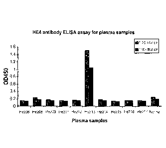
The results of the ELISA assay testing for the presence/amount of anti-HE4
antibodies
from the human sera samples are presented in FIGURE 1. As shown in FIGURE I,
one of the
ten patients with ovarian carcinoma (subject He213) had an 0D450 > 1.0 for
both dilutions, thus
indicating the presence of anti-HE4 antibodies. It is noted that the plasma
from patient He213
(with plasma that was positive for anti-HE4 abs), was found to be negative for
the presence of
HE4 antigen using the assay to detect the presence of HE4 antigen as described
in U.S. Patent
No. 7,270,960 (data not shown). However, cultured tumor cells derived from the
tumor of
patient He213 were shown to express the HE4 antigen (data not shown).
23
CA 2809368 2018-03-06
CA 02809368 2013-02-25
WO 2012/027631 PCT/US2011/049274
Conclusion:
The results described in this example demonstrate the successful development
of an
ELISA assay capable of detecting naturally occurring human anti-11E4
antibodies in human
serum. The results also demonstrate the presence of anti-HE4 antibodies in a
subject with
ovarian cancer. Based on our previous study on antibodies to mesothelian, (see
Hellstrom I.
et al., Cancer Epidemiol Biomarkers Prey. 17(6):1520-1526 (2008)), it is
expected that the
presence of, or an increase in titer of antibodies to HE4 identified in
ovarian cancer patients that
are undergoing, or having undergone treatment (via surgery, chemotherapy,
radiation and/or
immunotherapy), either alone, or in combination with a finding of a decreased
amount of HE4
antigen (as compared to the baseline amount at the start of the treatment), or
no circulating HE4
antigen, will indicate that the cancer therapy has been effective in damaging
the tumor. It is
further expected that a decrease in the titer of anti-HE4 antibodies, or no
detectable anti-HE4
antibodies to HE4 in ovarian cancer patients undergoing treatment, either
alone, or in
combination with increased amount of circulating HE4 antigen (as compared to
the baseline
amount at the start of treatment), will indicate that the cancer therapy has
been ineffective in
damaging the tumor, and/or indicative of a relapse.
EXAMPLE 3
This Example describes a study in which the ELISA assay developed for
detecting the
presence/amount of antibodies to HE4 (described in EXAMPLE 2) was used to
titrate the
amount of anti-HE4 antibody present in serum from 3 patients with ovarian
carcinoma, and 20
healthy female control subjects.
Methods:
Serum Samples Obtained from Ovarian Cancer Patients
As shown in TABLE 2 below, serum samples from three patients with early stage
(I/II)
ovarian carcinoma and 20 healthy human female subjects were obtained at the
Fred Hutchinson
Cancer Research Center (Seattle, Washington), 4 of which are shown in Table 2
below, Sera
were drawn from the subjects and asssayed using the methods as described in
Example 2,
Results:
The sera obtained as described above were assayed at 1:20 and 1:40 dilutions
using the
ELISA method described in EXAMPLE 2. The results are summarized in TABLE 2
below.
24
CA 02809368 2013-02-25
WO 2012/027631 PCT/US2011/049274
TABLE 2: Results of HE4 antibody analysis of serum from three ovarian Cancer
patients, and 4 healthy controls using the ELISA assay to detect/measure HE4
antibodies.
Patient Code Clinical 0D450 > 0.500
Disease/Condition (at least in the 1:20
dilution
1 00C17 Ovarian CarcinOma
206241 Ovarian Carcinoma
208448 Ovarian Carcinoma
1 healthy female control
2 healthy female control
3 healthy female control
4 healthy female control
5-24 (total of 24 Healthy female control 3 positive/21 negative
healthy female (of 24 total tested)
control subjects)
As summarized in TABLE 2, all three serum samples from patients with ovarian
carcinoma, and three of the 24 serum samples from healthy female subjects had
an 0D450 > 0.5
for the 1:20 dilution. As shown in TABLE 2, all of the three patients with
ovarian cancer had
anti-HE4 antibodies in their serum, as indicated by an 0D450? 0.5 at the 1:20
dilution. These
results are consistent with the results described in Example 2, and further
demonstrate the
successful development of an ELISA assay capable of detecting naturally
occurring human
anti-HE4 antibodies in human serum. The results also demonstrate the presence
of anti-HE4
antibodies in a subjects with both early and later stage ovarian cancer.
EXAMPLE 4
Reactivity with HE4 fragments displayed as phage fusion proteins
The epitope specificity of the human Anti HE4 antibodies was determined by
testing the
reactivity of human samples that displayed anti HE4 reactivity towards HE4
fragments expressed
as fusion proteins with phage coat protein pVIII in a phage ELISA.
Cloning of HE4 fragments in f88-4
cDNA, prepared from mRNA isolated from OvCar-3 cells, served as template for
PCR
amplification of the gene parts coding for the HE4 domains for cloning in the
phage display
vector f88-4. PCR primer pairs, listed in Table 3, were constructed for
amplification of the
CA 02809368 2013-10-25
coding regions indicated in Figure x. In the 5'-ends were restriction sites
for HindIII and PstI
inserted for cloning in fusion with the pVIII signal peptide and the pVIII
mature coat protein.
Table 3. PCR primers (SEQ ID NOS: 15-26, respectively, in order of appearance
used for
amplification of HE4 fragments
Primer Sequence 5-t0 3- WAP
W1 F 1 TGCTAAGCTTTGCC GAGAAGACTGGCGTGTGCCC N-WAP
W1 F2
TGCTAAGCTTTGCC AGCGAATGCGCCGACAACC N- WAP
W1 F 3
TGCTAAGCTTTGCC GACCAGAACTGCACGCAAG N - WAP
W1R1 CCTTCTGCAGG ATCATTGGGCAGAGAGCAG N- WAP
W 1 R2 CCTTCTGCAGG GTCCGAGACGCACTCTTGC N-WAP
W1R3 CCTTCTGCAGG GCTGCAGCACTTGAGGTTG N-WAP
W2 F 1
TGCTAAGCTTTGCC .AAGGAGGGTTCCTGCCCCCA C - WAP
W2 F2 TGCTAAGCTTTGCC AGCCAGTGTCCTGGCCAG C-WAP
W2 F3
TGCTAAGCTTTGCC CAGCTCGGCCTCTGTCGGGAC C-WAP
W2R1 CCTTCTGCAGG GAAATTGGGAGTGACACAGGA C-WAP
W2 R2 CCTTCTGCAGG GTCCACCTGGCACTGGTCC C-WAP
W 2 R 3 CCTTCTGCAGG ATTGCGGCAGCATTTCATCTG C-WAP
The HE4 fragments were separately amplified from 0,5 1 of cDNA in a reaction
mixture
containing 1 M of each forward and reverse primer, 75 mM Tris-HCl (pH 8.8 at
25 C), 20 mM
(NI-14)2SO4, 0.1% (v/v) Tween 20, 2 mM MgCl2, 0.02 u/ I Taq-polymerase
(Abgene, Surrey,
UK) and 0.1 mM of each deoxynucleotide in a final volume of 25 I with the
following
temperature cycle repeated 30 times: 30 seconds incubations at 95 C, 50 C and
72 C.
PCR products and 188-4, digested with HindIII and Pstl, were ligated together
and transfected
into E. coil JM109 where after clones were selected on LB plates with
tetracycline. Two clones
of each construct were amplified in E. coil JM109 and double-stranded DNA was
prepared for
DNA sequencing. DNA sequencing was performed using the Big dye terminator v1.1
cycle
sequencing kit and a 188-4 vector specific primer. Sequencing reactions were
sent to CyberGene
All (Huddinge, Sweden) for analysis. Sequence raw data was analyzed using the
free software
Chromas version 1.45 (Technelysium Pty Ltd., Australia). Nucleotide sequencing
verified
insertion in frame with the leader peptide and the mature phage coat protein
pVIII. The HE4
inserts demonstrated identity to the expected HE4 fragment sequences
(accession number
AY212888). The positions of the 1-1E4 fragments expressed as fusion proteins
with coat protein
26
pVIII using the primers shown in Tablel is shown in Figure 2. The amino acid
numbers refer to
positions is the HE4 amino acid sequence set forth in SEQ ID NO: 2.
Phage ELISA
Sequence verified phage clones were amplified, purified, concentrated with
PEG/NaC1 and
were diluted in PBS for use as coating antigen in the direct phage ELISA
assay, or alternatively
in in 1%BSA in PBS as antigen in the sandwhich phage ELISA.
In the direct phage ELISA the HE4 fragment phages were diluted in carbonate
buffer pH
9.2 and coated in microtiter wells. Binding of the human anti HE4 ab's was
determined after
dilution of the patient serum in PBS-1%BSA and incubation in HE4 phage coated
plates. The
bound hIg was determined by incubation with HRP Anti hIg Ab.
In the sandwich HE4 phage ELISA the patient samples were diluted with PBS-
1%BSA
and incubated in microtiter plates coated with anti-Human IgG for adsorption
of the hIgG. The
coated plates were incubated with the different HE4 pVIII phage particles in a
volume of 100
41/we1l were added. After two hours incubation, wells were washed and a rabbit
anti-M13
antibody (established in-house) was added. After incubation and washing, a HRP
labelled swine
anti rabbit antibody (Dako) was added. After the final wash TMB substrate was
added and the
plate was measured at 620 nm after 5 minute incubation. wt M-13 phage was used
as negative
control n both ELISA assays.
27
CA 2809368 2018-03-06