Note : Les descriptions sont présentées dans la langue officielle dans laquelle elles ont été soumises.
COMPOSITIONS AND METHODS FOR ENHANCING THE PLURI POTENCY OF
STEM CELLS
FIELD
This disclosure concerns compositions and methods for enhancing or prolonging
the
pi.uripotency of a stem cell, and the use of such pluripotent stem cells.
BACKGROUND
Mouse embryonic stem (ES) cells are prototypical pluripotent cells, which are
derived
from the inner cell mass of bIastocysts (Martin, Proe Nail Arad Sei USA
78:7634-7638, 1981;
Evans and Kaufman. Nature 292:154-156, 1981). ES cells have an unusual
capacity of
proliferating fora long time without losing their genome integrity and
katyotype (Suda et al., J
cell Phy.siol 133:197-201, 1987), and are capable of contributing to all the
cell types in animals
upon injection into mouse blastocysts (Niwa. Development 134:635-646, 2007).
The most
striking evidence of their potency has been demonstrated by injecting ES cells
into tetraploid
(4N) blastocysts, which produces healthy pups entirely from ES cells (Nagy et
al., Prot- Nail
Arad Sei USA 90:8424-8428, 1993). The ultimate test was to see if a single ES
cell can form an
entire healthy pup, though the success rate was extremely low (0.5%) (Wang and
Jaenisch, Der
Bit)/ 275:192-201, 2004).
It has recently been shown that Zscan4 (Zinc finger and scan domain-containing
protein
= 4), which is expressed specifically in 2-cell stage embryos and ES cells
(Falco et al., Dev BIN
307:539-550, 2007), is required for the maintenance of genome stability and
normal karyotype in
ES cells (Zalzman et aL, Nature 464:858-863, 2010). Although only a small
traction (-5%) of
undifferentiated ES cells express Zscan4 at a given time (Falco eta!,, Dev ma!
307:539-550,
2007), essentially all of the ES cells in culture undergo the transient Zscan4
state within 9
passages (Zalzman et al., Nature 464:858-863, 2010). Upon short hairpin RNA
(shRNA)-
mediated repression of Zscan4, after about 8 passages ES cells undergo massive
karyotype
deterioration. Prior studies have also shown that the Zscan4' state of ES
cells is associated with
telomere extension (Zalzman et al.. Nature 464:858-863, 20.10). Although ES
cells have the best
CA 2830600 2018-06-20
CA 02830600 2013-09-18
WO 2012/129342 PCT/US2012/030005
capacity to maintain their 2enome integrity in culture, it is also widely
recognized that even ES
cells, in long-term culture, gradually lose their developmental potency (i.e.,
ability to contribute
to tissues in chimeric mice).
SUMMARY
Disclosed herein is the finding that increasing the frequency of Zscan4
activation in mouse ES
cells not only enhances, but maintains their developmental potency in long-
term cell culture. In
particular, disclosed herein is the finding that particular Zscan4 protein
truncations and fusion
proteins increase the number of Zscan4 + cells and/or promote recurrent
activation of Zscan4 in
stem cells.
Provided herein are nucleic acid molecules, including vectors, encoding a
Zscan4-ERT2
fusion protein. Recombinant Zscan4-ERT2 fusion proteins are also provided.
Compositions and
cells (such as ES cell or iPS cells) comprising the Zscan4-ERT2 nucleic acid
molecules and
fusion proteins are also provided herein.
Further provided are nucleic acid molecules, including vectors, encoding a
Zscan4
protein with a C-terminal truncation of at least one zinc finger domain,
referred herein to as
Zscan4-AC. Recombinant Zscan4-AC proteins are also provided. Compositions and
cells (such
as ES cell or iPS cells) comprising the Zscan4-AC nucleic acid molecules and
proteins are also
provided herein.
Further provided are methods of enhancing or prolonging the pluripotency of a
stem cell
or a stem cell population; methods of increasing the frequency of Zscan4
positive cells in a stem
cell population; and methods of promoting genome stability or increasing
telomere length, or
both, in a stem cell or a stem cell population, by increasing the frequency of
Zscan4 activation in
the stem cell or stem cell population. In some embodiments, the methods
include contacting the
stem cell or stem cell population with a Zscan4-ERT2 nucleic acid molecule,
fusion protein or
composition as disclosed herein. In other embodiments. the methods include
contacting the stem
cell or stem cell population with a Zscan4-AC nucleic acid molecule, protein
or composition as
disclosed herein.
The foregoing and other objects and features of the disclosure will become
more apparent
from the following detailed description, which proceeds with reference to the
accompanying
figures.
2
CA 02830600 2013-09-18
WO 2012/129342 PCT/US2012/030005
BRIEF DESCRIPTION OF THE DRAWINGS
FIGS 1A-1F: Constitutive expression of a Zscan4c-ERT2 fusion protein increases
the number of Zscan4 ES cells. FIG. LA is a schematic of the structure of a
Zscan4c-ERT2
fusion protein. Zscan4c contains one SCAN domain and four C2H2 zinc finger
domains. FIG.
1B are fluorescence microscopy images of MC1-ZE3 cells, in which a Zscan4
promoter drives
the expression of Emerald marker (left), MC1-ZE3ZERT2 clone #15 cells, in
which the
Zscan4c-ERT2 fusion protein is constitutively expressed, cultured in the
absence of Tmx
(middle), and MC1-ZE3-ZERT2 clone #15 cells cultured in the presence of Tmx
for 3 days
(right). FIG. 1C is a graph showing flow-cytometry analysis of MC1-ZE3 ES
cells (left, control)
and MC1-ZE3-ZERT2 #15 ES cells (right) in the absence or presence of 1 j_tM
Tmx. Em
fluorescence levels (average S.E.M.; n=6) are shown. Note 3-fold increase of
Em + cells by the
constitute expression of a Zscan4c-ERT2 fusion protein even without Tmx. FIG.
1D is a graph
showing the results of quantitative RT-PCR analysis of endogenous Zscan4
expression measured
by using PCR primer pairs specific for 3'-UTR of Zscan4 in MC1-ZE3 ES cells
(left, control)
and MC1-ZE3-ZERT2 #15 ES cells (right) in the absence or presence of 1 ittM
Tmx. The fold-
induction of endogenous Zscan4 expression levels (average S.E.M.; n=6)
compared to that of
control MC1-ZE3 is shown. Note the 6fold increase of endogenous Zscan4 at the
RNA level by
the constitute expression of a Zscan4c-ERT2 fusion protein even without Tmx.
FIG. 1E is a
series of images of V6.5 parental ES cells (passage number 14), V6.5 ZERT2 #2
(p.20), V6.5
ZERT2 #7 (p.21), V6.5 ZERT2 #10 (p.20), V6.5 ZERT2 #18 (p.22) ES cell colonies
after
whole-mount RNA in situ hybridization of a Zscan4 full-length probe, which
detects both
endogenous and exogenous Zscan4 RNAs (upper panel) or a Zscan4 3'-UTR probe,
which
detects only endogenous Zscan4 RNAs (lower panel). FIG. 1F is a schematic
showing
comparisons of global expression profiles between V6.5 ZERT2 #18 ES cells and
Em + ES cells
(upper panel), and between Tmx- and Tmx+ conditions of V6.5 ZERT2 #18 ES cells
(lower
panel). Note that Zscan4-related genes (Zscan4c, BC061212, Tmeme92, and
Tcstv1/3) are
already upregulated in the V6.5 ZERT2 #18 ES cells even without Tmx.
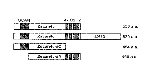
FIGS. 2A-2G: Zscan4 lacking the C-terminus increases the number of Zscan4 +
cells.
FIG. 2A is a schematic showing the structure of Zscan4c, Zscan4cERT2, Zscan4c-
AC and
Zscan4c-AN proteins. Zscan4c-AC was made by deleting four Zinc finger domains
at the C-
terminus of Zscan4c protein. Zscan4c-AN was made by deleting the SCAN domain
at the N-
terminus. These mutated genes were placed under the strong and constitutive
CAG promoter.
3
CA 02830600 2013-09-18
WO 2012/129342 PCT/US2012/030005
Each vector was transfected into MC1ZE16 ES cells (sister clones of MC1-ZE3).
Multiple
independent clones were isolated: ZDC-MCI-ZE16 #3, #4, #20 for Zscan4c-AC; ZDN-
MCI-
ZEI6 #5, #8, #15 for Zscan4c-AN. FIGS. 2B-2G are fluorescence microscopic
images of ZDC-
MC1-ZE16 #3, #4, #20 for Zscan4c-AC and ZDN-MC1-ZE16 #5, #8, #15 for Zscan4c-
AN. The
results demonstrate that the expression of Zscan4c-AC increases the number of
Zscan4 + cells,
whereas the expression of Zscan4c-AN does not change the number of Zscan4 +
cells.
FIGS. 3A-3B: Constitutive expression of a Zscan4c-ERT2 fusion protein
increases and
prolongs developmental potency of ES cells. FIG. 3A shows representative coat
colors of
chimeric mice generated by injecting various ES cells into blastocysts. The
higher chimerism
represents the higher contribution of injected ES cells to mice, indicating
the higher
developmental potency of ES cells. FIG. 3B is a graph showing the percent
distribution of
chimerism levels among "n" number of mice born from various ES cell lines.
FIGS. 4A-4E: Tetraploid (4N) complementation assays confirm the higher potency
of ES cells expressing a Zscan4c-ERT2 fusion protein. FIG. 4A is a table
showing
development of 4N blastocysts injected with multiple ES cells (10-15 ES
cells): V6.5 parental
ES cells (passage 18), V6.5 ZERT2 #7 (passage 22), V6.5 ZERT2 #10 (passage
22), V6.5
ZERT2 #18 (passage 19), and freshly isolated TA1 ES cells (passage 3). FIG. 4B
is a table
showing development of 4N blastocysts injected with single ES cells: V6.5
parental ES cells
(passage 18), V6.5 ZERT2 #2 (passage 21), V6.5 ZERT2 #18, and freshly isolated
TAI ES cells
(passage 4). FIG. 4C is an image of the embryos examined: only properly
developed embryos
were counted (the group on the right). FIG. 4D is a pair of images of two live
embryos derived
from single V6.5 ZERT2 #18 ES cells shown in FIG. 4A. FIG. 4E shows a proposed
model of
ES cell potency.
FIGS. 5A-5C are a table providing a list of genes upregulated in MC1-ZE7 Em
cells
compared to Em- cells.
FIGS. 6A-6C: Generation and characterization of V6.5 ZERT2 ES cell clones.
FIG. 6A
is a graph showing results of qRT-PCR analysis of Zscan4 expression levels by
a primer pair
detecting RNA from both endogenous Zscan4 and exogenous Zscan4 (transcripts
from a pCGA-
Zscan4-ERT2). The primer sequences are 5'-AGTCTGACTGATGAGTGCTTGAAGCC-3'
(SEQ ID NO: 15) and 5'-GGCCTTGTTGCAGATTGCTGTTG-3' (SEQ ID NO: 16). Data were
normalized by the expression of H2A, using primers 5'-
4
CA 02830600 2013-09-18
WO 2012/129342 PCT/US2012/030005
TTGCAGCTTGCTATACGTGGAGATG-3' (SEQ ID NO: 17) and 5'-
TGTTGTCCTTTCTTCCCGATCAGC-3' (SEQ ID NO: 18). The expression levels are shown
as
a fold change relative to the Zscan4 expression levels of a parental V6.5 ES
cells. FIG. 6B is a
graph of growth curves of V6.5 ZERT2 #18 ES cells in the presence (Tmx+) or
absence of
Tamoxifen (Tmx-). The presence of Tmx dramatically reduced the proliferation
of ES cells,
which was restored by removing the Tmx from the media even after long-term
culture with Tmx
(Tmx +>-). FIG. 6C is a series of images showing morphologies of cells in each
culture
condition.
FIG. 7A is a scatter plot showing genes expressed differentially between V 6.5
ZERT2
#18 ES cells and control V6.5 #2 ES cells. FIG. 7B is a scatter plot showing
genes expressed
differentially between V6.5 ZERT2 #18 ES cells cultured for 2 days in the
presence of Tmx and
control V6.5 #2 ES cells.
FIG. 8 is a table listing the top 50 genes upregulated in V6.5 ZERT2 #18 ES
cells
compared to V6.5 #2 ES cells. FIG. 9 is a table listing the top 50 genes
upregulated in V6.5
.. ZERT2 #18 ES cells cultured in the presence of Tmx for 2 days compared to
V6.5 #2 ES cells.
FIGS. 10A-10B: Derivation of new Fl (C57BL/6J X 129S6/SvEvTac) hybrid ES cell
lines. FIG. 10A is a table showing blastocysts obtained by mating C57BL/6J
females with
12956/SvEvTac males. Blastocysts were cultured in vitro on the mouse embryo
fibroblasts
(MEFs) feeders in 15% KSR medium (Invitrogen) supplemented with 50 nM PD98059
(MEK1
inhibitor). *Outgrowths showed undifferentiated (U), differentiated (D), and
mixed (U/D)
cellular phenotypes. FIG. 10B is table providing a summary of ES derivation
results.
FIGS. 11A-11B: Testing developmental potency of newly derived Fl hybrid ES
cell
lines by tetraploid complementation assays. FIG. 11A is a table of six ES cell
lines that
showed undifferentiated cellular phenotypes when injected into tetraploid (4N)
mouse
blastocysts. Success rates of obtaining live embryos at El 3.5 varied among ES
cell lines, ranging
from 15% to 60%. Clone #10 was selected for its highest success rate (named
TA1 ES cell line)
and was used for subsequent studies. FIG. 11B is a series of representative
images of 4N
placentas and E13.5 embryos derived from the TA1 ES cell line. Normal
appearance of female
and male gonads dissected from these embryos indicates their germline
competence.
5
CA 02830600 2013-09-18
WO 2012/129342 PCT/US2012/030005
FIG. 12: Testing transient overexpression of Zscan4 (i.e., unmodified Zscan4.
FIG. 12
includes a graph showing that the transient overexpression of Zscan4 was able
to increase the
developmental potency of ES cells.
SEQUENCE LISTING
The nucleic and amino acid sequences listed in the accompanying sequence
listing are
shown using standard letter abbreviations for nucleotide bases, and three
letter code for amino
acids, as defined in 37 C.F.R. 1.822. Only one strand of each nucleic acid
sequence is shown, but
the complementary strand is understood as included by any reference to the
displayed strand. In
the accompanying sequence listing:
SEQ ID NOs: 1 and 2 are nucleotide and amino acid sequences of human ZSCAN4.
SEQ ID NOs: 3 and 4 are nucleotide and amino acid sequences of mouse Zscan4a.
SEQ ID NOs: 5 and 6 are nucleotide and amino acid sequences of mouse Zscan4b.
SEQ ID NOs: 7 and 8 are nucleotide and amino acid sequences of mouse Zscan4c.
SEQ ID NOs: 9 and 10 are nucleotide and amino acid sequences of mouse Zscan4d.
SEQ ID NOs: 11 and 12 are nucleotide and amino acid sequences of mouse
Zscan4e.
SEQ ID NOs: 13 and 14 are nucleotide and amino acid sequences of mouse
Zscan4f.
SEQ ID NOs: 15-18 are nucleotide sequences of primers used for qRT-PCR
analysis of
Zscan4 and H2A.
SEQ ID NO: 19 is the nucleotide acid sequence of plasmid pPyCAGmZscan4c-ERT2.
SEQ ID NO: 20 is the nucleotide sequence of plasmid pPyCAG-hZscan4ERT2.
SEQ ID NO: 21 is the amino acid sequence of human ERT2.
SEQ ID NO: 22 is the amino acid sequence of a mouse Zscan4c-ERT2 fusion
protein.
SEQ ID NO: 23 is the amino acid sequence of a human ZSCAN4-ERT2 fusion
protein.
SEQ ID NO: 24 is the nucleotide sequence of plasmid pCAG-Zscan4-AC.
6
CA 02830600 2013-09-18
WO 2012/129342 PCT/US2012/030005
SEQ ID NO: 25 is the amino acid sequence of mouse Zscan4c-AC (lacking all four
zinc
finger domains).
DETAILED DESCRIPTION
I. Abbreviations
a.a. amino acid
eDNA complementary deoxyribonucleic acid
Em Emerald
ES embryonic stem
hCG human chorionic gonadotropin
ICM inner cell mass
LIF leukemia inhibitory factor
MEF murine embryonic fibroblast
ORF open reading frame
PFA paraformaldehyde
qPCR quantitative polymerase chain reaction
qRT-PCR quantitative reverse transcriptase polymerase chain
reaction
shRNA short hairpin ribonucleic acid
Tmx tamoxifen
II. Terms and Methods
Unless otherwise noted, technical terms are used according to conventional
usage.
Definitions of common terms in molecular biology may be found in Benjamin
Lewin, Genes V,
published by Oxford University Press, 1994 (ISBN 0-19-854287-9); Kendrew et
al. (eds.), The
Encyclopedia of Molecular Biology, published by Blackwell Science Ltd., 1994
(ISBN 0-632-
7
CA 02830600 2013-09-18
WO 2012/129342 PCT/US2012/030005
02182-9); and Robert A. Meyers (ed.), Molecular Biology and Biotechnology: a
Comprehensive
Desk Reference. published by VCH Publishers, Inc., 1995 (ISBN 1-56081-569-8).
In order to
facilitate review of the various embodiments of the disclosure, the following
explanations of
specific terms are provided:
Administration: To provide or give a subject an agent, such as an ES cell or
population
of ES cells, by any effective route. An exemplary route of administration
includes, but is not
limited to, injection (such as subcutaneous, intramuscular, intradenual,
intraperitoneal,
intravenous or intra-arterial).
Agent: Any protein, nucleic acid molecule, compound, cell, small molecule,
organic
compound, inorganic compound, or other molecule of interest. Contacting:
Placement in direct
physical association; includes both in solid and liquid form. As used herein,
"contacting" is used
interchangeably with "exposed." In some cases, "contacting" includes
transfecting, such as
transfecting a nucleic acid molecule into a cell.
Degenerate variant: A polynucleotide encoding a polypeptide, such as a Zscan4
polypeptide, that includes a sequence that is degenerate as a result of the
genetic code. There are
natural amino acids, most of which are specified by more than one codon.
Therefore, all
degenerate nucleotide sequences are included as long as the amino acid
sequence of the
polypeptide encoded by the nucleotide sequence is unchanged.
Differentiation: Refers to the process by which a cell develops into a
specific type of
20 cell (for example, muscle cell, skin cell etc.). Differentiation of
embryonic stem cells refers to
the development of the cells toward a specific cell lineage. As a cell becomes
more
differentiated, the cell loses potency, or the ability to become multiple
different cell types.
Embryonic stem (ES) cells: Pluripotent cells isolated from the inner cell mass
of a
developing blastocyst. ES cells can be derived from any organism, such as a
mammal. In one
embodiment, ES cells are produced from mice, rats, rabbits, guinea pigs,
goats, pigs, cows, non-
human primates or humans. Human and murine derived ES cells are exemplary. ES
cells are
pluripotent cells, meaning that they can generate all of the cells present in
the body (bone,
muscle, brain cells, etc.). Methods for producing murine ES cells can be
found, for example, in
U.S. Patent No. 5,670,372. Methods for producing human ES cells can be found,
for example. in
U.S. Patent No. 6,090,622, PCT Publication No. WO 00/70021 and PCT Publication
No. WO
8
00/27995. A number of human ES cell lines are known in the art and are
publically available.
For example. the National Institutes of Health (N1H) Human Embryonic Stem Cell
Registry
provides a list of a number of human ES eel, lines that have been developed (a
list can be found
online at the NIH Office of Extramural Research website).
Encapsulated: As used herein, a molecule "encapsulated" in a nanoparticle
refers to a
molecule (such as Zscan4-ERT2 fusion protein) that is either contained within
the nanoparticle
or attached to the surface of the nanoparticle, or a combination thereof.
ERT2: A protein comprising a mutated ligand binding domain of the human
estrogen
receptor that does not bind its natural ligand (170-estradiol) at
physiological concentrations. but
is highly sensitive to nanomolar concentrations of tamoxifen or its metabolite
4-hydroxy-
tamoxilen (40HT) (Feil el Bioehein ltiophys Res (.ommun 237(3):752-757.
1997). An
exemplary amino acid sequence for ERT2 is set forth herein as SEQ ID NO: 21,
and the
corresponding coding sequence is set forth herein as nucleotides 3989-4936 of
SEQ ID NO: 19.
ES cell therapy: A treatment that includes administration of ES cells to a
subject. In
particular examples, the ES cells are Zscan4 + ES cells.
Functional fragment or variant (of Zscan4): The disclosed Zscan4
polynucleotides and
polypeptides (such as those set forth as SEQ ID NOs: 1-14) include functional
fragments and
variants of Zscan4 that retain iscan4 biological activity. Functional
fragments and/or variants of
Zscan4 generally comprise at least about 80%, at least about 85%, at least
about 90%, at least
about 95% or at least about 99% sequence identity with one of the Zscan4
sequences set forth as
SEQ ID NOs 1-14. When less than the entire sequence is being compared for
sequence identity.
fragments will typically possess at least 80% sequence identity over the
length of the fragment.
and can possess. for example. sequence identities of at least 85%, 90%. 95% or
99%.
Fusion protein: A protein generated by expression of a nucleic acid sequence
engineered
from nucleic acid sequences encoding at least a portion of two different
(heterologous) proteins.
To create a fusion protein, the nucleic acid sequences must be in the same
reading frame and
contain no internal stop codons. In some embodiments herein, the fusion
protein is a Zscan4-
ERT2 fusion protein. In some examples, the fusion protein comprises a linker
between the two
different proteins.
9
CA 2830600 2018-06-20
CA 02830600 2013-09-18
WO 2012/129342 PCT/US2012/030005
Genome stability: The ability of a cell to faithfully replicate DNA and
maintain integrity
of the DNA replication machinery. An ES cell with a stable genome generally
defies cellular
senescence, can proliferate more than 250 doublings without undergoing crisis
or transformation,
has a low mutation frequency and a low frequency of chromosomal abnormalities
(e.g.. relative
to embryonal carcinoma cells), and maintains genomic integrity. Long telomeres
are thought to
provide a buffer against cellular senescence and be generally indicative of
genome stability and
overall cell health. Chromosome stability (e.g., few mutations, no chromosomal
rearrangements
or change in number) is also associated with genome stability. A loss of
genome stability is
associated with cancer, neurological disorders and premature aging. Signs of
genome instability
include elevated mutation rates, gross chromosomal rearrangements, alterations
in chromosome
number, and shortening of telomeres.
Heterologous: A heterologous polypeptide or polynucleotide refers to a
polypeptide or
polynucleotide derived from a different source or species. For example, a
mouse Zscan4 peptide
expressed in a human ES cell is heterologous to that ES cell.
Host cells: Cells in which a vector can be propagated and its DNA expressed.
The term
also includes any progeny of the subject host cell. It is understood that all
progeny may not be
identical to the parental cell since there may be mutations that occur during
replication.
However, such progeny are included when the term "host cell" is used.
Induced pluripotent stem (iPS) cells: A type of pluripotent stem cell
artificially derived
from a non-pluripotent cell, typically an adult somatic cell, by inducing a
"forced" expression of
certain genes. iPS cells can be derived from any organism, such as a mammal.
In one
embodiment, iPS cells are produced from mice, rats, rabbits, guinea pigs,
goats, pigs, cows, non-
human primates or humans. Human and murine derived iPS cells are exemplary.
iPS cells are similar to ES cells in many respects, such as the expression of
certain stem
cell genes and proteins, chromatin methylation patterns, doubling time,
embryoid body
formation, teratoma formation, viable chimera formation, and potency and
differentiability.
Methods for producing iPS cells are known in the art. For example, iPS cells
are typically
derived by transfection of certain stem cell-associated genes (such as Oct-3/4
(Pouf51) and
Sox2) into non-pluripotent cells, such as adult fibroblasts. Transfection can
be achieved through
viral vectors, such as retroviruses, lentiviruses, or adenoviruses. For
example, cells can be
transfected with 0ct3/4, Sox2, Klf4, and c-Myc using a retroviral system or
with OCT4, SOX2,
CA 02830600 2013-09-18
WO 2012/129342 PCT/US2012/030005
NANOG, and LIN28 using a lentiviral system. After 3-4 weeks, small numbers of
transfected
cells begin to become morphologically and biochemically similar to pluripotent
stem cells, and
are typically isolated through morphological selection, doubling time, or
through a reporter gene
and antibiotic selection. In one example, iPS from adult human cells are
generated by the method
of Yu et al.(Science 318(5854):1224, 2007) or Takahashi et al. (Cell
131(5):861-72, 2007).
Isolated: An isolated nucleic acid has been substantially separated or
purified away from
other nucleic acid sequences and from the cell of the organism in which the
nucleic acid
naturally occurs, i.e., other chromosomal and extrachromosomal DNA and RNA.
The term
"isolated" thus encompasses nucleic acids purified by standard nucleic acid
purification methods.
The term also embraces nucleic acids prepared by recombinant expression in a
host cell as well
as chemically synthesized nucleic acids. Similarly, "isolated" proteins have
been substantially
separated or purified from other proteins of the cells of an organism in which
the protein
naturally occurs, and encompasses proteins prepared by recombination
expression in a host cell
as well as chemically synthesized proteins. Similarly, "isolated" cells have
been substantially
separated away from other cell types.
Linker: One or more nucleotides or amino acids that serve as a spacer between
two
molecules, such as between two nucleic acid molecules or two peptides (such as
in a fusion
protein). In some examples a linker is 1 to 100 amino acids, such as 1 to 50
or 5 to 10 amino
acids.
Nanoparticle: A particle less than about 1000 nanometers (nm) in diameter.
Exemplary
nanoparticles for use with the methods provided herein are made of
biocompatible and
biodegradable polymeric materials. In some embodiments, the nanoparticles are
PLGA
nanoparticles. As used herein, a "polymeric nanoparticle" is a nanoparticle
made up of repeating
subunits of a particular substance or substances. "Poly(lactic acid)
nanoparticles" are
nanoparticles having repeated lactic acid subunits. Similarly, "poly(glycolic
acid) nanoparticles"
are nanoparticles having repeated glycolic acid subunits.
Non-human animal: Includes all animals other than humans. A non-human animal
includes, but is not limited to, a non-human primate, a farm animal such as
swine, cattle, and
poultry, a sport animal or pet such as dogs, cats, horses, hamsters, rodents,
such as mice, or a zoo
animal such as lions, tigers or bears. In one example, the non-human animal is
a transgenic
animal, such as a transgenic mouse, cow, sheep, or goat. In one specific, non-
limiting example,
11
CA 02830600 2013-09-18
WO 2012/129342 PCT/US2012/030005
the transgenic non-human animal is a mouse.
Operably linked: A first nucleic acid sequence is operably linked to a second
nucleic
acid sequence when the first nucleic acid sequence is placed in a functional
relationship with the
second nucleic acid sequence. For instance, a promoter is operably linked to a
coding sequence if
the promoter affects the transcription or expression of the coding sequence.
Generally, operably
linked nucleic acid sequences are contiguous and where necessary to join two
protein coding
regions, in the same reading frame.
Pharmaceutically acceptable carriers: The pharmaceutically acceptable carriers
of use
are conventional. Remington's Pharmaceutical Sciences, by E. W. Martin, Mack
Publishing Co.,
Easton, PA, 15th Edition (1975), describes compositions and formulations
suitable for
pharmaceutical delivery of the Zscan4 proteins (including fusion proteins),
Zscan4 nucleic acid
molecules, or cells herein disclosed.
In general, the nature of the carrier will depend on the particular mode of
administration
being employed. For instance, parenteral formulations usually comprise
injectable fluids that
include pharmaceutically and physiologically acceptable fluids such as water,
physiological
saline, balanced salt solutions, aqueous dextrose, glycerol or the like as a
vehicle. For solid
compositions (e.g., powder, pill, tablet, or capsule forms), conventional non-
toxic solid carriers
can include. for example, pharmaceutical grades of mannitol, lactose, starch,
or magnesium
stearate. In addition to biologically-neutral carriers, pharmaceutical
compositions to be
administered can contain minor amounts of non-toxic auxiliary substances, such
as wetting or
emulsifying agents, preservatives, and pH buffering agents and the like, for
example, sodium
acetate or sorbitan monolaurate.
Pluripotent/pluripotency: A "pluripotent" cell is a cell that can form all of
an
organism's cell lineages (endoderm, mesoderm and ectoderm), including germ
cells, but cannot
form an entire organisms autonomously. As used herein, enhancing or prolonging
pluripotency
refers to increasing the pluripotent capacity or duration of pluripotency of a
stem cell.
Polypeptide: A polymer in which the monomers are amino acid residues which are
joined together through amide bonds. When the amino acids are alpha-amino
acids, either the L-
optical isomer or the D-optical isomer can be used, the L-isomers being
preferred. The terms
"polypeptide" or "protein" as used herein are intended to encompass any amino
acid sequence
12
CA 02830600 2013-09-18
WO 2012/129342 PCT/US2012/030005
and include modified sequences such as glycoproteins. The term "polypeptide"
is specifically
intended to cover naturally occurring proteins, as well as those which are
recombinantly or
synthetically produced.
The term "polypeptide fragment" refers to a portion of a polypeptide which
exhibits at
least one useful epitope. The term "functional fragments of a polypeptide"
refers to all fragments
of a polypeptide that retain an activity of the polypeptide, such as a Zscan4.
Biologically
functional fragments, for example, can vary in size from a polypeptide
fragment as small as an
epitope capable of binding an antibody molecule to a large polypeptide capable
of participating
in the characteristic induction or programming of phenotypic changes within a
cell, including
affecting cell proliferation or differentiation. An "epitope" is a region of a
polypeptide capable of
binding an immunoglobulin generated in response to contact with an antigen.
Thus, smaller
peptides containing the biological activity of Zscan4, or conservative
variants of Zscan4, are thus
included as being of use.
The term "substantially purified polypeptide" as used herein refers to a
polypeptide
which is substantially free of other proteins, lipids, carbohydrates or other
materials with which it
is naturally associated. In one embodiment, the polypeptide is at least 50%,
for example at least
80% free of other proteins, lipids, carbohydrates or other materials with
which it is naturally
associated. In another embodiment, the polypeptide is at least 90% free of
other proteins, lipids,
carbohydrates or other materials with which it is naturally associated. In yet
another
embodiment, the polypeptide is at least 95% free of other proteins, lipids,
carbohydrates or other
materials with which it is naturally associated.
Conservative substitutions replace one amino acid with another amino acid that
is similar
in size, hydrophobicity, etc. Examples of conservative substitutions are shown
below:
Original Residue Conservative Substitutions
Ala Ser
Arg Lys
Asn Gin; His
Asp Glu
Cys Ser
Gln Asn
13
CA 02830600 2013-09-18
WO 2012/129342 PCT/US2012/030005
Glu Asp
His Asn; Gin
Ile Leu; Val
Leu Ile; Val
Lys Arg; Gln; Glu
Met Leu; Ile
Phe Met; Leu; Tyr
Ser Thr
Thr Ser
Trp Tyr
Tyr Trp; Phe
Val Ile; Leu
Variations in the cDNA sequence that result in amino acid changes, whether
conservative
or not, should be minimized in order to preserve the functional and
immunologic identity of the
encoded protein. Thus, in several non-limiting examples, a Zscan4 polypeptide
(or Zscan4 fusion
protein, such as Zscan4-ERT2). or other polypeptides disclosed herein,
includes at most two, at
most five, at most ten, at most twenty, or at most fifty conservative
substitutions. The
immunologic identity of the protein may be assessed by determining whether it
is recognized by
an antibody; a variant that is recognized by such an antibody is
immunologically conserved. Any
cDNA sequence variant will preferably introduce no more than twenty, and
preferably fewer
than ten amino acid substitutions into the encoded polypeptide. Variant amino
acid sequences
may be, for example, at least 80%, 90% or even 95% or 98% identical to the
native amino acid
sequence (such as a native Zscan4 sequence or a Zscan4-ERT2 sequence, such as
SEQ ID NO:
22 or 23).
Promoter: Nucleic acid control sequences which direct transcription of a
nucleic acid. A
promoter includes necessary nucleic acid sequences near the start site of
transcription. A
promoter also optionally includes distal enhancer or repressor elements. A
"constitutive
promoter" is a promoter that is continuously active and is not subject to
regulation by external
signals or molecules. In contrast, the activity of an "inducible promoter" is
regulated by an
external signal or molecule (for example, a transcription factor).
14
CA 02830600 2013-09-18
WO 2012/129342 PCT/US2012/030005
Recombinant: A recombinant nucleic acid or polypeptide is one that has a
sequence that
is not naturally occurring or has a sequence that is made by an artificial
combination of two
otherwise separated segments of sequence. This artificial combination is often
accomplished by
chemical synthesis or by the artificial manipulation of isolated segments of
nucleic acids, for
example, by genetic engineering techniques.
Sequence identity/similarity: The identity/similarity between two or more
nucleic acid
sequences, or two or more amino acid sequences, is expressed in terms of the
identity or
similarity between the sequences. Sequence identity can be measured in terms
of percentage
identity; the higher the percentage, the more identical the sequences are.
Sequence similarity can
be measured in terms of percentage similarity (which takes into account
conservative amino acid
substitutions); the higher the percentage, the more similar the sequences are.
Homologs or
orthologs of nucleic acid or amino acid sequences possess a relatively high
degree of sequence
identity/similarity when aligned using standard methods. This homology is more
significant
when the orthologous proteins or cDNAs are derived from species which are more
closely
related (such as human and mouse sequences), compared to species more
distantly related (such
as human and C. elegans sequences).
Methods of alignment of sequences for comparison are well known in the art.
Various
programs and alignment algorithms are described in: Smith & Waterman. Adv.
App!. Math.
2:482, 1981; Needleman & Wunsch, J. Mol. Biol. 48:443, 1970; Pearson & Lipman,
Proc. Natl.
Acad. Sci. USA 85:2444, 1988; Higgins & Sharp, Gene, 73:237-44, 1988; Higgins
& Sharp,
CABIOS 5:151-3, 1989; Corpet etal., Nuc. Acids Res. 16:1088190, 1988; Huang
etal. Computer
Appls. in the Biosciences 8, 155-65, 1992; and Pearson et al., Meth. Mol. Bio.
24:307-31, 1994.
Altschul et al., J. Mol. Biol. 215:403-10, 1990, presents a detailed
consideration of sequence
alignment methods and homology calculations.
The NCBI Basic Local Alignment Search Tool (BLAST) (Altschul et al., J. Mol.
Biol.
215:403-10, 1990) is available from several sources, including the National
Center for Biological
Information (NCBI, National Library of Medicine, Building 38A, Room 8N805.
Bethesda, MD
20894) and on the Internet, for use in connection with the sequence analysis
programs blastp,
blastn, blastx, tblastn and tblastx. Additional information can be found at
the NCBI web site.
Stem cell: A cell having the unique capacity to produce unaltered daughter
cells (self-
renewal; cell division produces at least one daughter cell that is identical
to the parent cell) and
CA 02830600 2013-09-18
WO 2012/129342 PCT/US2012/030005
to give rise to specialized cell types (potency). Stem cells include, but are
not limited to. ES
cells, EG cells, GS cells, MAPCs, maGSCs, USSCs, adult stem cells and induced
pluripotent
stem cells. In one embodiment, stem cells can generate a fully differentiated
functional cell of
more than one given cell type. The role of stem cells in vivo is to replace
cells that are destroyed
during the normal life of an animal. Generally, stem cells can divide without
limit. After
division, the stem cell may remain as a stem cell, become a precursor cell, or
proceed to terminal
differentiation. A precursor cell is a cell that can generate a fully
differentiated functional cell of
at least one given cell type. Generally, precursor cells can divide. After
division, a precursor cell
can remain a precursor cell, or may proceed to terminal differentiation.
Subject: Living multi-cellular vertebrate organisms, a category that includes
human and
non-human mammals.
Subpopulation: An identifiable portion of a population. As used herein, a
"subpopulation" of ES cells expressing Zscan4 is the portion of ES cells in a
given population
that has been identified as expressing Zscan4.
Telomere: Refers to the end of a eukaryotic chromosome, a specialized
structure
involved in the replication and stability of the chromosome. Telomeres consist
of many repeats
of a short DNA sequence in a specific orientation. Telomere functions include
protecting the
ends of the chromosome so that chromosomes do not end up joined together, and
allowing
replication of the extreme ends of the chromosomes (by telomerase). The number
of repeats of
telomeric DNA at the end of a chromosome decreases with age.
Transfecting or transfection: Refers to the process of introducing nucleic
acid into a
cell or tissue. Transfection can be achieved by any one of a number of
methods, such as, but not
limited to, liposomal-mediated transfection, electroporation and injection.
Vector: A nucleic acid molecule as introduced into a host cell, thereby
producing a
transformed host cell. A vector may include nucleic acid sequences that permit
it to replicate in a
host cell, such as an origin of replication (DNA sequences that participate in
initiating DNA
synthesis). For example, an expression vector contains the necessary
regulatory sequences to
allow transcription and translation of inserted gene or genes. A vector may
also include one or
more selectable marker genes and other genetic elements known in the art.
Vectors include. for
example, virus vectors and plasmid vectors.
16
CA 02830600 2013-09-18
WO 2012/129342 PCT/US2012/030005
Zscan4: A group of genes that have previously identified as exhibiting 2-
cellspecific
expression and ES cell-specific expression (PCT Publication No. WO
2008/118957) and have
been shown to promote telomere elongation and genome stability (Zalzman et
al., Nature
464(7290):858-863, 2010). In the context of the present disclosure, -Zscan4"
includes both
human ZSCAN4 and mouse Zscan4. In the mouse, the term "Zscan4" refers to a
collection of
genes including three pseudogenes (Zscan4-psl, Zscan4-ps2 and Zscan4-ps3) and
six expressed
genes (Zscan4a, Zscan4b, Zscan4c, Zscan4d, Zscan4e and Zscan4f). Among the six
paralogs, the
open reading frames of Zscan4c, Zscan4d, and Zscan4f encode a SCAN domain as
well as all
four zinc finger domains, suggesting their potential role as transcription
factors. Zscan4 refers to
Zscan4 polypeptides and Zscan4 polynucleotides encoding the Zscan4
polypeptides. Exemplary
sequences are provided herein (see SEQ ID NOs: 1-14).
Zscan4-AC: In the context of the present disclosure, "Zscan4-AC" includes any
mouse
or human Zscan4 protein having a C-terminal deletion of at least one zinc
finger domain. In
some embodiments, the Zscan4-AC protein includes a deletion of at least two,
such as three or all
four zinc finger domains. SEQ ID NO: 2 and SEQ ID NO: 8 provide the amino acid
sequences of
human ZSCAN4 and mouse Zscan4c, respectively, and delineate the N-terminal
SCAN domain
and C-terminal zinc finger (C2H2-type) domains. In addition, the nucleotide
and amino acid
regions of each domain of human ZSCAN4 and mouse Zscan4c are listed below.
Human ZSCAN4
Domain Nucleotides (SEQ ID NO: 1) Amino Acids (SEQ ID NO: 2)
SCAN 826-1074 44-126
C2H2-type 1 1630-1698 312-334
C2H2-type 2 1714-1782 340-362
C2H2-type 3 1798-1866 368-390
C2H2-type 4 1882-1950 396-418
17
Mouse Zscan4c
Domain Nucleotides (SEQ II) NO: 7) Amino Acids (SEQ ID NO: 8)
SCAN 309-557 37-119
C2H2-type 1. 1383-1451 395-417
C2H2-type 2 1470-1538 424-446
C2112-type 1554-1622 452-474
C2H2-type 4 1.638-1709 480-503
Zscan4-ERT2: A fusion protein made up of a Zscan4 amino acid sequence and an
ERT2
amino acid sequence. "Zscan4-ERT2" can also refer to a nucleic acid sequence
encoding a
2scan4-ERT2 fusion protein. Exemplary amino acid sequences for Zscan4
(including SEQ ID
NO: 2,8, 10 and 14) and ERT2 (SEQ ID NO: 21) are set forth herein. In some
embodiments, the
Zscan4 sequence is a functional fragment or variant of a known Zscan4 sequence
(such as SEQ
ID NO: 2, 8, 10 or 14) and/or the ERT2 sequence is a functional fragment or
variant of a known
ERT2 sequence (such as SEQ ID NO: 21). Any fragment or variant of Zscan4 or
ERT2 is
contemplated as long as the fragment or variant retains activity. In some
examples. the Zscan4-
ERT2 fusion protein comprises a linker (or spacer) between Zscan4 and ERT2.
Linkers are well
known in the art and an appropriate linker can be selected by one of ordinary
skill in the art. In
particular examples, the linker is encoded by the nucleotide sequence GCTAGC.
Unless otherwise explained, all technical and scientific terms used herein
have the same
meaning as commonly understood by one of ordinary skill in the art to which
this disclosure
belongs. The singular terms "a." "an." and "the include plural referents
unless context clearly
indicates otherwise. Similarly, the word "or- is intended to include "and"
unless the context
clearly indicates otherwise. Hence "comprising A or B" means including A. or
B. or A and B. It
is further to be understood that all base sizes or amino acid sizes, and all
molecular weight or
molecular mass values, given for nucleic acids or polypeptides are
approximate, and are
provided for description. Although methods and materials similar or equivalent
to those
described herein can be used in the practice or testing of the present
disclosure, suitable methods
and materials are described below. In case of conflict, the present
specification, including
explanations of terms, will control. In addition, the materials,
18
CA 2830600 2018-06-20
CA 02830600 2013-09-18
WO 2012/129342 PCT/US2012/030005
methods, and examples are illustrative only and not intended to be limiting.
III. Overview of Several Embodiments
The gold standard for examining the pluripotency of stem cells is to see
whether cells can
contribute to the entire body of an animal. It is disclosed herein that
increasing the frequency of
Zscan4 activation in mouse ES cells not only enhances, but also maintains
their developmental
potency in long-term cell culture. As the potency increases, even a whole
animal can be
produced from a single ES cell injected into a 4N blastocyst at an
unexpectedly high success
rate. Although Zscan4-activated cells express genes that are also expressed in
2-cell stage mouse
embryos, the transiently Zscan4-activated state of ES cells is not associated
with the high
potency of ES cells. While not wishing to be bound by theory, these findings
indicate that ES
cells acquire higher potency by going through the transient Zscan4 activation
state more
frequently than the regular state. Taken together, these results demonstrate
that the frequent
activation of Zscan4 can rejuvenate pluripotent stem cells.
Particularly disclosed herein is the finding that the constitutive presence of
Zscan4-ERT2,
even in the absence of its usual activator tamoxifen, can increase the
frequency of endogenous
Zscan4 activation in ES cells, resulting in the increase of developmental
potency of the ES cells.
ES cells cultured in the accelerated Zscan4 activation cycle exhibited
improved chimerism and
potency, which are evidenced by a high contribution to chimeric mice and
efficient production of
a whole mouse from a single ES cell. Further disclosed herein is the finding
that expression of C-
terminally truncated Zscan4 (lacking the zinc finger domains) increases the
number of Zscan44
cells, thus having an effect similar to Zscan4-ERT2.
Accordingly, provided herein are isolated nucleic acid molecules encoding a
Zscan4-
ERT2 fusion protein. In particular examples, the Zscan4 is mouse Zscan4c or
human ZSCAN4.
Further provided are vectors comprising a Zscan4-ERT2 coding sequence, cells
comprising such
vectors (such as ES cells, iPS cells or other stem cells), and compositions
that include the
Zscan4-ERT2 encoding nucleic acid molecules or vectors. Further provided are
recombinant
Zscan4-ERT2 fusion proteins, cells comprising Zscan4-ERT2 fusion proteins and
compositions
that include the Zscan4-ERT2 fusion proteins.
Further provided herein are isolated nucleic acid molecules encoding a
Zscan4AC protein
(a Zscan4 protein having a deletion of at least one zinc finger domain). In
particular examples,
19
CA 02830600 2013-09-18
WO 2012/129342 PCT/US2012/030005
the Zscan4 is mouse Zscan4c or human ZSCAN4. Further provided are vectors
comprising a
Zscan4-AC coding sequence, cells comprising such vectors (such as ES cells,
iPS cells or other
stem cells), and compositions that include the Zscan4-AC encoding nucleic acid
molecules or
vectors. Further provided are recombinant Zscan4-AC proteins, cells comprising
Zscan4-AC
proteins and compositions that include the Zscan4-AC proteins.
Also provided herein are methods of using the Zscan4-ERT2 or Zscan4-AC nucleic
acid
molecules and proteins. For example, methods of enhancing or prolonging the
pluripotency of a
stem cell or a stern cell population by contacting the stem cell or stem cell
population with a
Zscan4-ERT2 nucleic acid molecule or fusion protein are disclosed herein. In
other examples,
methods of enhancing or prolonging the pluripotency of a stem cell or a stem
cell population by
contacting the stem cell or stem cell population with a Zscan4-AC nucleic acid
molecule or
protein are provided. Similarly, methods of increasing the frequency of Zscan4-
positive cells in a
stem cell population, as well as methods of promoting genome stability and/or
increasing
telomere length in a stem cell or a stem cell population, are provided.
A. Compositions, Vectors and Cells Comprising Zscan4-ERT2
Provided herein are isolated nucleic acid molecules encoding a fusion protein,
wherein
the fusion protein includes a Zscan4 protein fused to an ERT2 protein. ERT2 is
a mutated
version of the ligand binding domain of human estrogen receptor. ERT2 does not
bind its natural
ligand (1713-estradiol) at physiological concentrations, but is highly
sensitive to nanomolar
concentrations of tamoxifen or its metabolite 4-hydroxytamoxifen (40HT).
In some embodiments, the nucleic acid molecule encoding the Zscan4-ERT2 fusion
protein includes human ZSCAN4, mouse Zscan4c, mouse Zscan4d or mouse Zscan4f,
or a
functional fragment or variant thereof. Functional fragments and variants of
Zscan4 include, for
example, any Zscan4 fragment or variant that retains one or more biological
activities of Zscan4,
such as the capacity to increase pluripotency of a stem cell, promote genomic
stability or
increase telomere length.
Exemplary nucleic acid sequences for a variety of Zscan4 polynucleotides are
known in
the art (see, for example. PCT Publication No. WO 2008/118957) and are set
forth herein, such
as SEQ ID NO: 1 (human ZSCAN4), SEQ ID NO: 7 (mouse Zscan4c), SEQ ID NO: 9
(mouse
Zscan4d) and SEQ ID NO: 13 (mouse Zscan4f). One skilled in the art will
appreciate that
sequences having at least 80%, at least 85%, at least 90%, at least 95%. at
least 96%. at least
97%, at least 98% or at least 99% sequence identity to these sequences and
retain Zscan4 activity
are contemplated and can be used in the compositions and methods provided
herein.
Zscan4 nucleic acid sequences from other species are also publically
available, including
dog ZSCAN4 (OenBank Accession Nos. Xlv1_541370 and XM_848557); cow ZSCAN4
(GenBank Accession No. XM_0)1789250); and horse ZSCAN4 (Gen Bank Accession No.
XM_001493944).
Fragments and variants of Zscan4 polynucleotides can readily be prepared by
one of skill
in the art using molecular techniques. In one embodiment, a fragment of a
Zscan4 nucleic acid
sequences includes at least 250, at least 500, at least 750, at least 1000, at
least 1500, or at least
2000 consecutive nucleic acids of the Zscan4 polynucleotide. In a further
embodiment. a
fragment of Zscan4 is a fragment that confers a function of Zscan4 when
expressed in a cell of
interest, such as, but not limited to, promoting pluripotency, enhancing
genome stability and/or
increasing telomere length. The Zscan4 nucleic acid sequences contemplated
herein include
sequences that are degenerate as a result of the genetic code. There are 20
natural amino acids,
most of which are specified by more than one codon. Therefore, all degenerate
nucleotide
sequences are included as long as the amino acid sequence of the Zscan4
polypeptide encoded by
the nucleotide sequence is functionally unchanged.
in some embodiments, the Zscan4 nucleic acid sequence portion of the nucleic
acid
molecule encoding the Zscan4-ERT2 fusion protein is at least 80%, at least
85%, at least 90%, at
least 95%, at least 96%. at least 97%, at least 98% or at least 99% identical
to SEQ 1D NO: 1, 7.
9 or 13. In some embodiments, the Zscan4 nucleic acid sequence comprises the
nucleic acid
sequence set forth in SEQ ID NO: 1,7, 9 or 13. In some embodiments, the Zscan4
nucleic acid
sequence consists of the nucleic acid sequence set forth in SEQ ID NO: 1, 7, 9
or 13.
In some examples, the Zscan4 portion of the Zscan4-ERT2 fusion protein
comprises
mouse Zscan4c. Thus, in particular examples, the Zscan4 nucleic acid sequence
is at least 80%,
at least 85%, at least 90%, at least 95%, at least 96%, at least 97%, at least
98% or at least 99%
identical to SEQ ID NO: 7. In other examples, the Zscan4 comprises human
ZSCAN4. In
particular examples, the Zscan4 nucleic acid sequence is at least 80%, at
least 85%, at least 90%,
at least 95%, at least 96%, at least 97%, at least 98% or at least 99%
identical to SEQ ID NO: I.
21
CA 2830600 2018-06-20
CA 02830600 2013-09-18
WO 2012/129342 PCT/US2012/030005
In some embodiments, the nucleic acid sequence encoding the ERT2 portion of
the
Zscan4-ERT2 fusion protein is at least 80%, at least 85%, at least 90%, at
least 95%, at least
96%, at least 97%, at least 98% or at least 99% identical to nucleotides 3989-
4936 of SEQ ID
NO: 19. In some examples, the nucleic acid sequence encoding ERT2 comprises or
consists of
nucleotides 3989-4936 of SEQ ID NO: 19.
In some embodiments, the nucleic acid molecule encoding the Zscan4-ERT2 fusion
protein includes a linker sequence between the Zscan4 and ERT2 coding
sequences. Linkers are
well known in the art and selection of an appropriate linker is well within
the capabilities of one
of ordinary skill in the art. In some examples, the linker is at least 2 amino
acids (aa), at least 3,
at least 5, at least 10, at least 20, at least 50 or at least 100 aa, such as
2 to 50 or 2 to 10 aa. In
particular examples disclosed herein, the linker includes the nucleic acid
sequence GCTAGC
(nucleotides 3983-3988 of SEQ ID NO: 19).
In some embodiments in which the Zscan4-ERT2 nucleic acid molecule encodes
mouse
Zscan4c, the nucleic acid molecule comprises a sequence at least 80%, at least
85%, at least
90%. at least 95%, at least 96%, at least 97%, at least 98% or at least 99%
identical to
nucleotides 2465-4936 of SEQ ID NO: 19. In particular examples, the nucleic
acid molecule
comprises, or consists of, the sequence of nucleotides 2465-4936 of SEQ ID NO:
19.
In other embodiments in which the Zscan4-ERT2 nucleic acid molecule encodes
human
ZSCAN4, the nucleic acid molecule comprises a sequence at least 80%, at least
85%, at least
90%, at least 95%, at least 96%, at least 97%, at least 98% or at least 99%
identical to
nucleotides 2479-4731 of SEQ ID NO: 20. In particular examples, the nucleic
acid molecule
comprises, or consists of, the sequence of nucleotides 2479-4731 of SEQ ID NO:
20. Also
provided are vectors that include a Zscan4-ERT2 encoding nucleic acid molecule
disclosed
herein. Any suitable expression vector, such as an expression (plasmid) vector
(e.g., pPyCAG-
BstXI-IP), or viral vector (e.g., an adenovirus, adenoassociated virus,
lentivirus or retrovirus
vector), is contemplated. Numerous expression vectors and viral vectors are
known in the art and
the selection of an appropriate vector is well within the capabilities of one
of ordinary skill in the
art.
In some embodiments, the vector comprises a nucleotide sequence that is at
least 80%, at
least 85%, at least 90%, at least 95%, at least 96%, at least 97%, at least
98% or at least 99%
identical to SEQ ID NO: 19 or SEQ ID NO: 20. In some examples, the vector
comprises a
22
nucleic acid sequence that is at least 95% identical to SEQ ID NO: 19 or SEQ
ID NO: 20_ In
specific non-limiting embodiments, the nucleic acid sequence of the vector
comprises, or
consists of, SEQ ID NO: 19 or SEQ ID NO: 20.
Further provided herein are isolated cells containing a Zscan4-ERT2 nucleic
acid
molecule or vector as described herein. In some embodiments, the cell is a
stem cell. In
particular examples, the stem cell is an ES cell or an iPS cell. The origin of
the stem cell can be
from any suitable species. In some examples. the stem cell is a mouse, rat,
human or non-human
primate stem cell.
Compositions comprising a nucleic acid molecule or vector encoding a
Zscan4ERT2
fusion protein are also provided herein. The compositions may further include
a carrier or
diluent, such as a pharmaceutically acceptable carrier or diluent. Zscan4-ERT2
fusion proteins
encoded by the nucleic acid molecules and vectors described herein are further
provided.
Also provided herein are recombinant Zscan4-ERT2 fusion proteins. In some
embodiments, the recombinant Zscan4-ERT2 fusion protein includes human ZSCAN4,
mouse
Zscan4c, mouse Zscan4d or mouse Zscan4f, or a functional fragment or variant
thereof.
Functional fragments and variants of Zscan4 include, for example. any Zscan4
fragment or
variant that retains one or more biological activities of Zscan4, such as the
capacity to increase
piuripotency of a stem cell, promote genomic stability or increase telomere
length.
Exemplary amino acid sequences for a variety of Zscan4 proteins are known in
the art
(see, for example, PCT Publication No. WO 2008/118957) and are set forth
herein, such as SEQ
11.) NO: 2 (human ZSCAN4), SEQ ID NO: 8 (mouse Zscan4c). SEQ ID NO: 10 (mouse
Zscan4d) and SEQ ID NO: 14 (mouse Zscan4t). One skilled in the art will
appreciate that
sequences having at least 80%, at least 85%, at least 90%. at least 95%, at
least 96%, at least
97%, at least 98% or at least 99% sequence identity to these sequences and
retain Zscan4 activity
are contemplated and can be used in the methods provided herein.
Zscan4 amino acid sequences from other species are publically available.
including dog
ZSCAN4 (GenBank Accession Nos. XP_541370 and X11_853650); cow ZSCAN4 (GenBank
Accession No. XP_)01789302); and horse ZSCAN4 (GenBank Accession No.
XP_001493994),
23
CA 2830600 2018-06-20
CA 02830600 2013-09-18
WO 2012/129342 PCT/US2012/030005
Fragments and variants of a Zscan4 protein can readily be prepared by one of
skill in the
art using molecular techniques. In one embodiment, a fragment of a Zscan4
protein includes at
least 50, at least 100, at least 150, at least 200, at least 250, at least
300, at least 350, at least 400,
at least 450 or at least 500 consecutive amino acids of the Zscan4
polypeptide. In a further
embodiment, a fragment of Zscan4 is a fragment that confers a function of
Zscan4, such as, but
not limited to, increasing pluripotency, enhancing genome stability or
increasing telomere length.
In some embodiments, the Zscan4 protein portion of the Zscan4-ERT2 fusion
protein
includes an amino acid sequence at least 80%, at least 85%, at least 90%, at
least 95%, at least
96%, at least 97%, at least 98% or at least 99% identical to the amino acid
sequence set forth in
SEQ ID NO: 2, 8, 10 or 14. In a further embodiment, the Zscan4 protein is a
conservative variant
of SEQ ID NO: 2, 8. 10 or 14, such that it includes no more than fifty
conservative amino acid
substitutions, such as no more than two, no more than five, no more than ten.
no more than
twenty, or no more than fifty conservative amino acid substitutions in SEQ ID
NO: 2, 8. 10 or
14. In another embodiment, the Zscan4 peptide portion of the Zscan4-ERT2
fusion protein has
an amino acid sequence comprising or consisting of the amino acid sequence set
forth in SEQ ID
NO: 2, 8, 10 or 14.
In some embodiments of the Zscan4-ERT2 fusion proteins. the Zscan4 comprises
mouse
Zscan4c. In some examples, the Zscan4c amino acid sequence is at least 80%, at
least 85%, at
least 90%, at least 95%, at least 96%, at least 97%, at least 98% or at least
99% identical to the
.. amino acid sequence of SEQ ID NO: 8.
In other embodiments of the Zscan4-ERT2 fusion proteins, the Zscan4 portion
comprises
human ZSCAN4. In some examples, the ZSCAN4 amino acid sequence is at least
80%, at least
85%, at least 90%, at least 95%, at least 96%, at least 97%, at least 98% or
at least 99% identical
to the amino acid sequence of SEQ ID NO: 2.
In some embodiments, the amino acid sequence of the ERT2 portion of the Zscan4-
ERT2
fusion protein is at least 80%, at least 85%, at least 90%, at least 95%, at
least 96%, at least 97%,
at least 98% or at least 99% identical to SEQ ID NO: 21. In some examples, the
amino acid
sequence of ERT2 comprises or consists of SEQ ID NO: 21.
In some embodiments, the Zscan4-ERT2 fusion protein includes a linker between
the
Zscan4 and ERT2 sequences. Linkers are well known in the art and selection of
an appropriate
24
CA 02830600 2013-09-18
WO 2012/129342 PCT/US2012/030005
linker is well within the capabilities of one of ordinary skill in the art. In
particular examples
disclosed herein, the linker includes the amino acid sequence Ala-Ser.
In some embodiments in which the Zscan4-ERT2 fusion protein includes mouse
Zscan4c, the amino acid sequence of the fusion protein is at least 80%, at
least 85%, at least
90%, at least 95%, at least 96%, at least 97%, at least 98% or at least 99%
identical to SEQ ID
NO: 22. In particular examples, the amino acid sequence of the Zscan4ERT2
fusion protein
comprises, or consists of, the amino acid sequence of SEQ ID NO: 22.
In other embodiments in which the Zscan4-ERT2 fusion protein includes human
ZSCAN4, the amino acid sequence of the fusion protein is at least 80%, at
least 85%, at least
90%, at least 95%, at least 96%, at least 97%, at least 98% or at least 99%
identical to SEQ ID
NO: 23. In particular examples, the amino acid sequence of the Zscan4-ERT2
fusion protein
comprises, or consists of, the amino acid sequence of SEQ ID NO: 23.
Further provided herein are isolated cells comprising a Zscan4-ERT2 fusion
protein
disclosed herein. In some embodiments, the cells are stem cells. In particular
examples, the stem cells are ES cells or iPS cells. The origin of the stem
cell can be from any
suitable species. In some examples, the stem cell is a mouse, rat, human or
non-human primate
stem cell.
Compositions comprising a Zscan4-ERT2 fusion protein are also provided herein.
The
compositions may further include a carrier or diluent, such as a
pharmaceutically acceptable
carrier or diluent, for example saline.
B. Compositions, Vectors and Cells Comprising Zscan4-AC
Also provided herein are isolated nucleic acid molecules encoding a Zscan4
protein with
a C-terminal truncation (referred to herein as Zscan4-AC). The C-terminally
truncated Zscan4
comprises a deletion of at least one zinc finger domain. Thus, in some
embodiments, the Zscan4-
AC protein has a deletion of one, two, three or four zinc finger domains.
In some embodiments, the nucleic acid molecule encoding the Zscan4-AC protein
includes C-terminally truncated human ZSCAN4, mouse Zscan4c, mouse Zscan4d or
mouse
Zscan4f. In particular embodiments, the Zscan4-AC protein is either human
ZSCAN4 or mouse
Zscan4c with a deletion of all four zinc finger domains. In one non-limiting
example, the
CA 02830600 2013-09-18
WO 2012/129342 PCT/US2012/030005
Zscan4-AC protein comprises the amino acid sequence of SEQ ID NO: 25 and/or is
encoded by
nucleotides 2465-3649 of SEQ ID NO: 24.
The Zscan4-AC nucleic acid sequences contemplated herein include sequences
that are
degenerate as a result of the genetic code. There are 20 natural amino acids,
most of which are
specified by more than one codon. Therefore, all degenerate nucleotide
sequences are included
as long as the amino acid sequence of the Zscan4-AC polypeptide encoded by the
nucleotide
sequence is functionally unchanged.
In some embodiments, the Zscan4-AC nucleic acid sequence is at least 80%, at
least
85%, at least 90%, at least 95%, at least 96%, at least 97%, at least 98% or
at least 99% identical
to nucleotides 2465-3649 of SEQ ID NO: 24. In some embodiments, the Zscan4-AC
nucleic acid
sequence comprises the nucleic acid sequence set forth as nucleotides 2465-
3649 of SEQ ID NO:
24. In some embodiments, the Zscan4-AC nucleic acid sequence consists of the
nucleic acid
sequence set forth as nucleotides 2465-3649 of SEQ ID NO: 24.
In some embodiments, the Zscan4-AC nucleic acid molecule is a human Zscan4-AC
nucleic acid molecule. In particular examples, the human Zscan4-AC nucleic
acid molecule
comprises a deletion of at least nucleotides 1630-1950, nucleotides 1714-1950,
nucleotides
1798-1950 or nucleotides 1882-1950 of SEQ ID NO: 1. In some embodiments, the
human
Zscan4-AC nucleic acid molecule is at least 80%, at least 85%, at least 90%,
at least 95%, at
least 96%, at least 97%, at least 98% or at least 99% identical to nucleotides
1-1629, nucleotides
1-1713, nucleotides 1-1797 or nucleotides 1-1881 of SEQ ID NO: 1. In some
examples, the
human Zscan4-AC nucleic acid molecule comprises or consists of nucleotides 1-
1629,
nucleotides 1-1713, nucleotides 1-1797 or nucleotides 1-1881 of SEQ ID NO: 1.
In some embodiments, the Zscan4-AC nucleic acid molecule is a mouse Zscan4AC
nucleic acid molecule. In particular examples, the mouse Zscan4-AC nucleic
acid molecule
comprises a deletion of at least nucleotides 1383-1709, nucleotides 1470-1709,
nucleotides
1554-1709 or nucleotides 1638-1709 of SEQ ID NO: 7. In some embodiments, the
mouse
Zscan4-AC nucleic acid molecule is at least 80%, at least 85%, at least 90%,
at least 95%. at
least 96%, at least 97%, at least 98% or at least 99% identical to nucleotides
1-1382, nucleotides
1-1469. nucleotides 1-1553 or nucleotides 1-1637 of SEQ ID NO: 7. In some
examples, the
mouse Zscan4-AC protein comprises or consists of nucleotides 1-1382,
nucleotides 1-1469,
nucleotides 1-1553 or nucleotides 1-1637 of SEQ ID NO: 7.
26
CA 02830600 2013-09-18
WO 2012/129342 PCT/US2012/030005
Also provided are vectors that include a Zscan4-AC encoding nucleic acid
molecule
disclosed herein. Any suitable expression vector, such as an expression
(plasmid) vector (e.g.,
pPyCAG-BstXI-IP), or viral vector (e.g., an adenovirus, adeno-associated
virus, lentivirus or
retrovirus vector), is contemplated. Numerous expression vectors and viral
vectors are known in
.. the art and the selection of an appropriate vector is well within the
capabilities of one of ordinary
skill in the art.
In some embodiments, the vector comprises a nucleotide sequence that is at
least 80%, at
least 85%, at least 90%, at least 95%, at least 96%, at least 97%, at least
98% or at least 99%
identical to SEQ ID NO: 24. In some examples, the vector comprises a nucleic
acid sequence
that is at least 95% identical to SEQ ID NO: 24. In specific non-limiting
embodiments, the
nucleic acid sequence of the vector comprises, or consists of, SEQ ID NO: 24.
Further provided herein are isolated cells containing a Zscan4-AC nucleic acid
molecule
or vector as described herein. In some embodiments, the cell is a stem cell.
In particular
examples, the stem cell is an ES cell or an iPS cell. The origin of the stem
cell can be from any
.. suitable species. In some examples, the stem cell is a mouse, rat, human or
non-human primate
stem cell.
Compositions comprising a nucleic acid molecule or vector encoding a Zscan4AC
protein
are also provided herein. The compositions may further include a carrier or
diluent, such as a
pharmaceutically acceptable carrier or diluent.
Zscan4-AC proteins encoded by the nucleic acid molecules and vectors described
herein
are further provided.
Also provided herein are recombinant Zscan4-AC proteins. In some embodiments,
the
recombinant Zscan4-AC protein includes C-terminally truncated human ZSCAN4,
mouse
Zscan4c, mouse Zscan4d or mouse Zscan4f.
In some embodiments, the Zscan4-AC protein includes an amino acid sequence at
least
80%, at least 85%, at least 90%, at least 95%, at least 96%, at least 97%, at
least 98% or at least
99% identical to the amino acid sequence set forth in SEQ ID NO: 25. In a
further embodiment,
the Zscan4-AC protein is a conservative variant of SEQ ID NO: 25, such that it
includes no more
than fifty conservative amino acid substitutions, such as no more than two, no
more than five, no
more than ten, no more than twenty, or no more than fifty conservative amino
acid substitutions
27
CA 02830600 2013-09-18
WO 2012/129342 PCT/US2012/030005
in SEQ ID NO: 25. In another embodiment, the Zscan4-AC protein has an amino
acid sequence
comprising or consisting of the amino acid sequence set forth in SEQ ID NO:
25.
In some embodiments, the Zscan4-AC protein is a human Zscan4-AC protein. In
particular examples, the human Zscan4-AC protein comprises a deletion of at
least amino acids
312-418, amino acids 340-418, amino acids 368-390 or amino acids 396418 of SEQ
ID NO: 2.
In some embodiments, the human Zscan4-AC protein is at least 80%, at least
85%, at least 90%,
at least 95%, at least 96%, at least 97%, at least 98% or at least 99%
identical to amino acids 1-
311, amino acids 1-339, amino acids 1-367 or amino acids 1-395 of SEQ ID NO:
2. In some
examples, the human Zscan4-AC protein comprises or consists of amino acids 1-
311, amino
acids 1-339, amino acids 1-367 or amino acids 1-395 of SEQ ID NO: 2.
In some embodiments, the Zscan4-AC protein is a mouse Zscan4-AC protein. In
particular examples, the mouse Zscan4-AC protein comprises a deletion of at
least amino acids
395-503, amino acids 424-503, amino acids 452-503 or amino acids 480-503 of
SEQ ID NO: 8.
In some embodiments, the mouse Zscan4-AC protein is at least 80%, at least
85%, at least 90%,
at least 95%, at least 96%, at least 97%, at least 98% or at least 99%
identical to amino acids 1-
394, amino acids 1-423, amino acids 1-451 or amino acids 1-479 of SEQ ID NO:
8. In some
examples, the mouse Zscan4-AC protein comprises or consists of amino acids 1-
394, amino
acids 1-423, amino acids 1-451 or amino acids 1-479 of SEQ ID NO: 8.
Further provided herein are isolated cells comprising a Zscan4-AC protein
disclosed
herein. In some embodiments, the cells are stem cells. In particular examples,
the stem cells are
ES cells or iPS cells. The origin of the stem cell can be from any suitable
species. In some
examples, the stem cell is a mouse, rat, human or non-human primate stem cell.
Compositions comprising a Zscan4-AC protein are also provided herein. The
compositions may further include a carrier or diluent, such as a
pharmaceutically acceptable
carrier or diluent, for example saline.
C. Recurrent Activation of Zscan4 in Stem Cells and Methods of Use
Disclosed herein is the finding that recurrent activation of Zscan4 enhances
the
pluripotency of stem cells. In particular, it is disclosed herein that
increasing the frequency of
Zscan4 activation in ES cells enhances and maintains developmental potency in
long-term
culture. The results described in the Examples below indicate that ES cells
acquire higher
28
CA 02830600 2013-09-18
WO 2012/129342 PCT/US2012/030005
potency by going through the transient Zscan4 activation state more frequently
than the regular
state.
Thus, provided herein are methods of enhancing or prolonging the pluripotency
of a stem
cell or a stem cell population by inducing frequent activation of Zscan4 in
the stem cell or stem
cell population. Methods of increasing the frequency of Zscan4-positive cells
in a stem cell
population by inducing frequent activation of Zscan4 are also provided.
Further provided are
methods of promoting genome stability or increasing telomere length, or both,
in a stern cell or a
stem cell population by promoting recurrent activation of Zscan4 in the stem
cell or stem cell
population.
In some embodiments of the methods disclosed herein, the methods include
contacting
the stem cell or stem cell population with (i) a nucleic acid molecule
encoding a Zscan4-ERT2
fusion protein or a composition thereof, (ii) a vector encoding a Zscan4-ERT2
fusion protein or a
composition thereof, or (iii) a Zscan4-ERT2 fusion protein or a composition
thereof.
In other embodiments of the methods disclosed herein, the methods include
contacting
the stem cell or stem cell population with (i) a nucleic acid molecule
encoding a Zscan4-AC
protein or a composition thereof, (ii) a vector encoding a Zscan4-AC protein
or a composition
thereof, or (iii) a Zscan4-AC protein or a composition thereof.
In other embodiments, a stem cell or stem cell population is contacted with an
agent that
promotes frequent activation of Zscan4. The agent can be, for example, any
nucleic acid
molecule, polypeptide, small molecule or other compound that results in
recurrent activation of
Zscan4 in a cell.
In some examples, the stem cell is an ES cell or an iPS. The methods can be
applied to
stem cells of any species, for example, mouse, rat, human or non-human primate
stem cells.
1. Enhancing or prolonging pluripotency of stem cells
Provided herein is a method of enhancing or prolonging the pluripotency of a
stem cell or
a stem cell population. In some embodiments, the method includes contacting
the stem cell or
stem cell population with a nucleic acid molecule or vector encoding a Zscan4-
ERT2 fusion
protein as disclosed herein. In other embodiments, the method includes
contacting the stem cell
or stem cell population with a Zscan4-ERT fusion protein disclosed herein.
29
CA 02830600 2013-09-18
WO 2012/129342 PCT/US2012/030005
In yet other embodiments, the method includes contacting the stem cell or stem
cell
population with a nucleic acid molecule or vector encoding a Zscan4-AC protein
as disclosed
herein. In other embodiments, the method includes contacting the stem cell or
stem cell
population with a Zscan4-AC protein disclosed herein.
Methods of delivering a nucleic acid molecule into a cell are well known in
the art. In
some examples, "contacting" the stem cell with a nucleic acid molecule or
vector includes
transfection (such as liposomal-mediated transfection), electroporation,
injection or any other
suitable technique for introducing a nucleic acid molecule into a cell.
Methods for delivery of proteins to cells are also well known in the art. In
some
examples, the Zscan4-ERT2 fusion protein or Zscan4-AC protein is encapsulated
by a
nanoparticle to aid in delivery to the cells. Suitable nanoparticles for use
with the disclosed
methods are known in the art and are described briefly below.
The nanoparticles for use with the methods described herein can be any type of
biocompatible nanoparticle, such as biodegradable nanoparticles, such as
polymeric
nanoparticles, including, but not limited to polyamide, polycarbonate,
polyalkene, polyvinyl
ethers, and cellulose ether nanoparticles. In some embodiments, the
nanoparticles are made of
biocompatible and biodegradable materials. In some embodiments, the
nanoparticles include, but
are not limited to nanoparticles comprising poly(lactic acid) or poly(glycolic
acid), or both
poly(lactic acid) and poly(glycolic acid). In particular embodiments, the
nanoparticles are
poly(D,L-lactic-co-glycolic acid) (PLGA) nanoparticles.
Other biodegradable polymeric materials are contemplated for use with the
methods
described herein, such as poly(lactic acid) (PLA) and polyglycolide (PGA).
Additional useful
nanoparticles include biodegradable poly(alkylcyanoacrylate) nanoparticles
(Vauthier et al., Adv.
Drug Del. Rev. 55: 519-48, 2003).
Various types of biodegradable and biocompatible nanoparticles, methods of
making
such nanoparticles, including PLGA nanoparticles, and methods of encapsulating
a variety of
synthetic compounds, proteins and nucleic acids, has been well described in
the art (see, for
example, U.S. Publication No. 2007/0148074: U.S. Publication No. 20070092575;
U.S. Patent
Publication No. 2006/0246139; U.S. Patent No. 5,753,234; U.S. Patent No.
7,081.489; and PCT
Publication No. WO/2006/052285).
CA 02830600 2013-09-18
WO 2012/129342 PCT/US2012/030005
Methods of assessing the pluripotency of a cell are known in the art. Example
2 below
describes exemplary methods that can be used to evaluate the potency of an ES
cell. In one
example, ES cells are injected into mouse blastocysts, transferred to uteri
and the extent of ES
cell potency is determined by the percent chimerism of the pups based on coat
color. In another
example, a 4N complementation assay is performed. In this assay, ES cells are
injected into a
tetraploid (4N) blastocyst. Potency of the ES cells is determined by the
ability of the ES cells to
produce live embryos.
In some examples, the pluripotency of a stem cell or a stem cell population is
increased
by at least 10%, at least 20%, at least 30%, at least 40%, at least 50%, at
least 60%, at least 70%,
at least 80%, at least 90% or at least 100%, as compared to the pluripotency
of a stem cell or a
stem cell population in the absence of increased Zscan4 activation frequency
(such as in the
absence of expression of an Zscan4-ERT2 fusion protein).
Also provided herein is a method for increasing the developmental potency of a
stem cell
or a stem cell population by transiently overexpressing Zscan4. In one
embodiment, the
overexpressed Zscan4 is mouse Zscan4c.
Further provided herein is a method of enhancing or prolonging the
pluripotency of a
stem cell or a stem cell population, by contacting the stem cell or stem cell
population with an
isolated nucleic acid molecule encoding a Zscan4 protein or a vector that
includes a nucleic acid
molecule encoding a Zscan 4 protein. In an embodiment employing a vector, the
vector includes
an inducible promoter.
2. Increasing the frequency of Zscan4 cells in a population
Also provided herein is a method of increasing the frequency of Zscan4-
positive cells in a
stem cell population. In some embodiments, the method includes contacting the
stem cell
population with a nucleic acid molecule or vector encoding a Zscan4-ERT2
fusion protein
disclosed herein.
In other embodiments, the method includes contacting the stem cell population
with a
Zscan4-ERT fusion protein disclosed herein. In yet other embodiments, the
method includes
contacting the stem cell population with a nucleic acid molecule or vector
encoding a Zscan4-AC
protein disclosed herein. In other embodiments, the method includes contacting
the stem cell
population with a Zscan4-AC protein disclosed herein.
31
Methods of delivering nucleic acid molecules encoding Zscan4-ERT2 or Zscan4-
AC, and
Zscan4-ERT2 or Zscan4-AC proteins to stem cells are known in the art and are
described above.
Methods of detecting Zscan4 cells in a cell population are routine and have
been
previously described (see for example, PCT Publication No. WO 2008/118957).
For example. antibodies specific for Zscan4 (which are commercially
available or can be produced according to standard procedures) can be used in
immunological
based assays to detect Zscan4 cells. For instance, fluorescence-activated cell
sorting can be used
to detect and quantify Zscan4 + cells in a population. As another example. a
Zscan4 reporter
construct can be used to detect expression of Zscan4 (such as the pZscan4-
Emerald vector as
described in PCT Publication No. WO 2008/118957 ).
In particular examples, the increase in frequency of Zscan4 cells in the
population is an
increase of at least 5%, at least 10%, at least 15%, at least 20%, at least
25%, at least 30%, at
least 40%. at least 50%, at least 50%. at least 75%. at least 90% or at least
100%. The increase is
relative to, for example, a population of cells that has not been contacted
with a Zscan4-ERT2
nucleic acid or fusion protein, or a Zscan4-AC nucleic acid or protein (and
thus has not
undergone frequent activation of 2scan4).
3. Promoting genorne stability and increasing telornere length
Methods of promoting genome stability or increasing telomere length. or both,
in a stem
cell or a stem cell population are further provided. In some embodiments, the
method includes
contacting the stem cell or stem cell population with a nucleic acid molecule
or vector encoding
a Zscan4-ERT2 fusion protein disclosed herein. In other embodiments, the
method includes
contacting the stem cell or stem cell population with a Zscan4-ERT fusion
protein disclosed
herein.
In yet other embodiments, the method includes contacting the stem cell or stem
cell
population with a nucleic acid molecule or vector encoding a Zscan4-AC protein
disclosed
herein. In other embodiments, the method includes contacting the stem cell or
stem cell
population with a Zscan4-AC protein disclosed herein.
Methods of delivering nucleic acid molecules encoding Zscan4-ERT2 or Zscan4-
AC, and
Zscan4-ERT2 or Zscan4-AC proteins to stem cells are known in the an and are
described above.
32
CA 2830600 2018-06-20
CA 02830600 2013-09-18
WO 2012/129342 PCT/US2012/030005
In particular examples, genome stability is increased in a stem cell by at
least 20%, at
least 40%, at least 50%, at least 60%, at least 75%, at least 80%, at least
90%, at least 95%, or at
least 98%, for example relative to stem cell that has not been contacted with
a Zscan4-ERT2 or
Zscan4-AC protein or a nucleic acid encoding a Zscan4-ERT2 or Zscan4-AC
protein (or
compared to a value or range of values expected in a stem cell that has not
undergone frequent
activation of Zscan4). Methods of measuring genome stability and telomere
length are routine in
the art, and the disclosure is not limited to particular methods. The
particular examples provided
herein are exemplary.
In some examples, genome stability in a stem cell is measured by detecting
cell
proliferation. Genome stability is increased if cell proliferation is
increased, for example relative
to a control cell (for example, a stem cell that has not been contacted with a
Zscan4-ERT or
Zscan4-AC protein or nucleic acid). For example. ES cell proliferation can be
detected by
growing ES cells in culture and measuring the doubling time of the cells after
each passage. In
one example, genome stability is increased if crisis (e.g., cell death) does
not occur at passage 8
or earlier.
In some examples, genome stability in a stem cell, such as an ES cell or iPS
cells, is
measured by performing karyotype analysis. Genome stability is increased if
the presence of
karyotype abnormalities (such as chromosome fusions and fragmentations) is
decreased or even
absent, for example relative to a cell that has not undergone frequent
activation of Zscan4. For
example, karyotype analysis can be performed in stem cells by inducing
metaphase arrests, then
preparing metaphase chromosome spreads.
In some examples, genome stability in stem cell is measured by measuring
telomere sister
chromatid exchange (T-SCE). Genome stability is increased if the presence of T-
SCE is
increased relative to a control (such as a stem cell that has not undergone
frequent activation of
Zscan4). For example, T-SCE can be measured in an stem cell by using telomere
chromosome-
orientation FISH (CO-FISH).
In some examples, genome stability in stem cell is measured by measuring
sister
chromatid exchange (SCE). Genome stability is increased if the presence of SCE
is decreased
relative to a control, such as a stem cell that has not undergone frequent
activation of Zscan4. For
example, SCE can be measured in a stem cell by detecting SCE in a metaphase
spread.
33
CA 02830600 2013-09-18
WO 2012/129342 PCT/US2012/030005
In some examples, telomere length is measured in stem cell. Telomere length is
increased
in a stem cell if the length of the telomeres is greater, for example relative
to telomere length in a
control cell that has not undergone frequent activation of Zscan4 (such as a
cell that has not been
contacted with a Zscan4-ERT2 or Zscan4-AC protein or nucleic acid). For
example. telomere
length can be detected in a stem cell by fluorescence in situ hybridization
(FISH), quantitative
FISH (Q-FISH), or telomere qPCR.
The following examples are provided to illustrate certain particular features
and/or
embodiments. These examples should not be construed to limit the disclosure to
the particular
features or embodiments described.
EXAMPLES
Example 1: Materials and Methods
This example describes the experimental procedures used for the studies
described in
Example 2.
ES cell culture
MC1 ES cells derived from 12956/SvEvTac and MC2 ES cells derived from C57BL/6J
(Olson etal., Cancer Res 63:6602-6606, 2003) were purchased from the
Transgenic Core
Laboratory of the Johns Hopkins University School of Medicine (Baltimore, MD).
V6.5 ES cells
(Eggan etal., Proc Natl Acad Sci USA 98:6209-6214, 2001) derived from an Fl
hybrid strain
(C57BL/6 x 129/Sv) were purchased from Thermo Scientific Open Biosystem. All
ES cell lines,
except for TA1 ES cell line (see below), were cultured at 37 C in 5% CO2 in
the complete ES
medium as previously described (Zalzman et al., Nature 464:858-863, 2010):
DMEM (Gibco),
15% FBS (Atlanta Biologicals), 1000 15/m1 leukemia inhibitory factor (LIF)
(ESGRO,
Chemicon), 1 mM sodium pyruvate, 0.1 mM non-essential amino acids (NEAA), 2 mM
GlutaMAXTm, 0.1 mM beta-mercaptoethanol, and penicillin/streptomycin (50 U/50
ug/m1). TA1
ES cell lines were cultured as described above. For all cell lines, media was
changed daily and
cells were passaged every 2 to 3 days routinely.
Derivation of TAI ES cell line
C57BL/6J females (The Jackson Laboratory, Bar Harbor, ME) and 129S6/SvEvTac
males (Taconic) were naturally mated to collect 2-cell embryos, which were
then cultured in
34
CA 02830600 2013-09-18
WO 2012/129342 PCT/US2012/030005
2
KSOM medium for 3 days at 37 C in 5% CO . Resulting blastocysts were
transferred onto
mouse embryo fibroblast (MEF) feeder cells treated with mitomycin C (Sigma)
and cultured for
7 days in the complete ES medium (described above) after replacing 15% FBS
with 15% KSR
(Invitrogen) and adding 50 nM PD98059 (MEK1 inhibitor). After picking inner
cell mass (ICM)
TM
clumps and dissociating them by ACCUTASE (Millipore), they were seeded onto
fresh feeder
cells and cultured in the same condition for an additional 7 days. Newly
derived ES cell lines
were directly tested for their developmental potency by 4N-complementation
(see below).
pCAG-Zscan4-ERT2 vector construction
Genes collectively called Zscan4 consist of 6 paralogous genes and 3
pseudogenes
clustered on a ¨850 kb region of chromosome 7 (Falco et al., Dev Biol 307:539-
550, 2007).
Among six paralogs named Zscan4a to Zscan4f, the open reading frames (ORFs) of
Zscan4c,
Zscan4d, and Zscan4f are very similar to each other and encode a SCAN domain
and four zinc
finger domains (Falco et al., Dev Biol 307:539550, 2007). To construct a pCAG-
Zscan4-ERT2
plasmid, an entire ORF (506 a.a.) of mouse Zscan4c gene (Falco et al., Dev
Biol 307:539-550,
2007) was fused with ERT2 (Feil et al., Proc Nall Acad Sci USA 93:10887-10890,
1996) (314
a.a.) and cloned into XhoI/NotI sites of pPyCAG-BstXI-IP (Niwa et al.. Gene
108:193-199,
1991). The resultant plasmid vector expresses Zscan4c-ERT2 fusion protein-IRES-
puromycin-
resistant protein under a strong CAG promoter.
Generation of ZE and ZERT2 ES cell clones
5
ES cells were grown in 6-well plates. For ZE ES cell clones, 5 x 10 ES cells
in
suspension were transfected with 1 ug of a linearized pZscan4-Emerald vector
(Zalzman et al.,
Nature 464:858-863, 2010) using EFFECTENETm (QIAGEN) according to
manufacturer's
protocol, and plated in 100 mm dishes. After selecting with 5 ug/mlblasticidin
for 8 days,
resulting ES cell colonies were picked, expanded, and frozen for further
analysis. For ZERT2 ES
cell clones, 5 x 10⁵ES cells in suspension were cotransfected with 0.5 jig of
a linearized pCAG-
Zscan4-ERT2 vector and 0.5 jig of PL452 (PGK promoter-Neo) (Liu et al., Genome
Res 13:476-
484, 2003) using EFFECTENETm (QIAGEN) according to manufacturer's protocol,
and plated in
100 mm dishes. After selecting with G418 for 8 days, resulting ES cell
colonies were picked,
expanded, and frozen for further analysis.
CA 02830600 2013-09-18
WO 2012/129342 PCT/US2012/030005
Quantitative reverse transcription polymerase chain reaction (qRT-PCR)
RNA was isolated from cells by TRIZOLTm (Invitrogen) in biological triplicate.
One jig
of total RNA was reverse transcribed by SuperScriptrm III (Invitrogen)
following the
manufacturer's protocol. 100 ng of oligo dT primers (Prome2a) was used per
reaction. For
qPCR, SYBRTm green master mix (Applied Biosystems) was used following the
manufacturer's
protocol. 96-well optical plates with a 25 IA total reaction volume were used,
10 ng of cDNA
was used per well. Plates were run on 7300 or 7500 system (Applied
Biosystems). Fold
induction was calculated by the AACt method (Livak et al., Methods 25:402-408,
2001) using
H2A as normalizer.
RNA isolation, cDNA preparation and qPCR analysis in mouse preimplantation
embryos
Four to six week-old B6D2F1 female mice were superovulated with 5 I.U. of PMSG
(Sigma) and 5 I.U. of human chorionic gonadotropin (hCG) (Sigma). Eggs or
embryos for qRT-
PCR experiments were collected after 20, 23, 30, 43, 55, 66, 80 and 102 hours
post hCG
injection for Mil (unfertilized oocytes), 1-cell, early and late-2 cell, 4-
cell, 8-cell, morula and
blastocyst embryos, respectively. Three sets of 10 synchronized eggs or
embryos were stored in
liquid nitrogen and mechanically ruptured by a freeze/thaw step for the cDNA
preparation
=
template. Oligo-dT primers and SuperScnptim III reverse transcriptase
(Invitrogen) were used
according to the manufacturer's instruction. Analysis was performed on the ABI
7300 Fast Real
Time PCR system (Applied Biosystems). Data was normalized by Chuk (Falco et
al., Reprod
Biomed Online 13:394-403, 2006) with the AACt method (Livak et al., Methods
25:402-408,
2001).
RNA in situ hybridization
Whole mount in situ hybridization was performed as previously described
(Carter et al.,
Gene Expr Patterns 8:181-198, 2008). Briefly, ES cells in triplicates, grown
for 3 days, were
fixed in 4% paraformaldehyde (PFA) at 4 C overnight. After digestion with
proteinase K. cells
were hybridized with 1 tig/m1 digoxigenin-labeled riboprobe at 62 C overnight.
Cells were then
washed, blocked, incubated with alkaline phosphatase-conjugated anti-
digoxigenin antibody, and
incubated with NBT/BCIP detection buffer for 30 minutes or overnight.
36
Double-fluorescence RNA in situ hybridization
Digoxigenin (DIG)- and biotin (1310)-labeled RNA probes were transcribed from
the
PCR product templates using RNA Labeling Mix (Roche). Ethanol-precipitated
probes were
resuspended in water and quantified by RNA 6000 Nano Assay on a 2100
Bloanalyzer (Agilent
Technologies). 103cells/well were seeded in glass chamber slides, cultured for
three days, fixed
with PEA, and permeabilized with 0.5% TritonX-100m. Cells were washed and
incubated with 1
g/m1 DIG and BIO probes for 12 hours at 60 C. in hybridization solution.
Probes were detected
by mouse anti-DIG antibody and by sheep anti-B1O, and visualized by
fluorophore-conjugated
secondary antibodies. Nuclei were stained with DAPI (blue).
Microarray analysis
DNA microairay analyses were carried out as described (Alba etal.. DNA Res
16:73-80,
2009). Briefly, universal Mouse Reference RNA (Stratagene) were labeled with
Cy5-dye, mixed
with Cy34abeld samples, and used for hybridization on the NIA Mouse 44K
Microarray v2.2
(Carter et al., Genome Blot 6:1(61, 2005) (manufactured by Agilent
Technologies #014117). The
intensity of each gene feature was extracted from scanned microarray images
using Feature
Extraction 9.5.1.1 software ( Agilent Technologies). Microarray data analyses
were carried out by
using an application developed in-house to perform ANOVA and other analyses
(NIA Array
Analysis software; online at lgsun.grc.nia.nih.gov/ANOVA/) (Sharov etal..
Bioinformarics
21:2548-2549, 2005). All the DNA mieroarmy data have been deposited to the
NCBJ Gene
Expression Omnibus and are accessible through GEO Series accession number
(GSE26278) and
the NIA Array Analysis software website (Sharov et al., Bioinformatics 21:2548-
2549, 2005).
ES cell injection into 2N or tetraploid (4N) blasfocysts
CD! females (Charles River. 8-12 week old) were used for superovulation by
PMSG
(Sigma) followed 48 hours later by hCG (Sigma) administration. After hCG
administration,
females were mated with males of the same strain and 2-cell embryos were
collected by flushing
oviducts. Recovered embryos were cultured in KSOM (Millipore) medium for 3
days at 37 C in
37
CA 2830600 2018-06-20
5% CO2. Collected 2-cell embryos were directly transferred into 0.3 M mannitol
solution and
aligned automatically by alternate current (AC) pulse in an electrofusion
chamber. Then two
direct current (DC) pulses with 140V/mm were applied for 40 II s using LF101
Electro Cell
Fusion Generator. Fused embryos (4N) that had one blastomere were collected at
60 minutes
cultivation and then culture continued in KSOM medium until they reached the
blastocyst stage.
A single ES cell or 10-15 ES cells were injected into 2N or 4N blastocysts to
assess their
developmental potency and then transferred to E2.5 recipient females. To study
the effects of
Tmx on ES cells. ES cells were cultured in the presence of 200 nM Ttnx for 2-3
days before
injection.
Example 2: Rejuvenation of pluripotent stem cells by frequent activation of
Zscan4
This example describes the finding that increasing the frequency of Zscan4
activation in
mouse ES cells not only enhances, but also maintains their developmental
potency in long-term
cell culture.
Commonality between transient Zscan4' state and 2-cell stage embryos
As a first step to characterize the Zscan4 state of ES cells, global gene
expression
profiles were compared between Zscan4 + and Zscan44 state of ES cells. In an
earlier study. a
reporter cell line. pZscan4-Emerald cells (hereafter called "MC1ZE"). was
established in which a
Z.scan4c-promoter-driven reporter green fluorescence protein GFP-Emerald (Ern)
recapitulates
the expression of endogenous Zscan4 (Zalzman etal.. Nature 464:858-863, 2010).
DNA
microarray analysis of FACS-sorted Ern+ and Ern cells was carried out. Em
cells showed a very
similar gene expression profile to the Ern- cells with only 161 differentially
expressed genes
(FIG. 5: see also PCT Publication No. WO 2008/118957 and Falco et al., Dent
Riot 307:539-550,
2007). Pluripotency-related markers remained unchanged in Em cells compared to
Ern cells.
but Testy! and Tcstv3 (two cell-specific transcript variant 1 and 3) genes
(Struwe and Solter,
Genflank accession AF067057.1: Zhang etal.. Nurleir Acids Res 34:4780-4790.
2006) were
among the most highly upregulated genes (FIG. 5). RNA whole-mount in situ
hybridization
revealed "Zscan4-like" expression for 7 other genes in the list (Tcstv1/3.
Eifl a, Pifl. AF067063.
EG668777. RP23-149D11.5. BC061212, and EG627488: see PCT Publication No. WO
2008/118957).
Furthermore. double-label fluorescence RNA in situ hybridization confirmed co-
38
CA 2830600 2018-06-20
expression of these genes with Zscan4. As Zscan4 is a 2-cell embryo marker
(Falco et al.. Dev
Biol 307:539-550, 2007). 6 genes were selected from the list based on
additional gene expression
information in preimplantation embryos (Ko et al.. Development 127:1737-1749.
2000: Sharov
et al., NIA Dial 1:E74, 2003) and were examined for their expression profiles
by qRT-PCR. All
six genes tested showed a high expression peak in 2-cell embryos: 2 genes
showed the highest
peak at the late 2-cell stage as Zscan4, whereas 4 others showed their highest
peak at the early 2-
cell stage (see PCT Publication No. WO 2008/118957).
Considering the fact that a large-scale screening of -250 transcription factor
genes by whole-
mount in situ hybridization identified only two other genes (Rhox9 and Whsc2
with a "Zscan4-
like" expression pattern (Carter etal.. Gene Erpr Patterns 8:181-198. 2008).
the high incidence
of finding 2-cell genes with a Zscan4-like expression pattern in ES cells
suggests that some of
4
the gene expression program in early-stage embryos are reactivated in the
1scan4 state of ES
cells.
Transient Zscan4 state is not associated with higher developmental potential
ES cells are thought to be equivalent to cells in the inner cell mass (ICM) of
blastocysts
(Nichols and Smith, Development 138:3-8, 2011: Yoshikawa etal.. Gene Erpr
Patients 6:213-
224. 2006). Commonality between Zscan4 state and 2-cell embryos suggest that
in standard cell
culture conditions, ES cells are a mixed population of -5% of 2-cell like
cells and -95% of ICM-
like cells. As it has been shown that by nuclear transplantation (cloning) the
2-cell nucleus has a
higher developmental potential than the ICM nucleus (Tsunoda et al..
Devekpmeni 107:407-411.
1989: Kono et Reprod Feral 93:165-172. 1991), the Zscan4' state may
represent high-
potential true stem cells among the regular ES cell population.
To test this notion. V6.5 ZE cells (clone #171 were generated and their
developmental
potency was assessed by transfecting a pZscan4-Emerald vector into V6.5 ES
cells derived from
an Fl hybrid strain (C57BU6 x 129/Sv). which has been extensively used for
testing
developmental potency (Eggan et al., Pror Nail Arad Sri USA 98:6209-6214.
2001: Wang and
Jaenisch, Dev Biol 275:192-201. 2004). To avoid cell damage caused by cell
sorting or long UV
exposure, Ere or Emf cells were separated manually by pipetting, single ES
cells were injected
into 2N blastocysts. and the subsequent embryo development was observed. Based
on the coat
colors, it was found that Emf ES cells were able to contribute to the tissues
of chimeric mice at a
relatively high rate (31%). whereas Ern' ES cells were not (0%). The results
indicate that,
39
CA 2830600 2018-06-20
CA 02830600 2013-09-18
WO 2012/129342 PCT/US2012/030005
contrary to expectations, Zscan4 cells are not associated with high
developmental potency
compared to Zscan4 cells.
Zscan4-ERT2 increases the frequency of endogenous Zscan4 cells in the absence
of Tmx
Intermittent and transient activation of Zscan4 is required for the long-term
maintenance
of ES cell cultures (Zalzman et al., Nature 464:858-863, 2010). It was
therefore hypothesized
that more frequent activation of Zscan4 further improves the quality of ES
cells, including their
developmental potency. A system to mimic the transient expression of Zscan4
was sought. To
this end, ERT2, the tamoxifen (Tmx)inducible system was selected (Feil et al.,
Proc Nail Acad
Sci USA 93:10887-10890, 1996). This system allows one to keep a transgene off
in the absence
of Tmx and turn it on in the presence of Tmx at will (Feil et al., Proc Nail
Acad Sci USA
93:10887-10890, 1996). First, the plasmid construct pCAG-Zscan4-ERT2 was made
in which
Zscan4c open reading frame (ORF) fused with ERT2 domain can be driven by a
strong
ubiquitous promoter CAG (Niwa et al., Gene 108:193-199, 1991) (FIG. 1A).
When the pCAG-Zscan4-ERT2 plasmid was transfected into MC1-ZE3 cells, it was
found that the constitutive expression of Zscan4-ERT2 in ES cells increased
the fraction of Em+
cells even in the Tmx- condition (FIG. 1B). Adding Tmx to the culture media
further increased
the fraction of Em-' cells, but also made ES cells (both Em and Em- cells)
flatter, resulting in the
flattening of ES cell colonies ¨ a deviation from the typical pluripotent ES
colony morphologies
(FIG. 1B). The results were further confirmed by quantitative assays for five
independent clones:
the constitutive expression of Zscan4-ERTs even in the absence of Tmx caused a
3-fold increase
of Em cells by the flow cytometry analysis (FIG. 1C) and 5-fold increase by
the qRT-PCR
analysis (FIG. 1D); and addition of Tmx to the medium caused further 2-fold
and 1.2fold
increase, respectively (FIGS. 1C-1D).
To further investigate this unexpected result, the pCAG-Zscan4-ERT2 plasmid
was
transfected into V6.5 ES cells (Eggan et al., Proc Natl Acad Sci USA 98:6209-
6214, 2001) and
multiple cell clones named V6.5 ZERT2 were isolated. Based on the qRT-PCR
analysis of
Zscan4 ORF, clone #18 was selected for the highest Zscan4 expression levels,
clones #7 and #10
were selected for the second and third highest Zscan4 levels, and clone #2 was
selected with the
background Zscan4 level (FIG. 6A). Based on genotyping by PCR, clone #2 did
not have any
copies of the pCAG-Zscan4ERT2 plasmid, and was thus used as a control (V6.5
#2). As
expected, Tmx'` conditions slowed down the proliferation of ES cells (FIG. 6B)
and made ES
CA 02830600 2013-09-18
WO 2012/129342 PCT/US2012/030005
cells flatter (FIG. 6C). When the Tmx was removed from the medium after 10
passages in the
Tmx 4 conditions, the cell proliferation and morphology returned to normal
(FIGS. 6B-6C),
suggesting that effects of Tmx on the V6.5 ZERT2 cells were reversible.
To check if the frequency of Zscan4 cells is increased even in the Tmx
condition, whole
.. mount in situ hybridization was carried out using a full-length Zscan4c
probe to detect both
endogenous and exogenous copies of Zscan4 as well as a 3'-UTR Zscan4c probe to
detect only
endogenous Zscan4. The results showed ¨3-fold increase of the number of Zscan4
+ cells in V6.5
ZERT2 ES cell clones (#7, #10, and #18) in the absence of Tmx compared to the
usual level of
Zscan4 cells in the control cells (V6.5 and V6.5 #2) (FIG. 1E). Further
comparison of global
gene expression profiles by DNA microanays confirmed that the expression of
Zscan4 was
upregulated by 3.6-fold in V6.5 ZERT2 #18 ES cells even in the Tmx- condition
(FIGS. 7 and 8).
Similarly, other key Zscan4-related genes identified in Falco et al. (Dev Biol
307:539-550,
2007), such as Tcstvl, Tcstv3, Tmem92, RP23-149D11.5, and BC061212, were also
upregulated
in V6.5 ZERT2 #18 ES cells in the Tmx- condition (FIG. 1F, FIG. 7, FIG. 8).
Adding Tmx
increased the expression of Zscan4 and other Zscan4-related genes only
slightly, but increased
that of Zscan4-unrelated genes significantly (FIG. 1G, FIG. 7 and FIG. 9).
Taken together, use of
constitutively expressing Zscan4-ERT2 without Tmx became an unexpected, but
attractive
strategy to enhance the naturally occurring Zscan4 effects by increasing the
number of
endogenous Zscan4 cells.
Zscan4 protein lacking the C-terminus (Zscan4c-AC) increases the number of
Zscanr cells
Based on the results described above, it was hypothesized that the effect of
ERT2 was
due to blocking the function of the Zscan4 zinc finger domains at the C-
terminus of the protein.
Thus, to evaluate whether C-terminally truncated Zscan4 has the same effect as
Zscan4-ERT2 of
inducing recurrent activation of Zscan4, vectors encoding either C-terminal
truncated (lacking all
four zinc finger domains) or N-terminal truncated (lacking the SCAN domain)
Zscan4 were
constructed. FIG. 2A provides a schematic of the structure of Zscan4c, Zscan4c-
ERT2, Zscan4c-
AC and Zscan4c-AN proteins. The amino acid sequence of Zscan4c-AC is set forth
herein as
SEQ ID NO: 25.
The mutated Zscan4c genes were placed under the strong and constitutive CAG
promoter. The sequence of the pCAG-Zscan4-AC vector is set forth herein as SEQ
ID NO: 24.
Each vector was transfected into MC1-ZE16 ES cells (sister clones of MC1-ZE3).
Multiple
41
CA 02830600 2013-09-18
WO 2012/129342 PCT/US2012/030005
independent clones were isolated: ZDC-MC1-ZE16 #3, #4, #20 for Zscan4c-AC; ZDN-
MC1-
ZEI6 #5, #8, #15 for Zscan4c-AN. Fluorescence microscopy was performed on each
cell clone.
The images of ZDC-MCI-ZEI6 #3. #4, #20 and ZDN-MCI-ZE16 #5. #8, #15 are shown
in
FIGS. 2B-2G. The results clearly show that the expression of Zscan4c-AC
increases the number
of Zscan4+ cells, whereas the expression of Zscan4c-AN does not change the
number of Zscan4+
cells. The results indicate that Zscan4c-AC functions in a manner similar to
Zscan4-ERT2 (Tmx-
condition).
Zscan4-ERT2 enhanced and prolonged developmental potency of ES cells in the
absence of
Tmx
To assess the effects of Zscan4-ERT2 on the developmental potency of ES cells.
various
ES cells were injected into mouse blastocysts, transferred to uteri, and their
development was
followed. The extent of ES cell potency was assessed by the percent chimerism
in the pups based
on coat colors: high (>70% chimerism), moderate (40%70%), low (<40%), and
albino (0%)
(FIG. 3A).
A V6.5 parental ES cell line at its early passage (p15) showed 18% high, 29%
moderate,
and 41% low chimerism, which are within the standard range for Fl hybrid ES
cell lines. It is
known that the developmental potency of ES cells generally becomes lower after
multiple
passages and/or plasmid transfection/drug selection. As expected, compared to
a V6.5 parental
ES cell line, a control V6.5 #2 ES cell line, which did not carry Zscan4-ERT2
but was generated
after transfection and drug selection, showed a slightly lower overall
potency, which was further
reduced over multiple passages (p21, p23, and p30) (FIG. 3B). By contrast,
V6.5 ZERT2 #18 ES
cells showed much higher developmental potency than parental V6.5 and control
V6.5 #2 ES
cells: 73% high and 27% moderate chimerism at passage 19 (FIG. 3B). Even more
surprising
was that such a high level of potency was maintained for an extended period of
time and
passages: for example, even at passage 30, more than 40% of pups derived from
V6.5 ZERT2
#18 ES cells showed "high" chimerism, whereas none of the pups derived from
control V6.5 #2
ES cells showed "high" chimerism (FIG. 3B). Five other ES cell lines of
different genetic
backgrounds and transgenes were tested, including a very early passage line
from freshly isolated
ES cells (TA1). Potency-wise none of these ES cell lines could even come close
to V6.5 ZERT2
#18 cell lines (FIG. 3B).
Interestingly, the exposure to Tmx for 2 to 3 days lowered the potency of both
V6.5 #2
42
CA 02830600 2013-09-18
WO 2012/129342 PCT/US2012/030005
and V6.5 ZERT2 #18 ES cells relative to that in the Tmx- condition, although
the V6.5 ZERT2
#18 ES cells still showed higher potency than V6.5 #2 ES cells (FIG. 3B).
These results seem to
be consistent with the observation made by the global expression profiling
(FIG. IF): Tmx'
conditions increased the expression of genes unrelated to naturally occurring
Zscane (i.e., Em)
state in V6.5 ZERT2 #18 ES cells.
Testing developmental potency of ES cells by the 4N complementation assay
It is widely recognized that the ultimate test for developmental potency is to
see if ES
cells alone injected into tetraploid (4N) blastocysts become an entire mouse
(Nagy et al.,
Development 110:815-821, 1990). Compared to early passage V6.5 ES cells
reported previously,
.. which has achieved 15-25 % pups alive at term (Eggan et al., Proc Nall Acad
Sci USA 98:6209-
6214, 2001), V6.5 ES cells at passage 18 only produced 2% live embryos (FIG.
4A). By
contrast, V6.5 ZERT2 #18 ES cells even at passage 19 showed a much higher
success rate ¨ 43%
live embryos (FIGS. 4A and 4C). Similarly, two other independent clones (V6.5
ZERT2 #7;
V6.5 ZERT2#10) also showed a high success rate of producing live embryos when
10-15 ES
cells were injected into 4N blastocysts (FIG. 4A).
To compare the high success rate of V6.5 ZERT2 #18 cells with those of the
best ES cells
possible, freshly isolated ES cells were established from blastocysts with the
same genetic
background ¨ Fl hybrid of C57BL/6J x 12956/SvEvTac and were cultured in the
best conditions
currently available (Wong et al., Methods Enzymol 476:265-283, 2010) (FIG. 10
and FIG. 11).
Of 20 blastocysts, 19 formed outgrowths in vitro, 13 of which continued to be
cultured for an
additional 7 days to form ES cell colonies, resulting in newly established ES
cell lines (FIG. 10).
Six clones out of 13 ES cell lines at the earliest passages (p3) were tested
for their potency by
injecting 10-15 ES cells into 4N blastocysts: one ES line, named "TA I",
showed the highest
efficiency (60%) of producing live embryo at E13.5 (FIG. 4A and FIG. 11).
Overall, these results
obtained by the 4N complementation assays indicate that the developmental
potency of V6.5
ZERT2 #18 ES cells even at the higher passage number is comparable to that of
freshly isolated
early passage ES cells.
To exclude the possibility that Zscan4-ERT2 affects only Fl hybrid ES cell
lines, MC2
ZERT2 #6 ES cells were generated by transfecting a Zscan4-ERT2 plasmid to an
MC2 ES cell
line (C57BL/6J) (Olson et al., Cancer Res 63:6602-6606, 2003). Consistent with
the reported
low potency of C57BL/6J-derived ES cells (Brook et al., Proc Natl Acad Sci USA
94:5709-5712,
43
CA 02830600 2013-09-18
WO 2012/129342 PCT/US2012/030005
1997; Eggan et al., Proc Nat! Acad Sci USA 98:6209-6214, 2001), both MC2 ES
cells at passage
17 and genetically modified MC2 ES cells at passage 12-13 did not produce any
live embryos
(FIG. 4A). By contrast, MC2 ZERT2 #6 ES cells, which were cultured for more
than 10
passages with the constitutive expression of Zscan4-ERT2, successfully
achieved the production
of 6% live embryos (FIG. 4A). The results thus suggest that the Zscan4-ERT2
construct can be
used as a universal tool to enhance the developmental potency of pluripotent
stem cells.
The unusually high developmental potency of V6.5 ZERT2 #18 cells prompted the
further examination of the potency of single ES cells. It has been shown once
that even a single
ES cell can form a live pup, although the success rate is extremely low (1
mouse/192 injected
blastocyst: 0.5%) (Wang and Jaenisch, Dev Biol 275:192-201, 2004). As expected
from the fact
that the same cell line was used as for the earlier study (Wang and Jaenisch,
Dev Biol 275:192-
201, 2004), the injection of a single parental V6.5 ES cell at passage 18 into
4N blastocysts
produced one live embryo (1%) (FIG. 4B). Furthermore, single control V6.5 #2
ES cells did not
produce any live embryos after injecting them into 77 tetraploid blastocysts
(FIG. 4B). By
contrast, of 44 tetraploid blastocysts that received a single V6.5 ZERT2 #18
cell, 3 (7%) became
complete embryos, 2 (5%) of which were alive at the time of dissection (FIGS.
4B and 4D). This
unusually high level of potency for V6.5 ZERT2 #18 ES cells was indeed
comparable to that of
early passage TA1 ES cells with 4% live embryos (FIG. 4B).
Discussion
It is disclosed herein that the constitutive presence of Zscan4-ERT2, without
its usual
activator Tmx, can increase the frequency of endogenous Zscan4 activation,
resulting in the
increase of developmental potency of ES cells. ES cells cultured in the
accelerated Zscan4
activation cycle show improved chimerism and potency, which are demonstrated
by high
contribution to chimeric mice and efficient production of a whole mouse from a
single ES cell.
How does the frequent activation of Zscan4 enhance and prolong the
developmental
potency of ES cells? Previously, it was demonstrated that the immortality of
ES cells is
maintained by an intermittent activation of Zscan4 (Zalzman etal., Nature
464:858-863, 2010).
The shRNA-mediated continuous repression of Zscan4 makes ES cells undergo
culture crisis
after multiple cell passages (Zalzman et al., Nature 464:858-863, 2010). It is
thus conceivable
that even in their regular proliferating condition ES cells gradually lose
their potency, which is
rapidly restored by the transient activation of Zscan4 (Zalzman et al., Nature
464:858-863,
44
CA 02830600 2013-09-18
WO 2012/129342 PCT/US2012/030005
2010). Consistent with the notion that drastic changes, including rapid
telomere extension by
telomere sister chromatid exchange (Zalzman etal., Nature 464:858-863, 2010),
are occurring in
ES cells in Zscan4 state, Zscan4' cells (Ern' cells in the experiments
described herein) did not
produce chimeric animals when injected into blastocysts. In standard ES cells,
the interval of
transient Zscan4 activation may be longer than ideal; thus, ES cells steadily
lose their average
potency, irrespective of the occasional activation of Zscan4 (FIG. 4E, upper
panel). More
frequent activation of Zscan4 by the presence of Zscan4-ERT2 may maintain or
even increase
ES cell potency (FIG. 4E, lower panel).
Activation of endogenous Zscan4 by Zscan4-ERT2 without Tmx was unexpected,
because ERT2-fusion proteins usually require Tmx for their activation. It is
speculated that this
may be related to a partial blocking of Zscan4 function, because the ERT2
domain is fused to the
C-terminus of Zscan4, near four zinc-finger (C2H2) domains, whereas a SCAN
domain is
located at the N-terminus (Falco et al., Dev Biol 307:539-550, 2007).
Considering the fact that
Zscan4 should not be constitutively active in ES cells, the unexpected finding
of Zscan4-ERT2
function provides an ideal means to increase the intermittent activation of
endogenous Zscan4
expression. Irrespective of the mechanism, the presence of Zscan4-ERT2 in ES
cells has
beneficial effects on the potency of ES cells in long-term culture.
In view of the many possible embodiments to which the principles of the
disclosure may
be appliefd, it should be recognized that the illustrated embodiments are only
examples of the
disclosure and should not be taken as limiting the scope of the disclosure.
Rather, the scope of
the disclosure is defined by the following claims. We therefore claim all that
comes within the
scope and spirit of these claims.
Example 3: Overexpression of Zscan4c alone can rejuvenate ES cells
To test whether transient overexpression of Zscan4 itself (i.e.. unmodified
Zscan4
protein) can increase the developmental potency of ES cells, we made a PB-
tetZsan4c-IRES-
beta-geo vector, in which the expression of the Zscan4c ORF is driven by the
Dox-inducible tet0
promoter (FIG. 12). The vector also contains beta-geo, G418-resitant gene, so
that only the ES
cells that contain Dox-inducible Zscan4c vector can be selected in the
presence of G418. This
piggyBAC vector was cotransfected with PB-CAG-rtTA vector (Dox-transactivator,
which is
necessary for the Dox-indelibility of te0 promoter) and PcyL43 transposase
vector (an enzyme
that facilitates the integration of piggyBAC vectors into the genome). After
the transfection, cells
CA 02830600 2013-09-18
WO 2012/129342
PCT/US2012/030005
were cultured in the presence of G418 and Dox+ for 6 days, and then cultured
in the absence of
Dox subsequently. These cells were named V6.5 tetZscan4 ESC. As a control,
parental V6.5 ES
cells were used. The expression of Zscan4 can be transiently increased by
adding Dox in the
culture media (shown in blue box, FIG. 12).
These cells were cultured and passaged every 3 days. At certain passages,
these cells
were injected into tetraploid (4N) blastocysts to see whether these cells can
form live mouse
embryos at E13.5. The percent fraction of live embryos out of the number of
injected blastocysts
represents the developmental potency of ES cells (y-axis of FIG. 12).
As expected, control V6.5 ES cells showed the highest developmental potency
(3%) at
.. the early passage (passage 12), which declined gradually over multiple
passages (FIG. 12). At
the passage 24, control V6.5 ES cells completely lost their potency. By
contrast, V6.5 tetZscan4c
ES cells showed the increase of developmental potency after the transient
Zscan4 overexpression
from 3% (passage 12) to 9% (passage 18). When cells began to lose their
developmental
potency, we added Dox to the culture medium and transiently overexpressed
Zscan4. As shown
in FIG. 12, the transient overexpression of Zscan4 was able to increase the
developmental
potency of ES cells. Subsequently, we were able to show that by occasionally
overexpressing
Zscan4. ES cells can main their developmental potency even after long term
cell culture (tested
up to 37 passages).
These data clearly demonstrated that the transient overexpression of Zscan4
alone can
increase the developmental potency (i.e., rejuvenate) ES cells.
46