Note : Les descriptions sont présentées dans la langue officielle dans laquelle elles ont été soumises.
STABLE FORMULATIONS OF
ANTIBODIES TO HUMAN PROGRAMMED DEATH RECEPTOR PD-1 AND
RELATED TREATMENTS
[0001]
FIELD OF THE INVENTION
[0002] The present invention relates to stable formulations of antibodies
against
human programmed death receptor PD-1, or antigen binding fragments thereof.
The present
invention further provides methods for treating various cancers and chronic
infections with
stable formulations of antibodies against human PD-1, or antigen binding
fragments thereof.
BACKGROUND OF THE INVENTION
[0003] Programmed Death 1 (PD-I), a member of the CD28 costimulatoty gene
family, is moderately expressed on naive T, B and NKT cells and up-regulated
by T/B cell
receptor signaling on lymphocytes, monocytes and myeloid cells (1). PD-1 has
two known
ligands with distinct expression profiles, PD-Ll (B7-H1) and PD-L2 (87-DC). PD-
L2
expression is relatively restricted and is found on activated dendritic cells,
macrophages and
monocytes and on vascular endothelial cells (1-3). In contrast, PD-L1 is
expressed more
broadly including on naive lymphocytes and its expression is induced on
activated B and T
cells, monocytes and dendritic cells. Furthermore, by mRNA, it is expressed by
non-lymphoid
tissues including vascular endothelial cells, epithelial cells and muscle
cells.
[0004] PD-1 is recognized as an important player in immune regulation and
the
maintenance of peripheral tolerance. In the mouse, this was shown to require
PD-L1
expression on peripheral tissues and ligation of PD-1 on potentially
autoreactive T cells to
negatively modulate T cell activation involving an ITIM sequence in the PD-1
cytoplasmic
domain (1, 4).
[0005] Depending on the specific genetic background, pdcd14-mice
spontaneously
develop lupus-like phenomena or dilated cadiomyopathy (5, 6). Furthermore,
antibody-
1
CA 2830806 2018-08-09
CA 02830806 2013-09-19
WO 2012/135408
PCT/1JS2012/031063
induced blockade of the PD-1 / PD-Li pathway was demonstrated to accelerate
the onset of
autoimmune insulitis and diabetes in NOD mice (7).
[0006] Human
cancers arising in various tissues were found to over-express PD-Li or
PD-L2. In large sample sets of e.g. ovarian, renal, colorectal, pancreatic,
liver cancers and
melanoma it was shown that PD-Li expression correlated with poor prognosis and
reduced
overall survival irrespective of subsequent treatment (15-26). Similarly, PD-1
expression on
tumor infiltrating lymphocytes was found to mark dysfunctional T cells in
breast cancer and
melanoma (27-28) and to correlate with poor prognosis in renal cancer (29).
Using primary
patient samples, it was shown that blockade of PD-1 or PD-Li in vitro results
in enhancement
of human tumor-specific T cell activation and cytokine production (30).
Consequently, in
several murine syngeneic tumor models, blockade of either PD-1 or PD-Li
significantly
inhibited tumor growth or induced complete regression.
[0007] A PD-1
blocking mAb (h409A11) was discovered and developed for use to
treat human cancer patients and chronic virus-infected patients (described in
co-pending
application W02008/156712).
[0008] Antigen-
specific T cell dysfunction or tolerance is exemplified by the
accumulated loss of the potential to produce Interleukin 2 (IL-2), Tumor
Necrosis factor
(TNF) a, perforM, interferon (IFN) y (8) and inability to mount a
proliferative response to T
cell receptor triggering (1). The PD-1 pathway controls antigen-specific T
cell tolerance and
was found to be exploited in viral infection and tumor development to control
and evade
effective T cell immunity.
[0009] In
chronic infection with LCMV (mouse), HIV, HBV or HCV (human),
antigen-specific T cells were found to express aberrantly high levels of PD-1
correlating with
their state of anergy or dysfunction (9). Blocking the PD-1 ¨ PD-Li
interaction in vivo
(LCMV) or in vitro (HIV, HCV, HBV) was shown to revive anti-viral T cell
activity (10-12).
PD-1 blockade in recently Simian Immunodeficiency Virus-infected macaques
resulted in
strong reduction of viral load and increased survival (13). Similarly,
reduction in viral load
was confirmed in second study using long-term SIV-infected rhesus macaques
(14).
[0010] Overall,
the PD-1/PD-L1 pathway is a well-validated target for the
development of antibody therapeutics for cancer treatment. Anti-PD-1
antibodies are also
useful for treating chronic viral infection. Memory CD8 T cells generated
after an acute viral
2
CA 02830806 2013-09-19
WO 2012/135408
PCT/1JS2012/031063
infection are highly functional and constitute an important component of
protective immunity.
In contrast, chronic infections are often characterized by varying degrees of
functional
impairment (exhaustion) of virus-specific T-cell responses, and this defect is
a principal
reason for the inability of the host to eliminate the persisting pathogen.
Although functional
effector T cells are initially generated during the early stages of infection,
they gradually lose
function during the course of a chronic infection. Barber et al. (Barber et
al., Nature 439: 682-
687 (2006)) showed that mice infected with a laboratory strain of LCMV
developed chronic
infection resulting in high levels of virus in the blood and other tissues.
These mice initially
developed a robust T cell response, but eventually succumbed to the infection
upon T cell
exhaustion. The authors found that the decline in number and function of the
effector T cells
in chronically infected mice could be reversed by injecting an antibody that
blocked the
interaction between PD-1 and PD-Ll.
[00111 PD-1 has
also been shown to be highly expressed on T cells from HIV infected
individuals and that receptor expression correlates with impaired T cell
function and disease
progression (Day et al., Nature 443:350-4 (2006); Trautmann L. et al., Nat.
Med. 12: 1198-
202 (2006)). In both studies, blockade of the PD-1 pathway using antibodies
against the
ligand PD-L1 significantly increased the expansion of HIV-specific, IFN-gamma
producing
cells in vitro.
100121 Other
studies also implicate the importance of the PD-1 pathway in controlling
viral infection. PD-1 knockout mice exhibit better control of adenovirus
infection than wild-
type mice (Iwai et al., Exp. Med. 198:39-50 (2003)). Also, adoptive transfer
of HBV- specific
T cells into HBV transgenic animals initiated hepatitis (Isogawa M. et al.,
Immunity 23:53-63
(2005)). The disease state of these animals oscillates as a consequence of
antigen recognition
in the liver and PD-1 upregulation by liver cells.
[0013]
Therapeutic antibodies may be used to block cytokine activity. A significant
limitation in using antibodies as a therapeutic agent in vivo is the
immunogenicity of the
antibodies. As most monoclonal antibodies are derived from non-human species,
repeated
use in humans results in the generation of an immune response against the
therapeutic
antibody. Such an immune response results in a loss of therapeutic efficacy at
a minimum,
and potentially a fatal anaphylactic response.
Accordingly, antibodies of reduced
immunogenicity in humans, such as humanized or fully human antibodies, are
preferred for
treatment of human subjects. Exemplary therapeutic antibodies specific for
human PD-1 are
3
disclosed in commonly-assigned U.S. Patent Application Publication No.
US2010/0266617,
and in International Patent Publication No. W02008/156712.
[0014] Antibodies for use in human subjects must be stored prior to use
and
transported to the point of administration. Reproducibly attaining a desired
level of antibody
drug in a subject requires that the drug be stored in a formulation that
maintains the
bioactivity of the drug. The need exists for stable formulations of anti-human
PD-1
antibodies for pharmaceutical use, e.g., for treating various cancers and
infectious diseases.
Preferably, such formulations will exhibit a long shelf-life, be stable when
stored and
transported, and will be amenable to administration at high concentrations,
e.g. for use in
subcutaneous administration, as well as low concentrations, e.g. for
intravenous
administration.
SUMMARY OF THE INVENTION
[0015] The present invention relates to stable formulations of antibodies
against
human programmed death receptor PD-1, or antigen binding fragments thereof.
The present
invention further provides methods for treating various cancers and chronic
infections with
stable formulations of antibodies against human programmed death receptor PD-
1, or antigen
binding fragments thereof.
[0016] In certain embodiments, the invention relates to a lyophilized
formulation of
an anti-human PD-1 antibody, or antigen binding fragment thereof, comprising:
a) said anti-
human PD-1 antibody, or antigen binding fragment thereof; b) histidine buffer;
c) polysorbate
80; and d) sucrose.
[0017] In certain embodiments, the formulation has a pH between 5.0 and
6.0 when
reconstituted.
[0018] In certain embodiments, the lyophilized formulation enables
reconstitution of
the antibody, or antigen binding fragment thereof, at a concentration of
between about 25
mg/mL and 100 mg/mL.
[0019] In certain embodiments, polysorbate 80 is present at a weight ratio
of
approximately 0.02% (w/v).
[0020] In certain embodiments, sucrose is present at a weight ratio of
approximately
7% (w/v).
[0021] In yet additional embodiments, the invention relates to a
lyophilized
pharmaceutical formulation of an anti-human PD-1 antibody, or antigen binding
fragment
thereof, made by lyophilizing an aqueous solution comprising: a) 25-100 mg/mL
anti-
4
CA 2830806 2018-08-09
CA 02830806 2013-09-19
WO 2012/135408
PCT/1JS2012/031063
antibody, or antigen binding fragment thereof; b) about 70 mg/mL sucrose; c)
about 0.2
mg/mL polysorbate 80; and d) about 10 mM histidine buffer at pH 5.0-6Ø
[0022] In
certain embodiments, the anti-human PD-1 antibody, or antigen binding
fragment thereof, is present at about 25 mg/mL in the aqueous solution. In
certain
embodiments, the aqueous solution has a pH of about 5.5.
[0023] In yet
additional embodiments, the invention relates to a lyophilized
pharmaceutical formulation of an anti-human PD-1 antibody, or antigen binding
fragment
thereof, that when reconstituted comprises: a) 25-100 mg/mL anti-human PD-1
antibody, or
antigen binding fragment thereof; b) about 70 mg/mL sucrose; c) about 0.2
mg/mL
polysorbate 80; and d) about 10 mM Histidine buffer at about pH 5.0- pH 6Ø
[0024] In
certain embodiments, the anti-human PD-1 antibody, or antigen binding
fragment thereof, is present at about 25 mg/mL in the reconstituted solution.
In certain
embodiments, the reconstituted solution has a pH of about 5.5.
[0025] In yet
additional embodiments, the invention relates to a liquid pharmaceutical
formulation of an anti-human PD-1 antibody, or antigen binding fragment
thereof comprising:
a) 25-100 mg/mL anti- antibody, or antigen binding fragment thereof; b) about
70 mg/mL
sucrose; c) about 0.2 mg/mL polysorbate 80; and d) about 10 mM histidine
buffer at pH 5.0-
6Ø
[0026] In yet
additional embodiments, the invention relates to a pharmaceutical
formulation of an anti-human PD-1 antibody, or antigen binding fragment
thereof comprising:
a) said anti-human PD-1 antibody, or antigen binding fragment thereof; b)
histidine buffer; c)
polysorbate 80; and d) sucrose. In certain embodiments, the formulation has a
pH between 5.0
and 6.0 when reconstituted. In certain embodiments, the polysorbate 80 is
present at a weight
ratio of approximately 0.02% (w/v). In certain embodiments, the sucrose is
present at a
weight ratio of approximately 7% (w/v).
[0027] In yet
additional embodiments, the invention relates to any of the formulations
described herein, wherein the antibody, or antigen binding fragment thereof;
comprises a light
chain comprising three CDR sequences selected from the group consisting of SEQ
ID NOs: 9,
10, 11, 15, 16, and 17.
[0028] In yet
additional embodiments, the invention relates to any of the formulations
described herein, wherein the antibody, or antigen binding fragment thereof;
comprises a
heavy chain comprising three CDR sequences selected from the group consisting
of SEQ ID
NOs: 12, 13, 14,18, 19, and 20.
CA 02830806 2013-09-19
WO 2012/135408
PCT/1JS2012/031063
[0029] In yet additional embodiments, the invention relates to any of the
formulations
described herein, wherein the antibody, or antigen binding fragment thereof,
comprises: i)
light chain comprising three CDR sequences SEQ ID NOs: 15, 16, and 17; and ii)
a heavy
chain comprising three CDR sequences SEQ ID NOs: 8, 19, and 20.
[0030] In yet additional embodiments, the invention relates to any of the
formulations
described herein, wherein the antibody, or antigen binding fragment thereof,
comprises a light
chain variable domain comprising amino acid residues 20 to 130 of SEQ ID
NO:32.
[0031] In yet additional embodiments, the invention relates to any of the
formulations
described herein, wherein the antibody, or antigen binding fragment thereof,
comprises a
heavy chain variable domain comprising SEQ ID NO:31.
[0032] In yet additional embodiments, the invention relates to any of the
formulations
described herein, wherein the antibody, or antigen binding fragment thereof,
comprises: i) a
light chain comprising amino acid residues 20 to 237 of SEQ ID NO: 36 and ii)
a heavy
chain comprising amino acid residues 20 to 466 of SEQ ID NO: 31.
[0033] In yet additional embodiments, the invention relates to any of the
formulations
described herein, wherein the antibody is selected from the group consisting
of h409A11,
h409A16, and h409A17.
[0034] In yet additional embodiments, the invention relates to a method of
treating
chronic infection in a mammalian subject in need thereof comprising:
administering an
effective amount of any of the formulations described herein.
[0035] In yet additional embodiments, the invention relates to a method of
treating
cancer in a mammalian subject in need thereof, the method comprising
administering an
effective amount of any of the formulations described herein. In certain
embodiments, the
effective amount comprises a dose of anti-human PD-1 antibody selected from
the group
consisting of the 1.0, 3.0, and 10 mg/kg.
BRIEF DESCRIPTION OF THE DRAWINGS
[0036] FIGURES 1A-B show stability data for lyophilized formulations of
h409A11
at pH 5.5 stored at 5 C (24 months).
[0037] FIGURES 2A-B show stability data for lyophilized formulations of
h409A11
at pH 5.5 stored at 25H conditions (25 C, 60% RH, 12 months).
[0038] FIGURES 3A-B show stability data for lyophilized formulations of
h409A11
at pH 5.5 stored at RH4 conditions (40 C, 75% RH, 6 months).
6
CA 02830806 2013-09-19
WO 2012/135408
PCT/1JS2012/031063
[0039] FIGURES 4A-B show stability data for lyophilized formulations of
h409A11
stored at 5 C (24 months).
[0040] FIGURES 5A-B show stability data for lyophilized formulations of
h409A1l
at pH 5.5 stored at 25H conditions (25 C, 60% RH, 6 months).
[0041] FIGURES 6A-B show stability data for lyophilized formulations of
h409A1 1
at pH 5.5 stored at RH4 conditions (40 C, 75% RH, 6 months).
[0042] FIGURES 7A-B show stability data for lyophilized formulations of
h409A11
stored at 5 C (24 months).
[0043] FIGURES 8A-B show stability data for lyophilized formulations of
h409A11
25H conditions (25 C, 60% RH, 6 months).
[0044] FIGURES 9A-B show stability data for lyophilized formulations of
h409A11
at RH4 conditions (40 C, 75% RH, 6 months).
DETAILED DESCRIPTION
[0045] The present invention provides formulations of anti-PD-1 antibodies
and uses
thereof for treating various cancers and infectious diseases.
[0046] Anti-PD-1 antibody h409A1 1 is an exemplary antibody in the stable
formulations described herein. Three humanized anti-PD-1 monoclonal antibodies
(i.e.,
h409A11, h409A16, and h509A17) suitable for the present formulations are
described in co-
pending patent publication W02008/156712. Additionally, formulations described
herein are
useful for treating certain cancers as well as chronic infections. Table 2
provides a list of the
corresponding CDR sequences for h409A11. Table 6 provides a list of sequences
of
exemplary anti-PD-1 antibodies.
[0047] In accordance with the present invention there may be employed
conventional
molecular biology, microbiology, protein expression and purification,
antibody, and
recombinant DNA techniques within the skill of the art. Such techniques are
explained fully
in the literature. See, e.g., Sambrook et al. (2001) Molecular Cloning: A
Laboratory Manual.
3"I ed. Cold Spring Harbor Laboratory Press: Cold Spring Harbor, New York;
Ausubel et al.
eds. (2005) Current Protocols in Molecular Biology. John Wiley and Sons, Inc.:
Hoboken,
NJ; Bonifacino et al. eds. (2005) Current Protocols in Cell Biology. John
Wiley and Sons,
Inc.: Hoboken, NJ; Coligan et al. eds. (2005) Current Protocols in Immunology,
John Wiley
and Sons, Inc.: Hoboken, NJ; Coico et al. eds. (2005) Current Protocols in
Microbiology,
John Wiley and Sons, Inc. : Hoboken, NJ; Coligan et al. eds. (2005) Current
Protocols in
7
CA 02830806 2013-09-19
WO 2012/135408
PCT/1JS2012/031063
Protein Science, John Wiley and Sons, Inc.: Hoboken, NJ; and Enna et al. eds.
(2005)
Current Protocols in Pharmacology, John Wiley and Sons, Inc.: Hoboken, NJ.;
Nucleic Acid
Hybridization, Hames & Higgins eds. (1985); Transcription And Translation,
Hames &
Higgins, eds. (1984); Animal Cell Culture Freshney, ed. (1986); Immobilized
Cells And
Enzymes, IRL Press (1986); Perbal, A Practical Guide To Molecular Cloning
(1984); and
Harlow and Lane. Antibodies: A Laboratory Manual (Cold Spring Harbor
Laboratory Press:
1988).
I. Definitions
[0048] As used
herein, the term "antibody" refers to any form of antibody that exhibits
the desired biological activity. Thus, it is used in the broadest sense and
specifically covers
monoclonal antibodies (including full length monoclonal antibodies),
polyclonal antibodies,
multispecific antibodies (e.g., bispecific antibodies), chimeric antibodies,
humanized
antibodies, fully human antibodies, etc. so long as they exhibit the desired
biological activity.
Adjuvant
[0049] As used
herein, the term "adjuvant" refers to a compound or mixture that
enhances the immune response to an antigen. An adjuvant can serve as a tissue
depot that
slowly releases the antigen and also as a lymphoid system activator that non-
specifically
enhances the immune response (Hood et al., Immunology, Second Ed., 1984,
Benjamin/Cummings: Menlo Park, California, p. 384). Often, a primary challenge
with an
antigen alone, in the absence of an adjuvant, will fail to elicit a humoral or
cellular immune
response. Adjuvants include, but are not limited to, complete Freund's
adjuvant, incomplete
Freund's adjuvant, saponin, mineral gels such as aluminum hydroxide, surface
active
substances such as lysolecithin, pluronic polyols, polyanions, peptides, oil
or hydrocarbon
emulsions, keyhole limpet hemocyanins, and potentially useful human adjuvants
such as N-
acetyl-muramyl-L-threonyl-D-isoglutamine (thr-MDP), N-acetyl-nor-muramyl-L-
alanyl-D-
isoglutamine, N-acetylmuramyl-L-alanyl-D-isoglutaminyl-L-alanine-2-(1'-2'-
dipalmitoyl-sn-
glycero-3-hydroxyphosphoryloxy)-ethylamine, BCG (bacille Calmette-Guerin) and
Corynebacterium parvum. Preferably, the adjuvant is pharmaceutically
acceptable.
Cytokine
[0050] The term
"cytokine" is a generic term for proteins released by one cell
population which act on another cell as intercellular mediators. Examples of
such cytokines
8
CA 02830806 2013-09-19
WO 2012/135408
PCT/1JS2012/031063
are lymphokines, monokines, chemokines, and traditional polypeptide hormones.
Examplary
cytokines include: human IL-2, IFN-7, IL-6, TNFa, IL-17, and IL-5.
Cytotoxic Agent
[0051] The term
"cytotoxic agent" as used herein refers to a substance that inhibits or
prevents the function of cells and/or causes destruction of cells. The term is
intended to
include radioactive isotopes (e.g., 1131, 1125, Y9 and Re186),
chemotherapeutic agents, and
toxins such as enzymatically active toxins of bacterial, fungal, plant or
animal origin, or
fragments thereof
Therapeutic Uses and Methods
[0052] The PD-1
blocking agents include those which specifically bind to human PD-
1, can be used to increase, enhance, stimulate or up-regulate an immune
response. Desirable
subjects include human patients in need of enhancement of an immune response
including
patients with cancer and/or a chronic viral infection.
Cancer
[0053] The
terms "cancer", "cancerous", or "malignant" refer to or describe the
physiological condition in mammals that is typically characterized by
unregulated cell
growth. Examples of cancer include but are not limited to, carcinoma,
lymphoma, leukemia,
blastoma, and sarcoma. More particular examples of such cancers include
squamous cell
carcinoma, myeloma, small-cell lung cancer, non-small cell lung cancer,
glioma, hodgkin's
lymphoma, non-hodgkin's lymphoma, gastrointestinal (tract) cancer, renal
cancer, ovarian
cancer, liver cancer, lymphoblastic leukemia, lymphocytic leukemia, colorectal
cancer,
endometrial cancer, kidney cancer, prostate cancer, thyroid cancer, melanoma,
chondrosarcoma, neuroblastoma, pancreatic cancer, glioblastoma multiforme,
cervical
cancer, brain cancer, stomach cancer, bladder cancer, hepatoma, breast cancer,
colon
carcinoma, and head and neck cancer.
[0054] PD-1
blocking agents include those used to treat cancer (i.e., to inhibit the
growth or survival of tumor cells). Preferred cancers whose growth may be
inhibited using
anti-PD-1 antibodies such as humanized anti-PD-1 antibody h409A 11 and include
cancers
typically responsive to immunotherapy, but also cancers that have not hitherto
been
associated with immunotherapy. Non-limiting examples of preferred cancers for
treatment
include melanoma (e.g., metastatic malignant melanoma), renal cancer (e.g.
clear cell
carcinoma), prostate cancer (e.g. hormone refractory prostate adenocarcinoma),
pancreatic
9
CA 02830806 2013-09-19
WO 2012/135408
PCT/1JS2012/031063
adenocarcinoma, breast cancer, colon cancer, lung cancer (e.g. non-small cell
lung cancer),
esophageal cancer, squamous cell carcinoma of the head and neck, liver cancer,
ovarian
cancer, cervical cancer, thyroid cancer, glioblastoma, glioma, leukemia,
lymphoma, and other
neoplastic malignancies. Malignancies that demonstrate improved disease-free
and overall
survival in relation to the presence of tumor-infiltrating lymphocytes in
biopsy or surgical
material, e.g. melanoma, colorectal, liver, kidney, stomach/esophageal,
breast, pancreas, and
ovarian cancer are encompassed in the methods and treatments described herein.
Such
cancer subtypes are known to be susceptible to immune control by T
lymphocytes.
Additionally, included are refractory or recurrent malignancies whose growth
may be
inhibited using the antibodies described herein. Particularly preferred
cancers include those
characterized by elevated expression of PD-1 and/or its ligands PD-Li and/or
PD-L2 in
tested tissue samples, including: ovarian, renal, colorectal, pancreatic,
breast, liver, gastric,
esophageal cancers and melanoma. Additional cancers that can benefit from
treatment with
anti-PD-1 antibodies such as humanized anti-PD-1 antibody h409A1 1 include
those
associated with persistent infection with viruses such as human
immunodeficiency viruses,
hepatitis viruses class A, B and C, Epstein Barr virus, human papilloma
viruses that are
known to be causally related to for instance Kaposi's sarcoma, liver cancer,
nasopharyngeal
cancer, lymphoma, cervical, vulval, anal, penile and oral cancers.
Chemotherapeutic Agent
100551 A
"chemotherapeutic agent" is a chemical compound useful in the treatment
of cancer. Anti-PD-1 antibodies can be used with any one or more suitable
chemotherapeutic
agent. Examples of such chemotherapeutic agents include alkylating agents such
as thiotepa
and cyclosphosphamide; alkyl sulfonates such as busulfan, improsulfan and
piposulfan;
aziridines such as benzodopa, carboquone, meturedopa, and uredopa;
ethylenimines and
methylamelamines including altretamine, triethylenemelamine,
trietylenephosphoramide,
triethylenethiophosphoramide and trimethylolomelamine; acetogenins (especially
bullatacin
and bullatacinone); a camptothecin (including the synthetic analogue
topotecan); bryostatin;
callystatin; CC-1065 (including its adozelesin, carzelesin and bizelesin
synthetic analogues);
cryptophycins (particularly cryptophycin 1 and cryptophycin 8); dolastatin;
duocarmycin
(including the synthetic analogues, KW-2189 and CBI-TMI); eleutherobin;
pancratistatin; a
sarcodictyin; spongistatin; nitrogen mustards such as chlorambucil,
chlomaphazine,
cholophosphamide, estramustine, ifosfamide, mechlorethamine, mechlorethamine
oxide
hydrochloride, melphalan, novembichin, phenesterine, prednimustine,
trofosfamide, uracil
CA 02830806 2013-09-19
WO 2012/135408
PCT/1JS2012/031063
mustard; nitrosureas such as carmustine, chlorozotocin, fotemustine,
lomustine, nimustine,
ranimustine; antibiotics such as the enediyne antibiotics (e.g. calicheamicin,
especially
calicheamicin gammal I and calicheamicin phin , see, e.g., Agnew, Chem. Intl.
Ed. Engl.,
33:183-186 (1994); dynemicin, including dynemicin A; bisphosphonates, such as
clodronate;
an esperamicin; as well as neocarzinostatin chromophore and related
chromoprotein enediyne
antibiotic chromomophores), aclacinomysins, actinomycin, authramycin,
azaserine,
bleomycins, cactinomycin, carabicin, caminomycin, carzinophilin, chromomycins,
dactinomycin, daunorubicin, detorubicin, 6-diazo-5-oxo-L-norleucine,
doxorubicin
(including morpholino-doxorubicin, cyanomorpholino-doxorubicin, 2-pyrrolino-
doxorubicin
and deoxydoxorubicin), epirubicin, esorubicin, idarubicin, marcellomycin,
mitomycins such
as mitomycin C, mycophenolic acid, nogalamycin, olivomycins, peplomycin,
potfiromycin,
puromycin, quelamycin, rodorubicin, streptonigrin, streptozocin, tubercidin,
ubenimex,
zinostatin, zorubicin; anti-metabolites such as methotrexate and 5-
fluorouracil (5-FU); folic
acid analogues such as denopterin, methotrexate, pteropterin, trimetrexate;
purine analogs
such as fludarabine, 6-mercaptopurine, thiamiprine, thioguanine; pyrimidine
analogs such as
ancitabine, azacitidine, 6-azauridine, carmofur, cytarabine, dideoxyutidine,
doxifluridine,
enocitabine, floxuridine; androgens such as calusterone, dromostanolone
propionate,
epitiostanol, mepitiostane, testolactone; anti-adrenals such as
aminoglutethimide, mitotane,
trilostane; folic acid replenisher such as frolinic acid; aceglatone;
aldophosphamide
glycoside; aminolevulinic acid; eniluracil; amsacrine; bestrabucil;
bisantrene; edatraxate;
defofamine; demecolcine; diaziquone; elformithine; elliptinium acetate; an
epothilone;
etoglucid; gallium nitrate; hydroxyurea; lentinan; lonidamine; maytansinoids
such as
maytansine and ansamitocins; mitoguazone; mitoxantrone; mopidamol; nitracrine;
pentostatin; phenamet; pirarubicin; losoxantrone; podophyllinic acid; 2-
ethylhydrazide;
procarbazine; razoxane; rhizoxin; sizofuran; spirogermanium; tenuazonic acid;
triaziquone;
2, 2',2"-trichlorotriethylamine; trichothecenes (especially T-2 toxin,
verraeurin A, roridin A
and anguidine); urethan; vindesine; dacarbazine; mannomustine; mitobronitol;
mitolactol;
pipobroman; gacytosine; arabinoside ("Ara-C"); cyclophosphamide; thiotepa;
taxoids, e.g.
paclitaxel and doxetaxel; chlorambucil; gemcitabine; 6-thioguanine;
mercaptopurine;
methotrexate; platinum analogs such as cisplatin and carboplatin; vinblastine;
platinum;
etopo side (VP-16); ifosfamide; mitoxantrone; vincristine; vinorelbine;
novantrone;
teniposide; edatrexate; daunomycin; aminopterin; xeloda; ibandronate; CPT-11;
topoisomerase inhibitor RFS 2000; difluoromethylomithine (DMF0); retinoids
such as
retinoic acid; capecitabine; and pharmaceutically acceptable salts, acids or
derivatives of any
11
CA 02830806 2013-09-19
WO 2012/135408
PCT/1JS2012/031063
of the above. Also included are anti-hormonal agents that act to regulate or
inhibit hormone
action on tumors such as anti-estrogens and selective estrogen receptor
modulators (SERMs),
including, for example, tamoxifen, raloxifene, droloxifene, 4-
hydroxytamoxifen, trioxifene,
keoxifene, LY117018, onapristone, and toremifene (Fareston); aromatase
inhibitors that
inhibit the enzyme aromatase, which regulates estrogen production in the
adrenal glands,
such as, for example, 4(5)-imidazoles, aminoglutethimide, megestrol acetate,
exemestane,
formestane, fadrozole, vorozole, letrozole, and anastrozole; and anti-
androgens such as
flutamide, nilutamide, bicalutamide, leuprolide, and goserelin; and
pharmaceutically
acceptable salts, acids or derivatives of any of the above.
Growth Inhibitory Agent
[0056] A
"growth inhibitory agent" when used herein refers to a compound or
composition which inhibits growth of a cell, especially cancer cell over
expressing any of the
genes identified herein, either in vitro or in vivo. Thus, the growth
inhibitory agent is one
which significantly reduces the percentage of cells over expressing such genes
in S phase.
Examples of growth inhibitory agents include agents that block cell cycle
progression (at a
place other than S phase), such as agents that induce G1 arrest and M-phase
arrest. Classical
M-phase blockers include the vincas (vincristine and vinblastine) taxanes, and
topo II
inhibitors such as doxorubicin, epirubicin, daunorubicin, and etoposide. Those
agents that
arrest G1 also spill over into S-phase arrest, for example, DNA alkylating
agents such as
dacarbazine, mechlorethamine, and cisplatin. Further information can be found
in The
Molecular Basis of Cancer, Mendelsohn and Israel, eds., Chapter 1, entitled
"Cell cycle
regulation, oncogens, and antineoplastic drugs" by Murakami et al. (WB
Saunders:
Philadelphia, 1995).
Antibody or Antibody Fragments in Combination with Additional Agents
[0057] Anti-PD-
1 antibody or antibody fragments can be used alone or in
combination with: other anti-neoplastic agents or immunogenic agents (for
example,
attenuated cancerous cells, tumor antigens (including recombinant proteins,
peptides, and
carbohydrate molecules), antigen presenting cells such as dendritic cells
pulsed with tumor
derived antigen or nucleic acids, immune stimulating cytokines (for example,
IL-2, IFNot2,
GM-CSF), and cells transfected with genes encoding immune stimulating
cytokines such as
but not limited to GM-CSF); standard cancer treatments (for example,
chemotherapy,
radiotherapy or surgery); or other antibodies (including but not limited to
antibodies to
VEGF, EGFR, Her2/neu, VEGF receptors, other growth factor receptors, CD20,
CD40, CD-
40L, CTLA-4, OX-40, 4-1BB, and ICOS).
12
CA 02830806 2013-09-19
WO 2012/135408
PCT/1JS2012/031063
Infectious Diseases
[0058]
Antagonist anti-PD-1 antibodies or antibody fragments can also be used to
prevent or treat infections and infectious disease. These agents can be used
alone, or in
combination with vaccines, to stimulate the immune response to pathogens,
toxins, and self-
antigens. The antibodies or antigen-binding fragment thereof can be used to
stimulate
immune response to viruses infectious to humans, including but not limited to:
human
immunodeficiency viruses, hepatitis viruses class A, B and C, Epstein Barr
virus, human
cytomegalovirus, human papilloma viruses, and herpes viruses. Antagonist anti-
PD-1
antibodies or antibody fragments can be used to stimulate immune response to
infection with
bacterial or fungal parasites, and other pathogens. Viral infections with
hepatitis B and C and
HIV are among those considered to be chronic viral infections.
[0059] As used
herein, the terms "PD-1 binding fragment," "antigen binding fragment
thereof," "binding fragment thereof' or "fragment thereof' encompass a
fragment or a
derivative of an antibody that still substantially retains its biological
activity of binding to
antigen (human PD-1) and inhibiting its activity (e.g., blocking the binding
of PD-1 to PDL1
and PDL2). Therefore, the term "antibody fragment" or PD-1 binding fragment
refers to a
portion of a full length antibody, generally the antigen binding or variable
region thereof.
Examples of antibody fragments include Fab, Fab', F(ab1)2, and Fv fragments;
diabodies;
linear antibodies; single-chain antibody molecules, e.g., sc-Fv; and
multispecific antibodies
formed from antibody fragments. Typically, a binding fragment or derivative
retains at least
10% of its PD-1 inhibitory activity. Preferably, a binding fragment or
derivative retains at
least 25%, 50%, 60%, 70%, 80%, 90%, 95%, 99% or 100% (or more) of its PD-1
inhibitory
activity, although any binding fragment with sufficient affinity to exert the
desired biological
effect will be useful. It is also intended that a PD-1 binding fragment can
include variants
having conservative amino acid substitutions that do not substantially alter
its biologic
activity.
[0060] A
"domain antibody" is an immunologically functional immunoglobulin
fragment containing only the variable region of a heavy chain or the variable
region of a light
chain. In some instances, two or more VH regions are covalently joined with a
peptide linker
to create a bivalent domain antibody. The two VH regions of a bivalent domain
antibody may
target the same or different antigens.
[0061] A
"bivalent antibody" comprises two antigen binding sites. In some instances,
the two binding sites have the same antigen specificities. However, bivalent
antibodies may
13
CA 02830806 2013-09-19
WO 2012/135408
PCMJS2012/031063
be bispecific. As used herein, the term "bispecific antibody" refers to an
antibody, typically a
monoclonal antibody, having binding specificities for at least two different
antigenic epitopes.
In one embodiment, the epitopes are from the same antigen. In another
embodiment, the
epitopes are from two different antigens. Methods for making bispecific
antibodies are
known in the art. For example, bispecific antibodies can be produced
recombinantly using
the co-expression of two immunoglobulin heavy chain/light chain pairs. See,
e.g., Milstein et
al. (1983) Nature 305: 537-39. Alternatively, bispecific antibodies can be
prepared using
chemical linkage. See, e.g., Brennan et al. (1985) Science 229:81. Bispecific
antibodies
include bispecific antibody fragments. See, e.g., Holliger et al. (1993) Proc.
Natl. Acad. Sci.
USA. 90:6444-48, Gruber et al. (1994)J. Immunol. 152:5368.
[0062] As used
herein, the term "single-chain Fv" or "scFv" antibody refers to
antibody fragments comprising the VH and VL domains of antibody, wherein these
domains
are present in a single polypeptide chain. Generally, the Fv polypeptide
further comprises a
polypeptide linker between the VH and VL domains which enables the sFy to form
the desired
structure for antigen binding. For a review of sFv, see Pluckthun (1994) THE
PHARMACOLOGY OF MONOCLONAL ANTIBODIES, vol. 113, Rosenburg and Moore eds.
Springer-Verlag, New York, pp. 269-315.
[0063] The
monoclonal antibodies herein also include camelized single domain
antibodies. See, e.g., Muyldermans etal. (2001) Trends Biochem. Sci. 26:230;
Reichmann et
al. (1999) 1 Irnmunol. Methods 231:25; WO 94/04678; WO 94/25591; U.S. Pat. No.
6,005,079). Single domain antibodies comprising two VH domains with
modifications such
that single domain antibodies are formed are also included.
[0064] As used
herein, the term "diabodies" refers to small antibody fragments with
two antigen-binding sites, which fragments comprise a heavy chain variable
domain (VH)
connected to a light chain variable domain (VL) in the same polypeptide chain
(VH-VL or VL-
VH). By using a linker that is too short to allow pairing between the two
domains on the same
chain, the domains are forced to pair with the complementary domains of
another chain and
create two antigen-binding sites. Diabodies are described more fully in, e.g.,
EP 404,097;
WO 93/11161; and Holliger et al. (1993) Proc. Natl. Acad. Sci. USA 90: 6444-
6448. For a
review of engineered antibody variants generally see Holliger and Hudson
(2005) Nat.
Biotechnol. 23:1126-1136.
[0065] As used
herein, the term "humanized antibody" refers to forms of antibodies
that contain sequences from non-human (e.g., murine) antibodies as well as
human
antibodies. Such
antibodies contain minimal sequence derived from non-human
14
CA 02830806 2013-09-19
WO 2012/135408
PCT/1JS2012/031063
immunoglobulin. In general, the humanized antibody will comprise substantially
all of at
least one, and typically two, variable domains, in which all or substantially
all of the
hypervariable loops correspond to those of a non-human immunoglobulin and all
or
substantially all of the FR regions are those of a human immunoglobulin
sequence. The
humanized antibody optionally also will comprise at least a portion of an
immunoglobulin
constant region (Fe), typically that of a human immunoglobulin. The humanized
forms of
rodent antibodies will generally comprise the same CDR sequences of the
parental rodent
antibodies, although certain amino acid substitutions may be included to
increase affinity,
increase stability of the humanized antibody, or for other reasons.
[0066] The
antibodies of the present invention also include antibodies with modified
(or blocked) Fe regions to provide altered effector functions. See, e.g., U.S.
Pat. No.
5,624,821; W02003/086310; W02005/120571; W02006/0057702; Presta (2006) Adv.
Drug
Delivery Rev. 58:640-656. Such modification can be used to enhance or suppress
various
reactions of the immune system, with possible beneficial effects in diagnosis
and therapy.
Alterations of the Fe region include amino acid changes (substitutions,
deletions and
insertions), glycosylation or deglycosylation, and adding multiple Fe. Changes
to the Fe can
also alter the half-life of antibodies in therapeutic antibodies, and a longer
half-life would
result in less frequent dosing, with the concomitant increased convenience and
decreased use
of material. See Presta (2005) 1 Allergy Clin. Immunol.116:731 at 734-35.
[0067] The
term "fully human antibody" refers to an antibody that comprises human
immunoglobulin protein sequences only. A fully human antibody may contain
murine
carbohydrate chains if produced in a mouse, in a mouse cell, or in a hybridoma
derived from a
mouse cell. Similarly, "mouse antibody" refers to an antibody which comprises
mouse
immunoglobulin sequences only. A fully human antibody may be generated in a
human being,
in a transgenic animal having human immunoglobulin germline sequences, by
phage display
or other molecular biological methods.
[0068] As used
herein, the term "hypervariable region" refers to the amino acid
residues of an antibody that are responsible for antigen-binding. The
hypervariable region
comprises amino acid residues from a "complementarity determining region" or
"CDR" (e.g.
residues 24-34 (CDRL1), 50-56 (CDRL2) and 89-97 (CDRL3) in the light chain
variable
domain and residues 31-35 (CDRH1), 50-65 (CDRH2) and 95-102 (CDRH3) in the
heavy
chain variable domain (Kabat et al. (1991) Sequences of Proteins of
Immunological Interest,
5th Ed. Public Health Service, National Institutes of Health, Bethesda, Md.)
and/or those
residues from a "hypervariable loop" (i.e. residues 26-32 (L1), 50-52 (L2) and
91-96 (L3) in
CA 02830806 2013-09-19
WO 2012/135408
PCT/1JS2012/031063
the light chain variable domain and 26-32 (H1), 53-55 (H2) and 96-101 (H3) in
the heavy
chain variable domain (Chothia and Lesk (1987) 1 Mol. Biol. 196: 901-917). As
used herein,
the term "framework" or "FR" residues refers to those variable domain residues
other than the
hypervariable region residues defined herein as CDR residues. The residue
numbering above
relates to the Kabat numbering system and does not necessarily correspond in
detail to the
sequence numbering in the accompanying Sequence Listing.
[0069]
"Conservatively modified variants" or "conservative substitution" refers to
substitutions of amino acids are known to those of skill in this art and may
be made generally
without altering the biological activity of the resulting molecule, even in
essential regions of
the polypeptide. Such exemplary substitutions are preferably made in
accordance with those
set forth in Table 1 as follows:
Table 1
Exemplary Conservative Amino Acid Substitutions
Original residue Conservative substitution
Ala (A) Gly; Ser
Arg (R) Lys, His
Asn (N) Gin; His
Asp (D) Glu; Asn
Cys (C) Ser; Ala
Gln (Q) Asn
Glu (E) Asp; Gin
Gly (G) Ala
His (H) Asn; Gin
Ile (I) Leu; Val
Leu (L) Ile; Val
Lys (K) Arg; His
Met (M) Leu; Ile; Tyr
Phe (F) Tyr; Met; Leu
Pro (P) Ala
Ser (S) Thr
Thr (T) Ser
Tip (W) Tyr; Phe
Tyr (Y) Trp; Phe
Val (V) Ile; Leu
16
CA 02830806 2013-09-19
WO 2012/135408
PCT/1JS2012/031063
[0070] In addition, those of skill in this art recognize that, in general,
single amino
acid substitutions in non-essential regions of a polypeptide do not
substantially alter
biological activity. See, e.g., Watson et al. (1987) Molecular Biology of the
Gene, The
Benjamin/Cummings Pub. Co., p. 224 (4th Edition).
[0071] The phrase "consists essentially of," or variations such as
"consist essentially
of" or "consisting essentially of," as used throughout the specification and
claims, indicate the
inclusion of any recited elements or group of elements, and the optional
inclusion of other
elements, of similar or different nature than the recited elements, that do
not materially
change the basic or novel properties of the specified dosage regimen, method,
or composition.
As a non-limiting example, a binding compound that consists essentially of a
recited amino
acid sequence may also include one or more amino acids, including
substitutions of one or
more amino acid residues, that do not materially affect the properties of the
binding
compound.
[0072] "Immune condition" or "immune disorder" encompasses, e.g.,
pathological
inflammation, an inflammatory disorder, and an autoimmune disorder or disease.
"Immune
condition" also refers to infections, persistent infections, and proliferative
conditions, such as
cancer, tumors, and angiogenesis, including infections, tumors, and cancers
that resist
eradication by the immune system. "Cancerous condition" includes, e.g.,
cancer, cancer cells,
tumors, angiogenesis, and precancerous conditions such as dysplasia.
[0073] The antibody, or binding composition derived from the antigen-
binding site of
an antibody, of the contemplated formulation or method binds to its antigen
with an affinity
that is at least two fold greater, preferably at least ten times greater, more
preferably at least
20-times greater, and most preferably at least 100-times greater than the
affinity with
unrelated antigens. In a preferred embodiment the antibody will have an
affinity that is
greater than about 109 liters/mol, as determined, e.g., by Scatchard analysis.
Munsen et al.
(1980) Analyt Biochein. 107:220-239.
Pharmaceutical Composition Definitions
[0074] The term "bulking agents" comprise agents that provide the
structure of the
freeze-dried product. Common examples used for bulking agents include
mannitol, glycine,
lactose and sucrose. In addition to providing a pharmaceutically elegant cake,
bulking agents
may also impart useful qualities in regard to modifying the collapse
temperature, providing
freeze-thaw protection, and enhancing the protein stability over long-term
storage. These
agents can also serve as tonicity modifiers.
17
CA 02830806 2013-09-19
WO 2012/135408
PCT/1JS2012/031063
[0075] The term
"buffer" encompasses those agents which maintain the solution pH in
an acceptable range prior to lyophilization and may include succinate (sodium
or potassium),
histidine, phosphate (sodium or potassium), Tris (tris (hydroxymethyl)
aminomethane),
diethanolamine, citrate (sodium) and the like. The buffer of this invention
has a pH in the
range from about 5.0 to about 6.0; and preferably has a pH of about 5.5.
Examples of buffers
that will control the pH in this range include succinate (such as sodium
succinate), gluconate,
histidine, citrate and other organic acid buffers. in arriving at the
exemplary formulation,
histidine, acetate and citrate buffers in the pH range of 5.0-6.0 were
explored for suitability.
Histine and acetate buffer systems performed better than the citrate system.
Histidine buffer
is a preferred buffer system, because acetate buffer systems are not
compatible with the
lyophilization process.
[0076] The term
"cryoprotectants" generally includes agents which provide stability to
the protein against freezing-induced stresses, presumably by being
preferentially excluded
from the protein surface. They may also offer protection during primary and
secondary
drying, and long-term product storage. Examples are polymers such as dextran
and
polyethylene glycol; sugars such as sucrose, glucose, trehalose, and lactose;
surfactants such
as polysorbates; and amino acids such as glycine, arginine, and serine.
[0077] The
terms "lyophilization," "lyophilized," and "freeze-dried" refer to a process
by which the material to be dried is first frozen and then the ice or frozen
solvent is removed
by sublimation in a vacuum environment. An excipient may be included in pre-
lyophilized
formulations to enhance stability of the lyophilized product upon storage.
[0078] The term
"Iyoprotectant' includes agents that provide stability to the protein
during the drying or 'dehydration' process (primary and secondary drying
cycles), presumably
by providing an amorphous glassy matrix and by binding with the protein
through hydrogen
bonding, replacing the water molecules that are removed during the drying
process. This
helps to maintain the protein conformation, minimize protein degradation
during the
lyophilization cycle and improve the long-term product stability. Examples
include polyols or
sugars such as sucrose and trehalose.
[0079] The term
"pharmaceutical formulation" refers to preparations which are in
such form as to permit the active ingredients to be effective, and which
contains no additional
components which are toxic to the subjects to which the formulation would be
administered.
[0080]
"Pharmaceutically acceptable" excipients (vehicles, additives) are those which
can reasonably be administered to a subject mammal to provide an effective
dose of the active
ingredient employed.
18
CA 02830806 2013-09-19
WO 2012/135408
PCT/1JS2012/031063
[0081]
"Reconstitution time" is the time that is required to rehydrate a lyophilized
formulation with a solution to a particle-free clarified solution.
[0082] A
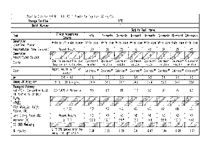
''stable" formulation is one in which the protein therein essentially retains
its
physical stability and/or chemical stability and/or biological activity upon
storage. Various
analytical techniques for measuring protein stability are available in the art
and are reviewed
in Peptide and Protein Drug Delivery, 247-301, Vincent Lee Ed., Marcel Dekker,
Inc., New
York, N.Y., Pubs. (1991) and Jones, A. Adv. Drug Delivery Rev. 10:29-90
(1993). Stability
can be measured at a selected temperature for a selected time period.
[0083] A
"stable" lyophilized antibody formulation is a lyophilized antibody
formulation with no significant changes observed at a refrigerated temperature
(2-8 C) for at
least 12 months, preferably 2 years, and more preferably 3 years; or at room
temperature (23-
27 C) for at least 3 months, preferably 6 months, and more preferably 1 year.
Typical
acceptable criteria for stability are as follows. No more than 10%, preferably
5%, of antibody
monomer is degraded as measured by SEC-HPLC. The rehydrated solution is
typically
colorless, or clear to slightly opalescent by visual analysis. The
concentration, pH and
osmolality of the formulation have no more than +/-10% change. Potency is
typically within a
range of 50-150% of the reference. No more than 10%, preferably 5% of clipping
is observed.
No more than 10%, preferably 5% of aggregation is formed.
[0084] A
"stable" pharmaceutical antibody formulation (including a lyophilized
formulation, a reconstituted liquid, as well as a liquid formulation that is a
"final" formulation
(i.e., has not been previously lyophilized)) is a pharmaceutical antibody
formulation with no
significant changes observed at a refrigerated temperature (2-8 C) for at
least 3 months,
preferably 6 months, and more preferably 1 year, and even more preferably up
through 2
years. Additionally, a "stable" liquid formulation includes one that exhibits
desired features
at temperatures including at 25 C and 40 C for periods including 1 month, 3
months, 6
months, 12 months, and/or 24 months. Typical acceptable criteria for stability
stability are as
follows. Typically, no more than about 10%, preferably about 5%, of antibody
monomer is
degraded as measured by SEC-HPLC. The pharmaceutical antibody formulation is
colorless,
or clear to slightly opalescent by visual analysis. The concentration, pH and
osmolality of the
formulation have no more than 41-10% change. Potency is typically within 50-
150 of the
reference. Typically, no more than about 10%, preferably about 5% of clipping
is observed.
Typically, no more than about 10%, preferably about 5% of aggregation is
formed.
[0085] An
antibody "retains its physical stability" in a pharmaceutical formulation if
it
shows no significant increase of aggregation, precipitation and/or
denaturation upon visual
19
CA 02830806 2013-09-19
WO 2012/135408
PCT/1JS2012/031063
examination of color and/or clarity, or as measured by UV light scattering,
size exclusion
chromatography (SEC) and dynamic light scattering. The changes of protein
conformation
can be evaluated by fluorescence spectroscopy, which determines the protein
tertiary
structure, and by FTIR spectroscopy, which determines the protein secondary
structure.
[0086] An antibody "retains its chemical stability" in a pharmaceutical
formulation, if
it shows no significant chemical alteration. Chemical stability can be
assessed by detecting
and quantifying chemically altered forms of the protein. Degradation processes
that often alter
the protein chemical structure include hydrolysis or clipping (evaluated by
methods such as
size exclusion chromatography and SDS-PAGE), oxidation (evaluated by methods
such as by
peptide mapping in conjunction with mass spectroscopy or MALDI/TOF/MS),
deamidation
(evaluated by methods such as ion-exchange chromatography, capillary
isoelectric focusing,
peptide mapping, isoaspartic acid measurement), and isomerization (evaluated
by measuring
the isoaspartic acid content, peptide mapping, etc.).
[0087] An antibody "retains its biological activity" in a pharmaceutical
formulation, if
the biological activity of the antibody at a given time is within a
predetermined range of the
biological activity exhibited at the time the pharmaceutical formulation was
prepared. The
biological activity of an antibody can be determined, for example, by an
antigen binding
assay.
[0088] The term "isotonic" means that the formulation of interest has
essentially the
same osmotic pressure as human blood. Isotonic formulations will generally
have an osmotic
pressure from about 270-328 mOsm. Slightly hypotonic pressure is 250-269 and
slightly
hypertonic pressure is 328-350 mOsm. Osmotic pressure can be measured, for
example, using
a vapor pressure or ice-freezing type osmometer.
[0089] Tonicity Modifiers: Salts (NaCl, KC1, MgCl2, CaC12, etc.) are used
as tonicity
modifiers to control osmotic pressure. In addition,
cryprotecants/lyoprotectants and/or bulking
agents such as sucrose, mannitol, glycine etc. can serve as tonicity
modifiers.
Analytical Methods
[0090] Analytical methods suitable for evaluating the product stability
include size
exclusion chromatography (SEC), dynamic light scattering test (DLS),
differential scanning
calorimetery (DSC), iso-asp quantification, potency, UV at 340 nm, UV
spectroscopy, and
FTIR. SEC (J. Pharm. Scien., 83:1645-1650, (1994); Pharm. Res., 11:485 (1994);
J. Pharm.
Bio. Anal., 15:1928 (1997); J. Pharm. Bio. Anal., 14:1133-1140 (1986))
measures percent
monomer in the product and gives information of the amount of soluble
aggregates. DSC
(Pharm. Res., 15:200 (1998); Pharm. Res., 9:109 (1982)) gives information of
protein
CA 02830806 2013-09-19
WO 2012/135408
PCT/1JS2012/031063
denaturation temperature and glass transition temperature. DLS (American Lab.,
November
(1991)) measures mean diffusion coefficient, and gives information of the
amount of soluble
and insoluble aggregates. UV at 340 nm measures scattered light intensity at
340 nm and
gives information about the amounts of soluble and insoluble aggregates. UV
spectroscopy
measures absorbance at 278 nm and gives information of protein concentration.
FTIR (Eur. J.
Pharm. Biopharm., 45:231 (1998); Pharm. Res., 12:1250 (1995); J. Pharm.
Scien., 85:1290
(1996); J. Pharm. Scien., 87:1069 (1998)) measures IR spectrum in the amide
one region, and
gives information of protein secondary structure.
[0091] The iso-
asp content in the samples is measured using the Isoquant Isoaspartate
Detection System (Promega). The kit uses the enzyme Protein Isoaspartyl
Methyltransferase
(PIMT) to specifically detect the presence of isoaspartic acid residues in a
target protein.
PIMT catalyzes the transfer of a methyl group from S-adenosyl-L-methionine to
isoaspartic
acid at the .alpha.-carboxyl position, generating S-adenosyl-L-homocysteine
(SAH) in the
process. This is a relatively small molecule, and can usually be isolated and
quantitated by
reverse phase HPLC using the SAH HPLC standards provided in the kit.
[0092] The
potency or bioidentity of an antibody can be measured by its ability to
bind to its antigen. The specific binding of an antibody to its antigen can be
quantitated by
any method known to those skilled in the art, for example, an immunoassay,
such as ELISA
(enzyme-linked immunosorbant assay).
[0093] A
"reconstituted" formulation is one that has been prepared by dissolving a
lyophilized protein formulation in a diluent such that the protein is
dispersed in the
reconstituted formulation. The reconstituted formulation is suitable for
administration, e.g.
parenteral administration), and may optionally be suitable for subcutaneous
administration.
Humanized Anti-PD-1 Antibodies
[0094] DNA
constructs encoding the variable regions of the heavy and light chains of
the humanized antibodies h409A11, h409A16 and h409A17 are described in
W02008/156712.
[0095] The
foregoing written specification is considered to be sufficient to enable one
skilled in the art to practice the invention. The present invention is not to
be limited in scope
by the culture deposited, since the deposited embodiment is intended as a
single illustration of
one aspect of the invention and any culture that is functionally equivalent is
within the scope
of this invention. The deposit of material herein does not constitute an
admission that the
written description herein contained is inadequate to enable the practice of
any aspect of the
invention, including the best mode thereof, nor is it to be construed as
limiting the scope of
21
CA 02830806 2013-09-19
WO 2012/135408
PCT/1JS2012/031063
the claims to the specific illustration that it represents. Indeed, various
modifications of the
invention in addition to those shown and described herein will become apparent
to those
skilled in the art from the foregoing description and fall within the scope of
the appended
claims.
[0096]
Sequences are provided for exemplary anti-human PD-1 antibodies; a
summary table of the sequences is provided in Table 6. CDRs are provided under
separate
sequence identifiers, as indicated in Table 2 for h409A11.
[0097]
Ordinarily, amino acid sequence variants of the humanized anti-PD-1 antibody
will have an amino acid sequence having at least 75% amino acid sequence
identity with the
original humanized antibody amino acid sequences of either the heavy or the
light chain more
preferably at least 80%, more preferably at least 85%, more preferably at
least 90%, and most
preferably at least 95, 98, or 99%. Identity or homology with respect to this
sequence is
defined herein as the percentage of amino acid residues in the candidate
sequence that are
identical with the humanized anti-PD-1 residues, after aligning the sequences
and introducing
gaps, if necessary, to achieve the maximum percent sequence identity, and not
considering
any conservative substitutions as part of the sequence identity. None of N-
terminal, C-
terminal, or internal extensions, deletions, or insertions into the antibody
sequence shall be
construed as affecting sequence identity or homology.
[0098] The
humanized antibody can be selected from any class of immunoglobulins,
including IgM, IgG, IgD, IgA, and IgE. Preferably, the antibody is an IgG
antibody. Any
isotype of IgG can be used, including IgGi, IgG2, IgG3, and IgG4. Different
constant domains
may be appended to the humanized VL and VH regions provided herein. For
example, if a
particular intended use of an antibody (or fragment) of the present invention
were to call for
altered effector functions, a heavy chain constant domain other than IgG1 may
be used.
Although IgG1 antibodies provide for long half-life and for effector
functions, such as
complement activation and antibody-dependent cellular cytotoxicity, such
activities may not
be desirable for all uses of the antibody. In such instances an IgG4 constant
domain, for
example, may be used.
[0099]
Likewise, either class of light chain can be used in the compositions and
methods herein. Specifically, kappa, lambda, or variants thereof are useful in
the present
compositions and methods.
[00100] CDR and
FR residues are determined according to the standard sequence
definition of Kabat. Kabat et al. (1987) Sequences of Proteins of
Immunological Interest,
National Institutes of Health, Bethesda Md.
22
CA 02830806 2013-09-19
WO 2012/135408 PCMJS2012/031063
[00101] The signal sequences, or nucleic acid sequences encoding the signal
sequences, may be appended to the N-terminus of the respective antibody chains
to create a
precursor protein for secretion from a host cell. Alternative signal sequences
may also be
used, and several can be found at "SPdb: a Signal Peptide Database." Choo et
al. (2005)
BMC Bioinformatics 6:249.
[00102]
TABLE 2
11409A11 CDR Sequences
Antibody CDR Sequence SEQ ID NO:
H409A11 Light chain CDR1 (equivalent to 15
hPD-1.09A light chain CDR1)
RASKGVSTSGYSYLH
H409A11 Light chain CDR2 (equivalent to 16
hPD-1.09A light chain CDR2)
LAS YLES
H409A11 Light chain CDR3 (equivalent to 17
hPD-1.09A light chain CDR3)
QHSRDLPLT
H409A11 Heavy chain CDR1 (equivalent to 18
hPD-1.09A heavy chain CDR1)
NYYMY
11409A11 Heavy chain CDR2 (equivalent to 19
hPD-1.09A heavy chain CDR2)
GINPSNGGTNFNEKFKN
H409A11 Heavy chain CDR3 (equivalent to 20
hPD-1.09A heavy chain CDR3)
RDYRFDMGFDY
Biological Activity of Humanized Anti-PD-1
[00103] Formulations of the present invention include antibodies and
fragments thereof
that are biologically active when reconstituted or in liquid form. As used
herein, the term
"biologically active" refers to an antibody or antibody fragment that is
capable of binding the
desired the antigenic epitope and directly or indirectly exerting a biologic
effect. Typically,
these effects result from the failure of PD-1 to bind its ligands. As used
herein, the term
"specific" refers to the selective binding of the antibody to the target
antigen epitope.
23
Antibodies can be tested for specificity of binding by comparing binding to PD-
1 to binding
to irrelevant antigen or antigen mixture under a given set of conditions.
Lyophilized Pharmaceutical Compositions
[00104] Lyophilized formulations of therapeutic proteins provide several
advantages.
Lyophilized formulations in general offer better chemical stability than
solution formulations,
and thus increased half-life. A lyophilized formulation may also be
reconstituted at different
concentrations depending on clinical factors, such as route of administration
or dosing. For
example, a lyophilized formulation may be reconstituted at a high
concentration (i.e. in a
small volume) if necessary for subcutaneous administration, or at a lower
concentration if
administered intravenously. High concentrations may also be necessary if high
dosing is
required for a particular subject, particularly if administered subcutaneously
where injection
volume must be minimized. One such lyophilized antibody formulation is
disclosed at U.S.
Pat. No. 6,267,958. Lyophilized formulations of another therapeutic protein
are disclosed at U.S.
Pat. No. 7,247,707.
[00105] Typically, the lyophilized formulation is prepared in anticipation
of
reconstitution at high concentration of drug product (DP, in an exemplary
embodiment
humanized anti-PD-1 antibody h409A11, or antigen binding fragment thereof),
i.e. in
anticipation of reconstitution in a low volume of water. Subsequent dilution
with water or
isotonic buffer can then readily be used to dilute the DP to a lower
concentration. Typically,
excipients are included in a lyophilized formulation of the present invention
at levels that will
result in a roughly isotonic formulation when reconstituted at high DP
concentration, e.g. for
subcutaneous administration. Reconstitution in a larger volume of water to
give a lower DP
concentration will necessarily reduce the tonicity of the reconstituted
solution, but such
reduction may be of little significance in non-subcutaneous, e.g. intravenous,
administration.
If isotonicity is desired at lower DP concentration, the lyophilized powder
may be
reconstituted in the standard low volume of water and then further diluted
with isotonic
diluent, such as 0.9% sodium chloride.
[00106] In an embodiment of the present invention, humanized anti-PD-1
antibody (or
antigen binding fragment thereof) is formulated as a lyophilized powder for
reconstituting and
utilizing for intravenous administration. Exemplary formulations are described
in Tables 3-4,
and in Figures 1-9. In certain embodiments, the antibody (or antigen binding
fragment
thereof) is provided at about 50 mg/vial, and is reconstituted with sterile
water for injection
prior to use. If desired, the reconstituted antibody may be aseptically
diluted with 0.9%
24
CA 2830806 2018-08-09
CA 02830806 2013-09-19
WO 2012/135408
PCT/1JS2012/031063
Sodium Chloride Injection USP in a sterile IV container. The target pH of the
reconstituted
formulation is 5.5 0.5. In various embodiments, the lyophilized formulation of
the present
invention enables reconstitution of the anti-PD-1 antibody to high
concentrations, such as
about 20, 25, 30, 40, 50, 60, 75, 100 or more mg/mL.
[00107] The present invention provides in certain embodiments, a
lyophilized
formulation comprising humanized anti-PD-1 antibody, a histidine buffer at
about pH 5.5, or
at about pH 5.0, for example at about 5.1, 5.2, 5.3, 5.4, 5.6, 5.7, 5.8, 5.9,
or 6Ø
[00108] When a range of pH values is recited, such as "a pH between pH 5.5
and 6.0,"
the range is intended to be inclusive of the recited values. Unless otherwise
indicated, the pH
refers to the pH after reconstitution of the lyophilized formulations of the
present invention.
The pH is typically measured at 25 C using standard glass bulb pH meter. As
used herein, a
solution comprising "histidine buffer at pH X" refers to a solution at pH X
and comprising
the histidine buffer, i.e. the pH is intended to refer to the pH of the
solution.
[00109] The formulation in Table 3 reflects the weight of the components in
a batch
formulation, as lyophilized in vials, and as reconstituted. Lyophilized
formulations are by
definition essentially dry, and thus the concept of concentration is not
useful in describing
them. Describing a lyophilized formulation in the terms of the weight of the
components in a
unit dose vial is more useful, but is problematic because it varies for
different doses or vial
sizes. In describing the lyophilized formulations of the present invention, it
is useful to
express the amount of a component as the ratio of the weight of the component
compared to
the weight of the drug substance (DS) in the same sample (e.g. a vial). This
ratio may be
expressed as a percentage. Such ratios reflect an intrinsic property of the
lyophilized
formulations of the present invention, independent of vial size, dosing, and
reconstitution
protocol.
[00110] In other embodiments, the lyophilized formulation of anti-human PD-
1
antibody, or antigen binding fragment, is defined in terms of the pre-
lyophilization solution
used to make the lyophilized formulation, such as the pre-lyophilization
solution. In one
embodiment the pre-lyophilization solution comprises antibody, or antigen-
binding fragment
thereof, at a concentration of about 25mg/mL. Such pre-lyophilization
solutions may be at pH
4.4 ¨ 5.2 (including about 4.4, 4.5, 4.6, 4.7, 4.8, 4.9, 5.0, 5.1. and 5.2),
e.g. preferably about
pH 4.8, or about pH 5.5.
[00111] In yet other embodiments, the lyophilized formulation of anti-human
PD-1
antibody, or antigen binding fragment, is defined in terms of the
reconstituted solution
CA 02830806 2013-09-19
WO 2012/135408
PCT/1JS2012/031063
generated from the lyophilized formulation, such as the reconstituted solution
disclosed at
Table 4.
[001121
Reconstituted solutions may comprise antibody, or antigen-binding fragment
thereof, at concentrations of about 10, 15, 20, 25, 30, 40, 50, 60, 75, 80, 90
or 100 mg/mL or
higher concentrations such as 150mg/mL, 200 mg/mL, 250 mg/mL, or up to about
300
mg/mL. Such reconstituted solutions may be at about pH 5.5, or range from
about pH 5.0 to
about 6.0
1001131 The
lyophilized formulations of the present invention are formed by
lyophilization (freeze-drying) of a pre-lyophilization solution. Freeze-drying
is accomplished
by freezing the formulation and subsequently subliming water at a temperature
suitable for
primary drying. Under this condition, the product temperature is below the
eutectic point or
the collapse temperature of the formulation. Typically, the shelf temperature
for the primary
drying will range from about -30 to 25 C (provided the product remains frozen
during
primary drying) at a suitable pressure, ranging typically from about 50 to 250
mTorr. The
formulation, size and type of the container holding the sample (e.g., glass
vial) and the
volume of liquid will dictate the time required for drying, which can range
from a few hours
to several days (e.g. 40-60 hrs). A secondary drying stage may be carried out
at about 0-
40 C, depending primarily on the type and size of container and the type of
protein employed.
The secondary drying time is dictated by the desired residual moisture level
in the product
and typically takes at least about 5 hours. Typically, the moisture content of
a lyophilized
formulation is less than about 5%, and preferably less than about 3%. The
pressure may be
the same as that employed during the primary drying step. Freeze-drying
conditions can be
varied depending on the formulation and vial size.
1001141 In some
instances, it may be desirable to lyophilize the protein formulation in
the container in which reconstitution of the protein is to be carried out in
order to avoid a
transfer step. The container in this instance may, for example, be a 3, 5, 10,
20, 50 or 100 cc
vial.
1001151 The
lyophilized formulations of the present invention are reconstituted prior to
administration. The protein may be reconstituted at a concentration of about
10, 15, 20, 25,
30, 40, 50, 60, 75, 80, 90 or 100 mg/mL or higher concentrations such as
150mg/mL, 200
mg/mL, 250 mg/mL, or 300 mg/mL up to about 500 mg/mL. High protein
concentrations are
particularly useful where subcutaneous delivery of the reconstituted
formulation is intended.
However, for other routes of administration, such as intravenous
administration, lower
concentrations of the protein may be desired (e.g. from about 5-50 mg/mL).
26
CA 02830806 2013-09-19
WO 2012/135408
PCT/1JS2012/031063
[00116]
Reconstitution generally takes place at a temperature of about 25 C to ensure
complete hydration, although other temperatures may be employed as desired.
The time
required for reconstitution will depend, e.g., on the type of diluent, amount
of excipient(s) and
protein. Exemplary diluents include sterile water, bacteriostatic water for
injection (BWFI), a
pH buffered solution (e.g. phosphate-buffered saline), sterile saline
solution, Ringer's solution
or dextrose solution.
[00117] The
lyophilized formulations of the present invention are expected to be stable
for at least about 36 months (based on the stability data from Figures 1-9).
In addition, the
liquid formulation is expected to exhibit stability for at least 24 months,
based on 24 months
of stability data from reconstituted h409A11formulation in polypropylene tubes
at 2-8 C.
[00118] In line
with the results shown in Figures 1-9, stability has been observed
through 2 years for a refrigerated reconstituted formulation of h409A11. 2mL
samples in
polypropylene tubes were stored at 5 C, and 25H and RH4 conditions and tested
at initial, 1,
3, 6, 9, 12, 18, and 24 month periods. This reconstituted h409A11 formulation
has the same
substituents in the same concentration as a liquid h409A11 formulation (i.e.,
a formulation
that was not lyophilized) and the stability is expected to be the same.
Liquid Pharmaceutical Compositions
[00119] A liquid
antibody formulation can be made by taking the drug substance (e.g.,
anti-humanized PD-1) which is in liquid form (e.g., h409A 1 1 in an aqueous
pharmaceutical
formulation) and buffer exchanging it into the desired buffer as the last step
of the
purification process. There is no lyophilization step in this embodiment. The
drug substance
in the final buffer is concentrated to a desired concentration. Excipients
such as sucrose and
polysorbate 80 are added to the drug substance and it is diluted using the
appropriate buffer to
final protein concentration. The final formulated drug substance is filtered
using 0.22 m
filters and filled into a final container (e.g. glass vials). Such a liquid
formulation is
exemplified by a final liquid formulation comprising 10 mM histidine pH 5.5,
7% sucrose,
0.02% polysorbate 80, and 25 mg/mL h409A11.
[00120] Various
literature references are available to facilitate selection of
pharmaceutically acceptable carriers or excipients. See, e.g., Remington's
Pharmaceutical
Sciences and US. Pharmacopeia.- National Formulary, Mack Publishing Company,
Easton,
PA (1984); Hardman et al. (2001) Goodman and Gilman 's The Pharmacological
Basis of
Therapeutics, McGraw-Hill, New York, NY; Gennaro (2000) Remington: The Science
and
Practice of Pharmacy, Lippincott, Williams, and Wilkins, New York, NY; Avis et
al. (eds.)
(1993) Pharmaceutical Dosage Forms: Parenteral Medications, Marcel Dekker, NY;
27
CA 02830806 2013-09-19
WO 2012/135408
PCT/1JS2012/031063
Lieberman, et al. (eds.) (1990) Pharmaceutical Dosage Forms: Tablets, Marcel
Dekker, NY;
Lieberman et al. (eds.) (1990) Pharmaceutical Dosage Forms: Disperse Systems,
Marcel
Dekker, NY; Weiner and Kotkoskie (2000) Excipient Toxicity and Safety, Marcel
Dekker,
Inc., New York, NY.
[00121] Toxicity is a consideration in selecting the proper dosing of a
therapeutic
agent, such as a humanized anti-PD-1 antibody (or antigen binding fragment
thereof).
Toxicity and therapeutic efficacy of the antibody compositions can be
determined by standard
pharmaceutical procedures in cell cultures or experimental animals, e.g., for
determining the
LD50 (the dose lethal to 50% of the population) and the ED50 (the dose
therapeutically
effective in 50% of the population). The dose ratio between toxic and
therapeutic effects is
the therapeutic index and it can be expressed as the ratio of LD50 to ED50.
Antibodies
exhibiting high therapeutic indices are preferred. The data obtained from
these cell culture
assays and animal studies can be used in formulating a range of dosage for use
in human. The
dosage of such compounds lies preferably within a range of circulating
concentrations that
include the ED50 with little or no toxicity. The dosage may vary within this
range depending
upon the dosage form employed and the route of administration utilized.
[00122] Suitable routes of administration may, for example, include
parenteral
delivery, including intramuscular, intradermal, subcutaneous, intramedullary
injections, as
well as intrathecal, direct intraventricular, intravenous, intraperitoneal.
Drugs can be
administered in a variety of conventional ways, such as intraperitoneal,
parenteral,
intraarterial or intravenous injection. Modes of administration in which the
volume of
solution must be limited (e.g. subcutaneous administration) require a
lyophilized formulation
to enable reconstitution at high concentration.
[00123] Alternately, one may administer the antibody in a local rather than
systemic
manner, for example, via injection of the antibody directly into a pathogen-
induced lesion
characterized by immunopathology, often in a depot or sustained release
formulation.
Furthermore, one may administer the antibody in a targeted drug delivery
system, for
example, in a liposome coated with a tissue-specific antibody, targeting, for
example,
pathogen-induced lesion characterized by immunopathology. The liposomes will
be targeted
to and taken up selectively by the afflicted tissue.
[00124] Selecting an administration regimen for a therapeutic depends on
several
factors, including the serum or tissue turnover rate of the entity, the level
of symptoms, the
immunogenicity of the entity, and the accessibility of the target cells in the
biological matrix.
Preferably, an administration regimen maximizes the amount of therapeutic
delivered to the
28
CA 02830806 2013-09-19
WO 2012/135408
PCT/1JS2012/031063
patient consistent with an acceptable level of side effects. Accordingly, the
amount of
biologic delivered depends in part on the particular entity and the severity
of the condition
being treated. Guidance in selecting appropriate doses of antibodies,
cytokines, and small
molecules are available. See, e.g., Wawrzynczak (1996) Antibody Therapy, Bios
Scientific
Pub. Ltd, Oxfordshire, UK; Kresina (ed.) (1991) Monoclonal Antibodies,
Cytokines and
Arthritis, Marcel Dekker, New York, NY; Bach (ed.) (1993) Monoclonal
Antibodies and
Peptide Therapy in Autoimmune Diseases, Marcel Dekker, New York, NY; Baert et
al.
(2003) New Engl. J. Med 348:601-608; Milgrom et al. (1999) New EngL J Med.
341:1966-
1973; Slamon et al. (2001) New Engl. J Med. 344:783-792; Beniaminovitz etal.
(2000) New
Engl. I Med. 342:613-619; Ghosh et al. (2003) New Engl. J. Med. 348:24-32;
Lipsky et al.
(2000) New EngL J. Med 343:1594-1602; Physicians' Desk Reference 2003
(Physicians'
Desk Reference, 57th Ed); Medical Economics Company; ISBN: 1563634457; 57th
edition
(November 2002).
[00125]
Determination of the appropriate dose is made by the clinician, e.g., using
parameters or factors known or suspected in the art to affect treatment or
predicted to affect
treatment. The appropriate dosage ("therapeutically effective amount") of the
protein will
depend, for example, on the condition to be treated, the severity and course
of the condition,
whether the protein is administered for preventive or therapeutic purposes,
previous therapy,
the patient's clinical history and response to the protein, the type of
protein used, and the
discretion of the attending physician. Generally, the dose begins with an
amount somewhat
less than the optimum dose and it is increased by small increments thereafter
until the desired
or optimum effect is achieved relative to any negative side effects. Important
diagnostic
measures include those of symptoms of, e.g., the inflammation or level of
inflammatory
cytokines produced. The antibody is suitably administered to the patient at
one time or
repeatedly. The antibody may be administered alone or in conjunction with
other drugs or
therapies.
[00126] A
pharmaceutical antibody formulation can be administered by continuous
infusion, or by doses at intervals of, e.g., one day, 1-7 times per week, one
week, two weeks,
three weeks, monthly, bimonthly, etc. A preferred dose protocol is one
involving the
maximal dose or dose frequency that avoids significant undesirable side
effects. A total
weekly dose is generally at least 0.05 pg/kg, 0.2 ps/kg, 0.5 ps/kg, 1 lug/kg,
10 jig/kg, 100
jig/kg, 0.2 mg/kg, 1.0 mg/kg, 2.0 mg/kg, 10 mg/kg, 25 mg/kg, 50 mg/kg body
weight or more.
See, e.g., Yang et al. (2003) New Engl. I Med 349:427-434; Herold et al.
(2002) New Engl.
J. Med. 346:1692-1698; Liu et al. (1999)1 Neurol. Neurosurg. Psych. 67:451-
456; Portielji
29
CA 02830806 2013-09-19
WO 2012/135408
PCT/1JS2012/031063
et al. (20003) Cancer Immunol. Immunother. 52:133-144. The desired dose of a
small
molecule therapeutic, e.g., a peptide mimetic, natural product, or organic
chemical, is about
the same as for an antibody or polypeptide, on a moles/kg basis.
[00127] In
certain embodiments, dosing will comprise administering to a subject
escalating doses of 1.0, 3.0, and 10 mg/kg of the pharmaceutical formulation,
i.e, a
formulation comprising h409A11, over the course of treatment. The formulation
comprising
h409A11 can be a reconstituted liquid formulation, or it can be a liquid
formulation not
previously lyophilized. Time courses can vary, and can continue as long as
desired effects are
obtained. In certain embodiments, dose escalation will continue up to a dose
of about
10mg/kg. In certain embodiments, the subject will have a histological or
cytological
diagnosis of melanoma, or other form of solid tumor, and in certain instances,
a subject may
have non-measurable disease. In certain embodiments, the subject will have
been treated
with other chemotherapeutics, while in other embodiments, the subject will be
treatment
naïve.
[00128] In yet
additional embodiments, the dosing regimen will comprise
administering a dose of 1, 3, or 10mg/kg of any of the pharmaceutical
formulations described
herein (i.e, a formulation comprising h409A11), throughout the course of
treatment. For such
a constant dosing regimen, the interval between doses will be about 14 days (
2 days). In
certain embodiments, the interval between doses will be about 21 days ( 2
days).
[00129] In
certain embodiments, the dosing regimen will comprise administering a
dose of from about 0.005mg/kg to about 10mg/kg, with intra-patient dose
escalation. In
certain embodiments, a dose of 5 mg/kg or 10 mg/kg will be administered at
intervals of
every 3 weeks, or every 2 weeks. In yet additional embodiments, a dose of
3mg/kg will be
administered at three week intervals for melanoma patients or patients with
other solid
tumors. In these embodiments, patients should have non-resectable disease;
however,
patients may have had previous surgery.
[00130] In
certain embodiments, a subject will be administered a 30 minute IV infusion
of any of the pharmaceutical formulations described herein. In certain
embodiments for the
escalating dose, the dosing interval will be about 28 days (( 1 day) between
the first and
second dose. In certain embodiments, the interval between the second and third
doses will be
about 14 days ( 2 days). In certain embodiments, the dosing interval will be
about 14 days
( 2 days), for doses subsequent to the second dose.
[00131] In
certain embodiments, the use of cell surface markers and/ore cytokine
markers, as described in co-pending patent publications W02012/018538 or
CA 02830806 2013-09-19
WO 2012/135408
PCT/1JS2012/031063
W02008/156712 will be used in bioassays for monitoring, diagnostic, patient
selection,
and/or treatment regimens involving blockade of the PD-1 pathway.
[00132]
Subcutaneous administration may performed by injected using a syringe, or
using other injection devices (e.g. the Inject-ease device); injector pens;
or needleless
devices (e.g. MediJector and BioJector8).
[00133] The
broad scope of this invention is best understood with reference to the
following examples, which are not intended to limit the inventions to the
specific
embodiments. The specific embodiments described herein are offered by way of
example
only, and the invention is to be limited by the terms of the appended claims,
along with the
full scope of equivalents to which such claims are entitled.
EXAMPLES
EXAMPLE 1
Antibody Production
[00134] h409Al1
is a humanized monoclonal antibody that binds to human PD-1 and
blocks the interaction between PD-1 and its ligands PDL1 and PDL2. The
antibody is an
IgG4/kappa isotype with a stabilizing S228P sequence alteration in the Fc
region. Table 2
provides a list of the CDR sequences. The theoretical molecular weights of the
heavy and
light chains derived from the amino acid sequences, excluding glycosylation,
are 49.3kDa and
23.7 kDa, respectively. The parental antibody (hPD-1.09A) was produced by
immunizing
mice with hPD-1 DNA. The h409A 1 1 antibody was generated by humanization of
the
parental murine anti-human PD-1 antibody by the Medical Research Council
(Cambridge,
UK) using CDR grafting technology, (e.g., U.S. Patent No. 5,225,539), as
described in co-
pending W02008/156712.
[00135] An
expression plasmid was constructed for expression of heavy and light
chains of h409A11. The nucleotide sequences encoding the heavy and light
chains, along
with their respective promoters and poly A signal sequence, were confirmed by
DNA
sequence analysis. The expression vector was subsequently used to transfect a
CHO cell
line. An antibody-expressing clone was selected for the generation of a Master
Seed Bank
(MSB), based on growth, productivity, and production stability. This MSB was
then used to
prepare the antibody and to generate the Master Cell Bank (MCB).
31
CA 02830806 2013-09-19
WO 2012/135408
PCT/1JS2012/031063
[00136] Cells
from the MCB were expanded in shake flasks, culture bags, and a seed
bioreactor to generate the inoculum for a production bioreactor to produce the
antibody
product. Further processing included three chromatography steps (protein A
affinity, cation
exchange and anion exchange chromatography), two orthogonal viral clearance
steps (low pH
viral inactivation and viral reduction filtration),
ultrafiltration/diafiltration, and a final 0.21.1m
filtration step.
h409A11 Structure and Features
[00137] h409A1
1 is a highly selective humanized monoclonal antibody that blocks the
interaction between human PD-1 and its ligands PD-Li and PD-L2. h409A1 1 is
heterogeneously glycosylated at asparagine 297 within the Fe domain of each
heavy chain,
yielding molecular weights typically ranging between 148.9 and 149.5 kDa,
depending on the
attached glycan chains. The amino acid sequences of the heavy and light chains
of h409Al1
are found in SEQ ID NO:31 and SEQ ID NO:36. The light chain without the leader
sequences
comprises amino acid residues 20 to 237 of SEQ ID NO: 36 and the heavy chain
without the
leader sequences comprises amino acid residues 20 to 466 of SEQ ID NO: 31.
Stable humanized PD-1 formulations
[00138] In
certain embodiments, stable humanized PD-1 e.g., h409A1l is an aqueous
solution stored under refrigerated conditions (temp. range: typically about 2-
8 C, but under
certain circumstances, the aqueous formulation may exhibit stability at other
temperatures
including at about 25 C and about 40 C for periods of up to about 12 months)
at a
concentration of? 25mg/mL in 10 mM Histidine buffer, pH 5.0-6Ø In certain
embodiments,
stable humanized PD-1 e.g., h409A1 1 is an aqueous solution at a concentration
of about 25
mg/mL in 10 mM Histidine buffer, pH 5.0-6Ø The stable formulation (Le., drug
substance)
is typically a clear to opalescent solution and may contain particulates.
[00139] In
certain embodiments, a liquid or frozen solution of h409Al1 is formulated
in histidine buffer (pH 5.5) containing sucrose and polysorbate 80.
[00140] An
additional exemplary formulation includes: h409A1 1 formulated in
histidine buffer (pH 5.5) containing sucrose and polysorbate 80 in lyophilized
form.
[00141] In
certain embodiments, stable humanized PD-1 formulation is provided as
lyophilized powder in vials intended for single-use.
[00142] In
certain embodiments, stable humanized PD-1 formulation is reconstituted
with water for injection (WFI) and aseptically diluted with appropriate
volumes of 0.9%
sodium chloride for injection in a sterile IV container to form an admixture
solution.
Biological Activity
32
CA 02830806 2013-09-19
WO 2012/135408
PCT/1JS2012/031063
[00143]
Biological activity of the humanized anti-PD-1 antibody is measured by its
ability to compete with PD-Li (natural ligand of PD-1) in binding to human PD-
1, quantified
in competitive ELISA relative to a reference material. The stable formulations
described
herein exhibit biological activity for long periods of time, including up to
at least about
eighteen months. The stability of several batches of h409A11, under various
storage
conditions are illustrated in Figures 1-9.
Stable Formulations of Humanized Anti-PD-1 Antibodies
[00144]
Lyophilized formulations of anti-PD-1 antibody are prepared as follows. An
exemplary batch formula for h409A1 1 antibody is provided in Table 3. The
final
concentration of antibody is 25 mg/mL. This batch formulation may be used to
prepare the
lyophilized 50 mg/vial units, as discussed with reference to Table 4, infra.
Polysorbate 80
from a vegetable source is used. Additional hydrochloric acid or sodium
hydroxide may be
added to adjust the pH to the desired value of approximately 5.5 ( 0.2). The
components are
brought to a final volume of 14 L with sterile water for injection (WFI).
Correspondingly
smaller lots may be prepared by proportional reduction of the amounts listed
in Table 3.
[00145] An
exemplary liquid antibody formulation is prepared by taking the drug
substance (e.g., anti-humanized PD-1 from a batch formula described herein)
which is in
liquid form (e.g., h409A11 in an aqueous formulation) and buffer exchanging it
into the
desired buffer as the last step of the purification process. In this instance,
there is no previous
lyophilization step. The drug substance in the final histidine buffer is
concentrated to a
desired concentration. Excipients such as sucrose and polysorbate 80 are added
to the drug
substance and it is diluted using the appropriate buffer to final protein
concentration. The
final formulated drug substance is filtered using 0.22tim filters and filled
into a final container
(e.g. glass vials). Such a liquid formulation includes final liquid
formulation comprising 10
mM histidine pH 5.5, 7% sucrose, 0.02% polysorbate 80, and 25 mg/mL h409A11.
[00146]
Table 3
Batch Formula of Representative 14.0 L Pre-lyophilization Solution for h409A1
1
Powder for Injection, 50 mg/vial
Component CompendialGrade Concentration Amount per Batch
(g)
(mg/mL)
h409A11 antibody N/A 25.0 350.0
33
CA 02830806 2013-09-19
WO 2012/135408 PCT/1JS2012/031063
L-Histidine USP 1.55 21.7
Polysorbate 80 NF 0.2 2.8
Sucrose NF 70 980
Hydrochloric acid a NF
Sodium Hydroxide a NF pH adjustment
Water for injection b USP 14.0 L @ q.s
a Hydrochloric acid and sodium hydroxide added if needed to adjust pH to 5.5
b Water removed by sublimation and desorption during lyophilization
[00147] The unit composition of an exemplary final lyophilized formulation
of
humanized anti-PD-1 is provided at Table 4.
Table 4
Unit Composition of Lyophilized Powder Formulation for Injection, 50 mg/vial
Component Grade Amount Concentration after Function
(mg/vial) Reconstitution
(mg/mL)b
h409A11 N/A 50 25 Drug
Substance/Active
Pharmaceutical
ingredient
L-Histidine USP 3.1 1.55 Buffer
Polysorbate 80 NF 0.4 0.2 Surfactant
Sucrose NF 140 70 Stabilizer/
Tonicity Modifier
Hydrochloric acid c NF pH adjustment
Sodium Hydroxide c NF pH adjustment
Sterile Water for Injection USP 2.0 mL
@ Solvent
(sWFI or WFI)d q.s.
a An excess fill of 0.4 mL is provided to ensure the recovery of 50 mg h409A11
per vial.
b Following reconstitution with 2.3 mL sterile water for injection.
C Hydrochloric acid and sodium hydroxide added if needed to adjust pH to 5.5
d Water removed by sublimation and desorption during lyophilization
34
CA 02830806 2013-09-19
WO 2012/135408
PCT/1JS2012/031063
[00148] The unit
formulation of Table 4 comprises 1/20,000th of the batch formulation
of Table 3 after lyophilization to remove the water. The 50 mg of DS is added
as 2.0 mL of
the 25 mg/mL batch formulation of Table 3. Each vial is filled with 2.4 mL and
reconstituted
with 2.3 mL sWFI, resulting in approximately 2.4 mL of reconstituted solution
due to
expansion volume of the lyophilized cake.
[00149] The drug
is packaged in sterile 20 mm neck, 6R DIN, Type 1 glass tubing
vials, closed with 20-mm gray butyl rubber stoppers and sealed with aluminum
crimp seals.
Vials are stored at 2 ¨ 8 C, and refrigerated when shipped.
[00150]
Compounding involves the following steps. Charge the required amount of
water for injection (WFI) into a tared compounding vessel. Charge and dissolve
with mixing,
sucrose, histidine, and polysorbate 80 from a vegetable source. Measure the pH
and adjust if
needed to bring the pH to about 5.4 --5.6. Use hydrochloric acid and/or sodium
hydroxide to
adjust the pH. Equilibrate the drug substance to ambient temperature and
charge the drug
substance slowly into the compounding vessel. Continue to mix gently to avoid
foaming.
Measure the pH again and adjust if needed to bring the pH to approximately
5.5. Charge WFI
to the final weight of the bulk solution with continued gentle mixing.
[00151]
Filtration involves the following steps. Connect clarifying filter (0.22 inn)
and
sterilizing filter (0.22 m) to the compounding vessel. Collect an aliquot of
the bulk solution
for bioburden testing after clarifying filtration step. Perform aseptic
filtration using a 0.22
pm filter into a sterile container. Remove aliquot of sample after aseptic
filtration for bulk
sterility testing. Perform filter integrity testing after product filtration.
[00152] Filling
involves the following steps. Using suitable filling equipment,
aseptically fill the product solution into sterilized Type I tubing glass
vials to achieve a target
fill volume of 2.4 mL. Perform fill weight checks during filling. Partially
seat sterilized lyo-
shape stoppers into filled vials. Load the filled vials into a suitable freeze-
dryer.
[00153]
Lyophilization, stoppering and capping involve the following steps.
Lyophilize the filled vials using an appropriate lyophilization cycle. After
lyophilization is
complete, backfill the vials with 0.22 p.m filtered nitrogen and fully
stopper. Unload the
stoppered vials from the lyophilizer and seal them.
1001541 The
resulting vials are inspected for visual defects and stored at 2-8 C.
Finished unit dosage vials are shipped under refrigerated conditions.
CA 02830806 2013-09-19
WO 2012/135408
PCT/1JS2012/031063
EXAMPLE 2
Stability Testing of Lyophilized Formulations of Humanized Anti-PD-1
Antibodies
[00155] Figures 1-9 provide data of stability testing of lyophilized
formulations of a
humanized anti-human PD-1 antibody under various storage conditions. Vials
were stored in
upright configurations. As discussed in more detail below, formulations of the
present
invention show stability through at least 24 months for antibodies lyophilized
at pH 5.5
(histidine buffer), as well as similar liquid formulations.
[00156] Stability was assessed as follows. Samples were lyophilized in 6R
DIN Type I
glass vials, and sealed with 20 mm bromobutyl lyo stoppers (Helvoet Rubber &
Plastic
Technologies BY, Hellevoetsluis, The Netherlands) and flip-off aluminum seals.
Vials were
placed on stability stations under the following storage conditions: 5 C (5 3
C), 25H (25,
60% relative humidity), or RH4 (40 C, 70% relative humidity). Samples were
obtained at an
initial time point, and for certain samples at a variety of time points
including 1, 2, 3, 6, 9,
12, 18, and 24 months.
[00157] The stability of the samples is illustrated by the various
characteristics
presented in the tables in FIGS 1-9. The lyophilized samples were visually
inspected,
reconstituted, and the reconstituted formulation was visually inspected. The
pH of the
samples after reconstitution was measured, and the protein concentration
determined by U.V.
absorbance. The samples were analyzed by CE-SDS technique in which protein was
denatured with sodium dodecyl sulfate (SDS) under reducing and non-reducing
conditions
and separated using capillary electrophoresis (CE). The proteins separate
based on their
apparent molecular weight. Under non-reducing conditions, all species other
than the main
IgG peak are classified as impurities. Under reducing conditions, the IgG is
resolved into the
heavy and light chains. All other species are classified as impurities.
[00158] Purity of the sample was further assessed by high performance size
exclusion
chromatography (HPSEC) in which the percentage of monomer was determined, as
well as
the percentages of high molecular weight species (possibly aggregates) and
late eluting peaks
(possibly degradation products).
[00159] Additional sample characterization data are provided in Figures 1-
9. High
performance ion-exchange chromatography (HP-IEX) was used to assess purity by
revealing
the presence of acidic or basic variants. Results are presented as a
percentage of total
observed material. The samples were further characterized for biological
function using an
enzyme-linked immunosorbent assay (ELISA) for binding to human PD-1. The
antibody
concentration necessary to achieve half-maximal binding is called EC50.
Potency of the test
36
CA 02830806 2013-09-19
WO 2012/135408
PCT/1JS2012/031063
sample was assessed by comparing binding curves of the test samples to a
reference material
(or control) by the ration of EC50's. Potency was expressed as percent
relative potency of
reference material (or control). Moisture
content of the lyophilized powder was also
determined by coulometric titration. Particulate matter count measurements
were performed
to count particles? 10 p.m and? 25 pm. The method used for these measurements
was based
on USP<788>.
[00160] These
results demonstrate high stability formulations of the present invention
over at least 24 months at about pH 5.5. The data reveal no trending over time
that would
reflect instability for samples the tested storage conditions.
[00161] EXAMPLE 3: INITIAL CLINICAL RESULTS
Phase 1 Study of h409A11 (Anti-PD-1 Monoclonal Antibody) in Patients With
Advanced Solid Tumors
[00162]
A phase 1 trial examined safety, PK, PD, and antitumor activity of h409A11.
An open-label, dose escalation study was conducted in patients with advanced
malignancy
refractory to standard chemotherapy. In the initial patient set, patients with
advanced solid
tumors were treated with a stable h409Al1 formulation as described herein.
There was no
limitation/restriction regarding surgery; however, patients were not currently
surgical
candidates. Cohorts of 3-6 patients were enrolled (3+3 design) at IV doses of
1, 3, or 10
mg/kg. Following an initial dose and 28-day Cycle 1, patients were allowed to
subsequently
receive multiple doses given every 2 wks. For phase 1 part A, three patients
were treated at 1
mg/kg, three patients were treated at 3mg/kg, and nine patients were treated
at 10 mg/kg and
all were dosed every 2 weeks. There was no intrapatient dose escalation.
Radiographic
assessment was conducted every 8 wks using RECIST 1.1 guidelines.
Nine patients, 3 at each dose level, completed the dose-limiting toxicity
(DLT) period
(28 d). Patients had non¨small cell lung cancer (NSCLC, n=3), rectal cancer
(n=2), melanoma
(MEL, n=2), sarcoma (n=1), or carcinoid (n=1). To date, a total of 63 doses
were
administered (median 7/patient; max 12) without DLT. Drug-related adverse
events (AEs)
across all doses included Grade 1 fatigue (n=3), nausea (n=2), diarrhea (n=1),
dysgeusia
(n=1), breast pain (n=1), and pruritus (N=1). One drug-related Grade 2 AE of
pruritus was
reported. No drug-related AEs > grade 3 were observed. PK data are shown in
Table 5. Based
on RECIST, 1 patient with MEL on therapy >6 mths had a partial response, and
preliminary
evidence of tumor size reduction (stable disease) was observed in 3 additional
patients with
37
CA 02830806 2013-09-19
WO 2012/135408
PCT/1JS2012/031063
advanced cancer. These results show that h409A1 I was well-tolerated without
DLT across 3
tested dose levels. ('i.e., I, 3, and 5 mg/kg). Evidence of antitumor activity
was observed.
[00163] TABLE 5. Mean (CV%) PK Parameter Values of MK-3475 Following
Single IV Dose of 1, 3, or 10 mg,/kg in Cycle 1
Dose N Cmax AUC(0-28day) t1/2a
(mg/kg) (ag/mL) (vig=day /mL) (day)
1 4 16.8 (23) 163 (20) b 15.1 (41)b
3 3 109(26) 990(23) 21.7 (11)
2 337(8) 2640 (30) 13.6 (28)
'PK sampling up to 28 days following first IV administration, therefore t112
not fully
characterized.
bN=3 due to subject discontinuation.
Table 6 provides a brief description of the sequences in the sequence listing.
Sequence Identifiers
SEQ ID Description
NO:
1 hPD-1.08A heavy chain variable region (DNA)
2 hPD-1.08A light chain variable region (DNA)
3 hPD-1.09A heavy chain variable region (DNA)
4 hPD-1.09A light chain variable region (DNA)
5 hPD-1.08A heavy chain variable region (AA)
6 hPD-1.08A light chain variable region (AA)
7 hPD-1.09A heavy chain variable region (AA)
8 hPD-1.09A light chain variable region (AA)
9 hPD-1.08A light chain CDR1 (AA)
10 hPD-1.08A light chain CDR2 (AA)
11 hPD-1.08A light chain CDR3 (AA)
12 hPD-1.08A heavy chain CDR1 (AA)
13 hPD-1.08A heavy chain CDR2 (AA)
14 hPD-1.08A heavy chain CDR3 (AA)
hPD-1.09A light chain CDR1 (AA)
16 hPD-1.09A light chain CDR2 (AA)
38
CA 02830806 2013-09-19
WO 2012/135408
PCT/1JS2012/031063
17 hPD-1.09A light chain CDR3 (AA)
18 hPD-1.09A heavy chain CDR1 (AA)
19 hPD-1.09A heavy chain CDR2 (AA)
20 hPD-1.09A heavy chain CDR3 (AA)
21 109A-H heavy chain variable region (DNA)
22 Codon optimized 109A-H heavy chain variable region (DNA)
23 Codon optimized 409A-H heavy chain full length (DNA)
24 KO9A-L-11 light chain variable region (DNA)
25 KO9A-L-16 light chain variable region (DNA)
26 KO9A-L-17 light chain variable region (DNA)
27 Codon optimized KO9A-L-11 light chain variable region (DNA)
28 Codon optimized KO9A-L-16 light chain variable region (DNA)
29 Codon optimized KO9A-L-17 light chain variable region (DNA)
30 109A-H heavy chain variable region (AA)
31 409A-H heavy chain full length (AA)
32 KO9A-L-11 light chain variable region (AA)
33 KO9A-L-16 light chain variable region (AA)
34 KO9A-L-17 light chain variable region (AA)
35 109A-H heavy chain full length (AA)
36 KO9A-L-11 light chain full length (AA)
37 KO9A-L-16 light chain full length (AA)
38 KO9A-L-17 light chain full length (AA)
[00164] As used herein, including the appended claims, the singular forms
of words
such as "a," "an," and "the," include their corresponding plural references
unless the context
clearly dictates otherwise. Unless otherwise indicated, the proteins and
subjects referred to
herein are human proteins and subject, rather than another species.
[00165] REFERENCES
1. Sharpe, A.H, Wherry, E.J., Ahmed R., and Freeman G.J. The function of
programmed cell
death 1 and its ligands in regulating autoimmunity and infection. Nature
Immunology (2007);
8:239-245.
39
CA 02830806 2013-09-19
WO 2012/135408
PCT/1JS2012/031063
2. Greenwald R.J., Freeman G.J., and Sharpe A.H. The B7 family revisited.
Annual Reviews
of Immunology (2005); 23:515-548.
3. Okazaki T and Honjo T. PD-1 and PD-1 ligands: from discovery to clinical
application.
International immunology (2007);19:813-824.
4. Chemnitz et al. SHP-1 and SHP-2 associate with immunoreceptor tyrosine-
based switch
motif of programmed death 1 upon primary human T cell stimulation, but only
receptor
ligation prevents activation. .1 Immunol. (2004): 173: 945-954.
5. Nishimura, H., Nose, M., Hiai, H. et al. Development of lupus-like
autoimmune diseases
by disruption of the PD-I gene encoding an ITIM motif-carrying immunoreceptor.
Immunity
(1999);11:141-151.
6. Okazaki T et al. Autoantibodies against cardiac troponin I are responsible
for dilated
cardiomyopathy in PD-1 deficient mice. Nature Medicine (2003): 9: 1477-1483.
7. Ansari MJ. The programmed death-1 pathway regulates diabetes in nonobesc
diabetic
(NOD) mice. J Exp. Med. (2003), Jul 7;198(1):63-9.
8. Riley J and June C. The road to recovery: translating PD-1 biology into
clinical benefit.
Trends in Immunology (2006): 28:48-50.
9. Barber DL. Restoring function in exhausted CD8 T cells during chronic viral
infection.
Nature (2006):439: 682-687.
10. Trautmann L et al. Upregulation of PD-1 expression on HIV-specific CD8+ T
cells leads
to reversible immune dysfunction. Nature Medicine (2006) 12: 1198-1202.
11. Petrovas C et al. PD-1 is a regulator of virus-specific CD8+ T cell
survival in HIV
infection. J Exp. Med. (2006): 203: 2281-2292.
CA 02830806 2013-09-19
WO 2012/135408
PCT/1JS2012/031063
12. Day CL et al. PD-1 expression on HIV-specific T cells is associated with T-
cell
exhaustion and disease progression, Nature. 2006 Sep 21;443(7109):350-4.
13. Velu V et al. 2009. Enhancing SW-specific immunity in vivo by PD-1
blockade. Nature
(2009) 458: 206-210.
14. Finnefrock et al. PD-1 blockade in rhesus macaques: inpact on chronic
infection and
prophylactic vaccination. I of Immunol. (2009): 182: 980-987.
15. Dong H et al. Tumor-associated B7-H1 promotes T-cell apoptosis: a
potential mechanism
of immune evasion. Nat Med. 2002 Aug;8(8):793-800.
16. Yang et al. PD-1 interaction contributes to the functional suppression of
T-cell responses
to human uveal melanoma cells in vitro. Invest Ophthalmol Vis Sci. 2008
Jun;49(6 (2008):
49: 2518-2525.
17. Ghebeh et al. The B7-H1 (PD-L1) T lymphocyte-inhibitory molecule is
expressed in
breast cancer patients with infiltrating ductal carcinoma: correlation with
important high-risk
propgnostic factors. Neoplasia (2006) 8: 190-198.
18. Hamanishi J et al. Programmed cell death 1 ligand 1 and tumor-infiltrating
CD8+ T
lymphocytes are prognostic factors of human ovarian cancer. Proceeding of the
National
Academy of Sciences (2007): 104: 3360-3365.
19. Thompson RH et al. Significance of B7-H1 overexpression in kidney cancer.
Clinical
genitourin Cancer (2006): 5:206-211.
20. Nomi, T. Sho, M., Akahori, T., etal. Clinical significance and therapeutic
potential of the
programmed death- 1 ligand/programmed death-1 pathway in human pancreatic
cancer.
Clinical Cancer Research (2007);13 :2151-2157.
21. Ohigashi Y et al. Clinical significance of programmed death-1 ligand-1 and
programmed
death-1 ligand 2 expression in human esophageal cancer. Clin. Cancer Research
(2005): 11:
2947-2953.
41
22. Inman et al. PD-Li (B7-H1) expression by urothelial carcinoma of the
bladder and BCG-
induced granulomata: associations with localized stage progression. Cancer
(2007): 109:
1499-1505.
23. Shimauchi T et al. Augmented expression of programmed death-1 in both
neoplasmatic
and nonneoplastic CD4+ T-cells in adult T-cell Leukemia/ Lymphoma. Int. J.
Cancer (2007):
121:2585-2590.
24. Gao et al. Overexpression of PD-Li significantly associates with tumor
aggressiveness
and postoperative recurrence in human hepatocellular carcinoma. Clinical
Cancer Research
(2009) 15: 971-979.
25. Nakanishi J. Overexpression of B7-H1 (PD-L1) significantly associates with
tumor grade
and postoperative prognosis in human urothelial cancers. Cancer Immunol
Immunother.
(2007) 56: 1173- 1182.
26. Hino et al. Tumor cell expression of programmed cell death-1 is a
prognostic factor for
malignant melanoma. Cancer (2010): 00: 1-9.
[00166] Citation of
the references herein is not intended as an admission that the reference is
pertinent prior art, nor does it constitute any admission as to the contents
or date of these
publications or documents.
**********************************************************
<160> 38 Sequences
42
CA 2830806 2018-08-09