Note : Les descriptions sont présentées dans la langue officielle dans laquelle elles ont été soumises.
CA 02835147 2013-11-05
WO 2012/159215
PCT/CA2012/050342
- 1 -
COMPOSITIONS AND METHODS FOR EFFICACIOUS AND
SAFE DELIVERY OF siRNA USING SPECIFIC CHITOSAN-
BASED NANOCOMPLEXES
CROSS-REFERENCE TO RELATED APPLICATIONS
[0001] This application claims priority from U.S. provisional patent
application 61/489,306 filed on May 24, 2011 and from U.S. provisional patent
application 61/489,302 filed on May 24, 2011, herewith incorporated in their
entirety.
TECHNICAL FIELD
[0002] The
present description relates to a composition and a method for the
efficient delivery of a therapeutic RNAi-inducing nucleic acid using specific
chitosan based nanocomplexes.
BACKGROUND
[0003] Gene
silencing by siRNA (short interfering RNA) is a developing field
in biology and has evolved as a novel post-transcriptional gene silencing
strategy with therapeutic potential. Based on the sequencing of the human
genome and the understanding of the molecular causes of diseases, the
possibility of turning off pathogenic genes at will is an appealing approach
for
treatment of a wide variety of clinical pathologies, such as diabetes,
atherosclerosis and cancer. With siRNAs, virtually every gene in the human
genome contributing to a disease becomes amenable to regulation, thus
opening opportunities for drug discovery. Whereas locally administered siRNAs
have already entered the first clinical trials, strategies for successful
systemic
delivery of siRNA are still in a preclinical stage of development.
Type II diabetes mellitus
[0004] Type ll
diabetes mellitus (T2DM) is a progressive metabolic disorder
with diverse pathologic manifestations and is often associated with lipid
metabolism and glycometabolic disorders (Bell et al., 2001, Nature, 414:788-
791). Type ll diabetes is characterized by a resistance to insulin action in
peripheral tissues such as muscle, adipose tissue and liver. It is also
characterized by a progressive failure in the ability of the islet 8-cell to
secrete
CA 02835147 2013-11-05
WO 2012/159215
PCT/CA2012/050342
- 2 -
insulin. The long term effects of diabetes result from its vascular
complications;
micro vascular complications, retinopathy, neuropathy and nephropathy. Macro
vascular complications are associated with type ll diabetes as well, and
include
cardiovascular and cerebrovascular complications.
[0005] The main
classes of anti-diabetic drugs known today are the
following. Biguanides are a class of drugs that help control blood glucose by
inhibiting hepatic glucose production, reducing intestinal absorption and
enhancing peripheral glucose uptake. This class includes metformin, a drug
that
lowers both glucose and blood triglycerides level. Sulfonylurea is a class of
drugs that helps in controlling or managing type ll diabetes by stimulating
the
release of endogenous insulin from the p-cells of the pancreas. This class
includes: tolbutamide, tolazamide, glisoxepide, glimipeide and glibomuride
among others. Glycosidase inhibitors stimulate the release of insulin from
pancreatic cells thus lowering blood sugar level and include repaglinide and
nateglinide.
[0006]
Unfortunately, these treatment modalities, even when combined, are
frequently constrained by safety, tolerability, weight gain, oedema and
gastrointestinal intolerance (Drucker et al., 2010, Nat Rev Drug Discov, 9:267-
268; Nauck et al., 2009, Diabetes Care, 32:84-90; Ng et al., 2010, Prim Care
Diabetes, 4:61-63; Truitt et al., 2010, Curr Med Res Opin, 26:1321-1331; and
Wajcberg and Tavaria, 2009, Expert Opin Pharmacother, 10:135-142). In
addition, as the disease progresses and p-cell function declines, efficacies
of
current treatments diminish (Turner et al., 1999, JAMA, 281:2005-2012).
[0007] The
discovery of the incretin effect has provided a new avenue of
treatment using a class of therapeutics capable of controlling T2DM with
minimal adverse effects. The incretin effect is mainly mediated by glucagon
like
peptide 1 (GLP-1) which regulates postprandial blood glucose level via the
stimulation of insulin secretion. GLP-1 has also indirect effects such as
delay of
gastric emptying, promoting satiety through its effect on the central nervous
system, promoting p-cell growth and inhibiting p-cell apoptosis as
demonstrated
in animal models (Nauck et al., 2002, J Clin Endocrinol Metab, 87:1239-1246;
CA 02835147 2013-11-05
WO 2012/159215
PCT/CA2012/050342
- 3 -
and Creutzfeldt et al., 1996, Diabetes Care, 19:580-586). However, the
potential
of GLP-1 in the clinic was hindered due to its rapid degradation by the
ubiquitous serine protease dipeptidyl peptidase IV (DPP-IV). The discovery
that
DPP-IV cleaves the His:Ala:Glu sequence at the N-terminal region of GLP-1
permitted the development of DPP-IV resistant GLP-1 analogues and the
development of DPP-IV inhibitors.
[0008] DPP-IV
inhibitors are a new class of drugs that inhibit the proteolytic
activity of dipeptidyl peptidase IV. The proteolytic activity of DPP-IV
decreases
blood level of glucoregulatory peptides, known as incretins. Inhibition of
dipeptidyl peptidase IV thereby potentiates the action of these incretin,
notably
glucagon like peptide 1 (GLP-1). These inhibitors include Sitagliptin,
Vildagliptin
and Saxagliptin and are orally administrated once daily.
Atherosclerosis
[0009]
Atherosclerosis is a chronic disease caused by the formation of
atherosclerotic plaque in arteries. Atherosclerosis represents a multitude of
cardiovascular diseases such as coronary heart disease, acute coronary
syndrome and angina pectoris (Lloyd-Jones et al., 2010, Circulation, 121:e46-
e215). In the United-States, the predicted economic cost of atherosclerosis
for
2010 was US$503 billion, mainly due to direct medical and indirect
productivity
costs (Lloyd-Jones et al., 2010, Circulation, 121:948-954). Although causal
factors for atherosclerosis remain unknown, increasing evidence suggest a high
role of dyslipidemia, hyperlipidemia and inflammation in the pathogenesis of
this
disease (Hanson et al., 2006, Nat Rev lmmunol, 6:508-519; Montecucco and
Mach, 2008, Clin Interv Aging, 3:341-349). Currently, the reduction of
morbidity
and mortality due to atherosclerosis and related pathologies - Cardiovascular
Diseases (CVD) - are mainly attributable to the aggressive clinical use of 3-
hydroxy-3-methylglutaryl coenzyme A (HMG-CoA) reducatase inhibitors
commonly named statin-based therapies (Vermissen et al., 2008, BMJ,
337:a2423). These therapies reduce low density lipoprotein cholesterol (LDL-
C).
Intervention studies have demonstrated reduced risk of CVD morbidity and
mortality when lipid lowering therapies were administered. Additionally, the
CA 02835147 2013-11-05
WO 2012/159215
PCT/CA2012/050342
- 4 -
decreased morbidity/mortality and LDL-C lowering demonstrate a log-linear
association (Law et al., 1994, BMJ, 308:367-372).
[0010] An
alternative approach to lowering LDL-C, and thus reducing
atherosclerosis, is the inhibition or blocking of very low density lipoprotein
(VLDL) secretion from the liver. This inhibition can be achieved through
apolipoprotein B (ApoB) targeting since ApoB is necessary for VLDL secretion
(Rutledge et al., 2010, Cell Biol, 88:251-267). ApoB is mainly expressed by
hepatocytes and entherocytes in humans.
[0011] In
humans, the ApoB gene is located on chromosome 2 (2q) and
spans over 43kb. ApoB mRNA consists of 28 introns and 29 exons and is
characterized by a 16 hour half life (Ludwig et al., 1987, DNA, 6:363-372;
Scott,
1989, Curr Opin Cell Biol, 1:1141-1147). The translation of ApoB mRNA yields a
protein with 4,536 amino acids and an apparent molecular weight of 517-
550kDa thus representing one of the largest monomeric proteins. The
importance of ApoB inhibition as an alternative therapy for atherosclerosis
and
its associated CVDs resides in the ability of ApoB to physically interact
through
its 8-sheet domains with lipids such as phospholipids, cholesterol and
cholesteryl esters to form large lipoproteins particles, namely VLDL, in the
liver
and cholymicrons in the intestine (reviewed in Rutledge et al., 2010, Biochem
Cell Biol, 88:251-267).
Cancer
[0012]
Classical cancer therapy includes the use of one or several
chemotherapeutic drugs. These treatment modalities are associated with
toxicity and severe side effects due to their non-specificity. Another major
problem associated with chemotherapy is the development of chemoresistance
with time. For example, resistance to chemotherapy is one of the major
problems associated with the management of breast cancer.
[0013] Cancer
cells employ a plethora of mechanisms to acquire resistance
to one or more chemotherapeutic agent. Major mechanisms of drug resistance
include (1) decreased intracellular uptake of soluble drugs, (2) genetic and
CA 02835147 2013-11-05
WO 2012/159215
PCT/CA2012/050342
- 5 -
phenotypic changes in cells that change the capacity of drugs to cause the
desired cell damage and (3) increased efflux of drugs by cell-surface
transporters, leading to multidrug resistance (MDR). In all these cases
resistance to a single chemotherapeutic entity is always associated with a
wide-
range drug resistance pattern against other chemotherapeutics.
[0014] One of
the most common and studied resistance mechanisms is the
reduction of intracellular drug concentration by transporter proteins that
pump
drugs out of cells before they reach the site of action, so that the cells
adapt to
low drug concentration without undergoing drug-induced cell death. Most of
these transporters are in the ATP-binding cassette transmembrane protein
super-family.
[0015] In
humans, 48 ABC genes (genes in the ATP-binding cassette family)
have been identified to date. In breast cancer, practically all MDR resistance
reported to date were closely related to one of the following: p-glycoprotein
(P-
gp), multidrug resistance-related protein (MRP), and breast cancer resistance
protein (BCRP).
[0016] The P-gp
is the most common protein involved in ATP-dependent
efflux of drugs in various cancer tissues. The over expression P-gp was
believed for some time to be the only protein capable of conferring MDR in
mammalian tumor cells. In breast cancer, 52% of chemotherapy-treated
patients had their P-gp up regulated due to therapy. The gene encoding P-gp is
termed ABCB1 (mdr1) and is located on chromosome 7 at the position q21.12.
ABCB1 is composed of 28 exons whose product yield a 1.2 kb mRNA. Protein
sequence analysis of P-gp revealed the presence of two extracytoplasmic
domains, each containing 6 putative transmembrane segments, and an ATP-
binding consensus motif.
[0017]
Furthermore, one class of interesting enzymes involved in
maintenance of genomic integrity and stability are DNA helicases. These
proteins play important roles in DNA replication, repair, recombination and
CA 02835147 2013-11-05
WO 2012/159215
PCT/CA2012/050342
- 6 -
transcription by an ATP dependant mechanism that unwinds duplex genomic
strands allowing the repair machinery access to damaged or mispaired DNA.
[0018] For
example, the RecQ family of helicases has been shown to play
an important role in recombination, repair and Holliday junction formation.
More
recently, these helicases have been implicated in the process of
posttranscriptional gene silencing (Cogoni and Macino, 1999, Science,
286:2342-2344). In this process, the helicase is required to separate the
double
stranded DNA before any hybridization and silencing mechanism could be
initiated. Other roles have been put forward for proteins of this family. For
example, RecQL1 is believed to play a role in nuclear protein transport since
it
interacts with both QIP1 and QIP2 proteins which function as nuclear
localization signals as demonstrated in a two hybrid screening (Seki et al.,
1997,
234:48-53).
[0019] The RecQ
family consists of five members and can be divided into
two groups according to whether they contain an additional carboxy- or amino-
terminus group. Mutations in these genes lead to increased incidence of cancer
as well as other physiologic abnormalities (Karow et al., 2000, Curr Opin
Genet
Dev, 10:32-38; Kawabe et al., 2000, Oncogene, 19:4767-4772). Such
abnormalities include Blooms syndrome (BLM), Wemer's syndrome (WRN) and
the Rothmund-Thompson syndrome (RecQ4). The human RecQL1 gene was
the first human member of this family to be identified and was shown to have
extensive homology with the E.coli DNA helicase, RecQ, and is located on
chromosome 12p11 (Puranam and Blackshear, 1994, J Biol Chem, 269:29838-
29845; Puranam et al., 1995, Genomics, 26:595-598).
[0020] RecQL1
over expression in cancerous cell lines such as AsPC1,
A549 and L5174T among others is believed to be driven in order to
compensate the high recombination rate in these cancerous cells, thus
preventing apoptosis (Futami et al., 2008, Cancer Sci, 99:71-80). RecQL1 gene
silencing using specific siRNA in these cell lines or in a murine Xenograft
model
lead to an increased cancerous cell death and tumor mass reduction (Futami et
al., 2008, Cancer Sci, 99:71-80).
CA 02835147 2013-11-05
WO 2012/159215
PCT/CA2012/050342
- 7 -
[0021] Another
class of enzymes involved in maintenance of homeostatic
stability and functional integrity are RNA helicases. These enzymes are
characterized by the presence of a centrally located "helicase domain",
consisting of eight conserved motifs. Based on these motifs, RNA helicases are
classified into families. These conserved motifs are required to perform the
NTP
hydrolysis and RNA unwinding functions (Linder et al., 2001, Trends Biochem
Sci., 26:339-341; Tanner and Linder, 2001, Mol Cell, 8:251-262). Another
function that has been associated with RNA helicases is disruption of RNA-
protein interactions (Jankowsky et al., 2001, Science, 291:121-125). These
enzymes are members of molecular complexes that can regulate both their
NTPase and helicase activities (Silverman et al., 2003, Gene, 312:1-16). The
intrinsic characteristics of these helicases play an important role in post
transcriptional events since the modulation of RNA secondary structure
regulates steps such as splicing (BaIvey et al., 1993, Bioessays, 15:165-169)
and translation (van der Velden and Thomas, 1999, Int J Biochem Cell Biol,
31:87-106).
[0022]
Dysregulation of RNA processing molecules such as RNA helicase
have been implicated in human pathologies and cancer development. Examples
of these helicases implicated in human pathologies include DDX1/5/6/9/10 and
DHX32 among others (Abdelhaleem, 2004, Anticancer Res, 2004, 24:3951-
3953; Abdelhaleem, 2004, Biocim Biophys Acta, 1704:37-46). These helicases
contain a characteristic DEAD box domain and are up-regulated in most
cancers (Abdelhaleem, 2004, Anticancer Res, 2004, 24:3951-3953;
Abdelhaleem, 2004, Biocim Biophys Acta, 1704:37-46).
[0023] There is
still a need today to be provided with alternative therapies by
sustaining siRNA delivery in vivo. Particularly, it would be highly desirable
to be
provided with an alternative means for treating type ll diabetes mellitus,
atherosclerosis and cancer.
SUMMARY
[0024] One aim
of the present description is to provide a composition
comprising chitosan and an RNA-inducing nucleic acid sequence wherein the
CA 02835147 2013-11-05
WO 2012/159215
PCT/CA2012/050342
- 8 -
chitosan has a molecular weight (Mn) of 5 kDa to 200kDa, a degree of
deacetylation (DDA) of 80% to 95%, and wherein the chitosan amine to nucleic
acid phosphate ratio (N:P) is below 20.
[0025] Another
aim of the present description is to provide a composition as
described herein for the treatment of diabetes mellitus, atherosclerosis or
cancer and/or related conditions in a patient.
[0026] In
accordance with the present description there is provided a method
of producing a composition for treating diabetes mellitus, atherosclerosis or
or
cancer and/or related conditions comprising admixing chitosan and an RNA-
inducing nucleic acid sequence in an acidic medium, wherein the chitosan has a
molecular weight (Mn) of 5 kDa to 200kDa, a degree of deacetylation (DDA) of
80% to 95%, and wherein the chitosan amine to nucleic acid phosphate ratio
(N:P) is below 20.
[0027] In
accordance with the present description, it is also provided the use
of a composition as defined herein for the treatment of diabetes mellitus,
atherosclerosis or cancer and/or related conditions in a patient; or in the
manufacture of a medicament for the treatment of diabetes mellitus,
atherosclerosis or cancer and/or related conditions in a patient.
[0028] One aim
of the present description is to provide a composition as
described herein for the treatment of cancer in a patient or the reversal of
chemoresistance or a combination of both. In accordance with the present
description there is provided a method of producing a composition for treating
cancer or sensitizing chemoresistant cancer to classical chemotherapy or both.
[0029] Another
aim of the present description is to provide a method of
treating diabetes mellitus, atherosclerosis or cancer and/or related
conditions in
a patient comprising administering to the patient an effective amount of a
composition as defined herein, more particularly a compostion comprsing
chitosan and an RNA-inducing nucleic acid sequence, wherein the chitosan has
a molecular weight (Mn) of 5 kDa to 200kDa, a degree of deacetylation (DDA) of
CA 02835147 2013-11-05
WO 2012/159215
PCT/CA2012/050342
- 9 -
80% to 95%, and wherein the chitosan amine to nucleic acid phosphate ratio
(N:P) is below 20.
[0030] It is
also provided a method for delivering a nucleic acid sequence
into a cell comprising the step of contacting the compositon as described
herein
with the cell.
[0031] In an
embodiment, the molecular weight of chitosan is 5 to 15 kDa,
the DDA from 90 to 95% and the N:P ratio is from 2 to 10; preferably the
molecular weight of chitosan is 10 kDa, the DDA is 92% and the N:P ratio is 5.
[0032] In a
further embodiment, the molecular weight of chitosan is 10 kDa,
40 kDa, 80 kDa, 150 kDa or 200 kDa.
[0033] In
another embodiment, the chitosan comprises block distribution of
acetyl groups or a chemical modification.
[0034] In a
further embodiment, chitosan has a polydispersity between 1.0
and 7Ø
[0035] In a
further embodiment, the RNA-inducing nucleic acid sequence is a
double stranded linear deoxyribonucleic acid sequence between 10 to 50
nucleotides; the RNA-inducing nucleic acid sequence is a double stranded
linear ribonucleic acid sequence between 10 to 50 nucleotides; the RNA-
inducing nucleic acid sequence is a hairpin structure of deoxyribonucleic or
ribonucleic acid sequence; and/or the RNA-inducing nucleic acid sequence is a
short interfering RNA, a short hairpin RNA or an RNAi-inducing vector.
[0036] In
another embodiment, the RNAi-inducing nucleic acid sequence is
chemically modified either on the sugar backbone, phosphate backbone and/or
the nucleotide base ring.
[0037]
Preferably, the RNA-inducing nucleic acid sequence targets a gene
involved in the pathogenesis of type ll diabetes, atherosclerosis or cancer;
such
as for example a gene involved in tumor development, metastasis or the
induction or acquisition of chemoresistance, a glycoregulating protein or an
CA 02835147 2013-11-05
WO 2012/159215
PCT/CA2012/050342
- 10 -
atherogenic protein; such as for example an incretin degrading enzyme; such as
for example dipeptydilpeptidase-IV (DPP-IV); such as for example
Apolipoprotein B (ApoB), Apolipoprotein E (ApoE), Apolipoprotein B 100 (ApoB
100), Apolipoprotein B 48 (ApoB 48), Neutrophil gelatinase-associated
lipocalin
(NGAL), Matrix metalloproteinase-9 (MMP-9), or Cholesteryl ester transfer
protein (CETP).
[0038] In
another embodiment, the RNAi-inducing nucleic acid sequence
targets a helicase protein, an RNA helicase, P68, DDX5, DDX32, DDX1, Akt,
PKB, a member of the ABC transporters, MDR1, MRP, a member of the RAS
family of proteins, SRC, HER2, EGFR, Abl, or Raf.
[0039] In
another emdodiment, the helicase protein is a member of the RecQ
family of helicases, such as for example RecQL1 DNA helicase. Additionally,
the RNAi-inducing nucleic acid sequence targets MDR1.
[0040] In
another embodiment, the diabetes mellitus related conditions are
insulin-dependent diabetes mellitus (type I diabetes), noninsulin-dependent
diabetes mellitus (type ll diabetes), insulin resistance, hyperinsulinemia,
diabetes-induced hypertension, obesity, damage to blood vessels, damage to
eyes, damage to kidneys, damage to nerves, damage to autonomic nervous
system, damage to skin, damage to connective tissue, and damage to immune
system.
[0041] In a
further embodiment, the atherosclerosis related conditions are
cardiovascular diseases, such as for example coronary heart diseases, acute
coronary syndromes or angina pectori.
[0042] In
another embodiment, the composition reduces ApoB plasma
levels; increases GLP-1 bioavailability, increases the control of glucose
metabolism in the patient; reduces the blood glucose level in the patient;
reduces the cholesterol level in the patient; reduces the low-density
lipoprotein
level in the patient; and/or reduces the weight gain in the patient.
CA 02835147 2013-11-05
WO 2012/159215
PCT/CA2012/050342
- 11 -
[0043] In a
further embodiment, the composition reduces ApoB plasma
levels of at least 35% and LDLNLDL cholesterol level of at least 20%.
[0044] In
another embodiment, the composition is formulated for a
subcutaneous administration, an intramuscular administration, an intravenous
administration, an intradermal administration, intramammary administration, an
intraperitoneal administration, an oral administration or a gastrointestinal
administration.
[0045] In a
particular embodiment, the composition is formulated for an
injection at a dose of lmg/kg.
[0046] In
another embodiment, the composition described herein can
comprise insulin, a glucosidase inhibitor, a sulfonylurea, a DPP-IV inhibitor
or a
hypoglycemic compound.
[0047] The
composition described herein can also be formulated for
concurrent administration with a suitable delivery reagent, insulin or a
hypoglycemic compound; such as a delivery agent being Mirus Transit TKO
lipophilic reagent, lipofectinO, lipofectamineTM, cellfectinO, polycations or
liposomes, or such as an hypoglycemic compound being metformin, acarbose,
acetohexamide, glimepiride, tolazamide, glipizide, glyburide, tolbutamide,
chlorpropamide, thiazolidinediones, alpha glucosidase inhibitors, biguanindine
derivatives, troglitazone, or a mixture thereof; such an sulfonylurea being
tolbutanide, tolazamide, glisoxepide, glimipeide or glibomuride, such as a DPP-
IV inhibitor being sitagliptin, vildagliptin or saxagliptin.
[0048] In an
embodiment, the cancer is breast cancer, glioma, large
intestinal cancer, lung cancer, small cell lung cancer, stomach cancer, liver
cancer, blood cancer, bone cancer, pancreatic cancer, skin cancer, head or
neck cancer, cutaneous or intraocular melanoma, uterine sarcoma, ovarian
cancer, rectal or colorectal cancer, anal cancer, colon cancer, fallopian tube
carcinoma, endometrial carcinoma, cervical cancer, vulval cancer, squamous
cell carcinoma, vaginal carcinoma, Hodgkin's disease, non-Hodgkin's
lymphoma, esophageal cancer, small intestine cancer, endocrine cancer,
CA 02835147 2013-11-05
WO 2012/159215
PCT/CA2012/050342
- 12 -
thyroid cancer, parathyroid cancer, adrenal cancer, soft tissue tumor,
urethral
cancer, penile cancer, prostate cancer, chronic or acute leukemia, lymphocytic
lymphoma, bladder cancer, kidney cancer, ureter cancer, renal cell carcinoma,
renal pelvic carcinoma, CNS tumor, glioma, astrocytoma, glioblastoma
multiforme, primary CNS lymphoma, bone marrow tumor, brain stem nerve
gliomas, pituitary adenoma, uveal melanoma, testicular cancer, oral cancer,
pharyngeal cancer, pediatric neoplasms, leukemia, neuroblastoma,
retinoblastoma, glioma, rhabdomyoblastoma or sarcoma.
[0049] In
another embodiment, the composition is formulated for concurrent
administration with at least one of a suitable delivery reagent and an anti-
cancer
compound.
[0050] The
suitable delivery agent can be Mirus Transit TKO lipophilic
reagent, LipofectinO, LipofectamineTM, CellfectinO, polycations or liposomes.
[0051] It is
also described that the compositon is formulated for concurrent
administration during a suitable anti-cancer therapy, such as an anti-cancer
therapy being at least one of a surgical procedure, chemotherapy, hormonal
therapy and localization radiation.
[0052] In a
preferred embodiment, the composition does not induce liver
toxicity and inflammation when administered.
[0053] The
composition described herein can further comprise a transfection
media having a pH varying from 5 to 7.1; can be formulated as a dried powder;
and/or is a particulate suspension in aqueous media.
[0054] In
another embodiment, the chitosan is dissolved in hydrochloric acid
prior to admixing with the RNA-inducing nucleic acid sequence.
[0055]
Preferably, the chitosan is dissolved in a glucosamine:HCI at a ratio of
1:1.
CA 02835147 2013-11-05
WO 2012/159215
PCT/CA2012/050342
- 13 -
[0056] In another embodiment, the admixing of chitosan with the RNA-
inducing nucleic acid sequence produces nanoparticles of spherical shape of
sizes below 200nm, preferably the size of 45 to 156 nm.
[0057] In an embodiment, the cell is a primary cell, a transformed cell
or an
immortalized cell.
[0058] In another embodiment, the chitosan is dissolved in hydrochloric
acid
prior to admixing with the RNAi-inducing nucleic acid sequence.
[0059] In another embodiment, the Mn of chitosan is 10 kDa, the DDA is of
80% or 92%, and wherein the chitosan amine to nucleic acid phosphate ratio
(N:P) is of 5 or 10.
BRIEF DESCRIPTION OF THE DRAWINGS
[0060] Reference will now be made to the accompanying drawings.
[0061] Fig. 1A illustrates environmental scanning electron microscopy
(ESEM) images of spherical chitosan/ dsODN nanoparticles and population size
distribution of (A) 92-10-5 chitosan/dsODN-DPP-IV nanoparticles, (B) 80-80-5
chitosan/dsODN-DPP-IV nanoparticles, (C) 80-10-10 chitosan/dsODN-DPP-IV
nanoparticles, (D) 92-10-5 chitosan/dsODN-ApoB nanoparticles, (E) 80-80-5
chitosan/dsODN-ApoB nanoparticles and (F) 80-10-10 chitosan/dsODN-ApoB
nanoparticles, and Fig. 1B illustrates environmental scanning electron
micrograph (ESEM) images of spherical chitosan/dsODN nanoparticles and
population size distribution: (A) 92-10-5 chitosan/dsODN-RecQL1
nanoparticles, (B) 80-40-5 chitosan/dsODN-RecQL1 nanoparticles, and (C) 80-
10-10 chitosan/dsODN-RecQL1 nanoparticles.
[0062] Fig. 2A illustrates environmental scanning electron microscopy
(ESEM) images of spherical chitosan/siRNA nanoparticles and population size
distribution of (A) 80-10-5 chitosan/siRNA-ApoB nanoparticles, (B) 80-40-5
ch itosan/si R NA-Apo B nanoparticles, (C) 92-10-5 chitosan/siRNA-ApoB
nanoparticles and (D) 92-40-5 chitosan/siRNA-ApoB nanoparticles, and Fig. 2B
illustrates environmental scanning electron micrograph (ESEM) images of
CA 02835147 2013-11-05
WO 2012/159215
PCT/CA2012/050342
- 14 -
spherical chitosan/siRNA nanoparticles and population size distribution: (A)
80-
10-5 chitosan/5iRNA-MDR1 nanoparticles, (B) 80-200-5 chitosan/5iRNA-MDR1
nanoparticles, (C) 92-10-5 chitosan/5iRNA-MDR1 nanoparticles and (D) 92-150-
chitosan/5iRNA-MDR1 nanoparticles.
[0063] Fig. 3A
illustrates a photographic representation of a polyacrylamide
gel electrophoresis of chitosan/dsODN nanoparticles possessing various N:P
ratios incubated at different pH values and during different time periods.
Chitosan 92-10 complexed with (A) dsODN-DPP-IV and (B) dsODN-ApoB and
incubated for 0.5h, 4h and 20h in pH6.5 (MES) and pH 8 (TAE) is shown; and
Fig. 3B illustrates a polyacrylamide gel electrophoresis of chitosan/dsODN
nanoparticles possessing various N:P ratios incubated at different pH and
during different time periods. Chitosan 92-10 complexed with dsODN-RecQL1
and incubated for 0.5h, 4h and 20h in pH6.5 (MES) and pH 8 (TAE). If
nanoparticles are not stable in the above-mentioned conditions, siRNA
mimicking dsODN are released and migrate in the gel.
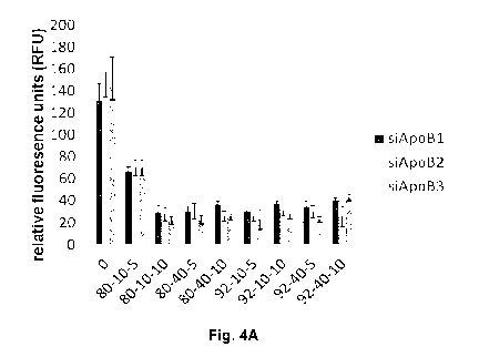
[0064] Fig. 4A
illustrates histograms of chitosan/siRNA nanoparticle stability
at a pH of 6.5, chitosan formulations at different DDA and MW were complexed
to three different anti-ApoB siRNA sequences (siApoB1, siApoB2 and siApoB3)
at N:P ratios of 5 and 10 and incubated for 20 hours, and following
nanoparticle
formation RibogreenTM, an RNA intercalating dye used for nucleic acid
quantitation, was added to each sample to measure the uncomplexed RNA
fraction so that high fluorescence values represent particle disassembly and
instability; Fig. 4B illustrates a histogram demonstrating the influence of MW
on
nanoparticle size, chitosan at a DDA of 92% and different MW was complexed
to anti-RecQL1 siRNA at different N:P ratio; Fig. 40 illustrates a histogram
demonstrating the influence of MW on nanoparticle size, chitosan at a DDA of
80% and different MW was complexed to anti-RecQL1 siRNA at different N:P
ratio; Fig .4D illustrates a histogram demonstrating the influence of MW on
nanoparticle size. Chitosan at a DDA of 72% and different MW was complexed
to anti-RecQL1 siRNA at different N:P ratio; and Fig. 4E illustrates a
histogram
demonstrating the effect of RecQL1 siRNA concentration on nanoparticle size,
and the effect of salt on nanoparticle size as measured by dynamic light
CA 02835147 2013-11-05
WO 2012/159215
PCT/CA2012/050342
- 15 -
scattering, chitosan with a DDA of 92%, a Molecular weight of 10 at an N:P
ratio
of 5 was complexed to increasing concentrations of anti-RecQL1 siRNA.
[0065] Fig. 5
illustrates the effect of DDA, MW and N:P ratio on nanoparticle
stability at different pH where low fluorescence indicates particle stability.
Chitosan with various DDA, MW was complexed to anti-MDR1 siRNA at
different N:P ratio to form nanoparticles. The latter were incubated at
different
pH and siRNA release was measured using the Ribogreen TM assay.
[0066] Fig. 6A illustrates results of nuclease protection assays of
chitosan/dsODN nanoparticles, (A) chitosan (92-10-5 or 80-10-10) complexed
with dsODN-DPP-IV, (B) dsODN-DPP-IV remaining after the DNAse I digestion,
(C) chitosan (92-10-5 or 80-10-10) complexed with dsODN-ApoB, (D) dsODN-
ApoB remaining after the DNAse I digestion, all digestions were assessed using
the signal intensity of the treated samples with the control. (i.e. OU DNAse I
=
100% intensity); and Fig. 6B illustrates nuclease protection assays results of
chitosan/dsODN nanoparticles: (A) chitosan (92-10-5, 80-40-5 or 80-10-10)
complexed with dsODN-RecQL1, and (B) dsODN-RecQL1 remaining after the
DNAse I digestion, all digestions were assessed using the signal intensity of
the
treated samples with the control. (i.e. OU DNAse I = 100% intensity).
[0067] Fig. 7A
illustrates histogram representations of the cellular uptake of
dsODN /nanoparticles 24 hours post-transfection in several cell lines: (A)
Chitosan (92-10-5, 80-80-5 or 80-10-10)/5'-6FAM labeled dsODN DPP-IV
uptake in HepG2 cell lines; and (B) Chitosan (92-10-5, 80-80-5 or 80-10-10)/5'-
6FAM labeled dsODN-ApoB uptake in HepG2, HEK293 and RAW264.7 cells,
DharmaFECTO #1 and 4 were used as positive uptake control; and Fig. 7B
illustrates a histogram showing the cellular uptake of dsODN/nanoparticles 24
hours post-transfection in several cell lines, chitosan (92-10-5, 80-40-5 or
80-
10-10)/5'-6FAM labeled dsODN RecQL1 uptake in AsPC1, LS174T and A549
cell lines, DharmaFECTTm #1 was used as positive uptake control.
[0068] Fig. 8
illustrates confocal imaging of chitosan/siRNA nanoparticle
uptake 24 hours post-transfection in (A) HepG2, (B) Caco-2 and (C) HT-29 cell
CA 02835147 2013-11-05
WO 2012/159215
PCT/CA2012/050342
- 16 -
lines transfected with chitosan/dsODN-DPP-IV nanoparticles, (D) HepG2, (E)
HEK293 and (F) RAW264.7 cell lines transfected with chitosan/dsODN-ApoB
nanoparticles. Chitosan 92-10 (DDA, Mn) was labeled with rhodamine (red) and
dsODN were 5' labeled with 6FAM (green). Chitosan 92-10 was complexed to
siRNA at an N:P ratio of 5. Cell membranes were stained prior to imaging with
CellMaskTm (blue), a membrane anchoring amphipatic dye, to differentiate
between internalized and membrane bound nanoparticles. Images shown
represent each separate channel with dsODN in green, chitosan in red,
membrane in blue, transmission DIC in grey and the merged images shown on
the bottom left quadrant.
[0069] Fig. 9
illustrates confocal imaging of chitosan/siRNA nanoparticle
uptake 24 hours post-transfection. LS174T cell lines transfected with
chitosan/siRNA-RecQL1 nanoparticles. Images were taken 24 hours post
transfection. Chitosan 92-10 (DDA, Mn) was labeled with rhodamine (red) and
siRNA were 5' labeled with 6FAM (green). Chitosan 92-10 was complexed to
siRNA-RecQL1 at an N:P ratio of 5. Cell membranes were stained prior to
imaging with CellMaskTm (blue). Images shown represent each separate
channels with siRNA in green, chitosan in red, membrane in blue, transmission
DIC in grey and the merge images shown on the bottom left quadrant.
[0070] Fig. 10
illustrates confocal imaging of chitosan/siRNA nanoparticle
uptake 24 hours post-transfection. MCF-7 MDR cell line transfected with
chitosan/5iRNA-MDR1 nanoparticles. Images were taken 24 hours post
transfection. Chitosan 92-10 (DDA, Mn) was labeled with rhodamine (red) and
siRNA were 5' labeled Cy3 (green). Chitosan 92-10 (A) chitosan 80-10 (B) and
chitosan 80-200 (C) were complexed to 5iRNA-cy3 at an N:P ratio of 5. Cell
membranes were stained prior to imaging with CellMaskTm (blue). Images
shown represent each separate channel with siRNA in green, chitosan in red,
membrane in blue, transmission DIC in grey and the merge images shown on
the bottom left quadrant.
[0071] Fig. 11A
illustrates histograms of real-time PCR (qPCR) analysis of
the inhibition DPP-IV and ApoB gene expression in specific cell lines, HepG2
CA 02835147 2013-11-05
WO 2012/159215
PCT/CA2012/050342
- 17 -
cells were transfected with: (A) chitosan (92-10-5, 80-80-5 and 80-10-10/siRNA-
DPP-IV); (B) chitosan (92-10-5/siRNA-ApoB) nanoparticles, the inhibition
percentage was obtained by comparing the transfected and non-transfected
cells, using the AACT method; and Fig. 11B illustrates a histogram showing
Real-time FOR (qPCR) analysis of the inhibition RecQL1 gene expression in
specific cell lines, LS174T cells were transfected with chitosan (92-10-5, 80-
40-
and 80-10-10/5iRNA-RecQL1), the inhibition percentage was obtained by
comparing the transfected and non-transfected cells, using the AACT method.
[0072] Fig. 12
illustrates a histogram showing DPP-IV enzymatic activity in
three different DPP-IV expressing cell lines. DPP-IV inhibition percentages
were
determined in comparison with siRNA-mock transfected cells. Values are
expressed as mean s.d., n=4 /group. *p < 0.05, ** p < 0.01.
[0073] Fig. 13
illustrates a histogram showing effects of chitosan/siRNA
administration on ApoB plasma levels. Protein levels were measured by ELISA,
for each treatment group. Columns and error bars represent the mean protein
level relative to the untreated atherosclerotic group, Da. The group Dp is the
normal negative control group fed a normal low fat diet.
[0074] Fig. 14
illustrates a histogram showing the therapeutic lowering of
LDLNLDL cholesterol after chitosan/siRNA administration. LDLNLDL
cholesterol levels were measured by a quantitative colorimetric ELISA kit on
samples taken the day of euthanasia. Columns and error bars represent the
mean cholesterol levels relative to the untreated atherosclerotic group, Da.
The
group Dp is the normal negative control group fed with a normal low fat diet.
[0075] Fig. 15
illustrates the reduction of liver cholesterol droplets in
Therapeutic NanoComplex (TNC) treated animal livers. Hematoxylin-eosin
stained paraffin fixed liver sections of (A) 01-1, (B) 02-1, (C) 03-1, (D) 04-
1,
(E) 05-1, (F) Da-2 day, (G) Da-3, (H) D8-1 and (I) Dp-1 mice demonstrating the
effects of chitosan/siRNA administration in cholesterol accumulation in the
liver.
Arrows (¨>) indicate cholesterol droplet accumulation. The Da group is the
CA 02835147 2013-11-05
WO 2012/159215
PCT/CA2012/050342
- 18 -
positive untreated atherosclerotic control while Dp is the normal negative
control
fed with a low fat diet.
[0076] Fig. 16
illustrates resorption of inflammation in TNC treated animal
liver. Safranin-O/fast-green/iron-hematoxylin stained paraffin fixed liver
section
of (A) 01-1, (B) 02-1, (C) 03-1, (D) 04-1, (E) 05-1, (F) Da-2 day, (G) Da-3,
(H)
D[3-1 and (I) Dp-1 mice demonstrating the resorption of the inflammatory
reaction related to the chitosan/siRNA administration or atherosclerosis
development. Circles (0) and arrows (¨>) indicate lymphoid infiltration.
[0077] Fig. 17
illustrates a histogram showing the weekly weight (g)
measurements of all animal groups. All animals were weighed on the first day
of
each week, before each chitosan/siRNA administration. Compared to the low fat
normal control Dp, a continual weight gain over 4 weeks was observed for all
animals fed with the high fat diet that was essentially unaffected by NTC
treatment.
[0078] Fig. 18
illustrates a histogram showing the percentage of weight gain
per week. All animals were weighed on the first day of each week, before
chitosan/siRNA administration. Weight gain consists in the relative difference
between the weight of the animal and its recorded weight the previous week
[(tn_
1-tn)/t n_1)]. This figure show immediate weight gain or loss following the
first TNC
administration.
DETAILED DESCRIPTION
[0079] In
accordance with the present disclosure, there is provided a novel
and specific composition of a non viral vector for the efficient delivery of
RNAi
inducing entities such as short interfering RNAs (siRNAs), short hairpin RNAs
(shRNAs), and RNAi-inducing vectors (i.e., vectors whose presence within a
cell
results in production of a siRNA or shRNA) to cells, tissues and organs in
mammals, e.g., human. In particular, the description provides chitosan
compositions with specific average molecular weight (Mn) and degree of
deacetylation (DDA) ranges comprising RNAi inducing entities with specific
chitosan to nucleic acid ratios.
CA 02835147 2013-11-05
WO 2012/159215
PCT/CA2012/050342
- 19 -
[0080] There is
thus provided compositions and methods of treating or
preventing diseases or conditions associated with excessive expression or
inappropriate expression of a target transcript; or inappropriate or excessive
activity of a polypeptide encoded by the target transcript.
[0081] The
compositions provided herein can be used in order to provide
symptomatic relief, by administering RNAi inducing entities using the
compositions disclosed herein to a subject at risk of, or, suffering from such
a
condition within an appropriate time window prior to, during, or after the
onset of
symptoms.
[0082] The
compositions and methods may be applied for a variety of
purposes, such as for example, but not limited to, studying the function of
the
transcript, studying the effect of different compounds of a cell or organism
in the
absence of, or with reduced activity of, the polypeptide encoded by the
transcript. Furthermore, the composition and methods may be applied in
clinical
therapy for type ll diabetes and its related pathologies, atherosclerosis and
its
related pathologies and cancer. Specifically, the compositions and methods
may be applied for the inhibition of incretin degrading enzymes (DPP-IV) or
any
glycoregulating protein in order to treat diabetes, applied for the inhibition
of
ApoB gene or any atherogenic protein (i.e ApoE) in order to treat
atherosclerosis, or for down-regulating the expression of RecQL1 DNA helicase
or DDX5 ¨ p68¨ RNA helicase respectively, but not limited to those, for
treating
cancer.
[0083]
Particulalry, the present description relates to the use of such nucleic
acids coupled with the compositions described herein as direct treatment of,
for
example, helicase over-expressing tumors or as radiosensitizing entities for
palliative medicine. Moreover the composition and methods described herein
can be used in conjunction with any other cancer treatment such as
radiotherapy, surgery, hormonal treatment or conventional chemotherapy. The
present description further provides compositions and methods for the
enhancement of radiotherapy or used in combination with other treatment
modalities.
CA 02835147 2013-11-05
WO 2012/159215
PCT/CA2012/050342
- 20 -
[0084] The
composition disclosed herein contains an RNAi inducing nucleic
acid and a chitosan that has the following physicochemical properties: N:P
ratio
below 25, a chitosan with number average molecular weight (Mn) in the range
of 5 kDa to 200kDa and a degree of deacetylation in the range of 80% DDA to
95% DDA. The present description demonstrate the effectiveness of
composition and methods to effectively transfect different cells line and
induce
gene silencing comparable to commercially available lipoplexes, where
transfection efficiency reached 80% at the mRNA level and cell uptake 95% in
some instance, without any apparent cytotoxicity.
[0085] RNA
interference (RNAi) is a process by which double-stranded RNA
directs sequence specific degradation of cellular transcripts such as
messenger
RNA (Sharp, 2001, Genes Dev, 15:485-490; Vance and Vaucheret, 2001,
Science, 292:2277-2280). This phenomenon was initially discovered in C.
elegans (Fire et al., 1998, Nature, 391:806-811). Naturally occurring RNAi is
mediated by small double stranded fragments between 21-25 nucleotide and
are termed small interfering RNA. These siRNA are generated by a dsRNA-
specific endonuclease, called Dicer by a process cleaving long double stranded
RNA (dsRNA) into a 21 base pair small interfering RNA (siRNA) consisting of a
core region of 19 base pair duplex region flanked by two nucleotide 3'over
hangs (Bernstein et al., 2001, Nature, 409:363-366). siRNA are then
incorporated into the RNA-induced silencing complex (RISC), and direct RISC
to recognize target mRNA with complementary sequences to the siRNA leading
to the cleavage of the specific transcript.
[0086]
Subsequently, RNAi was quickly recognized as having great potential
in clinical applications since it was discovered that RNAi can be triggered in
mammalian cells by introducing synthetic 21 nucleotide RNA duplexes (siRNA)
(Elbashir et al., 2001, Nature, 411:494-498), thus bypassing the requirement
of
Dicer mediated processing of long dsRNA.
[0087] For
example, by targeting and reducing the expression of ApoB, it is
possible to prevent excess formation of VLDL, thus diminishing the
accumulation of these atherogenic agents in the organism (Soutschek et al.,
CA 02835147 2013-11-05
WO 2012/159215
PCT/CA2012/050342
- 21 -
2004, Nature, 432:173-178). ApoB targeting at the mRNA level in non-human
primate using sequence specific siRNAs demonstrated significant reductions in
ApoB protein, serum cholesterol and low-density lipoprotein levels 24h post-
treatment (Zimmermann et al., 2006, Nature, 441:111-114). The therapeutic
effect of such treatment using lipid based nanoparticles (SNALP-siRNA) lasted
for 11 days at the highest siRNA dose, thus demonstrating an immediate, potent
and lasting biological effect of siRNA treatment. Unfortunately, these lipid-
based
vectors produced a high level of liver toxicity as indicated by elevated serum
levels of aspartate aminotransferase (AST) and alanine aminotransferase (ALT)
suggested hepatocyte necrosis (Zimmermann et al., 2006, Nature, 441:111-
114). Thus although these reports demonstrate the importance of ApoB as a
target for atherosclerotic and CVD therapies, they also highlight the current
inadequacies of siRNA delivery systems to attain a safe and efficacious
reduction in systemic ApoB.
[0088] Direct
delivery of RNAi in the form of synthetic small interfering RNA
continues to be problematic, suffering from poor cellular targeting and
uptake, a
short half life due to intracellular and/or extracellular nuclease degradation
(i.e.
RNAse) as well as limited blood stability and toxicity (Stein, 1996, Trends
Biotechnol, 14:147-149; Urban-Klein et al., 2004, Gene Therapy, 1-6; Katas and
Alper, 2006, J Control Release, 115:216-225). As a consequence, the
translation of RNAi into a clinical therapeutic is still pending resolution of
these
issues. RNAi has been shown to operate in a wide variety of different cell
types
when introduced into cells by means such as transfection. However,
transfection efficiency depends on the delivery vehicle carrying the small
interfering RNA molecule. The delivery vehicle, referred to as the vector,
should
be able to condense, protect and carry siRNA into target cells. Once in the
vicinity of the target, non-viral vectors should promote cellular uptake,
avoid
lysosomal sequestration and release their content in order to achieve the
desired biological effect.
[0089] Chemical
modification of synthetic siRNAs has provided resistance to
nuclease degradation and improved blood stability. For example, selective
addition of a phosphorothioate linkage or substitution with 2'-0-methyl on the
CA 02835147 2013-11-05
WO 2012/159215
PCT/CA2012/050342
- 22 -
02 position of specific riboses increases nuclease resistance of siRNAs
without
compromising activity (Corey, 2007, J Olin Invest, 117:3615-3622; Whitehead et
al., 2009, Nat Rev Drug Discov, 8:129-138; Judge et al., 2006, Mol Ther,
13:494-505) Nevertheless, some chemical modifications can increase
cytotoxicity and off target effects and reduce mRNA hybridization (Weyermann
et al., 2005, Eur J Pharm Biopharm, 59:431-438; Amarzguioui et al., 2003,
Nucleic Acids Res, 31:589-595). Despite progress achieved through chemical
modification to increase siRNA half life, transfection efficiency, cellular
targeting
and uptake remain as obstacles to effective delivery. Therefore, packaging
systems which can both protect and transport chemically unmodified/modified
siRNA to target cells are required. However, transfection efficiency depends
on
the delivery vehicle carrying the small interfering RNA molecule. The delivery
vehicle, referred to as the vector, should be able to condense, protect and
carry
siRNA into target cells. Once in the vicinity of the target, non-viral vectors
should promote cellular uptake, avoid lysosomal sequestration and release
their
content in order to achieve the desired biological effect. Such non-viral
vectors
are being tested in vitro and in vivo, demonstrating the potential translation
of
siRNA into a clinical reality. Nevertheless, major drawbacks are associated
with
such non-viral vectors. Low transfection efficiency, serum stability,
aggregation
and toxicity remain as major barriers to be addressed before commercialization
of non-viral vectors as powerful and non-toxic tools for drug delivery in the
clinic
becomes a reality. The major classes of non viral vectors are discussed below:
Calcium phosphate
[0090] The
major drawback of this vector is limited efficiency and its inability
to protect nucleic acids from nuclease degradation. Despite the improvement of
its ability to protect nucleic acids, its transfection efficiency remains low
thus
preventing its effective use in vivo.
Cationic lipids
[0091] Cationic
lipids form complexes with nucleic acids via electrostatic
interaction eventually forming multi lamellar lipid-nucleic acid complexes
CA 02835147 2013-11-05
WO 2012/159215
PCT/CA2012/050342
- 23 -
(lipoplexes). Liposome formulations usually include a cationic lipid and a
neutral
lipid such as DOPE (dioleoylphosphatidylethanolamine). The neutral lipid
contributes to the stability of the liposomic formulation and facilitates
membrane
fusion as well as contributing to the lysozomal escape by destabilizing the
endosome. Lipoplexes are one of the most efficient ways of delivering nucleic
acids into cultured cells. Despite their transfection efficiency, lipoplexes
are toxic
as observed in cultured cells and confirmed by several in vivo findings. The
toxicity is closely associated with the charge ratio of cationic lipids to
nucleic
acid in the complex as well as the administered dose. More biocompatible
formulations are being tested and developed in order to reduce lipoplexes
associated toxicity. Reduction of toxicity is mainly achieved via grafting
with
other polymers or reducing the total charge of the cationic polymer.
Cationic polymers
[0092] Cationic
polymers form nanoparticles of nanometric size through
interactions between oppositely charged polycation and polyanion species (i.e.
nucleic acids). These nanoparticles encapsulate nucleic acids, consequently
preventing cargo degradation from nucleases (Romoren et al., 2003, Int J
Pharm, 261:115-127). A large number of natural and synthetic cationic polymers
have been used as vehicles for gene delivery or silencing. Many of these
nanoparticles using cationic polymers have superior transfection efficiency
and
lower serum sensitivity compared to lipoplexes. Among naturally occurring
polycation are proteins such as histones, cationized human serum albumin and
chitosan, an aminopolysaccharide.
[0093] The
group of synthetic polycations includes poly-L-Lysine (PLL), poly-
L-Ornithine as well as polyamines such a polyethylenimine (PEI),
polypropylenimine and polyamidoamine dendimers.
[0094] An
advantage of polyplexes is that their formation does not require
interaction of multiple polycations, contrary to the need of multiple lipid
components of liposomes which make polyplex macroscopic properties easier
to control. Another major advantage of polycation is their block structure
CA 02835147 2013-11-05
WO 2012/159215
PCT/CA2012/050342
- 24 -
therefore allowing direct chemical modification to attain higher efficiency or
specific cell targeting. However, despite these advantages, many cationic
polymers have been found toxic because of high surface charge density since
high charge density nanoparticles appear to be more toxic. Furthermore, it has
been reported that the charge density in the polymer plays a more important
role in cytotoxicity than the total amount of charge. Toxicity may be
molecular
weight dependent as well, since the cytotoxicity of PEI increases linearly
with
molecular weight. Moreover, accumulation of non degradable polymer such as
PEI in the lysosome, a phenomenon called lysosomal sequestration, may yet be
an additional contributor to toxicity.
[0095] Chitosan
is a natural polymer of glucosamine and N-acetyl-
glucosamine monomers linked by 8-1, 4 glycosidic bonds derived from alkaline
deacetylation of chitin. Chitosan molecular weight and degree of deacetylation
dictate its biological and physicochemical properties. For example chitosan
biodegradability is affected by the amount and distribution of acetyl groups.
The
absence of these groups or their random rather than block distribution results
in
very low rate of degradation.
[0096] Chitosan
possesses a wide range of beneficial properties including
biocompatibility, biodegradability, mucoadhesive
properties,
antimicrobial/antifungal activity and very low toxicity. Therefore, it has
attracted
attention of the pharmaceutical and biomedical field and became one of the
most widely used non-viral vectors for nucleic acid packaging and
condensation.
[0097] Several
studies have addressed the effect of chitosan molecular
weight and degree of deacetylation (DDA) on uptake of chitosan- plasmid DNA
nanoparticle, nanoparticle trafficking and transfection efficiency on
different cell
lines. Huang et al. addressed this subject on A549 cells (2005, J Control
Release, 106:391-406). However this study only used seven formulations
(chitosan of 10,17,48,98 and 213kDa at 88% DDA, 213kDa at 61 and 46%
DDA) to study the effect of average molecular weight (Mn) and DDA on
transfection efficiency of pDNA without addressing the much smaller siRNA that
CA 02835147 2013-11-05
WO 2012/159215
PCT/CA2012/050342
- 25 -
is typically 21bp versus thousands of base pairs in plasmids. They found that
a
decrease in Mn and DDA produces lower transfection efficiency for plasmids.
However, the relationship between those two parameters is much more
complex and demands a fine balance between chitosan Mn and DDA to achieve
optimal stability. Their inability to draw a complex relationship is due to
their
limited number of formulations. Moreover, only one parameter at a time was
varied preventing them to see a coupling effect between Mn and DDA in relation
to the pH of the transfection media and to chitosan-to-DNA ratio (N:P).
Another
study addressing this complex relation for plasmid-chitosan polyplexes was
performed by Lavertu et al. (2006, Biomaterials, 27:4815-4824). In their
study,
they varied the molecular weight, for several distinct DDA levels and also
examined the chitosan-to-DNA ratio (NIP) and/or the pH of the transfection
media. This study demonstrated that such optimization achieved high
transfection efficiencies equivalent to broadly used commercial liposomes
(LipofectamineTM and FugeneTM) in HEK293 cells.
[0098] The DNA
binding capacity/affinity of chitosan increases when its
degree of deacetylation increases to create a higher charge density along the
chain to bind more tightly with pDNA to form nanoparticles (Ma et al., 2009,
Biomacromolecules, 10:1490-1499). Thus chitosan with a very low DDA are
unable to bind DNA efficiently and cannot form physically stable complexes to
transfect cells (Koping-Hoggard et al., 2003, J Gene Med, 5:130-141). As
mentioned hereinabove, DDA also exerts a dominant influence on
biodegradability where high DDAs are difficult to degrade. In this light, a
recent
study by Koping-Hoggard et al. (2001, Gene Ther, 8:1108-1121) suggested that
endosomal escape of the high Mn chitosan based complexes depends on
enzymatic degradation of chitosan and would occur less readily with high DDA
chitosans. The resulting degradation fragments are hypothesized to increase
endosome osmolarity and lead to membrane rupture. Thus, for highly
deacetylated chitosan (near 100% DDA), reduced degradability could result in
reduced endosomal escape.
[0099] The
influence of chitosan Mn on the ability to bind nucleic acids was
evaluated in several studies. Binding affinity between oppositely charged
CA 02835147 2013-11-05
WO 2012/159215
PCT/CA2012/050342
- 26 -
macromolecules is strongly dependant on the valence of each molecule, with a
low valence yielding only weak binding (Danielsen et al., 2004,
Biomacromolecules, 5:928-936). The reduction in chitosan valence for lower
molecular weight with shorter chains has been shown to reduce its affinity to
DNA (Ma et al., 2009, Biomacromolecules, 10:1490-1499). Although a high
level of complex stability is desirable extracellularly for protection against
enzymatic attack, MacLaughlin et al. (1998, J Control Release, 56:259-272)
suggested that a high Mn chitosan can form complexes that are overly stable to
transfect cells since they cannot be disassembled once inside the cell.
Furthermore, Lavertu et al. (2006, Biomaterials, 27:4815-4824) showed that Mn
does not appear to be a dominant factor in cellular uptake but does appear to
play a role in nucleic acid binding affinity and intracellular release. These
interpretations and the need for a finely balanced intermediate stability of
chitosan binding to nucleic acids were further supported by direct assessment
of
binding affinity by isothermal titration calorimetry (Ma et al., 2009,
Biomacromolecules, 10:1490-1499) and by live intracellular imaging of polyplex
trafficking and disassembly (Thibault et al., 2010, Mol Ther, 18:1787-1795).
[00100] The amine to phosphate ratio has been found to play an important
role in DNA binding and nanoparticle formation. For example, increasing the
N:P ratio enhances chitosan binding to DNA. For the same DDA, a lower Mn
chitosan requires a higher N:P ratio to completely bind plasmid DNA. Similarly
at equal Mn, a lower DDA requires a higher N:P ratio to completely bind DNA
(Koping-Hoggard, 2003, J Gene Med, 5:130-141; Kiang et al., 2004,
Biomaterials, 25:5293-5301). pH has been shown to play an important role in
transfection efficiency. Lavertu et al. (2006, Biomaterials, 27:4815-4824)
showed that complexes are more stable and an increase in transfection
efficiency is achieved in slightly acidic medium. This can be explained by the
fact that pH reduction increases chitosan protonation and consequently the
positive charge on the polyplex (zeta potential) and the binding affinity of
chitosan to DNA. The combined effect of the chitosan formulation parameters
(DDA, Mn, N:P and pH) was studied for plasmid DNA delivery in vitro by Lavertu
et al. (2006, Biomaterials, 27:4815-4824). They interestingly found that
CA 02835147 2013-11-05
WO 2012/159215
PCT/CA2012/050342
- 27 -
maximum transgene expression occurs for DDA: Mn values that run along a
diagonal from high DDA/low Mn to low DDA/high Mn (Lavertu et al., 2006,
Biomaterials, 27:4815-4824). Thus if one increases/decreases DDA, one must
correspondingly decrease/increase Mn to maintain maximal transfection.
[00101] As mentioned above, pH plays an important role in transfection
efficiency. Lavertu et al. (2006, Biomaterials, 27:4815-4824) showed that an
increase in pH displaces the Mn for the most efficient formulation with
plasmid
DNA toward higher Mn because of the neutralisation of chitosan at higher pH
resulting in reduced chitosan charge density. On the other hand, for a given
DDA, a change in N:P ratio from 5:1 to 10:1 displaces the Mn for the most
efficient formulation towards lower Mn, probably because of the stabilizing
effect
of increasing chitosan concentration. Thus, one can see the importance of
these
different formulation parameters on transfection efficiency and in the
development of a more efficient and stable chitosan-DNA formulations.
[00102] The structural differences between pDNA and siRNA are believed to
affect nanoparticle complexation/stability and the optimal parameters required
for effective delivery. Chitosan has been used for siRNA delivery both in
vitro
and in vivo (de Fougerolles et al., 2007, Nat Rev Drug Discov, 6:443-453;
Howard et al., 2006, Mol Ther, 14:476-484; Katas and Alper, 2006, J Control
Release, 115:216-225; Zimmermann et al., 2006, Nature, 441:111-114; and Liu
et al., 2007, Biomaterials, 28:1280-1288). However, and despite attempts to
identify optimal physico-chemical parameters for siRNA delivery, inconclusive
results have been observed in the literature due to experimental
discrepancies.
For example, nanoparticle formation, stability and protection of the siRNA
cargo
was evaluated at pH 7.9; a pH that is unrepresentative of the physiological
milieu. At this pH, chitosan is mainly deprotonated since its apparent pKa is
close to 6.5, and thus unable to efficiently bind the siRNA cargo. Since
complex
formation was tested under these conditions, several groups have used high
N:P ratios to compensate for the poor binding of chitosan to siRNA seen at pH
higher than chitosan pKa. The use of these high pH values (i.e 7.9) represents
an important design error and source of experimental discrepancy that led
these
investigators to use high N:P ratios to achieve nanoparticle complexation,
CA 02835147 2013-11-05
WO 2012/159215
PCT/CA2012/050342
- 28 -
stability and cargo protection. Unfortunately, the excess chitosan may
competitively affect transfection efficiency, create multiple non-specific
effects
and increase toxicity leading to incorrect conclusions.
[00103] For example, it was reported that intermediate DDA (80%) and high
Mn (64-170 kDa) were apparently more efficient than low molecular weight
chitosan (10kDa) in delivering siRNA (Katas et al., 2006, J Control Release,
115:216-225; and Liu et al., 2007, Biomaterials, 28:1280-1288). However, these
high molecular weight chitosans were found to be toxic (Howard et al., 2006,
Mol Ther, 14:476-484; and Richardson et al., 1999, Int J Pharm, 178:231-243).
Additionally, all previous reports evaluating complex formation, other physico-
chemical characteristics and transfection efficiency of chitosan/siRNA
nanoparticles uniformly concluded that formulations were efficient only at
very
high N:P ratios (N:P >25) (Howard et al., 2006, Mol Ther, 14:476-484; Katas et
al., 2006, J Control Release, 115:216-225; Liu et al., 2007, Biomaterials,
28:1280-1288). These reports did not recognize that a large portion of the
excess chitosan is actually soluble and not a structural component of the
nanoparticle (Ma et al., 2010, Biomacromolecules, 11:549-554). Such
formulations with very high N:P ratios (N:P >25) display significant practical
problems including limited dosing due to aggregation and non-specific toxic
effects of large quantities of soluble chitosan.
[00104] The use here of appropriate pH conditions near chitosan pKa as well
as near the physiological pH to asses nanoparticle physicochemical
characteristics revealed that such high N:P were not required to form
efficient
nanoparticle delivery vehicles, as demonstrated in the present disclosure
(Fig.
3).
[00105] Chitosan was used to deliver pharmacologically active compounds
through different administrational routes including intranasal, oral, intra-
peritoneal, and intramuscular routes. Chitosan/lnsulin was administered
through
intranasal routes in rat and sheep. These formulation involved the use of a
water soluble chitosan of molecular weight of 10kDa or greater, with no
specification on degree of deacetylation (Ilium, 1996, Danbiosyst UK Limited,
CA 02835147 2013-11-05
WO 2012/159215
PCT/CA2012/050342
- 29 -
United States, vol. 5554388; 1998, Danbiosyst UK Limited, United States, vol.
5744166).
[00106] Chitosan has also been used as adjuvant for the immunization of
mice through an intranasal route with soluble formulations (US patent
application publication no. 2003/0039665). These formulations involved
chitosan glutamate with a Mn ranging between 10-500kDa with a degree of
deacetylation between 50-90%.
[00107] Chitosan has also been used to deliver nucleic acids varying from
plasmid DNA to siRNA in vitro and in vivo as well. More than 40 examples of in
vivo studies using siRNA with various delivery vehicles have been reported (de
Fougerolles et al., 2007, Nat Rev Drug Discov, 6:443-453) to treat ocular
(Nakamura et al., 2004, Mol Vis, 10:703-711) and pulmonary targets (Howard et
al., 2006, Mol Ther, 14:476-484), or directed towards the nervous system
(Kumar et al., 2006, Plos Medicine, 3:505-514), liver (Soutschek et al., 2004,
Nature, 432:173-178), tumors (Grzelinski et al., 2006, Hum Gen Ther, 17:751-
766) and other organs by local or systemic delivery. In one example,
chitosan/siRNA nanoparticles mediated TNF-a knockdown in peritoneal
macrophages for anti-inflammatory treatment in an arthritis murine model
(Howard et al., 2006, Mol Ther, 14:476-484).
[00108] Several studies have examined the ability of chitosan to deliver siRNA
in vitro and in vivo. Katas et al. (2006, J Control Release, 115:216-225),
used
two different forms of chitosan salts (CS-HCI and CS-Glutamate) with a DDA of
84% to study the influence of chitosan parameters on transfection efficiency.
Four different high molecular weight chitosans were used (470kDa, 270kDa,
160kDa and 110kDa) and they found that increasing chitosan concentration
from 25pg/m1 (1.25:1) to 300pg/m1 (15:1) increased nanoparticle size from
approximately 150 nm to 450 nm (Katas et al., 2006, J Control Release,
115:216-225).
[00109]
Moreover, it was shown in their study that chitosan-glutamate yielded
smaller nanoparticles than chitosan-HCI. Katas et al. (2006, J Control
Release,
CA 02835147 2013-11-05
WO 2012/159215
PCT/CA2012/050342
- 30 -
115:216-225) found ¨under their experimental conditions- that complete binding
of siRNA to chitosan occurred only at an N:P ratio of 100:1 and above,
conditions of extreme excess of chitosan where most likely >95% of the
chitosan is soluble and not complexed to siRNA (Ma et al., 2010,
Biomacromolecules, 11:549-554). This large quantity of excess moderate DDA
(84%) chitosan is expected to cause sustained inflammation in vivo and to
increase adverse immunological responses (Jean et al., 2009, Gene Ther,
16:1097-1110). In their study, chitosan glutamate with a molecular weight of
470
kDa showed the highest gene silencing effect at 24 h post-transfection in
vitro
compared to its lower molecular weight or chitosan hydrochloride (Katas et
al.,
2006, J Control Release, 115:216-225). Ionic gelation of chitosan glutamate
with an average molecular mass of 470kDa showed a higher silencing efficiency
(82% mRNA knockdown) than chitosan¨siRNA nanoparticles formed by simple
complexation (51% mRNA knockdown) (Katas et al., 2006, J Control Release,
115:216-225).
[00110] Another group led by Howard et al. (2006, Mol Ther, 14:476-484),
delivered chitosan-siRNA nanoparticles in a transgenic EGFP mouse model via
the intranasal route of administration. For their study, they used chitosan at
84%
DDA and 114kDa at four different N:P ratios (N:P 6, 33, 71 and 285). Higher
N:P ratios resulted in smaller nanoparticles (N:P 6 = 223.6 nm vs N:P 33 =
181.6 nm) at low chitosan concentration of 250pg/m1 (Howard et al., 2006, Mol
Ther, 14:476-484). The same pattern was observed at higher chitosan
concentration (1mg/m1) where chitosan nanoparticles with a DDA of 84%, Mn of
114 and an N:P ratio of 33 had an average diameter of 328 nm compared to
139 nm for the formulation 84-114-285 (Howard et al., 2006, Mol Ther, 14:476-
484).
[00111] Their preliminary in vitro study showed that nanoparticle size depends
on the N:P ratio and increases in the size at lower N:P ratios, suggesting
high
N:P ratios to be required. This finding is in contradiction to the findings
presented herein, where it is demonstrated below the critical role of pH when
evaluating chitosan-siRNA complexation and stability. Based on their findings,
cell uptake and silencing efficiency were measured at the high N:P ratios of
36
CA 02835147 2013-11-05
WO 2012/159215
PCT/CA2012/050342
- 31 -
and 57 respectively in NIH 3T3 and H1299 cell lines. Chitosan formulations at
the high N:P ratio of 36 was used to study the silencing efficiency of EGFP
stable cell lines. Silencing efficiency was 77.9% and 86.9% in H1299 and
primary peritoneal mouse macrophage, respectively. The in vivo silencing
efficiency of the chitosan formulation 84-114 at N:P 36 achieved 43% silencing
efficiency in EGFP transgenic mouse model following a 30pg siRNA
injection/day for five days compared to untreated controls (Howard et al.,
2006,
Mol Ther, 14:476-484).
[00112] In
another in vivo study by Howard et al. (2009, Mol Ther, 17:162-
168), a 27 base-pair siRNA targeting TNF-a mRNA was complexed to chitosan
84-114 at the N:P ratio of 63 and injected in a collagen induced arthritis
(CIA)
mouse model. Their formulation achieved 43% silencing as measured by TNF-a
plasma levels.
[00113] Ji et al. (2009, Nanotechnology, 20:405103) suggested that 190kDa
and 310kDa chitosans at DDA ranging from 75% to 85% are suitable delivery
vehicles for siRNA. Similarly to the above studies, Ji et al. used chitosan
formulations at a high N:P ratio of 50 for knockdown experiments of the FHL2
oncogene in Lovo cells. Their formulations achieved 69% of mRNA knockdown.
[00114] In an
attempt to identify optimal parameters for chitosan delivery of
siRNA, Liu et al. (2007, Biomaterials, 28:1280-1288), tested a range of
chitosan
with different DDA, Mn and N:P ratios and stated that N:P ratio > 25 are
needed
for efficient silencing. They also found that low molecular weight chitosan-
siRNA
(10kDa) formulations prepared at N:P 50 showed no knockdown of endogenous
EGFP in H1299 human lung carcinoma cells, whereas chitosan formulations
prepared with higher Mn (64.8-170 kDa) at DDA of 80% showed greater gene
silencing ranging between 45% and 65%. The highest gene silencing efficiency
(80%) was achieved using chitosan/siRNA nanoparticles at the extreme N:P
150 with Mn of 114 and 170 kDa respectively and DDA of 84% that correlated
with their assessments of stable formation of nanoparticles with a diameter of
approximately 200 nm. Additionally, Liu et al. (2007, Biomaterials, 28:1280-
1288) found that a 95% DDA and 9kDa chitosan complexed to anti-EGFP
CA 02835147 2013-11-05
WO 2012/159215
PCT/CA2012/050342
- 32 -
siRNA at N:P ratio of 50 had an undesirable large size of 3500 nm as measured
by dynamic light scattering (DLS). Furthermore, they stated that this specific
formulation did not form complexes with siRNA at N:P ratio as high as 50
according to their gel retardation assays for stability testing conducted at
the
basic pH of 7.9 that was shown here to produce artifactual particle
disassembly.
In addition, this specific formulation showed no EGFP knockdown when
compared to the negative untreated control.
[00115] The above results found by others are in contrast to the novel
findings
presented herein where it is demonstrated that chitosan-siRNA nanoparticles
can be formed at moderate to low N:P ratios (below 25 and preferably 5) using
chitosan with a range of molecular weights (5 to 200kDa) at DDAs between of
80% and 95% and these nanoparticles achieve high levels of gene silencing,
good stability and small size ranges compared to previously reported systems.
[00116] Chitosan coated poly(isohexyl cynoacrylate) (PIHCA) nanoparticles
have also been used to deliver intravenously anti-RhoA siRNAs entities in a
xenografted aggressive breast cancer model (PiIle et al., 2006, Hum Gen Ther,
17:1019-1026). Administration of chitosan-coated-PI HCA-anti-RhoA si R NA
nanoparticles significantly reduced cancer aggressivity in vivo by knockdown
of
over-expressed RhoA in the cancer cells. Zhang et al. studied Nanogene 042, a
chitosan derived formulation, for de novo expression of siRNA targeting the
NS1
protein in lung tissues for the prevention and treatment of Respiratory
Syncitial
Virus (RSV) infections in a Balb/c model (Zhang et al., 2005, Nat Med, 11:56-
62). Zhang et al. used shRNA based plasmids and observed an efficient
silencing of the NS1 gene and an attenuation of RSV infection coupled with a
lowered viral titer load in vivo. Nanogene 042 showed higher transfection
efficiency and induced less inflammation compared to classical high MW
chitosan (Zhang et al., 2005, Nat Med, 11:56-62). However, the molecular
weight of Nanogene 042 is not disclosed in the stated reference.
[00117] For the purpose of the present description, the C57BL/6
(C57BL/6NCrI) mouse model is used for enabling different embodiments. The
C57BL/6 mouse model was developed by Charles River and Research Diets.
CA 02835147 2013-11-05
WO 2012/159215
PCT/CA2012/050342
- 33 -
The 057BL/6 mouse model can become obese when fed a fat rich diet
(D12492) with an apparent weight gain two weeks following with a fat rich diet
compared to lean control. The 057BL/6 mouse model is used in multipurpose
studies and hyperlipidemia research to study the level of LDL cholesterol in
circulation during a high-fat diet (Soutschek et al., 2004, Nature, 432:173-
178;
Crooke et al., 2005, J Lipid Res, 46:872-884; Bose et al., 2008, J Nutr,
138:1677-1683). The fat rich diet (D12492) is equivalent to six times more fat
than the control diet D12450B which contains only 10kcal% fat. In addition,
the
fat rich diet D12492 contains 300.8 (mg)/kg of cholesterol compared to 18
(mg)/kg for the control diet D12450B. Thereby, the feeding with such a high
fat
chow creates instability in the accumulation of LDL in arteries versus its
elimination in the liver, driving the development of atherosclerosis in the
C57BL/6 mouse model.
[00118] It has been found as described herein below that the compositions
described herein are effective gene transfer vectors when combined with siRNA
achieving in vitro transfection efficiencies similar to the commercial
liposome
DharmaFECTTm. Moreover, the compositions not only achieved comparable
efficiency in delivering siRNA into cells and similar silencing as
DharmaFECTTm,
but with lower toxicity.
[00119] Uptake efficiency using chitosan/dsODN nanoparticles achieved
levels comparable to or higher than the commercially used lipoplex
(DharmaFECTTm) with similar relative variation between cells type (Fig. 7A and
7B). Furthermore, these results are in accordance with confocal microscopy
data (Fig. 8), described below, where images show a cellular distribution of
chitosan and dsODN for all cell lines indicating a qualitative correlation to
the
FACS quantitative data. It is demonstrated herein the capability of the
formulations described to transfect and efficiently deliver different siRNA
into
multiple cell lines (see for example Figs. 7A and 8).
[00120] Results
disclosed herein clearly reveal the effectiveness of the
described chitosan-based formulations to efficiently deliver siRNA and knock
down specific genes at N:P ratios far below those used previously in the art.
In
CA 02835147 2013-11-05
WO 2012/159215
PCT/CA2012/050342
- 34 -
general, all of the low N:P ratio chitosan formulations used herein reached
high
level of gene silencing.
[00121] The results show nanoparticles of spherical shape (Figs. 1 and 2) with
mean diameters ranging between 45-156 nm (Table 2) depending on the
chitosan formulation (80-10-5, 80-40-5, 92-10-5, 92-40-5, 80-10-10, 80-80-5,
92-150-5 and 80-200-5) used and the extent of chemical modification of the
siRNA. No statistical differences in nanoparticle size were observed between
dsODN and un-modified siRNA-ApoB (Seq1, SEQ ID NO:5) and moderately
modified siRNA-ApoB complexed to chitosan (Seq2, SEQ ID NO:6 and SEQ ID
NO:7). Whereas, fully modified siRNA sequence yielded larger nanoparticles
when complexed to the different chitosans.
[00122] Results
obtained with specific formulations described herein are
consistent with dynamic light scattering results obtained (Table 2), thereby
indicating the robustness of the composition and method described herein.
Furthermore, the nanoparticles formed yield reproducible sizes below 200 nm
allowing for avoidance of renal clearance thus improving in vivo transfection
efficiency and increasing circulating nanoparticles half-life.
[00123] Chitosan/siRNA stability was evaluated using the Ribogreen assay TM,
a fluorescence based assay, to quantitate the released siRNA following
complex destabilization. The results show that chitosan/siRNA nanoparticle
with
an N:P ratio of 5 and 10 were stable for up to 20 hours at pH 6.5. Chitosan 80-
10-5 showed the least stability when compared to other formulations.
Increasing
the N:P ratio for chitosan 80-10 resulted in an improvement of nanoparticle
stability. Except for chitosan 80-10, increasing the N:P ratio above five did
not
result in an increase of nanoparticle stability (see for example Fig. 4A).
[00124] It is
demonstrated that the formulations described herein can achieve
levels of gene silencing comparable to the commercial liposome
DharmaFECTTm without any apparent cytotoxicity. The results disclosed herein
clearly reveal the effectiveness of the described chitosan-based formulations
to
efficiently deliver siRNA and knock down specific genes at N:P ratios (N:P=5)
CA 02835147 2013-11-05
WO 2012/159215
PCT/CA2012/050342
- 35 -
far below those used previously by others (N:P > 20) (see for example Fig. 11A
and 11B). In general, all of our low N:P ratio chitosan formulations reached
high
levels of gene silencing supporting the FACS data (see for example Fig. 7A and
7B). A tendency for the low molecular weight (10 kDa) and high DDA (92%)
chitosan to be most efficient (Figs. 11 and 12) and smaller (Fig. 4B) was
found
suggesting a particularly optimal formulation at NP ratio 5.
[00125] It has
also described that the composition described herein for the
treatment of atherosclerosis reduced in vivo ApoB plasma levels by
approximately 30% compared to the positive untreated control (called Da
below) (Fig. 13). It is also demonstrated that such a reduction resulted in
ApoB
serum levels similar to those of the non-atherosclerotic animal group negative
control, and is thus in the therapeutic range. It is also demonstrated in the
present description that the composition described herein for the treatment of
atherosclerosis produced a 20% reduction in LDL-cholesterol without any
apparent toxicity (Fig. 14). It is also demonstrated that chitosan based
therapeutic nanocomplexes containing siRNA (TNCs) did not result in any liver
toxicity as demonstrated by normal ALT/AST levels in serum.
[00126] It is further demonstrated that TNC treatment had a therapeutic effect
on cholesterol accumulation in the liver three weeks post injection, where
cholesterol accumulation in TNC treated animal liver was significantly reduced
(Fig. 15). Similarly, chitosan based TNCs induced transient immune cell
infiltration into the liver which resorbed rapidly without toxicity as
demonstrated
in another embodiment herein (Fig. 16). The lack of liver toxicity and the
rapid
resorption of immune cell infiltration indicated the possibility of increasing
the
injected dose to achieve yet higher ApoB and LDL-C plasma reduction.
[00127]
Furthermore, it is described that naked siRNA without chitosan
targeting ApoB induced an intense inflammatory response thus limiting their
dosing and potential for therapeutic use in an uncomplexed form. The lack of
toxicity/inflammation in TNCs treated animal at a tested dose of 1mg/kg anti-
ApoB siRNA coupled with their ability to reduce ApoB plasma levels by 35%
indicates their importance and potential use in a dose response study to
CA 02835147 2013-11-05
WO 2012/159215
PCT/CA2012/050342
- 36 -
determine the maximal tolerated dose (MTD) and achieve higher ApoB plasma
reduction.
[00128] It is demonstrated that TNC-treated animals had reduced ApoB
plasma levels for at least 8 weeks following the third and last injection.
Reductions in ApoB plasma levels for low N:P chitosan-based TNCs were
maintained for more than seven weeks after the last injection in the Cl animal
group (Figs. 13 and 16) without any apparent inflammation or liver toxicity.
These results indicate a particularly promising longevity of TNC treatment and
effective controlled release properties.
[00129] It is thus disclosed herein that low N:P chitosan ApoB siRNA TNCs
described herein, achieved a -35% reduction of ApoB plasma levels and a
-20% reduction in LDLNLDL cholesterol reduction at a 1mg/kg injected dose
(Figs. 13 and 14). These results suggest an effective therapeutic result has
been obtained since previously claimed successful results published using
liposomal delivery systems for ApoBsiRNA required higher doses to achieve
similar or higher ApoB/LDL-VLDL cholesterol reduction and these doses were
associated with liver toxicity and increased ALT and AST levels (Zimmermann
et al., 2006, Nature, 111-114; Soutschek et al., 2004, Nature 432:173-178).
For
example, the use of 5mg kg-1 of siRNA coupled with a lipid formulation (SNALP)
achieved a 73% reduction in ApoB plasma levels (Zimmermann et al., 2006,
Nature, 111-114); this fivefold higher injected concentration achieved 2.5
fold
higher ApoB plasma reduction compared to the results of the present invention.
Furthermore, the use of siRNA targeting ApoB in Ldlr -/+, Cetp -/+ mice model
using a second generation lipid LNP-OCD (LNP201) developed by Merck Inc.
showed an approximately 70% reduction in LDL at 3mg kg-1 (Tadin-Strapps et
al., 2011, J lipid Res, 52:1084-1097). Additionally, 50 mg kg-1 of naked
cholesterol modified siRNA were required to achieve 68% and 31% reduction in
ApoB plasma level depending on the siRNA sequence used (Soutschek et al.,
2004, Nature, 173-178). Additionally these studies were performed in normal
C57BL/6 mice fed with regular chow (lean control) on the contrary to enclosed
study where C57BL/6 mice groups were fed high fat diet to simulate
atherosclerosis until the completion of the study.
CA 02835147 2013-11-05
WO 2012/159215
PCT/CA2012/050342
- 37 -
[00130]
Furthermore, intraperitoneal administration of anti-ApoB antisense
oligonucleotiode (AOS) ISIS-147764, currently in phase III clinical trial,
required
at least 25mg kg-1 administered twice weekly to 057BL/6 feed with high fat in
order to achieve a 55% in ApoB plasma reduction level after six to eight week
of
treatment. Additionally, Crooke et al. reported a plasma cholesterol return to
normal following 50 mg kg-1 administration twice per week for six to eight
weeks
(Crooke et al., 2005, J Lipid Res, 46:872-884). The effect of ISIS-147764 on
cholesterol plasma reduction was observed on the fourth week of treatment (50
mg kg-1 twice/week).
[00131] The compositions and methods described herein demonstrate clearly
the efficiency of ApoB reduction using relatively low doses (1mg kg-1) when
compared to prior art. Additionally, it becomes clear in the present
description
that increasing dose using the present disclosure and disclosed TNCs will lead
to an enhanced ApoB and LDLNLDL-C plasma reduction since ApoB reduction
has been always shown to be dose-dependent (Zimmermann et al., 2006,
Nature, 441:111-114; Soutschek et al., 2004, Nature, 432:173-178; Crooke et
al., 2005, J Lipid Res, 46:872-884; and Crooke, 2005, Expert Opin Biol Ther,
5:907-917).
[00132] The present description provides methods for treatment of diabetes
mellitus and related conditions and symptoms. Such diabetes mellitus and
related conditions include insulin-dependent diabetes mellitus (type I
diabetes),
noninsulin-dependent diabetes mellitus (type ll diabetes), insulin resistance,
hyperinsulinemia, and diabetes-induced hypertension. Other diabetes-related
conditions include obesity and damage to blood vessels, eyes, kidneys, nerves,
autonomic nervous system, skin, connective tissue, and immune system. The
composition described herein can be used either alone or in combination with
insulin and/or hypoglycemic compounds.
[00133] The present description provides methods for treatment of cancer.
Such cancer include breast cancer, glioma, large intestinal cancer, lung
cancer,
small cell lung cancer, stomach cancer, liver cancer, blood cancer, bone
cancer,
pancreatic cancer, skin cancer, head or neck cancer, cutaneous or intraocular
CA 02835147 2013-11-05
WO 2012/159215
PCT/CA2012/050342
- 38 -
melanoma, uterine sarcoma, ovarian cancer, rectal or colorectal cancer, anal
cancer, colon cancer, fallopian tube carcinoma, endometrial carcinoma,
cervical
cancer, vulval cancer, squamous cell carcinoma, vaginal carcinoma, Hodgkin's
disease, non-Hodgkin's lymphoma, esophageal cancer, small intestine cancer,
endocrine cancer, thyroid cancer, parathyroid cancer, adrenal cancer, soft
tissue tumor, urethral cancer, penile cancer, prostate cancer, chronic or
acute
leukemia, lymphocytic lymphoma, bladder cancer, kidney cancer, ureter cancer,
renal cell carcinoma, renal pelvic carcinoma, CNS tumor, glioma, astrocytoma,
glioblastoma multiforme, primary CNS lymphoma, bone marrow tumor, brain
stem nerve gliomas, pituitary adenoma, uveal melanoma, testicular cancer, oral
cancer, pharyngeal cancer, pediatric neoplasms, leukemia, neuroblastoma,
retinoblastoma, glioma, rhabdomyoblastoma and sarcoma.
[00134] One
approach to circumvent MDR is the use of P-gp modulators or
reversal agents compounds that inhibit the transport activity of P-gp.
However,
their pharmacokinetic interaction with chemotherapeutics and toxicities limit
their usage in clinics. Alternatively, the expression of P-gp can be inhibited
by
RNA interference (RNAi). Unlike chemical regulators, this technology may
provide a more specific approach to dowregulation of P-gp and resistance
reversal.
[00135] Various
studies using siRNA or shRNA have demonstrated the
potential use of RNAi to overcome multidrug resistance phenotype. The first
studies showing the proof of principle of RNAi mediated reversal of resistance
by p-gp inhibition were published in 2003 (Nieth et al., 2003, FEBS letters
545(2-3):144-150) and (Wu et al., 2003, Cancer research 63(7):1515. Both
studies used a transient approach with siRNA to modulate multidrug resistant
phenotype in different cell models. Using 200 nM of siRNA, Hao et al. were
able
to suppress p-gp levels by 65% in MCF-7/ADR and A2780 Dx5, to highly
resistant MDR cell lines. Furthermore, they showed that MDR1 targeted siRNA
reversed resistance to p-gp transportable drugs (Doxorubicin) but did not
affect
the sensitivity to hydroxyurea a non P-gp substrate. These data suggest that
silencing of P-gp expression mediated by siRNA is specific. However, the most
pronounced transient MDR reversal of nearly 90% was achieved in the
CA 02835147 2013-11-05
WO 2012/159215
PCT/CA2012/050342
- 39 -
pancreatic carcinoma derived cell line (EPP85-181RDB) and gastric carcinoma
cell (ERG 85-257RDB) despite the use of smaller concentration of siRNA
(100nM) (Nieth et al., 2003, FEBS letters 545(2-3):144-150. Recently, DOnmez
et al. (2011, Biomedicine and Pharmacotherapy 65(2):85-89) revealed 89% in
gene silencing activity of MDR1 in doxorubicin-resistant MCF-7 cell although
the
concentration was lower as 20 nM. These data indicate that the efficacy of
RNAi
may be siRNA sequence-dependent as well as cell line-dependent.
[00136] In
addition to siRNA, stable antiMDR1/P-gp shRNA expression
vectors were used to modulate the MDR phenotype. In one study, shRNA
expression had similar efficiency compared to siRNA to down regulate
MDR1/P-gp in the paclitaxel-resistant SKOV-3TR and OVCAR8TR ovarian
cancer cell lines (Duan et al., 2004, Molecular cancer therapeutics 3(7):833).
Furthermore, Stege et al. (2004, Cancer gene therapy 11(11):699-706) reported
a complete reversal of P-gp expression by introducing a shRNA-expressing
vector (psiRNA/MDR-A) into an extremely high drug-resistant human gastric
carcinoma cell line EPG85-257RDB. Similarly, Yague et al. (2004, Gene
therapy 11(14):1170-1174) observed a complete reversal of doxorubicin
resistance in K562 leukaemic cells by introducing the shRNA-expressing vector
pSUPER. Using the same approach, Shi et al. (2006, Cancer biology & therapy
5(1):39-47) showed also a stable downregulation of MDR1/P-gp gene
expression and function induced by endogenous expression of shRNA which
expressed a novel containing MDR1-5iRNA expression cassette and EGFP
expression gene in human epidermoid carcinoma cell lines (KBv200).
[00137] In all
of the above mentioned studies, Lipofectamine 2000 (Li et al.,
2006, European journal of pharmacology, 536(1):93-97) and (DOnmez, Y. and
U. Gunduz, 2011, Biomedicine & Pharmacotherapy 65(2):85-89) and
oligofectamine (Nieth et al., 2003, FEBS letters 545(2-3):144-150, Wu et al.,
2003, Cancer research 63(7):1515, Stierle et al., 2005, Biochemical
pharmacology 70(10):1424-1430, and Stierle et al., 2007, Biochimie 89(8):1033-
1036), two commercially available liposomes, were used. To date, chitosan has
been used for the delivery of shRNA encoding plasmids targeting the MDR1
gene. In this study, nanoparticles were formed by complex coacervation (Yang
CA 02835147 2013-11-05
WO 2012/159215
PCT/CA2012/050342
- 40 -
et al., 2009, J Huazhong Univ Sci Technolog Med Sci. Apr,29(2):239-42). The
maximum mRNA reduction reported in the study was 52.6% with a time
dependent reversal of paclitaxel chemoresistance of up to 61.3%. No report to
date has described the use of chitosan for the delivery of anti-P-gp siRNA.
[00138] The composition described herein can be used either alone or in
combination with other anti-cancer compound such as Acivicin, Aclarubicin,
Acodazole Hydrochloride; Acronine, Adozelesin, Aldesleukin, Altretamine,
Ambomycin, Ametantrone Acetate; Aminoglutethimide, Amsacrine, Anastrozole,
Anthramycin, Asparaginase, Asperlin; Azacitidine, Azetepa, Azotomycin,
Batimastat, Benzodepa, Bicalutamide, Bisantrene Hydrochloride; Bisnafide
Dimesylate, Bizelesin, Bleomycin Sulfate; Brequinar Sodium; Bropirimine,
Busulfan, Cactinomycin, Calusterone, Caracemide, Carbetimer, Carboplatin,
Carmustine, Carubicin Hydrochloride; Carzelesin, Cedefingol, Chlorambucil,
Cirolemycin, Cisplatin, Cladribine, Crisnatol Mesylate, Cyclophosphamide,
Cytarabine, Dacarbazine, Dactinomycin, Daunorubicin Hydrochloride;
Decitabine, Dexormaplatin, Dezaguanine, Dezaguanine Mesylate, Diaziquone,
Docetaxel, Doxorubicin, Doxorubicin Hydrochloride; Droloxifene, Droloxifene
Citrate; Dromostanolone Propionate; Duazomycin, Edatrexate, Eflomithine
Hydrochloride; Elsamitrucin, Enloplatin, Enpromate, Epipropidine, Epirubicin
Hydrochloride; Erbulozole, Esorubicin
Hydrochloride; Estramustine,
Estramustine Phosphate Sodium; Etanidazole, Etoposide, Etoposide
Phosphate; Etoprine, Fadrozole Hydrochloride; Fazarabine, Fenretinide,
Floxuridine, Fludarabine Phosphate; Fluorouracil, Flurocitabine, Fosquidone,
Fostriecin Sodium; Gemcitabine, Gemcitabine Hydrochloride; Hydroxyurea,
Idarubicin Hydrochloride; Ifosfamide, Ilmofosine, Interferon a-2a, Interferon
a-
213; Interferon a-n1; Interferon a-n3; Interferon p-la, Interferon y-lb,
Iproplatin,
Irinotecan Hydrochloride; Lanreotide Acetate; Letrozole, Leuprolide Acetate;
Liarozole Hydrochloride; Lometrexol Sodium; Lomustine, Losoxantrone
Hydrochloride; Masoprocol, Maytansine, Mechlorethamine Hydrochloride;
Megestrol Acetate; Melengestrol Acetate; Melphalan, Menogaril,
Mercaptopurine,. Methotrexate, Methotrexate Sodium; Metoprine,.Meturedepa,
Mitindomide, Mitocarcin, Mitocromin, Mitogillin, Mitomalcin, Mitomycin,
Mitosper,
CA 02835147 2013-11-05
WO 2012/159215
PCT/CA2012/050342
- 41 -
Mitotane, Mitoxantrone Hydrochloride; Mycophenolic Acid; Nocodazole,
Nogalamycin, Ormaplatin, Oxisuran, Paclitaxel, Pegaspargase, Peliomycin,
Pentamustine, Peplomycin Sulfate; Perfosfamide, Pipobroman, Piposulfan,
Piroxantrone Hydrochloride; Plicamycin, Plomestane, Porfimer Sodium;
Porfiromycin, Prednimustine,. Procarbazine Hydrochloride; Puromycin,
Puromycin Hydrochloride; Pyrazofurin, Riboprine, Rogletimide, Safingol,
Safingol Hydrochloride; Semustine, Simtrazene, Sparfosate Sodium;
Sparsomycin, Spirogermanium Hydrochloride; Spiromustine, Spiroplatin,
Streptonigrin,. Streptozocin, Sulofenur, Talisomycin, Taxol, Taxotere,
Tecogalan
Sodium; Tegafur, Teloxantrone Hydrochloride; Temoporfin, Teniposide,
Teroxirone, Testolactone, Thiamiprine, Thioguanine, Thiotepa, Tiazofurin,
Tirapazamine, Topotecan Hydrochloride; Toremifene Citrate; Trestolone
Acetate; Triciribine Phosphate; Trimetrexate, Trimetrexate Glucuronate,
Triptorelin, Tubulozole Hydrochloride; Uracil Mustard; Uredepa, Vapreotide,
Verteporfin, Vinblastine Sulfate; Vincristine Sulfate; Vindesine, Vindesine
Sulfate; Vinepidine Sulfate; Vinglycinate Sulfate; Vinleurosine Sulfate;
Vinorelbine Tartrate; Vinrosidine Sulfate; Vinzolidine Sulfate; Vorozole,
Zeniplatin, Zinostatin, or Zorubicin Hydrochloride.
[00139] Other
anti-cancer drugs include: 20-epi-1,25 dihydroxyvitamin D3, 5-
ethynyluracil, abiraterone, aclarubicin, acylfulvene, adecypenol, adozelesin,
aldesleukin, ALL-TK antagonists; altretamine, ambamustine, amidox,
amifostine, aminolevulinic acid; amrubicin, amsacrine, anagrelide,
anastrozole,
andrographolide, angiogenesis inhibitors; antagonist ID, antagonist G,
antarelix,
anti-dorsalizing morphogenetic protein-1; antiandrogen, prostatic carcinoma;
antiestrogen, antineoplaston, antisense oligonucleotides, aphidicolin
glycinate,
apoptosis gene modulators; apoptosis regulators; apurinic acid; ara-CDP-DL-
PTBA, arginine deaminase, asulacrine, atamestane, atrimustine, axinastatin 1;
axinastatin 2; axinastatin 3; azasetron, azatoxin, azatyrosine, baccatin Ill
derivatives; balanol, batimastat, BCR/ABL antagonists; benzochlorins,
benzoylstaurosporine, beta lactam derivatives; beta-alethine, betaclamycin 6;
betulinic acid; bFGF inhibitor; bicalutamide, bisantrene,
bisaziridinylspermine,
bisnafide, bistratene A; bizelesin, breflate, bropirimine, budotitane,
buthionine
CA 02835147 2013-11-05
WO 2012/159215
PCT/CA2012/050342
- 42 -
sulfoximine, calcipotriol, calphostin
camptothecin derivatives, canarypox1L-2,
capecitabine, carboxamide-amino-triazole, carboxyamidotriazole, CaRest M3,
CARN 700, cartilage derived inhibitor, carzelesin, casein kinase inhibitors
(ICOS), castanospermine, cecropin B, cetrorelix, chlorins, chloroquinoxaline
sulfonamide,. cicaprost, cis-porphyrin, cladribine, clomifene analogues,
clotrimazole, collismycin A,. collismycin B, combretastatin A4, combretastatin
analogue, conagenin, crambescidin 816, crisnatol, cryptophycin
cryptophycin
A derivatives, curacin
cyclopentanthraquinones, cycloplatam, cypemycin,
cytarabine ocfosfate, cytolytic factor, cytostatin, dacliximab, decitabine,
dehydrodidemnin B, deslorelin, dexifosfamide, dexrazoxane, dexverapamil,
diaziquone, didemnin 13, didox, diethylnorspermine, dihydro-5-azacytidine,
dihydrotaxol, 9-, dioxamycin, diphenyl spiromustine, docosanol, dolasetron,
doxifluridine, droloxifene, dronabinol, duocarmycin SA, ebselen, ecomustine,
edelfosine, edrecolomab, eflomithine, elemene, emitefur, epirubicin,
epristeride,
estramustine analogue, estrogen agonists, estrogen antagonists, etanidazole,
etoposide phosphate, exemestane, fadrozole, fazarabine, fenretinide,
filgrastim,
finasteride, flavopiridol, flezelastine, fluasterone, fludarabine,
fluorodaunornicin
hydrochloride, forfenimex, formestane, fostriecin, fotemustine, gadolinium
texaphyrin, gallium nitrate, galocitabine, ganirelix, gelatinase inhibitors,
gemcitabine, glutathione inhibitors,.hepsulfarn, heregulin, hexamethylene
bisacetamide, hypericin, ibandronic acid, idarubicin, idoxifene, idramantone,
ilmofosine, ilomastat, imidazoacridones, imiquimod, immunostimulant peptides,
insulin-like growth factor-1 receptor inhibitor, interferon agonists,
interferons,
interleukins, iobenguane, iododoxorubicin, ipomeanol, 4-, irinotecan,
iroplact,
irsogladine, isobengazole, isohomohalicondrin 13, itasetron, jasplakinolide,
kahalalide
lamellarin-N triacetate, lanreotide, leinamycin, lenograstim,
lentinan sulfate, leptolstatin, letrozole, leukemia inhibiting factor,
leukocyte alpha
interferon, leuprolide+estrogen+progesterone, leuprorelin, levamisole,
liarozole,
linear polyamine analogue, lipophilic disaccharide peptide, lipophilic
platinum
compounds, lissoclinamide
lobaplatin, lombricine, lometrexol, lonidamine,
losoxantrone, lovastatin, loxoribine, lurtotecan, lutetium texaphyrin,
lysofylline,
lytic peptides, maitansine, mannostatin
marimastat, masoprocol, maspin,
matrilysin inhibitors, matrix metalloproteinase inhibitors, menogaril,
merbarone,
CA 02835147 2013-11-05
WO 2012/159215
PCT/CA2012/050342
- 43 -
meterelin, methioninase, metoclopramide, M I F
inhibitor, mifepristone,
miltefosine, mirimostim, mismatched double stranded RNA, mitoguazone,
mitolactol, mitomycin analogues, mitonafide, mitotoxin fibroblast growth
factor-
saporin, mitoxantrone, mofarotene, molgramostim, monoclonal antibody, human
chorionic gonadotrophin, monophosphoryl lipid A+myobacterium cell wall sk,
mopidamol, multiple drug resistance gene inhibitor,, multiple tumor suppressor
1-based therapy, mustard anti cancer compound, mycaperoxide B,
mycobacterial cell wall extract, myriaporone, N-acetyldinaline, N-substituted
benzamides, nafarelin, nagrestip, naloxone+pentazocine, napavin, naphterpin,
nartograstim, nedaplatin, nemorubicin, neridronic acid, neutral endopeptidase,
nilutamide, nisamycin, nitric oxide. modulators, nitroxide antioxidant,
nitrullyn,
06-benzylguanine, octreotide, okicenone, oligonucleotides, onapristone,
ondansetron, ondansetron, oracin, oral cytokine inducer, ormaplatin,
osaterone,
oxaliplatin, oxaunomycin, paclitaxel analogues, paclitaxel derivatives,
palauamine, palmitoylrhizoxin, pamidronic acid, panaxytriol, panomifene,
parabactin, pazelliptine, pegaspargase, peldesine, pentosan polysulfate
sodium, pentostatin, pentrozole, perflubron, perfosfamide, perilly1 alcohol,
phenazinomycin, phenylacetate, phosphatase inhibitors, picibanil, pilocarpine
hydrochloride, pirarubicin, piritrexim, placetin placetin
B, plasminogen
activator inhibitor, platinum complex, platinum compounds, platinum-triamine
complex, porfimer sodium, porfiromycin, propyl bis-acridone, prostaglandin J2,
proteasome inhibitors, protein A-based immune modulator, protein kinase C
inhibitor, protein kinase C inhibitors, microalgal, protein tyrosine
phosphatase
inhibitors, purine nucleoside phosphorylase
inhibitors, purpurins,
pyrazoloacridine, pyridoxylated hemoglobin polyoxyethylene conjugate, raf
antagonists, raltitrexed, ramosetron, ras farnesyl protein transferase
inhibitors,
ras inhibitors, ras-GAP inhibitor, retelliptine demethylated, rhenium Re 186
etidronate, rhizoxin, ribozymes, RII retinamide, rogletimide, rohitukine,
romurtide, roquinimex, rubiginone B1, ruboxyl, safingol, saintopin, SarCNU,
sarcophytol
sargramostim, Sdi 1 mimetics, semustine, senescence derived
inhibitor 1, sense oligonucleotides, signal transduction inhibitors, signal
transduction modulators, single chain antigen binding protein, sizofiran,
sobuzoxane, sodium borocaptate, sodium phenylacetate, solverol,
CA 02835147 2013-11-05
WO 2012/159215
PCT/CA2012/050342
- 44 -
somatomedin binding protein; sonermin, sparfosic acid; spicamycin
spiromustine, splenopentin, spongistatin 1; squalamine, stem cell inhibitor;
stem-cell division inhibitors; stipiamide, stromelysin inhibitors;
sulfinosine,
superactive vasoactive intestinal peptide antagonist; suradista, suramin,
swainsonine, synthetic glycosaminoglycans, tallimustine, tamoxifen methiodide,
tauromustine, tazarotene, tecogalan sodium; tegafur, tellurapyrylium,
telomerase inhibitors; temoporfin, temozolomide,
teniposide,
tetrachlorodecaoxide, tetrazomine, thaliblastine, thalidomide; thiocoraline,
thrombopoietin, thrombopoietin mimetic; thymalfasin, thymopoietin receptor
agonist, thymotrinan, thyroid stimulating hormone; tin ethyl etiopurpurin,
tirapazamine, titanocene dichloride; topotecan, topsentin, toremifene,
totipotent
stem cell factor; translation inhibitors; tretinoin, triacetyluridine,
triciribine,
trimetrexate, triptorelin, tropisetron, turosteride, tyrosine kinase
inhibitors;
tyrphostins, UBC inhibitors; ubenimex, urogenital sinus-derived growth
inhibitory
factor; urokinase receptor antagonists; vapreotide, variolin 6; vector system,
erythrocyte gene therapy; velaresol, veramine, verdins, verteporfin,
vinorelbine,
vinxaltine, vitaxin, vorozole, zanoterone, zeniplatin, zilascorb, and
zinostatin
stimalamer.
[00140] Anti-cancer supplementary potentiating compounds include: Tricyclic
anti-depressant drugs (e.g., imipramine, desipramine, amitryptyline,
clomipramine, trimipramine, doxepin, nortriptyline, protriptyline, amoxapine
and
maprotiline), non-tricyclic anti-depressant drugs (e.g., sertraline, trazodone
and
citalopram), Ca ++ antagonists (e.g., verapamil, nifedipine, nitrendipine and
caroverine), Calmodulin inhibitors (e.g., prenylamine, trifluoroperazine and
clomipramine), Amphotericin 6; Triparanol analogues (e.g., tamoxifen),
antiarrhythmic drugs (e.g., quinidine), antihypertensive drugs (e.g.,
reserpine),
Thiol depleters (e.g., buthionine and sulfoximine) and multiple drug
resistance
reducing compounds such as Cremaphor EL.
[00141] Other compounds which are useful in combination therapy for the
purpose of the invention include the antiproliferation compound, Piritrexim
Isethionate, the antiprostatic hypertrophy compound, Sitogluside, the benign
prostatic hyperplasia therapy compound, Tamsulosin Hydrochloride; the
CA 02835147 2013-11-05
WO 2012/159215
PCT/CA2012/050342
- 45 -
prostate growth inhibitor, Pentomone, radioactive compounds such as
Fibrinogen 1125, Fludeoxyglucose F 18, Fluorodopa F 18, Insulin 1125, Insulin
I
131, lobenguane I 123, lodipamide Sodium I 131, lodoantipyrine I 131,
lodocholesterol 1131, lodohippurate Sodium 1123, lodohippurate Sodium 1125,
lodohippurate Sodium I 131, lodopyracet 1125, lodopyracet I 131, lofetamine
Hydrochloride 1123, lomethin 1125, lomethin I 131, lothalamate Sodium 1125,
lothalamate Sodium I 131, lotyrosine I 131, Liothyronine 1125, Liothyronine I
131, Merisoprol Acetate Hg 197, Merisoprol Acetate Hg 203, Merisoprol Hg 197,
Selenomethionine Se 75, Technetium Tc 99m Antimony Trisulfide Colloid,
Technetium Tc 99m Bicisate, Technetium Tc 99m Disofenin, Technetium Tc
99m Etidronate, Technetium Tc 99m Exametazime, Technetium Tc 99m
Furifosmin, Technetium Tc 99m Gluceptate, Technetium Tc 99m Lidofenin,
Technetium Tc 99m Mebrofenin, Technetium Tc 99m Medronate, Technetium
Tc 99m Medronate Disodium, Technetium Tc 99m Mertiatide, Technetium Tc
99m Oxidronate, Technetium Tc 99m Pentetate, Technetium Tc 99m Pentetate
Calcium Trisodium, Technetium Tc 99m Sestamibi, Technetium Tc 99m
Siboroxime, Technetium Tc 99m Succimer, Technetium Tc 99m Sulfur Colloid,
Technetium Tc 99m Teboroxime, Technetium Tc 99m Tetrofosmin, Technetium
Tc 99m Tiatide, Thyroxine 1125, Thyroxine I 131, Tolpovidone I 131, Triolein I
125 and Triolein 1131.
[00142] As used herein, "treatment" and "treating" include preventing,
inhibiting, and alleviating diabetes mellitus and related conditions and
symptoms. The treatment may be carried out by administering a therapeutically
effective amount of the composition described herein. In other instances, the
treatment may be carried out by concurrently administering a therapeutically
effective amount of a combination of insulin and the composition described
herein. In still other instances, the treatment may involve concurrently
administering a therapeutically effective amount of a combination of a
hypoglycemic compound and the composition described herein when the
diabetes mellitus and related conditions to be treated is type ll diabetes,
insulin
resistance, hyperinsulinemia, diabetes-induced hypertension, obesity, or
CA 02835147 2013-11-05
WO 2012/159215
PCT/CA2012/050342
- 46 -
damage to blood vessels, eyes, kidneys, nerves, autonomic nervous system,
skin, connective tissue, or immune system.
[00143] Examples
of chitosan containing chemical modification are: chitosan-
based compounds having: (i) specific or non-specific cell targeting moieties
that
can be covalently attached to chitin and/or chitosan, or ionically or
hydrophobically adhered to a chitosan-based compound complexed with a
nucleic acid or an oligonucleotide, and (ii) various derivatives or
modifications of
chitin and chitosan which serve to alter their physical, chemical, or
physiological
properties. Examples of such modified chitosan are chitosan-based compounds
having specific or non-specific targeting ligands, membrane permeabilization
agents, sub-cellular localization components, endosomolytic (lytic) agents,
nuclear localization signals, colloidal stabilization agents, agents to
promote
long circulation half-lives in blood, and chemical derivatives such as salts,
0-
acetylated and N-acetylated derivatives. Some sites for chemical modification
of
chitosan include: 02(NH-CO-0H3 or NH2), 03(OH), or 06(CH2OH).
[00144] The compositions described herein are suitable drug delivery systems
with effective controlled release properties. The present compositions can be
administered with any known combination therapy, such as the co-
administration of a suitable delivery reagent such as, but not limited to,
Mirus
Transit TKO lipophilic reagent, LipofectinO, LipofectamineTM, CellfectinO,
polycations (e.g., polylysine) or liposomes.
[00145] Concurrent administration" and "concurrently administering" as used
herein includes administering a composition as described herein and insulin
and/or a hypoglycemic compound in admixture, such as, for example, in a
pharmaceutical composition, or as separate formulation, such as, for example,
separate pharmaceutical compositions administered consecutively,
simultaneously, or at different times.
[00146] Suitable hypoglycemic compounds include, for example, metformin,
acarbose, acetohexamide, glimepiride, tolazamide, glipizide, glyburide,
CA 02835147 2013-11-05
WO 2012/159215 PCT/CA2012/050342
- 47 -
tolbutamide, chlorpropamide, thiazolidinediones, alpha glucosidase inhibitors,
biguanindine derivatives, and troglitazone, and a mixture thereof.
[00147] Administration of the composition described herein can be a
parenteral administration which includes subcutaneous, intramuscular,
intradermal, intramammary, intravenous, and other administrative methods
known in the art.
[00148] The present invention will be more readily understood by referring to
the following examples.
EXAMPLE I
Reparation of Chitosan/ dsODN or siRNA based nanoparticles
formulations
[00149] Ultrapure chitosan samples were produced using quality controlled
manufacturing processes eliminate contaminants including proteins, bacterial
endotoxins, toxic metals, inorganic and organic impurities. All chitosans had
less than 50EU/g of bacterial endotoxins. Chitosan were selected having a 92%
and 80% of degree of deacetylation (Table 1). These chitosans were produced
by heterogeneous deacetylation resulting in a block rather than random
distribution of acetyl groups. Chitosans were chemically degraded using
nitrous
acid as described previously (Lavertu et al., 2006, Biomaterials, 27:4815-
4824;
Lavertu et al., 2003, J Pharmaceutical and Biomedical Analysis, 32:1149-1158)
to obtain specific molecular weights of 10kDa, 40kDa and 80kDa, the former at
both DDAs of 92% and 80% and the latter at 80% DDA (Table 1).
Table 1
Chitosan degree of deacetylation (DDA), average molecular weight (Mn), poly-
dispersity index (PDI)
Experiment Chitosan DDA Mn (kDa) Mw PDI
RecQL1 Confocal Rho-92-10 92.7 10 14 1.4
DLS, ESEM, 92-10 91.7 7.1 10.08 1.427
Protection,
RecQL1 Stability 80-40 82.5 38.37 53.4 1.392
Assay,FACS,
qPCR 80-10 84.4 10.82 14.525 1.343
CA 02835147 2013-11-05
WO 2012/159215 PCT/CA2012/050342
- 48 -
Experiment Chitosan DDA Mn (kDa) Mw PDI
92-10 92 7.46 9.32 1.25
Enzymatic test
DDP-IV 80-10 80 12.40 22.41 1.80
80-80 80.0 93.8 187.6 2.0
A B Protection 92-10 92.2 8.501 12.645 1.494
po
Assay,FACS, 80-80 80.8 71.535 118.03 1.65
DDP-IV
qPCR, in vivo 80-10 84.4 10.820 14.525 1.343
ApoB
Confocal Rho-92-10 92.7 10 14 1.4
DDP-IV
92-10 91.7 7.1 10.08 1.427
ApoB Stability Assay, 80-80 80.0 93.8 187.6 2.0
DDP-IV DLS, ESEM 80-10 80 12.40 22.41 1.80
80-10 84.4 10.820 14.525 1.343
80-40-5 82.5 38.375 53.410 1.392
92-40-5 92.7 60.6 37.9 1.6
[00150] Small interfering RNAs targeting the DPP-IV gene were purchased
from Dharmacon (Thermo scientific, Dharmacon RNAi Technologies, USA).
These siRNA sense and antisense strands are synthesized with 2 nucleotides
(UU) 3' overhangs. Candidates consisted in a pool of four sequences targeting
the DPP-IV sequence (DPP-IV Seq1: CACUCUAACUGAUUACUUA, SEQ ID
NO:1; DPP-IV Seq2: UAGCAUAUGCCCAAUUUAA, SEQ ID NO:2; DPP-IV Seq
3:
CAAGUUGAGUACCUCCUUA, SEQ ID NO:3; DPP-IV Seq 4:
UAUAGUAGCUAGCUUUGAU, SEQ ID NO:4). ApoB targeting siRNA sequence
was custom synthesized using the 2-ACE RNA chemistry by Dharmacon (ApoB
Seq1: GUCAUCACACUGAAUACCAAU, (antisense strands are synthesized
with 2 nucleotides (AC) 3' overhangs), SEQ ID NO:5; ApoB Seq 2 (sense): 5'
CUC UCA CAU ACA AUU GAA AdTdT 3', SEQ ID NO:7; ApoB seq 2
(antisense) 5' UUU CAA UUG UAU GUG AGA GUUoUoU 3' (oU-oU) = 2'-0-
methyl-uridine overhangs, SEQ ID NO:6; ApoB Seq3 (sense):
GGAAUCuuAuAuuuGAUCcA*A, SEQ ID NO:8; ApoB Seq3 (antisense):
uuGGAUcAAAuAuAAGAuUCc*c*U, SEQ ID NO:9; 2'0-Methyl modified
nucleotides are in lower case and phosphorothioate linkages are represented
by asterisks). These sequences were published by Soutschek, et al. (2004,
Nature, 432:173- 178), Zimmermann et al. (2006, Nature, 441:111-114) and
Strapps et al. (2010, Nucleic Acids Research, Vol. 38, No. 14).
CA 02835147 2013-11-05
WO 2012/159215
PCT/CA2012/050342
- 49 -
[00151] RecQL1 targeting siRNA sequence was custom synthesized using
the 2-ACE RNA chemistry by Dharmacon (Seq1: 5'-
GUUCAGACCACUUCAGCUUdTdT-3', SEQ ID NO:10). This sequence was
published by Futami et al. (2008, Cancer Sci, 99:71-80; 2008, Cancer Sci,
99:1227-1236). MDR1 targeting sequences were purchased presynthetised
from Dharmacon and are available through their catalogue under the product
number: M-003868-02-0010. Candidates consisted of four siRNA targeting the
MDR1 sequence: Seq 1 (sense): 5' GCUGAUCUAUGCAUCUUAUUU 3', SEQ
ID NO:11, Seq 1 (antisense) 5'AUAAGAUGCAUAGAUCAGCUU 3'; SEQ ID
NO:12, Seq 2 (sense): 5'GACCAUAAAUGUAAGGUUUUU 3', SEQ ID NO:13,
Seq 2 (Antisense): 5' AAACCUUACAUUUAUGGUCUU 3', SEQ ID NO:14, Seq
3 (sense): 5' GAAACUGCCUCAUAAAUUUUU 3', SEQ ID NO:15, Seq 3
(Antisense): AAAUUUAUGAGGCAGUUUCUU 3', SEQ ID NO:16, Seq 4
(sense): 5'UCGAGUCACUGCCUAAUAAUU3', SEQ ID NO:17, Seq 4
(Antisense): 5'UUAUUAGGCAGUGACUCGAUU 3', SEQ ID NO:18.
[00152] dsODN sequences were synthesized using the phosphoramidite
chemistry, (Integrated DNA Technologies, Inc) and used for , nanoparticle
stability and nuclease protection assays. For flow cytometry analysis, 6-
carboxyfluorescein (6FAM) 5' labeled dsODN were used (Integrated DNA
technologies, USA).
[00153] The rationale of dsODN use for physico-chemical characterization of
chitosan nanoparticles presented herein is their siRNA mimicking properties.
These mimicking properties are due to similarities at the structural level
(double
stranded structure, length (21 mers) and nucleotide over hangs) between siRNA
and dsODN. Additionally, charge densities are similar between siRNA and
dsODN due to identical phosphate residue number/spacing on their back bone.
Differences between siRNA and dsODN lie in the substitution of uracil to
thymine (U 4 T) in the dsODN sequences, and in the deoxyribosilation of
dsODN sugar back bone. The dsODN sequences were synthesized using the
phosphoramidite chemistry, (Integrated DNA Technologies, Inc) and used for
size and zeta potential determination, nanoparticles stability and nuclease
protection assays. For confocal microscopy, and flow cytometry analysis, 6-
CA 02835147 2013-11-05
WO 2012/159215
PCT/CA2012/050342
- 50 -
carboxyfluorescein (6FAM) 5' labeled dsODN were used (Integrated DNA
technologies, USA).
[00154] Chitosans with specific Mn and DDA were dissolved over night on a
rotary mixer at 0.5% (w/v) in hydrochloric acid using a glucosamine: HCI ratio
of
1:1 at a final concentration of 5 mg/mL. Sterile filtered solutions were then
diluted with deionized water to obtain the desired ratio (N:P) of amine
(chitosan
deacetylated groups) to phosphate (dsODNs or siRNA nucleic acids).
Nanoparticles (92-10-5, 92-150-5, 80-40-5, 80-10-10, 80-10-5, 80-200-5 and
80-80-5) were then prepared by rapid mixing (pippeting) of 100 pL of diluted
chitosan solution to 100 pL of dsODN or siRNA at a concentration of 0.05 pg/pL
respectively; a concentration of 0.33 pg/pL dsODN was used for stability and
nuclease protection assays whereas a concentration of 0.1 pg/pL was used for
DLS and ESEM. Nanoparticles were incubated for 30 minutes at room
temperature prior to use.
EXAMPLE II
Transfection experiments
[00155] For in
vitro transfection, High Glucose-Dulbecco's Modified Eagle's
Media (DMEM-HG) was prepared with 0.976 g/L of MES and 0.84 g/L of sodium
bicarbonate (NaHCO3) at pH 6.5. Transfection media without fetal bovine serum
(FBS) was equilibrated overnight at 37 C in a 5% CO2 incubator and pH
adjustment to a 6.5 value at 370 was performed using sterile HCI (1N) just
before transfection. For siRNA transfection performed in a 96 well plate,
chitosan/siRNA nanoparticles were prepared as described above, 30 minutes
before use. A 100p1 siRNA solution at a concentration of 0.05pg/p1 (3,704 nM)
was used for siRNA complexation with chitosan at a 1:1 ratio (v/v). Following
complexation, siRNA concentration becomes 0.025pg/p1 (1,852 nM) and
nanoparticles were incubated in a ghost plate containing DMEM-HG media, at a
final concentration of 0.00135 pg/pl equivalent to 100 nM per well (10
pmol/well)
of siRNA. For dsODN transfection performed in a 24 well plate, chitosan/dsODN
nanoparticles were prepared as described above, 30 minutes before use. A
100p1 dsODN solution at a concentration of 0.05pg/p1 (3,717 nM) was used for
CA 02835147 2013-11-05
WO 2012/159215
PCT/CA2012/050342
- 51 -
dsODN complexation with chitosan at a 1:1 ratio (v/v). Following complexation,
siRNA concentration becomes 0.025pg/p1 (1,858 nM) and nanoparticles were
incubated in a ghost plate containing DMEM-HG media, at a final concentration
of 0.00135 pg/pl equivalent to 600 nM per well (60 pmol/well) of dsODN. The
slight difference in molecular weight between dsODN used for FACS and siRNA
is due to the 6FAM labelling of dsODN. Plates containing nanoparticles were
equilibrated for 10 minutes at 37 C, 5% 002. Medium over cells was aspirated
and replenished with either 500 p1(24 well plates) or 100 pl per well (96 well
plate) of the equilibrated transfection medium at pH 6.5 containing dsODN or
siRNA based nanoparticles at a final concentration of 100 nM/well. FBS was
added four hours following transfection, to a final concentration of 10% per
well.
Cells were incubated with chitosan/siRNA nanoparticles until analysis at 24
hours post-transfection. DharmaFECTTm was used as a positive control and
both untreated cells and uncomplexed siRNA treated cells were used as
negative controls.
[00156] The commercially available liposome, DharmaFECTTm (Dharmacon
RNAi Technologies, Lafayette, CO, USA), was used as a positive control for
transfection efficiency in all tested cell lines. DharmaFECTTm/dsODN (flow
cytometry and confocal microscopy) or DharmaFECTTm/siRNA (qPCR)
lipoplexes (1:2 [w/v] ratio) were prepared following the manufacturer's
protocol.
[00157] The in vitro transfections involved HEK293, HepG2 (ApoB and DPP-
IV), HT-29 (DPP-1V), Caco-2 (DPP-1V), Raw264.7 (ApoB), A549, L5174T and
the AsPC1 cell lines, purchased from American Type Cell Culture (ATCC,
Manassas, VA). The MCF7-MDR cell line was a gift from Dr Hamid Morjani
(Paris, France). Cells were cultured in minimal essential medium (HepG2),
McCoys (HT-29), Dulbecco minimum essential media high glucose (HEK293
and RAW264.7) with 1.85g/L (HEK293) or 1.5g/I (RAW264.7) of sodium
bicarbonate, (L5174T), F12K (A549), RPMI-1640 (MCF-7 MDR) and RPMI-
1640 (A5PC1), and supplemented with 10% FBS (Cedarlane Laboratories,
Burlington, ON) at 37 C and 5% 002. HepG2 cells were supplemented with 8%
FBS. For transfection, cells were plated in 96-well or 24-well culture plates
CA 02835147 2013-11-05
WO 2012/159215
PCT/CA2012/050342
- 52 -
(Corning, NY, USA) so to obtain -50% to -70% of confluence the day of the
transfection.
EXAMPLE III
RNA extractions and gene expression analysis
[00158] Total RNA extraction was performed using the NucleoSpin0 RNA XS
kit from Machery-Nagel. Cells lysis was performed by adding 100p1 RA1 lysis
buffer supplemented with 2p1 TCEP and Streptomyces griseus chitosanase into
each well (Alameh et al., 2010, Int J Nanomedecine, 5:473-481). DNAse
treatment of sample was performed when sample were incubated with RA3
buffer before elution. RNA quantification and quality (integrity) assessment
were
performed using the Agilent Bioanalyzer 2100. RNA Integrity Number (RIN)
equal to 7.5 was considered as an acceptance threshold for qPCR analysis.
[00159] Reverse transcription of total RNA was performed using the first
strand cDNA transcriptor kit (Roche, Laval, CA). A total of 0.5-1pg of
RNA/sample was used for the reverse transcription reaction using oligodT
primers according to the manufacturer protocol. Gene quantification of
chitosan/siRNA treated cells was performed using the ABI PRISM 7900HT
Sequence Detection System. All reactions were run in triplicate and the
average
values of Cts were used for quantification. Gene expression level was
determined using assays with the Universal Probe Library (UPL) from
RocheTM. On the other hand, gene expression level for endogenous controls
(TBP, HPRT) was determined using the pre-validated TaqMan gene
expression assays. The relative quantification of target genes was determined
using the AACT method. Briefly, the Ct (threshold cycle) values of target
genes were normalized to an endogenous control gene (Endogenous control)
(ACT = Ct target ¨ Ct endoC) and compared with a calibrator: AACT = ACt Sample
- ACt Calibrator. Relative expression (RQ) was calculated using the Sequence
Detection System (SDS) 2.2.2 software (Applied Biosystems) and the formula is
RQ =
CA 02835147 2013-11-05
WO 2012/159215
PCT/CA2012/050342
- 53 -
EXAMPLE IV
Nanoparticles analysis
[00160] Size of chitosan/dsODN and chitosan/siRNA complexes was
determined by dynamic light scattering at an angle of 137 at 25 C using a
Malvern Zetasizer Nano ZS . Samples were measured in triplicates using
refractive index and viscosity of pure water in calculations. The zeta
potential
was measured in triplicates as well using laser Doppler velocimetry at 25 C
using the same instrument and the dielectric constant of water for
calculation.
For the size determination, reported as the intensity averaged diameter, 50p1
of
chitosan was mixed with 50p1 of dsODN or siRNA then completed to 500p1
using 10mM NaCI. For zeta measurement, nanoparticles were diluted 1:2 using
500p1 of 10mM NaCI. All formulations of chitosan/dsODN nanoparticles were in
the range of 45-156 nm, as measured by DLS. Chitosan/siRNA nanoparticles
had mean diameters in the range of 55-105 nm as measured by DLS when
complexed to siRNA sequence 1 (SEQ ID NO:5) and 2 (SEQ ID NO:6 and SEQ
ID NO:7) (Table 2). For siRNA sequence 3 (SEQ ID NO:8 and SEQ ID NO:9),
fully modified, chitosan-siRNA nanoparticles had mean diameters in the range
of 104-130 nm (Table 2). No statistical differences in nanoparticle size were
observed between dsODN and un-modified siRNA-ApoB (sequence 1; SEQ ID
NO:5) and moderately modified siRNA-ApoB complexed to chitosan (sequence
2; SEQ ID NO:6 and SEQ ID NO:7). However, fully modified siRNA sequence
yielded larger nanoparticles when complexed to the different chitosans.
Chitosan/dsODN and chitosan/siRNA nanoparticles showed higher size values
with increasing Mn. No statistically significant differences were observed
when
comparing DDAs for these specific formulations. As expected, the excess
chitosan in all formulations resulted in positively charged nanoparticles as
shown by zeta potentials in Table 2, wherein DLS permitted the determination
of
size and zeta potential, whereas ESEM measured size only.
CA 02835147 2013-11-05
WO 2012/159215
PCT/CA2012/050342
- 54 -
Table 2
Mean size ¨ by intensity ¨ and zeta potential, with standard deviation, of
nanoparticles
formed with with 5iRNA-RecQL1 or 5iRNA-MDR1 in chitosan formulations: 80-10-5,
80-
10-10, 80-40-5, 80-200-5, 92-10-5, 92-150; and siRNA-DPP-IV, ODN-ApoB or siRNA-
ApoB in chitosan formulations: 80-10-5, 80-10-10, 80-40-5 80-80-5, 92-10-5, 92-
40-5.
Sample Chitosan Size (nm) Zeta potential ESEM (nm)
(mV)
MDR1 80-10-5 70 2 12 3 62 9
80-200-5 156 35 18 3 131 5
92-10-5 71 15 15 2 64 8
92-150-5 140 49 17 5 123 6
RecQL1 80-10-10 91 7 18 2 73 9
80-40-5 86 9 18 1 97 12
92-10-5 63 8 23 1 54 6
DPP-IV (pool of siRNA 80-10-10 81 5 16 2 70-90
seq 1 to seq 4) 80-80-5 111 12 20 2 60-100
92-10-5 71 7 18 2 50-90
ApoB (ODN mimics 80-10-10 64 6 19 2 67 7
siRNA ApoB seq 1) 80-80-5 100 12 16 1 75 13
(mimics of SEQ ID NO:5) 92-10-5 45 4 21 2 66 5
ApoB (siRNA seq 1) 80-10-5 80 7 27 2 62 5
(SEQ ID NO:5) 80-40-5 105 6 24 5 90 7
92-10-5 55 3 28 2 60 3
92-40-5 69 4 23 5 65 14
ApoB (siRNA seq 2) 80-10-5 90 4 26 4 70 8
(SEQ ID NO:6 and SEQ 80-40-5 89 6 24 5 76 7
ID NO:7) 92-10-5 57 3 26 4 54 6
92-40-5 67 2 24 5 59 9
ApoB (siRNA seq 3) 80-10-5 139 7 19 3 89 7
(SEQ ID NO:8 and SEQ 80-40-5 130 2 25 2 100 9
ID NO:9) 92-10-5 105 3 22 5 78 5
92-40-5 104 4 27 3 80 6
[00161] Nanoparticles formed as described above were imaged using an
environmental scanning electron microscope (ESEM, Quanta 200 FEG, FEI
Company Hillsboro, OR, USA). Following nanoparticle formation, TNCs were
sprayed on silicon water substrate, and then sputter-coated with gold (Agar
Manual Sputter Coater, Marivac Inc.) as described previously (Lavertu et al.,
2003, J Pharm Biomed Anal, 32:1149-1158). Observations were performed at
20 kV in the high vacuum mode of the ESEM microscope. The average particle
size (+/- standard deviation) was determined by measuring the diameter of
more than 150 particles from at least 6 different fields for each fraction
using the
microscope XT Docu software (XT Docu, FEI Co). The robustness of size
CA 02835147 2013-11-05
WO 2012/159215
PCT/CA2012/050342
- 55 -
determination was analyzed by comparison of ESEM image analysis size
determination to DLS size data.
[00162] The results show nanoparticles of spherical shape (Figs. 1A, 1B, 2A
and 2B) with mean diameters ranging between 45-156 nm depending on the
chitosan formulation used (Table 2, ESEM). Results obtained with specific
formulations described herein are consistent with dynamic light scattering
results (Table 2), thereby indicating the robustness of the composition and
method described herein. Furthermore, the nanoparticles formed yield
reproducible sizes below 200 nm allowing for avoidance of renal clearance thus
improving in vivo transfection efficiency and increasing circulating
nanoparticles
half-life.
[00163] Formation and stability of chitosan/dsODN nanoparticles and
chitosan/siRNA nanoparticles were tested for up to 20 hours at pH 6.5 and 8
using different methods. Chitosan/dsODN nanoparticles were formed and were
stable up to 20 hours at an N:P ratios above 2 at slightly acidic pH (pH 6.5)
(Figs. 3A and 3B). At 4 hours following nanoparticle formation, no detectable
dsODN were observed at N:P ratio of 1 (pH 6.5) and higher, whereas complete
dsODN release was observed for the same N:P ratio at pH 8. Longer exposure
time, 20h, resulted in dsODN release at N:P ratio of 2 for ApoB dsODN while
higher N:P ratio (N:P 10) was able to maintain nanoparticle stability. At pH
values of 8, and for the same N: P ratio of 10, partial dsODN release was
observed. The specific chitosan formulations described herein assured
nanoparticle stability for a minimum period of 20h at N: P ratio above 2
(N:P>2).
Chitosan/siRNA stability was evaluated using the Ribogreen assayTM, a
fluorescence based assay, to quantitate the released siRNA following complex
destabilization. The results show that chitosan/siRNA nanoparticle with an N:P
ratio of 5 and 10 were stable for up to 20 hours at pH 6.5. Chitosan 80-10-5
showed the least stability when compared to other formulations. Increasing the
N:P ratio for chitosan 80-10 resulted in an improvement of nanoparticle
stability.
Except for chitosan 80-10, increasing the N:P ratio above five did not result
in
an increase of nanoparticle stability as demonstrated by the data (Figs. 4A
and
5). Thus, at lower N:P ratios nanoparticles were unstable and the complexation
CA 02835147 2013-11-05
WO 2012/159215
PCT/CA2012/050342
- 56 -
efficiency was not optimal. At a neutral pH, nanoparticles were stable at N:P
ratios between 2 and 5. At a more basic pH of 8, nanoparticles were unstable
with a clear requirement to higher N:P ratios and higher molecular weight for
increased stability.
[00164] The effect of chitosan parameters (DDA, MW and N:P ratio) was
studied using for example anti-RecQL1 siRNA. A clear effect of the molecular
weight is apparent with increased nanoparticle size when increasing chitosan
MW (Figs. 4B, 40 and 4D). The DDA had a very slight effect on nanoparticle
size. The N:P ratio seem to have a impact on nanoparticle size with higher
nanoparticle size at increasing N:P.
[00165] The effect of siRNA concentration on nanoparticle size was studied.
Our results show increased nanoparticle size with increased siRNA
concentrations (Fig. 4E).
[00166] The ability of chitosan to protect dsODN sequences at low N:P ratios
was assessed using a DNAse 1 protection assay. Nanoparticles of
chitosan/dsODN (6p1) were incubated in a buffer containing (pH 6.5) 20mM
MES, 1mM Mg012 and a concentration of 0, 0.5, 1, 2, 5 or 10 units of DNAse I.
Samples were incubated for 30 min at 37 C. The reaction was stopped by
adding 2p1 of EDTA (50mM) then heated at 72 C for 15 min. Samples were then
assessed by gel electrophoresis. Results demonstrate the ability of the
formulations to protect siRNA mimicking double stranded oligonucleotide (Figs.
6A and 6B). All digestions were assessed using the signal intensity of the
treated samples with the control (i.e. OU DNAse 1 = 100% intensity). The
protection is considerable and accounts for approximately 70% of complexes
when using 1 unit of DNAse l/pg of DNA whereas the negative control is
completely digested when 0.5 unit of DNAse 1 per pg of DNA is used. The
protection remains efficient when increasing DNAse 1 concentration to 5 units
per pg of DNA.
[00167] Cell uptake of RecQL1, DPP-1V and ApoB dsODN nanoparticles at
different DDA, Mn and N:P ratio was evaluated using FACS analysis of
CA 02835147 2013-11-05
WO 2012/159215
PCT/CA2012/050342
- 57 -
fluorescein labeled dsODN following chitosanase treatment of transfected cells
thus reducing any possible bias associated with membrane bound nanoparticles
as previously described (Alameh et al., 2010, Int J Nanomedicine, 5:473-481).
Interestingly, results obtained with dsODN/chitosan nanoparticles indicate the
cell line dependency of efficient uptake. The cell line dependency of chitosan
nanoparticles uptake was associated with different endocytic pathways in
previous work (Bishop, 1997, Rev Med Virol, 7:199-209; Huang et al., 2002,
Pharm Res, 19:1488-1494). FACS results show that in general, cell uptake
using these dsODN revealed no differences between formulations (Figs. 7A and
7B). The uptake efficiency using compositions presented herein ranged from
80% to 98% for RecQL1 (LS174T, A549 and AsPC1 cell lines), from 55% to
80% for ApoB (in HEK293, HepG2 and RAW264.7 cell lines). The uptake
efficiency of the DPP-IV dsODN nanocomplexes in HepG2 cell line ranged from
73% to 99% with no statistical differences between the different formulations
(92-10-5, 80-10-10 and 80-80-5). Uptake efficiency using chitosan/dsODN
nanoparticles achieved levels comparable to or higher than the commercially
used lipoplex (DharmaFECTTm) with similar relative variation between cells
type
(Fig. 7A and 7B). Furthermore, these results are in accordance with confocal
microscopy data (Figs. 8 to 10), described below, where images show a cellular
distribution of chitosan and dsODN for all cell lines indicating a qualitative
correlation to FACS quantitative data.
[00168] Confocal microscopy was used in order to assess particle uptake and
internalization into the different cell lines described herein (LS174T,MCF-7
MDR, HEK293, HepG2, Caco-2 and RAW264.7). Chitosan was labeled using
rhodamine whereas RecQL1-siRNA, DDP-IV-dsODN and ApoB-dsODN were
labeled using fluorescein. For MCF-7 MDR nanoparticle assessment, a Cy3
labeled siRNA was used. Following the labeling process, nanoparticles were
formed by mixing 1:1 volume of chitosan-rhodamine and siRNA mimicking
dsODN using the procedure described above. Results suggest that formulations
described in the present description were efficiently internalized into cells
with a
maximum release of siRNA or dsODN 24 hours post transfection. The enclosed
results indicate the lack of colocalisation at 24 hrs between siRNA or dsODN
CA 02835147 2013-11-05
WO 2012/159215
PCT/CA2012/050342
- 58 -
and chitosan demonstrating that complete release of the siRNA or dsODN
cargo was achieved 24h post transfection. Furthermore, the diffuse staining
pattern of siRNA or dsODN seen in most transfected cells is representative of
complexes that have escaped endocytic vesicles (Figs. 8 to 10), consistent
with
previous live cell imaging work using chitosan-plasmid DNA nanoparticles
(Thibault et al., 2010, Mol Ther, 18:1787-1795). Time course studies showed
that particle internalization starts within an hour post transfection with a
slow
release dynamics to reach a maximum 24 hours post transfection.
[00169] The above described results show the capability of the formulation
described in the present description to transfect and efficiently deliver
different
dsODN and siRNA into multiple cell lines (Figs. 8 to 11).
EXAMPLE V
Ex vivo siRNA delivery and gene expression inhibition
[00170] Chitosan specific formulations (92-10-5, 80-40-5, 80-10-10 and 80-
80-5) were assessed for the siRNA delivery and subsequent inhibition of gene
expression (RecQL1 mRNAs, DPP-IV, or ApoB mRNAs) in different cell lines.
Results show that RecQL1, DPP-IV and ApoB coding mRNAs were down-
regulated more than two fold when measured by quantitative real time FOR
(Figs. 11A and 11B). These results demonstrate that the formulation described
herein can achieve levels of gene silencing comparable to the commercial
liposome DhamaFECTTm without any apparent cytotoxicity as observed using
the alamar blue assay.
[00171] More specifically, regarding inhibiton of RecQL1 mRNAs in LS174T
cells, chitosan 92-10-5 showed a high level of silencing (-80%), similar to
the
current gold standard commercial formulation (-80%), used in the present
description as a positive control. Formulations 80-40-5 and 80-10-10 also
induced significant silencing but to a lower degree than 92-10-5 and also with
an increase of non-specific mock silencing, especially for formulation 80-10-
10
(Fig. 11B). The results disclosed herein clearly reveal the effectiveness of
the
described chitosan-based formulations to efficiently deliver siRNA and knock
down specific genes at N:P ratios far below (N:P=5) those used previously by
CA 02835147 2013-11-05
WO 2012/159215
PCT/CA2012/050342
- 59 -
others (N:P > 20). In general, all of our low N:P ratio chitosan formulations
reached high level of gene silencing supporting the FACS data (Fig. 7B).
[00172] It was found that 70% gene silencing at the messenger RNA level
(mRNA) of DPP-1V or ApoB mRNAs, can be achieved using the specific
formulation consisting of chitosan 92-10 with an N:P ratio of 5 (Fig. 11A).
However the 70% inhibition at the messenger level is translated to a reduction
of 50% of the enzymatic activity of DPP-1V (Fig. 12). This inhibition at the
enzymatic level is comparable to that achieved when using the commercial
lipoplex DharmaFECTTm.
EXAMPLE VI
In vivo efficiency analysis of chitosan/siRNA nanoparticles
[00173] The in vivo efficiency of siRNA-ApoB nanoparticles was evaluated in
a C57BL/6 mouse model. For each treatment modality, four animals (n= 4
except for the Da where n=2 and the Cl group where n=3) were injected with 1
mg kg-1 of siRNA targeting the ApoB gene. The 1 mg kg-1 siRNAs targeting the
ApoB gene were complexed to low molecular weight chitosan (LMW-CS) in a
final volume of 0.2 ml (injected volume For example, for a 39 g mouse a 39pg
siRNA ¨ calculated for a dose of 1mgkg-1 ¨ was administered following
complexation of a siRNA volume of 78p1 at 0.5pg/p1 (37,037 nM) at a 1:1 ratio
of
chitosan 92-10-5. The total volume of 156 pl was then administered. The siRNA
concentration following complexation becomes 0.25pg/p1 (18,518 nM).
Specifically, siRNA targeting the ApoB gene were complexed to chitosan
formulation 92-10 (DDA, Mn) at an N:P ratio of 5 (N:P 5). In total, five
groups
(Cl to 05, n=4/group) were TNC treated at different times following the
schedule in Table 3, wherein data for intravenous injections schedule of
chitosan/siRNA-ApoB nanoparticles at a dose of 1mg kg-1 anti-ApoB siRNA in
various 057BL/6 mice groups (n=4 animal per group) is disclosed. Each day
represents the only day in the week where injections were made or euthanasia
was performed. All the mice were injected once per week for three weeks with
the TNC 92-10-5 (Mn-DDA-N:P), with the exception of 2 mice from the Da
group which were injected with the TNC 92-10-5 just once and euthanized 2
CA 02835147 2013-11-05
WO 2012/159215 PCT/CA2012/050342
- 60 -
days later, to examine the rapidity of the therapeutic response. With the
exception of these 2 mice, all other mice were euthanized within the last week
of January 2011. The Da group served as the positive untreated atherosclerotic
control while Dp was the negative control group that received the normal low
fat
diet. The Dp group was the negative control group for the siRNA delivery
without chitosan and was injected with uncomplexed naked siRNA. The total
number of animals used for this study was 32.
Table 3
Animal study schedule
Groups
Cl C2 C3 C4 C5 Da D13 Dp
Day
(n=3) (n=4) (n=4) (n=4) (n=4) (n=4)
(n=4) (n=4)
23/11/10 Acclimation (All groups)
30/11/10 Injection
#1
07/11/10 Injection Injection
#2 #1
14/12/10 Injection Injection Injection
#3 #2 #1
21/12/10 Injection Injection Inject Injection
#3 #2 n#1 #1
28/12/10 Injection Inject Inject Injection
#3 n#2 n#1 #2
04/01/10 Inject Inject Injection
n#3 n#2 #3
Inject
11/01/11
n #3
18/01/11 Da-2day Da
Injection (n=2) n=
20/01/11
Euthanasia Da-
2day
26/01/11 Euthanasia (Cl, C2, C3)
Euthanasia (C4,
27/01/11 Euthanasia (Da, Dp, Dy)
C5)
[00174] All animals were acclimatized for two weeks before experimentation
as requested by the University of Montreal Animal Ethic Committee (CDEA).
Following the two week of acclimatization, high fat chow ¨ D12492 ¨ was fed to
CA 02835147 2013-11-05
WO 2012/159215
PCT/CA2012/050342
- 61 -
all treated groups including the Da positive group (untreated group, n=4) and
the Dp naked siRNA treated group (n=4) until the completion of the study which
corresponds to the day where animals were euthanatized (Table 3). The Dp
group (n=4) was fed regular chow ¨ D12450B ¨ and served as the normal
negative control (lean group). All treated animals were injected once a week
for
three weeks (Table 3). All C group animals were injected with 1 mg kg-1 of
ApoB
siRNA using the low N:P chitosan formulation 92-10-5. The last of the 3 weekly
injections occurred at 7, 6, 5, 4 and 3 weeks prior to euthanizing groups Cl,
02,
03, 04, 05, in order to examine the time course of treatment. Two of the 4
positive control atherosclerotic Da animals were injected with the above
formulation two days prior to euthanasia to examine the onset of treatment,
with
the other two remaining untreated. The D group was treated with uncomplexed
naked ApoBsiRNA at 1 mg kg-1 while the normal low fat diet group Dp was not
treated (details in Table 3)
[00175] During the experimental schedule, phlebotomy was performed once
per two weeks whereas animal weight measurement was performed once per
week before TNC injection until the completion of the study. At the end of the
experimental schedule and following the sacrifice of all animals (Table 3),
organs such as liver and intestine were removed for analysis.
[00176] Hematological, biochemical, serological and histological analysis
were performed on all animals. For instance, hematological and biochemical
analysis of sera were performed by VitaTech, Montreal, Canada. The
quantification of ApoB reduction in the sera was performed using an anti-ApoB
ELISA whereas the quantification of LDLNLDL cholesterol was performed using
a colorimetric assay. Staining of liver sections was performed using
hematoxylin-eosin staining in order to visualize fat vacuole. For the
evaluation of
immune cells infiltration into the liver, paraffin embedded sections were
stained
with Safranin-O/fast-green/iron-hematoxylin.
[00177] Hematological and biochemical analysis of all animals were
performed following serum collection the day of euthanasia. Alanine
aminotrasferase (ALT) and aspartate aminotrasferase (AST), two sensitive
CA 02835147 2013-11-05
WO 2012/159215
PCT/CA2012/050342
- 62 -
indicator of liver damage were quantified in treated and untreated animals. A
comparison of ALT and ASL plasma levels between the treated group (05) and
the positive control group (Da) did not show any significant difference
indicating
an absence of liver toxicity effects of treatment with low N:P chitosan-ApoB
siRNA TNCs (Table 4).
[00178] Moreover, results show that serum albumin levels were normal both
in treated and untreated groups also indicating normal liver function.
However,
total cholesterol quantification in siRNA-ApoB treated animals showed
potentially elevated serum levels similar to the positive control group (Table
4),
wherein 05-2 was administered chitosan/siARN-ApoB nanoparticles, whereas
Da-3 is a positive control for atherosclerosis development respectively. Only
one animal per group was used for haematological analysis because serum
volumes needed are high and require the sacrifice of one animal.
Table 4
Haematologic characterization of a treated (05-2) and untreated (Da-3) mice.
Mice (Group-Mice) C5-2 Da-3
Albumin (g/L) 35 35
Bilirubin (Total) (urnol/L) 0.4 0.7
Bilirubin (Conjugated) (urnol/L) 0.1 0
ALP (IU/L) 58 55
ALT (IU/L) 120 121
AST (IU/L) 213 222
GGT (IU/L) 0 0
Cholesterol (mg/dL) 220 209
Hemolysis 1+ 1+
lcterus Normal Normal
Lipemia Normal Normal
[00179] Taken together these results indicate the safety of the low N:P
chitosan based siRNA nanoparticles as they do not induce any liver damage.
[00180] Apolipoprotein B plasma concentration levels in pg/ml were assessed
using an anti-ApoB commercial ELISA kit (Uscn Life science Inc., China). The
determination of ApoB plasma levels varied between 597pg/mL and 1,433
pg/mL depending on the groups and controls tested. The results obtained show
that all treated groups had ApoB plasma levels that were -35% reduced from
CA 02835147 2013-11-05
WO 2012/159215
PCT/CA2012/050342
- 63 -
the positive atherosclerotic control group Da to reach levels similar to those
of
the normal negative control (Dp) (Fig. 13). The Da-2day group showed a similar
reduction two days following injection indicating a rapid silencing effect
following
TNC injection.
[00181] ApoB levels were decreased by 35% in animals receiving
uncomplexed siRNA (control group; Dp-1). Although this treatment modality
(D6-1) was similarly effective in ApoB plasma reduction as TNCs treatment
modalities (Fig. 13), it resulted in high inflammatory reactions in the liver
(Fig.
16H) thus limiting its dosing to achieve effective and therapeutic
silencing/ApoB
plasma reduction. Additionally, results show that reductions in ApoB plasma
levels for low N:P chitosan-based TNCs was maintained for more than seven
weeks after the last injection in the Cl animal group (Fig. 13) without any
apparent inflammation or liver toxicity. These results indicate a particularly
promising the longevity of TNC treatment and effective controlled release
properties.
[00182] The comparison between the Dp-1 and the C1-05 groups
toxicity/inflammatory profiles indicate the advantage of using these specific
LMW-TNCs over naked siRNA since no apparent toxicity/inflammation profile
was observed (Fig. 16 and Table 4).
[00183] The LDLNLDL cholesterol concentration was determined using a
commercial quantitative colorimetric detection kit BioAssay Systems, USA).
Results herein show that treated animals demonstrated a reduction in
LDLNLDL of -20% compared to the positive control (Da) (Fig. 14).
Interestingly, group C5 demonstrated a higher concentration of VLDL/LDL
compared to the untreated group despite the observed ApoB reduction (Fig.
13); a reduction comparable to other groups showing concomitant reduction of
both ApoB and VLDL/LDL plasma concentration. The comparison between
naked siRNA treated animals and TNCs treated animals show a similar
reduction in LDLNLDL cholesterol concentrations in accordance with previous
results where ApoB reduction was similar (Figs. 13 and 14).
CA 02835147 2013-11-05
WO 2012/159215
PCT/CA2012/050342
- 64 -
[00184]
Histological analysis of paraffin fixed liver sections stained with
hematoxylin-eosin reveal that TNC treated animals had lower cholesterol
accumulation compared to the positive control Da. Liver sections form TNC
treated groups, 03 and Dp, were found to have low levels of accumulated
cholesterol similar to the normal negative control group Dp that was fed the
low
fat diet. (Fig. 15). On the contrary, the group 04, 05 and Da2 presented fatty
livers similar to the positive control Da (Fig. 15) whereas 01 and 02 present
intermediate fatty livers. All together, results demonstrate that TNCs can
prevent excessive cholesterol accumulation in the liver through ApoB
inhibition
and LDLNLDL reduction therefore permitting the liver conversion of cholesterol
into bile in 01, 02, and 03 groups. The results observed in groups 04 and 05
appear to be due to an excessive accumulation of cholesterol before TNC
treatment. These results demonstrate the effectiveness of chitosan based TNCs
in the treatment of atherosclerosis.
[00185]
Histological analysis of paraffin fixed liver sections stained with
safranin-O/fast-green/iron-hematoxylin show that chitosan based TNCs reduced
the inflammatory reaction compared to naked ApoB siRNA treatment (Fig. 16).
Results show that 05 group presented a higher lymphoid cell infiltration rates
than the atherogenic control group thus indicating that inflammation was due
to
chitosan deposition in liver (Fig. 16). However, histological analysis of
liver from
groups 04, 03, 03 and Cl show a time dependent resorption of inflammation
(Fig. 16). Furthermore, the comparison of Da-2day and the positive untreated
control Da-3 show that chitosan effects of lymphoid cell infiltration is time
dependent (Fig. 16, F and G). It is estimated that nanoparticles dependent
inflammation within several weeks of treatment and is preserved during
approximately three weeks until the resorption.
[00186] Comparison of Figs. 15 and 16 allows the assessment of the
efficiency of the chitosan based nanoparticles to prevent cholesterol
accumulation in the liver without disruption of liver integrity as
demonstrated in
by the ALT/ASL profiles. Furthermore, the comparison between Figs. 13 and 14
pinpoint the longevity of the treatment thus confirming our previous
observations
of chitosan mediated slow release.
CA 02835147 2013-11-05
WO 2012/159215
PCT/CA2012/050342
- 65 -
[00187] The effect of treatment on weight gain was assessed by measuring
the weight of each animal/group once per week during the present study.
Results show that treatment did not affect weight gain (Fig. 17). However, it
was
noted that weight gain was slowed in the week following first TNC
administration. For example, group 04 and 05 received their first injection on
the 3rd and 4th week of investigation, respectively, which caused weight
stabilization for group 04 and weight loss for group 05. This effect is also
present in groups 02 and 03 on a smaller scale (Fig. 17). In fact, 05's mean
weight had an accelerated weight gain (mean weight) compared to all groups
from the beginning of the study until its first injection on 28-12-2010. The
effect
of this injection is observed on 04-01-2011 (5th week) where 05's weight
increase rate slowed drastically concordantly with what is observed in Fig.
18.
[00188] While the invention has been described in connection with specific
embodiments thereof, it will be understood that it is capable of further
modifications and this application is intended to cover any variations, uses,
or
adaptations of the invention, including such departures from the present
disclosure as come within known or customary practice within the art to which
the invention pertains and as follows in the scope of the appended claims.