Note : Les descriptions sont présentées dans la langue officielle dans laquelle elles ont été soumises.
CA 02836401 2013-11-15
WO 2012/154661
PCT/US2012/036756
ECHOGENICALLY ENHANCED DEVICE
CROSS REFERENCE TO RELATED APPLICATIONS
This application claims priority to provisional application Serial No.
61/483,094, filed May 6, 2011.
FIELD OF THE INVENTION
[0001] The present invention relates to devices with enhanced
echogenicity for better visualization in ultrasound imaging and methods for
enhancing echogenicity of a device.
BACKGROUND OF THE INVENTION
[0002] Ultrasound technology has advantages over other imaging
modalities. Along with the health advantage of reducing or eliminating
exposure to x-rays (fluoroscopy), the equipment needed is small enough to
move and it has advantages in diagnosing sub-surface tissue morphology.
Furthermore, ultrasound transducers can be made small enough to place inside
the body where they can provide better resolution than is currently available
with magnetic resonance imaging and x-ray computed tomography. Further,
device enhancements which increase their echogenicity to accommodate
ultrasound enable clinicians to quickly and properly treat patients, saving
time
and money.
[0003] Many interventional tools and instruments are designed with
polished surfaces that render the devices virtually invisible on ultrasound.
Interventional tools and instruments are herein referred to as "device(s)".
The
present invention relates to a device enhancement to increase echogenicity of
interventional devices. Interventional devices include, but are not limited
to,
septal puncture needles as well as implantable devices, such as, but not
limited
to, stents, filters, stent graphs, and/or heart valves.
[0004] Ultrasound image device enhancement or "echogenicity" has
been studied for many years. When sound waves contact a smooth surface,
the angle of incidence and reflection are the same. If the object is located
at a
steep angle most or all the sound waves bounce away from a transmitting/
receiver source. With such steep angles, even highly reflective devices can be
1
CA 02836401 2013-11-15
WO 2012/154661
PCT/US2012/036756
invisible by ultrasound if scattering does not direct sound back to a source
transducer. Conversely, if an object is perpendicular, the sound waves
reflecting directly back may cause a "white out" effect and prevent the
operator
from seeing around the object. This affect is referred to as specular
reflection.
[0005] Medical device manufacturers have tried a variety of techniques
to improve visibility of devices to ultrasound. Examples include roughening
the
surface of the device, entrapping gas, adhering particles to substrate
surfaces,
creating indentations or holes in the substrates and using dissimilar
materials.
SUMMARY OF THE INVENTION
[0006] An aspect of the present invention relates to an echogenically
enhanced interventional tool or device. The interventional tool or device to
be
imaged ultrasonically has a surface with one or more apertures and a polymeric
film in close contact with the surface of the tool or device which covers at
least
a portion of the one or more apertures.
[0007] Another aspect of the present invention relates to a method for
enhancing echogenicity of an interventional tool or device. In this method,
one
or more apertures are made in a surface of an interventional tool or device. A
polymeric film is then placed in close contact with the surface covering at
least
a portion of the one or more apertures.
BRIEF DESCRIPTION OF THE FIGURES
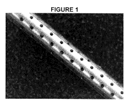
[0008] Figure 1 shows an interventional tool or device with a plurality of
apertures in its surface.
[0009] Figures 2A and 2B show the same interventional tool or device of
Figure 1 with a polymeric filrri in close contact with the surface of the
device so
that the apertures are closed.
[0010] Figure 3 is a bar graph showing results of a comparison of the dB
increase above control of a device of the present invention with a polymeric
film
covering apertures in the surface of the device as depicted in Figures 2A and
2B and another commercially available coated device.
[0011] Figure 4 is a plot of the reflected energy at various angles, which
reflects increased echogenic response.
2
CA 02836401 2013-11-15
WO 2012/154661
PCT/US2012/036756
DETAILED DESCRIPTION OF THE INVENTION
[0012] The present invention relates to an enhancement to increase
echogenicity of these interventional devices. The echogenically enhanced
device of the present invention comprises a device to be imaged ultrasonically
having a surface with one or more apertures. The interventional device of the
present invention further comprises a polymeric film in close contact with the
surface of the device which covers at least a portion of the one or more
apertures.
[0013] Examples of interventional tools or devices which can be
enhanced visually in ultrasound imaging in accordance with the present
invention include, but are not limited to, medical devices such as permanent
implantable or temporary indwelling devices, such as catheters, guide wires,
stents and other accessories and tools, surgical instruments, and needles such
as septal puncture needles. However, as will be understood by the skilled
artisan upon reading this disclosure, the techniques described herein for
visually enhancing a device via ultrasound imaging are adaptable to many
different fields and devices.
[0014] In accordance with the present invention, one or more apertures
are made in a surface of the interventional tool or device. The apertures of
the
present invention may be divots in the surface of an otherwise smooth device
surface, or holes through the surface of the device, or grooves formed in the
device surface, or any other topographical asperities in the otherwise smooth
surface of the device.
[015] In one embodiment, as depicted in Figure 1, a plurality of
apertures is made in the surface of the interventional tool or device.
[0016] In one embodiment, in addition to apertures in the surface of the
interventional device, the surface is also roughened. In one embodiment, the
surface roughness of the device has an average surface roughness of less
than 1 'JIM
[0017] In embodiments wherein the polymeric film is bonded to the
device, surface roughening may be useful to increase adhesion.
[0018] Echogenicity of this device is enhanced in accordance with the
present invention by positioning an echogenic polymeric film in close contact
with the surface of the device to cover at least a portion of the aperture or
apertures in the surface of the interventional tool or device. In one
embodiment, the polymeric film covers the entire aperture or apertures in the
3
CA 02836401 2013-11-15
WO 2012/154661
PCT/US2012/036756
surface of the interventional tool or device. In one embodiment, the polymeric
film surrounds the entire surface of the interventional tool or device. The
polymeric film covering may also restore luminal competency to a medical
device (needle, biopsy punch, etc) in which through-holes / apertures have
been added. n the case of divots or grooves, the polymeric film coveting,
especially the ePTFE film, may restore surface smoothness, which is
preferable in most endoluminal procedures.
[0019] In some embodiments of the present invention, the echogenic
response of the device may be adjustable. One adjustable embodiment
comprises a hollow device with through-hole apertures in the surface covered
by a thin polymeric film. The pressure within the device can be increased or
decreased to change the resonant characteristic of the polymeric film covering
said apertures so as to produce a change in the device's echogenic response
While viewed via ultrasound. In another embodiment, the tension of the
polymeric film covering the apertures of a device may be adjustable. By
increasing or decreasing the tension of this polymeric film, the echogenicity
of
the device can be adjusted. The shape of the apertures can be varied to
change the echogenicity that is achieved.
[0020] Any biocompatible polymeric film capable of an echogenic
response with minimal profile impact can be used. In one embodiment, the
polymeric film comprises a microporous fluoropolymer such as expanded
polytetrafluoroethylene (PTFE). In another embodiment, the polymeric film
may be a thin polyolefin film which may or may not be porous. The different
thickness of material will change the topography when the sleeve is
"activated."
Different topography will change the echogenicity of the object. The thickness
of said polymeric films should be less than 0.010". In another embodiment,
said polymeric film thickness is less than 0.006". In another embodiment, said
polymeric film thickness is less than 0.003.
[0021] Enhanced echogenicity of a device of the present invention was
demonstrated experimentally. Results are depicted in Figure 3 which shows a
comparison of the dB increase above control of a device of the present
invention and an Angiotech coated device.
[0022] The following non-limiting examples are provided to further
illustrate the present invention.
4
CA 02836401 2013-11-15
WO 2012/154661
PCT/US2012/036756
EXAMPLES
Example 1: Materials
[0023] A stainless steel needle with the dimensions of 0.040" diameter
and approximately 4.8" long was used as the test article for echogenic
enhancement. An unmodified needle was used as control to compare the
results of the modification. Echogenicity of a stainless steel needle with a
plurality of apertures covered by a polymeric film in accordance with the
present invention was also compared to an Angiotech coated needle
(Angiotech Pharmaceuticals, Inc., 1618 Station Street, Vancouver, BC Canada
V6A 166). The apertures are staggered 45 0.178 mm in diameter and spaced
0.38 mm apart.
Example 2: Methods
[0024] Three different methods were used to evaluate and compare the
treated samples.
[0025] All samples were subjected to an acoustic wave imaging system.
The testing apparatus consisted of a 7.5 MHz transmitting/receiving transducer
mounted onto a flat bar with a sample holder placed approximately 2.5 cm at
the transducer's focal length. The 7.5 MHz transducer produced a wave length
(A) of 200 microns. At 2.5 cm the width of the signal was approximately 1 mm.
The needle sample was placed into a holder that is perpendicular to the axis
of
the emitting transducer. This is 0 degrees. The sample holder is removable for
ease of changing out the sample. The holder is magnetically held in a
rotatable
goniometer for measuring the angle of the sample relative to the transmitting
and receiving transducer. The sample and transducer were submerged into a
room temperature water tank. Before collecting the data, every sample was
aligned with the transducer. This was accomplished by increasing the
attenuation setting on the pulser/receiver controller (approximately 40 dB) to
prevent saturation of the received signal. The operator then visually
monitored
the wave signal while manually rotating the goniometer and dialing the fine
adjustment knobs on the transducer to achieve a maximum return signal. The
attenuation was adjusted to a reference point of approximately 1 volt. The
attenuation setting and the goniometer indication were recorded. The
goniometer was rotated 10 degrees from the recorded indication. Since the
signal typically decreases off of perpendicular (specular reading) the
attenuation was reduced. The reduced level allowed a strong enough signal
CA 02836401 2013-11-15
WO 2012/154661
PCT/US2012/036756
during collection, without saturation of the receiver. The sample was rotated
through the entire angular rotation to ensure that the signal did not saturate
or
significantly move away from or closer to the transducer moving the signal out
of the data collection window. Significant time shift was an indication that
the
transducer was not aligned with the center or pivot of the sample. Once the
set-up was completed, the goniometer was moved to the 10 degree mark and
the collection of points was taken to 50 degrees at 2 degree increments.
Equipment connected to the transducer and test fixture measured reflection.
The software, Lab View, and hardware were used for data collection and later
analysis.
[0026] A second evaluation of samples was performed in a silicone
phantom submersible in a blood substitute from ATS laboratories to increase
attenuation and create a more realistic image environment. Using a 6.5 mHz
transducer ultrasound system, the samples were inserted into the phantom. A
still image was captured for each sample. These images were visually
compared to control images and inspected for consistency with the transducer
2D data. The data was collected at three different times. Between collections
two and three the transducer was rebuilt. Thus, while the absolute dB scale of
plots is not the same, the relative deltas are of importance.
[0027] The third evaluation was a surface analysis using an optical
comparator, Veeco Model NT3300. All raw data was further processed by the
machine software to better evaluate the samples. The macroscopic tilt and
cylindrical curvature were removed. A Gaussian filter (Fourier) was selected
to
filter frequencies below 20-1mm. Incomplete interior points were restored with
a
maximum of 3 or 5 pixels. All samples were masked at the edges to remove
large data drop out sections and anomalies associated with the filtering. 2D
samples were processed first followed by 3D samples.
[0028] Total roughness height, Rt or PV, which is the maximum peak to
valley height of the surface profile within the assessment length, was used to
depict the surface characteristics.
[0029] A comparison of the dB increase above control of a device of the
present invention and an Angiotech coated device is depicted in Figure 3.
6