Note : Les descriptions sont présentées dans la langue officielle dans laquelle elles ont été soumises.
CA 02842281 2014-01-17
WO 2013/020898 PCT/EP2012/065198
Constitutively active uPAR variants and their use for the generation and
isolation of
inhibitory antibodies
Background of the invention
The urokinase plasminogen activator receptor (uPAR, also named CD87) is a
membrane
glycoprotein anchored to the plasma membrane by a glycosylphosphatidylinositol
(GPI)
anchor. The urokinase plasminogen activator (uPA) and its receptor (uPAR) play
important
roles in physiological processes such as wound healing, inflammation, and stem
cell
mobilization, as well as in severe pathological conditions such as HIV-1
infection, tumor
invasion, and metastasis. The urokinase-type plasminogen activator receptor
(uPAR) is a
plasma membrane receptor overexpressed during inflammation and almost in all
human
cancers. The important role of uPAR in tumor cell adhesion, migration,
invasion, and
proliferation makes this receptor an attractive drug target in cancer
treatment. Several
therapeutic strategies inhibiting the uPA system have been or are currently
being developed
for suppression of tumor growth. Besides uPAR's well-established role in the
regulation of
pericellular proteolysis, it also modulates cell adhesion, migration, and
proliferation through
interactions with proteins present in the extracellular matrix, including
vitronectin (VN).
Although the importance of the interaction with VN is well documented to be
crucial for the
signaling activity of uPAR (Madsen et al, 2007; Smith et al, 2008), the
importance of this
interaction in vivo has never been addressed.
A direct VN interaction is both necessary and sufficient to initiate uPAR-
induced changes in
cell morphology, migration, and signaling independently of direct lateral
protein¨protein
interactions. The single interaction between uPAR and VN may be responsible
for many of
the proteolysis-independent biological effects initiated by uPAR. Development
of inhibitors of
the uPAR/vitronectin interaction is another attractive target and may possibly
start from the
uPAR-binding somatomedin B domain of vitronectin, which is a natural and
potent
uPAR/vitronectin interaction antagonist.
Several international applications disclose peptides ligand of urokinase
receptor, such as
W001/17544.
W097/35969 discloses peptides that are capable of binding to uPAR and
inhibiting the
binding of an integrin and vitronectin. The document does not refer to uPA
binding. The
1
CA 02842281 2014-01-17
WO 2013/020898 PCT/EP2012/065198
binding site of the peptides in uPAR was not determined and no data on the
function blocking
activity of the peptides are presented in the document.
W02008/073312 relates to urokinase-type plasminogen activator receptor epitope
and
monoclonal antibodies derived therefrom. The document discloses antibodies,
and antigen-
binding fragments thereof, specific for urokinase-type plasminogen activator
receptor (uPAR)
and their use for the treatment or prevention of cancer. In particular, the
disclosed antibodies
are specific for a particular epitope on uPAR. The antibodies described in
W02008/073312
recognize epitopes non-overlapping with those described in the present
invention.
Rabbani SA, et al (Neoplasia (2010) 12, 778-788) examined the effects of
administration of a
monoclonal anti-uPAR antibody (ATN-658) on prostate cancer progression in
vitro and in
vivo. ATN-658, a mouse IgG1 , is able to bind to D2D3 of uPAR with high
affinity (Kd ¨ 1
nM), does not inhibit the binding of uPA to uPAR, and is able to bind to uPAR
even when
uPA was also bound. The antibody used in this study (ATN-658) is that
described in
W02008/073312. The epitope recognized by the ATN-658 antibody does not overlap
with
those described in the present invention. The ATN-658 antibody is not a
competitive
antagonist of the uPAR/vitronectin interaction as it does not bind to the
vitronectin binding-
site in uPAR. The ATN-658 antibody binds to an epitope in uPAR similar or
identical to
another well-characterized monoclonal antibody R2 (Sidenius et al. JBC (2002)
277 27982-
90). ATN-658 binds to intact uPAR and the truncated D2D3 receptor equally
well. Thus, the
antibody therein described does not bind preferentially to intact uPAR.
W02005/116077 identifies antibodies or other ligands specific for the binary
uPA-uPAR
complexes, for ternary complexes comprising uPA-uPAR and for complexes of uPAR
and
proteins other than uPA such as integrins. The antibodies inhibit the
interaction of uPA and
uPAR with additional molecules with which the complex interact. Such
antibodies or other
ligands are used in diagnostic and therapeutic methods, particularly against
cancer. The
document refers to ligands that do not inhibit vitronectin binding but the
assembly of
vitronectin components; moreover, they recognize epitopes non-overlapping with
those herein
described.
W02006/094828 discloses antibodies that preferentially recognize truncated and
soluble
forms of uPAR receptor (D2 D3). The antibodies therein described do not bind
preferentially
to intact uPAR.
2
CA 02842281 2014-01-17
WO 2013/020898 PCT/EP2012/065198
CN101050237 discloses a compound that can block interactions between uPA and
uPAR, and
its application. The compound comprises ATF of uPA, ATF fragment, uPAR
fragment, anti-
ATF antibody, and anti-uPAR antibody. The compound can block the interactions
between
uPA and uPAR, and can be used to prepare medicine for preventing and treating
atherosclerosis.
Tressler RJ et al., (APMIS. 1999 Jan;107(1):168-73) discloses urokinase
receptor antagonists
based on the growth factor domains of both human and murine urokinase. Such
antagonists
show sub-nanomolar affinities for their homologous receptors. Further
modification of these
molecules by preparing fusions with the constant region of human IgG has led
to molecules
with high affinities and long in vivo half-lives. Smaller peptide inhibitors
have been obtained
by a combination of bacteriophage display and peptide analogue synthesis. All
of these
molecules inhibit the binding of the growth factor domain of uPA to the uPA
receptor and
enhance binding of the uPA receptor to vitronectin.
Gardsvoll H, et al (J Biol Chem, 2011 Sep 23;286(38):33544-56) proposes a
model of
cooperation between uPA and vitronectin to potentiate uPAR-dependent induction
of
lamellipodia on vitronectin matrices; this will have implications for drug
development
targeting uPAR function, i.e. epitope-mapped monoclonal antibodies. None of
the antibodies
investigated in this study block vitronectin binding to uPAR in the presence
of uPA.
There is thus the need of antibodies which bind preferentially to intact uPAR
and which are
potent inhibitors of uPAR-functions.
Description of the invention
The present invention concerns unique variants of the urokinase plasminogen
activator
receptor (uPAR) that display remarkably increased vitronectin (VN) binding
activity
(>10.000-fold increased apparent Kd), possibly caused by a more efficient
exposure of the VN
binding site.
Authors showed that monoclonal antibodies raised against said uPAR-variants
are potent
inhibitors of uPAR-functions. Mapping of the antibody binding epitopes shows
that these
antibodies bind to the VN binding site of uPAR classifying them as competitive
antagonists.
The authors also showed that the above uPAR-variants are used to isolate
synthetic antibodies
(scFv) from phage-display libraries which are functional inhibitors of uPAR.
The inhibited
functions are: cell adhesion (Fig. 11) and consequently cell migration and
cell proliferation
3
CA 02842281 2014-01-17
WO 2013/020898 PCT/EP2012/065198
(which are downstream events in respect of cell adhesion). These functions are
VN-dependent.
Detailed description of the invention
Object of the present invention is an urokinase plasminogen activator receptor
(uPAR) variant
molecule having an increased VN-binding activity with respect to the wild type
molecule.
The uPAR variant molecule according to the invention preferably comprises a
wild type uPAR
amino acid sequence linked to :
a) a growth factor-like domain (GFD) sequence of uPA at the N-terminal of
the wild type
uPAR sequence, and/or
b) a chain of the Fc region of an antibody molecule at the C-terminal of
the wild type
uPAR sequence,
wherein if said chain of the Fc region is present, the uPAR variant molecule
is a dimer.
For "wild type uPAR amino acid sequence" it is intended the sequence of the
full wild type
protein or fragments thereof maintaining a VN-binding activity.
In a preferred embodiment the wild type uPAR sequence comprises a sequence
consisting
essentially of the aa. 32-92 of mature huPAR of SEQ ID NO: 1 or a sequence
consisting
essentially of the aa. 32-93 of mature muPAR of SEQ ID NO: 2 or a polypeptide
encoded by
the correspondent regions from an uPAR orthologous gene, or functional mutants
or
derivatives or analogues thereof.
More preferably the wild type uPAR sequence comprises a sequence consisting
essentially of
the aa. 3-271 of mature huPAR of Seq ID NO: 1 or a sequence consisting
essentially of the aa.
3-270 of mature muPAR of Seq ID NO: 2 or a polypeptide encoded by the
correspondent
regions from an uPAR orthologous gene, or functional mutants or derivatives or
analogues
thereof.
Even more preferably, the wild type uPAR sequence comprises a sequence
consisting
essentially of the aa. 1-277 of mature huPAR of Seq ID NO: 1 or a sequence
consisting of
essentially the aa. 1-273 of mature muPAR of Seq ID NO: 2 or a polypeptide
encoded by the
correspondent regions from an uPAR orthologous gene, or functional mutants or
derivatives or
analogues thereof
In another preferred embodiment of the invention, the wild type uPAR sequence
comprises a
sequence consisting essentially of Seq ID NO: 1 or Seq ID NO: 2 or a
polypeptide encoded by
4
CA 02842281 2014-01-17
WO 2013/020898 PCT/EP2012/065198
the correspondent region from a uPAR orthologous gene, or functional mutants
or derivatives
or analogues thereof.
In the present invention, the GFD sequence of uPA preferably comprises a
sequence
consisting essentially of the aa. 11-42 of the GFD of human uPA of SEQ ID NO:
3 or a
sequence consisting essentially of the aa. 12-43 of the GFD of mouse uPA of
SEQ ID NO: 4
or a polypeptide encoded by the correspondent region from a GDF orthologous
gene, or
functional mutants or derivatives or analogues thereof.
In the uPAR variant molecule according to the invention the GFD sequence of
uPA preferably
consists essentially of the GFD sequence of human uPA of SEQ ID NO: 3 or of
the GFD
sequence of mouse uPA of SEQ ID NO: 4 or a polypeptide encoded by the
correspondent
region from a GFD orthologous gene, or functional mutants or derivatives or
analogues
thereof.
In the present invention, the chain of the Fc region is preferably of human
origin and
comprises a sequence consisting essentially of SEQ ID NO: 5 or the chain of
the Fc region is
preferably of mouse origin and comprises a sequence consisting essentially of
SEQ ID NO: 6
or a polypeptide encoded by the correspondent region from a chain of the Fc
region
orthologous gene, or functional mutants or derivatives or analogues thereof
In the uPAR variant molecule according to the invention, the human chain of
the Fc region
preferably consists essentially of SEQ ID NO: 5, or the mouse Fc region
preferably consists
essentially of SEQ ID NO: 6 or a polypeptide encoded by the correspondent
region from a
human chain of the Fc region orthologous gene, or functional mutants or
derivatives or
analogues thereof
In a preferred embodiment, the uPAR variant molecule of the invention further
comprises:
a) a first linker region between the GFD sequence of uPA and the N-terminal
of the wild
type uPAR sequence, and/or
b) a second linker region between the chain of the Fc region of an antibody
molecule
and the C-terminal of the wild type uPAR sequence.
Preferably, said first linker region consists essentially of the sequence of
SEQ ID NO: 7 or
SEQ ID NO: 8.
The second linker region preferably consists essentially of the sequence of
SEQ ID NO: 9,
SEQ ID NO: 10 or SEQ ID NO: 11.
5
CA 02842281 2014-01-17
WO 2013/020898 PCT/EP2012/065198
In a preffered embodiment, the uPAR variant molecule comprises a sequence
having
essentially the sequence of SEQ ID NOs: 12, 13, 14, 15, 16 or 17.
Another object of the invention is the use of the uPAR variant molecule
according to the
invention as antigen for obtaining a specific antibody molecule having an
antagonist activity
of uPAR functions or for selecting a recombinant or synthetic antigen-binding
fragments of
said antibody.
A further object of the invention is an antibody, recombinant or synthetic
antigen-binding
fragments thereof able to bind the urokinase plasminogen activator receptor
(uPAR) variants
as above described. Said antibody, recombinant or synthetic antigen-binding
fragments thereof
preferably have an antagonist activity of uPAR functions.
The antibodies are useful for therapeutic applications in humans. Typically,
the antibodies are
fully human or chimeric or humanized to minimize the risk for immune responses
against the
antibodies when administered to a patient. As described herein, other antigen-
binding
molecules such as, e.g., antigen- binding antibody fragments, antibody
derivatives, and multi-
specific molecules, can be designed or derived from such antibodies.
Antibody-binding fragments of such antibodies, as well as molecules comprising
such antigen-
binding fragments, including engineered antibody fragments, antibody
derivatives, bispecific
antibodies and other multispecific molecules, are also object of the
invention.
In a preferred embodiment, the antibody, recombinant or synthetic antigen-
binding fragments
thereof according to the invention, are able to bind to an epitope of uPAR
molecule, said
epitope comprising at least one of R89, R91 and Y92 amino acid residues.
Preferably, the antibody, recombinant or synthetic antigen-binding fragments
thereof
according to the invention comprise at least one heavy chain complementary
determining
region (CDRH3) amino acid sequence having at least 80% identity to an amino
acid sequence
selected from the group consisting of: aa. 90-102 of SEQ ID NO: 18, 19, 20, 21
or 25, aa. 90-
101 of SEQ ID NO: 24, and SEQ ID NO: 22 or 23, and/or at least one heavy chain
complementary determining region (CDRH2) amino acid sequence having at least
80%
identity to an amino acid sequence selected from the group consisting of: aa.
41-57 of SEQ ID
NO: 18, 19, 20, 21, 24 or 25, and/or at least one heavy chain complementary
determining
region (CDRH1) amino acid sequence having at least 80% identity to an amino
acid sequence
6
CA 02842281 2014-01-17
WO 2013/020898 PCT/EP2012/065198
selected from the group consisting of: aa. 22-26 of SEQ ID NO: 18, 19, 20, 21
or 24; aa. 17-26
of SEQ ID NO: 25.
Preferably, the antibody, recombinant or synthetic antigen-binding fragments
thereof
according the invention, comprise at least one light chain complementary
determining region
(CDRL3) amino acid sequence having at least 80% identity to an amino acid
sequence
selected from the group consisting of: aa. 80-87 of SEQ ID NO: 65, 66, 67, 68
or 69 , and SEQ
ID NO: 74 or 85 and/or at least one light chain complementary determining
region (CDRL2)
amino acid sequence having at least 80% identity to an amino acid sequence
selected from the
group consisting of: aa. 41-47 of SEQ ID NOs: 65, 66, 67, 68 and 69 and/or at
least one light
chain complementary determining region (CDRL1) amino acid sequence having at
least 80%
identity to an amino acid sequence selected from the group consisting of: aa.
15-23 of SEQ ID
NOs: 65, 66, 67, 68 and 69.
In a preferred embodiment, the antibody, recombinant or synthetic antigen-
binding fragments
thereof as described above comprises a CDRH1 amino acid sequence having at
least 80 %
identity to aa. 22-26 of SEQ ID NO: 18, 19, 20, 21 or 24, a CDRH2 amino acid
sequence
having at least 80 % identity to aa. 41-57 of SEQ ID NO: 18, 19, 20, 21 or 24,
respectively
and a CDRH3 amino acid sequence having at least 80 % identity to aa. 90-102 of
SEQ ID NO:
18, 19,20 or 21, or aa. 90-101 of SEQ ID NO: 24 respectively.
In another preferred embodiment, the antibody, recombinant or synthetic
antigen-binding
fragments thereof as described above comprises a CDRH1 amino acid sequence
having at
least 80 % identity to aa. 17-26 of SEQ ID NO: 25, a CDRH2 amino acid sequence
having at
least 80 % identity to aa. 41-57 of SEQ ID NO: 25, and a CDRH3 amino acid
sequence having
at least 80 % identity to aa. 90-102 of SEQ ID NO: 25.
In a still preferred embodiment, the antibody, recombinant or synthetic
antigen-binding
fragments thereof of the invention further comprises a CDRL1 amino acid
sequence having at
least 80 % identity to aa. 15-23 of SEQ. ID NO: 65, 66, 67, 68 or 69, a CDRL2
amino acid
sequence having at least 80 % identity to aa. 41-47 of SEQ ID NO: 65, 66, 67,
68 or 69
respectively and a CDRL3 amino acid sequence having at least 80 % identity to
aa. 80-87 of
SEQ ID NO: 65, 66, 67, 68 or 69 , respectively.
In a still preferred embodiment, the antibody, recombinant or synthetic
antigen-binding
fragments thereof of the invention further comprises a CDRH1 amino acid
sequence having at
7
CA 02842281 2014-01-17
WO 2013/020898 PCT/EP2012/065198
least 80 % identity to aa. 22-26 of SEQ ID No. 18, 19, 20, 21 or 24, a CDRH2
amino acid
sequence having at least 80 % identity to aa. 41-57 of SEQ ID No. 18, 19, 20,
21 or 24,
respectively, CDRH3 amino acid sequence having at least 80 % identity to aa.
90-102 of SEQ
ID NO: 18, 19, 20 or 21, or aa. 90-101 of SEQ ID NO: 24 respectively, a CDRL1
amino acid
sequence having at least 80 % identity to aa. 15-23 of SEQ ID NO: 66, 65, 68,
67 or 69
respectively, a CDRL2 amino acid sequence having at least 80% identity to aa.
41-47 of SEQ
ID NO: 66, 65, 68, 67 or 69 respectively and a CDRL3 amino acid sequence
having at least 80
% identity to aa. 80-87 of SEQ ID NO: 66, 65, 68, 67 or 69 respectively.
In another aspect, the antibody, recombinant or synthetic antigen-binding
fragments thereof
according the invention comprise a heavy chain variable region comprising an
amino acid
sequence having at least 80 % identity to an amino acid sequence selected from
the group
consisting of: SEQ. ID NOs: 18, 19, 20, 21, 24 and 25 and/or a light chain
variable region
comprising an amino acid sequence having at least 80 % identity to an amino
acid sequence
selected from the group consisting of: SEQ ID NOs: 65, 66, 67, 68 or 69.
In a preferred embodiment, the antibody, recombinant or synthetic antigen-
binding fragments
thereof according the invention comprise a heavy chain variable region
comprising an amino
acid sequence having at least 80 % identity to an amino acid sequence selected
from the group
consisting of: SEQ ID NOs: 18, 19, 20, 21 and 24 and a light chain variable
region
comprising an amino acid sequence having at least 80 % identity to an amino
acid sequence
selected from the group consisting of: SEQ ID NO: 66, 65, 68, 67 and 69
respectively.
In the present invention "at least 80 % identity" means that the identity may
be at least 80 %
or at least 85 % or 90% or 95% or 100% sequence identity to referred
sequences.
Preferably, the antibody, recombinant or synthetic antigen-binding fragments
thereof as
described above is a monoclonal antibody or a chimeric or a humanized, or a
deimmunized or
a fully human antibody.
Another object of the invention is the antibody, recombinant or synthetic
antigen-binding
fragments thereof as above described for use as a medicament, in particular
for use in the
treatment of cancer, preferably in the treatment of prostate cancer.
It is a further object of the invention a nucleic acid molecule encoding the
antibody,
recombinant or synthetic antigen-binding fragments thereof as defined above or
hybridizing
with the above nucleic acid, or consisting of a degenerated sequence thereof
It is a further
8
CA 02842281 2014-01-17
WO 2013/020898 PCT/EP2012/065198
object of the invention an expression vector encoding the antibody,
recombinant or synthetic
antigen-binding fragments thereof of the invention. It is a further object of
the invention a host
cell comprising the nucleic acid as described above. Preferably, the host cell
produces the
antibody, recombinant or synthetic antigen-binding fragments thereof of the
invention.
It is a further object of the invention a method of producing the antibody,
recombinant or
synthetic antigen-binding fragments thereof of the invention comprising
culturing the cell that
produces the antibody as described above and recovering the antibody from the
cell culture.
In the present invention mutants of the disclosed CDRs may be generated by
mutating one or
more amino acids in the sequence of the CDRs. It is known that a single amino
acid
substitution appropriately positioned in a CDR can be sufficient to improve
the affinity.
Researchers have used site directed mutagenesis to increase affinity of some
immunoglobulin
products by about 10 fold. This method of increasing or decreasing (i.e
modulating) affinity of
antibodies by mutating CDRs is common knowledge (see, e.g., Paul, W. E.,
1993). Thus, the
substitution, deletion, or addition of amino acids to the CDRs of the
invention to increase or
decrease (i.e. modulate) binding affinity or specificity is also within the
scope of this
invention.
For sake of brevity, the preferred antibodies according to the present
invention shall be
identified with the name 10H6 (comprising SEQ ID NO: 19 and SEQ ID NO: 65),
8B12
(comprising SEQ ID NO: 18 and SEQ ID NO: 66), 13D11 (comprising SEQ ID NO: 21
and
SEQ ID NO: 67), 19.10 (comprising SEQ ID NO: 20 and SEQ ID NO: 68), AL6
(comprising
SEQ ID NO: 24 and SEQ ID NO: 69) (as indicated in Fig. 9), OMD4 (comprising
SEQ ID
NO: 25) (as indicated in Fig. 22). While the present invention focuses on such
antibodies, as
an exemplification of the present invention, one of ordinary skill in the art
will appreciate that,
once given the present disclosure, other similar antibodies, and antibody
fragments thereof, as
well as antibody fragments of these similar antibodies may be produced and
used within the
scope of the present invention. Such similar antibodies may be produced by a
reasonable
amount of experimentation by those skilled in the art.
Still preferably, the antibody is a scFv, Fy fragment, a Fab fragment, a
F(ab)2 fragment, a
multimeric antibody, a peptide or a proteolytic fragment containing the
epitope binding region.
9
CA 02842281 2014-01-17
WO 2013/020898 PCT/EP2012/065198
a) SEQ ID NOs: 22 and/or 74 (herein identified with the name 3B6, as indicated
in table 3) or
b) SEQ ID NOs: 23 and/or 85 (herein identified with the name 3C10, as
indicated in table 3).
Kits or other articles that comprise the antibodies of the invention are also
part of the
invention.
A further object of the invention is a pharmaceutical composition comprising
at least one
antibody, recombinant or synthetic antigen-binding fragments thereof as above
described and
appropriated diluents and/or excipients. The composition comprises an
effective amount of the
antibody, recombinant or synthetic antigen-binding fragments thereof.
Pharmaceutical
compositions are conventional in this field and can be made by the person
skilled in the art
just based on the common general knowledge. Pharmaceutical compositions
comprising the
antibody and/or a fragment and/or a recombinant derivative and/or a conjugate
thereof in
admixture with at least one pharmaceutically acceptable excipient and/or
vehicle are included
in the scope of the present invention.
It is also an object of the invention a method of treating and/or preventing
cancer in a subject,
the method comprising administering to a subject in need thereof a
therapeutically effective
amount of the antibody, recombinant or synthetic antigen-binding fragments
thereof as
described above. It is an object of the invention a method of reducing and/or
inhibiting uPAR
comprising administering an effective amount of the antibody, recombinant or
synthetic
antigen-binding fragments thereof as described above.
The invention provides formulations comprising a therapeutically effective
amount of an
antibody as disclosed herein, a buffer maintaining the pH in the range from
about 4.5 to about
6.5, and, optionally, a surfactant. The formulations are typically for an
antibody as disclosed
herein, recombinant or synthetic antigen-binding fragments thereof of the
inventionas active
principle concentration from about 0.1 mg/ml to about 100 mg/ml. In certain
embodiments,
the antibody, recombinant or synthetic antigen-binding fragments thereof
concentration is
from about 0.1 mg/ml to 1 mg/ml; preferably from 1 mg/ml to 10 mg/ml,
preferably from 10
to 100 mg/ml. For the purposes herein, a "pharmaceutical composition" is one
that is adapted
and suitable for administration to a mammal, especially a human. Thus, the
composition can
be used to treat a disease or disorder in the mammal. Moreover, the antibody
in the
composition has been subjected to one or more purification or isolation steps,
such that
contaminant(s) that might interfere with its therapeutic use have been
separated therefrom.
CA 02842281 2014-01-17
WO 2013/020898 PCT/EP2012/065198
Generally, the pharmaceutical composition comprises the therapeutic protein
and a
pharmaceutically acceptable carrier or diluent. The composition is usually
sterile and may be
lyophilized. Pharmaceutical preparations are described in more detail below.
Therapeutic
formulations of the antibody/antibodies can be prepared by mixing the antibody
having the
desired degree of purity with optional physiologically acceptable carriers,
excipients or
stabilizers (Remington's Pharmaceutical Sciences 16th edition, Osol, A. Ed.,
1980), in the
form of lyophilized formulations or aqueous solutions. Acceptable carriers,
excipients, or
stabilizers are nontoxic to recipients at the dosages and concentrations
employed, and may
include buffers, antioxidants, preservatives, peptides, proteins, hydrophilic
polymers, chelating
agents such as EDTA, sugars, salt-forming counter-ions such as sodium; metal
complexes
(e.g., Zn-protein complexes); and/or non-ionic surfactants such as TWEEN ,
PLURONICS
or polyethylene glycol (PEG). The active ingredients may also be entrapped in
microcapsule
prepared, for example, by coacervation techniques or by interfacial
polymerization, for
example, hydroxymethylcellulose or gelatin-microcapsule and poly-
(methylmethacylate)
microcapsule, respectively, in colloidal drug delivery systems (for example,
liposomes,
albumin microspheres, microemulsions, nano-particles and nanocapsules) or in
macroemulsions. Such techniques are disclosed in Remington's Pharmaceutical
Sciences 16th
edition, Osol, A. Ed., 1980). The formulations to be used for in vivo
administration must be
sterile. This is readily accomplished by filtration through sterile filtration
membranes.
In another embodiment, for the prevention or treatment of disease, the
appropriate dosage of
the antibody/antibodies of the present invention, will depend on the type of
disease to be
treated, the severity and course of the disease, whether the antibody is
administered for
preventive or therapeutic purposes, previous therapy, the patient's clinical
history and response
to the antibody, and the discretion of the attending physician. The antibody
is suitably
administered to the patient at one time or over a series of treatments.
Depending on the type and severity of the disease, about 1 g/kg to 15 mg/kg
of antibody or
fragment thereof is an initial candidate dosage for administration to the
patient, whether, for
example, by one or more separate administrations, or by continuous infusion.
For repeated
administrations over several days or longer, depending on the condition, the
treatment is
sustained until a desired suppression of disease symptoms occurs. However,
other dosage
11
CA 02842281 2014-01-17
WO 2013/020898 PCT/EP2012/065198
regimens may be useful. The progress of this therapy is easily monitored by
conventional
techniques and assays. The antibody composition should be formulated, dosed,
and
administered in a fashion consistent with good medical practice. The
antibodies/derivatives of
the present invention can be administered by any appropriate route. This
includes (but is not
limited to) intraperitoneal, intramuscular, intravenous, subcutaneous,
intraarticular,
intratracheal, oral, enteral, parenteral, intranasal or dermal administration.
Factors for
consideration in this context include the particular disorder being treated,
the particular
mammal being treated, the clinical condition of the individual patient, the
cause of the
disorder, the site of delivery of the agent, the method of administration, the
scheduling of
administration, and other factors known to medical practitioners. The
"therapeutically
effective amount" of the antibody to be administered will be governed by such
considerations,
and is the minimum amount necessary to prevent, ameliorate, or treat a disease
or disorder.
The antibody need not be, but is optionally formulated with one or more agents
currently used
to prevent or treat the disorder in question. The effective amount of such
other agents depends
on the amount of antibody present in the formulation, the type of disorder or
treatment, and
other factors discussed above.
In the present invention an antibody refers to:
a) a monoclonal, a polyclonal or a chimeric, or a humanized, or a deimmunized,
or an affinity
matured antibody, or a fully human antibody or a scFv;
b) a recombinant or synthetic antigen-binding fragments thereof, as well as
molecules
comprising such antigen-binding fragments, including engineered antibody
fragments,
antibody derivatives, bispecific antibodies and other multispecific molecules.
The term "antibody" herein is used in the broadest sense and encompasses
various antibody
structures, including but not limited to monoclonal antibodies, polyclonal
antibodies,
multispecific antibodies (e.g., bispecific antibodies), and antibody fragments
so long as they
exhibit the desired antigen-binding activity.
An "antibody fragment" refers to a molecule other than an intact antibody that
comprises a
portion of an intact antibody that binds the antigen to which the intact
antibody binds.
Examples of antibody fragments include but are not limited to Fv, Fab, Fab',
Fab'-SH, F(ab')2;
diabodies; linear antibodies; single-chain antibody molecules (e.g. scFv); and
multispecific
antibodies formed from antibody fragments.
12
CA 02842281 2014-01-17
WO 2013/020898 PCT/EP2012/065198
The term "chimeric" antibody refers to an antibody in which a portion of the
heavy and/or
light chain is derived from a particular source or species, while the
remainder of the heavy
and/or light chain is derived from a different source or species.
The term "Fc region" herein is preferably used to define a C-terminal region
of an antibody,
preferably an immunoglobulin, more preferably a human IgG, heavy chain that
contains at
least a portion of the constant region, more preferably it is used to define
the human IgG hinge
and constant region (hFc) or mouse IgG hinge and constant region (mFc).
Similar sequences
from other immunoglobulin types and/or species which form dimers or oligomers
are included
in the term. The term also includes native sequence Fc regions and variant Fc
regions.
The terms "host cell," "host cell line," and "host cell culture" are used
interchangeably and
refer to cells into which exogenous nucleic acid has been introduced,
including the progeny of
such cells. Host cells include "transformants" and "transformed cells," which
include the
primary transformed cell and progeny derived therefrom without regard to the
number of
passages. Progeny may not be completely identical in nucleic acid content to a
parent cell, but
may contain mutations. Mutant progeny that have the same function or
biological activity as
screened or selected for in the originally transformed cell are included
herein.
A "human antibody" is one which possesses an amino acid sequence which
corresponds to that
of an antibody produced by a human or a human cell or derived from a non-human
source that
utilizes human antibody repertoires or other human antibody-encoding
sequences. This
definition of a human antibody specifically excludes a humanized antibody
comprising non-
human antigen-binding residues.
A "humanized" antibody refers to a chimeric antibody comprising amino acid
residues from
non-human HVRs and amino acid residues from human FRs. In certain embodiments,
a
humanized antibody will comprise substantially all of at least one, and
typically two, variable
domains, in which all or substantially all of the HVRs (e.g., CDRs) correspond
to those of a
non- human antibody, and all or substantially all of the FRs correspond to
those of a human
antibody. A humanized antibody optionally may comprise at least a portion of
an antibody
constant region derived from a human antibody. A "humanized form" of an
antibody, e.g., a
non-human antibody, refers to an antibody that has undergone humanization.
A "deimmunized" antibody is an antibody with reduced immunogenicity based on
disruption
of HLA binding, an underlying requirement for T cell stimulation.
13
CA 02842281 2014-01-17
WO 2013/020898 PCT/EP2012/065198
The term "monoclonal antibody" as used herein refers to an antibody obtained
from a
population of substantially homogeneous antibodies, i.e., the individual
antibodies comprising
the population are identical and/or bind the same epitope, except for possible
variant
antibodies, e.g., containing naturally occurring mutations or arising during
production of a
monoclonal antibody preparation, such variants generally being present in
minor amounts. In
contrast to polyclonal antibody preparations, which typically include
different antibodies
directed against different determinants (epitopes), each monoclonal antibody
of a monoclonal
antibody preparation is directed against a single determinant on an antigen.
Thus, the modifier
"monoclonal" indicates the character of the antibody as being obtained from a
substantially
homogeneous population of antibodies, and is not to be construed as requiring
production of
the antibody by any particular method. For example, the monoclonal antibodies
to be used in
accordance with the present invention may be made by a variety of techniques,
including but
not limited to the hybridoma method, recombinant DNA methods, phage-display
methods, and
methods utilizing transgenic animals containing all or part of the human
immunoglobulin loci,
such methods and other exemplary methods for making monoclonal antibodies
being
described herein.
"Percent (%) amino acid sequence identity" with respect to a reference
polypeptide sequence
is defined as the percentage of amino acid residues in a candidate sequence
that are identical
with the amino acid residues in the reference polypeptide sequence, after
aligning the
sequences and introducing gaps, if necessary, to achieve the maximum percent
sequence
identity, and not considering any conservative substitutions as part of the
sequence identity.
Alignment for purposes of determining percent amino acid sequence identity can
be achieved
in various ways that are within the skill in the art, for instance, using
publicly available
computer software such as BLAST, BLAST-2, ALIGN or Megalign (DNASTAR)
software.
Those skilled in the art can determine appropriate parameters for aligning
sequences,
including any algorithms needed to achieve maximal alignment over the full
length of the
sequences being compared.
The term "pharmaceutical formulation" refers to a preparation which is in such
form as to
permit the biological activity of an active ingredient contained therein to be
effective, and
which contains no additional components which are unacceptably toxic to a
subject to which
the formulation would be administered.
14
CA 02842281 2014-01-17
WO 2013/020898 PCT/EP2012/065198
As used herein, "treatment" (and grammatical variations thereof such as
"treat" or "treating")
refers to clinical intervention in an attempt to alter the natural course of
the individual being
treated, and can be performed either for prophylaxis or during the course of
clinical pathology.
Desirable effects of treatment include, but are not limited to, preventing
occurrence or
recurrence of disease, alleviation of symptoms, diminishment of any direct or
indirect
pathological consequences of the disease, preventing metastasis, decreasing
the rate of disease
progression, amelioration or palliation of the disease state, and remission or
improved
prognosis. In some embodiments, antibodies of the invention are used to delay
development of
a disease or to slow the progression of a disease.
The antibody, recombinant or synthetic antigen-binding fragments thereof of
the invention can
be conjugated to a molecole, said molecule is preferably a therapeutic agent.
The invention will be now described by non-limiting examples referring to the
following
figures:
Figure 1: Cartoon illustrating the domain structure of three uPAR variants,
uPAR-hFc,
uPAR-mFc and uPARmyc (as control) , respectively. (A) Cartoon illustrating the
structure of
uPAR-hFc - a soluble dimeric form of uPAR with a human Fc tag. uPAR-hFc
(Sequence 1
corresponding to SEQ ID NO:14) is composed of residues 1-277 (Sequence 1A,
corresponding to aa. 1-277 of SEQ ID NO: 1, domains D1, D2 and D3) of human
uPAR (full
sequence in Sequence 4 corresponding to SEQ ID NO: 1) , a linker region
(Sequence 1B
corresponding to SEQ ID NO: 9) and the hinge and constant regions (Fc) of a
human IgG
heavy chain (Sequence 1C corresponding to SEQ ID NO: 5). The presence of the
hFc-tag
results in the formation of homodimer where the two polypeptides are linked
together by
disulfide bonds.
(B) Cartoon illustrating the structure of uPAR-mFc - a soluble dimeric form of
uPAR with a
mouse Fc tag. uPAR-mFc (Sequence 2 corresponding to SEQ ID NO: 15) is composed
of
residues 1-277 of human uPAR (Sequence 1A, corresponding to aa. 1-277 of SEQ
ID NO: 1),
a linker region (Sequence 2A corresponding to SEQ ID NO: 10) and the hinge and
constant
regions (Fc) of a murine IgG heavy chain (Sequence 2B corresponding to SEQ ID
NO: 6). As
for uPAR-hFc, the presence of the mFc-tag results in the formation of
homodimer where the
two polypeptides are linked together by disulfide bonds. (C) Cartoon
illustrating the structure
of uPARmyc - a soluble monomeric form of uPAR with a C-terminal myc-tag.
uPARmyc
CA 02842281 2014-01-17
WO 2013/020898 PCT/EP2012/065198
(Sequence 3 corresponding to SEQ ID NO: 26) is composed of residues 1-274 of
human
uPAR (Sequence 3A corresponding to aa. 1-274 of SEQ ID NO: 1) and a C-terminal
myc-tag
(Sequence 3B corresponding to SEQ ID NO: 27). As indicated, mature wild-type
uPAR is
composed of three homologous domains termed D1, D2 and D3.
Figure 2: Forced dimerization of uPAR using immunoglobulin heavy chain
constant regions
increases the binding affinity for VN, but not for uPA. (A) Binding of uPAR-
hFc to
immobilized VN. 96-well plates coated with VN were incubated with increasing
concentrations of uPAR-hFc in the presence (black) or absence (grey) of excess
pro-uPA for 2
hours at room temperature. After washing, bound receptor was detected by
sequential
incubations with a monoclonal uPAR antibody (13F6) and a Eu3+-labeled goat-
anti mouse
antibody. The bound material was detected by measuring time-resolved
fluorescence intensity.
Specific binding was calculated by subtracting the non-specific binding
measured in uncoated
wells incubated with identical samples. The data shown are means SD from a
representative
experiment. The binding curve, equilibrium dissociation constant (Kd, in
nanomolar units) and
maximum binding capacity (Bmax, in CPS units (counts per second)) were
calculated by non-
linear regression (four-parameter fit) using the Prism 5.0 software suite.
Note that uPAR-hFc
has ¨10-fold higher affinity and ¨3-fold higher binding capacity than that of
the monomeric
uPARmyc shown in Panel B. (B) Binding of uPARmyc to immobilized VN. 96-well
plates
coated with VN were incubated with increasing concentrations of uPARmyc in the
presence
(black) or absence (grey) of excess pro-uPA for 2 hours at room temperature.
Binding of
uPARmyc was detected and analyzed exactly as described in panel A. (C) Binding
of uPAR-
hFc and uPARmyc to immobilized pro-uPA. 96-well plates coated with pro-uPA
were
incubated with increasing concentrations of uPAR-hFc (circles) and uPARmyc
(squares) for 2
hours at room temperature. Binding of uPARmyc was detected and analyzed
exactly as
described in panel A. Note that uPAR-hFc and uPARmyc bind to immobilized pro-
uPA with
very similar Kd and Bmax.
Figure 3: uPAR variant made by a chimeric molecule between the growth factor-
like domain
of uPA and uPAR through its N-terminal binding increases VN-binding and
reduces uPA-
binding. (A) Cartoon illustrating the domain structure of wild-type human uPAR
and the
uPAR variant GFD-uPAR chimera. Mature wild-type uPAR (Sequence 4 (SEQ ID NO:
1)) is
composed of 3 homologous protein domains (D1, D2 and D3) that is linked to
outer leaflet of
16
CA 02842281 2014-01-17
WO 2013/020898 PCT/EP2012/065198
the cell membrane by glycosylphosphatidylinositol (GPI) lipid anchor located
on the C-
terminal of uPAR. The GFDuPAR-chimera (Sequence 5 (SEQ ID NO: 16)) has the
same
sequence as wild-type uPAR (Sequence 4 (SEQ ID NO: 1)) but contains in
addition the
receptor-binding growth factor-like domain of uPA, GFD (Sequence 5A (SEQ ID
NO: 3)),
engineered onto the N-terminal of uPAR (Sequence 4 (SEQ ID NO: 1)) using a
short linker
sequence (Sequence 5B (SEQ ID NO: 7)). (B) Expression of GFDuPAR in 293 cells
promotes
cell adhesion to vitronectin. 293 cells expressing either wild type uPAR
(uPAR), uPAR
mutants with deficient VN-binding (uPARW32A and uPARR91A, (Madsen et al.,
2007)), the
GFDuPAR chimera (GFDuPAR) or no uPAR (mock) were allowed to adhere for 1 hour
at
37 C to wells coated with a VN-fragment deficient in integrin binding (VN(1-
66)RAD,
(Madsen et al., 2007)). After washing, the adherent cells were fixed, stained
with crystal violet
and quantified by measuring the absorbance at 530 nm. The specific cell
adhesion was
calculated by subtracting non-specific binding (measured in uncoated wells)
and is presented
in % of adhesion to poly-L-lysine. The data represents the mean SD of
independent
experiments (n = 3). Note that uPAR and GFDuPAR both promote robust cell
adhesion to VN
while the W32A and R91A mutant receptors, as well as mock-transfected cells,
fail to adhere.
(C) The GFDuPAR-chimera is deficient in promoting cell adhesion to immobilized
pro-uPA.
293 cells expressing the different uPAR variants were allowed to bind to wells
coated with
pro-uPA for 1 hour at 37 C and cell adhesion quantified as described in Panel
B. Note that
expression of uPAR, uPARW32A and uPARR91A induces firm cell adhesion to pro-
uPA
while the GFDuPAR-chimera does not, thus demonstrating that this chimera is
deficient in
pro-uPA binding.
Figure 4: Soluble GFDuPAR displays uPA-independent high-affinity binding to VN
and
reduced uPA binding. (A) Cartoon illustrating the domain organization of
GFDuPARmyc.
GFDuPARmyc (Sequence 6 (SEQ ID NO: 28)) is a secreted variant of GFDuPAR
(Figure 3A)
containing a C-terminal myc-tag. The composition of the GFDuPARmyc-chimera
(Sequence 6
(SEQ ID NO: 28)) is the receptor-binding growth factor-like domain of uPA, GFD
(Sequence
5A (SEQ ID NO: 3)), a short linker (Sequence 5B (SEQ ID NO: 7)), uPAR residues
1-274
(Sequence 3A corresponding to aa. 1-274 of SEQ ID NO: 1) and a C-terminal myc-
tag
(Sequence 3B (SEQ ID NO: 27)). (B) Binding of GFDuPARmyc and uPARmyc to
immobilized VN. 96-well plates coated with VN were incubated with increasing
17
CA 02842281 2014-01-17
WO 2013/020898 PCT/EP2012/065198
concentrations of GFDuPARmyc (black squares) and uPARmyc (grey circles) for 2
hours at
room temperature. After washing, the bound receptor was detected by sequential
incubations
with a monoclonal uPAR antibody (13F6) and a Eu3+-labeled goat-anti mouse
antibody. The
bound material was detected by measuring time-resolved fluorescence intensity.
Specific
binding was calculated by subtracting the non-specific binding measured in
uncoated wells
incubated with identical samples. The shown data are means SD from a
representative
experiment. The binding curve and dissociation constant (Kd) were calculated
by non-linear
regression (four-parameter fit) using the Prism 5.0 software suite. Note that
GFDuPARmyc,
but not uPARmyc, binds VN with high affinity. (C) Binding of GFDuPARmyc and
uPARmyc
to immobilized pro-uPA. 96-well plates coated with pro-uPA were incubated with
increasing
concentrations of GFDuPARmyc (black squares) and uPARmyc (grey circles) for 2
hours at
room temperature and bound receptor detected as described in panel B. The
binding curves,
Kd and Bmax were calculated by non-linear regression as above. Note that
GFDuPARmyc
binds uPA with ¨30-fold reduced affinity and ¨5-fold decreased binding
capacity, as
compared to uPARmyc.
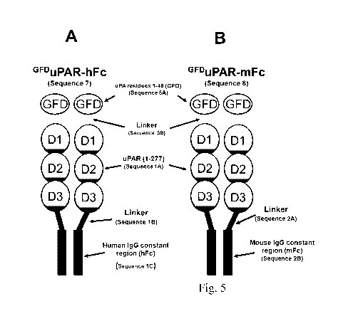
Figure 5: Other uPAR variants: forced dimerization and the addition of GFD
domain on the
N-terminal of uPAR synergize to increase the VN-binding activity of the
receptor
(A) Cartoon illustrating the domain structure of GFDuPAR-hFc. GFDuPAR-hFc
(Sequence 7
(SEQ ID NO: 12)) combines forced dimerization by addition of a C-terminal
human Fc-tag as
shown in Figure lA with the appending of the GFD-domain on the N-terminal as
shown in
Figure 3A. (B) Cartoon illustrating the domain structure of GFDuPAR-mFc.
GFDuPAR-mFc
(Sequence 8 (SEQ ID NO: 13)) is identical to GFDuPAR-hFc with the exception
that the Fc-
region originates from a mouse immunoglobulin (see Figure 1B). (C) GFDuPAR-mFc
binds
with extremely high affinity to immobilized VN. 96-well plates coated with VN
were
incubated with increasing concentrations of GFDuPAR-mFc for 2 hours at room
temperature.
After washing, the bound receptor was detected by sequential incubations with
a biotinylated
antibody specific for the constant region of mouse IgG and Eu3+-labeled
streptavidin. Bound
material was quantified by measuring time-resolved fluorescence. Specific
binding was
calculated by subtracting non-specific binding measured in uncoated wells
incubated with the
same samples. The data represents means SD and are from a representative
experiment. The
binding curve and Kd were calculated by non-linear regression.
18
CA 02842281 2014-01-17
WO 2013/020898 PCT/EP2012/065198
Figure 6: Direct comparison of VN-binding activity of different forms of
soluble uPAR (A)
GFDuPAR-hFc binds with high affinity to immobilized VN. 96-well plates coated
with VN
were incubated with increasing concentrations of GFDuPAR-hFc in the absence of
pro-uPA or
uPAR-hFc and uPARmyc in the presence and absence of excess pro-uPA. After
washing, the
bound receptor was detected by sequential incubations with a monoclonal uPAR
antibody
(13F6) and a Eu3+-labeled goat-anti mouse antibody. The data are from a single
experiment
and presented as means SD. The binding curve and Kd were calculated by non-
linear
regression. Note that GFDuPAR-hFc binds VN with higher affinity and capacity
than any
other form of uPAR tested. (B) Comparison of GFDuPAR-hFc and GFDuPARmyc
binding to
immobilized VN. 96-well plates coated with VN were incubated with increasing
concentrations of GFDuPAR-hFc and GFDuPARmyc in the absence of pro-uPA for 2
hours at
room temperature. After washing, the bound receptor was detected by sequential
incubations
with a monoclonal uPAR antibody (13F6) and a Eu3+-labeled goat-anti mouse
antibody. The
data are from a single experiment and are represented as means SD. The
binding curve and
Kd were calculated by non-linear regression. Note that the dimeric GFDuPAR-hFc
binds VN
with higher affinity (-4-fold) and capacity (-2.5-fold) than the monomeric
GFDuPARmyc.
Figure 7: Lack of specific requirements to the linker region connecting the
GFD and uPAR
domains in uPAR-hFc (A) Tested linker regions. To compare the possible effect
of different
linker length between the GFD and uPAR domains in GFDuPAR-hFc (see Figure 5A),
variants of GFDuPAR-hFc were made with the indicated linker sequences 5, 8, 16
or 20
residues long. The Linker 8 is identical to Sequence 5B (SEQ ID NO: 7) and to
Sequence 9B
(SEQ ID NO: 8) used in all the above experiments. (B) Binding to immobilized
VN. Wells
coated with VN were incubated with conditioned medium (diluted 10-fold) from
293 cell
transiently transfected with the indicated uPAR variants in the presence or
absence of 10 nM
pro-uPA. After washing, the bound material was detected by incubation with a
Eu3+-labeled
anti-human Fc antibody and measurement of time-resolved fluorescence. Note
that
independently of the linker length, all the GFDuPAR-hFc variants bind to VN
independently
of pro-uPA. (C) Binding to immobilized uPA. Wells coated with pro-uPA were
incubated with
conditioned medium (dilute 10-fold) from 293 cell transiently transfected with
the indicated
uPAR variants. After washing, the bound material was detected as described in
Panel B. Note
19
CA 02842281 2014-01-17
WO 2013/020898 PCT/EP2012/065198
that independently of the linker length all the GFDuPAR-hFc variants display
reduced binding
to uPA as compared to uPAR-hFc.
Figure 8: Cartoon illustrating two possible mechanisms by which the appending
of GFD on
uPAR may increase VN-binding and reduce uPA-binding. (A) Intra-molecular
binding. If
sterically allowed, the GFD-domain in GFDuPAR may bind to the uPA-binding
pocket in
uPAR leading to auto-saturation of the chimeric receptor. Auto-saturation of
uPAR may
induce a conformational change in the receptor leading to the efficient
exposure of the VN
binding site and prevent binding of uPA. (B) Inter-molecular binding. If auto-
saturation as
shown in panel A is not possible for sterical reasons, GFDuPAR will be hetero-
divalent and
thus display both uPA and uPAR binding activity. In this case, GFDuPAR is
likely to form
oligomers displaying reduced uPA binding activity and increased VN binding
activity.
Figure 9: Amino acid sequence of 8 antibody variable regions. The amino acid
sequence of
the variable regions of the heavy and light chains were deduced from the cDNA
sequence
obtained by PCR amplification as described in the materials and methods. The
amino acid
sequences are numbered according to the Kabat system. The complementarity
determining
regions (CDR) 1, 2 and 3 (from left to right) are underlined. Gaps introduced
in the sequences
to maintain alignment are indicated by hyphens. Punctuation, which corresponds
to an Xaa in
the sequence listing, indicates that the sequence is either unknown or
uncertain. Thus, Xaa can
be any naturally occurring amino acid.
Figure 10: Monoclonal antibodies raised against GFDuPAR-hFc recognize cell
surface
uPAR. 293 cells expressing human uPAR (huPAR), mouse uPAR (muPAR) or no uPAR
(mock) were stained with the monoclonal antibodies 8B12, 10H6, 13D11, 19.10,
13F6, AL6,
AL38 and BE18 raised against GFDuPAR-hFc. Bound antibody was detected using a
fluorescein labeled goat anti mouse antibody and the staining was analyzed by
flow cytometry.
The histograms show the staining intensity (X-axis, FL1-H) and frequency (Y-
axis, in % of
the most frequent intensity). Note that all eight antibodies stain cells
expressing human uPAR
specifically. The antibodies BR4 and AK17 have been described previously and
react
specifically with mouse uPAR (Tjwa et al., 2009).
Figure 11: Functional inhibitory activity of mAb 8B12. 293 cells expressing
human uPAR
were seeded in 96-well E-plates coated with Vitronectin (A and B) or
Fibronectin (C) and
transferred to a real time cell analyzer instrument (RTCA, xCELLigence, SP
Roche Corp.).
CA 02842281 2014-01-17
WO 2013/020898 PCT/EP2012/065198
The electric impedance (termed cell index, CI) was recorded at regular
intervals. After
approximately 2 hours, cells were added pro-uPA (B and C) or vehicle (A) and
the cell index
measurements continued. About one additional hour later, wells were added a
dilution curve
of 8B12 antibody at the final concentrations indicated in the graphs and the
cell index
measurements continued. The times at which pro-uPA and 8B12 were added are
indicated in
the graphs by stippled vertical lines. The curves show the normalized cell
index (NCI, Y-axis)
as a function of time (X-axis). All cell indexes were normalized to the cell
index measured
immediately prior to antibody addition. To determine IC50 values (panel D),
the NCI
measured one hour after antibody addition were calculated in % of the NCI for
untreated cells
at the same time point (ANCI, Y-axis) and graphed in function of antibody
concentration (X-
axis).
Figure 12: Epitope mapping by flow cytometry (I). 293 cells expressing human
uPAR (uPAR
WT), uPAR R83/89A were stained with the different antibodies as indicated and
the binding
analyzed by flow-cytometry. The staining of uPAR WT cells was conducted both
in presence
and absence of pro-uPA to detect possible effects of ligand occupancy on
antibody binding.
As negative control (Neg. Ct.), the staining profile of uPAR WT cells
receiving no primary
antibody is shown in all panels.
Figure 13: Epitope mapping by flow cytometry (II). As Figure 12 but different
uPAR variants
analyzed.
Figure 14: Epitope mapping by flow cytometry (III). As Figure 12 and 13 but
with different
uPAR variants analyzed.
Figure 15: Location of the binding epitope for the inhibitory antibodies in
uPAR. uPAR is
composed of three domains (D1, D2 and D3) where D1 is linked to D2 by a short
linker
region. This linker region contains residues that are critical for receptors
interaction with VN
(R91 and Y92, underlined) (Madsen et al., 2007) (Gardsvoll and Ploug, 2007).
The binding
site for the inhibitory antibodies generated in this example has overlapping
epitope(s) with
R89, R91 and Y92 being important hot-spots for binding. The structure of the
D2D3
truncation version of uPAR is shown below. This variant lacks residues 1-82 of
uPAR of SEQ
ID NO: 1.
Figure 16: Inhibition of Eu3 -uPA binding to 293/uPAR cells by mAb 8B12, 13F6,
R3 and
pro-uPA. mAb 8B12 does not interfere with the proteolytic functions of uPAR.
To investigate
21
CA 02842281 2014-01-17
WO 2013/020898 PCT/EP2012/065198
if the inhibitory antibody 8B12 is a specific inhibitor of the uPAR/VN-
interaction, or if it also
interferes with uPA binding to the receptor, we conducted in vitro binding
assays. Note that
mAb 8B12 displays no or little inhibitory activity documenting that this
antibody does not
interfere with the proteolytic functions of the receptor. The validity of the
assay is documented
by the fact that the R3 antibody, and un-labeled pro-uPA, displayed efficient
competitive
activity. (CPS ¨ Counts per second).
Figure 17: mAb 8B12 inhibits PC3 tumor growth in vivo. Male Balb C nu/nu mice
were
inoculated with (1 x 10⁶) PC-3 cells through the subcutaneous (s.c.) route.
Animals were
treated by bi-weekly injections with 10.0 mg/kg of mAb 8B12, mAb 13F6, a non-
immune
control mouse IgG (mIgG) or PBS via intraperitoneal route. Tumors were
measured twice
weekly, and tumor volume was determined as described in Materials and Methods.
No
differences were observed in the tumor growth between PBS and mIgG treated
animals and
data from these were pooled prior to statistical analysis. Significant
differences between
control animals and 8B12 treated animals are represented by asterisks (NS, Non-
Significant,
P>0.05, *P < .05, **P < .01 and ***P<.001). The difference in tumor volume
between control
and 8B12 treated animals (in %) is indicated.
Figure 18: mAb 8B12 reduces PC-3 tumor cell proliferation and promotes
apoptosis in vivo.
Male Balb C nu/nu mice were inoculated subcutaneously with PC-3 cells and
treated by bi-
weekly injections with 10.0 mg/kg of mAb 8B12, mAb 13F6 or a non-immune
control mouse
IgG via intraperitoneal route. Eight weeks after xenografting, the tumors were
harvested and
analyzed by immunohistochemistry (Panel A) as described in the Materials and
Methods
section. Ki-67 and activated Caspase-3 stainings are shown and nuclei are
counterstained with
DAPI. The quantification of the data is shown in Panel B. Note that the
treatment with the
inhibitory mAb 8B12 significantly reduces tumor cell proliferation and
increases apoptosis
when compared to treatment with control IgG. The non-inhibitory mAb 13F6, of
the same
isotype, does not display this activity documenting that it is the inhibitory
activity of the mAb
8B12 that is responsible for the anti-proliferative and pro-apoptotic effect.
The unit of the Y-
axis is number of positive cells per field.
Figure 19. Domain composition and VN-binding characteristics of mGFDmuPAR-Fc
morn
(A) Cartoon illustrating the domain organization of mGFDmuPAR-hFc . muPAR-
hFc
(Sequence 9 (SEQ ID NO: 17)) is composed of the receptor-binding growth factor-
like
22
CA 02842281 2014-01-17
WO 2013/020898 PCT/EP2012/065198
domain of murine uPA, mGFD (Sequence 9A (SEQ ID NO: 4)), a short linker
(Sequence 9B
(SEQ ID NO: 8)), mouse uPAR residues 1-273 (Sequence 9C corresponding to aa. 1-
273 of
SEQ ID NO: 2), another short linker (Sequence 9D (SEQ ID NO: 11)) and a C-
terminal
human Fc-tag (hFc, Sequence 1C (SEQ ID NO: 5)). A C-terminal of mouse Fc-tag
can be
equally used.
(B) Binding of mGFDmuPAR-hFc to immobilized VN. 96-well plates coated with VN
were
incubated with increasing concentrations of mGFDmuPAR-hFc for 2 hours at room
temperature.
After washing, the bound receptor was detected by incubation with a Fu3+-
labeled goat anti-
human Fc antibody. The bound material was detected by measuring time-resolved
fluorescence intensity. Specific binding was calculated by subtracting the non-
specific binding
measured in uncoated wells incubated with identical samples. The shown data
are means SD
from a representative experiment. The dissociation constant (Kd) was
calculated by non-linear
regression (four-parameter fit) using the Prism 5.0 software suite.
Figure 20. Antibodies raised against mGFDmuPAR-hFc inhibit cell adhesion to VN
mediated
by mouse uPAR. Inhibition of 293/muPAR cell adhesion to VN by cell culture
supernatants
from myeloma hybrids producing antibodies recognizing mGFD muPAR-hFc. 293
cells
expressing murine uPAR were seeded in VN-coated E-plates and cell adhesion
followed by
impedance measurements using an xCELLigence plate reader (Roche). When
adhesion arrived
at plateau (indicate by stippled vertical line), the wells were added
conditioned medium (final
concentration 30% v/v) from the 13 different myeloma hybrids derived from
splenocytes from
mice immunized with mGFDmuPAR-hFc. Note that the conditioned medium from 4
hybrids
results in a strong (OMD4, NE43 and 00F12) or intermediate reduction (NM23) in
cell
adhesion (measured as the normalized cell index) while conditioned medium from
the
remaining 9 hybrids displays little or no inhibitory activity.
Figure 21. The inhibitory antibodies OMD4 and NE43 bind to the VN binding site
in mouse
uPAR. To determine if the binding epitope of the generated antibodies falls in
the VN-binding
site of mouse uPAR (muPAR), in vitro binding assays were conducted on the
antigen used for
immunization (mGFDmu--
FAR-hFc) and a variant of this chimera containing a single amino acid
substitution in the VN binding site of muPAR (mGFDmuPAR-hFc R92A) as well as a
human
soluble receptor (suPAR) to determine if the antibodies also recognize human
uPAR.
23
CA 02842281 2014-01-17
WO 2013/020898 PCT/EP2012/065198
96-well elisa plates were coated with mGFDmuPAR-hFc mGFD,
muPAR-hFc R92A or human
soluble uPAR (suPAR), blocked and incubated with hybridoma supernatants
diluted 1:100 in
dilution buffer. After washing, bound antibody was probed by incubation with a
Eu3+-labeled
goat anti-mouse antibody and quantified by enhanced timeresolved fluorescence
intensity
measurements (Delfia). Specific binding was calculated by subtracting the
binding observed to
uncoated wells. Note that OMD4 and NE43 do not recognize the mGFDmuPAR-hFc
R92A
variant suggesting that these antibodies bind to the VN binding site in muPAR.
One of these
antibodies (OMD4) also recognizes the human receptor.
Figure 22. Amino acid sequence of mAb OMD4 raised against mGFDmuPAR-Fc heavy
chain
variable region. The amino acid sequences of the variable region of the OMD4
heavy chain
was deduced from the cDNA sequence obtained by PCR amplification as described
in the
Materials and Methods, Example 2. The amino acid sequence is numbered
according to the
Kabat system. The complementarity determining regions (CDR) 1, 2 and 3 (from
left to right)
are underlined.
Figure 23. Species specificity of the inhibitory activity of mAb OMD4, NE43,
00F12,
NM23, 8B12. 293 cells expressing human uPAR (Panel A) and mouse uPAR (Panel B)
were
seeded on VN-coated E-plates and cell adhesion monitored by impedance
measurement. Once
a plateau of cell adhesion was reached (vertical stippled line), wells were
added purified
antibody to a final concentration of 100 nM* and the resulting changes in cell
adhesion
recorded. Note that the adhesion of cells expressing human uPAR is inhibited
by mAb 8B12
and partially by mAb OMD4, while the remaining antibodies are without notable
effect. In
contrast, the adhesion of cells expressing murine uPAR is inhibited by mAb
NE43, 00F12,
NM23, partially by OMD4, but not at all by 8B12. 13F6 was used as a non-
inhibitory negative
control antibody binding human uPAR.
*The OMD4 antibody is IgA isotype and was used in the form of cell culture
supernatant
diluted 1:5. The concentration of this antibody in the supernatant is unknown
and may be low.
The partial effect observed with this antibody may therefore be attributed to
this.
Figure 24: Panning strategy for the isolation of scFv's recognizing ligand
occupied dimeric
uPAR
Figure 25: Reactivity of isolated scFy with cell surface uPAR. 293 cells
expressing human
uPAR were stained with the indicated scFy (200 nM). Bound antibody was
detected using a
24
CA 02842281 2014-01-17
WO 2013/020898 PCT/EP2012/065198
fluorescein labeled goat anti-human F(ab)2 antibody and the staining was
analyzed by flow
cytometry. The histograms show the staining intensity (X-axis, FL1-H) and
frequency (Y-axis,
in counts).
Figure 26: Inhibitory activity of scFy 3B6. The inhibitory activity of scFy
3B6 was assayed as
described for mAb 8B12 in Figure 11. The curves show the normalized cell index
(NCI, Y-
axis) as a function of time (X-axis). All cell indexes were normalized to the
cell index
measured immediately prior to antibody addition. To determine IC50 values
(panel D), the
NCI measured one hour after antibody addition were calculated in % of the NCI
for untreated
cells at the same time point (ANC, Y-axis) and graphed in function of antibody
concentration
(X-axis).
Figure 27: Inhibitory activity of scFy 3C10. The inhibitory activity of 3C10
was assayed
exactly as decribed for scFy 3B6 in Figure 26.
Figure 28: Comparison of the inhibitory activity of 8B12 with that of other
compounds
known to inhibit the uPAR/VN- interaction or uPAR function. The inhibitory
activity of the
SMB domain (Panel A), the peptide P7 (Panel B), antibodies R3 and R5 (Panel C)
as well as
the R2 antibody (Panel D) were measured as described for the 8B12 antibody in
Figure 11. To
determine the IC50 values, the NCI measured one hour after compound addition
were
calculated in % of the NCI for untreated cells at the same time point (ANCI, Y-
axis) and
graphed in function of compound concentration (X-axis). The inhibition curves
for 8B12 from
Figure 11 have been included in all four panels for comparison. The calculated
IC50 and max
inhibition constants for each of the tested compounds can be found in Table 2.
EXAMPLE 1
Materials and Methods
Construction of expression vectors
The expression vectors for recombinant proteins tagged with a human IgG
constant
region (hFc) are based on the pFRT/TO-Fc plasmid (Madsen et al., 2007),
however a number
of modifications were introduced to facilitate the shuffling of different
coding regions as well
as to improve protein yields. Firstly, an XhoI restriction site located in the
vector sequence
downstream of the hFc coding region was destroyed by site-directed mutagenesis
using oligos
dXu/dXd. Secondly, a linker encoding a cleavage sequence for the PreScission
protease, made
by annealing oligos PreF/PreR, was inserted in the XhoI site located at the
signal peptide/Fc
CA 02842281 2014-01-17
WO 2013/020898 PCT/EP2012/065198
junction. To remove the introns present in the Fc region of the construct,
which was found to
increase the yield of recombinant protein (our unpublished observations), the
vector was
transfected into CHO cells, RNA extracted, reverse transcribed, and the cDNA
amplified with
oligos hVNukpn/FcNr and cloned KpnI/NotI into pcDNA5/FRT-TO (Invitrogen corp.)
and
pEGFP-N1 (Clontech corp.) to generate pFRT/TO-hFc and pN 1 -hFc, respectively.
Expression
vectors for recombinant proteins tagged with a mouse IgG constant region (mFc)
was
generated by PCR amplification of a mouse IgG1 cDNA (clone IRAVp968B035D,
obtained
from imaGenes GmbH) with oligos mFcU/mFcD and cloned XhoI/NotI in pFRT/TO-hFc
and
pNl-hFc to generate pFRT/TO-mFc and pNl-mFc, respectively. Constructs encoding
soluble
uPAR tagged with a human Fc (uPAR-hFc, Sequence 1 (SEQ ID NO: 14)) and mouse
Fc
(uPAR-mFc, Sequence 2 (SEQ ID NO: 15)) were made by amplification of a full-
length
uPAR cDNA (Madsen et al., 2007) with oligos URskF/UpreR2D and cloned KpnI/XhoI
into
pFRT/TO-hFc and pFRT/TO-mFc to generate pFRT/TO-uPAR-hFc and pFRT/TO-uPAR-
mFc, respectively. The construct encoding soluble myc-tagged uPAR (uPARmyc,
Sequence 3
(SEQ ID NO: 26)) was generated by amplification of the uPAR cDNA with oligos
URskF/URMYCR and cloned KpnI/NotI into pcDNA5/FRT-TO to generate pFRT/TO-
uPARmyc. The expression vector encoding a chimera between the growth factor
domain of
uPA (GFD, Sequence 5A (SEQ ID NO: 3)) and full-length uPAR (Sequence 4 (SEQ
ID NO:
1)). GFDuPAR (Sequence 5 (SEQ ID NO: 16)) was generated in a two-step PCR
overlap
amplification procedure. Firstly, an uPA cDNA was amplified with oligos
ATFkpnF/GFD1r
and an uPAR cDNA with oligos UL817F012394. Secondly, the two PCR products were
mixed, co-amplified using oligos ATFkpnF/F012394 and cloned KpnI/NotI in
pcDNA5/FRT-
TO to generate pFRT/TO-GFDuPAR. The expression vector encoding soluble GFDuPAR
with a
C-terminal myc-tag (GFDuPARmyc, Sequence 6 (SEQ ID NO: 28)) was generated by
amplifying pFRT/TO-GFDuPAR with oligos ATFkpnF/URMYCR and cloning the product
KpnI/NotI in pcDNA5/FRT-TO to generate pFRT/TO-GDFuPARmyc. The expression
vectors
encoding soluble dimeric GFD uPAR-variants with a C-terminal human Fc-tag (GFD
uPAR-hFc,
Sequence 7 (SEQ ID NO: 12)) and mouse Fc-tag mFc (GFDuPAR-mFc, Sequence 8
(SEQ ID
NO: 13)) tags were generated by amplifying pFRT/TO-GFDuPAR with oligos
ATFkpnF/UpreR2D and cloning the product KpnI/XhoI in pFRT/TO-hFc and pFRT/TO-
mFc
to generate pFRT/TO-GDFuPAR-hFc and pFRT/TO- GDFuPAR-mFc, respectively.
Expression
26
CA 02842281 2014-01-17
WO 2013/020898 PCT/EP2012/065198
vectors encoding chimeras with different lengths of linker region between the
GFD and uPAR
domains in the chimera were made as described above replacing oligo uL8f with
uL5f, uLl2f,
uLl6f or uL20f. The region encoding GFD uPAR-hFc, and its variants with
different linker
length, were transferred KpnI/NotI to the pEGFP-N1 expression vector (Clontech
Corp.)
generating pNl-GFDuPAR-hFc used for transient expression experiments.
Expression and purification of recombinant proteins
The pFRT/TO-uPAR-hFc, pFRT/TO-GFDuPAR-hFc, pFRT/TO-uPAR-mFc, pFRT/TO-
GFD GFD
uPAR-mFc, pFRT/TO-uPARmyc pFRT/T 0- uPARmyc, expression vectors were
transfected into CHO Flp-In cells (Invitrogen Corp.) and the recombinant
proteins expressed
under serum-free conditions as previously described (Madsen et al., 2007).
Recombinant
tagged with human or mouse Fc tags were purified from the conditioned media by
standard
Protein A affinity chromatography and dialyzed extensively against PBS. The
conditioned
medium of pFRT/TO-uPARmyc and GFDuPARmyc transfected cells was concentrated
¨20-fold
and utilized for binding assays without further purification. Standard ELISA
assays were
employed to determine the concentrations of uPARmyc in the concentrated
conditioned media.
The GFDuPAR-hFc variants with different lengths of linker between the GFD and
uPAR
moiety were expressed by transient transfection of Phoenix cells cultured in
OptiMEM serum-
free media (Invitrogen Corp.) with the pN1 -GFDuPAR-hFc vector variants and
the conditioned
medium recovered after 6-8 days of culture.
Binding assays
Black 96-well immunoplates were coated with pro-uPA or VN (10 nM) diluted in
coating buffer (50 mM sodium carbonate, pH 9.6) at 4 C ON. Plates were washed
with wash
buffer (phosphate buffered saline containing 0.1% Tween-20 (PBS-T) and non-
specific
binding sites saturated with blocking buffer (PBS containing 2% bovine serum
albumin
(BSA)) for > 2 hours at RT. After washing with PBS-T, wells were incubated
with the
indicated concentrations of uPAR-hFc, uPAR-mFc and uPARmyc diluted in dilution
buffer
(PBS containing 1% BSA) in the presence or absence of pro-uPA as indicated.
The binding
was allowed to occur for 2 hours at RT after which unbound reagents were
removed by rinsing
with wash buffer. Bound uPAR-hFc and uPARmyc were detected by sequential
incubations
with an anti-uPAR monoclonal antibody (13F6, 1 g/m1) and a Eu3+-labeled goat-
anti mouse
antibody (1:5.000, Perkin Elmer Corp.). The Eu3+-label was detected by
measuring time-
27
CA 02842281 2014-01-17
WO 2013/020898 PCT/EP2012/065198
resolved fluorescence intensity using an Envision Xcite plate reader (Perkin
Elmer Corp.)
employing the DELFIA label protocol. To calculate the specific binding, non-
specific binding
measured in uncoated wells, incubated with identical samples, was subtracted
from the total
binding measured in coated wells.
Cell lines and cell adhesion assays
HEK293 Flp-In T-REx cells (293) expressing GFDuPAR were generated by
transfection
with the pFRT/TO-GFDuPAR vector according to published procedures (Madsen et
al., 2007).
293 cells transfected with empty vector and cells expressing uPARw32A and
uPARR91A have
been described previously (Madsen et al., 2007).
Oligonucleotide sequences
dXu: 5'-gtaaatgagcggccgcgtcgagtctagaggg-3' (SEQ ID NO: 29)
dXd: 5 '-ccctctagactcgacgcggccgctcattta-3 '(SEQ ID NO: 30)
PreF: 5 '-tcgagctggaagttctgttccaggggccca-3 ' (SEQ ID NO: 31)
PreR: 5 '-agctacccggggaccttgtcttgaaggtcg-3 ' (SEQ ID NO: 32)
hVNukpn: 5'-cggggtaccatggcacccctgaga-3' (SEQ ID NO: 33)
FcNr: 5'-ttgcggccgctcatttacccggagacag-3' (SEQ ID NO: 34)
mFcU: 5 '-gcctcgaggcaggagcaggacccagggattgtggttgtaa-3' (SEQ ID NO: 35)
mFcD: 5'-gcgcggccgctcatttaccaggagagtg-3' (SEQ ID NO: 36)
URskF: 5'-gcgtcgacggtacccgccaccatgggtcacccgccgctgctg-3' (SEQ ID NO: 37)
UpreR2D: 5'-gcctcgaggggcccctggaacagaacttccagatccaggtctgggtggttacagccact-3'
(SEQ ID NO:
38)
URMYCR: 5'-gcgcggccgctcacagatcctcttcagagatgagtttctgctctcctcctgggtggttacagccact-
3' (SEQ
ID NO: 39)
ATFkpnF: 5'-gcggtacccgccaccatgagagccctgctggcgcgc-3' (SEQ ID NO: 40)
GFD1r: 5 '-tgtgaaatagataagtcaaaagggggggccggggcg-3 ' (SEQ ID NO: 41)
uL8f: 5'-gggggggccggggcggctggaggactgcggtgcatgcagtgtaag-3' (SEQ ID NO: 42)
F012394: 5 '-tagtttagcggccgcttaggtccagaggagagt-3 ' (SEQ ID NO: 43)
UpreR2D: 5'-gcctcgaggggcccctggaacagaacttccagatccaggtctgggtggttacagccact-3'
(SEQ ID NO:
44)
URMYCR: 5'-gcgcggccgctcacagatcctcttcagagatgagtttctgctctcctcctgggtggttacagccact-
3' (SEQ
ID NO: 45)
28
CA 02842281 2014-01-17
WO 2013/020898 PCT/EP2012/065198
uL5f: 5'-gggggggccggggcgctgeggtgcatgcagtgtaag-3' (SEQ ID NO: 46)
uLl6f:
5 '-gggggggccggggcggctggagcaggagcaggtgctggtgctggaggactgcggtgcatgcagtgtaag-3 '
(SEQ ID NO: 47)
uL20f:
5'-
gggggggccggggcggctggagcaggagcaggtgctggtgctggagcaggtgctggtggtctgcggtgcatgcagtgta
ag-3'
(SEQ ID NO: 48)
Results
Forced dimerization of uPAR strongly enhances binding to vitronectin
Background and rationale
Several lines of evidence suggest that receptor-dimerization plays an
important role for
the interaction between uPAR and VN (Caiolfa et al., 2007; Sidenius et al.,
2002), however,
the dissociation constants determined by surface plasmon resonance (SPR) for
the interaction
between immobilized uPAR and soluble VN (¨ 1 04, (Gardsvoll and Ploug, 2007))
are
clearly insufficient to explain the high-affinity interaction predicted from
equilibrium binding
experiments using immobilized VN and soluble uPAR (Gardsvoll and Ploug, 2007;
Sidenius
et al., 2002). To directly address the role of uPAR-dimerization on VN-
binding, authors here
describe the construction, expression and purification of soluble forms of
recombinant dimeric
uPAR and the comparison of the ligand-binding characteristics of these with
those of
"conventional" soluble monomeric uPAR.
Construction and expression of dim eric uPAR
To directly determine the importance of receptor dimerization for the
interaction
between soluble uPAR and immobilized VN, authors constructed a soluble human
uPAR
tagged on the C-terminal with the hinge and constant region of a human IgG1
(hFc). The
resulting uPAR-hFc chimera is a covalent homo-dimer in which the two
polypeptides are held
together by disulphide bonds located in the hinge region of the Fc-tag (Figure
1A). The
uPAR-hFc chimera (Sequence 1 (SEQ ID NO: 14)) is composed of uPAR (residues 1
to 277,
Sequence 1A, corresponding to aa. 1-277 of SEQ ID NO: 1), a linker region
(LEVLFQGPLE,
Sequence 1B (SEQ ID NO: 9)) and the human Fc-tag (241 residues, Sequence 1C
(SEQ ID
NO: 5)). A similar construct was also made using the Fc-region of a mouse
immunoglobulin
and as illustrated in Figure 1B the sequence and predicted domain structure of
this chimera,
uPAR-mFc (Sequence 2 (SEQ ID NO: 15)), is identical to that of uPAR-hFc with
the
29
CA 02842281 2014-01-17
WO 2013/020898 PCT/EP2012/065198
exception of a slightly different linker region (Sequence 2A (SEQ ID NO: 10))
and the mouse
Fc-tag (Sequence 2B (SEQ ID NO: 6)). As a monomeric control receptor, authors
constructed
a soluble human uPAR with a C-terminal myc-tag (uPARmyc, Sequence 3 (SEQ ID
NO: 26))
illustrated in Figure 1C. This protein is composed of uPAR (residues 1 to 274,
Sequence 3A
corresponding to aa. 1-274 of SEQ ID NO:1) and a C-terminal myc-tag
(GGEQKLISEEDL,
Sequence 3B (SEQ ID NO: 27)). The recombinant proteins were expressed in
Chinese hamster
ovary (CHO) cells and purified from the conditioned media by standard Protein
A affinity
chromatography (uPAR-hFc and uPAR-mFc) or utilized without purification
(uPARmyc) after
quantification by ELISA.
Binding characteristics of dimeric uPAR
The VN-binding activity of uPAR-hFc (Figure 2A) and uPARmyc (Figure 2B) were
measured by incubating immobilized VN with increasing concentration of the
recombinant
receptors in the presence or absence of an excess of pro-uPA (the
catalytically inactive
zymogen form of uPA). After washing, bound receptor was revealed by sequential
incubations
with a mouse monoclonal anti-uPAR antibody (13F6), an Eu3+-labeled goat anti-
mouse
antibody and quantified by time-resolved fluorescence measurements. In the
absence of pro-
uPA, both uPAR-hFc and uPARmyc display poor binding to VN and the affinities
of the
interactions cannot be reliably estimated. However, in the presence of excess
pro-uPA, both
uPAR-hFc and uPARmyc display specific and dose dependent binding to VN. By non-
linear
regression analysis of the binding curves, the apparent dissociation constants
(Kd) of the
interaction between uPAR-hFc, uPARmyc and immobilized VN were calculated to be
¨10 nM
and ¨80 nM, respectively. In contrast, both the monomeric and dimeric soluble
receptors bind
immobilized uPA with comparable apparent affinities of ¨6 nM (uPAR-hFc) and
¨10 nM
(uPARmyc).
These data document that forced dimerization of uPAR, using an immunoglobulin
Fc-
tag, results in a ¨10-100-fold increase in the receptors apparent affinity for
VN as compared to
the monomeric receptor. The binding of both monomeric (uPARmyc) and dimeric
(uPAR-hFc
and uPAR-mFc) soluble uPAR is dependent upon concomitant occupancy by uPA as
no or
little binding is observed in its absence. The fact that forced dimerization
of uPAR fails to
increase the binding of the receptor to immobilized VN in the absence of uPA,
as well as to
CA 02842281 2014-01-17
WO 2013/020898 PCT/EP2012/065198
immobilized uPA, suggests that the increase in apparent affinity involves
unique
conformational changes and that it is not only a result of increased avidity.
Chimeras between uPA and uPAR display strong VN-binding and reduced uPA-
binding
Background and rationale
As described in the literature, and as illustrated in Figure 2, binding of
soluble uPAR to
VN is strongly dependent upon the concomitant occupancy of the receptor by
uPA. A
plausible explanation for this observation is that uPA-binding to uPAR induces
a
conformational change in the receptor leading to the exposure of the VN-
binding epitope in
the occupied receptor. With the aim of generating an uPAR-variant displaying
constitutive, i.e.
uPA-independent, VN-binding combined with deficient uPA-binding, authors
conceived that
this could be achieved through the construction of an appropriate uPA/uPAR-
chimera in
which the intra-molecular binding reaction predicted to occur in such a
chimera would lead to
the exposure of the VN-binding epitope as well as prevent the binding of uPA
in trans.
Construction of the uPA/uPAR chimeraGFDUPAR
To generate an uPAR-variant constitutively active in VN-binding and deficient
in
uPA-binding, authors engineered the growth factor-like domain of uPA (GFD)
onto the N-
terminal of human uPAR as illustrated in Figure 3A. The resulting chimera
(GrbupAR,
Sequence 5 (SEQ ID NO: 16)) is composed of the growth factor-like domain from
human uPA
(Sequence 5A (SEQ ID NO: 3)) connected by a short linker (Sequence 5B (SEQ ID
NO: 7)) to
the N-terminal of intact mature human uPAR (Sequence 4 (SEQ ID NO: 1)).
Binding characteristics of cell-surface GFDuPAR
To analyze the binding characteristics of GFDuPAR chimera, authors generated
293 cell
lines expressing GFDuPAR on the cell surface and compared their adhesion
characteristics with
that of cells expressing wild-type uPAR and uPAR-variants with specific
deficiency in VN-
binding (Figure 3). In these assays, authors found that GFDuPAR, like the wild-
type receptor,
promotes firm, integrin-independent, cell adhesion to VN (Figure 3B)
confirming that the
chimera retains full VN-binding activity. In addition, expression of GFDuPAR
failed to promote
cell binding to immobilized uPA suggesting that also the predicted loss of uPA-
binding
activity was attained in this chimera (Figure 3C).
Construction and binding characteristics of soluble GFDuPAR
31
CA 02842281 2014-01-17
WO 2013/020898 PCT/EP2012/065198
uPAR-mediated cell adhesion to VN does not require uPA-binding as long as on
the cell-
surface expression levels are sufficiently high (Madsen et al., 2007; Sidenius
and Blasi, 2000)
and authors therefore next generated a soluble variant of GFDuPAR for in vitro
binding
experiments. For this purpose, authors constructed a truncated version of
GFDuPAR carrying a
C-terminal myc-tag in place of the membrane anchoring sequence of wild-type
uPAR. The
constructed chimera illustrated in Figure 4A (GFDuPARmyc
Sequence 6 (SEQ ID NO: 28))
has the same N-terminal GFD-domain (Sequence 5A (SEQ ID NO: 3)) and linker-
sequence
(Sequence 5B (SEQ ID NO: 7)) as described above for GFDuPAR and the same uPAR
sequence (Sequence 3A (corresponding to aa. 1-274 of SEQ ID NO: 1)) and C-
terminal myc-
tag (Sequence 3B (SEQ ID NO: 27)) as described for uPARmyc in Figure 1C.
The GFDuPARmyc chimera was produced in CHO cells and its binding
characteristics
analyzed by in vitro binding assays to immobilized VN (Figure 4B) or
immobilized pro-uPA
(Figure 4C). As presented, the GFDuPARmyc chimera binds with high affinity (Kd
¨ 1.3 nM)
to immobilized VN in the absence of uPA. Assayed under identical conditions,
the control
receptor uPARmyc, lacking the N-terminal GFD-domain, fails to display any
appreciable
binding to immobilized VN even at concentrations up to 1 M. Appending the GFD-
domain
of uPA on the N-terminal of uPAR thus increases the measured binding affinity
of the receptor
G
to VN by at least three orders of magnitude. When the same proteins
(GFDuPARmyc and
uPARmyc) were tested for binding activity towards immobilized pro-uPA (Figure
4C),
authors found that the GFDuPARmyc chimera displayed a reduced binding affinity
(-30-fold)
and capacity (-5-fold) as compared to the control receptor (uPARmyc).
Conclusions
These data demonstrate that appending the GFD-domain of uPA on the N-terminal
of
uPAR increases the affinity of the receptor for VN by more than three orders
of magnitude.
Forced dimerization and uPA:uPAR-chimerism synergize to increase VN-binding
activity
Background and rationale
As documented above, the apparent affinity of uPAR for VN can be strongly
increased
by forced dimerization or by appending the GFD-domain of uPA on the N-terminal
of uPAR.
Finally authors here document that the combination of forced dimerization with
the appending
of the GFD-domain synergize to increase the apparent affinity for VN.
Construction and expression of dimericGFDUPAR
32
CA 02842281 2014-01-17
WO 2013/020898 PCT/EP2012/065198
Expression vectors encoding dimeric uPAR containing a GFD domain on the N-
terminal of uPAR were constructed by combining a C-terminal Fc tag as shown in
Figure 1A-
B with the engineering of the GFD domain of uPAR onto N-terminal of uPAR as
illustrated in
Figure 3A. The resulting chimeric proteins GFD
DuPAR-hFc (Sequence 7 (SEQ ID NO: 12)) and
GFDuPAR-mFc (Sequence 8 (SEQ ID NO: 13)) are illustrated in Figure 5A and B
respectively
and are composed of the GFD domain (Sequence 5A (SEQ ID NO: 3)) of uPA, a
linker region
(Sequence 5B (SEQ ID NO: 7)), uPAR residues 1-277 (Sequence lA corresponding
to aa. 1-
277 of SEQ ID NO: 1), another linker region (Sequence 1B (SEQ ID NO: 9) or
Sequence 2A
(SEQ ID NO: 10)) and a C-terminal Fc tag from a human (Sequence 1C (SEQ ID NO:
5)) or
mouse (Sequence 2B (SEQ ID NO: 6)) IgG.
Binding characteristics of dimeric GEDupAR -hFc and GFD uPAR-mFc
The GFDuPAR-hFc and GFDuPAR-mFc chimeras were expressed in CHO cells and
purified from the conditioned medium by Protein A affinity chromatography. To
determine
the VN-binding properties of the recombinant receptors, authors first measured
the binding of
GFDuPAR-mFc to immobilized VN (Figure 5C). As shown, GFDuPAR-mFc binds with
very
high affinity (¨ 20 pM) suggesting that forced dimerization (using an Fc-tag)
and ligand auto-
saturation (using the GFD domain) synergizes to increase the VN-binding
activity of uPAR.
To more directly compare the VN-binding characteristics of the different forms
of
soluble uPAR, additional binding experiments were conducted (Figure 6). When
compared to
uPAR-hFc in the presence of uPA, GFD
DuPAR-hFc has about 3-fold higher affinity and binding
capacity. When compared to monomeric soluble uPAR (uPARmyc) in the presence of
uPA,
GFD
uPAR-hFc displays about 600-fold higher affinity and 6-fold higher binding
capacity. Both
monomeric (uPARmyc) and dimeric (uPAR-hFc) forms of uPAR show no or little
binding to
VN in the absence of uPA and quantification of the differences in affinity is
therefore difficult.
However, when compared to the published values for the binding of VN to
immobilized uPAR
(1.3 M, (Gardsvoll and Ploug, 2007)), GFDuPAR-hFc display about 10.000-fold
higher
affinity. When compared directly to GFDuPARmyc (Figure 6B), GFDuPAR-hFc
display 3-fold
higher affinity and 2.5-fold higher binding capacity demonstrating that both
dimerization and
ligand auto-saturation contributes to the remarkable binding activity of
GFDuPAR-hFc and
presumably also GFDuPAR-mFc.
Requirements to the linker region connecting the GFD to uPAR
33
CA 02842281 2014-01-17
WO 2013/020898 PCT/EP2012/065198
To determine the importance of the length and sequence of the linker region
connecting the GFD domain to the N-terminal of uPAR in the GmuPAR-hFc chimera,
authors
generated variants of this with a shorter linker (5 residues) and longer
linkers (16 and 20
residues) and compared the VN and uPA binding activities with that of the
"standard" linker
(8 residues) used elsewhere in this study (Figure 7A). The variants were
expressed by
transient transfection in 293 cell line and the binding activity in the
conditioned medium was
measured (Figure 7). As shown, the addition of the GFD domain on the N-
terminus of uPAR
enhances VN binding (Figure 7B) and reduces uPA binding (Figure 7C)
independently on
the linker length applied suggesting that this sequence is very flexible in
terms of length.
Possible mechanisms explaining the high affinity ofGFDuPAR chimeras for VN
The concept behind the construction of GmuPAR was that the GFD domain
engineered
onto the N-terminus of uPAR would bind to the uPA binding cavity in uPAR in an
intra-
molecular fashion as illustrated in Figure 8A. Nevertheless, it is possible
that the intra-
molecular binding is prohibited by sterical constrains. In this case a single
molecule of
GFDuPAR may effectively display both uPAR and uPA binding activity and is
likely to self-
associate and oligomerize as shown in Figure 8B.
EXAMPLE 2
Materials and Methods
Antigen preparation
GFDuPAR-hF c and GFDuPAR-mF c were expressed and purified as described in
detail in
Example 1.
Immunization of mice
Three 2-month-old male C57B1/6 uPAR-/- mice (Ms#21574, Ms#1416 and Ms#1417)
were immunized by intraperitoneal (i.p.) injection with 67 lag GtuupAR -hFc in
200 1 of a 1:1
emulsion between 100 1 immunogen in PBS and 100 1 of Complete Freund's
Adjuvant
(CFA). The immunized animals were boosted 3 times, at 3-week intervals, by IP
injection of
671ag GmuPAR-hFc in 200 1 of a 1:1 emulsion between 100 1 immunogen in PBS and
100 1
of Incomplete Freund's Adjuvant (IFA). After a 7-weeks rest period, and 4 days
before the
fusion, Ms#21574 was subjected to a final pre-fusion boost using 200 g GtuupAR
-hFc in
200 1 PBS i.p.. At 3-week intervals, Ms#1416 and Ms#1417 received three
additional IP
boosts with 34lag GmuPAR-hFc in 200 1 of a 1:1 PBS/IFA emulsion. After a 7-
week rest
34
CA 02842281 2014-01-17
WO 2013/020898 PCT/EP2012/065198
period, 3 and 4 days before the fusions, Ms#1416 and Ms#1417 received a final
pre-fusion
_
boost using 250)ig GmupAR mFc in 200)il PBS i.p. plus 250 lug GFDuPAR-mFc in
20010 PBS
subcutaneously.
Fusion and hybridoma culture and cloning
Spleens were removed and the splenocytes fused to the mouse 5P2/0 myeloma cell
line
by the polyethylene glycol method using standard procedures (Galfre et al.,
1977). After
fusion, the cells were cultured for one day in non-selective medium (Iscove,
10% FBS,
lxHFCS) and then plated in 96-well plates (35000 splenocytes/well) in
selective HAT
medium (Iscove, 1 xHAT) supplemented with 1 xHFCS (Hybridoma Fusion and
Cloning
Supplement, Roche Corp.). Hybridomas positive for the production of
immunoglobulin
specific for the antigen were identified by ELISA (see below), expanded in 12-
well plates and
frozen. Selected hybridomas were sub-cloned by limiting dilution in 96-well
plates. Cells were
plated in HT medium (Iscove, lxHT) supplemented with lxHFCS at a density
ranging from
0.4 to 0.1 cells/well. When necessary, the sub-cloning procedure was repeated
until all sub-
clones scored positive by ELISA. The isotypes of the immunoglobulin produced
by the
different hybridomas were determined using a commercially available ELISA kit
(Mouse
Immunoglobulin Isotyping ELISA Kit, BD Pharmingen Corp.).
Screening of supernatants
Transparent 96-well plates (MAXI-SORP, NUNC Corp.) were coated with GFDuPAR-
hFc (1)ig/m1 in 0.1 M sodium carbonate buffer, pH 9.5). After washing with
PBST (PBS
containing 0.1% Tween-20), the wells were blocked with 3% BSA in PBST, washed
and
incubated with cell culture supernatants diluted 1:2 in PBST. Bound mouse
immunoglobulin
was detected using a peroxidase-conjugated goat-anti-mouse antibody followed
by washing
and colorimetric detection using ABTS in citrate buffer and plate reading at
415 nm.
Cloning of antibody variable chains
Heavy and light chain variable regions amplified essentially as described
before (Wang
et al., 2000). Briefly, total RNA was extracted using a kit (RNAeasy, Qiagen)
and first strand
cDNA generated by reverse transcription using a mixture of random hexamers and
oligo(dT)20 primers. The variable regions of the heavy chains were amplified
using a mixture
of forward primers MH1 and MH2 and the reverse primer IGG1. The variable
regions of the
light chains were amplified using the forward primer MK and the reverse primer
KC. PCR
CA 02842281 2014-01-17
WO 2013/020898 PCT/EP2012/065198
products were gel-purified and sequenced bi-directionally using primers MH1
and IGG1
(heavy chain PCR products) or MK and KC (light chain PCR products). Sequences
were
assembled and analyzed using IgBLAST (http://www.ncbi.nlm.nih.gov/igblast/).
Quantification of inhibitory activity
96-well E-Plates were coated with VN (5 tg/m1) or FN (10 tg/m1) over night at
4 C.
Plates were washed with PBS, added 0.1 ml serum free medium (DMEM, 0.1% bovine
serum
albumin, 25 mM Hepes pH 7.0) and transferred to real-time cell analyzer
instrument
(xCELLigence SP, Roche Corp.). Background impedance (cell index, CI) was
measured, the
plate removed from the instrument, the medium replaced with 15 x 103 293/uPAR
cells
suspended in 100 jai of serum free medium. The plate was returned to the
instrument and the
cell index recorded every three minutes. After 1.5-2 hours of incubation the
plate was removed
from the instrument and the wells added 10 IA of 200 nM pro-uPA or vehicle
control, and the
plate returned to the instrument. After another 1-1.5 hours of measurements
the plate was
removed again and wells added (20 1) of antibody diluted to yield the
indicated final
concentrations. The plate was returned to the instrument and measurements
conducted every 3
minutes for 2 hours and then every 15 minutes for 18 hours.
Cell lines and flow-cytometry (FACS)
293 Flp-In T-REx cells (Invitrogen Corp.) transfected with the indicated
receptors or
empty vector (mock), were harvested and sequentially stained with the
monoclonal antibodies
(10 tg/m1) and a fluorescein labeled secondary antibody. Fluorescence was
recorded by flow-
cytometry (FACSCalibur, BD Corp.) and the data analyzed using the software
package
FlowJo.
Oligonucleotide sequences
IGG1 5' -GGAAGATCTATAGACAGATGGGGGTGTCGTTTTGGC-3' (SEQ ID NO: 49)
MH1 5 '¨CTTCCGGAATTCSARGTNMAGCTGSAGSAGTC-3' ( corresponding to 5 '¨
CTTCCGGAATTC(G/C)A(A/G)GT(A/T/G/C)(A/C)AGCTG(G/C)AG(G/C)AGTC-3') (SEQ
ID NO: 50)
MH2 5'¨CTTCCGGAATTCSARGTNMAGCTGSAGSAGTCWGG-3' (corresponding to
5'-
CTT CCGGAATT C(G/C)A(A/G)GT(A/T/G/C)(A/C)AGCT G(G/C)AG(G/C)AGT C(A/T)GG-
3') (SEQ ID NO: 51)
36
CA 02842281 2014-01-17
WO 2013/020898 PCT/EP2012/065198
KC 5'¨GGTGCATGCGGATACAGTTGGTGCAGCATC-3' (SEQ ID NO: 52)
MK 5'¨GGGAGCTCGAYATTGTGMTSACMCARWCTMCA-3' (corresponding to 5'¨
GGGAGCTCGA(C/T)ATTGTG(A/C)T(G/C)AC(A/C)CA(A/G)(A/T)CT(A/C)CA-3') (SEQ
ID NO: 53).
The nomenclature IUPAC nomenclature is herein used for redundant nuclotide
positions (see:
http://www.bioinformatics.org/sms/iupac.html)
Cell binding
293/uPAR cells were seeded in FN-coated 96-well plates and allowed to adhere
for 2
hours. The cells were then incubated with a constant concentration of Eu3+-
labeled pro-uPA (4
nM, Eu3+uPA) in the presence/absence of increasing concentrations of the
inhibitors to be
tested (as indicated). Binding was allowed to occur for 2 hours at 4 C after
which the cells
were washed to remove unbound reagents. The Eu3+-label was solubilized using
Delfia
enhancement solution and quantified by time-resolved fluorescence intensity
measurements
using an EnVision plate reader (PerkinElmer). The specific binding was
calculated by
subtracting the binding observed in wells that did not receive cells but
otherwise treated
identically.
Xenograft experiments
Six-week-old male Balb C nu/nu mice were obtained from Charles River. Before
inoculation, PC-3 cells growing in serum-containing medium were washed with
phosphate
buffered saline (PBS), harvested by trypsinization, and pelleted at 1200 rpm
for 7 minutes.
Cells (1.0 x 10⁶) were resuspended in 200 1 of PBS with 20% Matrigel. Animals
were
anesthetized by intraperitoneal (i.p.) injection of Avertin and 1.0 x 10⁶
cells were inoculated
subcutaneously (s.c.) using a 26-gauge needle into the right flank of
anesthetized mice. 5 days
after xenografting, the animals were randomized into 2 control groups, where
animals were
treated twice a week i.p. with vehicle (n = 5, PBS), non-immune mouse IgG1 (n
= 5, 10
mg/kg), and two experimental groups where animals were treated with either mAb
8B12 (n =
5, 10 mg/kg) or mAb 13F6 (n = 5, 10 mg/kg). The animals were monitored twice a
week for 7
weeks for tumor development and growth. Tumor volume was determined according
to the
formula: tumor volume = shorter diameter2 x longer diameter/2. One mouse that
did not
develop palpable tumors, (one from the IgG control group) was excluded from
the data
analysis. There was no significant difference between tumor growth in PBS and
IgG1 treated
37
CA 02842281 2014-01-17
WO 2013/020898 PCT/EP2012/065198
animals (data not shown) and the data from these mice were pooled (n = 9) for
the comparison
with the experimental 8B12 (n = 5) and 13F6 (n = 5) groups. Results were
analyzed as the
mean SE, and comparisons of the experimental data were analyzed by unpaired,
two-tailed,
equal variance, t-test.
Immunohistochemical Analyses
For immunohistochemical analysis, primary tumors were excised, fixed in 4%
paraformaldehyde (Formalin) and embedded in optimal cutting temperature (OCT)
resin
(Killik, BIO-OPTICA). Tissue blocks were sectioned at 8 pm and mounted onto
positively
charged glass slides for immuno-staining. For Ki-67 staining, sections were
incubated with
acetone at 4 C for 1 minute. Slides were washed with PBS followed by blocking
in pre-
incubation buffer (PBS with 6% BSA and 10% FBS) for 1 h at RT. Slides were
incubated with
Ki-67 antibody (diluted 1:500) overnight at 4 C followed by washing with PBS.
For detection,
anti-rabbit Cy3 (1:200) and DAPI (1:2500) were used. Slides were mounted with
Vectamount
AQ. For detection of apoptotic cells, sections were incubated with 80% ethanol
at room
temperature for 1 minute. Slides were washed with PBS followed by blocking in
pre-
incubation buffer for 1 h at RT. Primary antibody (Cleaved caspase-3, 1:200)
incubation was
done overnight at 4 C followed by washing with PBS. Detection was done as for
Ki-67 stained
slides. For quantification of cell proliferation and apoptosis, a total of 24
sections per animal
were analyzed at 10X magnification, respectively. Data are shown as the
average number of
positive cells per field.
Results
Background and rationale
As described in Example 1 the uPA/uPAR-chimeras GFDuPAR-hFc, GFDupAR_ mFc and
GFD
uPARmyc display a dramatically increased (>10.000-fold) binding affinity for
VN as
compared to "conventional" forms of soluble uPAR. It is plausible that this
increased binding
is caused by a more efficient exposure of the VN binding site in these
chimeras. The presence
of an efficiently exposed VN binding site suggests that these chimeric
receptor can be
exploited for the generation and/or isolation of molecules that bind to the VN
binding site in
uPAR and have competitive antagonistic activity. In fact, in this example
authors show that
monoclonal antibodies raised against GFpuPAR-hFc frequently bind to the VN
binding site in
uPAR and often are potent inhibitors of uPAR function.
38
CA 02842281 2014-01-17
WO 2013/020898 PCT/EP2012/065198
Immunization
To generate monoclonal antibodies against oFoupAR -hFc, three C57B16
uPAR
animals were immunized with recombinant GFDuPAR-hFc according to well-
established
procedures (see materials and methods). All mice were initially immunized with
GFDuPAR-hFc
and received three post-immunization boosts with the same antigen. After a
seven-week rest
period, one animal (Ms#21574) received a pre-fusion boost with GFDuPAR-hFc and
spleens
were removed four days later and splenocytes fused to the mouse SP2/0 myeloma
cell line by
the polyethylene glycol method using standard procedures (Galfre et al.,
1977). The remaining
two mice (Ms#1416 and Ms#1417) were boosted other three times with reduced
amounts of
GFD GFD
uPAR-hFc and after a seven-week rest-period given a pre-fusion boost with uPAR-
mFc,
splenocytes were isolated and fused to the mouse SP2/0 myeloma as above. The
additional
boosts with reduced levels of antigen were done in an attempt to raise the
affinity of the
resulting antibodies. The final boost with GFDuPAR-mFc was done to reduce the
number of
antibodies reactive with the hFc portion of GFDuPAR-hFc as about half of the
positive hybrids
identified after the first fusion were found to recognize hFc (data not
shown).
Identification of positive hybrids, sub-cloning and isotyping
The three different fusions yielded a total of 17 (12, 3 and 2 from Ms#21574,
Ms#1416
and Ms#1417, respectively) hybrids that grew and displayed continuous
production of
immunoglobulin reactive with GFDuPAR-hFc and negative for binding to hFc. Of
the obtained
hybrids, 8 (5, 2 and 1 from Ms#21574, Ms#1416 and Ms#1417, respectively) were
subcloned
by limited dilution to ensure clonality. Immunoglobulin was purified from the
conditioned
medium by standard Protein A affinity chromatography and the isotypes
determined using a
commercial kit. The mouse ID, clone and subclone number and immunoglobulin
isotype is
shown in Table 1. To determine the sequence of the variable regions, RNA was
extracted from
growing hybridoma culture, reverse transcribed, amplified and sequenced. In
Figure 9, the
deduced amino acid sequence of the heavy and light chain variable regions are
shown
numbered according to the Kabat system with the complementarity determining
regions
(CDRs) underlined.
Reactivity of antibodies with cell surface uPAR
As the prime use of the antibodies is as inhibitory reagents binding to cell-
surface
uPAR, authors first tested the specificity of the antibodies by flow cytometry
on 293 cells
39
CA 02842281 2014-01-17
WO 2013/020898 PCT/EP2012/065198
transfected with empty vector or expressing human uPAR (huPAR) or mouse uPAR
(muPAR). As shown in Figure 10, all 8 monoclonal antibodies (mAb) (first and
second row
of histograms) bind specifically and efficiently to cells expressing human
uPAR. With the
exception of one antibody (19.10), all the antibodies display pronounced
species selectivity for
human uPAR as they fail to label cells expressing mouse uPAR efficiently. The
only
exception, 19.10, displays partial reactivity with mouse uPAR. Two mAbs
specific for mouse
uPAR (BR4 and AK17, (Tjwa et al., 2009)) were included in the analysis as
controls.
Inhibitory activity
To evaluate the activity of the different antibodies in inhibiting uPAR-
signaling in live
cells, authors quantified cell adhesion by impedance measurements using a real-
time cell
analyzer (RTCA, xCELLigence SP, Roche Corp.) (Figure 11). In these
experiments, authors
utilized 293 cells expressing uPAR (293/uPAR) as these display strong uPAR-
dependent cell
adhesion to VN (Madsen et al., 2007). When 293/uPAR cells are seeded in VN or
fibronectin
(FN) coated wells, the cells adhere and spread on the substrate resulting in a
time-dependent
increase in cell index. After approximately 1.5-2 hours of cell adhesion,
cells are either treated
with vehicle control (Figure 11A) or pro-uPA (Figure 11B). The treatment with
pro-uPA
saturates uPAR with ligand and enhances uPAR-dependent cell adhesion to VN
(Madsen et
al., 2007) as documented here by the robust increase in cell index observed
after pro-uPA
addition (compare black curves in Figure 11 panels A and B and note the fast
increase in cell
index upon pro-uPA addition). Treatment with pro-uPA does not modulate cell
adhesion to
FN, which is mediated by integrins (Madsen et al., 2007), and consistently the
treatment with
pro-uPA does not enhance the cell index in FN coated wells noticeably (Figure
11C).
Approximately one hour after pro-uPA (or vehicle) treatment, diluted amounts
of antibody
were added and the changes in cell index recorded over time. Inhibitory
activity was
quantified as the reduction in cell index observed one hour after addition of
the antibody
relative to vehicle treated cells and IC50 values calculated by non-linear
regression as
illustrated in Figure 11D. The data shown in Figure 11 show the analysis of
one antibody
(8B12) and a summary of the data obtained for all the antibodies can be found
in Table 2. Of
the eight antibodies characterized in this example, six (8B12, 10H6, 13D11,
19.10, AL38 and
BE18) were found to inhibit basal uPAR-mediated cell adhesion to VN with ICso
values in the
low nanomolar range. In the presence of pro-uPA, four of the six antibodies
(8B12, 10H6,
CA 02842281 2014-01-17
WO 2013/020898 PCT/EP2012/065198
13D11, 19.10) retained roughly unaltered inhibitory activity while two (AL38
and BE18) were
found to be non-inhibitory under these conditions. The ability to inhibit uPAR-
mediated cell
adhesion to VN in the presence of pro-uPA is a unique feature of 8B12, 10H6,
13D11 and
19.10 as the known inhibitory antibodies (R3 and R5) were found to be inactive
under these
conditions (see below). The inhibitory activity of the antibodies was highly
specific for cell
adhesion to VN, as they did not modulate cell adhesion to FN (Figure 11 panels
C and D and
data not shown).
Comparative analysis of the inhibitory activity of 8B12 with other known
inhibitors of the
uPAR/VN-interaction and/or uPAR function
Various inhibitors of the non-proteolytic activities of uPAR have been
described. These
include the uPAR-binding N-terminal domain of VN (the Somatomedin B domain,
SMB) that
represents the natural competitive antagonist of the uPAR/VN-interaction (Deng
et al., 1996),
a synthetic peptide (P7) isolated by phage display and shown to interfere with
VN-binding to
uPAR (W097/35969), two well-described conventional antibodies (R3 and R5)
known to
interfere with the uPAR/VN-interaction and uPAR-function (Sidenius and Blasi,
2000), and
the ATN-658 antibody (WO 2008/073312; W02005/116077) that has been shown to
reduce
tumor volume and skeletal lesions in a model of prostrate cancer (Rabbani et
al., 2010), reduce
small-volume and established disease in a model of colorectal cancer cell
growth in the liver
(Van Buren et al., 2009) and to reduce ovarian cancer metastasis (Kenny et
al., 2010). The
location of the minimal ATN-658 binding epitope in uPAR (268KSGCNHPDLD277, Seq
ID
no. 16 in WO 2008/073312, corresponding to aa. 268-277 of SEQ ID NO:1) is
close to the C-
terminal of uPAR and distinct from the epitope bound by the inhibitory
antibodies described in
this invention (R89, R91 and Y92). As a surrogate for ATN-658, authors used
the well-
described R2 antibody that binds uPAR in the exact same epitope as ATN-658
(residue D275
in the 268KSGCNHPDLD277 sequence, corresponding to aa. 275 of SEQ ID NO:1, is
critical
for R2 binding to uPAR (Gardsvoll et al., 2007)).
To compare the function inhibitory activity of the above-mentioned compounds,
authors
analyzed these in the same assay applied to determine the inhibitory activity
of the antibodies
described in this invention (see Figure 11). The results of these analyses are
graphically
presented in Figure 28 and numerically in Table 2.
41
CA 02842281 2014-01-17
WO 2013/020898 PCT/EP2012/065198
In terms of IC50 values, quantified in the absence of pro-uPA, the 8B12
antibody is 4-7 fold
more potent than the R3 and R5 antibodies, 34-fold more potent than the R2
antibody, 260-
fold more potent than the SMB domain and more than 2000-fold more potent than
the P7
peptide. Note that the maximal inhibition attained with all of these compounds
(with the
exception of the SMB-domain) are inferior to that attained with 8B12
suggesting that the
purely 1050-based comparison used here actually under-estimates the inhibitory
activity of
8B12 .
When quantified in the presence of pro-uPA, only the SMB domain and the R2
antibody were
found to be significantly (>20%) inhibitory. In terms of IC50 values, 8B12 was
found to be
25-fold more potent than R2 and 330-fold more potent than the SMB-domain.
Given the
extremely poor activity of the P7 peptide measured in the absence of pro-uPA,
this compound
was not tested in the presence of pro-uPA. Note that in addition to the 25-
fold difference in
IC50 between 8B12 and R2, the latter also display about 4-fold reduced maximal
inhibition
suggesting that also in the presence of pro-uPA the inhibitory activity of
8B12 is under-
estimated. As R2 and ATN-658 bind to the same epitope in uPAR it is plausible
that the
activity of 8B12 is similarly superior to this antibody, which has confirmed
in vivo efficacy.
Mapping of the binding epitopes in uPAR
To determine the molecular basis for the inhibitory activity of the
antibodies, authors
next aimed at mapping their binding epitopes in uPAR. Authors have previously
described a
complete functional alanine scan (the systematic substitution of individual
residues with
alanine) of uPAR in cell culture (Madsen et al., 2007). Detergent lysates of
cells expressing
the 255 different uPAR-mutants are available to the authors that therefore
conducted ELISA
assays to identify uPAR-mutants displaying reduced reactivity with the
different antibodies.
For a number of reasons, the data-quality of this screen was not sufficiently
high to determine
unequivocally the reactivity of the different monoclonal antibodies with the
different uPAR
alanine substitution mutants. Nevertheless, more than one of the inhibitory
antibodies seemed
to display reduced reactivity with uPAR-mutants having alanine substitution in
the region
close to R91 (data not shown). To investigate this finding in a more rigorous
manner, authors
conducted flow cytometry analysis (FACS) on 293 cells expressing selected uPAR
alanine
substitution mutants in this region (Figure 12, 13 and 14). Authors first
compared the
reactivity of the different antibodies with wild-type uPAR and a double
alanine substitution
42
CA 02842281 2014-01-17
WO 2013/020898 PCT/EP2012/065198
mutant of R83 and R89 (Figure 12). As shown, five of the eight antibodies
(8B12, 10H6,
13D11, 19.10 and AL6) displayed strongly reduced reactivity with the R83/89A
mutant
suggesting that the binding epitope for these antibodies includes R83 and/or
R89. The reason
for the reduction in staining intensity is not a result of a lower expression
level of this receptor
mutant as the three remaining antibodies (13F6, AL38 and BE18) stained wild
type and
mutant receptor equally well. In the same experiment, authors also addressed
the effect of pro-
uPA occupancy of the receptor on antibody recognition. The binding of most
antibodies,
including all of the inhibitory antibodies, was not notably affected by the
presence of uPA,
demonstrating that the binding sites for pro-uPA and these antibodies are non-
overlapping. In
contrast, the antibodies AL38 and BE18 displayed strongly reduced reactivity
in the presence
of pro-uPA, suggesting that these antibodies recognize epitopes overlapping
with the uPA
binding-site in uPAR. To determine if the reduction in recognition of the
R83/89A receptor
was due to the R83A and/or R89A mutation, authors next analyzed cells
expressing uPAR
mutants carrying discrete R83A and R89A substitutions as well as a alanine
substitution of
another arginine residue in this region (R91A) of uPAR and known to be
important for VN
binding to uPAR (Gardsvoll and Ploug, 2007; Madsen et al., 2007) (Figure 13).
As it can be
seen, the result of this experiment clearly shows that R91 and R89, but not
R83, are part of the
recognition epitope for the inhibitory antibodies 8B12, 10H6, 19.10 and 13D11
as well as for
the non-inhibitory antibody AL6. The binding of these antibodies to uPAR is
virtually
abrogated by the R91A mutation, strongly impaired by the R89A mutation and
unaffected by
the R83A substitution. Cells expressing R83A, R89A and R91A mutant receptors
were stained
equally well by the remaining antibodies (13F6, AL38 and BE18) documenting
that the
epitopes recognized by these antibodies lie outside of this region and that
the mutant receptors
are expressed equally well. To complete the analysis of this region of uPAR,
authors analyzed
another set of uPAR mutants (S88A, S90A and Y92A) as well as a distant
mutation (P218A)
and a deletion mutant of uPAR where residues 1-83 (domain D1) (corresponding
to aa. 1-83 of
SEQ ID NO:1) have been deleted (i.e. residues 84 to 283 are retained ¨ domains
D2 and D3)
(Figure 14). As it can be seen from the data, the result of this analysis
demonstrates that in
addition to R91 and R89 described above, also Y92 is important for binding of
the inhibitory
antibodies to uPAR. All antibodies recognize the truncated version of uPAR
lacking D1
(D2D3 see Figure 15) less well than the full-length receptor suggesting that
this receptor is
43
CA 02842281 2014-01-17
WO 2013/020898 PCT/EP2012/065198
expressed at lower levels on the cell surface. One antibody (13F6) recognizes
the D2D3
receptor better than the other antibodies, whereas remaining antibodies
recognize D2D3 less
well than the intact receptor.
The 8B12 antibody is a specific inhibitor of the VN-dependent functions of
uPAR and does not
interfere with the proteolytic functions of the receptor dependent on uPA-
binding.
The activity of 8B12 in inhibiting the uPAR-dependent cell adhesion to VN is
intact
even in the presence of uPA (see Figure 11) suggesting that this antibody is a
specific inhibitor
of the VN-dependent uPAR functions. This is consistent with its binding
epitope of this
antibody being centered on the VN-binding site in uPAR (R91) (see Figure 13,
14 and 15) that
is not involved in uPA-binding. To experimentally determine if 8B12 interferes
with uPA-
binding to uPAR, and thus with the proteolytic functions of the receptor,
authors conducted
binding assays in which uPAR-expressing 293 cells (293/uPAR) were incubated
with a fixed
concentration of Europium-labeled pro-uPA ('uPA) together with increasing
concentrations
of the compound to be tested. As shown in Figure 16, the antibodies 8B12 and
13F6 display
no or minimal competitive activity in this assay while the control antibody R3
and un-labeled
pro-uPA (self-competition) efficiently inhibited binding of Eu3+uPA to
293/uPAR cells.
These data document that 8B12 does not interfere with uPA binding to uPAR and
thus
demonstrate that this antibody is a selective inhibitor of the VN-dependent
uPAR-function.
This renders 8B12, and the other inhibitory antibodies described here (10H6,
13D11 and
19.10), unique. R3 and similar antibodies interfere with both uPA and VN
binding to uPAR
(see Figure 16 and Figure 28).
The 8B12 antibody reduces tumor growth in a xenograft model of prostate cancer
(PC3)
To determine the potential anti-tumor activity of mAb 8B12 in vivo, we
conducted
studies using a prostate cancer xenograft model. In this model, one million
PC3 cells were
inoculated in the right flank of male Balb C nu/nu mice through subcutaneous
route. The
xenografted animals were treated bi-weekly with mAb 8B12, the non-inhibitory
mAb 13F6, a
control mouse IgG or PBS (vehicle) by intraperitoneal injections and the
volume of the tumors
monitored by calibration. As shown in Figure 17, the animals treated with mAb
8B12
displayed significantly reduced tumor volumes as compared to control animals.
Treated
animals displayed a 30-40% reduction in tumor volume, which is comparable to
that observed
by others using an inhibitory anti-uPAR antibody ATN-658 (Rabbani SA, et al.
Neoplasia
44
CA 02842281 2014-01-17
WO 2013/020898 PCT/EP2012/065198
2010). A similar inhibition was not observed for the non-inhibitory antibody
13F6
demonstrating that the mechanism behind the inhibitory activity of 8B12 is its
inhibition of
VN-binding and not merely targeting of uPAR-expressing cells.
Treatment with 8B12 antibody reduces cell proliferation and increase apoptosis
in
xenografted PC3 tumors
To investigate the biological reason for the reduced PC-3 tumor growth in
animals
treated with 8B12, authors conducted immunohistochemistry analysis of sections
of tumors
taken from animals 8 weeks after xenografting (Figure 18). To evaluate tumor
cell
proliferation, authors stained for the proliferating cell antigen Ki-67 and to
evaluate apoptosis
they stained for activated (cleaved) Caspase-3. As illustrated in Figure 18A
and quantified in
Figure 18B, tumors taken from mice treated with 8B12 display a strong increase
in the number
of cells undergoing apoptosis as evidenced by cleaved Caspase-3 reactivity and
a marked
decrease in the number of proliferating cells as marked by Ki-67 positivity
suggesting that
mAb 8B12 suppresses tumor growth by promoting apoptosis and by reducing cell
proliferation. Treatment with the non-inhibitory mAb 13F6 antibody did not
cause any
significant changes in cell proliferation and apoptosis supporting that it is
not the simple
targeting of uPAR expressing cells that is responsible for the biological
activity of 8B12, but
rather that the inhibitory action on the uPAR/VN-interaction is required.
Conclusions
In this example, authors have shown that antibodies raised against GmupAR-hFc
frequently are functional inhibitors of uPAR. The more potent inhibitory
antibodies identified
herein (8B12, 10H6, 19.10 and 13D11) all bind uPAR in the same region, the
critical residues
being R91, R89 and Y92 (Figure 15). The binding site of the antibodies
coincides partially
with the published physical (Huai et al., 2008) and functional (Madsen et al.,
2007) binding
site for VN in the receptor demonstrating that functional inhibitory activity
of these antibodies
is mediated by competitive antagonism of the uPAR/VN-interaction. These data
furthermore
document that the VN binding site in uPAR is exposed in GFDuPAR-hFc and that
this region is
antigenic in mice. The authors have shown that 8B12 is a selective inhibitor
of the VN-
dependent uPAR functions. Furthermore, 8B12 inhibits tumor growth by reducing
tumor cell
proliferation and increasing apoptosis.
EXAMPLE 3
CA 02842281 2014-01-17
WO 2013/020898 PCT/EP2012/065198
Materials and Methods
Cloning ofmc"nuipAR-Fc
The expression vector encoding mGEDmuPAR-Fc was generated by amplification of
a
mouse uPA cDNA with oligos muPAkf/mGFDr and a mouse uPAR cDNA with oligos
muL8f/MUPPFCR. The two PCR products were mixed, co-amplified with oligos
muPAUMUPPFCR and cloned KpnI/XhoI in the vector pFRT/TO-Fc. The protein
encoded
by this vector (mGmmuPAR-Fc, Sequence 9 (SEQ ID NO: 17)) is composed of the 49
N-
terminal residues of mouse uPA including the growth factor domain (GFD,
Sequence 9A
(SEQ ID NO: 4)), a short linker (amino acids GGAGAAGG, Sequence 9B (SEQ ID NO:
8)),
residues 1-273 of mouse uPAR (Sequence 9C corresponding to aa. 1-273 of SEQ ID
NO: 2), a
second short linker (amino acids VELEVLFQGPIE, Sequence 9D (SEQ ID NO: 11))
and a
human Fc-tag (Sequence 1C (SEQ ID NO: 5)).
Oligonucleotide sequences
muPAkf: 5 '¨GGGGTACCATGAAAGTCTGGCTGGCGAG-3' (SEQ ID NO: 54)
mGFDr: 5'¨CGCCCCGGCCCCTCCTTTTGATGCATCTATCTCACA-3'(SEQ ID NO: 55)
muL8 f: 5 '¨GGAGGGGCCGGGGCGGCTGGAGGACTGCAGTGCATGCAGTGTGAG-3 '
(SEQ ID NO: 56)
MUPPFCR: 5'¨AGCGGCTGTAACAGCCCCGTCGACCG-3' (SEQ ID NO: 57)
Generation of antibodies
Monoclonal antibodies against mGmmuPAR-Fc were raised in uPAR mice mice as
described for human GEDuPAR-hFc variant (see Example 2, section Materials and
Methods).
Cell binding assay
x 103 293 cells expressing human uPAR (293/uPAR) suspended in DMEM
containing 0.1% BSA and 25 mM Hepes pH 7.0 (binding buffer) were seeded in
fibronectin
25 coated (10 g/ml in PBS) black 96-well ELISA plate (NUNC) wells and
allowed to adhere for
2-4 hours at 37 C. After gentle washing, cells were incubated with a fixed
concentration (4
nM) of Eurobium-labeled pro-uPA ('uPA) in the presence or absence of the
competitors to
be tested. Binding was allowed to occur for 1 hour at 4 C and unbound reagents
gently
removed by repeated washings using cold binding buffer. The cells were lysed
by addition of
30 0.1 ml Enhancement Solution (Perkin Elmer) and the Eu3+ label
quantified by time-resolved
fluorescence intensity measurement (Delfia, Perkin Elmer) using a EnVision
plate reader
46
CA 02842281 2014-01-17
WO 2013/020898 PCT/EP2012/065198
(Perkin Elmer). Specific binding was calculated by subtracting the binding
measured in wells
receiving no cells, but otherwise treated in the same way.
Results
Background and rationale
None of the inhibitory antibodies described above bind to mouse uPAR (see
Figure 10)
and are therefore unlikely to have any effect on uPAR-expressing cells of the
host in rodent
xenograft models of human cancer. The efficacy of mAb 812 in reducing tumor
growth thus
shows that the antibody is likely to be acting directly on the xenografted
human cancer cells.
Nevertheless, the species specificity of these antibodies impedes reliable pre-
clinical testing
because the possible positive or negative effects on host cells cannot be
addressed. To bypass
this limitation, authors set out to generate antibodies with similar
inhibitory activity, but
effective also on mouse uPAR.
Construction and production of a murine uPAR (n1GEDmupAR -hFc) displaying
constitutive
active VN-binding
With this aim, authors constructed a constitutively active mouse uPAR
(mGrnmupAR_
hFc, Figure 19A) essentially as described for GFDuPAR-hFc, but assembled using
the mouse-
derived sequences encoding GFD and uPAR. As predicted the resulting chimera
binds with
high affinity to immobilized VN (Kd = 0.82 nM, Figure 19B), demonstrating that
this strategy
is versatile and applicable to GFD-domains and uPAR's of different species
origin.
Antibodies raised against mGFD muPAR-hFc are potent inhibitors of mouse uPAR
mediated cell
adhesion to VAT
To generate monoclonal antibodies against mGFDmuPAR-hFc, five C57B16 uPAR-/-
animals were immunized with recombinant mGFDMUPAR-hFc as described above for
the
human GFDuPAR-hFc. Spleens from the two best responding animals were removed
and
splenocytes fused to the mouse 5P2/0 myeloma cell line by the polyethylene
glycol method
using standard procedures.
The two fusions yielded a total of 13 hybrids that grew and displayed
continuous
production of immunoglobulin reactive with mGFDmuPAR-hFc and negative for
binding to the
Fc tag (data not shown). To identify those hybrids producing inhibitory
antibody, the
conditioned medium were tested in cell adhesion assays to VN using 293 cells
expressing
47
CA 02842281 2014-01-17
WO 2013/020898 PCT/EP2012/065198
mouse uPAR (Figure 20). In this assay, the supernatant of four different
hybrids (00F12,
NM23, NE43 and OMD4) displayed evident inhibitory activity.
The inhibitory antibodies raised against constitutively active human uPAR
(GFDuPAR-hFc) were found to recognize an epitope in human uPAR coinciding with
the VN
binding-site including the functionally important Arg91 (R91). To determine if
the same
remarkable specificity is also observed for the inhibitory antibodies raised
against
om
constitutively active mouse uPAR (m muPAR-hFc), authors tested the reactivity
of the
antibodies in the hybridoma supernatants with immobilized mGmmuPAR-Fc and a
single point
mutant of this receptor in which Arg 92 (R92) (corresponding to Arg91, R91, in
human
uPAR) had been substituted with an alanine residue (mGFDmuPAR-Fc R92A). The
supernatants
of two of the hybridomas (OMD4 and NE43) displayed a clear preferential
binding to non-
substituted mGmmuPAR-Fc (Figure 21). As the supernatants of these two hybrids
were also
found to be inhibitory, this suggests that the produced antibodies are
competitive inhibitors of
the VN/muPAR interaction through binding to the VN binding site in muPAR. The
other two
inhibitory hybrids (00F12 and NM23) recognized substituted and non-
substituted
mGFD
muPAR-Fc equally well suggesting that their inhibitory activity is mediated
through
binding to different epitopes. In this assay, they also tested the reactivity
with human soluble
uPAR (suPAR) to determine if the generated antibodies cross-react with human
uPAR. One
antibody (OMD4) was found to display reactivity with human uPAR.
The four hybrids displaying inhibitory activity were selected for further
analysis and
subcloned by limited dilution to ensure clonality. Immunoglobulin was purified
from the
conditioned medium by standard Protein A affinity chromatography and the
isotypes
determined as described in Example 2. The mouse ID, clone and subclone number
and
immunoglobulin isotype are shown in Table 4. To determine the sequences of the
variable
regions, RNA was extracted from growing hybridoma culture, reverse
transcribed, amplified
and sequenced. In Figure 22 the deduced amino acid sequence of the heavy chain
variable
regions are shown numbered according to the Kabat system with the
complementarity
determining regions (CDRs) underlined.
Finally authors tested and compared the species-specificity of the generated
antibodies
with that of mAb 8B12 raised against GFDuPAR-hFc (Figure 23) and 13F6.
Consistent with
its inhibitory activity (Figure 20), the binding epitope dependence on R92
(Figure 21) and the
48
CA 02842281 2014-01-17
WO 2013/020898 PCT/EP2012/065198
reactivity with human uPAR (Figure 21), the antibody OMD4 was found to inhibit
cell
adhesion mediated by both human and mouse uPAR. All the other generated
antibodies were
found to display species specific inhibition with NM23, 00F12 and NE43 being
highly
selective inhibitors of mouse uPAR. The 13F6 antibody was included as a
negative control
reactive with human uPAR.
Conclusions
Authors have shown that constitutively active mouse uPAR variants can be
readily
generated and that these can be used to generate inhibitory antibodies with a
high frequency (4
out of 13); the inhibitory activity of these antibodies is frequently mediated
by direct binding
of the antibody to the VN binding site (2 of 4).
Moreover, the use of constitutively active mouse uPAR as antigen allows for
the generation of
inhibitory antibodies reactive specifically with the mouse receptor (00F12,
NM23 and NE43)
as well as antibodies reactive with both the mouse and human receptor (OMD4).
The latter antibodies will greatly facilitate future pre-clinical studies in
mouse models.
EXAMPLE 4
Materials and Methods
Antigen preparation
The expression and purification of uPAR-hFc is described in Example 1. VN(1-
66)-Fc
has been described previously (Madsen et al.). Pro-uPA was a kind gift of Jack
Henkin, Abbot
laboratories.
Panning procedure
For each round of panning, three NUNC immunotubes (A, B and C) were prepared
and
incubated as described below. All incubations were conducted in a total volume
of 4 ml and
coatings were done in 50 mM carbonate buffer, pH 9.6. Tubes were first coated
overnight with
anti-human Fc antibody (tube A: 150 lag/m1 and tube C: 15 lag/m1) or pro-uPA
(tube B: 150
lag/m1). The tubes were washed 3 times with PBS, blocked for 2 hours in PBS
containing 2%
milk (2% MPBS), and incubated with VN(1-66)-Fc (tube A: 150 lag/m1) and tube C
with a
mixture uPAR/Fc (15 lag/m1) and pro-uPA (15 lag/m1). Tubes were washed with
PBS to
remove unbound reagents and kept in 2% MPBS until use. In the first panning
step (negative
selection), 1013 t.u. (titration unit) of phage-library diluted in 4% MPBS was
added to tube A
and incubated for 2 hours at RT with 30 min of repeated inversion followed by
1.5 hr in
49
CA 02842281 2014-01-17
WO 2013/020898 PCT/EP2012/065198
upright position. In the second (negative) selection step, the supernatant of
tube A was
transferred to tube B and incubated as above. For the third (positive
selection) panning step,
the supernatant of tube B was transferred to tube C and incubated as above. At
the end of the
incubation, the supernatant was discarded and the tube washed 10 times with
PBS containing
0.1% Tween-20 and 10 times with PBS to remove weakly bound phages. Bound
phages were
eluted using 1 ml of 100 mM triethylamine for 5 min at RT with repeated
inversion. The
phage eluate was neutralized with 0.5 ml of 1M Tris HC1, pH 7.4 and used to
infect 10 ml of
growing TG1 culture (OD = 0.4). Infected TG1 cells were spread onto large
selection plates,
grown overnight at 30 C, and harvested by scraping. Phages were amplified by
VCS M13
(Stratagene Corp.) helper phage infection in liquid culture. Phages were
harvested from the
culture supernatant and concentrated by PEG precipitation. The 2nd and 3rd
rounds of panning
were conducted like the 1st round with the only exception that the
concentrations of bait
proteins used in the negative selection steps (i.e. anti human Fc antibody,
VN(1-66)-Fc and
pro-uPA) were all to 15 lag/ml.
Results
Background and rationale
In Examples 1, 2 and 3, authors have shown that engineered forms of uPAR
displaying
high VN-binding activity can be applied for the generation of natural
antibodies that are strong
inhibitors of uPAR function. The antibodies generated in Example 2 and 3 are
murine and may
thus be immunogenic in humans possibly limiting clinical use. Several ways
have been
developed to generate fully human antibodies and authors here exploit phage
display to isolate
human single chain variable fragments (scFv) antibodies that specifically
interact with uPAR
and inhibit its function. As the complex between uPAR-hFc and pro-uPA displays
high VN-
binding activity (see Figure 2A), authors reasoned that isolation of phages
binding to this
complex would enrich for phages binding to the VN-binding site in uPAR as
illustrated in the
cartoon in Figure 24.
Phage display scFv library
The synthetic human antibody phage display library applied here (ETH-2-Gold)
has
been described previously (Silacci et al., 2005) and has a complexity of 3
billion unique
sequences. The library is available from Philogen (http://www.philogen.corni)
and newer
libraries with even higher complexity are now available. The phage library was
handled and
CA 02842281 2014-01-17
WO 2013/020898 PCT/EP2012/065198
screened in close accordance with the detailed protocols available on the
Internet at URL
http://www.pharma.ethz.ch/institute groups/biomacromolecules/protocols/eth
Selection procedure
To isolate scFv-antibodies binding to ligand-occupied dimeric uPAR, authors
employed a panning strategy based on repeated rounds of negative and positive
selection to
enrich for phages binding to uPAR-hFc occupied by pro-uPA and to eliminate
phages binding
to the non-uPAR components of the positive enrichments step. The panning
procedure is
illustrated in Figure 24. In a first negative selection step, phages binding
to human Fc (hFc),
the goat anti-human Fc-antibody (anti-hFc) used for capture and pro-uPA are
removed by
adsorption of suspended phages to immobilized hFc, anti-hFc and pro-uPA. Non-
adsorbed
phages are transferred to the second positive selection step in which the
complex between
uPAR-hFc and pro-uPA bound to immobilized anti-hFc antibody was used to
capture phages.
Identification of positive clones
A total of 564 clones were picked after 2 and 3 rounds of selection and small-
scale
scFy production induced by IPTG addition to liquid cultures grown in 96-well
plates. The
bacterial supernatants were assayed by ELISA for the presence of scFy binding
activity
towards the proteins and protein complexes used in the positive and negative
panning steps.
Of the analyzed supernatants, 59% (n = 335) scored positive (ELISA signal
greater than 3-fold
over background) for binding to the positive bait. Of these, only 12% (n = 41)
scored positive
also with the negative bait. These data demonstrate that the negative
selection procedure is
effective in removing phages reactive with non-desired components of the
protein complex
used for the positive selection.
Sequence analysis of positive clones
Plasmid DNA was isolated only from clones reactive with the positive bait and
subjected to sequencing. Of these, 225 clones yielded high-quality sequence
information of the
heavy and light chain complement determining 3 regions (HC-CDR3 and LC-CDR3)
and the
analysis was restricted to these. A total of 13 unique sequences were found
with a single
sequence accounting for about 90% (n=200) of all the clones. Manual inspection
for evident
sequence homology suggests that the 13 unique sequences can be grouped into 6
different
classes (A-F) assumed to have similar binding specificity (Table 3). One
representative clone
51
CA 02842281 2014-01-17
WO 2013/020898 PCT/EP2012/065198
of each of the 13 unique sequences was selected and scFv expressed and
purified according to
standard protocols.
scFv 's isolated using the pro-uPA:uPAR-hFc complex bind cell surface uPAR and
their
binding is modulated by pro-uPA.
To determine the specificity of the scFv, authors conducted FACS analysis on
293
cells expressing human uPAR and the histogram data shown in Figure 25 and
summarized in
Table 3. Four clones (1G5, 3D9, 2H10 and 3C10) gave strong positive staining,
two clones
gave intermediate staining (1C1 and 3B6), and two clones only weak staining
(2G5 and 105).
The remaining 7 clones were negative (data not shown) and not further
analyzed. The staining
was conducted in the presence or absence of pro-uPA to determine if the
exposure of the scFv
binding epitopes was modulated by ligand occupancy. In one clone (3B6) the
presence of pro-
uPA increased the staining intensity and in three clones (1C1, 3C10 and 2G5)
it reduced it.
scFv 's isolated using the pro-uPA:uPAR-hFc complex inhibit uPAR function
Initial testing showed that three scFv's (1C1, 3B6 and 3C10) inhibited uPAR
mediated
293 cell adhesion to VN (data not shown), however, because of difficulties in
expression and
purification of scFv 1C1, only 3B6 (which is very similar to 1C1 in sequence)
and 3C10 were
analyzed in more detail. To quantify the inhibitory activity, authors
conducted real time cell
assays exactly as described for the monoclonal antibodies in Example 2. As
shown in Figure
26A, 3B6 inhibits uPAR mediated cell adhesion to VN in a dose-dependent
manner. Also in
the presence of pro-uPA (Figure 26B), the scFv 3B6 reduced cell adhesion,
however, with
reduced efficiency. 3B6 did not affect cell adhesion to FN documenting its
specificity (Figure
26C). By non-linear regression analysis of dose response curves (Figure 26D),
the ICso
concentrations were calculated to be 561 nM and 2220 nM in the absence and
presence of pro-
uPA, respectively. Similarly, scFv 3C10 inhibited uPAR-mediated cell adhesion
in the
absence of pro-uPA in a dose-dependent manner (Figure 27A). This scFv was
however
without notable activity when assayed in the presence of pro-uPA (Figure 27B).
Again, the
inhibitory activity was specific for uPAR-mediated cell adhesion to VN, as it
had no effect on
adhesion to FN (Figure 27C). From non-linear regression analysis of the dose
response curve
(Figure 27D), the IC50 concentration in the absence of pro-uPA was calculated
to be 108 nM.
TABLES
Table 1: Monoclonal antibodies
52
CA 02842281 2014-01-17
WO 2013/020898 PCT/EP2012/065198
Mouse Clone Subclone Isotype
#21574 13F6 13F6.1.4 IgG1 lc
#21574 8B12 8B12.3 IgG1 lc
#21574 10H6 10H6.3.76.1 IgG1 lc
#21574 13D11 13D11.78.26 IgG1 lc
#21574 19.10 19.10.3 IgG2b lc
#1416 AL6 AL6.1.1 IgG2b lc
#1416 AL38 AL38.27 IgG1 lc
#1417 BE18 BE18.4.2 IgG1 lc
Table indicating the mouse ID, clone number, subclone number, and the isotype
of the
monoclonal antibodies described in this example.
Table 2: Inhibitory activity of monoclonal antibodies and activity comparison
with other
published inhibitors of the uPAR/VN-interaction and/or of uPAR-function
Antibody/ % max IC50 (veh. pre- % max inhibition IC50 (pro-uPA pre-
reagent inhibition treat.) treat.)
13F6 <20 non inhibitory <20 non inhibitory
8B12 90 1.8 (1.4 - 2.3) 89 2.4 (2.0 - 2.8)
10H6 39 4.0 (3.4 - 4.7) 78 3.7 (3.2 - 4.2)
13D11 38 3.7 (2.8 - 4.9) 82 4.9 (4.7 - 5.3)
19.10 67 1.9 (1.4 - 2.4) 79 3.6 (3.4 - 3.9)
AL6 <20 non inhibitory <20 non inhibitory
AL38 68 2.4 (1.8 - 3.2) <20 non inhibitory
BE18 37 5.8 (3.2 - 10.4) <20 non inhibitory
R2 65 62 (47 - 82) 21 104 (47 - 235)
R3 53 8.2 (7.5 9.0)) <20 non inhibitory
R5 56 14 (12 - 18) <20 non inhibitory
SMB 100* 469 (387 - 569) 100* 800 (745 - 859)
P7 38 4683 (1465 - Not tested Not tested
14983)
53
CA 02842281 2014-01-17
WO 2013/020898 PCT/EP2012/065198
Summary of the inhibitory activity of different monoclonal antibodies. "Non-
inhibitory"
indicates that less than 20% inhibition was observed at the highest tested
antibody
concentration (300 nM). The IC50 values (i.e. the concentration required to
attain half-maximal
inhibition) and their associated 95% confidence intervals (indicated in
parentheses) were
determined as shown for antibody 8B12 in Figure 11. The unit of the measures
is nanomolar
(nM). Values are shown for cells pre-treated with pro-uPA (pro-uPA pre-treat.)
and vehicle
pre-treated cells (veh. pre-treat.).
Table 3: Sequences of isolated scFv 's
#Found Clone Class CDR3 VH CDR3 VL FACS (-) FACS (+)
200 1C1 A E/YDPL/F S/SPSPSA/V ++ +(+)
(SEQ ID NO: 72) (SEQ ID NO: 73)
1 3B6 A E/WDPA S/SMMKTP/V +(+) ++
(SEQ ID NO: 22) (SEQ ID NO: 74)
5 2B10 B K/RFGL/F S/LPLNST/V - -
(SEQ ID NO: 75) (SEQ ID NO: 76)
5 2A3 B K/RWGR/F S/EPYLT/V- -
(SEQ ID NO: 77) (SEQ ID NO: 78)
2 1G5 C K/SKGLPY/F S/HSLNPP/V +++ +++
(SEQ ID NO: 79) (SEQ ID NO: 80)
2 3D9 C K/SKGVPY/F S/QHRAQPN +++ +++
(SEQ ID NO: 81) (SEQ ID NO: 82)
1 2H10 C K/SQGLPY/F S/ADQAPV/V +++ +++
(SEQ ID NO: 83) (SEQ ID NO: 84)
1 3C10 C K/TKGLPH/F S/AATGGP/V +++ ++(+)
(SEQ ID NO: 23) (SEQ ID NO: 85)
4 1E6 D K/VGKN/F S/WDKVKP/V - -
(SEQ ID NO: 86) (SEQ ID NO: 87)
1 2G5 D K/VGRN/F S/VSNRTP/V (+) -
(SEQ ID NO: 88) (SEQ ID NO: 89)
1 105 D K/GRFV/F S/VWPWPR/V + +
(SEQ ID NO: 90) (SEQ ID NO: 91)
1 106 E K/RGPKS/F S/MASSRP/V - -
(SEQ ID NO: 92) (SEQ ID NO: 93)
1 1C3 F K/VFAHG/F S/LPPLHP/V - -
(SEQ ID NO: 94) (SEQ ID NO: 95)
Table showing the sequences of the heavy (VH) and light (VL) chain
complementarity
determining regions 3 (CDR3) of the isolated scFv's numbered according to
(Silacci et al.,
2005). The sequences between the dashes are the regions hyper-mutated in the
phage library.
54
CA 02842281 2014-01-17
WO 2013/020898 PCT/EP2012/065198
A total of 13 unique sequences were found among the 255 clones analyzed. For
each unique
sequence the number of clones having this sequence is shown (#Found) together
with the
name of the representative clone used for biochemical characterization. The
unique sequences
have been grouped into classes (A to F) based on homology between the
sequences. The
reactivity with cell surface uPAR (see Figure 25) in the absence (FACS(-)) and
in the presence
(FACS(+)) of pro-uPA is presented in arbitrary units representing no (-), low
(+), intermediate
(++) and high (+++) reactivity. Conserved residues within each class are
underlined.
Table 4: Anti mGFD nutPAR-hFc monoclonal antibodies
Mouse Clone Subclone Isotype
#1676 NE43 NE43-3 IgG1 lc
#1676 NM23 NM23-1 IgG1 lc
#1679 OMD4 OMD4-6 IgA
#1679 00F12 00F12-3 IgG1 lc
Table indicating the mouse ID, clone number, subclone number, and the isotype
of the anti-
mGFDmuPAR-Fc monoclonal antibodies.
SEQUENCES
Wild-type human uPAR
MGHPPLLPLLLLLHTCVPASWGLRCMQCKTNGDCRVEECALGQDLCRTTIVRLVVE
EGEELELVEKSCTHSEKTNRTLSYRTGLKITSLTEVVCGLDLCNQGNSGRAVTYS
RSRYLECISCGSSDMSCERGRHQSLQCRSPEEQCLDVVTHWIQEGEEGRPKDDR
HLRGCGYLPGCPGSNGFHNNDTFHFLKCCNTTKCNEGPILELENLPQNGRQCYS
CKGNSTHGCSSEETFLIDCRGPMNQCLVATGTHEPKNQSYMVRGCATASMCQH
AHLGDAFSMNHIDVSCCTKSGCNHPDLDVQYRSGAAPQPGPAHLSL TITLLMTARL W
GGTLLWT (SEQ ID NO: 58)
Amino acid sequence of wild-type human uPAR with the signal peptide (Met-22 -
G1y-1) in
cursive, a C-terminal peptide (Ala284 ¨ Thr313) removed during synthesis upon
addition of the
glycolipid membrane anchor attached to G1y283 in cursive and underlined, and
the mature
protein (Leul ¨ G1y283) in bold.
The minimal essential region of human uPAR (Trp32-Tyr92) expected to be
required for the
generation of the antibodies described herein is shown in bold and underlined,
corresponding
to aa 32-92 of SEQ ID NO: 1.
CA 02842281 2014-01-17
WO 2013/020898 PCT/EP2012/065198
Wild-type mouse uPAR
MGLPRRLLLLLLLATTCVPASQGLQCMQCESNQSCLVEECALGQDLCRTTVLREWQ
DDRELEVVTRGCAHSEKTNRTMSYRNIGSMIISLTETVCATNLCNRPRPGARGRA
FPQGRYLECASCTSLDQSCERGREQSLQCRYPTEHCIEVVTLQSTERSLKDQDYT
RGCGSLPGCPGTAGFHSNQTFHFLKCCNYTHCNGGPVLDLQSFPPNGFQCYSCE
GNNTLGCSSEEASLINCRGPMNQCLVATGLDVLGNRSYTVRGCATASWCQGSH
VADSFPTHLNVSVSCCHGSGCNSPTGGAPRPGPA QLSLIASLLLTLGLWGVLLWT (SEQ
ID NO: 96)
Amino acid sequence of wild-type mouse uPAR with the signal peptide (Met-23 -
G1y-1) in
cursive, a C-terminal peptide (G1y276 ¨ Thr304) removed during synthesis upon
addition of the
glycolipid membrane anchor attached to G1y275 in cursive and underlined, and
the mature
protein (Leul ¨ Gly275) in bold.
The minimal essential region of mouse uPAR (Trp32-Tyr93) expected to be
required for the
generation of the antibodies described herein is shown in bold and underlined,
corresponding
to aa 32-93 of SEQ ID NO:2.
Wild-type mature mouse uPAR
LQ CM Q CE SNQ SCLVEECALGQDLCRTTVLREWQDDRELEVVTRGCAHSEKTNRTMS
YRMG S MIIS LTETVCATNLCNRPRP GARGRAFP Q GRYLE CAS CT S LD Q S CERGRE Q SL
QCRYPTEHCIEVVTLQSTERSLKDQDYTRGCGSLPGCPGTAGFHSNQTFHFLKCCNYT
HCNGGPVLDLQSFPPNGFQCYSCEGNNTLGCS SEEASLINCRGPMNQCLVATGLDVL
GNRSYTVRGCATASWCQGSHVADSFPTHLNVSVSCCHGSGCNSPTG (SEQ ID NO: 2)
Sequence 1: uPAR-hFc
LRCMQCKTNGDCRVEECALGQDLCRTTIVRLWEEGEELELVEKSCTHSEKTNRTLSY
RTGLKITS LTEVVCGLDLCNQGNS GRAVTYSRS RYLECIS CGS SDMSCERGRHQSLQC
RSPEEQCLDVVTHWIQEGEEGRPKDDRHLRGCGYLPGCPGSNGFHNNDTFHFLKCCN
TTKCNEGPILELENLPQNGRQCYSCKGNSTHGCS SEETFLIDCRGPMNQCLVATGTHE
PKNQSYMVRGCATASMCQHAHLGDAFSMNHIDVSCCTKSGCNHPDLDLEVLFQGPL
ELEVLFQGPIEPKSCDKTHTCPPCPAPELLGGPSVFLFPPKPKDTLMISR TPEVTCVVVDVS
HEDPEVKFNWYVDGVEVHNAKTKPREEQYNSTYRVVSVLTVLHQDWLNGKEYKCKVSNK
ALPAPIEKTISKAKGQPREPQVYTLPPSRDELTKNQVSLTCLVKGFYPSDIAVEWESNGQPE
56
CA 02842281 2014-01-17
WO 2013/020898 PCT/EP2012/065198
NNYKATPPVLDSDGSFFLYSKLTVDKSRWQQGNVFSCSVMHEALHNHYTQKSLSLSPGK
(SEQ ID NO: 14)
uPAR residues (Sequence lA corresponding to aa. 1-277 of SEQ ID NO:1) are
shown in plain
text, the linker region (Sequence 1B (SEQ ID NO: 9)) is underlined and the C-
terminal human
Fc-tag (Sequence 1C (SEQ ID NO: 5)) is in cursive.
Sequence 1A: uPAR residues 1 to 277, corresponding to aa. 1-277 of SEQ ID NO:
1
LRCMQCKTNGDCRVEECALGQDLCRTTIVRLWEEGEELELVEKSCTHSEKTNRTLSY
RTGLKITS LTEVVCGLDLCNQGNS GRAVTYSRS RYLECIS CGS SDMSCERGRHQSLQC
RSPEEQCLDVVTHWIQEGEEGRPKDDRHLRGCGYLPGCPGSNGFHNNDTFHFLKCCN
TTKCNEGPILELENLPQNGRQCYSCKGNSTHGCS SEETFLIDCRGPMNQCLVATGTHE
PKNQSYMVRGCATASMCQHAHLGDAFSMNHIDVS CCTKSGCNHPDLD
The expected minimal functional sequence (residues 3 to 271) is underlined.
Sequence 1B: Linker
Sequence: LEVLFQGPLELEVLFQGPIE (SEQ ID NO: 9)
There are no predicted specific requirements to the length or sequence of this
linker. Possibly
it may be entirely omitted.
Sequence 1C: Human IgG hinge and constant region (hFc)
PKSCDKTHTCPPCPAPELLGGPSVFLFPPKPKDTLMISRTPEVTCVVVDVSHEDPEVKF
NWYVDGVEVHNAKTKPREEQYNSTYRVVSVLTVLHQDWLNGKEYKCKVSNKALPA
PIEKTISKAKGQPREPQVYTLPPSRDELTKNQVSLTCLVKGFYPSDIAVEWESNGQPEN
NYKATPPVLDSDGSFFLYSKLTVDKSRWQQGNVFSCSVMHEALHNHYTQKSLSLSPG
K (SEQ ID NO: 5)
Similar sequences from other immunoglobulin types and/or species are likely to
work equally
well as long as they form dimers or oligomers.
Sequence 2: uPAR-mFc
LRCMQCKTNGDCRVEECALGQDLCRTTIVRLWEEGEELELVEKSCTHSEKTNRTLSY
RTGLKITS LTEVVCGLDLCNQGNS GRAVTYSRS RYLECIS CGS SDMSCERGRHQSLQC
RSPEEQCLDVVTHWIQEGEEGRPKDDRHLRGCGYLPGCPGSNGFHNNDTFHFLKCCN
TTKCNEGPILELENLPQNGRQCYSCKGNSTHGCS SEETFLIDCRGPMNQCLVATGTHE
PKNQ SYMVRGCAT ASMCQHAHLGDAF SMNHIDVS CCTKS GCNHPDLDLEVLFQGPL
EAGAGPRDCGCKPCICTVPEVSSVFIFPPKPKD VLTITLTPKVTCVVVDISKDDPEVQFSWF
57
CA 02842281 2014-01-17
WO 2013/020898 PCT/EP2012/065198
VDDVEVHTAQTKPREEQFNSTFRSVSELPIMHQDWLNGKEFKCRVNSAAFPAPIEKTISKT
KGRPKAP QVYTIPPPKEQMAKDKVSLTCMITDFFPEDITVEWQWNGQP AENYKNTQPIMD
TDGSYFVYSKLNVQKSNWEAGNTFTCSVLHEGLHNHHTEKSLSHSPGK (SEQ ID NO: 15)
The mature sequence of uPAR-mFc is composed of human uPAR residues 1-277
(Sequence
lA corresponding to aa. 1-277 of SEQ ID NO:1) shown in plain text, a
LEVLFQGPLEAGAG
linker is underlined (Sequence 2A (SEQ ID NO: 10)) and the hinge and constant
region of a
mouse IgG1 (Sequence 2B (SEQ ID NO: 6)) is shown in cursive.
Sequence 2A: Linker.
LEVLFQGPLEAGAG (SEQ ID NO: 10)
There are no predicted specific requirements to the length or sequence of this
linker. Possibly
it may be entirely omitted.
Sequence 2B: Mouse IgG hinge and constant region (mFc).
PRDCGCKP CICTVPEV S SVFIFPPKPKDVLTITLTP KVT CVVVDISKDDPEVQF S WFVD
DVEVHTAQTKPREEQFNSTFRSVSELPIMHQDWLNGKEFKCRVNSAAFPAPIEKTISKT
KGRPKAPQVYTIPPPKEQMAKDKVSLTCMITDFFPEDITVEWQWNGQPAENYKNTQP
IMDTDGSYFVYSKLNVQKSNWEAGNTFTCSVLHEGLHNHHTEKSLSHSPGK (SEQ ID
NO: 6)
Sequence of the mouse IgG hinge and constant region (mFc) tag consisting
residues 216-441
of a mouse immunoglobulin heavy chain (numbered according to (Adetugbo,
1978)). Similar
sequences from other immunoglobulin types and/or species are likely to work
equally well as
long as they form dimers or oligomers.
Sequence 3: uPARmyc
LRCMQCKTNGDCRVEECALGQDLCRTTIVRLWEEGEELELVEKSCTHSEKTNRTLSY
RTGLKITSLTEVVCGLDLCNQGNSGRAVTYSRSRYLECISCGSSDMSCERGRHQSLQC
RSPEEQCLDVVTHWIQEGEEGRPKDDRHLRGCGYLPGCPGSNGFHNNDTFHFLKCCN
TTKCNEGPILELENLPQNGRQCYSCKGNSTHGCSSEETFLIDCRGPMNQCLVATGTHE
PKNQSYMVRGCATASMCQHAHLGDAFSMNHIDVSCCTKSGCNHP GGEQKLISEEDL
(SEQ ID NO: 26)
58
CA 02842281 2014-01-17
WO 2013/020898 PCT/EP2012/065198
The polypeptide sequence of soluble human uPAR (residues 1 to 274, Sequence 3A
corresponding to aa. 1-274 of SEQ ID NO: 1) is shown in plain text and the C-
terminal myc-
tag (GGEQKLISEEDL, Sequence 3B corresponding to SEQ ID NO: 27) in cursive.
Sequence 3A: uPAR residues 1-274, corresponding to aa. 1-274 of SEQ ID NO: 1)
LRCMQCKTNGDCRVEECALGQDLCRTTIVRLWEEGEELELVEKSCTHSEKTNRTLSY
RTGLKIT S LTEVVCGLDLCNQ GNS GRAVTYSRS RYLECIS CGS SDMSCERGRHQSLQC
RSPEEQCLDVVTHWIQEGEEGRPKDDRHLRGCGYLPGCPGSNGFHNNDTFHFLKCCN
TTKCNEGPILELENLPQNGRQCYSCKGNSTHGCS SEETFLIDCRGPMNQCLVATGTHE
PKNQ SYMVRGCAT AS MC QHAHLGDAF S MNHIDVS CCTKSGCNHP
The expected minimal functional sequence (residues 3 to 271 (aa. 3-271 of SEQ
ID NO: 1)) is
underlined.
Sequence 3B: myc-tag
GGEQKLISEEDL (SEQ ID NO: 27)
The sole purpose of this C-terminal tag is for immunological detection and/or
purification. The
sequence may be eliminated without functional consequences.
Sequence 4: Wild-type mature human uPAR
LRCMQCKTNGDCRVEECALGQDLCRTTIVRLWEEGEELELVEKSCTHSEKTNRTLSY
RTGLKIT S LTEVVCGLDLCNQ GNS GRAVTYSRS RYLECIS CGS SDMSCERGRHQSLQC
RSPEEQCLDVVTHWIQEGEEGRPKDDRHLRGCGYLPGCPGSNGFHNNDTFHFLKCCN
TTKCNE GPILELENLP QN GRQ CY S CKGN S THGC S SEETFLIDCRGPMNQCLVATGTHE
PKNQSYMVRGCATASMCQHAHLGDAFSMNHIDVS CCTKS GCNHPDLDVQYRS G-
(GPI-anchor) (SEQ ID NO: 1)
Mature human uPAR (residues 1-283) is linked to the cell membrane by
glycolipid anchor
attached to the C-terminal residue (G1y283).
Sequence 5: GFDuPAR
SNELHQVPSNCDCLNGGTCVSNKYFSNIHWCNCPKKFGGQHCEIDKSKGGAGAA
GGLRCMQCKTNGDCRVEECALGQDLCRTTIVRLWEEGEELELVEKSCTHSEKTNRTL
SYRT GLKIT SLTEVVCGLDLCNQ GNS GRAVTYSRSRYLECIS CGS SDMSCERGRHQSL
QCRSPEEQCLDVVTHWIQEGEEGRPKDDRHLRGCGYLPGCPGSNGFHNNDTFHFLKC
CNTTKCNEGPILELENLPQNGRQCYSCKGNSTHGCS SEETFLIDCRGPMNQCLVAT GT
59
CA 02842281 2014-01-17
WO 2013/020898 PCT/EP2012/065198
HEPKNQSYMVRGCATASMCQHAHLGDAFSMNHIDVSCCTKSGCNHPDLDVQYRSG-
(GPI-anchor) (SEQ ID NO: 16)
The polypeptide sequence of GFDuPAR is shown with the GFD-domain of human uPA
(residues 1 to 48, Sequence 5A (SEQ ID NO: 3)) in bold, an 8-residue GGAGAAGG
linker
(Sequence 5B (SEQ ID NO: 7)) is underlined and mature human uPAR (residues 1-
283,
Sequence 4 (SEQ ID NO: 1)) is shown as plain text. The mature GFDuPAR
polypeptide is
tethered to the cell membrane by a GPI-anchor attached on the C-terminal
residue of uPAR
(Gly283).
Sequence 5A: The growth factor-like domain (GFD) of human uPA (residues 1 to
48)
SNELHQVPSNCDCLNGGTCVSNKYFSNIHWCNCPKKFGGQHCEIDKSK (SEQ ID NO:
3)
The predicted minimal sequence is underlined.
Sequence 5B: Linker sequence
GGAGAAGG (SEQ ID NO: 7)
The length and sequence of this linker is likely to affect the biochemical
properties of
GFDuPAR as it may determine if the binding of the GFD-domain to the uPAR-
domains of the
chimera occurs in cis and/or in trans (see Figure 8) Experimentally, linkers
5, 8, 16 and 20
residues long all work well (see Figure 7) suggesting that the length and
amino acid
composition of this linker is very flexible.
Sequence 6: GFDuPARmyc
SNELHQVPSNCDCLNGGTCVSNKYFSNIHWCNCPKKFGGQHCEIDKSKGGAGAA
GGLRCMQCKTNGDCRVEECALGQDLCRTTIVRLWEEGEELELVEKSCTHSEKTNRTL
SYRTGLKIT SLTEVVCGLDLCNQ GNS GRAVTYSRSRYLECIS CGS SDMS CERGRHQ SL
QCRSPEEQCLDVVTHWIQEGEEGRPKDDRHLRGCGYLPGCPGSNGFHNNDTFHFLKC
CNTTKCNEGPILELENLPQNGRQCYS CKGNSTHGC S SEETFLIDCRGPMNQCLVAT GT
HEPKNQSYMVRGCATASMCQHAHLGDAF SMNHIDVSCCTKSGCNHPGGEQKLISEE
DL (SEQ ID NO: 28)
The polypeptide sequence of GFDuPARmyc is shown with the GFD-domain of human
uPA
(residues 1 to 48, Sequence 5A (SEQ ID NO: 3)) in bold, an 8-residue GGAGAAGG
linker
(Sequence 5B (SEQ ID NO: 7)) is underlined, human uPAR residues 1-274
(Sequence 3A
CA 02842281 2014-01-17
WO 2013/020898 PCT/EP2012/065198
corresponding to aa. 1-274 of SEQ ID NO: 1)) is shown as plain text and a C-
terminal myc-tag
(Sequence 3B (SEQ ID NO: 27)) in cursive.
Sequence 7: GFDuPAR-hFc
SNELHQVPSNCDCLNGGTCVSNKYFSNIHWCNCPKKFGGQHCEIDKSKGGAGAA
GGLRCMQCKTNGDCRVEECALGQDLCRTTIVRLWEEGEELELVEKSCTHSEKTNRTL
SYRT GLKIT SLTEVVCGLDLCNQ GNS GRAVTYSRSRYLECIS CGS SDMSCERGRHQSL
QCRSPEEQCLDVVTHWIQEGEEGRPKDDRHLRGCGYLPGCPGSNGFHNNDTFHFLKC
CNTTKCNEGPILELENLPQNGRQCYS CKGNSTHGCS SEETFLIDCRGPMNQCLVAT GT
HEPKNQSYMVRGCATASMCQHAHLGDAFSMNHIDVSCCTKSGCNHPDLDLEVLFQG
PLELEVLFQGPIEPKSCDKTHTCPPCPAPELLGGPSVFLFPPKPKDTLMISRTPEVTCVVVD
VSHEDPEVKFNWYVDGVEVHNAKTKPREEQYNSTYRVVSVLTVLHQDWLNGKEYKCKVSN
KALPAPIEKTISKAKGQPREPQVYTLPPSRDELTKNQVSLTCLVKGFYPSDIAVEWESNGQP
ENNYKATPPVLDSDGSFFLYSKLTVDKSRWQQGNVFSCSVMHEALHNHYTQKSLSLSPG
(SEQ ID NO: 12)
The GFDuPAR-hFc polypeptide is composed of the GFD domain (Sequence 5A (SEQ ID
NO:
3)) shown in bold, a linker (Sequence 5B (SEQ ID NO: 7)) is underlined, uPAR
residues 1-
277 (Sequence lA corresponding to aa. 1-277 of SEQ ID NO: 1)) in plain text, a
linker
(Sequence 1B (SEQ ID NO: 9)) in underlined cursive and a human Fc-tag
(Sequence 1C
(SEQ ID NO: 5)) in cursive.
Sequence 8: GFDuPAR-mFc
SNELHQVPSNCDCLNGGTCVSNKYFSNIHWCNCPKKFGGQHCEIDKSKGGAGAA
GGLRCMQCKTNGDCRVEECALGQDLCRTTIVRLWEEGEELELVEKSCTHSEKTNRTL
SYRT GLKIT SLTEVVCGLDLCNQ GNS GRAVTYSRSRYLECIS CGS SDMSCERGRHQSL
QCRSPEEQCLDVVTHWIQEGEEGRPKDDRHLRGCGYLPGCPGSNGFHNNDTFHFLKC
CNTTKCNEGPILELENLPQNGRQCYS CKGNSTHGCS SEETFLIDCRGPMNQCLVAT GT
HEPKNQSYMVRGCATASMCQHAHLGDAFSMNHIDVSCCTKSGCNHPDLDLEVLFQG
PLEAGAGPRDCGCKPCICTVPEVSSVFIFPPKPKDVLTITLTPKVTCVVVDISKDDPEVQFS
WFVDDVEVHTAQTKPREEQFNSTFRSVSELPIMHQDWLNGKEFKCRVNSAAFPAPIEKTIS
KTKGRPKAPQVYTIPPPKEQMAKDKVSLTCMITDFFPEDITVEWQWNGQPAENYKNTQPI
MDTDGSYFVYSKLNVQKSNWEAGNTFTCSVLHEGLHNHHTEKSLSHSPGK (SEQ ID NO:
13)
61
CA 02842281 2014-01-17
WO 2013/020898 PCT/EP2012/065198
The GFDuPAR-mFc polypeptide is composed of the GFD domain (Sequence 5A (SEQ ID
NO:
3)) shown in bold, a linker (Sequence 5B (SEQ ID NO: 7)) is underlined, uPAR
residues 1-
277 (Sequence lA corresponding to aa. 1-277 of SEQ ID NO: 1)) in plain text, a
linker
(Sequence 2A (SEQ ID NO: 10)) in underlined cursive and a mouse Fc-tag
(Sequence 2B
(SEQ ID NO: 6)) in cursive.
Sequence 9: mGFDmuPAR-Fc
GSVLGAPDESNCGCQNGGVCVSYKYFSRIRRCSCPRKFQGEHCEIDASKGGAGA
AGGLQ CM Q CE SNQ SCLVEECALGQDLCRTTVLREWQDDRELEVVTRGCAHSEKTNR
TMSYRMGSMIISLTETVCATNLCNRPRP GARGRAF P Q GRYLECAS CT SLD Q SCERGRE
Q SLQCRYPTEHCIEVVTLQ STERSLKDEDYTRGCGSLPGCPGTAGFHSNQTFHFLKCC
NYTHCNGGPVLDLQ SFPPNGFQCY SCEGNNTLGCS SEEASLINCRGPMNQCLVAT GL
DVLGNRSYTVRGCATASWCQGSHVADSFPTHLNVSVSCCHGSGCNSP VELEVLFQGPI
EPKSCDKTHTCPPCPAPELLGGPSVFLFPPKPKDTLMISRTPEVTCVVVDVSHEDPEVKFN
WYVDGVEVHNAKTKPREEQYNSTYRVVSVLTVLHQDWLNGKEYKCKVSNKALPAPIEKTIS
KAKGQPREPQVYTLPPSRDELTKNQVSLTCLVKGFYPSDIAVEWESNGQPENNYKATPPVL
DSDGSFFLYSKLTVDKSRWQQGNVFSCSVMHEALHNHYTQKSLSLSPGK (SEQ ID NO: 17)
The mGFDmuPAR-Fc polypeptide is composed of the mouse GFD domain (Sequence 9A
(SEQ
ID NO: 4)) shown in bold, a linker (Sequence 9B (SEQ ID NO: 8)) is underlined,
mouse
uPAR residues 1-273 (Sequence 9C corresponding to aa. 1-273 of SEQ ID NO: 2)
in plain
text, a linker (Sequence 9D (SEQ ID NO: 11)) in underlined cursive and a human
Fc-tag
(Sequence 1C (SEQ ID NO: 5)) in cursive.
Sequence 9A: The growth factor-like domain (GFD) of mouse uPA (residues 1 to
49)
GSVLGAPDESNCGCQNGGVCVSYKYFSRIRRCSCPRKFQGEHCEIDASK (SEQ ID NO:
4)
The predicted minimal sequence (residues 12-43) is underlined.
Sequence 9B: Linker sequence
GGAGAAGG (SEQ ID NO: 8)
The length and sequence of this linker is likely to affect the biochemical
properties of
mGFDmuPAR-Fc. Experimentally, linkers 5, 8, 16 and 20 residues long all work
well in the
human variant (see Figure 7), suggesting that in practice the length and amino
acid
composition of this linker is very flexible.
62
CA 02842281 2014-01-17
WO 2013/020898 PCT/EP2012/065198
Sequence 9C: mouse uPAR residues 1 to 273 corresponding to aa. 1-273 of SEQ
ID NO: 2
LQCMQCESNQSCLVEECALGQDLCRTTVLREWQDDRELEVVTRGCAHSEKTNRTMS
YRMGSMIISLTETVCATNLCNRPRP GARGRAFP Q GRYLECAS CT SLDQ SCERGREQSL
QCRYPTEHCIEVVTLQSTERSLKDEDYTRGCGSLPGCPGTAGFHSNQTFHFLKCCNYT
HCNGGPVLDLQSFPPNGFQCYSCEGNNTLGCSSEEASLINCRGPMNQCLVATGLDVL
GNRSYTVRGCATASWCQGSHVADSFPTHLNVSVSCCHGSGCNSP
The expected minimal functional sequence (residues 3 to 270) is underlined.
Sequence 9D: linker
VELEVLFQGPIE (SEQ ID NO: 11)
There are no predicted specific requirements to the length or amino acid
composition of this
sequence.
REFERENCES
Adetugbo, K. (1978). Evolution of immunoglobulin subclasses. Primary structure
of a murine
myeloma gammal chain. J Biol Chem 253, 6068-6075.
Caiolfa, V.R., Zamai, M., Malengo, G., Andolfo, A., Madsen, C.D., Sutin, J.,
Digman, M.A.,
Gratton, E., Blasi, F., and Sidenius, N. (2007). Monomer dimer dynamics and
distribution of
GPI-anchored uPAR are determined by cell surface protein assemblies. J Cell
Biol 179, 1067-
1082.
Deng, G., Curriden, S.A., Wang, S., Rosenberg, S., and Loskutoff, D.J. (1996).
Is plasminogen
activator inhibitor-1 the molecular switch that governs urokinase receptor-
mediated cell
adhesion and release? J Cell Biol 134, 1563-1571.
Galfre, G., Howe, S.C., Milstein, C., Butcher, G.W., and Howard, J.C. (1977).
Antibodies to
major histocompatibility antigens produced by hybrid cell lines. Nature 266,
550-552.
Gardsvoll, H., Hansen, L.V., Jorgensen, T.J., and Ploug, M. (2007). A new
tagging system for
production of recombinant proteins in Drosophila S2 cells using the third
domain of the
urokinase receptor. Protein Expr Purif 52, 384-394.
Gardsvoll, H., and Ploug, M. (2007). Mapping of the Vitronectin-binding Site
on the
Urokinase Receptor: INVOLVEMENT OF A COHERENT RECEPTOR INTERFACE
CONSISTING OF RESIDUES FROM BOTH DOMAIN I AND THE FLANKING
INTERDOMAIN LINKER REGION. J Biol Chem 282, 13561-13572.
63
CA 02842281 2014-01-17
WO 2013/020898 PCT/EP2012/065198
Huai, Q., Zhou, A., Lin, L., Mazar, A.P., Parry, G.C., Callahan, J., Shaw,
D.E., Furie, B.,
Furie, B.C., and Huang, M. (2008). Crystal structures of two human
vitronectin, urokinase and
urokinase receptor complexes. Nat Struct Mol Biol 15, 422-423.
Kenny, H.A., Leonhardt, P., Ladanyi, A., Yamada, S.D., Montag, A., Im, H.K.,
Jagadeeswaran, S., Shaw, D.E., Mazar, A.P., and Lengyel, E. (2010). Targeting
the urokinase
plasminogen activator receptor inhibits ovarian cancer metastasis. Clin Cancer
Res 17, 459-
471.
Madsen, C.D., Ferraris, G.M., Andolfo, A., Cunningham, 0., and Sidenius, N.
(2007). uPAR-
induced cell adhesion and migration: vitronectin provides the key. J Cell Biol
177, 927-939.
Rabbani, S.A., Ateeq, B., Arakelian, A., Valentino, M.L., Shaw, D.E.,
Dauffenbach, L.M.,
Kerfoot, C.A., and Mazar, A.P. (2010). An anti-urokinase plasminogen activator
receptor
antibody (ATN-658) blocks prostate cancer invasion, migration, growth, and
experimental
skeletal metastasis in vitro and in vivo. Neoplasia 12, 778-788.
Sidenius, N., Andolfo, A., Fesce, R., and Blasi, F. (2002). Urokinase
regulates vitronectin
binding by controlling urokinase receptor oligomerization. J Biol Chem 277,
27982-27990.
Sidenius, N., and Blasi, F. (2000). Domain 1 of the urokinase receptor (uPAR)
is required for
uPAR-mediated cell binding to vitronectin. FEBS Lett 470, 40-46.
Silacci, M., Brack, S., Schirru, G., Marlind, J., Ettorre, A., Merlo, A.,
Viti, F., and Neri, D.
(2005). Design, construction, and characterization of a large synthetic human
antibody phage
display library. Proteomics 5, 2340-2350.
Tjwa, M., Sidenius, N., Moura, R., Jansen, S., Theunissen, K., Andolfo, A., De
Mol, M.,
Dewerchin, M., Moons, L., Blasi, F., et at. (2009). Membrane-anchored uPAR
regulates the
proliferation, marrow pool size, engraftment, and mobilization of mouse
hematopoietic
stem/progenitor cells. J Clin Invest 119, 1008-1018.
Van Buren, G., 2nd, Gray, M.J., Dallas, N.A., Xia, L., Lim, S.J., Fan, F.,
Mazar, A.P., and
Ellis, L.M. (2009). Targeting the urokinase plasminogen activator receptor
with a monoclonal
antibody impairs the growth of human colorectal cancer in the liver. Cancer
115, 3360-3368.
Wang, Z., Raifu, M., Howard, M., Smith, L., Hansen, D., Goldsby, R., and
Ratner, D. (2000).
Universal PCR amplification of mouse immunoglobulin gene variable regions: the
design of
degenerate primers and an assessment of the effect of DNA polymerase 3' to 5'
exonuclease
activity. J Immunol Methods 233, 167-177.
64