Note : Les descriptions sont présentées dans la langue officielle dans laquelle elles ont été soumises.
CA 02856881 2014-05-23
WO 2013/078424 PCT/US2012/066410
VERSATILE AND SENSITIVE BIOSENSOR
Related Application
[0001] This claims the benefit of priority to U.S. Provisional Patent
Application No. 61/563,130, filed November 23, 2011 which application is
hereby
incorporated herein by reference in its entirety.
Field of the Invention
[0002] The field of the invention is analytical devices for characterizing or
detecting a wide
range of analytes, including nucleic acids, proteins, and small molecules.
Background
[0003] The development of universal sensors that can detect a broad range of
different
molecular targets is highly desirable. For example, such versatile platforms
have the
potential to provide a single solution for tests that are run using different
types of
instrumentation. However, to date, very few universal detection systems have
been
developed and none have sufficient sensitivity for direct sample analysis or
clinical use.
Furthermore, detection methods that are rapid and more sensitive than those
currently
available will fulfill unmet needs in screening for drugs of abuse, medical
diagnosis, point-of-
care testing, and environmental monitoring.
[0004] Electrochemical detection is an attractive modality for such universal
sensors, as it
does not rely on complex and relatively fragile optical systems and the sensor
surface may be
fabricated as a compact and relatively inexpensive microchip containing an
array of sensors
with different specificities that may be read essentially simultaneously.
Sensing approaches
that report on changes in the electrostatics of a sensor-immobilized monolayer
have been
developed with a variety of readout strategies, including field-effect
transistors (Tian, B. et al
(2010) Science 13:830-834). microcantilevers (Wu, G., et al. (2001) Nat.
Biotechnol. 19:856-
860), and electrochemical sensors (Drummond, T. G.; Hill, M. G.; and Barton,
J. K. (2008)
Nat. Biotechnol. 21:1192-1199). However, an effective method that can
sensitively detect a
wide variety of analytes has remained elusive. Electrochemical signaling
methods have
attracted particular attention for fast, sensitive, portable, and cost-
effective detection. One
electrochemical system has shown promise for versatile detection, but with a
limited
-1-
CA 02856881 2014-05-23
WO 2013/078424 PCT/US2012/066410
sensitivity towards nucleic acids analytes that require complex and time
consuming
enzymatic amplification of target sequences prior to detection (Lai, R. L. et
al (2006) Proc.
Natl. Acad. Sci. 103:4017-4021).
Summary
[0005] The devices and methods described herein provide a new approach to
electrochemical detection that affords excellent sensitivity to a wide range
of analytes,
including nucleic acids, proteins, and small molecules. In certain
embodiments, a probe
sequence or probe aptamer is immobilized on a sensor surface that detects
local charge. This
probe sequence or probe aptamer is exposed to a pseudoligand, or neutralizer,
which
complexes with and has a charge opposed to that of the probe sequence or probe
aptamer,
thereby reducing the magnitude of the total or overall charge that is present
in the local
environment of the probe. In certain embodiments, a sample that may contain an
analyte is
contacted with the probe. The analyte of interest, if present in the sample,
forms a complex
with the probe sequence or probe aptamer. The formation of the complex
displaces the
neutralizer, thereby changing the charge state of the local environment of the
probe by, for
example, generating a higher charge density near the test surface that is
subsequently
detected. The neutralizer may contain one or more sequence mismatches in order
to improve
the efficiency of displacement from the probe sequence or probe aptamer by the
analyte.
[0006] Any suitable sensor surface may be used. In certain embodiments, the
sensor
surface provides a response that is charge dependent. In certain embodiments,
the sensor
surface is a nanostructured electrochemical detection electrode. In certain
embodiments, the
sensor surface is a field-effect transistor, a microcantilever, or an
electrochemical sensor. In
certain embodiments, at least two sensor surfaces with different probes
affixed are used,
which are capable of forming complexes with different analytes.
[0007] The analyte can be any substance or chemical of interest in an analytic
procedures,
including without limitation nucleic acids, proteins, and small molecules. In
one
embodiment, the analyte of interest may be a small molecule, including but not
limited to a
therapeutic drug, a drug of abuse, environmental pollutant, and free
nucleotides. In such an
embodiment, the probe may be an aptamer configured to bind the small molecule,
and can
include a neutralizer that complexes with the probe and is displaced by the
small molecule.
[0008] In certain embodiments, the relative stabilities between the probe and
pseudoligand,
the probe and the analyte, and the analyte and the pseudoligand can be
modified by
-2-
CA 02856881 2014-05-23
WO 2013/078424 PCT/US2012/066410
manipulating the temperature. In certain embodiments, the relative stabilities
between the
probe and pseudoligand, the probe and the analyte, and the analyte and the
pseudoligand can
be modified by the composition of a buffer solution in which the complexes
form.
[0009] In certain embodiments, the analyte of interest may be a target nucleic
acid,
including but not limited to DNA, RNA, and peptide nucleic acid (PNA). In such
embodiments the probe may be a nucleic acid sequence that is at least
partially
complementary to the analyte nucleic acid, and can include a neutralizer that
complexes with
the probe sequence and is displaced by the target nucleic acid. In certain
embodiments, the
probe may comprise a nucleic acid, such as DNA or PNA. In certain embodiments,
the
pseudoligand may comprise a PNA.
[0010] In certain embodiments, the analyte of interest may be a protein or
protein fragment.
In such embodiments the probe may be an aptamer configured to bind to the
protein or
protein fragment, and can include a neutralizer that complexes with the probe
aptamer and is
displaced by the protein or protein fragment. In certain embodiments, the
analyte of interest
may be an uncharged molecule. In certain embodiments, the analyte is a small
molecule with
a molecular weight of less than about 500 daltons.
[0011] In certain embodiments, the analyte of interest binds to the
neutralizer with high
affinity. In such embodiments, formation of a complex between the neutralizer
and the
analyte frees the neutralizer from the probe, thereby causing charge near the
sensor surface to
increase in magnitude. In such embodiments, the neutralizer may incorporate
one or more
base pair mismatches in order to reduce its affinity for the probe.
Brief Description of the Drawings
[0012] The foregoing and other objects and advantages will be apparent upon
consideration
of the following detailed description, taken in conjunction with the
accompanying drawings,
in which like reference characters refer to like parts throughout.
[0013] FIG. lA illustrates the prior art method for electrostatic detection
using an
electronegative probe on a sensor surface.
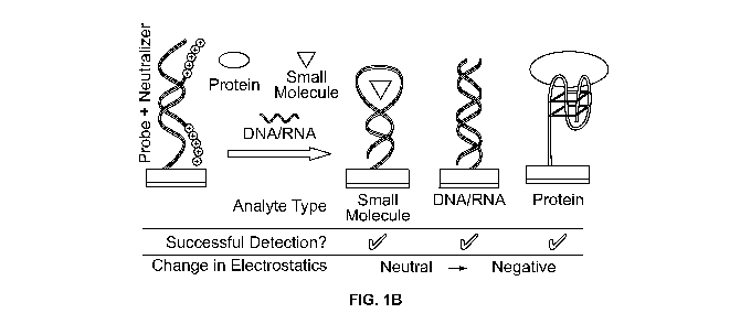
[0014] FIG. 1B illustrates an embodiment wherein a large change in the charge
near the
detection surface is generated when an analyte displaces a pseudoligand from a
charged
probe immobilized on the detection surface.
[0015] FIG. 2 shows an illustrative process for detecting an analyte in
accordance with
certain embodiments.
-3-
CA 02856881 2014-05-23
WO 2013/078424 PCT/US2012/066410
[0016] FIG. 3A illustrates an embodiment wherein charged reporter ions are
drawn towards
the probe on release of the neutralizer and binding of a target.
[0017] FIG. 3B shows an exemplary sensor chip containing multiple
nanostructured
detection electrodes.
[0018] FIG. 3C shows an exemplary nanostructured detection electrode.
[0019] FIG. 4 shows an exemplary system for detecting an analyte in accordance
with
certain embodiments.
[0020] FIG. 5 shows a list of illustrative probe and neutralizer sequences.
[0021] FIG. 6A shows an illustrative ATP biosensor, wherein displacement of a
neutralizer
from a DNA probe aptamer on a detection electrode occurs when ATP complexes
with a
probe aptamer.
[0022] FIG. 6B shows illustrative differential pulse voltammograms for an ATP
biosensor
in the absence of neutralizer (aptamer only), after neutralizer has complexed
with the probe
aptamer (+ neutralizer), and following displacement of the neutralizer from
the probe aptamer
by ATP (+ ATP).
[0023] FIG. 6C shows the relationship between signal strength and ATP
concentration for
an illustrative ATP biosensor.
[0024] FIG. 6D shows the time dependence of the signal change observed from an
illustrative ATP biosensor on the addition of ATP.
[0025] FIG. 7A shows an illustrative cocaine biosensor, wherein displacement
of a
neutralizer from a probe aptamer on a detection electrode occurs when cocaine
complexes
with the probe aptamer.
[0026] FIG. 7B shows illustrative differential pulse voltammograms for a
cocaine biosensor
in the presence of cocaine (+ cocaine) and in the absence of cocaine (-
cocaine).
[0027] FIG. 7C shows the relationship between signal strength and cocaine
concentration
for an illustrative cocaine biosensor.
[0028] FIG. 8A shows an illustrative nucleic acid biosensor, wherein
displacement of a
neutralizer from a probe sequence immobilized on a detection electrode occurs
when a target
nucleic acid complexes with the probe sequence.
[0029] FIG. 8B shows differential pulse voltammograms for a DNA biosensor in
the
absence of neutralizer (probe only), after neutralizer has complexed with the
probe sequence
(+ neutralizer), and following displacement of the neutralizer from the probe
sequence by
1pM of a complementary 20mer DNA target (+ DNA target).
-4-
CA 02856881 2014-05-23
WO 2013/078424 PCT/US2012/066410
[0030] FIG. 8C shows the relationship between signal strength and
complementary 20mer
DNA target concentration for a DNA biosensor.
[0031] FIG. 8D shows the relationship between signal strength and total E.
coli RNA
concentration for a biosensor utilizing a probe complementary to rpoB.
[0032] FIG. 8E shows the relationship between signal strength and lysates
obtained from
different concentrations of E. coli for an illustrative biosensor utilizing a
probe
complementary to rpoB.
[0033] FIG. 9A shows an illustrative protein biosensor, wherein displacement
of a
neutralizer from a probe aptamer immobilized on a detection electrode occurs
when a protein
(in this instance thrombin) complexes with the probe aptamer.
[0034] FIG. 9B shows differential pulse voltammograms for an illustrative
thrombin
biosensor in the absence of neutralizer (aptamer only), after neutralizer has
complexed with
the probe aptamer (+ neutralizer), and following displacement of the
neutralizer from the
probe aptamer by 100 pM thrombin (+ thrombin).
[0035] FIG. 9C shows differential pulse voltammograms for a thrombin biosensor
in the
presence of the nonspecific blocking protein bovine serum albumin (+ BSA) and
absence of
bovine serum albumin (- BSA).
[0036] FIG. 9D shows the relationship between signal strength and thrombin
concentration
for a thrombin biosensor. The horizontal dashed line shows the signal
generated by 100nM
bovine serum albumin, a nonspecific protein.
[0037] FIG. 10 shows an illustrative nucleic acid biosensor, wherein
displacement of a
neutralizer from a probe sequence immobilized on a detection electrode occurs
when a target
nucleic acid complexes with the neutralizer.
Detailed Description
[0038] The principles underlying prior art assays are illustrated in FIG. 1A.
In prior art
assays, as illustrated in FIG. 1A, a non-complexed probe molecule is affixed
to the surface of
a detection electrode. As a result, the charge of the sensor is determined
solely by the probe
molecule prior to introduction of the analyte, the analyte being a ligand that
binds to and
forms a stable complex with the probe molecule. Following binding by the
analyte, the
charge state at the electrode surface is changed by the charge of the analyte.
This approach
leads to significant limitations in prior art charge-based sensing methods.
First, the
background signal may be large due to the inherent charge of the probe
molecule, which is
-5-
CA 02856881 2014-05-23
WO 2013/078424 PCT/US2012/066410
often an electronegative nucleic acid. As a result the ratio of the signal
resulting from analyte
complexing with the probe to the background signal may be small if the charge
of the probe
is very large relative to the charge of the analyte, resulting in limited
assay sensitivity. As a
result, such assays often require tedious, expensive, and error prone
amplification of the
analyte (by methods such as PCR) prior to analysis. Uncharged analytes that do
not produce
a significant change in the charge at the electrode surface when they complex
with the probe,
particularly low molecular weight analytes, may be undetectable. Finally, the
detection
signal is often a reduction in charge magnitude at the electrode surface as a
result of complex
formation between the affixed probe and the analyte. This leads to a "signal-
off' assay
structure in which presence of the analyte is indicated by a lack of signal, a
configuration that
often results in a high rate of false-positive determinations. Prior art
methods of sensing are,
essentially, dependent upon the nature of the analyte for their signal
amplitude, their signal-
to-background ratio, and their sign of signal change. Unfortunately, the
choice of analyte is a
not a variable that the assay designer can change, but rather is a requirement
of the test.
[0039] The embodiments illustrated in FIG. 1B introduce a new freedom in the
design of
the electrostatic character of the sensor surface. A probe molecule is affixed
to the surface of
an electrode and is complexed with a neutralizer, the neutralizer being a
pseudoligand that
has an affinity for the probe and neutralizes the probe's charge on complex
formation. In
certain embodiments, a device may be supplied to the user with the neutralizer
already
complexed to the probe. In certain embodiments, the user may apply the
neutralizer to a
probe-containing element such as a detection electrode prior to the addition
of analyte. In yet
another embodiment the user may apply the neutralizer to a probe-containing
element
essentially simultaneously with the addition of the analyte. The neutralizer
acts as a
pseudoligand that forms a reversible complex with the probe and can be
displaced by a ligand
or analyte that forms a complex with the probe. Towards this end the
neutralizer may
incorporate base pair mismatches with the probe such that the analyte of
interest binds the
probe more strongly, rapidly and/or robustly, leading to displacement of the
neutralizer. The
affinity of the neutralizer for the probe may also be modified using
temperature changes or
changes in buffer composition. Such changes in buffer composition include, but
are not
limited to, changes in ionic strength, presence or absence of multivalent
cations, presence or
absence of organic solvents, presence or absence of chaotropes, and presence
or absence of
hydrophilic polymers.
-6-
CA 02856881 2014-05-23
WO 2013/078424 PCT/US2012/066410
[0040] The neutralizer may be any molecule that forms a complex with the probe
molecule.
In certain embodiments, such a complex has a reduced charge magnitude compared
to the
affixed probe such that the neutralizer is displaced from such a complex on
the addition of
target analyte. The neutralizer may be a nucleic acid analog that incorporates
a neutral or a
positively charged backbone structure. The neutralizer may also be a nucleic
acid analog that
incorporates a negatively charged backbone structure but has a net positive
charge. In certain
embodiments, the neutralizer is a conjugate of peptide nucleic acid and
cationic amino acids
that specifically bind to an electronegative probe so that the charge of the
neutralizer-probe
complex is less electronegative than that of the probe alone. In other
embodiments the
neutralizer may incorporate morpholino nucleic acid analogs or
methylphosphonate nucleic
acid analogs.
[0041] The use of a displaceable pseudoligand allows various embodiments to
overcome
the limitations of traditional charge-sensing assays. In certain embodiments,
the background
signal in the assay is suppressed through charge compensation that is
engineered by the assay
designer, thereby enhancing the signal detection. In such embodiments, the
signal changes
that correspond to the presence of an analyte are determined not only by the
molecular charge
of the analyte ligand, but also by the inherent charge of the probe molecule,
which is
unmasked upon release of the neutralizer. This permits the detection of
analytes that do not
produce significant changes in the charge of the probe upon complex formation,
permitting
the use of assays with a range of low molecular weight analytes that could not
previously be
addressed by electrochemical detection. Such low molecular weight molecules
typically
have a molecular weight of less than about 500 daltons, and may include but
are not limited
to nucleotides and nucleotide analogs, drugs of abuse, therapeutic drugs, and
environmental
contaminants.
[0042] In certain embodiments, the suppression of background signal also
greatly improves
the analyte-specific signal to background signal ratio. Surprisingly, this
reduction in the
analyte-specific signal to background signal permits direct detection of
nucleic acid analytes,
removing the need for expensive and time-consuming PCR amplification of
samples prior to
characterization or detection.
[0043] In certain embodiments, the sign and amplitude of signal is determined
not only by
the charge of the analyte but also by that of the probe. This permits design
of a "signal-on"
assay in which presence of the analyte is indicated by a magnitude increase in
the measured
-7-
CA 02856881 2014-05-23
WO 2013/078424 PCT/US2012/066410
signal. Such signal-on assays generally show a low rate of false positive
results relative to
assays with a signal-off structure.
[0044] FIG. 2 shows an illustrative process 200 for detecting an analyte in
accordance with
certain embodiments. The process begins at step 202. At step 204, a reversible
first complex
is formed between a probe affixed to a sensor surface and a pseudoligand. The
pseudoligand
may be partially complementary to the probe, and has a charge that is opposed
to that of the
probe. At step 206, a sample that may contain the analyte is contacted with
the first complex
at the sensor surface. If the analyte is present in the sample, at step 208,
the pseudoligand is
displaced by the analyte and a second complex is formed. In certain
embodiments, the
pseudoligand is displaced by the analyte and a second complex is formed
between the analyte
and the probe. In certain embodiments, the pseudoligand is displaced by the
analyte and a
second complex is formed between the analyte and the pseudoligand. At step
210, the
presence of the second complex is detected, which indicates that the analyte
is present in the
sample. The process ends at step 212. It is understood that the steps of
process 200 are
merely illustrative and that certain steps may be performed simultaneously
and/or performed
in another suitable order without departing from the scope of the invention.
In certain
embodiments, process 200 also includes a step of communicating the result of
the detection
(not shown), for example, by displaying an indicator (e.g., displaying a text,
symbol, or color-
coded indicator) to a user of the process. In certain embodiments, the step of
communicating
the results includes storing the result in a local or remote memory associated
with the process
200, or by sending a message to a user of the process 200.
[0045] FIG. 3A shows an illustrative embodiment which was tested using an
electrocatalytic reporter system that provides a signal proportional to the
magnitude of the
charge change at electrode surfaces. To measure the change of charge at the
sensor surface, a
[Ru(NH3)6]3V[Fe(CN)6]3- catalytic reporter system 300 was used to generate a
signal that can
be monitored by differential pulse voltammetry (DPV) in the presence of, for
example, a low
molecular weight molecule 310 such as a nucleotide and nucleotide analog, a
drug of abuse, a
therapeutic drug, and an environmental contaminant. In this illustrative
system, the primary
electron acceptor 308, which may be any suitable electron acceptor such as
[Ru(NH3)6]3', is
electrostatically attracted to the electrode surface 302 in proportion to the
amount of
phosphate-bearing nucleic acid 304. When [Fe(CN)6]3- is used during
electrochemical
readout, the Ru(III) is chemically regenerated by [Fe(CN)6]3- forming a redox
cycle, which
amplifies the signal significantly. This illustrative embodiment is free of
covalent labels and
-8-
CA 02856881 2014-05-23
WO 2013/078424 PCT/US2012/066410
does not require preprocessing of the samples. High catalytic currents would
be expected
when only DNA aptamer probes 304 are immobilized on the sensors 302 due to
electrostatic
affinity of Ru(III) for the phosphate groups of the DNA backbone, however in
the tested
assays these currents would be strongly attenuated in the presence of the
neutralizer 306.
This reporter system may be used with nanostructured microelectrodes that can
be fabricated
on the surface of a chip.
[0046] FIGS. 3B and 3C show an illustrative sensor used in an example
embodiment. To
fabricate the sensor, photolithographic patterning was used to produce a
microelectronic chip
320 with an array of sensors 322. The chips used in this study possessed
twenty sensors.
With the use of a silicon wafer 324 coated with a gold (Au) layer 326 and a
Si02 layer 328 as
a base, contact pads and leads were patterned onto individual chips. An
overlayer of Si3N4
was then used to passivate the surface of the chip. To provide a template for
the growth of
electrodeposited sensors, photolithography was then used to open 5 um
apertures 330 in the
Si3N4. Gold electrodeposition was then employed to grow fractal
microstructures 332, the
size and morphology of which can be modulated by deposition time, potential,
Au
concentration, supporting electrolyte, and overcoating protocol by methods
known in the art.
As nanostructures increase the sensitivity of the assay significantly, Au
structures were
coated with a thin layer of Pd to form finely nanostructured sensors (FIG.
3C). It is
understood that the materials, dimensions, and processes used to generate the
sensors are
merely illustrative and that other suitable materials, processes, or
dimensions may be used
without departing from the scope of the disclosure.
[0047] In certain embodiments, chips were fabricated using several inch
silicon wafers that
were passivated using a thick layer of thermally grown silicon dioxide. A 25
nm Ti was then
deposited. A 350 nm gold layer was subsequently deposited on the chip using
electron-
beam-assisted gold evaporation, and patterned using standard photolithography
and a lift-off
process. A 5 nm Ti layer was then deposited. A 500 nm layer of insulating
Si3N4 was
deposited using chemical vapor deposition. 5 mm apertures were then imprinted
on the
electrodes using standard photolithography, and 0.4 mm x 2 mm bond pads were
exposed
using standard photolithography.
[0048] To fabricate the assay test sites in certain embodiments, chips were
cleaned by
sonication in acetone for 5 min, rinsed with isopropyl alcohol and deionized
(DI) water, and
dried with a flow of nitrogen. Electrodeposition was performed at room
temperature; 5 gm
apertures on the fabricated electrodes were used as the working electrode and
were contacted
-9-
CA 02856881 2014-05-23
WO 2013/078424
PCT/US2012/066410
using the exposed bond pads. Au (gold) sensors were made using a deposition
solution
containing 50 mM solutions of HAuC14 and 0.5 M HC1. 100 gm and 20 gm Au
structures
were formed using DC potential amperometry at 0 mV for 100 seconds and 0 mV
for
20 seconds respectively. After washing with DI water and drying, the Au
sensors were
coated with Pd to form nanostructures by replating in a solution of 5 mM
H2PdC14 and 0.5 M
HC104 at -250 mV for 10 seconds (for 100 micron structure) and for 5 seconds
(for 20
micron structure).
[0049] In certain embodiments, an exemplary protocol for preparing the assays
was used.
In this protocol, thiolated aptamers and thiolated DNA probes were deprotected
using
dithiothreitol (DTT) followed by purification with HPLC. HPLC-purified probes
were
subsequently lyophilized and stored at -20 C. Phosphate buffer solution (25
mM, pH 7)
containing 5 gM thiolated probe, 25 mM NaC1, and 50 mM MgC12 was incubated
with
sensors for 1 hour in a dark humidity chamber at room temperature to
immobilize the probe
on the test surface. The chip was then washed twice for 5 minutes with
phosphate buffer
solution (25 mM) containing 25 mM NaCl. Sensors were then incubated with a
phosphate
buffer solution (25 mM) containing 10 gM neutralizer and 25 mM NaC1 for 30
minutes at
room temperature, followed by washing three times for 5 minutes with the same
buffer. For
the purposes of demonstrating detection, the chips were then treated with
different analytes
followed by washing.
[0050] In certain embodiments, electrochemical experiments were carried out
using a
Bioanalytical Systems (West Lafayette, Indiana) Epsilon potentiostat with a
three-electrode
system featuring a Ag/AgC1 reference electrode and a platinum wire auxiliary
electrode.
Electrochemical signals were measured in a 25 mM phosphate buffer solution (pH
7)
containing 25 mM NaC1, 10 gM [Ru(NH3)6]C13, and 4 mM K3[Fe(CN)6]. Differential
pulse
voltammetry (DPV) signals were obtained with a potential step of 5 mV, pulse
amplitude of
50 mV, pulse width of 50 msec, and a pulse period of 100 msec. Signal changes
corresponding to replacement of the neutralizer by specific target were
calculated with
background-subtracted currents: AI% = -
Ibefore)/Ibefore x 100 (where Iafter = current after
replacement of neutralizer, 'before = current before replacement of
neutralizer). In these
illustrative embodiments, scanning electron microscope images were obtained
using an
Aspex (Delmont, Pennsylvania) 3025 SEM.
[0051] FIG. 4 shows an exemplary system for detecting an analyte in accordance
with
certain embodiments. The detection system 400 has a detection chamber 402 that
includes
-10-
CA 02856881 2014-05-23
WO 2013/078424 PCT/US2012/066410
one or more electrodes. In FIG. 4, the detection chamber includes a working
electrode 404,
a counterelectrode 406, and a reference electrode 408. However, any suitable
number or
types of electrodes may be used. The detection chamber 402 also has an inlet
410 for flowing
in a sample for contacting with the working electrode 404, and outlet 412 for
flowing out the
sample. If the sample contains the analyte of interest, the analyte may form a
probe-analyte
complex 414 on the surface of the working electrode 404. In certain
embodiments, once a
sample enters the detection chamber 402 through the inlet 410, a certain
amount of time may
be allotted to facilitate formation of the probe-analyte complex 414. In
certain embodiments,
a sample containing the analyte may flow out through the outlet after enough
time has been
allotted for the probe-analyte complex 414 to form. A washing solution may
subsequently
flow in through the sample chamber 402 to remove undesirable materials that
may be present
in the sample.
[0052] The detection system 400 shown in FIG. 4 incorporates a illustrative
three-electrode
potentiostat configuration, however it is to be understood that any suitable
configuration of
components could be used. The counterelectrode 406 is connected to resistor
418, which is
in turn connected to the output of control amplifier 416. A detection module
420 is
connected across the resistor 418 to provide a current measurement. The
detection module
420 may be configured to provide real-time current measurement in response to
any input
waveform. The reference electrode 408 is connected to the inverting terminal
of control
amplifier 416. A signal generator 422 is connected to the noninverting
terminal of the control
amplifier 416. This configuration maintains constant potential at the working
electrode while
allowing for accurate measurements of the current. In certain embodiments, the
detection
chamber 402 may contain a plurality of electrodes for detecting multiple
analytes. For
example, the detection chamber 402 may include multiple working electrodes
each with a
different type of probe affixed for complexing with different targets present
in the sample. In
certain embodiments, the detection system 400 may be configured to
individually address the
working electrodes one at a time while utilizing a common counterelectrode and
reference
electrode.
[0053] A control and communication unit 424 is operably coupled to the
detection module
420 and the signal generator 422. The control and communication unit 424 may
synchronize
the input waveforms and output measurements, and may receive and store the
input and
output in a memory. In certain embodiments, the control and communication unit
424 may
be a separate unit that interfaces with the detection system 400. For example,
the detection
-11-
CA 02856881 2014-05-23
WO 2013/078424 PCT/US2012/066410
system 400 may be a disposable cartridge with a plurality of input and output
terminals that
can connect to an external control and communication unit 424. In certain
embodiments, the
control and communication unit may be operably coupled to a display unit that
displays the
output as a function of input. In certain embodiments, the control and
communication unit
424 may transmit the input and output information to a remote destination for
storage and
display. For example, the control and communication unit 424 could be a mobile
device or
capable of being interfaced with a mobile device. In certain embodiments, the
control and
communication unit 424 could provide power to the detection system 400. The
system 400
maybe powered using any suitable power source, including a battery or a
plugged-in AC
power source.
[0054] In certain embodiments, a detection system may be provided as an assay
composition for use in drug screening. The assay composition may have a
reversible first
complex comprising a probe molecule affixed to a sensor surface that forms a
complex with a
complementary or partially-complementary pseudoligand. The probe may have an
affinity
for a drug that is greater than that for the pseudoligand. Consequently, the
pseudoligand may
be displaced by the drug, forming a second complex. The first and second
complexes may
have first and second charge states, respectively. In certain embodiments, the
detection
system may be provided as a kit, which includes a device with a sensor surface
and a probe
affixed to the sensor surface. The kit may also have a pseudoligand capable of
forming a
reversible complex with the pseudoligand. The pseudoligand in the kit may be
already
complexed with the probe, or may be separately included with the kit for later
complexation.
[0055] In various embodiments, the neutralizers were synthesized using a solid
phase
synthesis approach on a Prelude automated peptide synthesizer (Protein
Technologies, Inc.;
Tucson, Arizona). In these embodiments, synthesis products were confirmed by
mass
spectroscopy.
[0056] In certain embodiments, to examine the ability of the assay to detect
small
molecules, ATP was selected as a model analyte or binding ligand. Illustrative
ATP probe
and aptamer sequences are shown in FIG. 5. An illustrative configuration of
the neutralizer
assay 600 for ATP detection is shown in FIG. 6A. In these embodiments,
thiolated ATP-
binding aptamers 602 are first immobilized onto sensors 604 with Pd on their
surfaces, and a
partially complementary neutralizer 606 is then introduced. The PNA portion
608 of the
neutralizer 606 is primarily complementary to the aptamer 602. However, two
mismatches
610 were introduced to permit facile release of the neutralizer 606 upon ATP
612 addition.
-12-
CA 02856881 2014-05-23
WO 2013/078424 PCT/US2012/066410
The presence of the neutralizer 606 strongly reduces the charge at the sensor
604 surface,
which would be restored by the displacement of the neutralizer 610 by a target
molecule.
[0057] In FIG. 6B, DPV graph 620 shows signals obtained at the sensors before
neutralization 622, after neutralization 624, and after ATP introduction 626.
For ATP
detection, sensors were incubated with a phosphate buffer solution (25 mM)
containing
25 mM NaC1 and different concentrations of ATP for 10 minutes at room
temperature. Scans
of uncomplexed immobilized aptamers revealed high catalytic current,
consistent with the
strong electrostatic attraction of Ru(III) to the DNA backbone of the aptamer.
The observed
current is reduced by > 80% when the neutralizer molecule is hybridized to the
aptamer.
Surprisingly, the reduction peak also shifts to more negative potentials. This
may be due to
slowing the kinetics of electron transfer when the neutralizer is present.
When the analyte
ATP binds to the aptamer probe a structural change of the aptamer causes
release of the
neutralizer, leading to an increase in catalytic current. As shown in
concentration graph 630
of FIG. 6C, the observed change in current before ATP binding and after ATP
binding was
directly related to the concentration of ATP in solution. The horizontal
dashed lines 632
show the signal generated in the absence of ATP.
[0058] To evaluate the time dependence of the sensor response in certain
embodiments,
ATP was introduced into the [Ru(NH3)6[3 V[Fe(CN)6[3- catalytic solution and
signal changes
were measured in real time. FIG. 6D shows illustrative time graph 640, which
contains data
that indicate that signal changes in the presence 642 of ATP occur within 1
min, and the
signal change in absence 644 of ATP is not significant even after 20 min.
These results
clearly indicate that sensor response is rapid, and that the probe-neutralizer
complex is stable.
[0059] FIG. 7A shows an illustrative embodiment of a cocaine assay 700 that
was carried
out using a similar approach as described above for ATP, with aptamers 702
that were
immobilized onto sensors 704 and specific for a cocaine molecule 712. DPV
graph 720 of
FIG. 7B shows the initial high current of the cocaine-binding aptamer
decreased after
neutralizer hybridization 722. Illustrative sequences for the aptamer 702 and
neutralizer 706
used in the cocaine assay 700 are shown in FIG. 5. In this embodiment, the
neutralizer 706
has a portion 708 that is complementary to the aptamer 702, and two mismatches
710 that
permit facile release of the neutralizer 706 upon cocaine 712 addition. When
the aptamer-
neutralizer complex was challenged with cocaine 712, the resulting structural
change of the
aptamer 702 released the neutralizer 706 and resulted in a high catalytic
current 724. For
cocaine detection in this illustrative embodiment, sensors 704 were incubated
with a
-13-
CA 02856881 2014-05-23
WO 2013/078424 PCT/US2012/066410
phosphate buffer solution (25 mM) containing 25 mM NaC1 and different
concentrations of
cocaine for 2 min at room temperature. Concentration graph 730 of FIG. 7C
shows the
observed change in current (in percentage points) on the y-axis against
cocaine concentration
(in [tg/mL) on the x-axis. The horizontal dashed lines 732 show the signal
generated in the
absence of cocaine. The observed change in current before cocaine binding and
after cocaine
binding was directly related to the concentration of cocaine in solution. The
signal change
for 1 [tg/mL cocaine was > 60% higher than that in the absence of cocaine or
in the presence
of a non-target analyte. This level of sensitivity is comparable with
commercial tests and is
ample for drug screening.
[0060] Having established a high level of performance with small molecule
analytes, assay
performance was evaluated to determine if it provided clinically-relevant
(femtomolar or
better) sensitivity against nucleic acid analytes, as other attempts to
develop universal
detection systems have not been successful in achieving good sensitivity with
this analyte
class.
[0061] FIG. 8A illustrates a schematic representation 800 of an exemplary
assay applied
toward nucleic acids targets 812. A thiolated-DNA probe 802 was immobilized on
the sensor
804. The catalytic current 822 for the uncomplexed DNA probe 802 was initially
high, and
was significantly suppressed 824 upon addition of neutralizer 806, as shown in
DPV graph
820 of FIG. 8B. After exposure to 1 pM complementary oligonucleotide 20-mer
target the
current 826 increased by >300%. Sequences for the DNA probe 802, DNA probe
neutralizer
806, and DNA target 812 are shown in FIG. 5. The neutralizer 806 has a portion
808 that is
complementary to the DNA probe 802, and two mismatches 810 that permit facile
release of
the neutralizer 806 upon DNA target 812 addition.
[0062] In certain embodiments, concentration dependence of a nucleic acid
assay was
studied using the 20-mer synthetic target DNA and a noncomplementary target,
which was
used to evaluate background levels and evaluate specificity. An illustrative
sequence for the
noncomplementary target is shown in FIG. 5. For this synthetic target DNA,
sensors were
incubated with a phosphate buffer solution (25 mM) containing 25 mM NaC1, 10
mM MgC12,
and different concentrations of target for 30 minutes at room temperature. To
identify the
detection limit of the exemplary assay, the concentration of target DNA varied
between
lOpM and 10 aM, as shown in concentration graph 830 of FIG. 8C. Signal
increased with
increasing concentration of target within a range spanning 5 orders of
magnitude. The
horizontal dashed line 832 indicates the average currents of 100 nM the
noncomplementary
-14-
CA 02856881 2014-05-23
WO 2013/078424 PCT/US2012/066410
target. The signal change for 10 aM of DNA target, while above that of the 100
nM of the
noncomplementary target, was not high enough to be statistically significant,
indicating that
the detection limit of this assay for 20-mer target is between 100 and 10 aM.
[0063] Performance of certain embodiments of assay with complex heterogeneous
samples
in the form of E. coli total RNA was also evaluated. The DNA probe was
designed for RNA
polymerase mRNA (rpoB), a transcript that is highly expressed in bacteria
and is not
conserved between species. Illustrative sequences for the DNA probe and the
neutralizer are
shown in FIG. 5. As shown in the concentration graph 840 of FIG. 8D,
concentration
dependence of the signal produced by this illustrative assay on the
concentration of a
heterogeneous mixture E. coli total RNA, with the horizontal dashed line 842
showing the
signal observed in the absence of E. coli RNA. In the cases of E. coli total
RNA sensors were
incubated with sterile and RNase-free PBS containing different concentrations
of target for
30 minutes at room temperature. 10 pg/iut E. coli total RNA was successfully
detected,
indicating that the illustrative assay can achieve high levels of sensitivity
with an excess of
non-complementary material.
[0064] Certain embodiments of the assay that use unprocessed bacterial lysates
are also
provided and tested. In testing these embodiments, unprocessed bacterial
lysates were
generated by placing suspensions containing known quantities of E. coli into a
lysis chamber,
where strong electrical fields lysed the bacteria. This lysate was then used
without further
purification or amplification. Illustrative sequences used for the bacterial
lysate probe and
the bacterial lysate neutralizer are shown in FIG. 5. The concentration graph
850 of FIG. 8E
illustrates the dependence of the assay signal on the quantity of E. coli used
to produce the
unprocessed lysates, and shows that signal increased with increasing
concentration of E. coli
bacteria in the initial suspensions. In these studies sensors were incubated
with E.coli lysate
in sterile and RNase-free PBS for 30 minutes at room temperature. The
horizontal dashed
line 852 in FIG. 8E represents average signal for lysate from 150 cfu/iut
Staphylococcus
saprophyticus, the components of which were noncomplementary to the probe. The
detection
limit for E. coli bacteria was, surprisingly, 0.15 cfu/ L. This sensitivity
detection limit is
unprecedented in direct analysis on unprocessed samples, and is clinically
relevant for the
detection of bacteria found in clinical samples. The kinetics of the assay in
these illustrative
embodiments allowed rapid detection of the presence of bacteria with high
sensitivity and
specificity, requiring less than 30 minutes from sample acquisition to result.
-15-
CA 02856881 2014-05-23
WO 2013/078424 PCT/US2012/066410
[0065] Certain embodiments were characterized to verify that the assay format
described
herein in connection with various embodiments could detect protein biomarkers,
using
thrombin as a model system. The thrombin binding aptamer is a well-
characterized sequence
that is known to fold into a G-quartet structure and bind thrombin at exosite
I. Illustrative
sequences for the thrombin binding aptamer and the thrombin aptamer
neutralizer are shown
in FIG. 5. FIG. 9A shows an illustrative protein detection method 900
according to certain
embodiments. A thiolated thrombin-binding aptamer 902 was deposited onto a
sensor
surface 904 and subsequently complexed with thrombin aptamer neutralizer 906,
which at
least partially neutralizes the charge near the sensor surface 904. The
neutralizer 906 has a
portion 908 that is complementary to the aptamer 902, and two mismatches 910
that permit
facile release of the neutralizer 906 upon thrombin 912 addition. Binding of
thrombin 912 to
the thrombin binding aptamer 902, which forms an aptamer-thrombin complex 914,
displaces
the thrombin aptamer neutralizer 906 and restores the charge, resulting in a
detectable change
in sensor response.
[0066] In FIG. 9B, DPV graph 920 shows electrocatalytic currents of the
aptamer alone
922, the aptamer-neutralizer complex 924, and the aptamer-thrombin complex
926. The
electrocatalytic current was clearly suppressed upon neutralizer binding to
the thrombin
aptamer. When treated with 100 pM thrombin, a large increase in catalytic
current was
observed. Conversely, DPV graph 930 of FIG. 9C shows that the signal change
was
negligible when treated with 100 nM BSA (a nonspecific protein), indicating
that the assay
was specific for thrombin. As shown in concentration graph 940 of FIG. 9D, the
observed
signal was directly proportional to thrombin concentration, and showed that as
low as 10 fM
thrombin was clearly detectable. The horizontal dashed lines 942 show the
signal generated
in the absence of thrombin.
[0067] FIG. 10 shows an illustrative embodiment of a nucleic acid assay 1000
in which the
analyte displaces the pseudoligand by forming a complex with the pseudoligand
rather than
the probe. In this embodiment, the neutralizer 1006 has a greater affinity for
the analyte
1012 than it does for the probe 1002, which results in the formation of a
complex 1014
between the analyte 1012 and the neutralizer 1006. In assay 1000, the
neutralizer 1006 is
specific for the analyte 1012 and incorporates one or more base pair
mismatches 1010 with
the probe 1002, and a portion 1008 that is complementary to the probe 1002. In
this
embodiment, the signal observed after the neutralizer 1006 hybridizes to the
probe 1002 is
increased by the removal of the mismatched neutralizer 1006 from the probe
1002 due to
-16-
CA 02856881 2014-05-23
WO 2013/078424 PCT/US2012/066410
hybridization with a perfectly-matched target 1012 that is in solution. This
leads to charge
restoration at the electrode detection surface 1004 in the absence of an
analyte-probe
complex, based on removal of the neutralizer 1006 and restoration of the
charge of the probe
1002 that is affixed to the electrode detection surface 1004. It is to be
understood that,
although this embodiment is described in the context of nucleic acid
detection, the
embodiment is not so limited, and may be used to detect a wide variety of
analytes.
[0068] Thus, specific embodiments and applications of a sensitive biosensor
applicable to a
wide range of biological molecules have been disclosed. It should be apparent,
however, to
those skilled in the art that many more modifications besides those already
described are
possible without departing from the inventive concepts herein. The inventive
subject matter,
therefore, is not to be restricted except in the spirit of the appended
claims. Moreover, in
interpreting both the specification and the claims, all terms should be
interpreted in the
broadest possible manner consistent with the context. In particular, the terms
"includes",
"including", "contains", "containing", "has", "have", having", "comprises" and
"comprising," as used herein, should be interpreted as referring to elements,
components, or
steps in a non-exclusive manner, indicating that the referenced elements,
components, or
steps may be present, or utilized, or combined with other elements,
components, or steps that
are not expressly referenced. The term "plurality," as used herein means more
than one, and
may include any defined or undefined subset of two or more steps, elements, or
components.
Furthermore, where a definition or use of a term in a reference, which is
incorporated by
reference herein, is inconsistent or contrary to the definition of that term
provided herein, the
definition of that term provided herein applies and the definition of that
term in the reference
does not apply.
[0069] The application of which this description and claims form part may be
used as a
basis for priority in respect of any subsequent application. The claims of
such subsequent
application may be directed to any feature or combination of features
described herein. They
may take the form of product, method or use claims and may include, by way of
example and
without limitation, one or more of the following claims.
-17-