Note : Les descriptions sont présentées dans la langue officielle dans laquelle elles ont été soumises.
CA 02865602 2016-01-28
1
A LAYERED IMPLANT COMPRISING A BIOACTIVE MATERIAL
FIELD OF THE INVENTION
The invention relates to an implant comprising at least two layers made of
fibers and at
least one layer of bioactive material arranged between said at least two
layers.
BACKGROUND
The use of reinforced composites made of particulate fillers or reinforcing
fibers is
already known. The state-of-the-art fiber reinforced composites yield high
strength
properties and by selecting the multiphase resin matrix for the composite, the
handling
characteristics of the composite can be considerably improved.
On the other hand, a lot of development has occurred with bioactive materials,
namely
bioactive ceramics and glass and sol-gel processed silica. These materials can
be used
to achieve attachment of e.g. bone to a biomaterial surface after the material
has been
put in contact with tissue. An additional advantage of bioactive glass is its
antimicrobial
effect on the microbes existing for instance in infected sinuses of a bone.
Document
WO 2004/049904 discloses bioactive, resorbable scaffolds for tissue
engineering. The
scaffolds are made of bioactive glass meshes that comprise interwoven
bioactive glass
fibers and may comprise incubating cells such as fibroblasts and
chondroblasts.
From a surgical perspective, individual replacement of bone, cartilage and
soft tissues
are insufficient in tumour, traumatologic and tissue reconstruction surgery
despite the
increasing advances in biomaterials research and their clinical application
methods and
tissue engineering. The need and indications for development of new kinds of
materials
result from disadvantages of the use of allografts. Risks for transmittable
diseases (HIV,
Creutzfeld-Jacob's disease, etc.) are related to allografting. Metals are not
bioactive or
osteoconductive, and their use results in stress shielding phenomena and bone
atrophy
of the adjacent bone. Metal implants cause also severe problems in magnetic
resonance imaging (MRI) when diagnosing diseases of patients. These main
disadvantages are well documented in large clinical series.
There thus still exists a need for alternative implants for medical uses.
CA 02865602 2016-01-28
2
OBJECTS AND SUMMARY OF THE INVENTION
An object of the present invention is to provide a biologically compatible
material that
does not have the above-listed drawbacks, or at least those disadvantages are
minimised. Specifically, an object of the present invention is to provide a
material useful
for medical, dental and surgical uses, such as for bone grafting in repair of
bone defects
and fixation of fractured pieces of bone.
In accordance with one aspect, the present invention provides an implant
comprising at
least two layers made of fibers and bioactive material arranged between said
at least
two layers, wherein
- at least one of the layers is at least mainly formed of a mesh,
- made of glass fibers having a diameter of 3-100 pm, and wherein
- the mesh size is selected such that the bioactive material is retained
within the implant,
- the layers are embedded in a matrix made of a resin selected from the group
consisting of substituted and unsubstituted dimethacrylates and methacrylates,
- the layers are attached to each other along the contour of the implant,
characterised in that the bioactive material is selected from the group
consisting of
bioactive glass, hydroxyapatite, tricalciumphosphate and mixtures thereof in
particle
form.
BRIEF DESCRIPTION OF THE DRAWING
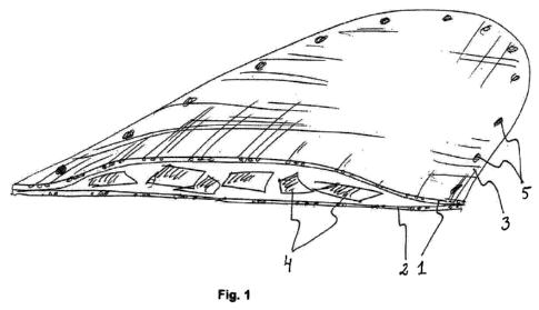
Figure 1 schematically shows an implant according to a first embodiment.
Figure 2 schematically shows an implant according to a second embodiment.
Figure 3 schematically shows the implant according to the second
embodiment,
from a different angle.
Figure 4 schematically shows an implant according to a third embodiment.
Figure 5 schematically shows an implant according to a fourth embodiment.
CA 02865602 2016-01-28
2a
Figure 6 schematically shows the implant according to the fourth
embodiment,
from a different angle.
Figure 7 schematically shows an implant according to a fifth embodiment.
DETAILED DESCRIPTION OF THE INVENTION
The invention relates to an implant comprising at least two layers made of
fibers and
at least one layer of bioactive material arranged between said at least two
layers.
A typical implant according to this invention comprises at least two layers
made of
fibers and bioactive material arranged between said at least two layers. A
least
CA 02865602 2014-08-26
WO 2013/178637 PCT/EP2013/060980
3
one of the layers is at least mainly formed of a mesh made of glass fibers
having a
diameter of 3-100 pm, and the mesh size is selected such that the bioactive
material is retained within the implant. Moreover, the layers are embedded in
a
matrix made of a resin selected from the group consisting of polyesters,
epoxies,
acrylates and mixtures thereof, and the layers are attached to each other
along the
contour of the implant. Furthermore, the bioactive material is selected from
the
group consisting of bioactive glass, hydroxyapatite, tricalciunnphosphate and
mixtures thereof
The implant according to this invention thus takes advantage of the capillary
effect,
as at least one of the surfaces is formed at least mainly of a mesh. Indeed,
the
structure of the implant, due to the use of at least one mesh and a bioactive
material, is such that the capillary effect is enhanced, thus leading to
improved
bone ingrowth, as fluids can penetrate inside of the implant better than if
both
surfaces were made of a tightly woven cloth or a film. In addition, the
openings of
the mesh allow the penetration of the body fluids to occur from various
directions
of the implant which means that the fluid penetration is not sensitive to the
direction of blood flow from arteries.
The implant may have both its outer surfaces made of a mesh or one of the
surfaces may be made of a film or a tightly woven cloth. When the other
surface is
not made of a mesh, it is typically the surface that will be on the outside
once the
implant is in its place. The implant may also comprise more than two layers,
such
as three, four or five layers. According to an embodiment, the layer thickness
is
about 500-700 pm. The thickness of the implant depends for example on the
thickness of the bone it intends to replace. Most typically, a maximum
thickness of
10 mm is achieved with five layers. When several layers are used, the
intermediate (i.e. the inner layers as opposed to the outermost layers) are
preferably made of mesh. According to a preferred embodiment, all the layers
are
impregnated with a resin, i.e. embedded in a matrix. The resin chosen may be
the
same or different for each layer. Furthermore, when several layers are used,
only
the two outermost may be attached to each other along the contour of the
implant
or all or some of the other layers (intermediate layers) may be attached to
each
other in a similar manner.
CA 02865602 2014-08-26
WO 2013/178637 PCT/EP2013/060980
4
In this specification, by curing it is meant polymerisation and/or
crosslinking. By
matrix, it is understood the continuous phase of a composition and by uncured
matrix it is meant a matrix that is in its deformable state but that can be
cured, i.e.
hardened, to an essentially non-deformable state. The uncured matrix may
already
comprise some long chains but it is essentially not yet polymerised and/or
crosslinked. By prepreg, it is meant a semi-manufactured product, that is, a
product that is not or only partly polymerised, but yet still deformable. The
curing of
a resin leads to a composite material, wherein the cured resin forms the
matrix.
The layers of the implant are at least mainly formed of a mesh, meaning that
at
least 55 % of the surface of the layer is made of mesh. Preferably, at least
60, 65,
70, 75, 80, 85, 90 or 95 % of the surface is made of mesh. As will be
explained
later, the layers may also comprise zones where the layer is in another form
than
mesh, such as tightly woven cloth or continuous fibers. Typically these zones
are
used for cutting or bending the implant. Most preferably the layers are made
of a
mesh except for these zones. Sometimes the contour of the layers may be made
of continuous fibers. This may be used for example in implant where they are
attached to the bone in an area where the bone (and thus the attachment) is
under
significant stress. Therefore, the continuous fibers reinforce the contour
where
attachment to the bone takes place.
According to one embodiment of the invention, the fibers are selected from the
group consisting of inert glass fibers and bioactive glass fibers. According
to
another embodiment, the glass fibers are made of a glass composition of E-
glass,
S-glass, R-glass, C-glass or bioactive glasses.
According to yet another embodiment, the diameter of the fibers is 4-25 pm.
The
diameter of the fibers can be for example from 3, 5, 6, 10, 15, 20, 25, 30,
40, 45,
50, 60, 70 or 80 pm up to 5, 6, 10, 15, 20, 25, 30, 40, 45, 50, 60, 70, 80, 90
or 100
pm. Fibers in the nanometer scale, i.e. with a cross-sectional diameter
varying
between 200 - 1000 nm can also be used.
The bioactive material can be in any form suitable for inserting between two
layers
consisting mainly of a mesh. It may be for example in the form of a monolith
or in
particle form. By particles, it is meant entities wherein the largest
dimension is no
CA 02865602 2014-08-26
WO 2013/178637 PCT/EP2013/060980
more than five times larger than the smallest dimension. It may thus also be
in the
form of chopped, short fibers. When particles are used, their size is smaller
than
the mesh size of the layers, in order for the layer to be able to retain them
inside
the implant. The bioactive material may also be in the form of a monolith or
just
5 two, three or four large particles. Some possible particles sizes are 10-
1000 pm.
The particle size can be for example from 10, 20, 50, 100, 150, 200, 250, 300,
400, 500, 650, 700 or 800 pm up to 20, 50, 100, 150, 200, 250, 300, 400, 500,
650, 700, 800, 900 or 1000 pm.
The bioactive material may also be in the form of a fluid having a viscosity
such
that the layers of mesh are impermeable to the fluid, that is, the implant may
comprise such bioactive material in addition to those listed in the
independent
claim. The fluid can be a highly viscous fluid or a colloid in fluid form. By
colloid, it
is meant a substance microscopically dispersed evenly throughout another
substance. The bioactive material may naturally also be in several of these
forms,
for example a combination of particles in a fluid. Preferably, the bioactive
material
is bioactive glass.
According to an embodiment, mesh size is optimized by weaving process of the
mesh and viscosity and amount of impregnation resin of the mesh. According to
an embodiment, the mesh size is preferably 1 to 5 micrometers less than the
smallest diameter of the particles. The mesh size may be for example 9-999 pm.
The mesh size may thus be for example from 1, 2, 3, 5, 7, 9, 10, 15, 20, 50,
100,
150, 200, 250, 300, 400, 500, 650, 700, 800 or 900 pm up to 2, 3, 5, 7, 9, 10,
15,
20, 50, 100, 150, 200, 250, 300, 400, 500, 650, 700, 800, 900, 950 or 1000 pm.
According to a further embodiment, the two layers of mesh are attached to each
other also along at least one cutting line. The cutting line may be formed for
example of unidirectional continuous fibers.
The attachment zone, i.e. the part of the implant where the layers are
attached
together, can be varied in width. The advantage of a large attachment zone is
that
the implant can be cut smaller to fit to the intended use, yet it still
remains
functional as the bioactive material is retained within the implant.
CA 02865602 2014-08-26
WO 2013/178637 PCT/EP2013/060980
6
The positioning of the attachment zone is also important and can be varied
depending on the intended use. For example, the implant may be made such that
it has more than one part (for example two, three, four, five or six parts),
each part
being separated from the other parts by an attachment zone, i.e. a cutting
line.
The attachment zones between the parts can be used for example for easier
bending of the implant or for cutting one or more parts out from the implant.
Thus a
versatile implant can be made whereby the user will have to decide what size
it
needed only just before implanting the implant. This is especially important
for
emergency operations and is also believed to reduce costs as it will no longer
be
necessary to keep a stock of different sizes of implants. The shelf-life of
these
implants is believed to be approximately one year, depending naturally of the
components used.
The contour of the implant, i.e. the attachment zone along the contour may
also
contain holes that extend through both layers of the mesh to ease the
attaching of
the implant to place with for example bone screws. Similar holes may be also
provided in a cutting line if needed. Moreover, when a large attachment zone
along the contour of the implant is used, it may be equipped with a series of
holes
at different distances from the edges such that the implant is still easily
attachable
even when cut to a smaller size.
The implant may be homogenous in its structure and materials or it may consist
of
different materials and/or properties at different locations. It is for
example possible
to vary one or more of the following: the mesh size, the matrix material, the
amount of matrix, the fiber material, the fiber diameter or the bioactive
material.
This could lead to for example different strengths at different locations of
the
implant.
A preferred matrix material is an acrylate polymer. The matrix is formed when
the
resin is cured. According to an embodiment, the matrix resin is selected from
the
group consisting of substituted and unsubstituted dinnethacrylates and
methacrylates. Some especially advantageous matrix materials (monomers) are
methyl acrylate, methyl methacrylate, methacrylate functionalized dendrimers,
glycidyl dimethacrylate (bis-GMA), triethylene glycol dimethacrylate (TEGDMA)
CA 02865602 2014-08-26
WO 2013/178637 PCT/EP2013/060980
7
and urethane dimethacrylate (UDMA). The materials may be used as blends and
they may form interpenetrating polymer networks (IPNs). They may also be
functionalised with bioactive molecules that allow for a drug-like contact
effect.
Combinations of monomers and polymers are also suitable to be used, including
modifications of resin systems by antimicrobial side group containing iodine
which
offers additional benefit in increasing radio opacity of the resin system.
The viscosity of the resin is such that it does not obstruct the mesh
structure.
Some examples of resin viscosity and mesh size are given below.
The implant may further comprise modifier particles. These modifier particles
may
for example be bioactive and for example improve the osteoconductivity of the
implant. The particles may be in the form of particulate fillers or fibers.
The weight
fraction of these modifier particles in the implant can be for example 10-60
wt-%,
such as from 5, 10, 15, 20, 35 or 50 wt-% up to 10, 15, 20, 35, 50, 55, 60 or
75 wt-
%.
According to one embodiment, the modifier particles are selected from the
group
consisting of bioactive ceramics, bioactive glass, silica gel, titanium gel,
silica
xerogel, silica aerogel, natrium silica glass, titanium gels, bioactive glass
ionomer,
hydroxyapatite, Ca/P-doped silica gel and mixtures thereof. Any combination of
said materials may naturally also be used. When rapid mineralization is
needed, it
is preferred to have bioactive glass with sol-gel processed silica particles.
The implant according to the present invention may further comprise additional
particulate filler material, such as metal oxides, ceramics, polymers and
mixtures
thereof. Metal oxides may for example be used as radio or X-ray opaque
materials
or as colouring materials.
The implant may also comprise therapeutically active agents or cells such as
stem
cells, proteins such as growth factors and/or signalling molecules. Several
kinds of
cells including hematopoietic bone marrow cells, fibroblasts, osteoblasts,
regenerative cells, stem cells, like embryonic stem cells, mesenchymal stem
cells
or adipose stem cells can be seeded to the implant. The embryonic stem cells
may
or may not be of a human origin. Stem cells seeded to the implant can be
cultured
CA 02865602 2014-08-26
WO 2013/178637 PCT/EP2013/060980
8
in bioreactors ex vivo, in other parts of the body before inserting the formed
tissue
into its final place, or directly at the place where regenerative and
reconstructive
treatment is needed. The implant may contain also additives enhancing its
processability, such as polymerisation initiators. The materials of the
implant can
be either bioresorpable, biodegradable, biostable or a mixture of these.
The implant may also contain, between the layers, interconnective parts that
are
rigid and essentially non-compressible. These interconnective parts thus
ensure
that when the material is bent, the layers do not come into contact with each
other,
as they should remain spaced apart. This then ensures that the properties of
the
implant remain essentially intact with respect to the capillary effect and
bone
ingrowth.
The size and shape of the implant is selected according to the intended use.
The
diameter of the implant can be for example from 10 to 350 mm. The shape can be
any suitable shape such as circular, elliptic, square etc. The implant may
also
have a cross-section that is essentially symmetrical with respect to the two
layers,
i.e. they are equally spaced apart along essentially the whole width of the
implant.
The implant may also have different shapes as will be explained in more detail
in
connection with the drawing. The implant may thus have an essentially flat
upper
(or lower) surface and an extension on the other surface. Such forms are
especially suitable for cranial uses for filling in bur holes after surgery.
The implant may be used for reconstitution of bones following a trauma, a
defect
or a surgery of diseases. Implant reconstruction of damaged or missing parts
of
skeleton is performed by providing immediate repair of an anatomical shape and
adequate mechanical support of the remaining pieces of bone with simultaneous
penetration of blood and bone forming cells from the adjacent tissues to the
implant. Typically the needs are in repairs of calvarial bone defects after
neurosurgical operations and traumas, in reconstructions of bony orbital
floors and
jaw bones, but the implant can be used also in orthopaedics and spine surgery
as
well as in fixation of fragmented pieces of bone. In the presence of long
bones
weakened by diseases, or when parts of the cortical bone are lost, the implant
can
be used to reinforce the long bones and cover openings where cortical bone is
CA 02865602 2014-08-26
WO 2013/178637 PCT/EP2013/060980
9
lost. In tissue engineering applications, the implant fabricated to the
desired form
can be loaded with stem cells and the tissue formed in bioreactor or in
adjacent
tissues of the patient before application of the implant to the final
location.
The implant is preferably manufactured as follows. A two-piece mould is
produced
from translucent mould material to give the shape for the implant's both
sides.
Typically, the implant's outer surface is made thicker and not mesh-like
whereas
the inner surface which is going to be in contact with the blood circulation
of the
damaged tissues, is made mesh-like. In the cases where better permeability of
the
implant by fluids and/or tissue is preferred, the outer surface is also made
of
mech-like material. Fiber fabric for the outer surface is typically fully
impregnated
with the monomer resin system and the fiber fabric is placed to the mold.
Particles
of bioactive glass are poured on the inner surface of the outer surface layer
thus
formed. To produce the mesh-like inner surface for the implant, a mesh-like
fibre
fabric is impregnated with monomer resin. By varying the amount of monomer
resin and its viscosity in the fibre fabric, sizes of the openings in the
inner laminate
can be varied. Some examples of suitable viscosities are as follows. The
viscosity
of the monomer resin glycidyl dimethacrylate and triethylene glycol
dimethacrylate
may vary from 550 Pa.s of pure glycidyl dimethacrylate to 50 Pa.s of
triethylene
glycol dimethacrylate. Mixture of 50%:50% of glycidyl dimethacrylate and
triethyle
glycol dimethacrylate may have a viscosity of 180 Pa.s and the resin can be
used
to impregnate a fiber mesh having size of the openings to be 300 micrometers.
By
increasing the proportion of glycidyl dimethacrylate, the viscosity of the
mixture
increases and larger openings of the fiber mesh can be used to have the final
mesh (opening) size of 300 micrometers. The viscosities are given for a
temperature of 25 C.
The mesh-like fabric is placed on top of the implant's outer layer laminate
and
bioactive particles, followed by closing the mould system. Through the
translucent
mould material, the initial polymerization of the monomer resin system is
initiated
with light. A photosensitive initiator and activator system in the monomer
resin of
the implant will initially become polymerised. The mould is opened and the
initially
polymerised implant is released from the mould and the curing is completed in
CA 02865602 2014-08-26
WO 2013/178637 PCT/EP2013/060980
vacuum and at elevated temperature before finishing the implant (rounding the
contours etc).
Some embodiments of the invention are explained in more detail in the enclosed
drawing, which is not to be construed as limiting the claims. The reference
signs
5 are also not to be construed as limiting the claims.
DETAILED DESCRIPTION OF THE DRAWING
In the following, the same reference signs are used of the same or similar
components in different embodiments and/or Figures.
Figure 1 schematically shows an implant according to a first embodiment. In
this
10 embodiment, the implant consists of two layers, a first upper layer 1
and a second
lower layer 2 made of a fiber mesh. The layers are attached to each other
along
the contour 3 of the implant and bioactive particles 4 are arranged between
the
layers. The contour 3 also contains holes 5 that extend through both layers 1
and
2 to ease the attaching of the implant to place with for example bone screws.
Figure 2 schematically shows an implant according to a second embodiment. In
this embodiment, the implant is an orbital plate consisting of two layers, a
first
upper layer 1 and a second lower layer 2 made mainly of open hole woven fiber
reinforced composite mesh. The layers also have a cutting line 6 made of
unidirectional long fibers 7. The layers are attached to each other along the
contour 3 of the implant as well as along the cutting line 6. Bioactive
particles 4 are
arranged between the layers. Figure 3 schematically shows the implant
according
to the second embodiment, from a different angle, i.e. perpendicularly to the
layers. In this Figure, it can be seen that the cutting line 6 consists of
continuous
unidirectional fibers 7 extending from one end of the implant to the other.
This
Figure also shows how the mesh size of the layers is smaller than the size of
the
particle 4. The Figure also shows the width of the attachment zone along the
contour 3.
CA 02865602 2014-08-26
WO 2013/178637 PCT/EP2013/060980
11
Figure 4 schematically shows an implant according to a third embodiment. In
this
embodiment, the cutting line 6 is made of the same material as the rest of the
layers and formed by simply attaching the layers to each other.
Figure 5 schematically shows an implant according to a fourth embodiment. In
this
embodiment, the implant is a fixation stub for bone flaps following a
craniotomy.
The attachment zone 3 is quite large in this embodiment, in order to allow for
good
adhesion of the implant to the bone. The attachment zone 3 also has two holes
8,
8' for fixation screws, shown as half holes in this Figure. The first, upper
layer 1 is
in this embodiment essentially flat and the second, lower layer 2 forms an
extension 9 under the first layer 1. The size and shape of the extension 9 is
essentially identical to the bur holes in the calvarial bone. These extensions
also
contain bioactive particles 4 to enhance bone ingrowth.
Figure 6 schematically shows the implant according to the fourth embodiment,
from a different angle and the two holes 8, 8' for fixation screws can be seen
clearly.
Figure 7 schematically shows an implant according to a fifth embodiment. In
this
embodiment, the implant is a covering plate for bone defects of long bones.
The
implant contains also interconnective parts 10 ensuring that when the material
is
bent, the layers do not come into contact with each other in areas where they
should remain spaced apart in order for allowing good bone ingrowth.