Note : Les descriptions sont présentées dans la langue officielle dans laquelle elles ont été soumises.
CA 02867129 2011-09-11
SELECTIVE CELL TARGETING USING ADENOVIRUS AND CHEMICAL
DENIERS =
=
[0001]
CROSS-REFERENCE TO RELATED APPLICATIONS
[0002] This application claims the benefit of U.S. Provisional Application No.
61/610,416
filed Mar 13, 2012,
BACKGROUND OF THE INVENTION
[0003] Cancer is a debilitating disease that accounts for more than half a
million deaths each
year. There is a profound need for more effective, selective and safe
treatments for cancer.
' Existing
treatments for this pervasive, life threatening disease, such as chemotherapy
and =
surgery, rarely eliminate all malignant cells, and often exhibit deleterious
side-effects that can
outweigh therapeutic benefit.
[0004] One approach that has the potential to address many of the shortcomings
of current
cancer treatments is oncolytic adenoviral therapy [Pesonen, S. et aL,
Molecular Pharrnacetaics,.
8(1): p. 12-28 (2010)]. These viruses are designed to replicate specifically
in cancer cells, but
leave normal cells unharmed. One way to engineer tumor selectivity is to
target adenoviras
infection to receptors upregulated on tumor cells, for example EGFR family
members (Zhang H,
Berezov A, Wang Q, Zhang G, Drebin J, Murali R, et al. ErbB receptors: from
oncogenes to
targeted cancer therapies. J Clin Invest. 2007;117(8):2051-8. PMCM: 1934579),
CEACAM (Li
HJ, Everts M, Pereboova L, Komarova S, Idan A, Curiel DT, et aL Adenovirus
tumor targeting
and hepatic untargeting by a coxsackie/adenovirus receptor ectodomain anti-
carcinoembryonic
antigen bispecific adapter. Cancer Res. 2007;67(11):5354-61), EpCAM (Haisma
HT, Pined
HM, Rijswijk A, der Meulen-Muileman 1, Sosnowski BA, Ying W, et al. Tumor-
specific gene
transfer via an adenoviral vector targeted to the pan-carcinoma antigen EpCAM.
Gene Then =
1999;6(8):1469-7), and HLA-Al/MAGE-Al (de Vrij 3, Uil TG, van den Hengel SK,
Cramer SJ,
1
81782188
Koppers-Lalic D, Verweij MC, et at. Adenovirus targeting to HLA-Al/MAGE-Al-
positive tumor
cells by fusing a single-chain T-cell receptor with minor capsid protein IX.
Gene Ther.
2008;15(13):978-89). For review of various strategies of adenovirus targeting,
see (Noureddini
SC, Curiel DT. Genetic targeting strategies for adenovirus. Mol Pharm.
2005;2(5):341-7; Nicklin
SA, Wu E, Nemerow GR, Baker AH. The influence of adenovirus fiber structure
and function on
vector development for gene therapy. Mol Ther. 2005;12(3):384-93).
100051 Adenovirus (Ad) is a self-replicating biological machine. It
consists of a linear
double-stranded 36 kb DNA genome sheathed in a protein coat. Ad requires a
human host cell
to replicate. It invades and hijacks the cellular replicative machinery to
reproduce and upon
assembly induces lytic cell death to escape the cell and spread and invade
surrounding cells
(Fig. 1). No ab initio system has come close to mimicking the autonomy and
efficiency of Ad,
however, Applicants have developed new strategies to systematically manipulate
the Ad
genome to create novel adenoviruses. Henceforth, with the ability to
manipulate the Ad
genome, Applicants can take the virus by the horns and redesign it to perform
the functions of
tumor-specific infection, replication, and cell killing.
100061 Currently adenoviral vectors rely on a single cellular receptor
for their uptake, which
significantly limits their therapeutic potential. Ad5 infection is mediated
primarily through
interactions between the fiber protein on the outer viral capsid and the
coxsackie and adenovirus
receptor (CAR) on human epithelial cells. Unfortunately, many cancer cells do
not express CAR,
such as mesenchymal and deadly metastatic tumor cells. Since viral
replication/killing is limited
by the ability to infect cells, there is a need for viruses that infect tumor
cells via receptors other
than CAR, ideally those specifically upregulated on tumor cells. The present
invention addresses
these and other needs in the art by providing viral compositions and methods
that chemically link
viral capsids via chemical adapters to a broad variety of cellular receptors.
Provided herein is a
novel, inducible, genetically encoded chemical adapter system that retargets
infection to multiple
cell types, and is not lost upon viral replication. The compositions provided
herein can be used to
customize an oncolytic virus to target different cellular receptors over the
course of infection.
2
CA 2867129 2018-04-06
81782188
BRIEF SUMMARY OF THE INVENTION
[0007] In one aspect, a recombinant nucleic acid encoding a capsid-
dimerizing agent
binder conjugate and a ligand-dimerizing agent binder conjugate are provided.
[0007a] In another aspect, a recombinant nucleic acid encoding a capsid-
dimerizing agent
binder conjugate and a ligand-dimerizing agent binder conjugate, wherein the
capsid-
dimerizing agent binder conjugate comprises an adenoviral fiber protein and a
first dimerizing
agent binder inserted into the HI loop of the adenoviral fiber protein, and
said ligand-
dimerizing agent binder conjugate comprises a ligand and a second dimerizing
agent binder is
provided.
[0008] In another aspect, a recombinant adenovirus including a recombinant
nucleic
acid provided herein including embodiments thereof is provided.
[0009] In another aspect, a recombinant adenovirus including a capsid-
dimerizing agent
binder conjugate is provided.
[0010] In another aspect, a cell including a recombinant adenovirus
provided herein
including embodiments thereof is provided.
[0010a] In another aspect, a recombinant adenovirus comprising a capsid-
dimerizing agent
binder conjugate, wherein the capsid dimerizing agent binder conjugate
comprises an
adenoviral fiber protein and a dimerizing agent binder inserted into the H1
loop of the
adenoviral fiber protein is provided.
[0011] In another aspect, a method of forming an adenoviral cancer cell
targeting
construct is provided. The method includes infecting a cell with a recombinant
adenovirus
provided herein, thereby forming an adenoviral infected cell. The adenoviral
infected cell is
allowed to express the recombinant nucleic acid, thereby forming a ligand-
dimerizing agent
binder conjugate and a recombinant adenovirus including a capsid-dimerizing
agent binder
conjugate. The recombinant adenovirus and the ligand-dimerizing agent binder
conjugate
are contacted with a dimerizing agent. The recombinant adenovirus and the
ligand-
3
CA 2867129 2018-04-06
81782188
dimerizing agent binder conjugate are allowed to bind to the dimerizing agent,
thereby forming
the adenoviral cancer cell targeting construct.
[0012] In another aspect, a method of targeting a cell is provided. The
method includes
contacting a cell with a recombinant adenovirus provided herein including
embodiments thereof.
[0013] In another aspect, a method of targeting a cancer cell in a cancer
patient is provided.
The method includes administering to a cancer patient a recombinant adenovirus
provided herein.
The recombinant adenovirus is allowed to infect a cell in the cancer patient,
thereby forming an
adenoviral infected cell. The adenoviral infected cell is allowed to express
the recombinant
nucleic acid, thereby forming a ligand-dimerizing agent binder conjugate and a
recombinant
adenovirus including a capsid-dimerizing agent binder conjugate. The cancer
patient is
administered with a dimerizing agent. The recombinant adenovirus and the
ligand-dimerizing
agent binder conjugate are allowed to bind to the dimerizing agent, thereby
founing an adenoviral
cancer cell targeting construct. The adenoviral cancer cell targeting
construct is allowed to bind to
a cancer cell, thereby targeting the cancer cell in the cancer patient.
10013a1 In another aspect, there is provided a recombinant nucleic acid
encoding a capsid-
dimerizing agent binder conjugate and a ligand-dimerizing agent binder
conjugate, wherein the
capsid-dimerizing agent binder conjugate comprises an adenoviral fiber protein
and a FRB protein
inserted into the H1 loop of the adenoviral fiber protein between Thr546 and
Pro547, and said
ligand-dimerizing agent binder conjugate comprises a ligand and a FKBP
protein, wherein the
FRB protein is at least 90% identical across the whole sequence to the FRB
protein encoded by
SEQ ID NO: 69 and maintains FRB protein activity, and the FKBP protein is at
least 90%
identical across the whole sequence to the FKBP protein encoded by SEQ ID NO:
66 and
maintains FKBP protein activity.
10013b1 In another aspect, there is provided a recombinant adenovirus
comprising a recombinant
nucleic acid as described herein.
[0013c] In another aspect, there is provided an in vitro method of
forming an adenoviral
cancer cell targeting construct, said method comprising: (i) infecting a cell
with a recombinant
adenovirus as described herein, thereby forming an adenoviral infected cell;
(ii) allowing said
3a
Date recue/Date received 2023-04-06
81782188
adenoviral infected cell to express said recombinant nucleic acid, thereby
forming a ligand-
dimerizing agent binder conjugate and a recombinant adenovirus comprising a
capsid-dimerizing
agent binder conjugate; (iii) contacting said recombinant adenovirus and said
ligand-dimerizing
agent binder conjugate with a dimerizing agent; (iv) allowing said recombinant
adenovirus and said
ligand-dimerizing agent binder conjugate to bind to said dimerizing agent,
thereby forming said
adenoviral cancer cell targeting construct.
[0013d] In another aspect, there is provided use of the recombinant
adenovirus as described
herein for targeting a cancer cell in a cancer patient, wherein said
recombinant adenovirus is for
administration to said cancer patient to infect a cell in said cancer patient,
wherein said adenoviral
infected cell expresses said recombinant nucleic acid to foun a ligand-
dimerizing agent binder
conjugate and a recombinant adenovirus comprising a capsid-dimerizing agent
binder conjugate
prior to the administration of a dimerizing agent to said cancer patient,
wherein said recombinant
adenovirus and said ligand-dimerizing agent binder conjugate bind to said
dimerizing agent to form
an adenoviral cancer cell targeting construct that binds to a cancer cell to
target said cancer cell in
said cancer patient.
10013e1 In another aspect, there is provided use of the recombinant
adenovirus as described
herein for targeting a cell, wherein said recombinant adenovirus is for the
infection of a first cell to
form an adenoviral infected cell that expresses said recombinant nucleic acid
to form a ligand-
dimerizing agent binder conjugate and a recombinant adenovirus comprising a
capsid-dimerizing
agent binder conjugate prior to the contact of said ligand-dimerizing agent
binder conjugate and said
recombinant adenovirus with a dimerizing agent, wherein said recombinant
adenovirus and said
ligand-dimerizing agent binder conjugate bind to said dimerizing agent to form
an adenoviral cell
targeting construct that binds to a second cell to target said second cell.
[0014] In another aspect, a method of targeting a cell is provided. The
method includes
contacting a first cell with a recombinant adenovirus provided herein. The
recombinant
adenovirus is allowed to infect the first cell, thereby forming an adenoviral
infected cell. The
adenoviral infected cell is allowed to express the recombinant nucleic acid,
thereby forming a
3b
Date recue/Date received 2023-04-06
CA 02867129 2014-09-11
WO 2013/138505 PCT/US2013/031002
ligand-dimerizing agent binder conjugate and a recombinant adenovirus
comprising a capsid-
dimerizing agent binder conjugate. The ligand-dimerizing agent binder
conjugate and the
recombinant adenovirus are contacted with a dimerizing agent. The recombinant
adenovirus and
the ligand-dimerizing agent binder conjugate are allowed to bind to the
dimerizing agent, thereby
forming an adenoviral cell targeting construct. The adenoviral cell targeting
construct is allowed
to bind to a second cell, thereby targeting the cell.
BRIEF DESCRIPTION OF THE DRAWINGS
[0015] Figure 1. General rationale of oncolytic viral cancer therapy.
[0016] Figure 2. Structural features of adenovirus and a map of the adenovirus
genome with
transcriptional units in boxes and labeled genes.
100171 Figure 3. Outline of the Adsembly and Ad-SlicR adenovirus genome
manipulation
strategies developed by Applicants. Figure 3A upper panel: The Ad genome is
organized into
early (E1-4) and late (L1-5) transcription units that express multiple genes
via alternative
splicing. Arrows represent multi-gene transcriptional units used by the
adenovirus with
functional organization reminiscent of operons. The genome is split into
transcriptional and
functional units ('parts') and cloned into plasmids (Figure 3A lower panel).
The Library of parts
includes mutants, alternate serotypes and transgenes. Systematic multi-site
specific in vitro re-
assembly (Adsembly or Ad-SLIC) and reconstitution of virus is performed.
Figure 3B: the
Adenovirus genome (Figure 3B top panel) is separated into components (Figure
3B second panel
from top). Mutagenesis is performed on individual vectors to build library
parts (Figure 3B third
panel form the top) and the virus is assembled in vitro (Figure 3B bottom
panel) to generate
novel adenoviruses.
[0018] Figure 4. Ribbon representation of the adenovirus fiber protein trimer.
The N terminus
(left) is bound to the surface of the capsid, with the C-terminal knob domain
farthest away from
the virus core. The flexible H1 loop the knob domain has been used for
peptides insertions to
impart new properties to fiber.
[0019] Figure 5. Structure of immunosuppressive anti tumor drug and antibiotic
rapamycin
and rapalog AP21967.
[0020] Figure 6. Genome assembly strategy utilizing the building and
combination of
components to systematically create combination mutations in novel
adenoviruses.
4
CA 02867129 2014-09-11
WO 2013/138505 PCT/US2013/031002
[0021] Figure 7. Ad 122 is a viable adenovirus expressing fiber with the FRB
insertion.
Ad-122 is a viable adenovirus expressing fiber with the FRB insertion. Figure
7A: Western blot
using anti-fiber antibody 4D2 (Abeam) on lysates from Ad-122 and Wt Ad5
infected 293 E4
cells 48 h p.i. Figure 7B: Bright field and GFP fluorescence images of 293 E4
cells infected
with Ad-122 48 h p.i. showing significant CPE.
[0022] Figure 8. Genetic configurations to express FRB-Fiber and FKBP from Ad5
E3 region.
Figure 8A) Wildtypc Ad5 E3 region. Figure 8B) FRB insertion into fiber gene.
Figure 8C) Co-
translational expression of FKBP using Furin-2A auto-cleavage sequence. Figure
8 D) Co-
transcriptional expression of FKBP using IRES element on fiber transcript.
Figure 8E)
Replacement of E3B encoded proteins (RIDa, RIDr3, 14.7k) with FKBP.
[0023] Figure 9. AD-178 expresses FKBP during infection. Lysates collected
from infected
293 E4 cells 24 and 60 h p.i. and probed with anti-fiber (top panel of Figure
9) and anti-FKBP
antibody ab2918 (Abeam; bottom panel of Figure 9).
[0024] Figure 10. Ribbon model of FRB-fiber knob-domain in complex with
rapamycin/VHH-FKBP and VHH target. Ad5 knob trimer (PDB ID 1KNB) with FRB
domain
in complex with FKBP (PDB ID 1NSG) as a C-terminal fusion of VHH, binding its
target (PDB
ID 3EBA). Figure 10A) Model from 'top down view. Figure 10B) Model from 'side'
view,
showing that the binding interface of the VHH is facing away from the virus
particle if it is fused
to the N-terminus of FKBP.
[0025] Figure 11. Immunofluorescence to detect fiber and CEAVHH-FKBP
localization in
infected 293 E4 cells. 293 E4 cells infected with either Ad-177 (CEAVHH-FKBP,
FRB-fiber)
or Ad-199 (CEAVHH-FKBP, wt fiber) and 500 nM rap or solvent only (Et0H) added
30 h p.i.
Cells fixed at 36 h and stained with anti-fiber antibody 4D2 or anti-FBKP
antibody ab2918
(Abeam).
[0026] Figure 12. FKBP fusion protein does not detectibly accumulate when
controlled by 5'
1RES on fiber gene. 293 E4 cells infected with recombinant adenoviruses. Cells
harvested, and
soluble proteins probed for fiber and FKBP expression by immunoblot. (Figure
12 top panel)
FRB-fiber accumulates during infection. (Figure 12 bottom panel) VHH-FKBP (-32
kDa) is not
detectible.
[0027] Figure 13. Representative 1mageXpress images of rapamycin-induced EGFR-
retargeted Ad5 infection of MDA MB 468. Ad-178 expressing a GFP-reporter was
prepared in
5
CA 02867129 2014-09-11
WO 2013/138505 PCT/US2013/031002
the presence or absence of 500 nM rapamycin by infection of 293 E4 cells, and
supernatant was
used to infect MDA MB 468 in culture. Figure 13 left panel represents
infections with undiluted
viral supernatant; Figure 13 right panel represents infections with 1/16
dilution of viral
supernatant.
[0028] Figure 14. Representative ImageXpress images of rapamycin-induced EGFR-
retargeted Ad5 infection of MDA MB 453. Ad-178 expressing a GFP-reporter was
prepared in
the presence or absence of 500 nM rapamycin by infection of 293 E4 cells, and
supernatant was
used to infect MDA MB 453 in culture. Figure 14 left panel represents
infections with undiluted
viral supernatant; Figure 14 right panel represents infections with 1/8
dilution of viral
supernatant.
[0029] Figure 15. Representative ImageXpress images of rapamycin-induced EGFR-
retargeted Ad5 infection of MDA MB 231. Ad-178 expressing a GFP-reporter was
prepared in
the presence or absence of 500 nM rapamycin by infection of 293 E4 cells, and
supernatant was
used to infect MDA MB 231 in culture. Figure 15 left panel represents
infections with undiluted
viral supernatant; Figure 15 right panel represents infections with 1/8
dilution of viral
supernatant.
[0030] Figure 16. Representative ImageXpress images of rapamycin-induced EGFR-
retargeted Ad5 infection of HS578T. Ad-178 expressing a GFP-reporter was
prepared in the
presence or absence of 500 nM rapamycin by infection of 293 E4 cells, and
supernatant was used
to infect HS5781 in culture. Figure 16 left panel represents infections with
undiluted viral
supernatant; Figure 16 right panel represents infections with 1/4 dilution of
viral supernatant.
[0031] Figure 17. Representative ImageXpress images of rapamycin-induced EGFR-
retargeted Ad5 infection of U87. Ad-178 expressing a GFP-reporter was prepared
in the
presence or absence of 500 nM rapamycin by infection of 293 E4 cells, and
supernatant was used
to infect U87 in culture. Figure 17 left panel represents infections with
undiluted viral
supernatant; Figure 17 right panel represents infections with 1/8 dilution of
viral supernatant.
[0032] Figure 18. Infection of a panel of breast cancer cell lines by
rapamycin-induced EGFR-
retargeted adenovirus. Ad-178 expressing a GFP-reporter was prepared in the
presence or
absence of 500 nM rapamycin by infection of 293 E4 cells, and supernatant was
diluted 50-fold
used to infect cells in culture. % infected cells determined 24 h p.i. by
ImageXpress analysis of
GFP positive nuclei. Each pair of columns in the histogram shows infection of
a breast cancer
6
CA 02867129 2014-09-11
WO 2013/138505 PCT/US2013/031002
cell line with Ad-178 expressing a GFP-reporter prepared in the absence (left
column) or in the
presence (right column) of rapamycin. The histogram shows from left to right
infection of MDA
MB468 cells (90% without rapamycin; 96% plus rapamycin), MDA MB415 cells (69%
without
rapamycin; 55% plus rapamycin), MDA MB453 (16% without rapamycin; 73% plus
rapamycin),
MDA M1B231 (16% without rapamycin; 78% plus rapamycin), BTS49 (37% without
rapamycin;
74% plus rapamycin), and HS578 (0% without rapamycin; 28% plus rapamycin),
respectively.
[0033] Figure 19. Infection of a panel of cancer cell lines by rapamycin-
induccd EGFR-
retargeted adenovirus. An Ad-178 expressing a GFP-reporter was prepared in the
presence or
absence of 500 nM rapamycin by infection of 293 E4 cells, and supernatant was
diluted 50-fold
used to infect different cancer cells in culture. % infected cells determined
24 h p.i. by
ImageXpress analysis of GFP positive nuclei. Each pair of columns in the
histogram shows
infection of a cancer cell line with Ad-178 expressing a GFP-reporter prepared
in the absence
(left column) or in the presence (right column) of rapamycin. The histogram
shows from left to
right infection of U2OS osteosarcoma cell line (52% without rapamycin; 24%
plus rapamycin),
H1299 lung carcinoma cell line (78% without rapamycin; 78% plus rapamycin),
A549 lung
carcinoma cell line (37% without rapamycin; 66% plus rapamycin), and U87
glioblastoma cell
line (11% without rapamycin; 50% plus rapamycin), respectively.
100341 Figure 20. Rapamycin concentration optimization for EGFR-retargeting
with Ad-178
to infect MDA MB 453. Ad-178 expressing a GFP-reporter was prepared in the
presence or
absence of various rapamycin concentration during infection of 293 E4 cells,
and supernatant
was used to infect MDA MB 453 cells in culture. % infected cells determined 24
h p.i. by FACS
analysis of GFP positive cells. Percent GFP positive cells were 54.13% at 0 nM
rap, 58.96% at
10 nM rap, 68.23% at 25 nM rap, 76.75% at 50 nM rap, 70.73% at 100 nM rap, and
71.76% at
500 nM rap, respectively.
[0035] Figure 21. EGFR-dependent infection of Ad-178. Infection quantified by
FACS,
counting cells expressing adenovirus-delivered GFP gene, >30k events each.
Figure 21A:
Adenovirus with genetically encoded FRB domain insertion in fiber, and EGFRVHH-
FKBP
fusion protein prepared in the presence or absence of 50 nM rapamycin and used
to infect MDA
MB 453 cells with or without shRNA-mediated EGFR knockdown. Figure 21B:
Adenovirus
with only genetically encoded FRB domain insertion in fiber, prepared in the
presence or
absence of 50 nM rapamycin and used to infect MDA MB 453 cells with or without
shRNA-
7
CA 02867129 2014-09-11
WO 2013/138505 PCT/US2013/031002
mediated EGFR knockdown. Figure 21C: Verification of stable, shRNA-mediated
EGFR
knockdown in MDA MB 453 cells by protein immunoblot.
100361 Figure 22. Rapamycin induced EGFR-retargeting of Ad-178 enhances cell
killing of
HS578T. CPE assay using WST-1 reagent for % metabolic activity vs uninfected
cells 9 days
post infection. 50 nM rapamycin added to cells at time points indicated in
figure legend. Data
points shown are averages of samples in triplicate.
100371 Figure 23. Targeted infection of cell lines by control Ad, or by Ad
encoding ligands
fused to FKBP. The viruses encoded either the CEACAM single domain antibody
fragment
fused to FKBP (CEAVE1H-FKBP), the EGFR single domain antibody fragment fused
to FKBP
(EGFRVHH-FKBP), or domain 4 of protective antigen fused to FKBP (D4-FKBP). The
adcnoviruscs were prepared in the presence or absence of 100 nM rapamycin by
infection of 293
E4 cells, and supernatant was used to infect the targeted cell lines: Figure
23A shows infection of
MDA MB231. Figure 23B shows infection of MDA MB453. Figure 23C shows infection
of
MDA MB468. Figure 23D shows infection of HS578T. Figure 23E shows infection of
B1474.
.. Figure 23F shows infection of MCF7. Figure 23G shows infection of CHO Kl.
Figure 23H
shows infection of CHO R1.1. Numbers on top of the columns represent % of GFP
(i.e.
infected) cells.
[0038] Figure 24. Targeted infection of cell lines using AP21967 and mutant
FRB domain-
containing Ad. The adenoviruses were prepared in the presence or absence of
100 nM rapamycin
.. or 100 nM AP21967 by infection of 293 E4 cells, and supernatant was used to
infect the targeted
cell lines. Figure 24A shows infection of MDA MB453. Figure 24B shows
infection of MDA
=MB468. Figure 24C shows infection of MDA HS578T. Figure 24D shows infection
of MDA
MCF7. Numbers on top of the columns represent % of GFP (i.e. infected) cells.
[0039] Figure 25. Targeted infection of cell lines using AP21967 and mutant
FRB domain-
.. containing Ad. The EGFR-targeted adenovirus containing the FRB-mutant in
the capsid was
prepared with a range of concentration of AP21967 or 100 nM rapamycin were
prepared by
infection of 293 E4 cells, and supernatant was used to infect the MDA MB 453.
Numbers on top
of the columns represent % of GFP (i.e. infected) cells.
[0040] Figure 26. Targeted infection of cell lines ectopically expressed
ligand-FKBP fusion,
EGFRVHH-FKBP. The ligand-FKBP fusion (or GFP as a control) was transiently
expressed in
293 E4 cells, and infected with Ad-122. The virus was prepared in the presence
of absence of
8
CA 02867129 2014-09-11
WO 2013/138505 PCT/US2013/031002
100 nM rapamycin, and the supernatant was used to infect the MDA MB 231.
Numbers on top
of the columns represent % of GFP (i.e. infected) cells.
100411 Figure 27A-C. Targeted infection of cell lines by control Ad, or by Ad
encoding
ligands fused to FKBP. The adenoviruses were prepared in the presence or
absence of 100 nM
rapamycin by infection of 293 E4 cells, and supernatant was used to infect the
targeted cell lines.
Figure 27A upper panel shows infection of MDA MB231. Figure 27A middle panel
shows
infection of MDA MB453. Figure 27A lower panel shows infection of MDA MB468.
Figure
27B upper panel shows infection of HS578T. Figure 27B middle panel shows
infection of
BT474. Figure 27B lower panel shows infection of MCF7. Figure 27C upper panel
shows
infection of CHO Kl, Figure 27C lower panel shows infection of CHO R1.1.
Numbers on top of
the columns represent % of GFP (i.e. infected) cells.
DETAILED DESCRIPTION OF THE INVENTION
I. Definitions
100421 "Nucleic acid" refers to deoxyribonucicotides or ribonucleotidcs and
polymers thereof
in either single- or double-stranded form, and complements thereof. The term
encompasses
nucleic acids containing known nucleotide analogs or modified backbone
residues or linkages,
which are synthetic, naturally occurring, and non-naturally occurring, which
have similar binding
properties as the reference nucleic acid, and which are metabolized in a
manner similar to the
reference nucleotides. Examples of such analogs include, without limitation,
phosphorothioates,
phosphoramidates, methyl phosphonates, chiral-methyl phosphonates, 2-0-methyl
ribonucleotides, peptide-nucleic acids (PNAs).
100431 The terms "Ad5" and " Adcnoviral gcnomc" as used herein refer to the
nucleic
sequence as set forth in SEQ ID NO:108.
100441 Unless otherwise indicated, a particular nucleic acid sequence also
implicitly
encompasses conservatively modified variants thereof (e.g., degenerate codon
substitutions) and
complementary sequences, as well as the sequence explicitly indicated.
Specifically, degenerate
codon substitutions may be achieved by generating sequences in which the third
position of one
or more selected (or all) codons is substituted with mixed-base and/or
deoxyinosine residues
(Batzer et al., Nucleic Acid Res. 19:5081(1991); Ohtsuka et al., 1 Biol. Chem.
260:2605-2608
(1985); Rossolini et al., Mol. Cell. Probes 8:91-98 (1994)). The term nucleic
acid is used
interchangeably with gene, cDNA, mRNA, oligonucleotide, and polynucleotide.
9
CA 02867129 2014-09-11
WO 2013/138505 PCT/US2013/031002
[0045] A particular nucleic acid sequence also implicitly encompasses "splice
variants."
Similarly, a particular protein encoded by a nucleic acid implicitly
encompasses any protein
encoded by a splice variant of that nucleic acid. "Splice variants," as the
name suggests, are
products of alternative splicing of a gene. After transcription, an initial
nucleic acid transcript
may be spliced such that different (alternate) nucleic acid splice products
encode different
polypeptides. Mechanisms for the production of splice variants vary, but
include alternate
splicing of exons. Alternate polypeptides derived from the same nucleic acid
by read-through
transcription are also encompassed by this definition. Any products of a
splicing reaction,
including recombinant forms of the splice products, are included in this
definition. An example
of potassium channel splice variants is discussed in Leicher, etal., J. Biol.
Chem.
273(52):35095-35101 (1998).
[0046] Construction of suitable vectors containing the desired therapeutic
gene coding and
control sequences may employ standard ligation and restriction techniques,
which are well
understood in the art (see Maniatis et al., in Molecular Cloning: A Laboratory
Manual, Cold
Spring Harbor Laboratory, New York (1982)). Isolated plasmids, DNA sequences,
or
synthesized oligonucleotides may be cleaved, tailored, and re-ligated in the
form desired.
[0047] Nucleic acid is "operably linked" when it is placed into a functional
relationship with
another nucleic acid sequence. For example, DNA for a presequence or secretory
leader is
operably linked to DNA for a polypeptide if it is expressed as a preprotein
that participates in the
secretion of the polypeptide; a promoter or enhancer is operably linked to a
coding sequence if it
affects the transcription of the sequence; or a ribosome binding site is
operably linked to a coding
sequence if it is positioned so as to facilitate translation. Generally,
"operably linked" means that
the DNA sequences being linked are near each other, and, in the case of a
secretory leader,
contiguous and in reading phase. However, enhancers do not have to be
contiguous. Linking is
accomplished by ligation at convenient restriction sites. If such sites do not
exist, the synthetic
oligonucleotide adaptors or linkers are used in accordance with conventional
practice.
[0048] The terms "identical" or percent "identity," in the context of two or
more nucleic acids
or polypeptide sequences, refer to two or more sequences or subsequences that
are the same or
have a specified percentage of amino acid residues or nucleotides that are the
same (i.e., about
60% identity, preferably 65%, 70%, 75%, 80%, 85%, 90%, 91%, 92%, 93%, 94%,
95%, 96%,
97%, 98%, 99%, or higher identity over a specified region, when compared and
aligned for
maximum correspondence over a comparison window or designated region) as
measured using a
CA 02867129 2014-09-11
WO 2013/138505 PCT/US2013/031002
BLAST or BLAST 2.0 sequence comparison algorithms with default parameters
described
below, or by manual alignment and visual inspection (see, e.g., NCBT web site
or the like). Such
sequences are then said to be "substantially identical." This definition also
refers to, or may be
applied to, the compliment of a test sequence. The definition also includes
sequences that have
deletions and/or additions, as well as those that have substitutions. As
described below, the
preferred algorithms can account for gaps and the like. Preferably, identity
exists over a region
that is at least about 25 amino acids or nucleotides in length, or more
preferably over a region
that is 50-100 amino acids or nucleotides in length.
[0049] For sequence comparison, typically one sequence acts as a reference
sequence, to
which test sequences are compared. When using a sequence comparison algorithm,
test and
reference sequences are entered into a computer, subsequence coordinates are
designated, if
necessary, and sequence algorithm program parameters are designated.
Preferably, default
program parameters can be used, or alternative parameters can be designated.
The sequence
comparison algorithm then calculates the percent sequence identities for the
test sequences
relative to the reference sequence, based on the program parameters.
[0050] A "comparison window", as used herein, includes reference to a segment
of any one of
the number of contiguous positions selected from the group consisting of from
20 to 600, usually
about 50 to about 200, more usually about 100 to about 150 in which a sequence
may be
compared to a reference sequence of the same number of contiguous positions
after the two
sequences are optimally aligned. Methods of alignment of sequences for
comparison are well-
known in the art. Optimal alignment of sequences for comparison can be
conducted, e.g., by the
local homology algorithm of Smith & Waterman, Adv. App!. Math. 2:482 (1981),
by the
homology alignment algorithm of Needleman & Wunsch, J. Mol. Biol. 48:443
(1970), by the
search for similarity method of Pearson & Lipman, Proc. Nat'l. Acad. Sci. USA
85:2444 (1988),
by computerized implementations of these algorithms (GAP, BESTFIT, FASTA, and
TFASTA
in the Wisconsin Genetics Software Package, Genetics Computer Group, 575
Science Dr.,
Madison, WI), or by manual alignment and visual inspection (see, e.g., Current
Protocols in
Molecular Biology (Ausubel et al., eds. 1995 supplement)).
[0051] A preferred example of algorithm that is suitable for determining
percent sequence
identity and sequence similarity are the BLAST and BLAST 2.0 algorithms, which
are described
in Altschul etal., Nuc. Acids Res. 25:3389-3402 (1977) and Altschul et al.õI.
Mol, Biol.
215:403-410 (1990), respectively. BLAST and BLAST 2.0 are used, with the
parameters
11
CA 02867129 2014-09-11
WO 2013/138505 PCT/US2013/031002
described herein, to determine percent sequence identity for the nucleic acids
and proteins of the
invention. Software for performing BLAST analyses is publicly available
through the National
Center for Biotechnology Information, as known in the art. This algorithm
involves first
identifying high scoring sequence pairs (HSPs) by identifying short words of
length W in the
query sequence, which either match or satisfy some positive-valued threshold
score T when
aligned with a word of the same length in a database sequence. T is referred
to as the
neighborhood word score threshold (Altschul et al., supra). These initial
neighborhood word
hits act as seeds for initiating searches to find longer HSPs containing them.
The word hits are
extended in both directions along each sequence for as far as the cumulative
alignment score can
be increased. Cumulative scores are calculated using, for nucleotide
sequences, the parameters
M (reward score for a pair of matching residues; always > 0) and N (penalty
score for
mismatching residues; always <0). For amino acid sequences, a scoring matrix
is used to
calculate the cumulative score. Extension of the word hits in each direction
are halted when: the
cumulative alignment score falls off by the quantity X from its maximum
achieved value; the
cumulative score goes to zero or below, due to the accumulation of one or more
negative-scoring
residue alignments; or the end of either sequence is reached. The BLAST
algorithm parameters
W, T, and X determine the sensitivity and speed of the alignment. The BLASTN
program (for
nucleotide sequences) uses as defaults a wordlength (W) of 11, an expectation
(E) of 10, M=5,
N=-4 and a comparison of both strands. For amino acid sequences, the BLASTP
program uses
as defaults a wordlength of 3, and expectation (E) of 10, and the BLOSUM62
scoring matrix (see
Henikoff & Henikoff, Proc. Nall. Acad. Sci. USA 89:10915 (1989)) alignments
(B) of 50,
expectation (E) of 10, M=5, N=-4, and a comparison of both strands.
[0052] The terms "polypeptide," "peptide" and "protein" are used
interchangeably herein to
refer to a polymer of amino acid residues. The terms apply to amino acid
polymers in which one
or more amino acid residue is an artificial chemical mimetic of a
corresponding naturally
occurring amino acid, as well as to naturally occurring amino acid polymers
and non-naturally
occurring amino acid polymer.
[0053] The term "amino acid" refers to naturally occurring and synthetic amino
acids, as well
as amino acid analogs and amino acid mimetics that function in a manner
similar to the naturally
occurring amino acids. Naturally occurring amino acids are those encoded by
the genetic code,
as well as those amino acids that are later modified, e.g., hydroxyproline, y-
carboxyglutamate,
and 0-phosphoserine. Amino acid analogs refers to compounds that have the same
basic
12
CA 02867129 2014-09-11
WO 2013/138505 PCT/US2013/031002
chemical structure as a naturally occurring amino acid, i.e., an a carbon that
is bound to a
hydrogen, a carboxyl group, an amino group, and an R group, e.g., homoserine,
norleucine,
methionine sulfoxide, methionine methyl sulfonium. Such analogs have modified
R groups
(e.g., norleucine) or modified peptide backbones, but retain the same basic
chemical structure as
a naturally occurring amino acid. Amino acid mimetics refers to chemical
compounds that have
a structure that is different from the general chemical structure of an amino
acid, but that
functions in a manner similar to a naturally occurring amino acid.
100541 Amino acids may be referred to herein by either their commonly known
three letter
symbols or by the one-letter symbols recommended by the IUPAC-ITJB Biochemical
Nomenclature Commission. Nucleotides, likewise, may be referred to by their
commonly
accepted single-letter codes.
100551 "Conservatively modified variants" applies to both amino acid and
nucleic acid
sequences. With respect to particular nucleic acid sequences, conservatively
modified variants
refers to those nucleic acids which encode identical or essentially identical
amino acid
sequences, or where the nucleic acid does not encode an amino acid sequence,
to essentially
identical sequences. Because of the degeneracy of the genetic code, a large
number of
functionally identical nucleic acids encode any given protein. For instance,
the codons GCA,
GCC, GCG and GCU all encode the amino acid alanine. Thus, at every position
where an
alanine is specified by a codon, the codon can be altered to any of the
corresponding codons
described without altering the encoded polypeptide. Such nucleic acid
variations are "silent
variations," which are one species of conservatively modified variations.
Every nucleic acid
sequence herein which encodes a polypeptide also describes every possible
silent variation of the
nucleic acid. One of skill will recognize that each codon in a nucleic acid
(except AUG, which is
ordinarily the only codon for methionine, and TGG, which is ordinarily the
only codon for
tryptophan) can be modified to yield a functionally identical molecule.
Accordingly, each silent
variation of a nucleic acid which encodes a polypeptide is implicit in each
described sequence
with respect to the expression product, but not with respect to actual probe
sequences.
100561 As to amino acid sequences, one of skill will recognize that individual
substitutions,
deletions or additions to a nucleic acid, peptide, polypeptide, or protein
sequence which alters,
adds or deletes a single amino acid or a small percentage of amino acids in
the encoded sequence
is a "conservatively modified variant" where the alteration results in the
substitution of an amino
acid with a chemically similar amino acid. Conservative substitution tables
providing
13
CA 02867129 2014-09-11
WO 2013/138505 PCT/US2013/031002
functionally similar amino acids are well known in the art. Such
conservatively modified
variants are in addition to and do not exclude polymorphic variants,
interspecies homologs, and
alleles of the invention.
100571 The following eight groups each contain amino acids that are
conservative substitutions
for one another: 1) Alanine (A), Glyeine (G); 2) Aspartic acid (D), Glutamic
acid (E); 3)
Asparagine (N), Glutamine (Q); 4) Arginine (R), Lysine (K); 5) Isoleucine (I),
Leucine (L),
Methionine (M), Valine (V); 6) Phenylalanine (F), Tyrosine (Y), Tryptophan
(W); 7) Serine (S),
Threonine (T); and 8) Cysteine (C), Methionine (M) (see, e.g., Creighton,
Proteins (1984)).
100581 The term "recombinant" when used with reference, e.g., to a cell,
virus, nucleic acid,
protein, or vector, indicates that the cell, virus, nucleic acid, protein or
vector, has been modified
by the introduction of a heterologous nucleic acid or protein or the
alteration of a native nucleic
acid or protein, or that the cell is derived from a cell so modified. Thus,
for example,
recombinant cells express genes that are not found within the native (non-
recombinant) form of
the cell or express native genes that are otherwise abnormally expressed,
under expressed or not
expressed at all.
100591 The phrase "stringent hybridization conditions" refers to conditions
under which a
probe will hybridize to its target subsequence, typically in a complex mixture
of nucleic acids,
but to no other sequences. Stringent conditions are sequence-dependent and
will be different in
different circumstances. Longer sequences hybridize specifically at higher
temperatures. An
.. extensive guide to the hybridization of nucleic acids is found in Tijssen,
Techniques in
Biochemistry and Molecular Biology¨Hybridization with Nucleic Probes,
"Overview of
principles of hybridization and the strategy of nucleic acid assays" (1993).
Generally, stringent
conditions are selected to be about 5-10 C lower than the thermal melting
point (Tm) for the
specific sequence at a defined ionic strength pH. The Tm is the temperature
(under defined ionic
strength, pH, and nucleic concentration) at which 50% of the probes
complementary to the target
hybridize to the target sequence at equilibrium (as the target sequences are
present in excess, at
Tm, 50% of the probes are occupied at equilibrium). Stringent conditions may
also be achieved
with the addition of destabilizing agents such as formamide. For selective or
specific
hybridization, a positive signal is at least two times background, preferably
10 times background
hybridization. Exemplary stringent hybridization conditions can be as
following: 50%
formamide, 5x SSC, and 1% SDS, incubating at 42 C, or, 5x SSC, 1% SDS,
incubating at 65 C,
with wash in 0.2x SSC, and 0.1% SDS at 65 C.
14
CA 02867129 2014-09-11
WO 2013/138505
PCT/US2013/031002
[0060] Nucleic acids that do not hybridize to each other under stringent
conditions are still
substantially identical if the polypeptides which they encode are
substantially identical. This
occurs, for example, when a copy of a nucleic acid is created using the
maximum codon
degeneracy permitted by the genetic code. In such cases, the nucleic acids
typically hybridize
under moderately stringent hybridization conditions. Exemplary "moderately
stringent
hybridization conditions" include a hybridization in a buffer of 40%
formamide, 1 M NaCl, 1%
SDS at 37 C, and a wash in IX SSC at 45 C. A positive hybridization is at
least twice
background. Those of ordinary skill will readily recognize that alternative
hybridization and
wash conditions can be utilized to provide conditions of similar stringency.
Additional
guidelines for determining hybridization parameters are provided in numerous
reference, e.g.,
and Current Protocols in Molecular Biolou, ed. Ausubel, et al., John Wiley &
Sons.
100611 For PCR, a temperature of about 36 C is typical for low stringency
amplification,
although annealing temperatures may vary between about 32 C and 48 C depending
on primer
length. For high stringency PCR amplification, a temperature of about 62 C is
typical, although
high stringency annealing temperatures can range from about 50 C to about 65
C, depending on
the primer length and specificity. Typical cycle conditions for both high and
low stringency
amplifications include a denaturation phase of 90 C - 95 C for 30 sec -2 min.,
an annealing
phase lasting 30 sec. - 2 min , and an extension phase of about 72 C for 1 - 2
min. Protocols and
guidelines for low and high stringency amplification reactions are provided,
e.g., in Innis et al.
(1990) PCR Protocols, A Guide to Methods and Applications, Academic Press,
Inc. N.Y.).
[0062] The terms "transfection", "transduction", "transfecting" or
"transducing" can be used
interchangeably and are defined as a process of introducing a nucleic acid
molecule or a protein
to a cell. Nucleic acids arc introduced to a cell using non-viral or viral-
based methods. The
nucleic acid molecule can be a sequence encoding complete proteins or
functional portions
thereof. Typically, a nucleic acid vector, comprising the elements necessary
for protein
expression (e.g., a promoter, transcription start site, etc.). Non-viral
methods of transfection
include any appropriate method that does not use viral DNA or viral particles
as a delivery
system to introduce the nucleic acid molecule into the cell. Exemplary non-
viral transfection
methods include calcium phosphate transfection, liposomal transfection,
nucleofection,
sonoporation, transfection through heat shock, magnetifection and
electroporation. For viral-
based methods, any useful viral vector can be used in the methods described
herein. Examples
of viral vectors include, but are not limited to retroviral, adenoviral,
lentiviral and adeno-
CA 02867129 2014-09-11
WO 2013/138505 PCT/US2013/031002
associated viral vectors. In some aspects, the nucleic acid molecules are
introduced into a cell
using a adenoviral vector following standard procedures well known in the art.
The terms
"transfection" or "transduction" also refer to introducing proteins into a
cell from the external
environment. Typically, transduction or transfection of a protein relies on
attachment of a
peptide or protein capable of crossing the cell membrane to the protein of
interest. See, e.g.,
Ford et al. (2001) Gene Therapy 8:1-4 and Prochiantz (2007) Nat. Methods 4:119-
20.
[0063] Expression of a transfected gene can occur transiently or stably in a
host cell. During
"transient expression" the transfected nucleic acid is not integrated into the
host cell genome, and
is not transferred to the daughter cell during cell division. Since its
expression is restricted to the
transfected cell, expression of the gene is lost over time. In contrast,
stable expression of a
transfected gene can occur when the gene is co-transfected with another gene
that confers a
selection advantage to the transfected cell. Such a selection advantage may be
a resistance
towards a certain toxin that is presented to the cell. Expression of a
transfected gene can further
be accomplished by transposon-mediated insertion into to the host genome.
During transposon-
mediated insertion, the gene is positioned in a predictable manner between two
transposon linker
sequences that allow insertion into the host gcnomc as well as subsequent
excision.
[0064] "FKBP" or an "FKBP protein or polypeptide" as referred to herein
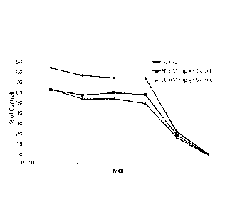
includes any of the
naturally-occurring forms of the FKBP protein, or variants thereof that
maintain FKBP protein
activity (e.g. within at least 50%, 80%, 90%, 95%, 96%, 97%, 98%, 99% or 100%
activity
compared to FKBP). In some embodiments, variants have at least 90%, 95%, 96%,
97%, 98%,
99% or 100% amino acid sequence identity across the whole sequence or a
portion of the
sequence (e.g. a 50, 100, 150 or 200 continuous amino acid portion) compared
to a naturally
occurring FKBP protein as set forth in SEQ ID NO:66.
[0065] "FRB" or an "FRB protein or polypeptide" as referred to herein includes
any of the
naturally-occurring forms of the FRB protein, or variants thereof that
maintain FRB protein
activity (e.g. within at least 50%, 80%, 90%, 95%, 96%, 97%, 98%,
or 100% activity
compared to FRB). In some embodiments, variants have at least 90%, 95%, 96%,
97%, 98%,
99% or 100% amino acid sequence identity across the whole sequence or a
portion of the
sequence (e.g. a 50, 100, 150 or 200 continuous amino acid portion) compared
to a naturally
occurring FRB protein as set forth in SEQ ID NO:69.
16
CA 02867129 2014-09-11
WO 2013/138505 PCT/US2013/031002
[0066] "EGFR" refers to the epidermal growth factor receptor corresponding to
the amino acid
sequence as set forth in SEQ TD NO:21.
[0067] "VHH" refers to a single domain antibody consisiting of a single
monomeric variable
antibody domain that is capable of selectively binding to a specific antigen
(e.g. EGFR). VHH
single-domain antibodies may be engineered from heavy-chain antibodies found
in camelids.
The terms VHH or VHH are used interchangeably throughout and are used
according to their
common meaning in the art. An "EGFR VHH" or "a EGFR VHH protein" as provided
herein
refers to a VI-1H single domain antibody specifically binding to EGFR. In some
embodiments,
the EGFR VHH has the sequence set forth in SEQ m NO:4. In further embodiments,
EGFR
VHH is operably linked to FKBP to form a ligand-dimerizing agent binder
conjugate. In some
further embodiments, the ligand-dimerizing agent binder conjugate has the
sequence set forth in
SEQ ID NO: 6.
[0068] "CEA" or CEACAM5- as provided herein refers Carcinoembryonic antigen-
related cell
adhesion molecule 5 also known in the art as CD66. "CEA VHH" or "a CEA VHH
protein" as
.. provided herein refers to a VHH single domain antibody specifically binding
to CEA. In some
embodiments, the CEA VHH has the sequence set forth in SEQ ID NO: 1. In
further
embodiments, the CEA VHH is operably linked to FKBP to form a ligand-
dimerizing agent
binder conjugate. In some further embodiments, the ligand-dimerizing agent
binder conjugate
has the amino acid sequence set forth in SEQ ID NO: 3.
[0069] A "protective antigen domain 4 (D4) protein" provided herein refers to
the Bacillus
anthracis protective antigen domain 4 as set forth in SEQ ID NO:94. In some
embodiments, D4
is operably linked to FKBP to form a ligand-dimerizing agent binder conjugate.
In some further
embodiments, the ligand-dimerizing agent binder conjugate has the amino acid
sequence set
forth in SEQ ID NO: 9.
.. [0070] A "control" sample or value refers to a sample that serves as a
reference, usually a
known reference, for comparison to a test sample. For example, a test sample
can be taken from
a test condition, e.g., in the presence of a test compound, and compared to
samples from known
conditions, e.g., in the absence of the test compound (negative control), or
in the presence of a
known compound (positive control). A control can also represent an average
value gathered
from a number of tests or results. One of skill in the art will recognize that
controls can be
designed for assessment of any number of parameters. For example, a control
can be devised to
17
CA 02867129 2014-09-11
WO 2013/138505 PCT/US2013/031002
compare therapeutic benefit based on pharmacological data (e.g., half-life) or
therapeutic
measures (e.g., comparison of side effects). One of skill in the art will
understand which controls
are valuable in a given situation and be able to analyze data based on
comparisons to control
values. Controls are also valuable for determining the significance of data.
For example, if
.. values for a given parameter are widely variant in controls, variation in
test samples will not be
considered as significant.
100711 As used herein, the term "cancer" refers to all types of cancer,
neoplasm, or malignant
tumors found in mammals, including leukemia, carcinomas and sarcomas.
Exemplary cancers
include cancer of the brain, breast, cervix, colon, head & neck, liver,
kidney, lung, non-small cell
lung, melanoma, mesothelioma, ovary, sarcoma, stomach, uterus and
Medulloblastoma.
Additional examples include, Hodgkin's Disease, Non-Hodgkin's Lymphoma,
multiple myeloma,
neuroblastoma, ovarian cancer, rhabdomyosarcoma, primary thrombocytosis,
primary
macroglobulinemia, primary brain tumors, cancer, malignant pancreatic
insulanoma, malignant
carcinoid, urinary bladder cancer, premalignant skin lesions, testicular
cancer, lymphomas,
.. thyroid cancer, neuroblastoma, esophageal cancer, genitourinary tract
cancer, malignant
hypercalccmia, endometrial cancer, adrenal cortical cancer, neoplasms of the
endocrine and
exocrine pancreas, and prostate cancer.
100721 The term "leukemia" refers broadly to progressive, malignant diseases
of the blood-
forming organs and is generally characterized by a distorted proliferation and
development of
leukocytes and their precursors in the blood and bone marrow. Leukemia is
generally clinically
classified on the basis of (1) the duration and character of the disease-acute
or chronic; (2) the
type of cell involved; myeloid (myelogenous), lymphoid (lymphogenous), or
monocytie; and (3)
the increase or non-increase in the number abnormal cells in the blood-
leukemic or aleukemic
(subleukemic). The P3g8 leukemia model is widely accepted as being predictive
of in vivo anti-
leukemic activity. It is believed that a compound that tests positive in the
P388 assay will
generally exhibit some level of anti-leukemic activity in vivo regardless of
the type of leukemia
being treated. Accordingly, the present invention includes a method of
treating leukemia, and,
preferably, a method of treating acute nonlymphocytic leukemia, chronic
lymphocytic leukemia,
acute granulocytic leukemia, chronic granulocytic leukemia, acute
promyelocytic leukemia, adult
T-cell leukemia, aleukemic leukemia, a leukocythemic leukemia, basophylic
leukemia, blast cell
leukemia, bovine leukemia, chronic myelocytic leukemia, leukemia cutis,
embryonal leukemia,
eosinophilic leukemia, Gross' leukemia, hairy-cell leukemia, hemoblastic
leukemia,
18
CA 02867129 2014-09-11
WO 2013/138505 PCT/US2013/031002
hemocytoblastic leukemia, histiocytic leukemia, stem cell leukemia, acute
monocytic leukemia,
leukopenic leukemia, lymphatic leukemia, lymphoblastic leukemia, lymphocytic
leukemia,
lymphogenous leukemia, lymphoid leukemia, lymphosarcoma cell leukemia, mast
cell leukemia,
megakaryocytic leukemia, micromyeloblastic leukemia, monocytic leukemia,
myeloblastic
leukemia, myelocytic leukemia, myeloid granulocytic leukemia, myelomonocytic
leukemia,
Naegeli leukemia, plasma cell leukemia, multiple myeloma, plasmacytic
leukemia,
promyelocytic leukemia, Rieder cell leukemia, Schilling's leukemia, stem cell
leukemia,
sublcukcmic leukemia, and undifferentiated cell leukemia.
[0073] The term "sarcoma" generally refers to a tumor which is made up of a
substance like
the embryonic connective tissue and is generally composed of closely packed
cells embedded in
a fibrillar or homogeneous substance. Sarcomas which can be treated with a
combination of
antineoplastic thiol-binding mitochondrial oxidant and an anticancer agent
include a
chondrosarcoma, fibrosarcoma, lymphosarcoma, melanosarcoma, myxosarcoma,
osteosarcoma,
Abemethy's sarcoma, adipose sarcoma, liposarcoma, alveolar soft part sarcoma,
ameloblastic
sarcoma, botryoid sarcoma, chloroma sarcoma, chorio carcinoma, embryonal
sarcoma, Wilms'
tumor sarcoma, endometrial sarcoma, stromal sarcoma, Ewing's sarcoma, fascial
sarcoma,
fibroblastic sarcoma, giant cell sarcoma, granulocytic sarcoma, Hodgkin's
sarcoma, idiopathic
multiple pigmented hemorrhagic sarcoma, inamunoblastic sarcoma of B cells,
lymphoma,
immunoblastic sarcoma of T-cells, Jensen's sarcoma, Kaposi's sarcoma, Kupffer
cell sarcoma,
angiosarcoma, leukosarcoma, malignant mesenchymoma sarcoma, parostcal sarcoma,
reticulocytic sarcoma, Rous sarcoma, serocystic sarcoma, synovial sarcoma, and
telangiectaltic
sarcoma.
[0074] The term "melanoma" is taken to mean a tumor arising from the
melanocytic system of
the skin and other organs. Melanomas which can be treated with a combination
of antineoplastic
thiol-binding mitochondrial oxidant and an anticancer agent include, for
example, aeral-
lentiginous melanoma, amelanotic melanoma, benign juvenile melanoma,
Cloudman's
melanoma, S91 melanoma, Harding-Passey melanoma, juvenile melanoma, lentigo
maligna
melanoma, malignant melanoma, nodular melanoma, subungal melanoma, and
superficial
spreading melanoma.
[0075] The term "carcinoma" refers to a malignant new growth made up of
epithelial cells
tending to infiltrate the surrounding tissues and give rise to metastases.
Exemplary carcinomas
which can be treated with a combination of antineoplastic thiol-binding
mitochondrial oxidant
19
CA 02867129 2014-09-11
WO 2013/138505 PCT/US2013/031002
and an anticancer agent include, for example, acinar carcinoma, acinous
carcinoma, adenocystic
carcinoma, adenoid cystic carcinoma, carcinoma adenomatosum, carcinoma of
adrenal cortex,
alveolar carcinoma, alveolar cell carcinoma, basal cell carcinoma, carcinoma
basocellulare,
basaloid carcinoma, basosquamous cell carcinoma, bronchioalveolar carcinoma,
bronchiolar
carcinoma, bronchogenic carcinoma, cerebriform carcinoma, cholangiocellular
carcinoma,
cherionic carcinoma, colloid carcinoma, corned carcinoma, corpus carcinoma,
cribriform
carcinoma, carcinoma en cuirasse, carcinoma cutaneum, cylindrical carcinoma,
cylindrical cell
carcinoma, duct carcinoma, carcinoma durum, embryonal carcinoma, cncephaloid
carcinoma,
epiermoid carcinoma, carcinoma epitheliale adenoides, exophytic carcinoma,
carcinoma ex
ulcere, carcinoma fibrosum, gelatiniforni carcinoma, gelatinous carcinoma,
giant cell carcinoma,
carcinoma gigantocellulare, glandular carcinoma, granulosa cell carcinoma,
hair-matrix
carcinoma, hcmatoid carcinoma, hepatocellular carcinoma, Hurthle cell
carcinoma, hyaline
carcinoma, hypemephroid carcinoma, infantile embryonal carcinoma, carcinoma in
situ,
intraepidermal carcinoma, intraepithelial carcinoma, Krompecher's carcinoma,
Kulchitzlcy-cell
carcinoma, large-cell carcinoma, lenticular carcinoma, carcinoma lenticulare,
lipomatous
carcinoma, lymphoepithelial carcinoma, carcinoma medullare, medullary
carcinoma, melanotic
carcinoma, carcinoma molle, mucinous carcinoma, carcinoma muciparum, carcinoma
mucocellulare, mucoepidermoid carcinoma, carcinoma mucosum, mucous carcinoma,
carcinoma
myxomatodes, nasopharyngeal carcinoma, oat cell carcinoma, carcinoma
ossificans, osteoid
carcinoma, papillary carcinoma, periportal carcinoma, preinvasive carcinoma,
prickle cell
carcinoma, pultaceous carcinoma, renal cell carcinoma of kidney, reserve cell
carcinoma,
carcinoma sarcomatodes, schneiderian carcinoma, scirrhous carcinoma, carcinoma
scroti, signet-
ring cell carcinoma, carcinoma simplex, small-cell carcinoma, solanoid
carcinoma, spheroidal
cell carcinoma, spindle cell carcinoma, carcinoma spongiosum, squamous
carcinoma, squamous
cell carcinoma, string carcinoma, carcinoma telangiectaticum, carcinoma
telangicctodes,
transitional cell carcinoma, carcinoma tuberosum, tuberous carcinoma,
vernicous carcinoma, and
carcinoma villosum.
100761 By "therapeutically effective dose or amount" herein is meant a dose
that produces
effects for which it is administered. The exact dose and formulation will
depend on the purpose
of the treatment, and will be ascertainable by one skilled in the art using
known techniques (see,
e.g., Lieberman, Pharmaceutical Dosage Forms (vols. 1-3, 1992); Lloyd, The
Art, Science and
Technology of Pharmaceutical Compounding (1999); Remington: The Science and
Practice of
Pharmacy, 20th Edition, Gcnnaro, Editor (2003), and Pickar, Dosage
Calculations (1999)).
CA 02867129 2014-09-11
WO 2013/138.505 PCT/US2013/031002
[0077] The term "pharmaceutically acceptable salts" or "pharmaceutically
acceptable carrier"
is meant to include salts of the active compounds which are prepared with
relatively nontoxic
acids or bases, depending on the particular substituents found on the
compounds described
herein. When compounds of the present invention contain relatively acidic
functionalities, base
addition salts can be obtained by contacting the neutral form of such
compounds with a sufficient
amount of the desired base, either neat or in a suitable inert solvent.
Examples of
pharmaceutically acceptable base addition salts include sodium, potassium,
calcium, ammonium,
organic amino, or magnesium salt, or a similar salt. When compounds of thc
present invention
contain relatively basic functionalities, acid addition salts can be obtained
by contacting the
neutral form of such compounds with a sufficient amount of the desired acid,
either neat or in a
suitable inert solvent. Examples of pharmaceutically acceptable acid addition
salts include those
derived from inorganic acids like hydrochloric, hydrobromic, nitric, carbonic,
monohydrogencarbonic, phosphoric, monohydrogenphosphoric,
dihydrogenphosphoric, sulfuric,
monohydrogensulfuric, hydriodic, or phosphorous acids and the like, as well as
the salts derived
from relatively nontoxic organic acids like acetic, propionic, isobutyric,
maleic, malonic,
benzoic, succinic, suberic, fumaric, lactic, mandelic, phthalic,
benzenesulfonic, p-tolylsulfonic,
citric, tartaric, methanesulfonic, and the like. Also included are salts of
amino acids such as
arginate and the like, and salts of organic acids like glucuronic or
galactunoric acids and the like
(see, e.g., Berge et al, Journal of Pharmaceutical Science 66:1-19 (1977)).
Certain specific
compounds of the present invention contain both basic and acidic
functionalities that allow the
compounds to be converted into either base or acid addition salts. Other
pharmaceutically
acceptable carriers known to those of skill in the art are suitable for the
present invention.
[0078] A "subject," "individual," or "patient," is used interchangeably
herein, which refers to a
vertebrate, preferably a mammal, more preferably a human. Mammals include, but
are not
limited to, murines, simians, humans, farm animals, sport animals, and pets.
Tissues, cells and
their progeny of a biological entity obtained in vitro or cultured in vitro
are also encompassed.
Compositions
[0079] Provided herein, inter alia, are adenoviral compositions useful for
infecting a broad
variety of different cell types (e.g. cancer cells). For example, the
compositions provided herein
may be used to retarget adenovirus infection to receptors upregulated on tumor
cells (e.g. EGFR,
CEA, ErbB). Using the compositions provided herein including embodiments
thereof, the
heterogeneity of tumors can be overcome by designing recombinant adenoviruses
that are able to
21
CA 02867129 2014-09-11
WO 2013/138505 PCT/US2013/031002
infect tumor cells through more than one receptor. The viral compositions
provided herein
express polypeptide binding pairs (as listed in Table 2, e.g. FKBP and FRB)
capable of
dimerizing in the presence of a chemical dimerizing agent (e.g. rapamycin) and
thereby forming
a ternary complex. The ternary complex enables the virus to bind to a specific
cellular surface
receptor. The components of the ternary complex may completely or partially be
encoded by the
adenoviral genome and are therefore not lost during viral replication
providing for the ability of
the virus of subsequent re-infection. Thus, in one aspect, a recombinant
nucleic acid encoding a
capsid-dimerizing agent binder conjugate and a ligand-dimerizing agent binder
conjugate are
provided. The capsid-dimerizing agent binder conjugate includes a dimerizing
agent binder (e.g.
FRB) operably linked to a viral capsid protein (e.g. fiber). A dimerizing
agent binder as
provided herein is an agent capable of binding a dimerizing agent. A
dimerizing agent binder
includes without limitation a protein, a compound or a small molecule. In some
embodiments,
the dimerizing agent binder is a FRB protein. Non limiting examples of
dimerizing agent
binders are set forth in Table 2 provided herein. Binding of the dimerizing
agent binder to the
dimerizing agent may occur through non-covalent intermolecular interactions
such as hydrogen
bonding, electrostatic interactions, hydrophobic and Van der Waals forces. The
capsid-
dimerizing agent binder conjugate includes a viral capsid protein. The term
capsid refers to any
component (e.g. capsid proteins or polypeptides) forming the shell of a virus,
wherein the capsid
can include one or more of these components. The capsid includes any
appropriate structural
components of the viral shell. In some embodiments, the capsid protein is an
adenoviral capsid
protein. Non-limiting examples of capsid proteins are L3 II (hexon) (e.g.
encoding major
structural proteins that form the triangular faces of the capsid), Li Ina
(e.g. encoding minor
structural proteins that help to stabilize the capsid), L2 III (penton) (e.g.
encoding major
structural proteins that form the vertex of the capsid where the fiber
protrudes), L2 pVIT (e.g.
encoding core structural proteins with homology to histone H3 and associate
with viral DNA in
the capsid), and L5 IV (Fiber) (e.g. encoding major structural proteins that
extend from the
penton base and are responsible for receptor binding). In some embodiments,
the adenoviral
capsid protein is a fiber protein.
100801 Upon expression in a cell the dimerizing agent binder and the viral
capsid protein form
a capsid-dimerizing agent binder conjugate, which is capable of binding to a
dimerizing agent
(e.g. rapamycin) through the dimerizing agent binder (e.g. FRB) and is
incorporated into the viral
capsid by the capsid protein (e.g. fiber). Thus, in some embodiments, the
capsid-dimerizing
agent binder conjugate includes a capsid protein and a dimcrizing agent
binder. In other
22
CA 02867129 2014-09-11
WO 2013/138505 PCT/US2013/031002
embodiments, the capsid protein is operably linked to the dimerizing agent
binder. Through
binding to the dimerizing agent the capsid-dimerizing agent binder conjugate
may connect to the
ligand-dimerizing agent binder conjugate. The ligand-dimerizing agent binder
conjugate
includes a cell surface receptor-specific ligand (e.g. EGFR VHH) operably
linked to a second
dimerizing agent binder (e.g. FKBP). A ligand as provided herein is a protein
with the capability
of binding a molecule expressed on the surface of a cell. Non-limiting
examples of ligands and
corresponding cellular receptors are set forth in Table 3. In some
embodiments, the ligand is a
EGFR VHH protein. In a further embodiment, the dimerizing agent binder is
FKBP. In some
embodiments, the ligand is a CEA VHH protein. In a further embodiment, the
dimerizing agent
binder is FKBP. In some embodiments, the ligand is a protective antigen domain
4 (D4) protein.
In a further embodiment, the dimerizing agent binder is FKBP. In some
embodiments, the
ligand-dimerizing agent binder conjugate includes a ligand and a dimerizing
agent binder. In
some embodiments, the ligand is operably linked to the dimerizing agent
binder. In some
embodiments, the ligand is an antibody. In some further embodiments, the
antibody is a single
domain antibody. In some embodiments, a plurality of ligands is operably
linked to the
dimerizing agent binder, wherein the plurality of ligands are individually
different. The plurality
of ligands may be operably linked to one or both termini of the dimerizing
agent binder. In some
embodiments, the plurality of ligands is operably linked in tandem to one or
both termini of the
dimerizing agent binder. In some embodiments, the dimerizing agent binder is
an immunophilin
protein. In some further embodiments, the immunophilin protein is a FKBP
protein. In some
further embodiments, the FKBP protein is a human FKBP protein. In some further
embodiments, the human FKBP protein is FKBP12. In other embodiments, the
ligand is capable
of binding a cell. In other embodiments, the cell is a tumor cell.
[0081] Provided herein, inter alia, are recombinant adenoviruses expressing
the recombinant
nucleic acid described above. Thus, in another aspect, a recombinant
adenovirus including a
recombinant nucleic acid provided herein including embodiments thereof is
provided. In some
embodiments, the adenovirus is a replication incompetent adenovirus. In other
embodiments, the
adenovirus is a replication competent adenovirus. Where the adenovirus is a
replication
competent adenovirus, the adenovirus is capable of infecting a cell by binding
to a specific
cellular surface receptor (e.g. EGFR), replicating inside said cell thereby
producing new viral
progeny capable of infecting additional cells. In contrast, a replication
incompetent adenovirus,
is capable of entering a cell by binding to a specific cellular receptor and
expressing the
adcnoviral genome inside said cell. However, a replication incompetent virus
lacks genes
23
CA 02867129 2014-09-11
WO 2013/138505 PCT/US2013/031002
necessary to produce new viral progeny and therefore is not capable of
subsequent infection of
additional cells.
[0082] In another aspect, a recombinant adenovirus including a capsid-
dimerizing agent binder
conjugate is provided. As described above a capsid-dimerizing agent binder
conjugate includes a
capsid protein (e.g. fiber) operably linked to a dimerizing agent binder (e.g.
FRB). The binding
of the dimerizing agent binder (e.g. FRB) to the dimerizing agent (e.g.
rapamycin) therefore
connects the recombinant adenovirus to the dimerizing agent. Thus, the
recombinant adenovirus
including a capsid-dimerizing agent binder conjugate may be bound to a
dimerizing agent. A
dimerizing agent as provided herein is an agent capable of binding a
dimerizing agent binder of a
capsid-dimerizing agent binder conjugate and a dimerizing agent binder of a
ligand-dimerizing
agent binder conjugate. In some embodiments, the dimerizing agent binds a
dimerizing agent
binder of a capsid-dimerizing agent binder conjugate. The dimerizing agent may
bind a
dimerizing agent binder of a ligand-dimerizing agent binder conjugate and a
dimerizing agent
binder of a ligand-dimerizing agent binder conjugate. Thus, in some
embodiments, the
dimerizing agent is further bound to a ligand-dimerizing agent binder
conjugate. A dimerizing
agent may bind the dimerizing agent binder through non-covalent intermolecular
interactions
such as hydrogen bonding, electrostatic interactions, hydrophobic and Van der
Waals forces. In
some embodiments, the dimerizing agent binds covalently to the dimerizing
agent binder. The
dimerizing agent as provided herein may be a naturally occurring substance
(e.g. rapamycin,
.. abscisic acid) or a synthetic substance (e.g. a small molecule, a
compound). Examples of
dimerizing agents according to the invention provided herein are listed in
Table 2. In some
embodiments, the dimerizing agent is a compound. In some further embodiments,
the compound
is rapamycin. Rapamycin refers, in the customary sense, to CAS Registry No.
53123-88-9.
Rapamycin inhibits the mTOR kinase and is used as an immunosuppressing agent
and anti-
cancer treatment. In some further embodiments, the dimerizing agent is a
rapalog. A rapalog as
provided herein is a rapamycin analog does not inhibit cellular mTOR kinase
activity. In some
further embodiment, the rapalog is AP21967. In some embodiments, the
dimerizing agent is an
anti-cancer drug.
[0083] As described above, the compositions provided herein include a
recombinant
adenovirus including a recombinant nucleic acid including a capsid-dimerizing
agent binder and
a ligand-dimerizing agent binder conjugate. Therefore, the recombinant
adenovirus may further
include a ligand-dimerizing agent binder conjugate. In other embodiments, the
ligand-
24
CA 02867129 2014-09-11
WO 2013/138505 PCT/US2013/031002
dimerizing agent binder conjugate is ectopically expressed. Wherein the ligand-
dimerizing agent
binder conjugate is ectopically expressed, the nucleic acid encoding the
ligand-dinterizing agent
binder conjugate does not form part of the recombinant nucleic acid included
in the recombinant
adenovirus. Where the ligand-dimerizing agent binder conjugate is ectopically
expressed it may
be encoded by the genome of the cell infected with the recombinant adenovirus.
[0084] In some embodiments, the recombinant adenovirus includes a plurality of
ligand-
dimerizing agent binder conjugates, whcrcin each ligand-dimerizing agent
binder conjugate may
be different. For example where the recombinant adenovirus includes a
plurality of ligand-
dimerizing agent binder conjugates, the recombinant adenovirus may include a
first ligand-
dimerizing agent binder conjugate, a second ligand-dimerizing agent binder
conjugate and a third
ligand-dimerizing agent binder conjugate with each ligand-dimerizing agent
binder conjugate
being different. Thus, the first ligand-dimerizing agent binder conjugate may
include a first
ligand and a first dimerizing agent binder, the second ligand-dimerizing agent
binder conjugate
may include a second ligand and a second dimerizing agent binder, wherein the
first ligand is
different from the second ligand and the first dimerizing agent binder is the
same or different
from the second dimcrizing agent binder. For example, the first ligand- EGFR
VEIH may be
operably linked to the first dimerizing agent binder FKBP and the second
ligand CEA VHH may
be operably linked to the second dimerizing agent binder AB1 or FKBP.
[0085] Moreover, the recombinant adenovirus may include a plurality of capsid-
dimerizing
agent binder conjugates, wherein each capsid-dimerizing agent binder conjugate
may be
different. For example where the recombinant adenovirus includes a plurality
of capsid-
dimerizing agent binder conjugates, the recombinant adenovirus may include a
first capsid-
dimerizing agent binder conjugate, a second capsid-dimerizing agent binder
conjugate and a
third capsid-dimerizing agent binder conjugate with each capsid-dimerizing
agent binder
conjugate being different. Thus, the first capsid-dimerizing agent binder
conjugate may include
a first capsid protein and a first dimerizing agent binder, the second capsid-
dimerizing agent
binder conjugate may include a second capsid protein and a second dimerizing
agent binder,
wherein the first and second capsid protein may be the same or different and
the first and second
dimerizing agent binder may the same or different. For example, the first
capsid protein fiber
may be operably linked to the first dimerizing agent binder FRB and the second
capsid protein
fiber may be operably linked to the second dimerizing agent binder PYLl. Thus,
in one
embodiment, the recombinant adenovirus includes a first capsid-dimerizing
agent binder
CA 02867129 2014-09-11
WO 2013/138505 PCT/US2013/031002
conjugate (e.g. fiber/FRB), a first ligand-dimerizing agent binder conjugate
(e.g. EGFR
VHH/FKBP), a second capsid-dimerizing agent binder conjugate (e.g. fiber/PYL1)
and a second
ligand-dimerizing agent binder conjugate (e.g. CEA VHH/AB1). In the presence
of a first
dimerizing agent (i.e. rapamycin) the first capsid-dimerizing agent binder
conjugate and the first
ligand-dimcrizing agent binder conjugate arc connected through the binding of
FRB and FKBP
to rapamycin. In the presence of a second dimerizing agent (i.e. abscisic
acid) the second capsid-
dimerizing agent binder conjugate and the second ligand-dimerizing agent
binder conjugate are
connected through the binding of AB1 and Pyll to abscisic acid. Therefore, in
the presence of
rapamycin the recombinant adenovirus infects cells expressing the EGF receptor
and in the
presence of abscisic acid the same virus may infect cells expressing CEA.
Thus, the same
recombinant adenovirus is capable of infecting different cell types depending
on the presence
of dimerizing agent administered.
[0086] In another aspect, a cell including a recombinant adenovirus provided
herein including
embodiments thereof is provided. In some embodiments, the cell is a cancer
cell in a cancer
patient. In other embodiments, the cell is a non-cancer cell in a cancer
patient. In some
embodiments, the cell is a cell in an organism. In some further embodiments,
the organism is a
mammal. In some further embodiments, the mammal is a human. In other
embodiments, the
cell is a cell in a culture vessel. In some further embodiments, the cell is a
transformed cell.
III. Methods
[0087] In another aspect, a method of forming an adenoviral cancer cell
targeting construct is
provided. The method includes infecting a cell with a recombinant adenovirus
provided herein,
thereby forming an adenoviral infected cell. The adenoviral infected cell is
allowed to express
the recombinant nucleic acid, thereby forming a ligand-dimerizing agent binder
conjugate and a
recombinant adenovirus including a capsid-dimerizing agent binder conjugate.
The recombinant
adenovirus and the ligand-dimerizing agent binder conjugate are contacted with
a dimerizing
agent. The recombinant adenovirus and the ligand-dimerizing agent binder
conjugate are
allowed to bind to the dimerizing agent, thereby forming the adenoviral cancer
cell targeting
construct. As described above, the recombinant nucleic acid may include a
plurality of capsid-
dimerizing agent binder conjugates and ligand-dimerizing agent binder
conjugates, thereby
enabling the adenovirus expressing the recombinant nucleic acid to bind to
plurality of different
cellular surface receptors.
26
CA 02867129 2014-09-11
WO 2013/138505 PCT/US2013/031002
[0088] In another aspect, a method of targeting a cell is provided. The method
includes
contacting a cell with a recombinant adenovirus provided herein including
embodiments thereof.
In some embodiments, the cell is a cancer cell.
[0089] In another aspect, a method of targeting a cancer cell in a cancer
patient is provided.
The method includes administering to a cancer patient a recombinant adenovirus
provided
herein. The recombinant adenovirus is allowed to infect a cell in the cancer
patient, thereby
forming an adenoviral infected cell. The adenoviral infected cell is allowed
to express the
recombinant nucleic acid, thereby forming a ligand-dimerizing agent binder
conjugate and a
recombinant adenovirus including a capsid-dimerizing agent binder conjugate.
The cancer
patient is administered with a dimerizing agent. The recombinant adenovirus
and the ligand-
dimerizing agent binder conjugate are allowed to bind to the dimerizing agent,
thereby forming
an adenoviral cancer cell targeting construct. The adenoviral cancer cell
targeting construct is
allowed to bind to a cancer cell, thereby targeting the cancer cell in the
cancer patient. In some
embodiments, the cell is a cancer cell. In other embodiments, the cell is a
non-cancer cell.
100901 In another aspect, a method of targeting a cell is provided. The method
includes
contacting a first cell with a recombinant adenovirus provided herein. The
recombinant
adenovirus is allowed to infect the first cell, thereby forming an adenoviral
infected cell. The
adenoviral infected cell is allowed to express the recombinant nucleic acid,
thereby forming a
ligand-dimerizing agent binder conjugate and a recombinant adenovirus
comprising a capsid-
dimerizing agent binder conjugate. The ligand-dimerizing agent binder
conjugate and the
recombinant adenovirus are contacted with a dimerizing agent. The recombinant
adenovirus and
the ligand-dimerizing agent binder conjugate are allowed to bind to the
dimerizing agent, thereby
forming an adenoviral cell targeting construct. The adenoviral cell targeting
construct is allowed
to bind to a second cell, thereby targeting said cell. In some embodiments,
the first cell and the
second cell form part of an organism.
IV. Specific Embodiments
[0091] Cancer is a debilitating disease that accounts for more than half a
million deaths each
year. There is a profound need for more effective, selective and safe
treatments for cancer.
Existing treatments for this pervasive, life threatening disease, such as
chemotherapy and
surgery, rarely eliminate all malignant cells, and often exhibit deleterious
side-effects that can
outweigh therapeutic benefit. The present invention provides powerful
recombinant viruses that
27
'8 17 8 2 1 88
are capable of infecting tumor cells via disparate receptors. These viruses
will enable a new safe
form of effective, self-amplifying therapy that breaks the paradigm of
systemic genotoxic
treatments for cancer.
100921 One approach that has the potential to address many of the shortcomings
of current
cancer treatments is oncolytic adenoviral therapy [Pesonen, S. et al.,
Molecular Pharmaceutics,.
8(1): p. 12-28 (2010)]. These viruses are designed to replicate specifically
in cancer cells, but
leave normal cells unharmed. This selectivity can be engineered by exploiting
the functional
overlap between adenoviml, early onco-proteins, such as El A, and tumor
mutations in the Rb
tumor suppressor pathway which drives deregulated cell cycle entry and
pathological DNA
replication [Poznic, M., .1 Word, 34(2): p. 305-12 (2009)1.
100931 Adenovirus (Ad) is a self-replicating biological machine. It consists
of a linear double-
stranded 36 kb DNA genome sheathed in a protein coat. Ad requires a human host
cell to
replicate. It invades and hijacks the cellular replicative machinery to
reproduce and upon
assembly induces lytic cell death to escape the cell and spread and invade
surrounding cells (Fig.
1), No ab initio system has come close to mimicking the autonomy and
efficiency of Ad,
however, Applicants have developed two new strategies to systematically
manipulate the Ad
genome to create novel adenoviruses as described in published application
PCT/US2011/048006.
Henceforth, with the ability to
manipulate the Ad genome, Applicants can take the virus by the horns and
redesign it to perform
the functions of tumor-specific infection, replication, and cell killing.
100941 Currently adenoviral vectors rely on a single cellular receptor for
their uptake, which
significantly limits their therapeutic potential. Ad5 infection is mediated
primarily Through
interactions between the fiber protein on the outer viral capsid and the
coxsackie and adenovirus
receptor (CAR) on human epithelial cells. Unfortunately, many cancer cells do
not express
CAR, such as mesenehymal and deadly metastatic tumor cells. Since viral
replication/killing
will be limited by the ability to infect cells, Applicants need viruses that
infect tumor cells via
receptors other than CAR, ideally those specifically upregulated on tumor
cells. Provided herein
are genetically-encoded switchable targeting moieties that enable Ad5 to
infect cancer cells
regardless of their CAR-expression. Applicants used a known property of the
cancer drug
rapamycin (rap) to dimerize heterologous proteins with FKBP and FRB domains
and engineered
viruses that express a FRB-fiber capsid protein fusion together with
retargeting ligands fused to
FKBP. These viruses are induced to infect any cell type via multiple
retargeting ligands upon
28
CA 2867129 2019-08-30
CA 02867129 2014-09-11
WO 2013/138505 PCT/US2013/031002
rap treatment. This represents a rational and powerful combination of chemical
and viral
weapons as a novel cancer therapy. Tn addition, a major goal is to overcome
tumor heterogeneity
by engineering viruses that are able to infect tumor cells through more than
one more mechanism
and receptor. Applicants achieved this by using a known property of the cancer
drug rapamycin
(rap) to dimerize heterologous proteins with FKBP and FRB domains. Rapamycin
inhibits the
mTOR kinase and is used as an immunosuppressing agent and anti-cancer
treatment. By
engineering FRB mutations, rapalogs of rapamycin that do not inhibit cellular
mTOR kinase
activity can also be used to induce infection of any cell type upon
administration of a rapalog.
These viruses can be induced to infect any cell type via multiple retargeting
ligands upon rap
treatment.
[0095] Cancer continues to be an intractable disease without safe and reliably
effective
treatments. In the last century, Applicants' knowledge about the origins of
cancer and cancer
biology has greatly advanced. However, despite Applicants' new understanding
of cancer as a
genetic disease, the standard of care for non-resectable disseminated disease
remains genotoxic
therapies, such as chemotherapy and irradiation, which often have intolerable
and toxic side-
effects. While drugs have been developed to target oncogcnic proteins,
Applicants have nothing
to treat the genetic loss of tumor-suppressors. One approach to treat these
cancers is oncolytic
viral cancer therapy (Fig. 1) [Pesonen, S. et al., Molecular Pharmaceutics,.
8(1): p. 12-28
(2010)].
[0096] Adenovirus (Ad) has been studied for more than half a century, and has
contributed
significantly to Applicants' understanding of key mechanisms at the heart of
mammalian cell
biology such as splicing, critical growth regulatory hubs, transcription, and
the cell cycle. Ad is
a small double-stranded 36 kb DNA virus, sheathed in a protein capsid coat
(Fig. 2). Ad
particles primarily interact with host cells through protein interactions
between the knob-domain
of fiber on the surface of the capsid and a cell surface molecule (Fig. 2).
Serotype 5 of
adenovirus species C (Ad5) infects cells via fiber interactions with coxsackie
virus and
adenovirus receptor (CAR), primarily found at epithelial cell junctions. Like
all viruses, it is
entirely dependent on host cells for its propagation. After depositing its
genome into the host
cell nucleus, a program is coordinated by virus proteins to activate the cell
cycle in quiescent
cells in order to replicate virus DNA. At the end of the Ad5 life cycle, after
progeny virions
have been assembled in the cell nucleus, the membranes of the cell are lysed,
releasing the next
generation of viruses.
29
CA 02867129 2014-09-11
WO 2013/138505 PCT/US2013/031002
[0097] Manipulating Adenovirus Tropism
[0098] Ad5 infection is mostly limited to cells that have CAR, which is
expressed along with
cadhcrin at epithelial cell tight junctions [Tomko, R.P. et al., Proceedings
of the National
Academy of Sciences, 94(7): p. 3352-3356 (2009); Bergelson, J.M., et al.,
Science, 275(5304): p.
1320-1323 (1997)]. Unfortunately, it is metastases that kill most cancer
patients, in which an
epithelial to mesenchymal transition (EMT) results in downregulation of
cadherin and CAR,
instigating invasion and spread to distant sites [Anders, M., et al., Br J
Cancer, 100(2): P. 352-9
(2009)]. Thus, many malignant cells do not express CAR and are not susceptible
to infection by
Ad5 [Anders, M., et al., Br J Cancer, 100(2): p. 352-9 (2009); Dietel, M., et
al., Journal of
Molecular Medicine, 89(6): p. 621-630(2011); Matsumoto, K., et al., Urology,
66(2): p. 441-446
(2005)]. A number of approaches have been taken to retarget Ad5 to different
cellular receptors,
including: chemical modification of purified adenovirus particles and
infection with recombinant
divalent "bridging" proteins to form complexes between fiber and receptor
(reviewed in [Rein,
D.T., M. Breidenbach, and D.T. Curiel, Future Oncology, 2(1): p. 137-143
(2006)]). The
disadvantage of these approaches is the restriction to the first round of
infection, since following
virus replication the chemical/recombinant targeting moiety is lost. This
drawback can be
overcome by directly modifying the fiber gene to encode targeting sequences,
however this
approach is not systematic because Applicants cannot predict the folding of de
novo sequences
and the correct assembly with virus particles. To date, this approach has only
been useful for the
insertion of small peptides.
[0099] Adsembly and AdSLIC are enabling technologies to systematically design
new
optimized adeno viruses from libraries of genomic building blocks and
heterologous parts.
101001 The potential of adenoviral vectors in several applications is hindered
by the ability to
engineer and combine multiple genetic modifications rapidly and
systematically. To
systematically re-design adenovirus as an oncolytic agent, Tools are needed to
enable precise
modification of its components. The 36 kb Ad5 genome is difficult to
manipulate due to its size
and abundance of restriction enzyme recognition (RER) sites. To date, a
majority of
recombinant Ads have been limited to the backbones that were digested and
selected for fewer
RER sites in the 1980s, and continue to remain due to the legacy of shuttle
vectors. These
backbones have accumulated a number of mutations distant from wild type
sequences.
Traditional cloning techniques with complex sequences are still time consuming
and not
systematic.
CA 02867129 2014-09-11
WO 2013/138505 PCT/US2013/031002
[0101] To overcome the limitations of Ad5 and current methodologies,
Applicants have
developed two new technologies, named `Adsembly' and `AdSlicR' , which enable
the rapid de
novo assembly of adenoviral genomes in vitro from genomic component parts and
heterologous
elements in a single hour. Using a bioinformatics approach, Applicants split
the adenoviral
genome (36 kb) into 5 units, based on evolutionarily conserved sequences
between species,
transcriptional and functional modules. Each of these 5 units comprise
compatible sections of a
genomic building "parts library", the functions and diversity of which can be
altered by
engineering mutations or heterologous elements and further expanded by adding
equivalent units
from disparate adenovirus serotypes, mutants and species. In order to create a
new adenovirus
with unique properties, one of each of the units is selected from the library
and rapidly
reassembled into a complete genome in vitro using Adsembly or Ad-SlicR.
Adsembly can be
used to assemble a novel genome (in 1 hour) via multi-site specific
recombination, which upon
transfection, self-excises from a plasmid backbone and replicates to produce
novel viruses. Ad-
SlicR, which utilizes the same library genome building blocks, is a
complementary strategy to
erase inserted recombination sequences for more potent viral replication (if
necessary) and
clinical use. The ease of manipulation of multiple genomic fragments as small
modular plasmid
units and the systematic approach of these technologies now allows for rapid
and precise
construction of novel adenoviruses.
[0102] Adenovirus Targeting: A Genetically Encoded Switch
[0103] Oncolytic viral therapy has the potential to destroy a tumor mass of
unlimited size, but
only if the virus crosses the tumor vasculature and infection spreads from one
cell to another.
The fiber of Ad5 recognizes the epithelial cell junction molecule CAR [Tomko,
R.P. et al.,
Proceedings of the National Academy of Sciences, 94(7): p. 3352-3356 (2009);
Bergelson, J.M.,
et al., Science, 275(5304): p. 1320-1323 (1997)], which is expressed in
variable levels on tumors
[Rein, D.T., M. Breidenbach, and D.T. Curiel, Future Oncology, 2(1): p. 137-
143 (2006);
Dmitriev, I., et al., J. ViroL, 72(12): p. 9706-9713 (1998); Bauerschmitz,
G.J., S.D. Barker, and
A. Hemminki, In! J Oncol, 21(6): p. 1161-74 (2002); Breidenbach, M., et al.,
Hum Gene Ther.,
15(5): p. 509-18 (2004); Cripc, T.P., et al., Cancer Res, 61(7): p. 2953-60
(2001); Fechncr, 1-i., et
al., Gene Ther, 7(22): p. 1954-68 (2000); Hemmi, S., et al., Hum Gene Ther,
9(16): p. 2363-73
(1998); Hemminki, A. and R.D. Alvarez, BioDrugs, 16(2): p. 77-87 (2002);
Kanerva, A., et al.,
Clin Cancer Res, 8(1): p. 275-80 (2002); Li, Y., et al., Cancer Res, 59(2): p.
325-30 (1999);
Miller, C.R., et al., Cancer Res, 58(24): p. 5738-48 (1998); Rein, D.T., et
al., Mt J. Cancer,
31
CA 02867129 2014-09-11
WO 2013/138505 PCT/US2013/031002
111(5): p. 698-704 (2004)] and the loss of which is associated with increased
metastasis [Anders,
M., et al., Br J Cancer, 100(2): p.352-9 (2009); Dietel, M., et al., Journal
of Molecular
Medicine, 89(6): p. 621-630 (2011); Matsumoto, K., et al., Urology, 66(2): p.
441-446 (2005)1.
Ad5 is not a naturally blood-borne virus and does not actively target and
cross the vasculature.
Both of these factors limit the potential of adenoviral vectors for gene
expression and therapy.
[0104] Attempts to retarget adenoviral uptake include the use of chemical
adapters that link
viral capsids to retargeting ligands. One example is fiber biotinylation to
provide a chemical
linker for high affinity binding to avidin-retargeting ligands [Liu, Y., P.
Valadon, and J.
Schnitzer, Virology Journal, 7(1): p. 316 (2010)]. However, retargeting is
only achieved with
exogenous virus, since the chemical modifications are lost upon viral
replication. Genetically
encoding retargeting adapter fusions to viral coat proteins is desirable, but
also more challenging.
Unfortunately, the incorporation of large ligands in capsid proteins disrupts
their
folding/assembly [Belousova, N., etal., J. Virol., 76(17): p. 8621-8631
(2002)]. To avoid
misfolding, smaller polypeptides can be inserted into the fiber Hi loop (Fig.
6) [Belousova, N.,
et al., Virol, 76(17): p. 8621-31(2002)]. For example, RGD peptides enhance
integrin-assisted
uptake, but arc not sufficient to alter viral tropism. Fiber fusions to single
chain antibodies
(scFVs) are attractive as well, but the former require processing in the
ER/cytosol while fiber
assembles in the nucleus [Kontermann, R.E., Curr Opin Mol Ther, 12(2): p. 176-
83 (2010)].
Thus, despite ongoing efforts to retarget infection, in vivo studies and gains
have been
disappointing [Waehler, R., S.J. Russell, and D.T. Curiel, Nat Rev Genet,
8(8): p. 573-87
(2007)].
[0105] An ideal virus would cross the blood/endothelium layer and infect tumor
cells via
disparate receptors. The ideal system would be a genetically encoded chemical
adapter that
could be used to switch viral tropism within the body via any multiple
retargeting moieties,
without compromising viral replication and safety. Provided herein is a novel,
inducible,
genetically encoded chemical adapter system that retargets infection to
multiple cell types, and is
not lost upon viral replication. The present invention therefore overcomes the
limitations of
prior approaches and has several advantages. Any unanticipated toxicities
associated with
receptor-retargeting can be stopped by drug withdrawal. In addition, multiple
retargeting ligands
can be expressed within a single virus to target tumor cell receptors (e.g.
EGFR) and the
vasculature (e.g. Von Willebrand factor/transferrin).
32
CA 02867129 2014-09-11
WO 2013/138505 PCT/US2013/031002
[0106] Applicants retargeted the adenovirus viral coat protein fiber to
alternate cellular
receptors using a known property of the immunosuppressive and anti-tumor drug
rapamycin
(rap; Fig. 7). Rapamycin can be used to induce heterodimers of heterologous
proteins if one is
fused to FKBP domains (e.g. a retargeting ligand) and the other (e.g. fiber)
to the FRB domain of
mTOR [Chen, J., et al., Proc Nat! Acad Sci USA, 92(11): p. 4947-51 (1995)].
Upon treatment
with rap, fiber will heterodimerize with the retargeting ligand enabling the
virus to infect the cell
type of choice. Rapamycin is a macrolide antibiotic that is FDA approved and
has ideal
pharmacokinctic profiles in mammals. The high affinity and stability of rap-
induced
heterodimerization has been used with great success in several applications
including phage
display of receptor-ligand complexes [de Wildt, R.M., et al., Proc Nat! Acad
Sci USA, 99(13):
p. 8530-5 (2002)1 transcriptional activation and reconstitution of bi-
functional proteins
[Clackson, T., Chem Biol Drug Des, 67(6): p. 440-2 (2006)]. A novel
application of this system
is provided, which also takes advantage of Applicants' previous studies of rap
as a rational
combination with oncolytic viruses [O'Shea, C., et al., Embo J, 24(6): p. 1211-
21 (2005)].
[0107] Develop a genetically encoded small molecule-controlled system for
retargeting
adenovirus to tumor cell receptors.
[0108] The reliance of current adenoviral vectors on a single cellular
receptor for their uptake
limits their therapeutic potential via systemic delivery. To overcome this
problem, Applicants
designed novel Ads with the rapamycin-induced, genetically encoded FRB/FKBP
heterodimer to
enable retargeting of adenovirus to tumor cell receptors. Ultimately, this
system enables
targeting of receptors in angiogenic tumor vasculature to eliminate aggressive
tumors (e.g.
TEMs, TVMs), and upregulated markers in high-risk tumors such as breast cancer
(e.g. EGFR,
HER2, TfR).
[0109] Insertion of FRB domain into fiber Ifl loop.
[0110] The fiber protein which infers tropism to adenovirus is generally not
permissible to
large insertions or modifications, because the correct folding and assembly of
fiber trimers into
adenovirus particles arc critical for viable progeny. To date, insertion of
sequences in the C-
terminal Ad5 fiber knob-domain has been effectively limited to peptides
[Belousova, N., et al., J.
Virol., 76(17): p. 8621-8631(2002)]. Using Adsembly (described in Fig. 5) the
90 amino acid
FRB domain was inserted into the flexible HI loop of fiber, which accommodates
insertions of
33
CA 02867129 2014-09-11
WO 2013/138505 PCT/US2013/031002
up to 100 amino acids without deleterious effects [Belousova, N., et al., J.
ViroL, 76(17): p.
8621-8631 (2002)].
[0111] Experimental Approach
[0112] The wild type E3 component plasmid, E3_001, (Table 1) from the genome
parts library
which Applicants designed was used as the template for insertion of the FRB
sequence into the
fiber gene. E3-001 was PCR amplified to generate a product with SLIC-
compatible ends for
insertion between fiber 'Thr546 and Pro567. The 90 aa FRB domain of mTOR
(G1u2025-
G1n2114) was PCR amplified from mTOR cDNA and combined via SLIC to generate E3-
002
(Fig. 8). Wildtype E2, L3, and E4 components were combined with E3-002 and E1-
009
(containing a MIV-driven GFP gene) using the Adsembly strategy to generate the
Ad-122
genome (Fig. 8). Applicants transfected the Ad-122 genome into 293 E4 cells,
and harvested
virus. Unlike the insertion of many large ligands such as TfR, FRB did not
inhibit viral
replication or assembly and robust infection as evidenced by GFP fluorescence
from the El
reporter and observed cytopathic effect (CPE). I confirmed expression of FRB-
fiber by Western
blot as indicated by predicted migration of FRB-fiber (72.4 kDa) versus wt
fiber (61.6 kDa; Fig.
9).
[0113] Expression of FKBP retargeting moieties from adenovirus genome.
101141 The expression of FKBP from the virus genome would ideally have similar
timing and
levels matching that of fiber, to enable efficient dimerization with fiber in
the presence of rap.
Applicants adopted several strategies to express FKBP from the genome as
summarized in figure
10.
101151 Experimental Approach
[0116] The E3-002 plasmid, carrying the FRB insertion in fiber, was used as
the template to
introduce the sequences necessary for the strategies summarized in figure 10.
The first approach
was to express FKBP from the adenovirus genome by co-translationally
expressing it from the
fiber transcript (Fig. 10C). The FKBP sequence was placed downstream of the
fiber coding
sequence following an inserted Furin-2a sequence. The Furin-2a sequence is an
optimized Furin
protease recognition site followed by the foot-and-mouth disease virus 2a auto-
cleavage site
[Fang, J., et al., Mol Ther, 15(6): p. 1153-1159 (2007)]. It should generate
two distinct
polypeptides in equimolar amount; the FRB-Fiber molecule with a residual
arginine on its C-
terminus, and the FKBP protein with residual proline on its N- terminus.
34
CA 02867129 2014-09-11
WO 2013/138505 PCT/US2013/031002
101171 The sequence was cloned successfully, generating E3-016, and used the
Adsembly
strategy to create a full length genome with El -009, E3-016 and wild type E2,
L3, and E4.
Similar to the preparation of Ad-122, the isolated supernatant was applied to
293 E4 cells, but
there was no productive viral replication indicated by either fluorescence or
CPE. Therefore, an
alternative approach was approached to express FKBP using an IRES element
inserted on the 3'
end of the fiber gene before the polyA sequence used for the fiber transcript
(Fig. 10D). The E3
component (E3-015) was cloned successfully, and generated a complete genome
using E1-009,
E3-015 and wildtype E2, L3, and E4 (Fig. 8). This virus was able to replicate
in 293 E4 cells,
however no FKBP expression could be detected by Western blot as late as 60 h
p.i. (data not
shown), indicating that the efficiency of an IRES on the fiber transcript is
not ideal to express
FKBP. The final approach was to utilize adenovirus transcriptional
architecture to express
FKBP. Since the genes in the E3 transcription unit of adenovirus arc
dispensable for virus
replication in cell culture, the sequence on the E3B transcript encoding
RIDot, RIM, and 14.7k
was replaced with FKBP. The E3 component (E3-048) was cloned and used Adsembly
with El-
009, wild type E2, L3, and E4 to generate Ad-178 (Fig. 8). This virus was able
to replicate in
293 E4 cells, as evidenced by fluorescence and CPE. Western blot analysis of
infected cell
lysates revealed that the FKBP protein accumulated in infected 293 E4 cells
(Fig. 11).
Therefore, this strategy was used to create novel viruses that express FRB-
fiber and FKBP
retargeting moieties.
101181 Targeting moieties for fusion to FKBP.
101191 An ideal targeting protein for fusion to FKBP is a stable molecule with
strong affmity
for a specific cancer cell surface molecule. A number of approaches were
explored including:
BN peptide, EGF peptide, TGRt, anthrax toxin PA, Tf, F3 peptide, and VEGF
(Table 3). As a
proof of principle, VHHs, were first explored as described below. A similar
experimental
approach will be adapted for other retargeting moieties listed in Table 3. A
class of proteins
which best fits these criteria are the heavy chain domains (VHH) from single-
domain antibodies
(sdAbs). Camelids and sharks encode sdAbs which have specificity for their
specific target from
one variable chain domain, instead of the two (conventionally) that most other
mammals have
(e.g. rodents, humans) [Kontermann, R.E., Curr Opin Mol Thar, 12(2): p. 176-83
(2010)].
Although small single-chain variable fragments (scFVs) have been more widely
used, the
smaller and more stable VHHs have the distinct advantage of not requiring post-
translational
disulfide bond formation to function. FKBP was fused to VHHs with specificity
to cancer cell
CA 02867129 2014-09-11
WO 2013/138505 PCT/US2013/031002
receptors to impart ideal adenovirus targeting. To demonstrate this effect
with the rap-inducible
retargeting system, recently identified VHHs with specificity to
carcinoembryonic antigen-
related cell adhesion molecule 5 (CEA also CEACAM5) [Vaneycken, I., et
al.õTournal of
Nuclear Medicine, 51(7): p. 1099-1106 (2010); Behar, G., et al., FEBS Journal,
276(14): p.
3881-3893 (2009)1, a biomarker for gastrointestinal, breast, lung and ovarian
carcinomas [Duffy,
M.J., Clin Chem, 47(4): p. 624-630 (2001)1, and epidermal growth factor
receptor (EGFR)
[Gainkam, L.O., et al., Journal of nuclear medicine: official publication,
Society of Nuclear
Medicine, 49(5): p. 788-95 (2008)1 upregulated in many cancers of epithelial
origin such as
breast, head and neck, prostate, lung, and skin are used. Based on the
structural modeling (Fig.
13), the VHH domains (CEAVHH, EGFRVE1H) are fused to the N terminus of FKBP
for the
least steric hindrance for VHH/target interactions and the FKBP/rap/FRB
dimerization interface.
[0120] The gene sequences encoding CEAVHH and EGFRVHH were human codon
optimized
and synthesized by Blue Heron Biotechnologies based on protein sequences
identified by Behar
et al. and Roovers et al., respectively [Behar, G., et al., FEBS Journal,
276(14): p.3881-3893
(2009); Roovers, R., et al., cancer Immunology, Immunotherapy, 56(3): p. 303-
317 (2007)].
Using SL1C, the VH1-1 sequences were fused to the N-terminus of FKBP with an
inserted
GSGSGST linker sequence. These fusion proteins were cloned into E3 components
with the
approach described in herein to generate Ad-177 and Ad-178 (Table 1). Figure
11 shows the
expression the EGFRVE11-1-FKBP fusion protein from the Ad-178 infected cells,
which is similar
to CEAVHH-FKBP expression from Ad-177 (data not shown). The gene sequence
encoding PA
domain 4 were human codon optimized and synthesized by Blue Heron
Biotechnologies based
on Uniprot accession P13423. Using SLIC, the PA domain 4 was fused to the N-
terminus of
FKBP with an inserted GSGSGST linker sequence. This fusion protein was cloned
into an E3
component with the approach described in herein to generate Ad-281 (Table 1).
101211 Immunofluorescence to detect rapamycin-induced colocalization of FRB-
fiber and
VHH-FKBP fusion proteins.
[0122] Detection of the colocalization of proteins in cells by
immunofluorescence or via
fluorescently tagged proteins is one approach to evaluate if proteins have the
potential for
interaction. A difference of FKBP localization to FRB-fiber (or vice versa) in
the presence of
.. rap versus the absence would suggest that rap was inducing their
association. 293 E4 cells grown
on microscope slides for direct imaging of adenovirus expressed proteins are
infected. FRB-
fiber and VE1H-FKBP fusion proteins are evaluated in cells which have 500 nM
rap versus
36
CA 02867129 2014-09-11
WO 2013/138505 PCT/US2013/031002
solvent control to evaluate any differences in localization due to presence of
the drug. As
controls, a virus with the Adsembly strategy are constructed that expresses
CEAVHH-FKBP and
wt fiber (Ad-199) as a control for FRB-dependent rap-induced colocalization of
FKBP. Non-
confocal IF imaging in infected 293 E4 cells shows colocalization of FRB-fiber
and VHH-FKBP
signals in the presence of rap (Fig. 14).
[0123] Co-immunoprecipitation (ColP) of FRB-fiber and VHH-FKBP fusion proteins
via
rapamycin induced heterodimerization.
[0124] CoIP of FRB-fiber through IP of FKBP (and vice versa) from the lysates
of infected
293 E4 cells have been performed. Viruses used are the experimental group, Ad-
177 or Ad-178,
to evaluate rap-induced FRB-FKBP association; Ad-122 (no FKBP, FRB-fiber) to
evaluate any
background of endogenous FKBP heterodimcrization, and Ad-199 (CEAVHI-1-FKBP,
wt fiber)
as a negative control (for complete virus list, see Table 1). 293 E4 cells are
infected with a
multiplicity of infection (MOI) of 10, and media are replaced 4 hours after
addition of virus.
The cells are treated with 500 nM rap or solvent control (Et01-1) at 24 h
p.i., and are collected for
lysis 36 h p.i. Total cell extract arc used for IP.
101251 To demonstrate that the FRB/FKBP interaction is biologically relevant
and occurring
on the surface of adenovirus particles, CoIP of the VHH-FKBP through non-fiber
adenovirus
capsid proteins from the lysates of infected 293 E4 cells are performed. In
addition, purification
of Ad-177 and Ad-178 by CsC1 gradient ultracentrifugation and anion exchange
with and
without rap is performed to see if the VHH-FKBP is
(nonimmuno)precipitated/retained through
these processes in the presence of rap.
[0126] Rapamycin induced retargeting of virus tropism.
[0127] The dimerization induced by rap on FKBP-retargeted viruses should
enable them to
infect via disparate receptors based on the affinity of the targeting moiety
to a cellular receptor.
That principle are demonstrated with the Ad-177, Ad-178 viruses directed to
CEA and EGFR
respectively. Ad-177 and Ad-178 arc used to infect 293 E4 cells and treated
with 500 15 nM rap
or solvent control. Viruses are harvested from the media 48 h p.i. and
directly used to infect a
panel of cancer cell lines. Cells will infected in duplicate and at different
dilutions of the
infectious media (le: undiluted, 2-fold, 4-fold, etc). Infection are
determined by quantifying the
number of GFP positive cells by FACS and high-throughput imaging.
37
CA 02867129 2014-09-11
WO 2013/138505 PCT/US2013/031002
[0128] The FKBP-ligand fusion can also be expressed ectopically in infected
cells to enable
targeting of recombinant adenovirus with an inserted FRB-domain in the capsid.
If an FKBP-
ligand and dimerizing agent is present with the recombinant adenovirus, the
targeted complex
should assemble, even if the FKBP-ligand was not expressed from the adenovirus
genome. We
will transiently express EGFRVHH-FKBP ectopically (from a plasmid), then
infect cells with
Ad-122. Ad-122 alone was not targeted in the presence of rapamycin, but in the
additional
presence of EGFRVHH-FKBP, there was enhanced infection (Fig. 26). The ability
to express the
FKBP-ligand ectopically will enable rapid screening of ligand candidates or
ligands in a library,
without the need to assemble new recombinant adenovirus genomes. For example,
in a multi-
well format, each well of cells could transiently express a pool of ligand-
FKBPs, each would be
infected with Ad-122, and the resulting viral supes (supernatant) could be
applied to cells in
culture to quantify enhancement of targeting. The identity of the members in
the ligand-FKBP
pool can be further tested individually. Further, effective ligand-FKBP clones
can be
mutagenized and re-screened to enhance the effectiveness of targeted
adenovirus infection.
[0129] Validate retargeting specificity.
The effects observed from rapamycin-preparation of viruses should be confirmed
that the
interaction is gained via interaction with the VHH-targeting moiety to verify
the system. In the
cases of viruses that exhibit retargeting and different tropisms, specificity
is verified by using
different VHH fusions, by knocking down CAR and the cellular target of the VHH
via shRNA
(e.g. EGFR knockdown for Ad-178), or blocking the cellular target with
exogenous antibodies or
VHH (e.g. add excess exogenous EGFRVHH to block before Ad-178 infection).
Alternative
chemical-induced dimer systems such as orthogonal FRB/FKBP mutants that
utilize rap analogs
(rapalogs) may also be necessary if endogenous mTOR or FKBP interfere with
virus component
assembly. Alternatively other dimerization systems could be explored [Hubbard,
K.E., et al.,
Genes & Development, 24(16): p. 1695-1708 (2010)].
101301 Rapalog induced retargeting of virus tropism
101311 Since rapamycin may exhibit undesirable biological effects, such as
growth and
proliferation arrest (Jacinto E, Hall MN. Tor signalling in bugs, brain and
brawn. Nat Rev Mol
Cell Biol. 2003;4(2):117-26), an biologically orthogonal molecule with the
same
heterodimerizing capability is used to retarget adenovirus infection. The
rapalog AP21967 (Fig.
7) is able to form stable heterodimers with FKBP and a mutant FRB domain (mTOR
mutation
38
CA 02867129 2014-09-11
WO 2013/138505 PCT/US2013/031002
T2098L), but not with the wt FRB domain (Bayle JH, Grimley JS, Stankunas K,
Gestwicld JE,
Wandless TJ, Crabtree GR. Rapamycin analogs with differential binding
specificity permit
orthogonal control of protein activity. Chem Biol. 2006;13(1):99-107). The
recombinant
adenovirus encoding EGFRVHH-FKBP and the mutant FRB-modified fiber (Ad-220)
was
assembled and tested for targeting using either rapamycin or AP21967. Both
rapamycin and
AP21967 were able to retarget Ad-220, while the control virus was only
targeted with rapamycin
and not AP21967.
V. Material and Methods
[0132] Adsembly
[0133] Modified adenoviruses were made with the below referenced components.
Gateway
DONR vectors were employed. In the example of human Ad5, the El module was
obtained by
PCR and inserted into the vector pDONR P1P4 using SLIC. The pDONR P1P4 vector
backbone
including attL1 and attL4 recombination sites was amplified using PCR and
combined with the
Ad5 El module by SL1C. In order to generate an alternate counter-selection
cassette, vector
pDONR P1P4 was modified. This vector backbone including attP1 and attP4
recombination
sites was amplified using PCR and combined with the PheSA294o mutations and a
Tetracycline
resistance cassette (the pLac-Tet cassette from pENTR L3-pLac-Tet-L2) to
create a new DONR
vector. The attRl-PheSA294c,Tet(r)-attR4 fragment from the new DONR vector was
then
amplified by PCR and inserted into the Adsembly DEST vector. See "MultiSite
Gateway Pro
Plus", Cat# 12537-100; and Sone, T. et al. J Biotechnol. 2008 Sep 10;136(3-
4):113-21.
[0134] In the example of human Ad5, E3 module was inserted into the pDONR
P5P3r vector
by gateway BP reaction. The E4 module was inserted into pDONR P3P2 vector by
gateway BP
reaction. The attR5-ccdB-Cm(r)-attR2 fragment from the pDONR P5P2 vector was
amplified by
PCR and inserted into the Adsembly DEST vector. See "MultiSite Gateway Pro
Plus", Cat#
12537-100; and Sone, T. et al. Biotechno/. 2008 Sep 10;136(3-4):113-21.
[0135] The vector backbone for the Adsembly DEST vector is composed of parts
from three
different sources. The Amp(r) cassette and lacZ gene was amplified from
plasmid pUC19. This
was combined with the pl5A origin of replication, obtained from plasmid pSB3K5-
I52002, part
of the BioBricksiGEM 2007 parts distribution. The pl5A ori, which maintains
plasmids at a
lower (10-12) copy number is necessary to reduce El toxicity. Lastly, in order
to create a self-
excising virus, the mammalian expression cassette for the enzyme ISceI was
PCR'd from
39
CA 02867129 2014-09-11
WO 2013/138505 PCT/LTS2013/031002
plasmid pAdZ5-CV5-E3+. This cassette was cloned into the vector backbone to
create the
vector called p I 5A-SceI. This is the vector used to start genome assembly.
Tn the example of
human Ad5, the gene modules were all obtained from either DNA purified from
wild type Ad5
virus or the plasmid pAd/CMVN5/DEST (Invitrogen).
101361 Regarding the DEST vector in the example of human Ad5, the E2 and L3
modules
were inserted into plasmid pl5A-SceI by 3-fragment SLIC. The counterselection
marker
expressing ccdB and Chlor(r) flanked by attR5 and attR2 sites was obtained by
PCR from
plasmid pDONR P5P2. The second counterselection marker (PheS-Tet), was
obtained by PCR
from the vector pDONR P1 P4 PheSA294G-Tet (see above). The two counter-
selection markers
were inserted on the right (ccdB/Cm) and left (PhcS/Tet) sidcs of pl5A-Scel E2-
L4 by SLIC
after cutting with unique restriction enzymes engineered to the ends of the E2
and L4 modules to
create the DEST vector (pDEST E2-L5).
[0137] Regarding the multisite gateway entry vector containing adenoviral gene
modules, in
the example of human Ad5, the El module were inserted into pDONR PIP4 by SLIC.
The E3
module was inserted into pDONR P5P3R by gateway BP reaction. The E4 module was
inserted
into pDONR P3P2 by gateway BP reaction.
101381 Regarding Amp(r) cassette: plasmid pUC19, the p15A on: plasmid pSB3K5-
152002
was part of the BioBricksiGEM 2007 parts distribution. Regarding the
adenoviral gene modules,
either the DNA purified from Ad5 particles, or plasmid pAd/CMVN5/DEST
(Invitrogen). The
DONR vectors pDONR P1P4, P5P2, P5P3R, P3P2 were received from Jon Chesnut
(Invitrogen).
The PheS gene was derived from DH5alpha bacterial genomic DNA and subsequently
mutated
by quick change to create the PheSA29.4G mutant. Regarding the Tet(r) gene,
the plasmid pENTR
L3-pLac-Tet-L2 was received from Jon Chesnut (Invitrogen).
101391 Regarding an embodiment of the Adscmbly method, 20 fmol of a dual DEST
vector,
typically containing a core module flanked by two counterselection cassettes,
is combined with
10 fmol of each remaining entry vector containing gene modules. In the example
of Ad5, this
includes combining 20 fmol of the E2-L3 dual DEST vector with 10 fmol each of
an El module
entry vector, an E3 module entry vector, and an E4 module entry vector. In
some cases,
increasing the amount of one or more of the entry vectors may increase
efficiency (e.g. using 50
fmol of the El module entry vector for Ad5). These vectors are combined with 2
pl of LR
Clonase Ii (Invitrogen) in a final volume of 10 pl. The reaction is incubated
at 25 C overnight
CA 02867129 2014-09-11
WO 2013/138505 PCT/US2013/031002
(12-16 hours). The reaction is stopped by the addition of 1 111 of proteinase
K (Invitrogen) and
incubation at 37 C for 10 minutes. Five ul of the reaction is then transformed
into high
competency bacteria (>1e9 cfu/ g) that are sensitive to the ccdB gene product
and plated onto
YEG-Cl agar plates (as described in Kast, P. Gene, 138 (1994) 109-114; when
using PheSA294e,
counterselection) or other appropriate media for the counterselection used in
the vector.
Colonies are subsequently isolated and screened for complete genomes. Complete
genomes are
directly transfected into 293 E4 cells, resulting in infectious particles 5-9
days post-transfection.
101401 Regarding PCRs, all PCRs were performed using the Phusion enzyme (NEB).
PCRs to
obtain the ADENO VIRAL GENE modules from Ad5 were performed with lx HF buffer,
200
p.M each dNTP, 0.5 p.M each primer, and 10 ng of template. For the E2-L2
module, 3% DMSO
was also added. Template was either plasmid pAd/PL-DEST (Invitrogen; for E2-
L2, L3-L4, and
E4 modules) or Ad5 genomic DNA (for El and E3 modules). PCR conditions were as
follows.
E2-L2 and L3-L4: 98 C 30 sec - 10 cycles of 98 C 10 sec, 65 C 30 sec (decrease
temp 1 C
every 2 cycles), 72 C 7 min - 29 cycles of 98 C 10 sec, 60 C 30 sec, 72 C
8 mM - 72 C 10
min ¨4 C hold. E3: 98 C 30 sec -10 cycles of 98 C 10 sec, 70 C 30 sec
(decrease temp 0.5
C every cycle), 72 C 2 mM 30 sec - 25 cycles of 98 C 10 sec, 68 C 30 sec,
72 C 2 mM 30
see¨ 72 C 10 min -4 C hold. E4: 98 C 30 sec -6 cycles of 98 C 10 sec, 63 C 30
sec
(decrease temp 0.5 C every cycle), 72 C 2 min - 29 cycles of 98 C 10 sec,
60 C 30 sec, 72 C
2 min - 72 C 5 min - 4 C hold. Regarding obtaining viral genomic DNA from
purified virus,
up to 100 I of purified virus is added to 300 1 of lysis buffer containing
10 mM Tris pH 8, 5
mM EDTA, 200 mM NaCl, and 0.2% SDS. Mix is incubated at 60 C for 5 mM,
followed by
addition of 5 I of proteinase K stock (-20 mg/mL) and further incubated at 60
C for 1 hour.
Samples are then placed on ice for 5 min, followed by spinning at 15K x g for
15 min.
Supernatant is removed and added to an equal volume of isopropanol, mixed
well, and spun at
15K x g for 15 min at 4 C. Pellet is washed with 70% ethanol and respun for
15 min at 4 C.
The pellet is dried and resuspended for use.
101411 Regarding SLTC, linear fragments are exonuclease treated for 20min at
room temp in
the following 20 1 reaction: 50 mM Tris pH 8, 10 mM MgCl2, 50 p.g/mL BSA, 200
mM Urea, 5
mM DTT, and 0.5 p3 T4 DNA polymerase. The reaction is stopped by addition of 1
10.5 M
EDTA, followed by incubation at 75 C for 20min. An equal amount of T4-treated
DNAs are
then mixed to around 20 I in volume in a new tube. For SLIC combining 2
fragments, 10 1 of
each reaction is used. For SLIC combining 3 fragments, 7 1 of each reaction
is used.
41
CA 02867129 2014-09-11
WO 2013/138505 PCT/US2013/031002
101421 Fragments are annealed by heating to 65 C for 10 min, followed by a
slow cool down
decreasing the temperature 0.5 C every 5 seconds down to 25 C. After
annealing, 5 pi of the
reaction is transformed and clones are screened.
[0143] Retargeted Virus Preparation
[0144] Regarding virus production, concentration and purification, 293 E4
cells are infected
with infectious particles, and approximately 48 hours post-transfection when
CPE is apparent,
the cells are collected and isolated by centrifugation at 500 x g for 5
minutes. The cells are lysed
in TMN buffer (10 mM TrisC1 pH 7.5, 1 mM MgCl2, 150 mM NaCl) via 3x
freeze/thaws, and
the cell debris was removed by two rounds of centrifugation at 3K x g and 3.5K
x g for 15
minutes. A cesium chloride gradient (0.5 g/mL) is used to band virus particles
via
ultracentrifugation at 37K x g for 18-24 hours. The band is collected and
dialyzed in a 10k
MWCO Slide-A-Lyzerg dialysis cassette (Thermo Scientific) in TMN with 10%
glycerol
overnight (12-18 h) at 4 C, then stored at -80 C. The titer of the purified
virus is determined
versus a titered wildtype standard by a cell-based serial dilution infection
ELISA with anti-
adcnovirus type 5 primary antibody (ab6982, Abcam), and ImmunoPure anti-rabbit
alkaline
phosphatase secondary antibody (Thermo Scientific).
[0145] Regarding insertion of the FRB domain of mTOR into the adenovirus
fiber, the FRB
domain was inserted into the Hl-loop region of the fiber gene in the Adsembly
entry vector
pENTR E3-L5 by SLIC. The 90aa FRB domain of mTOR (amino acids Glu2025-Gln2114)
was
PCR amplified from pRK5 mTOR-myc (R. Shaw) for insertion into PCR amplified
pENTR E3-
L5 with ends flanking the adenovirus fiber HI-loop between Thr546 and Pro547
to generate the
resulting vector, pENTR E3-L5 (FRB-Fiber).
101461 Regarding mutation of the FRB domain of mTOR to be specific for AP21967
binding,
the FRB domain was mutated using standard techniques in the Adsembly entry
vector pENTR
E3-L5 (FRB-Fiber) and pENTR E3-L5 (ARID, EGFRVHH-FKBP, FRB-Fiber). The residue
Thr2098 (mTOR numbering) was mutated to Leu. This resulted in the Adsembly
entry vectors
pENTR E3-L5 (FRB*-Fiber) and pENTR E3-L5 (ARID, EGFRVHH-FKBP, FRB*-Fiber).
101471 Regarding adenovirus-encoded fluorescent reporter for infection, the
sequence for GFP
was inserted 5' of the adenovirus El A gene to generate the fusion described
by Zhao, L.J. et al. J
Biol Chem. 2006 Dec 1;281(48):36613-23. The GFP gene was PCR amplified with
the
42
CA 02867129 2014-09-11
WO 2013/138505 PCT/US2013/031002
described C-linker sequence for insertion into PCR amplified pENTR El with
ends flanking the
start codon of adenovirus El A to generate the resulting plasmid, pENTR El
(GEP-E l A).
[0148] Regarding expression of FKBP from virus gcnomc, the FKBP sequence was
inserted
by SLIC into pENTR E3-L5, replacing the adenovirus RIDa, RIDfl, and 14.7K
genes. The
FKBP gene was PCR amplified from pcDNA-FKBP12-Crluc (S. Gambhir) for insertion
into
PCR amplified pENTR E3-L5 and pENTR E3-L5 (FRB-Fiber) lacking the sequence
from the
start codon of RIDa to the stop codon of 14.7 to generate the resulting
vectors, pENTR E3-L5
(ARID, FKBP) and pENTR E3-L5 (ARID, FKBP, FRB-Fiber). Alternative FKBP
insertion
locations were constructed, but did not appear to lead to accumulation of FKBP
during infection
via immunoblot (C-term 1RES-driven expression on El transcript, C-term IRES-
driven
expression on fiber transcript, or Fiber-Furin2A-FKBP autocleavage sequence).
101491 Regarding retargeting moiety genetic fusion with FKBP, 3D modeling in
PyMol of
FRB-Fiber in rapamycin-dependent complex with FKBP revealed an advantage to
fuse the
targeting moiety to the N-terminus of FKBP. in the case of the camelid
antibody variable heavy
chain (VH14) with EGFR binding specificity (EGFRVHEI), EGFRVHH was gene
synthesized by
Blue Heron Biotech, and inserted at the N-terminus of FKBP in pENTR E3-L5
(ARID, FKBP)
and pENTR E3-L5 (ARID, FKBP, FRB-Fiber) by SLIC with a GSGSGST linker
sequence, to
generate the plasmids pENTR E3-L5 (ARID, EGFRVHH-FKBP) and pENTR E3-L5 (ARID,
EGFRVHH-FKBP, FRB-Fiber).
[0150] Retargeting experiments
[0151] Regarding rapamycin-induced retargeting adenovirus infection via EGFR
by
EGFRVHH-FKBP, a virus was constructed using Adsembly with pENTR El (GFP-E1A),
pENTR E3-L5 (ARID, EGFRVHH-FKBP, FRB-Fiber), pENTR E4, and pDEST E2-L5,
referred
to hereafter as Ad-178, to infect a panel of cancer cell lines. Ad-178 was
used to infect 293 E4
cells at MOT 10. Twenty-four hours following the infection 50 nM rapamycin was
added to the
medium. The concentration of rapamycin was optimized by testing a range of
rapamycin
concentrations with Ad-178 to infect MDA MB 453. Forty-eight hours following
infection, the
media containing infectious particles was collected and filtered through a
0.22 gm pore filter.
The filtered media was used in serial dilution to infect a panel of cancer
cell lines in black-walled
96-well plates or 6-well plates, and virus similarly prepared without the
addition of rapamycin
43
CA 02867129 2014-09-11
WO 2013/138505 PCT/US2013/031002
was used to infect an identical set of cells in parallel as the control. The
media was replace 3
hours post-infection.
[0152] Regarding rapalog-induced retargeting adenovirus infection via EGFR by
EGFRVHH-
FKBP, a virus was constructed using Adsembly with pENTR El (GFP-E1A), pENTR E3-
L5
(ARID, EGFRVHH-FKBP, FRB*-Fiber), pENTR E4, and pDEST E2-L5, referred to
hereafter as
Ad5-220. Ad5-220 was used to infect 293 E4 cells at MCI 10. Twenty-four hours
following the
infection 100 nM AP21967 was added to the medium. Fourty-cight hours following
infection,
the media containing infectious particles was collected and filtered through a
0.22 i.tm pore filter.
The filtered media was used to infect cancer cell lines 12-well plates, and
virus similarly
prepared with the addition of rapamycin or without the addition of AP21967
were used to infect
an identical set of cells in parallel as controls. The media was replace 1
hour post-infection.
101531 Regarding rapamycin-induced retargeting adenovirus infection with
ectopically
expressed ligand-FKBP, 293 E4 cells were transiently transfected with EGFRVHH-
FKBP (or
GFP as a control), and 24 hours following transfection, were infected with Ad-
122 at MOT 10.
Twenty-four hours following the infection 100 nM rapamycin was added to the
medium. Fourty-
eight hours following infection, the media containing infectious particles was
collected and
filtered through a 0.22 um pore filter. The filtered media was used to infect
MDA MB 231 cells
seeded on 12-well plates. The media was replaced 1 hours post-infection.
[0154] Regarding quantification of the infection efficiency, 96-well plates
were quantified by
high-content imaging by counting /0GFP-positive cells using ImageXpress
software on an 25
ImageXpress Micro. The infection efficiency of 6-well or 12-well plates was
quantified by
counting %GFP-positive cells by FACS using a FACSean (BD Biosciences).
[0155] Regarding the specificity of the EGFRVHE retargeted adenovirus to EGFR,
the
effective shRNA B sequence from Engelman, J.A. et al. J Clin Inv. 2006 Oct
2;116(10):2695-
706 was cloned under the control of an H1 promoter in the pLentiX2 puro vector
(Addgene), and
used to generate lentivirus to mediate EGFR knockdown in MDA MB 453 breast
cancer cells.
MDA MB 453s were transduccd with the anti-EGFR shRNA construct or a control
lentivirus
encoding an shRNA directed against the luciferase gene, and were selected
under 2 g/mL
puromycin. Knockdown efficiency was quantified by immunoblotting for EGFR in
total protein.
The retargeting assay as described above was repeated on the selected MDA MB
453 in 6-well
44
CA 02867129 2014-09-11
WO 2013/138505 PCT/US2013/031002
plates, and the infection efficiencies were quantified by FACS using a FACScan
(BD
Riosciences).
VI. Tables
101561 Table 1: Summary of designed novel adenoviruses.
Ad-Serotype Components
E2,
Adenoyirus El L3 E3 E4
Ad-122 (SEQ ID NO 70) GFP-ElA wt FRB-fiber wt
AE3-RIDa, AE3-RIDI3, AE3-14.7K
replaced with CEAVHH-FKBP
Ad-177 (SEQ ID NO 71) GFP-ElA wt fusion; FRB-fiber
wt
AE3-RIDa, AE3-RIDI3, AE3-14.7K
replaced with EGFRVHH-FKBP
Ad-178 (SEQ ID NO 72) GFP-ElA wt fusion; FRB-fiber
wt
AE3-RIDa, AE3-RID13, AE3-14.7K
replaced with CEAVHH-FKBP
Ad-199 (SEQ ID NO 67) GFP-ElA wt fusion; wt fiber
wt
1E3-RIDa, AE3-R1DI3, AE3-14.7K
replaced with EGFRVHH-FKBP
Ad-200 (SEQ ID NO 68) GFP-ElA wt fusion; wt fiber
wt
AE3-RIDa, AE3-RID13, AE3-14.7K
replaced with PA D4-FKBP fusion;
Ad-281 (SEQ ID NO 109) GFP-ElA wt FRB fiber wt
AE3-RIDa, AE3-RIDP, AE3-14.7K
replaced with EGFRVHH-FKBP
Ad-220 (SEQ ID NO 110) GFP-ElA wt fusion; FRB (mTOR
T2098L) fiber wt
101571 Table 2. Examples of dimerizing agents (DA), dimerizing agent binders
of the capsid
dimcrizing-agent binder conjugate (DABC) and dimerizing agent binders of the
ligand-
dimerizing agent binder conjugate (DABL).
DA DABC DABL
rapamycin FRB FKBP12
FRB (with mTOR 12098L
AP21967 FKBP12
mutation)
abscisic acid (ABA) PYL1 AB1
2,4-dichlorophenoxyacetic acid
(CFA)
Tin 1 IAA7
inositol hexakisphosphate (I HP)
indole-3-acetic acid (Auxin/IAA)
0
b.)
=
[01581 Table 3. Examples of ligands included in the ligand-dimerizing agent
binder conjugate and corresponding cell surface receptors bound by
7:4
- - - .
1 r
such ligands.
t...)
oo
=al
=
uri
Uniprot Accession Uniprot Accession
Retargeting Element Receptor Receptor
Notes
Number\Sequence Number
Widely expressed in brain, glia I cells, astrocytes, neuronal
apelin Q9ULZ1 (SEQ ID NO:73) APLNR P35414
(SEQ ID NO:10) subpopulations, spleen, thymus, ovary, small intestine,
and colon.
BDKRB1 is expressed in tissue injury, at sites of
P30411 (SEQ ID NO:11)
bradykinin P01042 (SEQ ID NO:74) BDKRB1,
52 inflammation. 52 is ubiquitously expressed, widespread in
P46663 (SEQ ID NO:12)
normal smooth muscle and neurons.
calcitonin P01258 (SEQ ID NO:75) CALCR P30988
(SEQ ID NO:13) Receptor found on osteoclasts.
g_
005586 (SEQ ID NO:14)
2
Q12879 (SEQ ID NO:15)
o
0
NMDAR1, 2A,
..1
4:. conantokin peptides e.g. P07231 (SEQ ID
NO:76) Q13224 (SEQ ID NO:16) Found
upregulated in invasive tumor cells. r;
2B, 2C, 2D
.
ro Q14957 (SEQ ID NO:17)
015399 (SEQ ID NO:18)
0
1...
Ab
i
P32238 (SEQ ID NO:19) Receptor
found in CNS and gastrointestinal tract, o
cholecystokinin P06307 (SEQ ID NO:77) CCKAR, CCKBR
P32239 (SEQ ID NO:20)
upregulated in some colorectal and pancreatic tumors.
1-
-
P01133 (SEQ ID NO:78) Receptor
ubiquitously expressed, upregulated in
EGF peptide, TGFa EGFR P00533 (SEQ ID NO:21)
P01135 (HQ ID NO:79) numerous
tumors.
EDNRA (ETA), P25101 (SEQ ID NO:22)
Receptors present in blood vessels, nerves, and brain
endothelin P05305 (SEQ ID NO:80)
EDNRB (ETB) P24530 (SEQ ID NO:23)
tissue.
KDEPQRRSARLSAKPAPPKPE
13 peptide PKPKKAPAKK (SEQ ID Nucleolin P19338
(SEQ ID NO:24) Receptor on cell surface of endothelial cells.
NO:81)
Factor XI P03951 (SEQ ID NO:82) F11 receptor Q9Y624
(SEQ ID NO:25) Receptor is a epithelial tight junction adhesion molecule.
"CI
Receptor present on the surface of myeloid lineage cells
n
Fc fragment of IgA FCAR (CD98) P24071
(SEQ ID NO:26) such as neutrophils, monocytes, macrophages, and
eosinophils.
c.i)
r.)
FCER1A, P12319 (SEQ ID NO:27)
Receptors bind alpha and gamma polypeptide
with =
Fc fragment of IgE
=¨k
FCER1G P20491 (SEQ ID NO:27)
respectively with high affinity. e...)
Fc fragment fragment of IgG FCGR1A (CD64) P12314 (SEQ ID
NO:29) Receptor is monocyte/macrophage
specific. ta
=
r...)
Uniprot Accession Uniprot Accession
Retargeting Element Receptor Receptor
Notes 0
Number\Sequence Number
ra
=
P47211 (SEQ ID NO:30)
7:4
galanin P22466 (SEQ ID NO:83) GALR1, R2, R3 043603
(SEQ ID NO:31) Receptors expressd in CNS and some solid
tumors. ---...
i¨L
060755 (SEQID NO:32)
t...)
oo
fal
gastric inhibitory
=
P09681 (SEQ ID NO:84) GIPR P48546 (SEQ ID NO:33)
Receptors found on beta cells in pancreas.
Loo
polypeptide (GIP)
gastrin releasing
P07492 (SEQ ID NO:85) Highly
expressed in pancreas, also in stomach, adrenal
peptide (GRP), GRPR P30550 (SEQ ID NO:34)
P21591 (SEQ ID NO:86) cortex,
and brain.
bombesin
glucagon (GCG) P01275 (SEQ ID NO:87) GCGR P47871
(SEQ ID NO:35) Receptor found on hepatocytes.
CGNKRTRGC (SEQ ID Receptor
normally found in mitochondria, but is on cell
LyP-1 peptide ClQBP Q07021 (SEQ ID NO:36)
NO:88) surface
of lymphatic, myeloid, and cancer cells in tumors.
Found expressed and functional in lung carcinoma cells,
neuromedin P08949 (SEQ ID NO:89) NMBR P28336 (HQ ID
NO:37)
related to GRPR.
Q03431 (SEQ ID NO:38)
g
parathyroid hormone P01270 (HQ ID NO:90) PTH1R, 2R Receptor
in osteoblasts and kidney. 0
P49190 (SEQ ID NO:39)
.
0
_
.
Q91UDO (SEQ ID NO:91) Receptor
establishes cell-cell junctions between epithelial ..1
4:. poliovirus VP3, TIGIT PVR (C0155)
P15151 (SEQ ID NO:40) r;
--4 Q495A1 (SEQ ID NO:92) cells.
io
Receptors found in ma millary glands, ovaries, pituitary
0
1...
glands, heart, lung, thymus, spleen, liver, pancreas,
Ala
1
prolactin P01236 (SEQ ID NO:93) PRLR
P16471 (SEQ ID NO:41) 0
kidney, adrenal gland, uterus, skeletal muscle, skin, and
.
areas of CNS.
I-.
I-
TEMS found in umbilical vein endothelial cells and tumor
ANTXR1
endothelial cells. CMG2 in prostate, thymus, ovary, testis,
protective antigen P58335 (SEQ ID NO:42)
P13423 (SEQ ID NO:94) (TEM8), R2
pancreas, colon, heart, kidney, lung, liver, peripheral
(domain 4) Q9H6X2 (SEQ ID NO:43)
(CMG2) blood
leukocytes, placenta, skeletal muscle, small
intestine, and spleen. Involved in a ngiogenesis.
protein C (PROC) P04070 (SEQ ID NO:95) EPCR Q9UNN8
(SEQ ID NO:44) Receptor found on endothelial cells.
terminal
Beta-D-galactopyranoside moieties on cell surface
ricin 6-chain P02879 (SEQ ID NO:96)
galactose "CI
glycoproteins and glycolipids found on most cells.
n
residues
secretin P09683 (SEQ ID NO:97) SCTR P47872
(SEQ ID NO:45) Receptor ubiquitously expressed. ,"-
zi=
I.
ri)
Receptor found in renal epithelial tissues, CNS neurons
"
shigatoxin B subunit Q8HA13 (SEQ ID NO:98) CD77
Q9NPC4 (SEQ ID NO:46) =
and endothelium, pancreas cancer, colon cancer.
=-k
r...)
P20366 (SEQ ID NO:99) NK1R, K2R, P25103 (SEQ ID
NO:47) Receptors binds family of
neuropeptides known as "-o--
tachykinin peptides
r..i4
Q9UHFO (SEQ ID NO:100) K3R P21452 (SEQ ID NO:48)
tachykinins. 5
=
ri..)
Uniprot Accession Uniprot Accession
Retargeting Element Receptor Receptor Notes
Number\Sequence Number
P29371 (SEQ ID NO:49)
7:4
tetanus toxin B- Q7L0.13 (SEQ ID NO:50)
P04958 (SEQ ID NO:101) SV2A, 26 Receptors found on
neuronal cells.
(heavy) chain Q7L112 (SEQ ID NO:51)
oo
Receptor has high affinity for activated thrombin, and is
thrombin (F2) P00734 (SEQ ID NO:102) F2R P25116 (SEQ ID NO:52)
found mostly in smooth muscle and heart.
CD36 found on platelets and monocytes/macrophages.
C047 is broadly distributed, abundant in some epithelia
rombospondin-1 C036, C047, P16671 (SEQ ID NO:53)
and the brain, and has been found in ovarian tumors.
P07996 (SEQ ID NO:103)
(TSP1) integrins Q08722 (SEQ ID NO:54) TSP1 can
bind to fibrinogen, fibronectin, laminin, type V
collagen and integrins alpha-V/beta-1, alpha-V/beta-3 and
alpha-11 b/beta-3.
Receptor is found in endothelial cells and colon, and is
Transferrin, TER TFRC (CD71), P02786 (SEQ ID NO:55)
P02787 (SEQ ID NO:104) constitutively
endocytosed. It is upregulated by cancer
binding peptides TFR2 Q9UP52 (SEQ ID NO:56)
drug a rabinoside cytosine.
0
VPAC1 found in CNS, liver, lung, intestine, and T-
vasoactive intestinal P32241 (SEQ ID NO:57)
P01282 (SEQ ID NO:105) VIPR1, R2 lymphocytes. VPAC2
found in CNS, pancreas, skeletal
peptide P41587 (SEQ ID NO:58)
muscle, heart, kidney, adipose tissue, testis, and stomach.
VEGFR1 found in normal lung, placenta, liver, kidney,
Ala
P17948 (SEQ ID NO:59) heart, arid brain.
Specifically expressed in vascular 0
VEGF P15692 (SEQ ID NO:106) VEGFR1, R2, R3 P35968
(SEQ ID NO:60) endothelial cells and peripheral blood monocytes. VEGFR3
P35916 (SEQ ID NO:61) is expressed in corneal
epithelial cells and vascular
smooth muscle cells.
P07359 (SEQ ID NO:62)
GPIbB,
von Willebrand (GPIbA, P13224 (SEQ ID NO:63)
P04275 (SEQ ID NO:107) GP9, and GP5) Receptor complex found
on platelets.
factor P14770 (SEQ ID NO:64)
in concert
P40197 (SEQ ID NO:65)
scFvs Any* Can be designated by
directed evolution of antibodies.
VHH Any* Can be designated by
directed evolution of antibodies.
"t:1
rji
CA 02867129 2014-09-11
WO 2013/138505 PCT/US2013/031002
V11. Embodiments
[0159] Embodiment 1. A recombinant nucleic acid encoding a capsid-dimerizing
agent binder
conjugate and a ligand-dimerizing agent binder conjugate.
[0160] Embodiment 2. The recombinant nucleic acid of embodiment 1, wherein
said capsid-
dimerizing agent binder conjugate comprises a capsid protein and a dimerizing
agent binder.
[0161] Embodiment 3. The recombinant nucleic acid of embodiment 2, wherein
said capsid
protein is operably linked to said dimerizing agent binder.
101621 Embodiment 4. The recombinant nucleic acid of embodiment 3, wherein
said capsid
protein is an adenoviral capsid protein.
[0163] Embodiment 5. The recombinant nucleic acid of embodiment 4, wherein
said
adenoviral capsid protein is a fiber protein.
[0164] Embodiment 6. The recombinant nucleic acid of embodiment 3, wherein
said
dimerizing agent binder is a FRB protein.
[0165] Embodiment 7. The recombinant nucleic acid of embodiment 1, wherein
said ligand-
dimerizing agent binder conjugate comprises a ligand and a dimerizing agent
binder.
[0166] Embodiment 8. The recombinant nucleic acid of embodiment 7, wherein
said ligand is
operably linked to said dimerizing agent binder.
[0167] Embodiment 9. The recombinant nucleic acid of embodiment 7, wherein
said ligand is
capable of binding a cell.
[0168] Embodiment 10. The recombinant nucleic acid of embodiment 9, wherein
said cell is a
tumor cell.
[0169] Embodiment 11. The recombinant nucleic acid of embodiment 7, wherein
said ligand
is an antibody.
[0170] Embodiment 12. The recombinant nucleic acid of embodiment 11, wherein
said
antibody is a single domain antibody.
[0171] Embodiment 13. The recombinant nucleic acid of embodiment 7, wherein
said
dimerizing agent binder is an immunophilin protein.
49
CA 02867129 2014-09-11
WO 2013/138505 PCT/US2013/031002
[0172] Embodiment 14. The recombinant nucleic acid of embodiment 13, wherein
said
immunophilin protein is a FKBP protein.
[0173] Embodiment 15. The recombinant nucleic acid of embodiment 14, wherein
said FKBP
protein is a human FKBP protein.
[0174] Embodiment 16. The recombinant nucleic acid of embodiment 15, wherein
said human
FKBP protein is FKBP12.
[0175] Embodiment 17. A recombinant adenovirus comprising a recombinant
nucleic acid of
one of embodiments 1-16.
[0176] Embodiment 18. The recombinant adenovirus of embodiment 17, wherein
said
adenovirus is a replication incompetent adenovirus.
[0177] Embodiment 19. The recombinant adenovirus of embodiment 17, wherein
said
adenovirus is a replication competent adenovirus.
[0178] Embodiment 20. A recombinant adenovirus comprising a capsid-dimerizing
agent
binder conjugate.
[0179] Embodiment 21. The recombinant adenovirus of embodiment 20, wherein
said capsid-
dimerizing agent binder conjugate is bound to a dimerizing agent.
[0180] Embodiment 22. The recombinant adenovirus of embodiment 21, wherein
said
dimerizing agent is a compound.
[0181] Embodiment 23. The recombinant adenovirus of embodiment 22, wherein
said
compound is rapamycin.
[0182] Embodiment 24. The recombinant adenovirus of embodiment 21, wherein
said
dimerizing agent is an anti-cancer drug.
101831 Embodiment 25. The recombinant adenovirus of embodiment 21, wherein
said
dimerizing agent is further bound to a ligand-dimerizing agent binder
conjugate.
[0184] Embodiment 26. A cell comprising a recombinant adenovirus of any one of
embodiments 20-25.
[0185] Embodiment 27. A method of forming an adenoviral cancer cell targeting
construct,
said method comprising: (i) infecting a cell with a recombinant adenovirus of
embodiment 17,
CA 02867129 2014-09-11
WO 2013/138505 PCT/US2013/031002
thereby forming an adenoviral infected cell; (ii) allowing said adenoviral
infected cell to express
said recombinant nucleic acid, thereby forming a ligand-dimerizing agent
binder conjugate and a
recombinant adenovirus comprising a capsid-dimerizing agent binder conjugate;
(iii) contacting
said recombinant adenovirus and said ligand-dimerizing agent binder conjugate
with a
dimerizing agent; (iv)allowing said recombinant adenovirus and said ligand-
dimerizing agent
binder conjugate to bind to said dimerizing agent, thereby forming said
adenoviral cancer cell
targeting construct.
[0186] Embodiment 28. A method of targeting a cell, said method comprising
contacting a
cell with a recombinant adenovirus of any one of embodiments 20-25.
.. [0187] Embodiment 29. The method of embodiment 28, wherein said cell is a
cancer cell.
[0188] Embodiment 30. A method of targeting a cancer cell in a cancer patient,
said method
comprising: (i) administering to a cancer patient a recombinant adenovirus of
embodiment 17;
(ii) allowing said recombinant adenovirus to infect a cell in said cancer
patient, thereby forming
an adenoviral infected cell; (iii) allowing said adenoviral infected cell to
express said
recombinant nucleic acid, thereby forming a ligand-dimerizing agent binder
conjugate and a
recombinant adenovirus comprising a capsid-dimerizing agent binder conjugate;
(iv)
administering to said cancer patient a dimerizing agent; (v) allowing said
recombinant
adenovirus and said ligand-dimerizing agent binder conjugate to bind to said
dimerizing agent,
thereby forming an adenoviral cancer cell targeting construct; (vi) allowing
said adenoviral
.. cancer cell targeting construct to bind to a cancer cell, thereby targeting
said cancer cell in said
cancer patient.
[0189] Embodiment 31. The method of embodiment 30, wherein said cell is a
cancer cell.
[0190] Embodiment 32. The method of embodiment 30, wherein said cell is a non-
cancer cell.
[0191] Embodiment 33. A method of targeting a cell, said method comprising:
(i) contacting a
first cell with a recombinant adenovirus of embodiment 17; (ii) allowing said
recombinant
adenovirus to infect said first cell, thereby forming an adenoviral infected
cell; (iii) allowing said
adenoviral infected cell to express said recombinant nucleic acid, thereby
forming a ligand-
dimerizing agent binder conjugate and a recombinant adenovirus comprising a
capsid-dimerizing
agent binder conjugate; (iv) contacting said ligand-dimerizing agent binder
conjugate and said
recombinant adenovirus with a dimerizing agent; (v) allowing said recombinant
adenovirus and
said ligand-dimerizing agent binder conjugate to bind to said dimerizing
agent, thereby forming
51
CA 02867129 2014-09-11
an adenoviral cell targeting construct; (vi) allowing said adenoviral cell
targeting construct to
bind to a second cell, thereby targeting said cell.
101921 Embodiment 34. The method of embodiment 33, wherein said first cell and
said
second cell form part of an organism.
[0193] Embodiment 35. The method of embodiment 33, wherein said first cell and
said
second cell form part of tissue culture vessel.
SEQUENCE LISTING IN ELECTRONIC FORM
In accordance with Section 111(1) of the Patent Rules, this
description contains a sequence listing in electronic form in ASCII
text format (file: 63198-1721 Seq 28-AUG-14 vl.txt).
A copy of the sequence listing in electronic form is available from
the Canadian Intellectual Property Office.
52