Note : Les descriptions sont présentées dans la langue officielle dans laquelle elles ont été soumises.
81783442
Mouse Heterozygous for Urokinase-type Plasminogen Activator Transgene
Technical Field
The present invention relates to a mouse with liver damage, which is prepared
by introducing a DNA fragment that contains a liver-specific promoter/enhancer
and
cDNA encoding an urokinase-type plasminogen activator operably linked under
the
control thereof, into ES cells and then using the ES cells, wherein the DNA
fragment is
introduced in a heterozygous form.
Background Art
Experimentation using human cells is generally desired to study human
diseases.
In particular, studies of diseases, in which many drug-metabolizing enzymes
confirmed
to have species specificity, viruses, the hosts of which are limited to
humans, and the like
are involved, require to use human cells, and particularly human hepatocytes.
However,
the supply of human hepatocytes is limited and in vitro proliferation of human
hepatocytes while keeping their differentiation status is very difficult. The
use of in vivo
environment is relatively efficient for the proliferation of human
hepatocytes.
Specifically, a gene accelerating the death of mouse hepatocytes is introduced
into mice
that have been produced from immunodeficient mice as the genetic background to
produce transgenic mice, human hepatocytes are transplanted into the
transgenic mice,
and then human hepatocytes are proliferated. In this manner, the replacement
of most
mouse hepatocytes by human hepatocytes has been attempted.
Liver disease caused by the infection of human liver with viruses is a disease
difficult to treat in recent years in medical practice. Animal species
susceptible to these
viruses that infect human hepatocytes are limited to humans and chimpanzees.
Tests
using human hepatocytes are required to develop remedies against these viral
infections.
Also, hepatocytes play important roles in drug metabolism. Elucidation of the
metabolic
- 1 -
CA 2871586 2018-05-25
CA 02871586 2014-10-24
Is
pathways of individual drugs 'in hunians is considered to lead to the
development of new
pharmaceutical products. However, species specificity is present in many
drug-metabolizing enzymes, and thus elucidation of the drug metabolic pathways
in
humans requires to conduct tests using human hepatocytes.
Regarding Hepatitis C virus (HCV), about 1500,000 carriers of Hepatitis C
virus
(HCV carriers), and about 400,000 to 500,000 patients other than these
carriers are
estimated to be treated in Japan. The number of chronic hepatitis C patients
receiving
interferon administration is said to be annually 30,000 to 40,000. In these
days, new
antiviral agents targeting various sites of viral genome are under
development.
However, the advancement thereof is significantly inhibited because of the
lack of
reliable HCV animal models with high reproducibility. This can be said for not
only
HCV, but also other types of viral hepatitis such as hepatitis type B virus
(HBV). Hosts
for these viruses are only humans and chimpanzees. Therefore, development of
small
model animals produced by replacing human hepatocytes by a host's hepatocytes
is
desired for large-scale development and study of antiviral agents using
animals.
Fatty liver is developed due to the accumulation of neutral fat in the liver.
In
recent years, the incidence of non-alcoholic steatohepatitis (NASH) that is
hepatitis
resulting from the accumulation of fat in the liver is increasing. This
disease may
proceed to diseases with poor prognosis such as chronic hepatitis, hepatic
cirrhosis, and
hepatocellular carcinoma. Meanwhile, the absence of effective remedies against
such
liver diseases has been suggested (Non-patent Literature 1). The development
of such
remedies also requires the presence of optimum animal models.
If the use of model animals having human hepatocytes as a result of
replacement
becomes possible for the study of the above diseases, this will contribute to
many studies
for drug development. However, the preparation of the model animals requires
efficient
proliferation of human hepatocytes after transplantation thereof into host
animals and
successful replacement thereof by the host's hepatocytes.
Several examples of transplantation of human hepatocytes into transgenic mice
have been reported, wherein human hepatocytes are transplanted into the
transgenic mice
- 2 -
CA 02871586 2014-10-24
=
in which an urokinase-type Plasminogen activator (hereinafter, referred to as
"uPA")
gene is expressed liver-specifically, so as to damage mouse hepatocytes. uPA
transgenic
mice prepared using the genomic sequence of uPA (Non-patent Literature 2) and
uPA
transgenic mice prepared using the cDNA of uPA (Non-patent Literature 3) have
been
reported. All of these uPA transgenic mice are required to have the uPA gene
in a
homozygous form, since the engraftment of transplanted human hepatocytes is
difficult
when the mice have the uPA gene in a heterozygous form. However, the
preparation of
transgenic mice having the uPA gene in a homozygous form requires at least two
generations and at least 6 months. Moreover, homozygous mice are obtained in a
proportion of about only 25% with respect to the total number of the thus
obtained mice.
It has been difficult to prepare transgenic mice having a large quantity of
the uPA gene in
a homozygous form within a short period. It has also been difficult to prepare
a
cross-bred line with another transgenic mouse due to a similar reason.
Moreover, in
transgenic mice produced using a conventional uPA genomic sequence, the
recombination of the uPA gene introduced into the liver takes place over time,
and the
loss of the uPA gene is observed. Since mouse cells lacking the uPA gene
regenerate
hepatocytes again, it has been difficult for human hepatocytes to engraft
after
transplantation thereof into heterozygous mice. Furthermore, in homozygous
mice,
mouse hepatocytes are regenerated due to the loss of the uPA gene, and thus a
gradual
decrease in human hepatocytes that have engrafted is frequently observed among
mice.
Hence, uPA transgenic mice that can be produced efficiently in large quantity
and
enables easy preparation of a cross-bred line with another transgenic mouse
have been
desperately desired in the art.
Prior Art Literature
Non-patent Literature
Non-patent Literature 1 N Engl J Med. 346:1221-31 (2002)
Non-patent Literature 2 Cell 66: 245-256 (1991)
Non-patent Literature 3 BBRC 377: 248-252 (2008)
- 3 -
CA 02871586 2014-10-24
As
=
Summary of the Invention "
The present invention provides mice with liver damage, having a high degree of
damage to the original mouse hepatocytes while having the uPA gene in a
heterozygous
form, and a method for efficiently preparing the mice.
As a result of intensive studies to achieve the above object, the present
inventors
have discovered that transgenic mice having a high degree of damage to the
original
mouse hepatocytes while having the uPA gene in a heterozygous form can be
efficiently
prepared by introducing a DNA fragment that contains a liver-specific
promoter/enhancer and cDNA encoding uPA operably linked under the control
thereof,
into mouse ES cells and then using the ES cells. The present inventors have
also
discovered that no or almost no loss of the introduced uPA gene takes place
over time in
the transgenic mice.
The present inventors have further discovered that human hepatocytes
transplanted into immunodeficient mice with liver damage can engraft, which
are
prepared using the above transgenic mice.
The present invention is based on these findings.
Specifically, the present invention encompasses the following [1] to [14].
[1] A method for preparing a mouse with liver damage, which has an uPA gene in
a
heterozygous form, comprising the following steps of:
(i) transforming mouse ES cells with a DNA fragment containing a liver-
specific
promoter/enhancer and cDNA that encodes a urokinase-type plasminogen activator
operably linked under the control thereof;
(ii) injecting the transformed mouse ES cells obtained in step (i) into a host
embryo;
(iii) transplanting the host embryo obtained in step (ii) via the injection of
the ES cells
into the uterus of a surrogate mother mouse, so as to obtain a chimeric mouse;
and
(iv) crossing the chimeric mice obtained in step (iii), so as to obtain a
transgenic mouse
in which the DNA fragment is introduced in a heterozygous form.
[2] The method of [1], further comprising step (v) of obtaining a transgenic
mouse in
which the serum ALT level of the 2- to 3-week-old transgenic mouse is 30
(Karmen unit)
- 4 -
CA 02871586 2014-10-24
or more.
[3] The method of [1] or [2], wherein the liver-specific promoter is an
albumin promoter.
[4] A mouse with liver damage prepared by the method of [1] to [3] and a
portion
thereof.
[5] An immunodeficient mouse with liver damage, which is obtained by crossing
the
mouse with liver damage of [4] with a SCID mouse.
[6] A method for preparing a chimeric mouse characterized by having a chimeric
liver
containing human hepatocytes, comprising transplanting human hepatocytes into
the
immunodeficient mouse with liver damage of [5].
[7] A chimeric mouse prepared by the method of [6], which has a chimeric liver
containing human hepatocytes.
[8] A chimeric mouse, which is immunodeficient, has a DNA fragment containing
a
liver-specific promoter/enhancer and cDNA that encodes a urokinase-type
plasminogen
activator operably linked under the control thereof, in a heterozygous form,
and has a
chimeric liver containing human hepatocytes.
[9] The chimeric mouse of [7] or [8], wherein human hepatocytes account for at
least
10% of all hepatocytes in the chimeric liver.
[10] The chimeric mouse of [7] or [8], wherein the human hepatocytes retain
their
functions and properties for at least 2 weeks in the chimeric liver.
[11] A method for screening for a substance that affects human liver
functions,
comprising the following steps (a) to (c) of:
(a) administering a test substance to the chimeric mouse of any one of [7] to
[10];
(b) measuring one or more values selected from the group consisting of the
human
albumin concentration, the body weight curve, the liver-weight-to-body-weight
ratio, the
total albumin level, the total protein level, the ALT level, the AST level,
and the total
bilirubin level in the chimeric mouse to which the test substance is
administered in (a);
and
(c) selecting a test substance that causes an increase or a decrease in any
one or more of
the human albumin concentration, the body weight curve, the
- 5 -
CA 02871586 2014-10-24
liver-weight-to-body-weight iatio, the total albumin level, the total protein
level, the
ALT level, the AST level, and the total bilirubin level measured in (b),
compared with
the human albumin concentration, the body weight curve, the
liver-weight-to-body-weight ratio, the total albumin level, the total protein
level, the
ALT level, the AST level, and the total bilirubin level of the chimeric mouse
to which no
test substance is administered.
[12] A method for evaluating the toxicity of a test substance against human
hepatocytes,
comprising the following steps (a) to (c) of:
(a) administering a test substance to the chimeric mouse of any one of [7] to
[10];
(b) measuring one or more values selected from the group consisting of the
human
albumin concentration, the body weight curve, the liver-weight-to-body-weight
ratio, the
total albumin level, the total protein level, the ALT level, the AST level,
and the total
bilirubin level in the chimeric mouse to which the test substance is
administered in (a);
and
(c) evaluating the effect of the test substance on human hepatocytes using, as
an
indicator, an increase or a decrease in any one or more of the human albumin
concentration, the body weight curve, the liver-weight-to-body-weight ratio,
the total
albumin level, the total protein level, the ALT level, the AST level, and the
total bilirubin
level measured in (b), compared with the human albumin concentration, the body
weight
curve, the liver-weight-to-body-weight ratio, the total albumin level, the
total protein
level, the ALT level, the AST level, and the total bilirubin level of the
chimeric mouse to
which no test substance is administered.
[13] A method for screening for a substance effective for treatment of viral
hepatitis,
comprising the following steps (a) to (d) of:
(a) inoculating a hepatitis virus into the chimeric mouse of any one of [7] to
[10];
(b) administering a test substance to the chimeric mouse inoculated with the
hepatitis
virus in (a);
(c) measuring one or more values selected from the group consisting of the
human
albumin concentration, the body weight curve, the liver-weight-to-body-weight
ratio, the
- 6 -
81783442
- a method for preparing a mouse with liver damage that has a urokinase-type
plasminogen activator (uPA) gene in a heterozygous form, comprising the
following steps:
(i) transforming mouse ES cells with a DNA fragment containing a mouse albumin
promoter
or a mouse albumin enhancer or both and cDNA comprising the nucleotide
sequence from
position 104 to 1405 of SEQ ID NO: 11 or the mouse uPA gene that encodes uPA
operably
linked under the control thereof; (ii) injecting the transformed mouse ES
cells obtained in step
(i) into a host blastocyst; (iii) crossing a chimeric mouse obtained by
transplantation of the
host blastocyst obtained in step (ii) into the uterus of a surrogate mother
mouse with a mouse,
so as to obtain a transgenic mouse into which the DNA fragment is introduced
in a
heterozygous form; and (iv) obtaining a transgenic mouse, wherein the serum
alanine
aminotransferase (ALT) level of the transgenic mouse at at least 6 weeks old
or at at least 8
weeks old is higher than that of a mouse having no uPA gene;
- a method for preparing an immunodeficient transgenic mouse with liver damage
that has a uPA gene in a heterozygous form, which comprises a step of crossing
an
immunodeficient mouse with (i) a transgenic mouse with liver damage having a
uPA transgene
in a heterozygous form, or offspring thereof with liver damage having a uPA
transgene in a
heterozygous form, wherein a DNA fragment containing cDNA that encodes uPA is
introduced
in a heterozygous form, wherein the cDNA that encodes uPA is operably linked
to an albumin
promoter and a mouse albumin enhancer, and wherein the serum alanine
aminotransferase (ALT)
level of the transgenic mouse or offspring thereof increases at least from
when it is 6 weeks old
to when it is 8 weeks old as compared with that of a mouse having no uPA gene,
or (ii) a
transgenic mouse with liver damage having a uPA transgene in a heterozygous
form, or offspring
thereof with liver damage having a uPA transgene in a heterozygous form,
wherein a DNA
fragment containing cDNA that encodes uPA is introduced in a heterozygous
form, wherein the
cDNA that encodes uPA is operably linked to an albumin promoter and a mouse
albumin
enhancer, and wherein the serum ALT level of the transgenic mouse at at least
6 weeks old or at
at least 8 weeks old is higher than that of a mouse having no uPA gene;
- use of an immunodeficient transgenic mouse with liver damage as a recipient
for
transplantation with human hepatocytes, thereby obtaining a chimeric mouse
having a chimeric
- 7a -
CA 2871586 2018-05-25
817g3442
liver that contains human hepatocytes, wherein the immunodeficient transgenic
mouse with liver
damage is produced by the method of the invention or is an offspring thereof
with liver damage
having a uPA transgene in a heterozygous form;
- a method for screening for a substance that affects human liver functions,
comprising the following steps (a) to (c): (a) administering a test substance
to a chimeric
mouse, wherein the chimeric mouse is prepared by transplanting human
hepatocytes into an
immunodeficient transgenic mouse with liver damage or immunodeficient
offspring thereof
with liver damage, wherein the immunodeficient transgenic mouse with liver
damage is
produced by the method of the invention; (b) measuring one or more values
selected from the
group consisting of the human albumin concentration, the body weight curve,
the liver-
weight-to-body-weight ratio, the total albumin level, the total protein level,
the alanine
aminotransferase (ALT) level, the aspartate aminotransferase (AST) level, and
the total
bilirubin level in the chimeric mouse to which the test substance is
administered in (a); and (c)
selecting a test substance that causes an increase or an decrease in any one
or more of the
human albumin concentration, the body weight curve, the liver-weight-to-body-
weight ratio,
the total albumin level, the total protein level, the ALT level, the AST
level, and the total
bilirubin level measured in (b), compared with the human albumin
concentration, the body
weight curve, the liver-weight-to-body-weight ratio, the total albumin level,
the total protein
level, the ALT level, the AST level, and the total bilirubin level of the
chimeric mouse to
which no test substance is administered;
- a method for evaluating the toxicity of a test substance against human
hepatocytes, comprising the following steps (a) to (c): (a) administering a
test substance to a
chimeric mouse, wherein the chimeric mouse is prepared by transplanting human
hepatocytes
into an immunodeficient transgenic mouse with liver damage or immunodeficient
offspring
thereof with liver damage, wherein the immunodeficient transgenic mouse with
liver damage
is produced by the method of the invention; (b) measuring one or more values
selected from
the group consisting of the human albumin concentration, the body weight
curve, the liver-
weight-to-body-weight ratio, the total albumin level, the total protein level,
the alanine
aminotransferase (ALT) level, the aspartate aminotransferase (AST) level, and
the total
- 7b -
CA 2871586 2018-05-25
81783442
bilirubin level in the chimeric mouse to which the test substance is
administered in (a); and (c)
evaluating the effect of the test substance on human hepatocytes using, as an
indicator, an
increase or a decrease in any one or more of the human albumin concentration,
the body
weight curve, the liver-weight-to-body-weight ratio, the total albumin level,
the total protein
level, the ALT level, the AST level, and the total bilirubin level measured in
(b), compared
with the human albumin concentration, the body weight curve, the liver-weight-
to-body-
weight ratio, the total albumin level, the total protein level, the ALT level,
the AST level, and
the total bilirubin level of the chimeric mouse to which no test substance is
administered; and
- a method for screening for a substance effective for treatment of viral
hepatitis, comprising the following steps (a) to (d): (a) inoculating a
hepatitis virus into a
chimeric mouse, wherein the chimeric mouse is prepared by transplanting human
hepatocytes
into an immunodeficient transgenic mouse with liver damage or immunodeficient
offspring
thereof with liver damage, wherein the immunodeficient transgenic mouse with
liver damage
is produced by the method of the invention; (b) administering a test substance
to the chimeric
mouse inoculated with the hepatitis virus in (a); (c) measuring one or more
values selected
from the group consisting of the human albumin concentration, the body weight
curve, the
liver-weight-to-body-weight ratio, the total albumin level, the total protein
level, the alanine
aminotransferase (ALT) level, the aspartate aminotransferase (AST) level, the
total bilirubin
level, the viral load, and the amount of a virus-derived protein of the
chimeric mouse to which
the test substance is administered in (b); and (d) selecting a test substance
causing a change in
any one or more of the human albumin concentration, the body weight curve, the
liver-
weight-to-body-weight ratio, the total albumin level, the total protein level,
the ALT level, the
AST level, the total bilirubin level, the viral load, and the amount of a
virus-derived protein
measured in (c), compared with the human albumin concentration, the body
weight curve, the
liver-weight-to-body-weight ratio, the total albumin level, the total protein
level, the ALT
level, the AST level, the total bilirubin level, the viral load, and the
amount of a virus-derived
protein in the chimeric mouse to which no test substance is administered.
- 7c -
CA 2871586 2018-05-25
81783442
This description includes all or part of the contents as disclosed in the
description and/or drawings of Japanese Patent Application No. 2012-102814,
from which the
present application claims the priority.
Brief Description of the Drawings
Fig. 1 is a schematic view showing an uPA gene insertion vector for fertilized
eggs, "mAlb uPAInt2". SV40pA: SV40 polyA signal; mAlbPro/En: mouse albumin
enhancer/promoter; uPA cDNA: the ORF portion of mouse uPA; exon-intron-exon:
the 2nd
exon, intron, and the 3rd exon of rabbit p globin; polyA + About 50 bp: polyA
signal in the 3rd
exon of rabbit 13 globin.
Fig. 2 is a schematic view showing an uPA gene insertion vector for ES cells,
"mAlb uPAInt2ES". SV40pA: SV40 polyA signal; mAlbPro/En: mouse albumin
enhancer/promoter; uPA cDNA: the ORF portion of mouse uPA; exon-intron-exon:
the 2nd
exon, intron, and the 3rd exon of rabbit 3-globin; polyA + About 50 bp: polyA
signal in the 3rd
exon of rabbit P-globin.
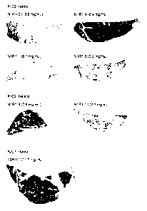
Fig. 3 shows the results of measuring, the ALT levels and so on in uPA
- 7d -
CA 2871586 2018-05-25
CA 02871586 2014-10-24
transgenic mice prepared via ES cell.
Fig. 4 shows the results of measuring human albumin concentrations in mouse
blood (top) and body weights (bottom) of #1C2 mice (up to 14 weeks old) after
transplantation of human hepatocytes into the mice. The solid lines denote
homozygous
mice and dotted lines denote heterozygous mice.
Fig. 5 shows the results of measuring human albumin concentrations in mouse
blood (top) and body weights (bottom) of #2C7 mice (up to 14 weeks old) after
transplantation of human hepatocytes into the mice. The solid lines denote
homozygous
mice and dotted lines denote heterozygous mice.
Fig. 6 shows the immunostaining images of chimeric mouse liver sections
prepared using #1C2 homozygous, #1C2 heterozygous, and #2C7 homozygous mice
immuno stained with a human cytokeratin 8/18 antibody.
Fig. 7 shows the results of measuring the replacement rates in the livers of
chimeric mice prepared using 14-week-old (top) and 30-week-old (bottom) #1C2
homozygous, #1C2 heterozygous, and 2C7 homozygous mice and human albumin
concentrations in the mouse blood.
Fig. 8-1 shows human albumin concentrations in mouse blood (left) before HCV
inoculation and each viral copy number (right) in mouse serum after
inoculation of
chimeric mice prepared using #1C2 homozygous and #1C2 heterozygous mice. Solid
lines denote homozygous mice and dotted lines denote heterozygous mice.
Fig. 8-2 shows human albumin concentrations in mouse blood (left) before HBV
inoculation and each viral copy numbers (right) in mouse serum after
inoculation of
chimeric mice prepared using #1C2 homozygous and #1C2 heterozygous mice. Solid
lines denote homozygous mice and dotted lines denote heterozygous mice.
Fig. 9-1 shows human albumin concentrations in mouse blood (left) before HCV
inoculation and HCV copy numbers (right) in mouse serum after inoculation of
chimeric
mice prepared using #2C7 homozygous mice.
Fig. 9-2 shows human albumin concentrations in mouse blood (left) before HBV
inoculation and HBV copy numbers (right) in mouse serum after inoculation of
chimeric
- 8 -
CA 02871586 2014-10-24
mice prepared using #2C7 horn. ozygius mice.
Fig. 10 shows the results of measuring human albumin concentrations in mouse
blood (top) and body weights (bottom) of #1C2 mice (up to 30 weeks old) after
transplantation of human hepatocytes into the mice. Solid lines denote
homozygous mice
and dotted lines denote heterozygous mice.
Fig. 11 shows the results of measuring human albumin concentrations in mouse
blood (top) and body weights (bottom) of #2C7 mice (up to 30 weeks old) after
transplantation of human hepatocytes into the mice. Solid lines denote
homozygous mice
and dotted lines denote heterozygous mice.
Modes for Carrying Out the Invention
The present invention will be described below in detail.
The mouse with liver damage of the present invention has a DNA fragment
containing a liver-specific promoter/enhancer and cDNA that encodes a
urokinase-type
plasminogen activator operably linked under the control thereof, in a
heterozygous form,
whereby uPA is expressed liver-specifically, and at least 20%, at least 30%,
at least 40%,
at least 50%, at least 60%, at least 70%, at least 80%, at least 90%, at least
95% or more
of original mouse liver cells (particularly, hepatocytes) are damaged, the
proliferation
thereof is suppressed, and/or the cells are necrotized.
The mouse with liver damage of the present invention has a high degree of
damage against original mouse hepatocytes while having the uPA gene in a
heterozygous
form, and thus are not required to have the uPA gene in a homozygous form
unlike
conventionally known uPA transgenic mice.
The mouse with liver damage of the present invention can be prepared on the
basis of a conventionally known method for preparing transgenic animals (Proc.
Natl.
Acad. Sci. U.S.A. 77: 7380-7384 (1980)) by introducing a DNA fragment
containing a
liver-specific promoter/enhancer and cDNA that encodes uPA operably linked
under the
control thereof into mouse ES cells and then using the thus obtained ES cells.
The term "promoter/enhancer" refers to DNA having a sequence capable of
- 9 -
CA 02871586 2014-10-24
,
providing the functions of both the piomoter and the enhancer.
Examples of a "liver-specific promoter" include, but are not particularly
limited
to, as long as it can induce the expression of a gene ligated to the 3' side
in a
liver-specific manner, an albumin promoter, an a-fetoprotein promoter, an
ai-anti-trypsin promoter, a transferrin transthyretin promoter, a serum
amyloid A
promoter, a transthyretin promoter, and a hepatocyte nuclear factor 6 (HNF-6)
promoter.
A preferable example thereof is an albumin promoter.
The "liver-specific promoter/enhancer" may be any one of an endogenous
promoter/enhancer, an exogenous promoter/enhancer, a promoter/enhancer of the
same
species, a promoter/enhancer of a different species, an artificial
promoter/enhancer, as
long as it enables the expression of a target gene liver-specifically.
Preferably, a
mouse-derived promoter/enhancer is used. A mouse-derived liver-specific
promoter/enhancer is known in the art. For example, an albumin
promoter/enhancer
can be used. A mouse-derived albumin promoter/enhancer is known (Herbst RS et
al,
Proc Natl Acad Sci U.S.A. 1989 Mar; 86 (5): 1553-7; Heckel JL et al., Cell
1990 Aug 10;
62(3): 447-56), and can be obtained by performing PCR using primers specific
to the
albumin promoter/enhancer and a mouse genomic library as a template.
uPA-encoding cDNA may be any one of endogenous cDNA, exogenous cDNA,
cDNA of the same species, and cDNA of a different species. Preferably, mouse-
derived
cDNA is used. The uPA-encoding cDNA can be obtained by a general technique
known
by persons skilled in the art, specifically by performing reverse
transcription PCR using
RNA extracted from the liver as a template and primers specific to an uPA-
encoding
gene. The uPA-encoding gene was registered under Accession No. NM008873 in the
above published database. In the present invention, the gene information can
be used (in
this Description, the uPA-encoding gene is represented by SEQ ID NO: 11). In
addition,
in the Description, the term "uPA gene" described in the present invention
refers to
uPA-encoding cDNA. These terms can be used interchangeably.
The term "a liver-specific promoter/enhancer and cDNA that encodes uPA
operably linked under the control thereof" means that uPA-encoding cDNA is
arranged
- 10 -
CA 02871586 2014-10-24
=
so that uPA is expressed under the control of the liver-specific
promoter/enhancer.
The DNA fragment containing a liver-specific promoter/enhancer and cDNA that
encodes uPA operably linked under the control thereof is introduced into ES
cells
(embryonic stem cells).
The DNA fragment can be introduced into ES cells by a calcium phosphate
method, an electrical pulse method, a lipofection method, an aggregation
method, a
microinjection method, a particle gun method, a DEAE-dextran method, or the
like
(examples thereof are not limited thereto).
ES cells prepared by introducing the DNA fragment can be cultured ex vivo, so
that cells into which the DNA fragment has been introduced successfully and/or
cells in
which the introduced DNA fragment has not been lost can be screened for. Next,
the thus
obtained ES cells are injected into a host embryo, and preferably a mouse
blastocyst, the
resultant is transplanted into the uterine horn of a surrogate mother mouse
for generation,
and thus transgenic mice (chimeric mice) are born. As a surrogate mother
mouse, in
general, a female pseudopregnant mouse produced by crossing with a male mouse
subjected to vasectomy is used.
The resulting transgenic mice (chimeric mice) are confirmed for the
incorporation of the above DNA fragment and then crossed with wild-type mice
for the
birth of Fl mice. Among Fl mice that are born as a result of this crossing,
mice having
the above DNA fragment (heterozygote) in somatic cells are transgenic mice
capable of
transmitting the above DNA fragment to germ cells.
The mouse with liver damage of the present invention may be a mouse of any
generation of the above transgenic mice, as long as the introduced DNA
fragment is a
heterozygote. The selection of a heterozygote can be tested by screening
chromosomal
DNA separated and extracted from the Fl mouse tail by Southern hybridization
or a PCR
method, for example.
Moreover, from the thus obtained transgenic mice, 2- to 3-week-old transgenic
mice exhibiting the serum ALT (alanine aminotransferase) level of 30 (Karmen
unit) or
higher are selected. Preferably, 3-week-old or 4 week-old transgenic mice
exhibiting the
- 11 -
CA 02871586 2014-10-24
serum ALT level of 30 (Karmen unif) or higher, further preferably 6 week-old
transgenic
mice exhibiting the serum ALT level of 45, 50 or 55 (Karmen unit) or higher,
particularly
preferably 8-week-old transgenic mice exhibiting the serum ALT level of 60,
65, or 70
(Karmen unit) or higher are selected. A serum ALT level can be an indicator
for the
degree of liver damage. The higher the serum ALT level, the higher the degree
of liver
damage.
The above ES cells and blastocysts to be used for preparing the mouse with
liver
damage of the present invention are not particularly limited and ES cells and
blastocysts
from various mouse lines can be used. For example, cells from 129SvEv mice,
C57BL/6J mice, or the like can be used.
According to the method for preparing the mouse with liver damage of the
present invention, the resulting mouse can have a transgene in a heterozygous
form.
Therefore, the transgenic mice can be efficiently prepared in large numbers,
and desired
transgenic mice can be selected and prepared efficiently from the thus
obtained
transgenic mice by screening for mice having a high degree of liver damage
while
having the uPA gene in a heterozygous form.
Moreover, the mouse with liver damage of the present invention has mouse
productivity higher than that of conventional transgenic mice having the uPA
gene in a
homozygous form, since the mouse with liver damage of the present invention
can have
the uPA gene in a heterozygous form. Specifically, first, many heterozygous
female mice
should be produced in order to obtain many homozygous mice. Thereafter,
homozygous
mice should be obtained by external fertilization or natural mating of the
heterozygous
mice. This process requires two generations and at least 6 months in total.
Moreover, the
thus obtained homozygous mice accounting for only about 25% of the total
number of
the thus obtained mice are obtained. In contrast, many heterozygous mice can
be
obtained in the second generation (via single generation) by performing
external
fertilization or natural mating with wild-type mice that can be purchased in
large
numbers from breeders. The time period required for this process is at least 3
months.
Moreover, about 50% of the total number of mice obtained herein are
heterozygous mice,
- 12 -
CA 02871586 2014-10-24
indicating that a large number of necessary mice can be produced within a
short time
period highly efficiently. Also, when a cross-bred line produced with another
genetically
mutated mouse (e.g., gene deficiency or introduced genes) is used for an
experiment,
mice that can be efficiently used for the experiment can be obtained if
heterozygous uPA
transgenic mice can be used. For example, when mice each having the introduced
uPA
gene in a heterozygous form and another type of gene mutation in a
heterozygous form
are used with each other to produce a mouse having the uPA gene and another
type of
gene mutation in a double homozygous form, the thus obtained mice having both
genes
in a homozygous form account for only 6% of the thus obtained mice.
Furthermore,
female and male homozygous mice should be obtained and then breeding and
production
should be performed in order to obtain a considerable number of mice to be
used for an
experiment. Meanwhile, the thus obtained mice having the uPA gene in a
heterozygous
form and another type of gene mutation in a homozygous form account for about
12.5%.
This indicates that mice required for an experiment can be obtained at this
time point
with production efficiency higher than that of the production of mice having
both genes
in a homozygous form. This means that a considerable number of mice that can
be used
for an experiment can be obtained earlier by a single generation than the
production of
mice having both genes in a homozygous form. As described above, the fact that
heterozygous mice can be used enables to obtain high production efficiency, so
as to
contribute to save the space for an animal room to be used for keeping and
obtaining
mice necessary for the experiment, resulting in a shorter period required for
production,
a drastic reduction in the number of mice to be used, and the reduction of
experimenters'
efforts.
In the present invention, examples of the "mouse with liver damage" include
portions of the mouse. The term "a portion(s) of the mouse" refers to, mouse-
derived
tissues, body fluids, cells, and disrupted products thereof or extracts
therefrom, for
example (the examples thereof are not particularly limited to them). Examples
of such
tissues include, but are not particularly limited to, heart, lungs, kidney,
liver, gallbladder,
pancreas, spleen, intestine, muscle, blood vessel, brain, testis, ovary,
uterus, placenta,
- 13 -
CA 02871586 2014-10-24
marrow, thyroid gland, thyMus gland, and mammary gland. Examples of body
fluids
include, but are not particularly limited to, blood, lymph fluids, and urine.
The term
"cells" refers to cells contained in the above tissues or body fluids, and
examples thereof
include cultured cells, sperm cells, ova, and fertilized eggs obtained by
isolation or
culture thereof. Examples of cultured cells include both primary cultured
cells and cells
of an established cell line. Examples of the portions of the mouse also
include tissues,
body fluids, and cells at the developmental stage (embryonic stage), as well
as the
disrupted products or extracts thereof. In addition, an established cell line
from the
mouse with liver damage of the present invention can be established using a
known
method (Primary Culture Methods for Embryonic Cells (Shin Seikagaku fikken
Kozo
(New Biochemical Experimental Lecture Series), Vol. 18, pages 125-129, TOKYO
KAGAKU DOZIN CO., LTD., and Manuals for Mouse Embryo Manipulation, pages
262-264, Kindai Shuppan)).
The present invention further provides an immunodeficient mouse with liver
damage. The immunodeficient mouse with liver damage of the present invention
can be
used as a host mouse for transplantation of human hepatocytes. The
immunodeficient
mouse with liver damage of the present invention can be obtained by crossing
the above
mouse with liver damage with an immunodeficient mouse.
Examples of the "immunodeficient mouse" may be any mouse that does not
exhibit rejection against hepatocytes (in particular, human hepatocytes) from
a different
animal origin, and include, but are not limited to, SCID (severe combined
immunodeficiency) mice exhibiting deficiency in T- and B-cell lines, mice
(NUDE mice)
that have lost T cell functions because of genetic deletion of the thymus
gland, and mice
(RAG2 knockout mice) produced by knocking out the RAG2 gene by a known gene
targeting method (Science, 244: 1288-1292, 1989). A preferable example thereof
is a
SCID mouse.
The immunodeficient mouse with liver damage of the present invention has a
gene that specifies the phenotype of immunodeficiency in a homozygous form.
The
immunodeficient mouse with liver damage of the present invention may also have
a
- 14 -
CA 02871586 2014-10-24
DNA fragment containing the uPA 'gene from the above mouse with liver damage
in
either a heterozygous form or a homozygous form. Even when the immunodeficient
mouse with liver damage of the present invention has the uPA gene in a
heterozygous
form, human hepatocytes transplanted into the mouse can engraft for long
periods of
time. Examples of the genotype of the immunodeficient mouse with liver damage
of the
present invention include, but are not limited to, uPA (+/-)/SCID (+/+) and
uPA
(+/+)/SCID (+/+).
Heterozygous mice or homozygous mice can be selected by screening, as
described above, chromosomal DNAs separated and extracted from the tails of
the thus
obtained offspring by Southern hybridization or a PCR method.
In the present invention, examples of the "immunodeficient mouse with liver
damage" include portions of the mouse. The term "a portion of the mouse" is as
defined
above.
Moreover, the present invention provides a chimeric mouse having human
hepatocytes. The chimeric mouse of the present invention is immunologically
deficient,
which is prepared by introducing, in a heterozygous form, a DNA fragment
containing
cDNA that encodes an urokinase-type plasminogen activator operably linked
under the
control of the liver-specific promoter and enhancer region, and has a chimeric
liver
containing human hepatocytes.
The chimeric mouse of the present invention can be prepared by transplanting
human hepatocytes into the above immunodeficient mouse with liver damage of
the
present invention.
As human hepatocytes to be used for transplantation, human hepatocytes
isolated from normal human liver tissue by a conventional method such as a
collagenase
perfusion method can be used. The thus separated hepatocytes can also be used
by
thawing after cryopreservation. Alternatively, the chimeric mouse hepatocytes,
which are
defined as the human hepatocytes separated by a technique such as a
collagenase
perfusion method from a chimeric mouse liver, in which mouse hepatocytes have
been
replaced by human hepatocytes, can be used in a fresh state, and the
cryopreserved
- 15 -
CA 02871586 2014-10-24
chimeric mouse hepatocytes are also available after thawing.
Such human hepatocytes can be transplanted into the liver via the spleen of
the
above immunodeficient mouse with liver damage. Such human hepatocytes can also
be
directly transplanted via the portal vein. The number of human hepatocytes to
be
transplanted may range from about 1 to 2,000,000 cells and preferably range
from about
200,000 to 1,000,000 cells. The gender of the immunodeficient mouse with liver
damage
is not particularly limited. Also, the age on days of the immunodeficient
mouse with
liver damage upon transplantation is not particularly limited. When human
hepatocytes
are transplanted into a young mouse (early weeks of age), human hepatocytes
can more
actively proliferate as the mouse grows. Hence, about 0- to 40-day-old mice
after birth,
and particularly about 8- to 40-day-old mice after birth are preferably used.
Mice after transplantation can be maintained by a conventional method. After
transplantation, blood is collected periodically from the mouse tail, and then
the human
albumin concentration in mouse blood is measured. Since human albumin
concentration
correlates with the replacement rate of human hepatocytes in the mouse liver,
the degrees
of the engraftment and the proliferation of human hepatocytes can be inferred
with the
value of human albumin concentration. A mouse inferred to have a replacement
rate of
70% or more on the basis of the blood human albumin concentration can be used
as a
chimeric mouse with a high degree of replacement for pharmacokinetic studies,
infection
studies with hepatitis virus, or the like. In the case of mice, when about 1
to 10 x 105
human hepatocytes are transplanted, the mouse is maintained for about 40 to
100 days,
and thus a blood human albumin concentration ranging from 100,000 to
30,000,000
ng/mL can be obtained.
The thus transplanted human hepatocytes account for at least 10%, 20% or more,
30% or more, 40% or more, 50% or more, 60% or more, 70% or more, 80% or more,
90% or more, 95% or more, or even a higher percentage of hepatocytes in the
liver of the
chimeric mouse.
Transplanted human hepatocytes retain the functions and the properties of
normal human hepatocytes in the liver of the chimeric mouse for at least 2
weeks, 3 or
- 16-
CA 02871586 2014-10-24
more weeks, 4 or more weeks, 5 or more weeks, 10 weeks, 20 weeks, 30 weeks,
and 40
weeks, and most preferably for a period during which the mouse survives.
Examples of "the functions and the properties of human hepatocytes" include,
but are not limited to, drug-metabolizing functions, protein synthesis,
gluconeogenesis,
urea synthesis, bile synthesis, lipid synthesis, glucose metabolism,
detoxication, and
infectiveness against hepatitis virus.
Transplanted human hepatocytes retain 50% or more, 60% or more, 70% or more,
80% or more, 90% or more, 95% or more, or even a higher percentage of the
functions
and the properties in the normal human liver.
The present invention further provides a method for screening for a substance
that affects human liver functions, with the use of the above chimeric mouse.
An example of the method is an evaluation method comprising the following
steps of:
(a) administering a test substance to the above chimeric mouse;
(b) measuring one or more values selected from the group consisting of the
human
albumin concentration, the body weight curve, the liver-weight-to-body-weight
ratio, the
total albumin level, the total protein level, the ALT level, the AST level,
and the total
bilirubin level in the chimeric mouse to which the test substance is
administered in (a);
and
(c) selecting a test substance that causes an increase or an decrease in any
one or more of
the human albumin concentration, the body weight curve, the
liver-weight-to-body-weight ratio, the total albumin level, the total protein
level, the
ALT level, the AST level, and the total bilirubin level measured in (b),
compared with
the human albumin concentration, the body weight curve, the
liver-weight-to-body-weight ratio, the total albumin level, the total protein
level, the
ALT level, the AST level, and the total bilirubin level of the chimeric mouse
to which no
test substance is administered.
Examples of the "test substance" in the method of the present invention are
not
particularly limited and include natural compounds, organic compounds,
inorganic
- 17-
CA 02871586 2014-10-24
compounds, proteins, antibodies, peptides, and single compounds such as an
amino acid,
and nucleic acids, as well as compound libraries, expression products from
gene libraries,
cell extracts, cell culture supernatants, products of fermenting
microorganisms, extracts
from marine creatures, plant extracts, extracts from prokaryotic cells,
extracts from
eukaryotic single cells, and extracts from animal cells. These products may be
purified
products or crude products such as plant, animal, or microbial extracts. Also,
a method
for producing a test substance is not particularly limited. A test substance
to be used
herein may be a substance isolated from a natural product, synthesized
chemically or
biochemically, or prepared by genetic engineering techniques.
The above test substance can be adequately labeled and then used as necessary.
Examples of labels include radiolabels and fluorescent labels. Examples of the
test
substance include, in addition to the above test samples, mixtures of a
plurality of types
of these test samples.
In this method, examples of a method for administering a test substance to
mice
are not particularly limited. Such an administration method can be adequately
selected
from among oral administration or parenteral administration such as
subcutaneous,
intravenous, local, transdermal, and enteral (intrarectal) administration,
depending on the
type of a test substance to be administered.
The rate of replacement by human hepatocytes in the mouse liver can be
predicted by measuring the human albumin concentration in mouse blood by
ELISA,
immunonephelometry, or the like. For prediction, a correlation curve of human
albumin
concentrations and replacement rates should be prepared in advance as
described below.
Blood is collected before the autopsy of a chimeric mouse, and then the human
albumin
concentration is determined. Frozen sections or paraffin sections are prepared
from the
entire liver or partial hepatic loves collected upon autopsy. Immunostaining
is performed
using an antibody specific to human hepatocytes, such as a human specific
cytokeratin
8/18 (hCK8/18) antibody. Photographs of the sections are taken under a
microscope, the
proportion of the hCK8/18-positive area per liver section is calculated to
give a
replacement rate. Human albumin concentrations and replacement rates are
plotted on a
- 18-
CA 02871586 2014-10-24
graph, thereby finding a correlation equation. The human albumin concentration
in
mouse blood is entered to the correlation equation, so that a replacement rate
can be
roughly calculated. Furthermore, the body weight is measured over time, the
health
status of the mouse can be predicted. A biochemical test is performed for
blood collected
upon autopsy. For example, a total albumin level, a total protein level, and
the like are
measured, and thus the health status of the mouse can be clarified. The degree
of liver
damage of a chimeric mouse can be clarified by measuring the liver weight, the
body
weight, ALT, AST, and the total bilirubin levels, for example. Specifically,
the effects of
a test substance against human hepatocytes can be determined using increases
or
decreases in these numerical figures as indicators.
The present invention further provides a method for evaluating hepatotoxicity
of
a test substance against human hepatocytes, with the use of the above chimeric
mouse.
An example of this method is an evaluation method comprising the following
steps of:
(a) administering a test substance to the above chimeric mouse;
(b) measuring one or more values selected from the group consisting of the
human
albumin concentration, the body weight curve, the liver-weight-to-body-weight
ratio, the
total albumin level, the total protein level, the ALT level, the AST level,
and the total
bilirubin level in the chimeric mouse to which the test substance is
administered in (a);
and
(c) evaluating the effect of the test substance on human hepatocytes using, as
an
indicator, an increase or a decrease in any one or more of the human albumin
concentration, the body weight curve, the liver-weight-to-body-weight ratio,
the total
albumin level, the total protein level, the ALT level, the AST level, and the
total bilirubin
level measured in (b), compared with the human albumin concentration, the body
weight
curve, the liver-weight-to-body-weight ratio, the total albumin level, the
total protein
level, the ALT level, the AST level, and the total bilirubin level of the
chimeric mouse to
which no test substance is administered.
Examples of the "test substance" and the "administration method" thereof
- 19 -
CA 02871586 2014-10-24
,
. .
include those defined above.
As described above, the degree of liver damage of a chimeric mouse can be
analyzed on the basis of human albumin concentration, body weight curve,
liver-weight-to-body-weight ratio, total albumin level, total protein level,
ALT level,
AST level, and total bilirubin level. With the use of increases or decreases
in these
numerical figures as indicators, the toxicity of the test substance against
human
hepatocytes can be determined and evaluated.
The present invention further provides a method for screening for a substance
effective for treatment of viral hepatitis, with the use of the above chimeric
mouse.
An example of this method is an evaluation method comprising the following
steps of:
(a) inoculating a hepatitis virus into the above chimeric mouse;
(b) administering a test substance to the chimeric mouse inoculated with the
hepatitis
virus in (a);
(c) measuring one or more values selected from the group consisting of the
human
albumin concentration, the body weight curve, the liver-weight-to-body-weight
ratio, the
total albumin level, the total protein level, the ALT level, the AST level,
the total
bilirubin level, the viral load, and the amount of a virus-derived protein of
the chimeric
mouse to which the test substance is administered in (b); and
(d) selecting a test substance causing a change in any one or more of the
human albumin
concentration, the body weight curve, the liver-weight-to-body-weight ratio,
the total
albumin level, the total protein level, the ALT level, the AST level, the
total bilirubin
level, the viral load, and the amount of a virus-derived protein measured in
(c),
compared with the human albumin concentration, the body weight curve, the
liver-weight-to-body-weight ratio, the total albumin level, the total protein
level, the
ALT level, the AST level, the total bilirubin level, the viral load, and the
amount of a
virus-derived protein in the chimeric mouse to which no test substance is
administered.
Examples of hepatitis viruses to be used for inoculation include hepatitis
type A
virus, hepatitis type B virus, hepatitis type C virus, hepatitis type D virus,
and hepatitis
- 20 -
CA 02871586 2016-05-02
72813-390
type E virus. Viruses can be inoculated via intravascular or intraperitoneal
administration.
The above chimeric mouse to be used in this method is preferably a mouse that
satisfies at least one of the following conditions: 3 or more weeks have
passed after the
transplantation of human hepatocytes; the blood human albumin concentration is
1
mg/mL or higher; and human hepatocytes' account for 10% or more of all
hepatocytes.
Examples of the "test substance" and the "administration method" thereof
include those defined above.
The degree of liver damage due to hepatitis viruses can be found on the basis
of
human albumin concentration, body weight curve, liver-weight-to-body-weight
ratio,
total albumin level, total protein level, ALT level, AST level, total
bilirubin level, viral
load, and the amount of a virus-derived protein_ With the use of changes in
these
numerical figures as indicators, the effectiveness of a test substance in
treatment of viral
hepatitis can be determined and evaluated.
Hereafter, the present invention is described in greater detail with reference
to
the following examples.
Example 1. Preparation of uPA transgenic mice using DNA microinjection method
(1) Preparation of vector containing an uPA gene and an albumin promoter
Regarding a uPA gene, total RNA was extracted from mouse liver by an AGPC
method (acid-guanidinium-isothiocyanate-phenol-chloroform), and then dissolved
in
RNase-free water. A reverse transcription reaction was performed using the
above-obtained total RNA, a uPA gene-specific primer (the antisense sequence
having a
length from the 1341st to the 1360th nucleotides) prepared based on the
sequence of the
uPA gene (Accession No.: NM008873 (SEQ ID NO: 11)) registered in the published
database, and Long Range Reverse Transcriptase (Qiagen) at 25 C for 10
minutes, and
then performed at 42 C for 90 minutes. After 5 minutes of reverse
transcriptase
-21-
CA 02871586 2014-10-24
inactivation treatment at 85 C, RNaseH (Invitrogen) was added, the resultant
was treated
at 37 C for 20 minutes to digest mRNA, and thus only cDNA remained. PCR was
performed using the thus synthesized cDNA as a template. The above reaction
solution
in an amount 1/10 the total amount thereof was added. As an enzyme, Phusion
DNA
polymerase (Fynnzymes) was used. A PCR primer (the sense sequence having a
length
from the 39th nucleotide to the 61st nucleotide) was prepared based on the
sequence of
the uPA gene (Accession No.: NM008873). The fragment amplified by PCR has a
length
of nucleotide Nos. 39-1360. The thus obtained DNA fragment was introduced into
an
expression plasmid having a mouse albumin promoter/enhancer described later,
thereby
constructing "mAlb uPAInt2." The configuration of the "mAlb uPAInt2" gene is
shown
in Fig. 1. The 2nd exon, intron, and the 3rd exon of rabbit p globin, the ORF
portion of
mouse uPA, and polyA signal in the 3rd exon of rabbit p globin were ligated
downstream
of the mouse albumin enhancer/promoter.
(2) Microinjection of DNA into the pronuclei of fertilized eggs
The concentration of the DNA fragment was adjusted to 3 ng/p,L, and then the
DNA fragment was injected into pronuclear stage fertilized eggs collected from
CB-17/Icr and Scid-beige cross-bred mice. DNA was injected into such a
fertilized egg
by microinjection. 635 out of 748 fertilized eggs, into which DNA had been
injected,
survived, and 469 eggs thereof differentiated into the 2-cell stage embryos.
The 2-cell
stage embryos were transplanted into the uterine tubes of recipient ICR mice
treated in
advance to be in pseudo-pregnancy. 108 offspring were obtained. Whether or not
the thus
obtained offspring contained the uPA gene was analyzed by PCR (The Tokyo
Metropolitan Institute of Medical Science). As a result of PCR, it was
confirmed that one mouse contained the target DNA. A uPA transgenic mouse line
was
established from the one mouse.
(3) Measurement and analysis of serum ALT levels in uPA transgenic mice
Blood was collected from the thus obtained mice having the uPA gene, so as to
obtain the serum. Subsequently, the effect due to the expression of the uPA
gene in the
liver, and specifically, the damage of hepatocytes were analyzed by measuring
the ALT
- 22 -
CA 02871586 2014-10-24
levels. The ALT levels were measured using "Transaminase CII-Test Wako" (Wako
Pure
Chemical Industries, Ltd., cat# 431-30901). After serum collection, serum
samples were
preserved at -80 C until measured.
The method for measuring ALT was performed on 1/20 the scale of the standard
procedure 1 included with "Transaminase CII-Test Wako". First, a substrate
enzyme
solution for ALT: 10 mL of a substrate buffer for ALT was added to 1 vial of
an enzyme
agent for ALT and the enzyme agent was dissolved in the buffer. Furthermore, a
chromogenic solution: 40 mL of a color former solution was added to 1 vial of
color
former and the color former was dissolved in the solution.
Next, a CORNING 25850 96-well U-bottomed plate was prepared on ice. A
serum sample (1 4) was added to the plate. The plate was removed from ice, 25
1AL of
the substrate enzyme solution was added, and then heated at 37 C for 5
minutes. STND
(x 1/2 dilution: 1 pL, x 1: 1, 2 L) was added to empty wells, and then 25 !IL
of the
chromogenic solution was added to each well. The substrate enzyme solution (25
L)
was added to wells containing STND, and then heated at 37 C for 20 minutes. A
stop
solution (100 I) was added to each well. The solution was stirred well with a
plate
mixer, and then absorbance was measured at 570 nm within 60 minutes after
stirring. A
calibration curve was prepared using the measurement value of STND, thereby
calculating the values representing the activity in samples.
No mouse with a high ALT level was confirmed from among mice subjected to
measurement. The uPA transgenic mouse of interest must exhibit a condition
wherein the
expression level of the uPA gene is optimum for the subsequent hepatocyte
transplantation. Mice exhibiting such optimum expression level should be
selected
after preparation of many uPA transgenic mice. It was revealed that this
method is not a
suitable for a method for probabilistically preparing such optimum mice in
this case,
because of the limited efficiency of the preparation of transgenic mice.
Example 2. Preparation of uPA transgenic mice via ES cells
(1) Establishment of ES cells prepared by inserting the uPA gene
In this example, a uPA gene insertion vector "mAlb uPAInt2ES" (Fig. 2) was
- 23 -
CA 02871586 2015-01-15
72813-390
constructed and used. This vector was constructed by introducing a neomycin
resistance gene
that is expressed under the control of an SV40 promoter to the "mAlb uPAInt2"
plasmid in
order to impart drug selectivity in ES cells. The vector was introduced by
electroporation into
ES cells obtained from a 129SvEv mouse, followed by selective culture using
G418. The thus
obtained G418-resistant colonies were subjected to the testing by PCR for ES
cells into which
the gene had been introduced. This is as described specifically below.
A uPA gene insertion vector DNA (25-30 g) was linearized by cleavage with a
restriction enzyme, and then purified. The DNA was suspended in an
electroporation buffer
(20 mM HEPES pH7.0, 137 mM NaCl, 5 mM KCI, 6 mM D-glucose, 0.7 mM Na2HPO4)
containing 3x10⁶ mouse ES cells. Gene transfer was performed under the
conditions of Field
Strength 185V/cm and Capacitance 500 F. Selective culture was performed with
G418
(Geniticin) (SIGMA, G-9516) with a final concentration of 200 g/mL, 24 hours
after gene
transfer. ES cells were cultured using a Dulbecco's modified Eagle's medium
(DMEM)
(Gibco/BRL, 11965-084) culture solution supplemented with fetal bovine serum
having a
final concentration of 15% (Hyclone, SH30071), L-glutamine having a final
concentration
of 2 mM (Gibco/BRL, 25030-081), non-essential amino acids each having a final
concentration of 100 M (Gibco/BRL, 11140-050), HEPES having a final
concentration of
10 mM (Gibco/BRL, 15630-080), penicillin/streptomycin each having a final
concentration
of 100 U/mL (Gibco/BRL, 15140-122), P-mercaptoethanol having a final
concentration
of 10011M (SIGMA, M-7522), and ESGRO (LIF) having a final concentration of
1000 U/mL
(Gibco/BRL, 13275-029) (hereinafter referred to as "ES medium").
Moreover, as feeder cells for ES cells, MEF (Mouse Embryonic Fibroblast)
cells isolated from E14.5 embryos were used. A culture solution used herein
was a DMEM
(Gibco/BRL, 11965-084) culture solution supplemented with fetal bovine serum
having a
final concentration of 10% (Hyclone, SH30071), L-glutamine having a final
concentration of
2 mM (Gibco/BRL, 25030-081), non-essential amino acids each having a final
concentration
of 100 M (Gibco/BRL, 11140-050), and penicillin/streptomycin
- 24 -
CA 02871586 2014-10-24
each having a final concentration of 100 U/mL (Gibco/BRL, 15140-122)
(hereinafter,
referred to as "MEF medium"). MEF cells cultured to confluency in a 150-cm2
flask
were removed with trypsin/EDTA (0.05%/1 mM, Gibco/BRL, 25300-047) and then
plated again on four 10 cm dishes, two 24-well plates, two 6-well plates, six
25cm2-flasks, and two 75 cm2 flasks at optimal concentrations.
(2) Adjustment of ES cells for genotype analysis
On day 5 after gene transfer, G418-resistant colonies that had appeared were
passaged to a 24-well plate, as described below. Specifically, G418-resistant
colonies
were transferred to a 96-well microplate containing 150 L of a trypsin/EDTA
solution
using Pipetman (Gilson). After 20 minutes of treatment within an incubator at
37 C,
pipetting was performed with Pipetman to obtain single cells. The cell
suspension was
transferred to a 24-well plate and then culture was continued. Two days later,
cells on the
24-well plate were divided into two groups, cells for cryopreservation and
cells for DNA
extraction. Specifically, 500 fit of trypsin/EDTA was added to cells, cells
were treated
for 20 minutes within an incubator at 37 C, and then 500 pit of ES medium was
added.
Gentle pipetting was performed with Pipetman, so as to obtain single cells.
Subsequently,
a half of the cell suspension was transferred to a 24-well plate containing 1
mL of ES
medium. One mL of ES medium was also added to the original 24-well plate. Two
days
later, the medium in one of the 24-well plates was removed. 1 mL of medium for
freezing prepared by adding fetal bovine serum having a final concentration of
10% and
dimethyl sulfoxide (DMSO) having a final concentration of 10% (Sigma, D-5879)
to an
ES medium was added. The resultant was sealed and then cryopreserved at -70 C.
ES cells into which the gene had been introduced were tested by PCR as
described below. Specifically, medium was removed from each well of the 24-
well plates
in which cells had grown to confluency. After washing with PBS, 2501_tt of a
dissolution
buffer (1% SDS, 20 mM EDTA, 20 mM Tris pH7.5) and 5 111, of proteinase K (20
mg/mL) were added and then the resultant was shaken well, followed by heating
at 52 C
for dissolution. DNA was extracted from a dissolved sample by
phenol/chloroform
extraction, and then the resultant was used as template DNA for PCR.
- 25 -
CA 02871586 2014-10-24
(3) Method for analyzing the genotype of ES cells: ES cells into which the uPA
gene had
been introduced were selected by the following procedure.
PCR primers used herein were set in rabbit 1-2, globin. The sequences are: a
sense
primer: GGGCGGCGGTACCGATCCTGAGAACTTCAGGGTGAG (SEQ ID NO: 1)
and an antisense primer: GGGCGGCGGTACCAATTCTTTGCCAAAATGATGAGA
(SEQ ID NO: 2). Reaction was performed according to the method included with
AmpiTaqGold (ABI). After 9 minutes of activation of the enzyme at 95 C, the
cycle of
PCR [94 C for 30 seconds (denaturation), 63 C for 30 seconds (annealing), and
72 C for
1 minute (extension)] was repeated 40 times. After the completion of the
reaction, the
reaction solution was subjected to 2% agarose gel electrophoresis, so as to
confirm PCR
products.
Clones for which gene transfer had been confirmed by PCR analysis were
thawed by heating the previously cryopreserved 96-well plate to 37 C, and then
passaged to a 24-well plate. Clones in the 24-well plate were cultured for 24
hours at
37 C, medium exchange was performed to remove DMSO and liquid paraffin. When
each clone reached 75% to 90% confluency, respectively, clones were passaged
from the
24-well plate to a 6-well plate. Moreover, when clones that had grown to 75%
to 90%
confluency were obtained in 2 wells of the 6-well plate, clones in one well
were
cryopreserved and clones in the other well were used for injection into
blastocysts and
DNA extraction.
Cryopreservation was performed as follows. Specifically, cells were rinsed
twice
with PBS, 0.5 mL of Trypsin was added, and then the temperature was kept at 37
C for
15 to 20 minutes to perform trypsin treatment. Furthermore, 0.5 mL of ES cell
medium
was added, pipetting was performed 35 to 40 times, and thus the mass of ES
cells was
completely dissociated. The cell suspension was transferred to a 15 mL
centrifugal tube.
Wells were further washed with 1 mL of ES cell medium, and then the resultants
were
collected in a tube. The tube was centrifuged at 1,000 rpm for 7 minutes. The
medium
was removed and then suspended again in 0.25 mL of ES cell medium. 0.25 mL of
2x
frozen medium was added. The contents of the wells were transferred to a
cryogenic vial,
- 26 -
CA 02871586 2014-10-24
frozen at -80 C, and then preserved in liquid nitrogen.
Regarding cells for injection into blastocysts and DNA extraction, the mass of
ES cells was completely dissociated, one-quarter thereof was used for
injection into
blastocysts, one-third of the remaining cells and two-third of the same were
each
passaged into a 60 mm dish coated with gelatin. When the former cells grew to
confluency, genomic DNA for PCR analysis was extracted. When the latter cells
grew to
continency, the cells were divided into three groups and then frozen.
(4) Preparation of chimeric mice using ES cells having the uPA gene
Regarding ES cell clones for which gene transfer had been confirmed, chimeric
embryos were prepared using the blastocysts of C57BL/6J mice as host embryos.
The
chimeric embryos were each transplanted into the uterine horn of a
pseudopregnant
mouse to obtain offspring. Host embryos were collected by perfusion of the
uterine tube
and the uterus with Whitten's medium supplemented with 100 M trypsin/EDTA on
day
3 of pregnancy. 8-cell-stage embryos or morulae were cultured for 24 hours in
Whitten's
medium. The thus obtained blastocysts were used for injection. ES cells used
for
injection were dispersed by TE treatment on day 2 or 3 of passage, and then
left to stand
at 4 C until the mieromanipulation of these cells. As a pipette for injection
of ES cells,
glass capillary tubing (Sutter, inner diameter of about 20 gm) was used. A
pipette for
holding embryos used herein was processed as follows. A glass microtubule with
an
outer diameter of 1 mm (NARISHIGE) was pulled thin using a micropipette puller
(Sutter, P-97/IVF), and then its tip with an outer diameter ranging from 50 pm
to 100 p.m
was cut using a microforge (De Fonburun), and then processed to have an
aperture of 10
p.m to 20 m. The pipette for injection and the pipette for holding were
connected to a
micromanipulator (Lica) with a piezo system (Primetech PAMS-CT150) connected
thereto. As a chamber used for micromanipulation, perforated slide glass to
which cover
glass had been adhered with bees wax was used. Two drops of Hepes-buffered
Whitten's
medium supplemented with about 10 L of 0.3% BSA were placed thereon, and then
the
top face was covered with mineral oil (Sigma). One drop contained about 100 ES
cells,
and the other drop contained about 20 expanded blastocysts. About 15 ES cells
were
- 27 -
CA 02871586 2014-10-24
injected per embryo. Micromanipulation was always performed under an inverted
microscope. Manipulated embryos were transplanted into the uterine horns of
recipient
ICR female mice on day 2 of pseudo pregnancy. Recipient female mice that had
not
delivered offspring even on the predicted delivery date were subjected to
cesarean
section. The resulting offspring were raised by surrogate parents. As a result
of injection
of 45 clones of ES cells into the blastocysts of C57BL/6J mice, male chimeric
mice were
obtained from 39 clones.
(5) Test of the transmission of the uPA gene to the germ line
Chimeric mice were crossed with C57BL/6J mice, and then whether or not
ES-cell-derived offspring were obtained was tested. If the germ cells of the
chimeric
mice were derived from ES cells, the thus delivered offspring would have wild-
type hair
color. If the germ cells of the chimeric mice were derived from the
blastocysts of
C57BL/6J mice, the thus delivered offspring would have black hair color. As a
result of
crossing, offspring having wild-type hair color were delivered in 25 lines,
and thus the
transmission of ES cells to the germ line was confirmed.
Next, DNA was extracted from the tail portions of these mice having wild-type
hair color and then subjected to PCR to examine if the uPA gene had been
transmitted.
As a result, the transmission of the uPA gene was confirmed for ES-cell-
derived
offspring of 14 lines.
(6) Measurement and analysis of serum ALT levels in uPA transgenic mice
Blood was collected from the thus obtained mice having the uPA gene, so as to
obtain the serum. Subsequently, the effect due to the expression of the uPA
gene in the
liver, and specifically, the damage of hepatocytes were analyzed by measuring
the ALT
levels. The ALT levels were measured using "Transaminase CII-Test Wako" (Wako
Pure
Chemical Industries, Ltd., cat# 431-30901). After serum collection, serum
samples were
preserved at -80 C until measured.
The method for measuring ALT was performed on 1/20 the scale of the standard
procedure 1 included with "Transaminase CII-Test Wako". First, a substrate
enzyme
solution for ALT: 10 mL of a substrate buffer for ALT was added to 1 vial of
an enzyme
- 28 -
CA 02871586 2014-10-24
agent for ALT and the enzyme agent was dissolved in the buffer. Furthermore, a
chromogenic solution: 40 mL of a color former solution was added to 1 vial of
color
former and the color former was dissolved in the solution.
Next, a CORNING 25850 96-well U-bottomed plate was prepared on ice. A
serum sample (1 p,L) was added to the plate. The plate was removed from ice,
25 tiL of
the substrate enzyme solution was added, and then heated at 37 C for 5
minutes. STND
(x 1/2 dilution: 1 1.xL, x 1: 1, 2 L) was added to empty wells, and then 25
.t,L of the
chromogenic solution was added to each well. The substrate enzyme solution (25
L)
was added to wells containing STND, and then heated at 37 C for 20 minutes. A
stop
solution (100 til) was added to each well. The solution was stirred well with
a plate
mixer, and then absorbance was measured at 570 nm within 60 minutes after
stirring. A
calibration curve was prepared using the measurement value of STND, thereby
calculating the values representing the activity in samples.
Of 14 mouse lines measured, 3 lines of heterozygous mice were confirmed to
have high ALT levels (Fig. 3).
The following experiment of human hepatocyte transplantation was performed
using 2 lines (#1C2 and #2C7) of mice with high ALT levels from among the thus
obtained 3 lines.
Example 3. Preparation of chimeric mice
(1) Immunodeficient mice with liver damage
uPA-Tg mice (hemizygote, +/-) prepared in Example 2 above were back-crossed
twice with SCID-bg mice, thereby obtaining mice having the genotype of
uPA-Tg(+/-)SCID(+/+). Sperm cells were collected from the male mice, external
fertilization was performed with unfertilized eggs of SCID mice (homozygote,
+/+), and
then the fertilized eggs were returned into surrogate mother mice. Among
offspring
delivered, mice having a Tg gene therein were selected and then subjected to
natural
mating, so that mice having both genotypes (uPA-Tg(+/-)/SCID (+/+)) were
obtained.
uPA-Tg (+/-) and uPA-Tg(-/-) were distinguished from each other by a genome
PCR
method using sequences specific to the transgene as primers.
- 29 -
CA 02871586 2014-10-24
6
Forward primer
5'-GGGCGGCGGTACCGATCCTGAGAACTTCAGGGTGAG-3' (SEQ ID NO: 3)
Reverse primer
5'-GGGCGGCGGTACCAATTCTTTGCCAAAATGATGAGA-3' (SEQ ID NO: 4)
In addition, SCID (+/+), SCID (+/-), and SCID(-/-) were distinguished from
each other by a PCR-RFLP method.
Next, the thus obtained uPA-Tg(+/-)/SCID(+/+) mice were crossed each other,
thereby obtaining uPA-Tg(+/+)/SCID(+/+) and uPA-Tg(+/-)/SCID(+/+). uPA-Tg(+/+)
and uPA-Tg(+/-) were distinguished from each other by a Southern blotting
method.
About 5-mm tail portions were cut from 8- to 10-day-old mice, and then
solubilized with
a 3 SDS, proteinase K solution. Protein components mixed therein were removed
by
phenol and chloroform extraction. RNA mixed therein was denatured using DNAse-
free
RNase A, and then macromolecular genomic DNA was precipitated by isopropanol
precipitation. The above genomic DNA was washed with 70% ethanol, air dried,
and
then dissolved again in TE. Genomic DNA extracted from a specimen, positive
control
genomic DNA, and negative control genomic DNA (5 i.tg each) were completely
digested
with EcoRl. The thus generated DNA fragments were separated by agarose
electrophoresis, and then transferred to a nylon membrane. A DNA fragment
appropriate
as a probe for Southern hybridization was purified (379 bp) from uPA cDNA
probe/TA
using restriction enzyme EcoRl. The above DNA fragment was labeled with [32P]
by a
random prime method. The DNA fragment transferred to the nylon membrane was
hybridized with the RI-laded uPA cDNA probe. Non-specifically bound probes
were
removed by washing. Radioactive signals from the foreign gene introduced in
mAlb-uPA-Int2 Tg mouse candidates were exposed to an X-ray film and thus
detected.
Wild-type-locus-derived 1.5-kb specific signals and mutant-locus-derived 0.4
kb (wt: 1.5
kb) specific signals were detected, thereby determining the genotype of
individual
mAlb-uPA-Int2 Tg mice.
(2) Transplantation of human hepatocytes
As human hepatocytes, hepatocytes (Lot No.BD85, boy, 5 years old) purchased
-30-
CA 02871586 2014-10-24
from BD Gentest were used. The cryopreserved hepatocytes were thawed and used
according to a conventionally known method (Chise Tateno et al, Near-
completely
humanized liver in mice shows human-type metabolic responses to drugs. Am J
Pathol
165: 901-912, 2004).
2- to 4-week-old 7 #1C2 homozygous, 4 #1C2 heterozygous, 4 #2C7
homozygous, and 7 #2C7 heterozygous uPA-Tg/SCID mice were ether-anesthetized.
An
about 5-mm incision was made in a flank, and then 2.5x10⁵ human hepatocytes
were
injected via the inferior splenic pole. The spleen was returned to the
peritoneal cavity
and then the site was sutured. One #1C2 homozygous mouse died on day 30 of
transplantation.
2 lit of blood was collected from mouse tail vein on weeks 3 and 6 after
transplantation and then every week, and then added to 200 pt of LX-Buffer.
Human
albumin concentrations in mouse blood were measured by immunonephelometry
using
an autoanalyzer JEOL BM6050 (JEOL Ltd.). As a result, increases in human
albumin
concentration were observed for #1C2 homozygous, #1C2 heterozygous, and #2C7
homozygous mice. Specifically, mice with human albumin concentrations higher
than 7
mg/mL were observed (Fig. 4 and Fig. 5). No increase in human albumin
concentration
was observed for #2C7 heterozygous mice (Fig. 5). A smooth gain in body weight
was
observed for all mice. The body weights of most #2C7 heterozygous mice were at
high
levels (Fig. 5).
Chimeric mice (13- to 15-week-old) were anatomized 10 to 12 weeks after
transplantation, and then liver and blood were collected. The frozen sections
of 7 liver
lobes were prepared, and then immunostaining was performed using a human-
specific
cytokeratin 8/18 (hCK8/18) antibody (Fig. 6). The hCK8/18 positive area per
area of a
frozen section was determined, thereby obtaining a replacement rate. As a
result, #1C2
homozygous mice and #2C7 homozygous mice having a replacement rate of 70% or
more were confirmed (Fig. 7). #1C2 heterozygous mice having a replacement rate
of
60% or more were observed (Fig. 7). A correlation between human albumin
concentrations in mouse blood and replacement rates was confirmed in a manner
similar
- 31 -
CA 02871586 2014-10-24
to those in conventionally known chimeric mice (Chise Tateno et al, described
above)
(Fig. 7). In terms of correlation, no clear difference was confirmed between
#1C2 and
#2C7, and, between heterozygous mice and homozygous mice (Fig. 7).
Human hepatocytes were transplanted into 2- to 4-week-old 28 #1C2
homozygous, 28 #1C2 heterozygous, 18 #2C7 homozygous, and 15 #2C7 heterozygous
uPA-Tg/SCID mice. As a result, chimeric mice having high replacement rates,
wherein
the human albumin concentration in mouse blood was 7 mg/mL or higher, were
#1C2
homozygous mice (61%), #1C2 heterozygous mice (36%), #2C7 homozygous mice
(28%), and #2C7 heterozygous mice (0%) (Fig. 8-1, 2, Fig. 9-1, 2, Fig. 10, and
Fig. 11).
Several 14- or 15-week-old mice (6 #1C2 homozygous mice, 6 #1C2
heterozygous mice, and 4 #2C7 homozygous mice) were inoculated with HBV or HCV
(1x10⁴ copies/mouse) via orbital venous plexus (Fig. 8-1, 2, 9-1, 2). On week
1 after
inoculation, blood was collected via orbital venous plexus every week, HBV and
HCV
viral titers were determined by a real-time quantitative PCR method and a real-
time
quantitative RT-PCR method.
RNA was extracted from 5 !IL of serum collected from each mouse inoculated
with HCV using SepaGene RV-R (Sanko Junyaku Co., Ltd. (currently, EIDIA Co.,
Ltd.),
Tokyo, Japan). RNA was dissolved in 10 j.tL of Nuclease-free water (Life
Technologies
Corporation, Carlsbad, CA, USA) containing 1 mM DTT (Promega, Tokyo, Japan)
and
0.4 U/IAL ribonuclease inhibitor (Takara Bio Inc. Shiga, Japan). The thus
dissolved RNA
was preserved at -80 C until the quantification of serum HCV RNA level.
TaqMan EZ RT-PCR Core Reagents (Life Technologies Corporation) and 2.5 L
of a dissolved undiluted RNA solution or diluted RNA solution were used for a
PCR
solution. PCR was performed under conditions of 50 C for 2 minutes
(Uracil-N-Glycosylase treatment)-->60 C for 30 minutes (reverse transcription
reaction)¨>95 C for 5 minutes (PCR initial activation)¨>[95 C for 20 seconds
(denaturation)¨>62 C for 1 minute (annealing-extension reaction)P50 cycles.
Reaction
and analyses were performed using ABI Prism 7500 (Life Technologies
Corporation).
Primers and a probe used herein are as follows.
- 32 -
CA 02871586 2014-10-24
=
Forward primer: 5'-CGGGAGAGCC-ATAGTGG-3' (SEQ ID NO: 5)
Reverse primer: 5'-AGTACCACAAGGCCTTTCG-3' (SEQ ID NO: 6)
Probe: 5'-CTGCGGAACCGGTGAGTACAC-3' (SEQ ID NO: 7)
(5'-end: FAM, 3'-end: TAMRA)
As an HCV RNA standard, serum obtained from HCV-infected chimeric mice
was used. The serum had been subjected to the determination of the HCV RNA
level
based on artificial HCV RNA, and preserved at -80 C until the quantification
of serum
HCV RNA. When serum HCV RNA concentrations were measured, RNA was extracted
from the serum, subjected to 10-fold serial dilution for use as the HCV RNA
standard.
The determination limit of the determination of serum HCV RNA using the HCV
RNA
standard ranges from 2.1x10⁴ copies/mL to 2.1x10⁷ copies/mL in serum.
DNA was extracted from 10 vtL of serum collected from mice inoculated with
HBV using SMI TEST EX-R&D (Code: R-35, Medical and Biological Laboratories
Co.,
Ltd., Nagano, Japan). DNA was dissolved in 20 vtL of Nuclease-free water. The
thus
dissolved DNA was preserved at -20 C or lower until the quantification of HBV
DNA.
lit of dissolved DNA and TaqMan PCR Core Reagents Kit with AmpliTaq
Gold (Applied Biosystems, Tokyo, Japan) were used for a PCR solution. PCR was
performed under conditions of 50 C for 2 minutes (primer annealing)¨>95 C for
10
minutes (PCR initial activation)¨>[95 C for 20 seconds (denaturation)¨>60 C
for 1
minute (annealing-extension reaction)]x53 cycles. Reaction was performed using
ABI
Prism 7500 (Applied Biosystems, Tokyo, Japan). For analysis, each HBV DNA
level
calculated herein was the average level of the levels found from two wells. In
addition,
the thus calculated HBV DNA levels of higher than 0 and less than
4.0x10⁴copies/mL
were considered to be PCR positive. The thus calculated HBV DNA levels of 0
were
considered to be PCR negative. Specifically, when both the levels found from 2
wells
subjected to measurement were PCR positive, the result was denoted as "+" (HBV
positive). When both the levels found from two wells were PCR negative, the
result was
denoted as "-" (HBV negative). When one of the levels found from two wells was
positive, the result was denoted as " " (HBV false-positive).
- 33 -
CA 02871586 2014-10-24
Primers and a probe used herein had the following sequences.
Forward primer: 5-CACATCAGGATTCCTAGGACC-3 (SEQ ID NO: 8)
Reverse primer: 5-AGGTTGGTGAGTGATTGGAG-3 (SEQ ID NO: 9)
Probe: 5-CAGAGTCTAGACTCGTGGTGGACTTC-3 (SEQ ID NO: 10)
(5'-end: FAM, 3'-end: TAMRA)
As an HBV standard, a plasmid, into which an HBV gene (nucleotides 1 to
2182) had been inserted and the copy number of which had been calculated in
terms of
the concentration at 0D260 nm, was used. The determination limit for the
determination
of serum HBV DNA ranged from 4.0x10⁴ copies/mL to 4.0x10⁹ copies/mL. Specimens
exhibited 4.0x10⁹ copies/mL or more were diluted and then deremination was
performed
with the measurement range.
As a result, HBV infection and HCV infection were confirmed for all the
infected mice (Fig. 8-1,2, Fig. 9-1, 2). HCV was detected on week 1 after
inoculation for
7 out of 8 cases of HCV-inoculated mice, and the HCV level reached a plateau
around
week 4 after inoculation (Fig. 8-1, Fig. 9-1). HCV was detected on week 4
after
inoculation for 1 out of 8 cases (Fig. 8-1, Fig. 9-1). Regarding HBV, HBV was
detected
in mouse serum on week 3 after inoculation for 7 out of 8 cases, and all the
mice were
infected on week 4. The level reached plateau on week 8 after inoculation
(Fig. 8-2, Fig.
9-2). In terms of infection efficiency and viral growth rate, no clear
difference was
confirmed between #1C2 and #2C7, and, between #1C2 heterozygous and #1C2
homozygous mice (Fig. 8-1,2, Fig. 9-1, 2). In addition, in the case of #2C7
heterozygous
mice, chimeric mice having high replacement rates; that is, human albumin
concentrations in mouse blood of 7 mg/mL or higher, were not obtained. Hence,
none of
these mice were used for the infection experiment.
The remaining mice (22 #1C2 homozygous mice, 22 #1C2 heterozygous mice,
14 #2C7 homozygous mice, and 15 #2C7 heterozygous mice) were maintained until
they
were 30 weeks old. Human albumin concentration was measured once a week. These
mice were anatomized on week 30, and the replacement rates in liver were
similarly
determined. The human albumin concentrations were found to continuously
increase
- 34 -
CA 02871586 2015-01-15
even after the mice were 14 weeks old or older (Fig. 10 and Fig. 11). Until
week 30, no
decrease in human albumin concentration in mouse blood was observed for most
of these
mice. A correlation between human albumin concentrations and replacement rates
was
observed at autopsy (Fig. 7).
Industrial Applicability
According to the present invention, mice with liver damage having a high
degree
of damage against mouse's original hepatocytes, while having the uPA gene in a
heterozygous form, and a method for efficiently preparing the mice with liver
damage
can be provided.
SEQUENCE LISTING IN ELECTRONIC FORM
In accordance with Section 111(1) of the Patent Rules, this
description contains a sequence listing in electronic form in ASCII
text format (file: 72813-390 Seq 05-01-2015 vl.txt).
A copy of the sequence listing in electronic form is available from
the Canadian Intellectual Property Office.
The sequences in the sequence listing in electronic form are
reproduced in the following table.
SEQUENCE TABLE
<110> Tokyo Metropolitan Institute of Medical Science.
CHUGAI SEIYAKU KABUSHIKI KAISHA
PhoenixBio Co., Ltd.
<120> urokinase-type plasminogen activator-transgenic mice
<130> PH-5543-PCT
<140> CA 2871586
<141> 2013-04-25
<150> JP 2012-102814
<151> 2012-04-27
<160> 11
<170> PatentIn version 3.5
CA 02871586 2015-01-15
<210> 1
<211> 36
<212> DNA
<213> Artificial sequence
<220>
<223> primer
<400> 1
gggcggcggt accgatcctg agaacttcag ggtgag 36
<210> 2
<211> 36
<212> DNA
<213> Artificial sequence
<220>
<223> primer
<400> 2
gggcggcggt accaattctt tgccaaaatg atgaga 36
<210> 3
<211> 36
<212> DNA
<213> Artificial sequence
<220>
<223> primer
<400> 3
gggcggcggt accgatcctg agaacttcag ggtgag 36
<210> 4
<211> 36
<212> DNA
<213> Artificial sequence
<220>
<223> primer
<400> 4
gggcggcggt accaattctt tgccaaaatg atgaga 36
<210> 5
<211> 17
<212> DNA
<213> Artificial sequence
<220>
<223> primer
36
CA 02871586 2015-01-15
<400> 5
cgggagaecc atagtgg 17
<210> 6
<211> 19
<212> DNA
<213> Artificial sequence
<220>
<223> primer
<400> 6
agtaccacaa ggcctttcg 19
<210> 7
<211> 21
<212> DNA
<213> Artificial sequence
<220>
<223> primer
<400> 7
ctgcggaacc ggtgagtaca c 21
<210> 8
<211> 21
<212> DNA
<213> Artificial sequence
<220>
<223> primer
<400> 8
cacatcagga ttcctaggac c 21
<210> 9
<211> 20
<212> DNA
<213> Artificial sequence
<220>
<223> primer
<400> 9
aggttggtga gtgattggag 20
<210> 10
<211> 26
<212> DNA
<213> Artificial sequence
37
CA 02871586 2015-01-15
<220>
<223> primer
<400> 10
cagagtctag actcgtggtg gacttc 26
<210> 11
<211> 2343
<212> DNA
<213> Mus musculus
<400> 11
gtagccccag agctctgtct gtcatccatc cagtccttgc gtgtctgcca gcgcccttcc 60
gctgcagtca ccgaactgct gtctagagcc cagcggcact accatgaaag tctggctggc 120
gagcctgttc ctctgcgcct tggtggtgaa aaactctgaa ggtggcagtg tacttggagc 180
tcctgatgaa tcaaactgtg gctgtcagaa cggagqtqta tgcgtgtcct acaagtactt 240
ctccagaatt cgccgatgca gctgcccaag gaaattccag ggggagcact gtgagataga 300
tgcatcaaaa acctgctatc atggaaatgg tgactcttac cgaggaaagg ccaacactga 360
taccaaaggt cggccctgcc tggcctggaa tgcgcctgct gtocttcaga aaccctacaa 420
tgcccacaga cctgatgcta ttagcctagg cctggggaaa cacaattact gcaggaaccc 480
tgacaaccag aagcgaccct ggtgctatgt gcagattggc ctaaggcagt ttgtccaaga 540
atgcatggtg catgactgct ctcttagcaa aaagccttct tcgtctgtag accaacaagg 600
cttccagtgt ggccagaagg ctctaaggcc ccgctttaag attgttgggg gagaattcac 660
tgaggtggag aaccagocct ggttcgcagc catctaccag aagaacaagg gaggaagtcc 720
tccctccttt aaatgtggtg ggagtctcat cagtccttgc tgggtggcca gtgccgcaca 780
ctgcttcatt caactcccaa agaaggaaaa ctacgttgtc tacctgggtc agtcgaagga 840
gagctcctat aatcctggag agatgaagtt tgaggtggag cagctcatct tgcacgaata 900
ctacagggaa gacagcctgg cctaccataa tgatattgcc ttgctgaaga tacgtaccag 960
cacgggccaa tgtgcacagc catccaggtc catacagacc atctgcctgc ccccaaggtt 1020
tactgatgct ccgtttggtt cagactgtga gatcactggc tttggaaaag agtctgaaag 1080
tgactatctc tatccaaaga acctgaaaat gtccgtcgta aagcttgttt ctcatgaaca 1140
gtgtatgcag ccccactact atggctctga aattaattat aaaatgctgt gtgctgcgga 1200
cccagagtgg aaaacavtt cctgcaaggg cgattctgiga ggaccgctta tctgtaacat 1260
cgaaggccgc ccaactctga gLgggaLtgt gagctggggc cgaggatgtg cagagaaaaa 1320
caagcccggt gtctacacga gggtctcaca cttcctggac tggattcaat cccacattgg 1380
agaagagaaa qgtctggcct tctgatggcc ctcaggtagc tgagggaaga aacagatggg 1440
tcacttgttc ccatgctgac cgtcctctct gcaacagagt cgtcaaatgg agggaagaag 1500
ctgaaaagac aggttttgca ttgatcctct gctgtgctgc ccaccagggt gagcgccaat 1560
agcattaccc tcagacacag gcctgggtgc tggccatcca gaccctoccg accaggatgg 1620
aaagttggtc ctgactcagg atgctataga ccaggagttg cctttttatg gactaaagcc 1620
atctgcagtt tagaaaacat ctcctqggca agtgtaggag gagagctgtt tcccttaatg 1740
ggtcattcat gagatctgct gttgggaaat aaatgatttc ccaattagga agtgcaacag 1800
ctgaggtatt gtgagggtgc ttqtccaata tgagaacggt agcttgagga gtagagacac 1860
taacggcttg agggaacagc tctagcatcc catgaatgga tcaggaaatg ttatatttgt 1920
gtqtatgttt gttcactctg cacaggctgt gagtataagc ctgagcaaaa gctggtgtat 1980
ttctgtatct aactgcaagt ctaggtattt ccctaactcc agactgtgat gcggggccat 2040
ttggtclAcc atqtqatgct ccacgtgaat gtatcattcc cgggcgtgac ccgtgactag 2100
cactaaatgt cggtttcact ttttatatag atgtccactt cttggccagt tatctttttt 2160
tttttttttt tttttttttt tttttttttt ttactaatta gcctagttca tccaatcctc 2220
actgggtggg gtaaggacca cttctacata cttaatattt aataattatg ttctgctatt 2280
tttatttata tctattttta taattctgag taaaggtgat caataaatgt gatttttctg 2340
aag 2343
38