Note : Les descriptions sont présentées dans la langue officielle dans laquelle elles ont été soumises.
CA 02892748 2015-05-26
WO 2014/085527
PCT/US2013/072137
DESCRIPTION
HUMANIZED MONOCLONAL ANTIBODIES AGAINST ACTIVATED PROTEIN C
AND USES THEREOF
SEQUENCE LISTING
Pursuant to 37 C.F.R. 1.821(c), a sequence listing is submitted herewith as an
ASCII
compliant text file named "BAYRP0002USP l_ST25", created on November 25, 2013
and
having a size of ¨25 kilobytes. The content of the aforementioned file is
hereby incorporated
by reference in its entirety.
PRIORITY CLAIM
This application claims benefit of priority to U.S. Provisional Application
Serial No.
61/731,368, filed November 29, 2012, the entire contents of which are hereby
incorporated
by reference.
BACKGROUND
1. Introduction
Blood coagulation is a process consisting of a complex interaction of various
blood
components, or factors, which eventually give rise to a fibrin clot.
Generally, blood
components participating in the coagulation "cascade" are proenzymes or
zymogens¨
enzymatically inactive proteins that are converted into an active form by
action of an
activator. Regulation of blood coagulation is largely accomplished
enzymatically by
proteolytic inactivation of the pro-coagulation factors Va and VIIIa achieved
by activated
protein C (aPC) (Esmon, 1989).
Protein C is the precursor to aPC, a potent natural anticoagulant. Protein C
is activated
by thrombin in complex with thrombomodulin (TM). The activation is augmented
by
endothelial cell protein C receptor (EPCR). TM and EPCR can be down-regulated
due to
inflammatory mediators, such as tumor necrosis factor, reviewed by Esmon
(1999). TM and
EPCR have also been found to be reduced in some forms of septic shock,
meningococcemia
in particular. Since EPCR and TM are expressed on endothelium, it is not
possible to directly
determine how well they are functioning without removal of blood vessels.
1
CA 02892748 2015-05-26
WO 2014/085527
PCT/US2013/072137
aPC functions as an anticoagulant by proteolytically cleaving and
downregulating
pro-coagulant factors. aPC also serves important functions as an anti-
apoptosis agent, an anti-
inflammatory molecule and a cytoprotectant. Bleeding disorders where
homeostatis is
dysregulated through a loss of a key factor, such as the absence of Factor
VIII in hemophilia,
or in trauma patients where the wound process results in a temporary loss of
hemostasis, can
be treated by the removal of aPC. Such treatment, however, could result in
unwanted
detrimental consequences of removing the beneficial functions of aPC in
addition to the
removal of the anti-coagulant activity. Therefore it is desirable to have a
therapeutic that
selectively targets the anti-coagulant activity of aPC while leaving other
functions of the
molecule intact.
SUMMARY
Thus, there is provided an antibody comprising (a) a heavy chain comprising
heavy
chain CDRs represented by SEQ ID NOS: 1, 2 and 3; and (b) a light chain
comprising light
chain CDRs represented by SEQ ID NOS: 4, 5 and 6. The antibody maybe a
humanized
antibody, and may have the following sequence composition:
TABLE 1 ¨ Antibody Sequences
FR2 CDR1 FR2 CDR2 FR3 CDR3 FR4
Light Chain CDR
SEQ ID NO: 1 2 3
Heavy Chain CDR
SEQ ID NO: 4 5 6
Light Chain Framework
SEQ ID NO: 7 8 9 10
Heavy Chain Framework
SEQ ID NO: 11 12 13 14
The heavy chain framework regions may be represented by SEQ ID NOS: 7, 8, 9
and
10, or having 5 or fewer conservative amino acid substitutions, and/or the
light chain
framework regions may be represented by SEQ ID NOS: 11, 12, 13 and 14, or
having 5 or
fewer conservative amino acid substitutions. For example residue 14 of SEQ ID
NO: 8 may
be substituted with Ala, and/or residues 11, 13 and 31 of SEQ ID NO: 9 may be
substituted
with Serine, Valine and Isoleucine, respectively; and/or the heavy chain may
comprise SEQ
2
CA 02892748 2015-05-26
WO 2014/085527
PCT/US2013/072137
ID NOS: 16-24. Also for example, residue 4 of SEQ ID NO: 11 may be substituted
with
Leucine; and/or residue 12 of SEQ ID NO: 13 may be substituted with Arginine;
and/or the
light chain comprises SEQ ID NOS: 26-30. The antibody may be a single-chain
antibody or
an antibody fragment, such as a Fab', Fab, F(ab')2, a single domain antibody,
Fv, or scFv.
Also provided is a pharmaceutical composition comprising any of the foregoing
embodiments dispersed in a pharmaceutically acceptable carrier.
The disclosure also provides an expression construct, cell or cell line
comprising a
nucleic acid encoding an antibody comprising (a) a heavy chain comprising
heavy chain
CDRs represented by SEQ ID NOS: 1, 2 and 3; and (b) a light chain comprising
light chain
CDRs represented by SEQ ID NOS: 4, 5 and 6. The antibody may be a humanized
antibody.
The heavy chain framework regions may be represented by SEQ ID NOS: 7, 8, 9
and 10, or
having 5 or fewer conservative amino acid substitutions, and/or the light
chain framework
regions may be represented by SEQ ID NOS: 11, 12, 13 and 14, or having 5 or
fewer
conservative amino acid substitutions. For example residue 14 of SEQ ID NO: 8
may be
substituted with Ala, and/or residues 11, 13 and 31 of SEQ ID NO: 9 may be
substituted with
Serine, Valine and Isoleucine, respectively; and/or the heavy chain may
comprise SEQ ID
NOS: 16-24. Also for example, residue 4 of SEQ ID NO: 11 may be substituted
with
Leucine; and/or residue 12 of SEQ ID NO: 13 may be substituted with Arginine;
and/or the
light chain comprises SEQ ID NOS: 26-30. The antibody may be a single-chain
antibody or
an antibody fragment, such as a Fab', Fab, F(ab')2, a single domain antibody,
Fv, or scFv.
Also provided is method of inhibiting activated protein C anticoagulant
activity in a
subject, comprising administering an effective amount of an antibody according
to the
description above.
Also provided is a method of inhibiting activated protein C amidolytic
activity in a
subject comprising administering an effective amount of an antibody according
to the
description above.
Also provided is a method of treating a subject in need of blood coagulation
comprising administering an effective amount of an antibody according to the
description
above.
Also provided is a method of treating a subject suffering from sepsis
comprising
administering an effective amount of an antibody according to the description
above. The
method may further comprise administration of activated protein C.
Also provided is a method of treating a subject suffering from hemophilia
comprising
administering an effective amount of an antibody according to the description
above.
3
CA 02892748 2015-05-26
WO 2014/085527
PCT/US2013/072137
Also provided is a method of modulating hemostasis in a subject, comprising
administering an effective amount of an antibody according to the description
above. The
subject may be a trauma patient.
Also provided is a method of modulating thrombosis in a subject, comprising
administering an effective amount of an antibody according to the description
above.
Yet another embodiment includes a kit comprising an antibody according to the
description above. The antibody may be labeled, such as with a fluorophore, a
radiolabel, a
chemilluminescent label, a dye, a quantum dot, a bead or a chromophore. The
kit may further
comprise a buffer or diluent, and/or instructions on the use of said antibody.
The antibody
may be present in an aqueous suspension, or be lyophilized.
It is contemplated that any embodiment discussed in this specification can be
implemented with respect to any compound, method, or composition, and vice
versa.
Other objects, features and advantages will become apparent from the following
detailed description. It should be understood, however, that the detailed
description and the
specific examples, while indicating specific embodiments, are given by way of
illustration
only, since various changes and modifications within the spirit and scope will
become
apparent to those skilled in the art from this detailed description.
BRIEF DESCRIPTION OF THE DRAWINGS
The following drawings form part of the present specification and are included
to
further demonstrate certain aspects of the present disclosure. The disclosure
may be better
understood by reference to one or more of these drawings in combination with
the detailed
description of specific embodiments presented herein.
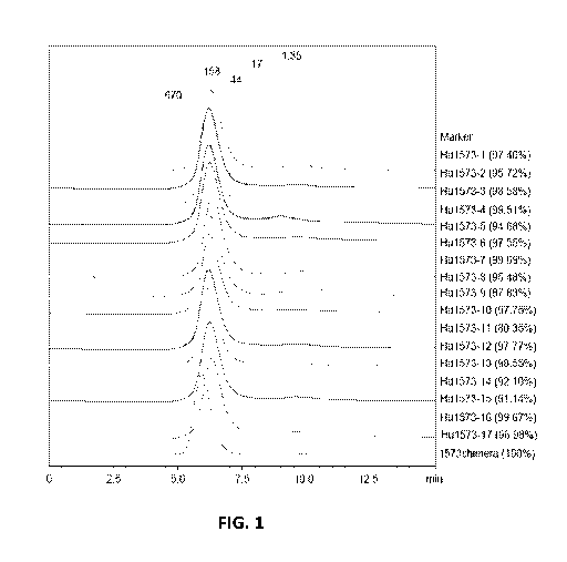
FIG. 1 - SEC analysis of 1573 humanized antibodies.
FIG. 2 - Immobilization of capture antibody to CM5 chip using amine coupling
method. 9200 RU of anti-mouse FC IgG signal (top figure) and 6400 RU of anti-
human FC
IgG (bottom figure) were generated respectively. The running buffer was HBS-EP
running
buffer: 10 mM HEPES, pH 7.4, 150 mM NaC1, 3.4 mM EDTA, 0.005% surfactant P20.
FIG. 3 - SPR sensor-grams of binding of human aPC to1573 antibodies: human aPC
=was injected over 1573 antibody at concentration of 0, 1.25, 2.5, 5, 10, 20
nM,
4
CA 02892748 2015-05-26
WO 2014/085527
PCT/US2013/072137
respectively, at 30 n1/min for 180 s of association phase and 500 s of
dissociation phase.
FIG. 4 - SPR sensor-grams of binding of cyno aPC to1573 antibodies: cyno aPC
was
injected over 1573 antibody at concentration of 0, 5, 10, 20, 40, 80 WM
respectively at 30
nl/min for 180 s of association phase and 420 s of dissociation phase.
FIG. 5 - 1573 humanized antibodies binding ELISA of human PC and aPC.
FIG. 6 - 1573 humanized antibodies binding ELISA of monkey PC and aPC.
DESCRIPTION
The present disclosure relates to the discovery of monoclonal antibodies that
selectively bind to activated protein C, but not unactivated protein C, and
specifically inhibit
the anti-coagulation activity of activated protein C.
Whenever appropriate, terms used in the singular will also include the plural
and vice
versa. In the event that any definition set forth below conflicts with the
usage of that word in
any other document, including any document incorporated herein by reference,
the definition
set forth below shall always control for purposes of interpreting this
specification and its
associated claims unless a contrary meaning is clearly intended (for example
in the document
where the term is originally used). The use of "or" means "and/or" unless
stated otherwise.
The use of "a" herein means "one or more" unless stated otherwise or where the
use of "one
or more" is clearly inappropriate. The use of "comprise," "comprises,"
"comprising,"
"include," "includes," and "including" are interchangeable and are not
limiting. For example,
the term "including" shall mean "including, but not limited to."
The term "Protein C" or "PC" as used herein refers to any variant, isoform,
and/or
species homolog of Protein C in its zymogen form that is naturally expressed
by cells and
present in plasma and is distinct from the activated form of Protein C.
The term "activated Protein C" or "aPC" as used herein refers to an activated
form of
Protein C that is characterized by the removal and absence of a 12 amino acid
activation
peptide present in Protein C as a result of a thrombin cleavage site.
As used herein, an "antibody" refers to a whole antibody and any antigen
binding
fragment (i.e., "antigen-binding portion") or single chain thereof The term
includes a full-
length immunoglobulin molecule (e.g., an IgG antibody) that is naturally
occurring or formed
by normal immunoglobulin gene fragment recombinatorial processes, or an
immunologically
active portion of an immunoglobulin molecule, such as an antibody fragment,
that retains the
5
CA 02892748 2015-05-26
WO 2014/085527
PCT/US2013/072137
specific binding activity. Regardless of structure, an antibody fragment binds
with the same
antigen that is recognized by the full-length antibody. For example, an anti-
aPC monoclonal
antibody fragment binds to an epitope of aPC. The antigen-binding function of
an antibody
can be performed by fragments of a full-length antibody. Examples of binding
fragments
encompassed within the term "antigen-binding portion" of an antibody include
(i) a Fab
fragment, a monovalent fragment consisting of the VL, VH, CL and CHi domains;
(ii) a F(ab')2
fragment, a bivalent fragment comprising two Fab fragments linked by a
disulfide bridge at
the hinge region; (iii) a Fd fragment consisting of the VH and CHi domains;
(iv) a Fy fragment
consisting of the VL and VH domains of a single arm of an antibody, (y) a dAb
fragment
(Ward et al., (1989) Nature 341:544-546), which consists of a VH domain; (vi)
an isolated
complementarity determining region (CDR); (vii) minibodies, diaboidies,
triabodies,
tetrabodies, and kappa bodies (see, e.g., Ill et al., Protein Eng 1997;10:949-
57); (viii) camel
IgG; and (ix) IgNAR . Furthermore, although the two domains of the Fy
fragment, VL and
VH, are coded for by separate genes, they can be joined, using recombinant
methods, by a
synthetic linker that enables them to be made as a single protein chain in
which the VL and
VH regions pair to form monovalent molecules (known as single chain Fy (scFy);
see e.g.,
Bird et al. (1988) Science 242:423-426; and Huston et al. (1988) Proc. Natl.
Acad. Sci. USA
85:5879-5883). Such single chain antibodies are also intended to be
encompassed within the
term "antigen-binding portion" of an antibody. These antibody fragments are
obtained using
conventional techniques known to those with skill in the art, and the
fragments are analyzed
for utility in the same manner as are intact antibodies.
Furthermore, it is contemplated that an antigen binding fragment can be
encompassed
in an antibody mimetic. The term "antibody mimetic" or "mimetic" as used
herein is meant a
protein that exhibits binding similar to an antibody but is a smaller
alternative antibody or a
non-antibody protein. Such antibody mimetic can be comprised in a scaffold.
The term
"scaffold" refers to a polypeptide platform for the engineering of new
products with tailored
functions and characteristics.
As used herein, the term "anti-aPC antibody" refers to an antibody that
specifically
binds to an epitope of aPC. When bound in vivo to an epitope of aPC, the anti-
aPC
antibodies disclosed herein augment one or more aspects of the blood clotting
cascade.
As used herein, the terms "inhibits binding" and "blocks binding" (e.g.,
referring to
inhibition/blocking of binding of aPC substrate to aPC) are used
interchangeably and
encompass both partial and complete inhibition or blocking of a protein with
its substrate,
6
CA 02892748 2015-05-26
WO 2014/085527
PCT/US2013/072137
such as an inhibition or blocking by at least about 10%, about 20%, about 30%,
about 40%,
about 50%, about 60%, about 70%, about 80%, about 90%, about 95%, about 96%,
about
97%, about 98%, about 99%, or about100%. As used herein, "about" means +/- 10%
of the
numerical value indicated.
In reference to the inhibition and/or blocking of binding of aPC substrate to
aPC, the
terms inhibition and blocking also include any measurable decrease in the
binding affinity of
aPC to a physiological substrate when in contact with an anti-aPC antibody as
compared to
aPC not in contact with an anti-aPC antibody, e.g., the blocking of the
interaction of aPC with
its substrates, including Factor Va or with Factor VIIIa, by at least about
10%, about 20%,
about 30%, about 40%, about 50%, about 60%, about 70%, about 80%, about 90%,
about
95%, about 96%, about 97%, about 98%, about 99%, or about 100%.
The terms "monoclonal antibody" or "monoclonal antibody composition" as used
herein refer to a preparation of antibody molecules of single molecular
composition. A
monoclonal antibody composition displays a single binding specificity and
affinity for a
particular epitope. Accordingly, the term "human monoclonal antibody" refers
to antibodies
displaying a single binding specificity that have variable and constant
regions derived from
human germline immunoglobulin sequences. The human antibodies can include
amino acid
residues not encoded by human germline immunoglobulin sequences (e.g.,
mutations
introduced by random or site-specific mutagenesis in vitro or by somatic
mutation in vivo).
An "isolated antibody," as used herein, is intended to refer to an antibody
which is
substantially free of other biological molecules, including antibodies having
different
antigenic specificities (e.g., an isolated antibody that binds to aPC is
substantially free of
antibodies that bind antigens other than aPC). In some embodiments, the
isolated antibody is
at least about 75%, about 80%, about 90%, about 95%, about 97%, about 99%,
about 99.9%
or about 100% pure by dry weight. In some embodiments, purity can be measured
by a
method such as column chromatography, polyacrylamide gel electrophoresis, or
HPLC
analysis. An isolated antibody that binds to an epitope, isoform or variant of
human aPC can,
however, have cross-reactivity to other related antigens, e.g., from other
species (e.g., aPC
species homologs). Moreover, an isolated antibody can be substantially free of
other cellular
material and/or chemicals. As used herein, "specific binding" refers to
antibody binding to a
predetermined antigen. Typically, an antibody that exhibits "specific binding"
binds to an
antigen with an affinity of at least about 105 M-1 and binds to that antigen
with an affinity that
is higher, for example at least two-fold greater, than its binding affinity
for an irrelevant
7
CA 02892748 2015-05-26
WO 2014/085527
PCT/US2013/072137
antigen (e.g., BSA, casein). The phrases "an antibody recognizing an antigen"
and "an
antibody specific for an antigen" are used interchangeably herein with the
term "an antibody
which binds specifically to an antigen."
As used herein, the term "minimal binding" refers to an antibody that does not
bind to
and/or exhibits low affinity to a specified antigen. Typically, an antibody
having minimal
binding to an antigen binds to that antigen with an affinity that is lower
than about 102 MA
and does not bind to a predetermined antigen with higher affinity than it
binds to an irrelevant
antigen.
As used herein, the term "high affinity" for an antibody, such as an IgG
antibody
refers to a binding affinity of at least about 107M-1, in at least one
embodiment at least about
108M-1, in some embodiments at least about 109M-1, 1010M-1, 1011M-1 or
greater, e.g., up to
1013M-1 or greater. However, "high affinity" binding can vary for other
antibody isotypes.
For example, "high affinity" binding for an IgM isotype refers to a binding
affinity of at least
about 107M-1. As used herein, "isotype" refers to the antibody class (e.g.,
IgM or IgG1) that
is encoded by heavy chain constant region genes.
"Complementarity-determining region" or "CDR" refers to one of three
hypervariable
regions within the variable region of the heavy chain or the variable region
of the light chain
of an antibody molecule that form the N-terminal antigen-binding surface that
is
complementary to the three-dimensional structure of the bound antigen.
Proceeding from the
N-terminus of a heavy or light chain, these complementarity-determining
regions are denoted
as "CDR1," "CDR2," and "CDR3," respectively [Wu TT, Kabat EA, Bilofsky H, Proc
Natl
Acad Sci USA. 1975 Dec;72(12):5107 and Wu TT, Kabat EA, J Exp Med. 1970 Aug
1;132(2):211]. CDRs are involved in antigen-antibody binding, and the CDR3
comprises a
unique region specific for antigen-antibody binding. An antigen-binding site,
therefore, can
include six CDRs, comprising the CDR regions from each of a heavy and a light
chain V
region.
The term "epitope" refers to the area or region of an antigen to which an
antibody
specifically binds or interacts, which in some embodiments indicates where the
antigen is in
physical contact with the antibody. Conversely, the term "paratope" refers to
the area or
region of the antibody on which the antigen specifically binds. Epitopes
characterized by
competition binding are said to be overlapping if the binding of the
corresponding antibodies
are mutually exclusive, i.e., binding of one antibody excludes simultaneous
binding of
8
CA 02892748 2015-05-26
WO 2014/085527
PCT/US2013/072137
another antibody. The epitopes are said to be separate (unique) if the antigen
is able to
accommodate binding of both corresponding antibodies simultaneously.
The term "competing antibodies," as used herein, refers to antibodies that
bind to
about, substantially or essentially the same, or even the same, epitope as an
antibody against
aPC as described herein. "Competing antibodies" include antibodies with
overlapping
epitope specificities. Competing antibodies are thus able to effectively
compete with an
antibody as described herein for binding to aPC. In some embodiments, the
competing
antibody can bind to the same epitope as the antibody described herein.
Alternatively
viewed, the competing antibody has the same epitope specificity as the
antibody described
herein.
As used herein, "conservative substitutions" refers to modifications of a
polypeptide
that involve the substitution of one or more amino acids for amino acids
having similar
biochemical properties that do not result in loss of a biological or
biochemical function of the
polypeptide. A "conservative amino acid substitution" is one in which the
amino acid residue
is replaced with an amino acid residue having a similar side chain. Families
of amino acid
residues having similar side chains have been defined in the art. These
families include
amino acids with basic side chains (e.g., lysine, arginine, histidine), acidic
side chains (e.g.,
aspartic acid, glutamic acid), uncharged polar side chains (e.g., glycine,
asparagine,
glutamine, serine, threonine, tyrosine, cysteine), nonpolar side chains (e.g.,
alanine, valine,
leucine, isoleucine, proline, phenylalanine, methionine, tryptophan), 13-
branched side chains
(e.g., threonine, valine, isoleucine), and aromatic side chains (e.g.,
tyrosine, phenylalanine,
tryptophan, histidine). Antibodies of the present disclosure can have one or
more
conservative amino acid substitutions yet retain antigen binding activity.
For nucleic acids and polypeptides, the term "substantial homology" indicates
that
two nucleic acids or two polypeptides, or designated sequences thereof, when
optimally
aligned and compared, are identical, with appropriate nucleotide or amino acid
insertions or
deletions, in at least about 80% of the nucleotides or amino acids, usually at
least about 85%,
in some embodiments about 90%, 91%, 92%, 93%, 94%, or 95%, in at least one
embodiment
at least about 96%, 97%, 98%, 99%, 99.1%, 99.2%, 99.3%, 99.4%, or 99.5% of the
nucleotides or amino acids. Alternatively, substantial homology for nucleic
acids exists when
the segments will hybridize under selective hybridization conditions to the
complement of the
strand. Also included are nucleic acid sequences and polypeptide sequences
having
9
CA 02892748 2015-05-26
WO 2014/085527
PCT/US2013/072137
substantial homology to the specific nucleic acid sequences and amino acid
sequences recited
herein.
The percent identity between two sequences is a function of the number of
identical
positions shared by the sequences (i.e., % homology = # of identical positions
/ total # of
positions x 100), taking into account the number of gaps, and the length of
each gap, which
need to be introduced for optimal alignment of the two sequences. The
comparison of
sequences and determination of percent identity between two sequences can be
accomplished
using a mathematical algorithm, such as without limitation the A1ignXTM module
of
VectorNTITm (Invitrogen Corp., Carlsbad, CA). For A1ignXTM, the default
parameters of
multiple alignment are: gap opening penalty: 10; gap extension penalty: 0.05;
gap separation
penalty range: 8; % identity for alignment delay: 40. (further details found
at the world-wide-
web at invitrogen.com/site/us/en/home/LINNEA-Online-Guides/LINNEA-Communities/
Vector-NTI-Community/S equenc e-analys is-and-data-management-s o ftware-for-P
Cs/
AlignX-Module-for-Vector-NTI-Advance.reg.us.html).
Another method for determining the best overall match between a query sequence
(a
sequence of the present disclosure) and a subject sequence, also referred to
as a global
sequence alignment, can be determined using the CLUSTALW computer program
(Thompson et al., Nucleic Acids Res, 1994, 2(22): 4673-4680), which is based
on the
algorithm of Higgins et al., Computer Applications in the Biosciences
(CABIOS), 1992, 8(2):
189-191). In a sequence alignment the query and subject sequences are both DNA
sequences. The result of said global sequence alignment is in percent
identity. Parameters
that can be used in a CLUSTALW alignment of DNA sequences to calculate percent
identity
via pairwise alignments are: Matrix = IUB, k-tuple = 1, Number of Top
Diagonals = 5, Gap
Penalty = 3, Gap Open Penalty = 10, Gap Extension Penalty = 0.1. For multiple
alignments,
the following CLUSTALW parameters can be used: Gap Opening Penalty = 10, Gap
Extension Parameter = 0.05; Gap Separation Penalty Range = 8; % Identity for
Alignment
Delay = 40.
The nucleic acids can be present in whole cells, in a cell lysate, or in a
partially
purified or substantially pure form. A nucleic acid is "isolated" or "rendered
substantially
pure" when purified away from other cellular components with which it is
normally
associated in the natural environment. To isolate a nucleic acid, standard
techniques such as
the following can be used: alkaline/SDS treatment, CsC1 banding, column
chromatography,
agarose gel electrophoresis and others well known in the art.
CA 02892748 2015-05-26
WO 2014/085527
PCT/US2013/072137
I. Activated Protein C (aPC) and Antibodies
A. Activated Protein C
Protein C is activated by thrombin complexed with thrombomodulin on
endothelium.
Unlike the few-second transient life of active thrombin in vivo, human aPC has
about a 20
minute half-life in circulation after its generation (Berg, et al., 2003).
Therefore, one can
feasibly measure a level of aPC in plasma to study its regulation under
various pathophysical
conditions.
B. Antibodies to aPC
Previously, a murine antibody HAPC1573 was developed which enhanced FL-aPC
binding on the endothelial cells. HAPC1573 facilitated aPC internalization on
endothelium
through the interaction of Gla domain of aPC and EPCR on the cells, and this
internalization
could be blocked by either EPCR blocking Ab or Gla domain blocking Ab
(HPC1575).
HAPC1573 also dramatically altered the kinetic parameters of aPC toward its
chromogenic
substrate, Spectrozyme PCa. This profound change of aPC toward small peptide
substrate in
the presence of HAPC1573 indicated that this mAb recognized an epitope near
active site of
aPC and the interaction of Ab and antigen dramatically increased the affinity
of APC toward
small peptide substrate but decreased the off rate of product from aPC
catalytic site.
HAPC1573 also almost completely diminished the prolongation effect of aPC in
factor Xa
initiated one-stage plasma clotting assay, suggesting that the interaction of
HAPC1573 and
aPC prevents aPC from cleaving factor Va. Surprisingly, HAPC1573 did not
inhibit but
actually enhanced aPC cleaving histone H3 and H4. Consistently, HAPC1573 did
not inhibit
but slightly enhanced aPC cytoprotection activity on endothelium against
histone H3 and H4.
Finally, their results show that HAPC1573, recognizes aPC, but not Protein C.
See U.S.
Patent 8,153,766.
Recent studies have shown that anticoagulant activity of aPC is dispensable
for its
cytoprotective function, but aPC cleavage activity toward PAR1 might be
essential for its
anti-apoptotic effect (Mosnier et al., 2004). However, the cytoprotection
effect of aPC has
been shown not only in endothelial cells which express EPCR, but also on other
cells such as
neuron and keratinocytes which do not express EPCR on their cell surfaces (Guo
et al., 2004;
Berg et al., 2003), indicating other mechanisms than PAR1 mediated aPC
signaling might
exist.
11
CA 02892748 2015-05-26
WO 2014/085527
PCT/US2013/072137
C. Applications of the Technology
The ability to distinguish between Protein C and aPC demonstrates the utility
of
antibodies in a convenient ELISA method for measuring aPC level in plasma in
vivo.
Typically, it takes less than 4 hours to measure a plasma sample containing 1
ng/ml APC
with this method compared to 19 hours or even weeks with enzyme capture assays
(Gruber
and Griffen, 1992; Liaw et al., 2003).
Also, as discussed above, HAPC1573 altered aPC cleavage activity toward a
chromogenic peptide substrate and also blocked aPC anticoagulant activity in a
plasma
clotting assay, suggesting this mAb recognizes an epitope near the aPC active
site and alters
its catalytic activity upon antibody-antigen binding. At the same time,
HAPC1573 actually
enhanced aPC cleaving extracellular histones, and enhanced APC cytoprotection
activity on
endothelium against histones. This indicates that APC anticoagulant activity
for cleaving
activated factor V and VIII is not required for its cytoprotection activity by
cleaving
extracellular histones. Cleaving extracellular histones independent from its
anticoagulant
activity might be one of the molecular mechanisms of aPC regulation
inflammation and
cytoprotection.
Thus, such antibodies against aPC can, for example, be used in treatment of
hemophilia A patients. aPC cleaves both factor VIIIa and factor Va and thus
negatively
affects blood clotting. In hemophilia A patients, factor VIII levels are low
and the
inactivation of factor Va by aPC is probably a major pathway to regulate
hemostasis and
thrombosis in these patients. Recent clinical reports demonstrated factor V
Leiden mutant
which is resistant to aPC cleavage was beneficial to hemophilia A patients
regarding their
bleeding symptom (van't Zant et al., 1997). Blocking aPC anticoagulant
activity toward
factor Va in vivo with an antibody is an alternative approach for hemophilia A
treatments,
especially for those patients who have high level factor VIII inhibitors so
that the factor VIII
replacement therapy would not be very effective.
In other embodiments, another possible clinical application for antibodies
against aPC
is in the treatment of trauma patients wherein homeostasis is disrupted,
excessive bleeding is
likely, and surgical intervention is delayed to regain homeostatis. Treatment
with antibodies
can selectively restore the pro-coagulant state without eliminating the
cytoprotective or anti-
inflammatory activities of APC.
Yet another clinical application of antibodies against aPC is in combination
with aPC
in sepsis treatment. Its bleeding side effect in patients is due to aPC
anticoagulant activity.
Because HAPC1573 blocked aPC anticoagulant activity while maintaining, and
even
12
CA 02892748 2015-05-26
WO 2014/085527
PCT/US2013/072137
enhancing, aPC cytoprotective effect, the mAb-aPC complex can be a better
therapeutic than
aPC alone regarding its bleeding side effect.
II. Antibody Structure
Antibodies comprise a large family of glycoproteins with common structural
features.
An antibody is comprised of four polypeptides that form a three dimensional
structure.
Typically, an antibody is comprised of two different polypeptides, the heavy
chain and the
light chain. An antibody molecule is comprised of one or more of these units,
each unit
comprising two heavy chains and two light chains. An antibody molecule
typically consists
of three functional domains: the Fc, Fab, and antigen-binding site.
There are five different types of heavy chain polypeptides designated as a, 6,
c, y, and
1..t. There are two different types of light chain polypeptides designated K
and L An antibody
typically contains only one type of heavy chain and only one type of light
chain, although any
light chain can associate with any heavy chain.
The carboxyl terminal of each heavy chain polypeptide is known as the constant
(Fc)
region. The amino terminal of each heavy and light chain polypeptide is known
as the
variable (V) region. Within the variable regions of the chains are
hypervariable regions
known as complementarity determining regions (CDRs). The variable regions of
one heavy
chain and one light chain associate to form an antigen-binding site. Each
heavy chain and
each light chain includes three CDRs. The six CDRs of an antigen-binding site
define the
amino acid residues that form the actual binding site for the antigen. CDR
variability
accounts for the diversity of antigen recognition.
Antibodies against aPC may be defined by sequences set forth in the following
table:
TABLE 1 ¨ Antibody Sequences
FR2 CDR1 FR2 CDR2 FR3 CDR3 FR4
Light Chain CDR
SEQ ID NO: 1 2 3
Heavy Chain CDR
SEQ ID NO: 4 5 6
Light Chain Framework
SEQ ID NO: 7 8 9 10
Heavy Chain Framework
SEQ ID NO: 11 12 13 14
13
CA 02892748 2015-05-26
WO 2014/085527
PCT/US2013/072137
III. Antibodies against aPC
A. Antibody Fragments
Thus, in one embodiment, such molecules will comprise fragments (such as
(F(ab'),
F(ab')2) that are produced, for example, by the proteolytic cleavage of the
mAbs, or single-
chain immunoglobulins producible, for example, via recombinant means. Such
antibody
derivatives are monovalent. In one embodiment, such fragments can be combined
with one
another, or with other antibody fragments or receptor ligands to form
"chimeric" binding
molecules. Significantly, such chimeric molecules can contain substituents
capable of binding
to different epitopes of the same molecule, or they can be capable of binding
to an activated
protein C epitope and a "non-activated protein C" epitope.
A single-chain variable fragment (scFv) is another form of antibody fragment.
It
comprises a fusion of the variable regions of the heavy and light chains of
immunoglobulins,
linked together with a short (usually serine, glycine) linker. This chimeric
molecule retains
the specificity of the original immunoglobulin, despite removal of the
constant regions and
the introduction of a linker peptide. These molecules were created
historically to facilitate
phage display where it is highly convenient to express the antigen binding
domain as a single
peptide. Alternatively, scFy can be created directly from subcloned heavy and
light chains
derived from a hybridoma. Single chain variable fragments lack the constant Fc
region found
in complete antibody molecules, and thus, the common binding sites (e.g.,
protein A/G) used
to purify antibodies. These fragments can often be purified/immobilized using
Protein L since
Protein L interacts with the variable region of kappa light chains.
Flexible linkers generally are comprised of helix- and turn-promoting amino
acid
residues such as alaine, serine and glycine. However, other residues can
function as well.
Tang et al. (1996) used phage display as a means of rapidly selecting tailored
linkers for
single-chain antibodies (scFvs) from protein linker libraries. A random linker
library was
constructed in which the genes for the heavy and light chain variable domains
were linked by
a segment encoding an 18-amino acid polypeptide of variable composition. The
scFy
repertoire (approx. 5 x 10⁶ different members) was displayed on filamentous
phage and
subjected to affinity selection with hapten. The population of selected
variants exhibited
significant increases in binding activity but retained considerable sequence
diversity.
Screening 1054 individual variants subsequently yielded a catalytically active
scFy that was
produced efficiently in soluble form. Sequence analysis revealed a conserved
proline in the
14
CA 02892748 2015-05-26
WO 2014/085527
PCT/US2013/072137
linker two residues after the VH C terminus and an abundance of arginines and
prolines at
other positions as the only common features of the selected tethers.
The recombinant antibodies against aPC can also involve sequences or moieties
that
permit dimerization or multimerization of the receptors. Such sequences
include those
derived from IgA, which permit formation of multimers in conjunction with the
J chain.
Another multimerization domain is the Ga14 dimerization domain. In other
embodiments, the
chains can be modified with agents such as biotin/ayidin, which permit the
combination of
two antibodies.
In a separate embodiment, a single-chain antibody can be created by joining
receptor
light and heavy chains using a non-peptide linker or chemical unit. Generally,
the light and
heavy chains will be produced in distinct cells, purified, and subsequently
linked together in
an appropriate fashion (i.e., the N-terminus of the heavy chain being attached
to the C-
terminus of the light chain via an appropriate chemical bridge).
Cross-linking reagents are used to form molecular bridges that tie functional
groups of
two different molecules, e.g., a stablizing and coagulating agent. However, it
is contemplated
that dimers or multimers of the same analog or heteromeric complexes comprised
of different
analogs can be created. To link two different compounds in a step-wise manner,
heterobifunctional cross-linkers can be used that eliminate unwanted
homopolymer
formation. An exemplary hetero-bifunctional cross-linker contains two reactive
groups: one
reacting with primary amine group (e.g., N-hydroxy succinimide) and the other
reacting with
a thiol group (e.g., pyridyl disulfide, maleimides, halogens, etc.). Through
the primary amine
reactive group, the cross-linker can react with the lysine residue(s) of one
protein (e.g., the
selected antibody or fragment) and through the thiol reactive group, the cross-
linker, already
tied up to the first protein, reacts with the cysteine residue (free
sulfhydryl group) of the other
protein (e.g., the selective agent).
A cross-linker haying reasonable stability in blood can be employed. Numerous
types
of disulfide-bond containing linkers are known that can be successfully
employed to
conjugate targeting and therapeutic/preventative agents. Linkers that contain
a disulfide bond
that is sterically hindered can prove to give greater stability in vivo,
preventing release of the
targeting peptide prior to reaching the site of action. These linkers are thus
one group of
linking agents.
Another cross-linking reagent is SMPT, which is a bifunctional cross-linker
containing a disulfide bond that is "sterically hindered" by an adjacent
benzene ring and
methyl groups. It is believed that steric hindrance of the disulfide bond
serves a function of
CA 02892748 2015-05-26
WO 2014/085527
PCT/US2013/072137
protecting the bond from attack by thiolate anions such as glutathione which
can be present in
tissues and blood, and thereby help in preventing decoupling of the conjugate
prior to the
delivery of the attached agent to the target site. The SMPT cross-linking
reagent, as with
many other known cross-linking reagents, lends the ability to cross-link
functional groups
such as the SH of cysteine or primary amines (e.g., the epsilon amino group of
lysine).
Another possible type of cross-linker includes the hetero-bifunctional
photoreactive
phenylazides containing a cleavable disulfide bond such as sulfosuccinimidy1-2-
(p-azido
salicylamido) ethyl-1,3'-dithiopropionate. The N-hydroxysuccinimidyl group
reacts with
primary amino groups and the phenylazide (upon photolysis) reacts non-
selectively with any
amino acid residue.
In addition to hindered cross-linkers, non-hindered linkers also can be
employed in
accordance herewith. Other useful cross-linkers, not considered to contain or
generate a
protected disulfide, include SATA, SPDP and 2-iminothiolane (Wawrzynczak &
Thorpe,
1987). The use of such cross-linkers is well understood in the art. Another
embodiment
involves the use of flexible linkers. U.S. Patent 4,680,338, describes
bifunctional linkers
useful for producing conjugates of ligands with amine-containing polymers
and/or proteins,
especially for forming antibody conjugates with chelators, drugs, enzymes,
detectable labels
and the like. U.S. Patents 5,141,648 and 5,563,250 disclose cleavable
conjugates containing a
labile bond that is cleavable under a variety of mild conditions. This linker
is particularly
useful in that the agent of interest can be bonded directly to the linker,
with cleavage resulting
in release of the active agent. Particular uses include adding a free amino or
free sulfhydryl
group to a protein, such as an antibody, or a drug.
U.S. Patent 5,856,456 provides peptide linkers for use in connecting
polypeptide
constituents to make fusion proteins, e.g., single chain antibodies. The
linker is up to about 50
amino acids in length, contains at 5 least one occurrence of a charged amino
acid (e.g.,
arginine or lysine) followed by a proline, and is characterized by greater
stability and reduced
aggregation. U.S. Patent 5,880,270 discloses aminooxy-containing linkers
useful in a variety
of immunodiagnostic and separative techniques.
B. Antibody Conjugates
Further provided are antibody conjugates. For both diagnostic and therapeutic
purposes, one can link or covalently bind or complex an agent to an antibody.
Such a
molecule or moiety can be, but is not limited to, at least one effector or
reporter molecule. A
reporter molecule is defined as any moiety which can be detected using an
assay. Non-
16
CA 02892748 2015-05-26
WO 2014/085527
PCT/US2013/072137
limiting examples of reporter molecules which have been conjugated to
antibodies include
enzymes, radiolabels, haptens, fluorescent labels, phosphorescent molecules,
chemiluminescent molecules, chromophores, luminescent molecules, photoaffinity
molecules, colored particles or ligands, such as biotin.
Certain examples of antibody conjugates are those conjugates in which the
antibody is
linked to a detectable label. "Detectable labels" are compounds and/or
elements that can be
detected due to their specific functional properties, and/or chemical
characteristics, the use of
which allows the antibody to which they are attached to be detected, and/or
further quantified
if desired. Another such example is the formation of a conjugate comprising an
antibody
linked to a cytotoxic or anti cellular agent, and can be termed
"immunotoxins."
Antibody conjugates are used as diagnostic agents. Antibody diagnostics
generally
fall within two classes, those for use in in vitro diagnostics, such as in a
variety of
immunoassays, and/or those for use in vivo diagnostic protocols, generally
known as
"antibody-directed imaging."
1 5 Many
appropriate imaging agents are known in the art, as are methods for their
attachment to antibodies (see, for e.g., U.S. Patents 5,02 1,23 6; 4,938,948;
and 4,472,509,
each incorporated herein by reference). The imaging moieties used can be
paramagnetic ions;
radioactive isotopes; fluorochromes; NMR-detectable substances; X-ray imaging.
In the case of paramagnetic ions, one might mention by way of example ions
such as
chromium (III), manganese (II), iron (III), iron (II), cobalt (II), nickel
(II), copper (II),
neodymium (III), samarium (III), ytterbium (III), gadolinium (III), vanadium
(II), terbium
(III), dysprosium (III), holmium (III) and/or erbium (III). Ions useful in
other contexts, such
as X-ray imaging, include but are not limited to lanthanum (III), gold (III),
lead (II), and
especially bismuth (III).
In the case of radioactive isotopes for therapeutic and/or diagnostic
application, one
might mention astatine211, 14carbon, 51chromium, 36chlorine, 57cobalt,
58cobalt, copper67,
152Eu, gallium67, 3hydrogen, iodine123, iodine125, iodineni, indiumm, 59iron,
32phosphorus,
rhenium186, rhenium188, 75selenium, 35sulphur, technicium99m and/or yttrium90.
1251 is often
being commonly used in certain embodiments, and technicium99m and/or indium111
are also
often used due to their low energy and suitability for long range detection.
Radioactively
labeled monoclonal antibodies can be produced according to well-known methods
in the art.
For instance, monoclonal antibodies can be iodinated by contact with sodium
and/or
potassium iodide and a chemical oxidizing agent such as sodium hypochlorite,
or an
enzymatic oxidizing agent, such as lactoperoxidase. Monoclonal antibodies can
be labeled
17
CA 02892748 2015-05-26
WO 2014/085527
PCT/US2013/072137
with technetium99m by ligand exchange process, for example, by reducing
pertechnate with
stannous solution, chelating the reduced technetium onto a Sephadex column and
applying
the antibody to this column. Alternatively, direct labeling techniques can be
used, e.g., by
incubating pertechnate, a reducing agent such as SNC12, a buffer solution such
as sodium-
potassium phthalate solution, and the antibody. Intermediary functional groups
which are
often used to bind radioisotopes which exist as metallic ions to antibody are
diethylenetriaminepentaacetic acid (DTPA) or ethylene diaminetetracetic acid
(EDTA).
Among the fluorescent labels contemplated for use as conjugates include Alexa
350,
Alexa 430, AMCA, BODIPY 630/650, BODIPY 650/665, BODIPY-FL, BODIPY-R6G,
BODIPY-TMR, BODIPY-TRX, Cascade Blue, Cy3, Cy5,6-FAM, Fluorescein
Isothiocyanate, HEX, 6-JOE, Oregon Green 488, Oregon Green 500, Oregon Green
514,
Pacific Blue, REG, Rhodamine Green, Rhodamine Red, Renographin, ROX, TAMRA,
TET,
Tetramethylrhodamine, and/or Texas Red.
Another type of antibody conjugates contemplated are those intended primarily
for
use in vitro, where the antibody is linked to a secondary binding ligand
and/or to an enzyme
(an enzyme tag) that will generate a colored product upon contact with a
chromogenic
substrate. Examples of suitable enzymes include urease, alkaline phosphatase,
(horseradish)
hydrogen peroxidase or glucose oxidase. Secondary binding ligands are biotin
and/or avidin
and streptavidin compounds. The use of such labels is well known to those of
skill in the art
and are described, for example, in U.S. Patents 3,817,837; 3,850,752;
3,939,350; 3,996,345;
4,277,437; 4,275,149 and 4,366,241; each incorporated herein by reference.
Yet another known method of site-specific attachment of molecules to
antibodies
comprises the reaction of antibodies with hapten-based affinity labels.
Essentially, hapten-
based affinity labels react with amino acids in the antigen binding site,
thereby destroying
this site and blocking specific antigen reaction. However, this can not be
advantageous since
it results in loss of antigen binding by the antibody conjugate.
Molecules containing azido groups can also be used to form covalent bonds to
proteins through reactive nitrene intermediates that are generated by low
intensity ultraviolet
light (Potter & Haley, 1983). In particular, 2- and 8-azido analogues of
purine nucleotides
have been used as site-directed photoprobes to identify nucleotide binding
proteins in crude
cell extracts (Owens & Haley, 1987; Atherton et al., 1985). The 2- and 8-azido
nucleotides
have also been used to map nucleotide binding domains of purified proteins
(Khatoon et al.,
1989; King et al., 1989; and Dholakia et al., 1989) and can be used as
antibody binding
agents.
18
CA 02892748 2015-05-26
WO 2014/085527
PCT/US2013/072137
Several methods are known in the art for the attachment or conjugation of an
antibody
to its conjugate moiety. Some attachment methods involve the use of a metal
chelate
complex employing, for example, an organic chelating agent such as described
in U.S.
Patents 4,472,509 and 4,938,948, each incorporated herein by reference).
Monoclonal
antibodies can also be reacted with an enzyme in the presence of a coupling
agent such as
glutaraldehyde or periodate. Conjugates with fluorescein markers are prepared
in the
presence of these coupling agents or by reaction with an isothiocyanate. In
U.S. Patent
4,938,948, imaging of breast tumors is achieved using monoclonal antibodies
and the
detectable imaging moieties are bound to the antibody using linkers such as
methyl-p-
hydroxybenzimidate or N-succinimidy1-3-(4-hydroxyphenyl)propionate.
In other embodiments, derivatization of immunoglobulins by selectively
introducing
sulfhydryl groups in the Fc region of an immunoglobulin, using reaction
conditions that do
not alter the antibody combining site are contemplated. Antibody conjugates
produced
according to this methodology are disclosed to exhibit improved longevity,
specificity and
sensitivity (U.S. Patent 5,196,066, incorporated herein by reference). Site-
specific
attachment of effector or reporter molecules, wherein the reporter or effector
molecule is
conjugated to a carbohydrate residue in the Fc region have also been disclosed
in the
literature (O'Shannessy et al., 1987). This approach has been reported to
produce
diagnostically and therapeutically promising antibodies which are currently in
clinical
evaluation.
In another embodiment, one may choose to modify the immunoglobulins to improve
their stability and half-life in vivo. PEGylation is one such
process that involves covalent attachment of polyethylene glycol (PEG) polymer
chains to the
antibody. PEGylation is routinely achieved by incubation of a reactive
derivative of PEG
with the target molecule. The covalent attachment of PEG can "mask" the
antibody from the
host's immune system (reduced immunogenicity and antigenicity), and increase
the
hydrodynamic size (size in solution) of the agent which prolongs its
circulatory time by
reducing renal clearance. PEGylation can also provide water solubility. Other
polymers used
to modify antibodies include polyethyleneimine and polylysine, often linked
through succinic
acid groups.
C. Immunodetection Methods
In still further embodiments, also provided are immunodetection methods for
binding,
purifying, removing, quantifying and/or otherwise generally detecting
biological components
19
CA 02892748 2015-05-26
WO 2014/085527
PCT/US2013/072137
using antibodies that react immunologically with such components. Some
immunodetection
methods include enzyme linked immunosorbent assay (ELISA), radioimmunoassay
(RIA),
immunoradiometric assay, fluoroimmunoassay, chemiluminescent assay,
bioluminescent
assay, and Western blot to mention a few. The steps of various useful
immunodetection
methods have been described in the scientific literature, such as, e.g.,
Doolittle and Ben-Zeev
(1999); Gulbis and Galand (1993); De Jager et al. (1993); and Nakamura et al.
(1987), each
incorporated herein by reference.
In general, the immunobinding methods include obtaining a sample containing a
target of interest, and contacting the sample with a first antibody that
reacts immunologically
with the target under conditions effective to allow the formation of
immunocomplexes. The
binding of the antibody to the target can then be assessed using a variety of
different formats.
In one format, the antibody can be linked to a solid support, such as in the
form of a
column matrix, and the sample suspected of containing the target will be
applied to the
immobilized antibody. The unwanted components will be washed from the column,
leaving
the target immunocomplexed to the immobilized antibody to be eluted.
The immunobinding methods also include methods for detecting and quantifying
the
amount of an target in a sample and the detection and quantification of any
immune
complexes formed during the binding process. Here, one would obtain a sample
suspected of
containing a target, and contact the sample with an antibody against the
target, and then
detect and quantify the amount of immune complexes formed under the specific
conditions.
In terms of antigen detection, the biological sample analyzed can be any
sample that
is suspected of containing a target, such as, for example, a body fluid like
blood, serum,
plasma, mucous, urine, saliva, tears or semen. Alternatively, a tissue can be
used.
Contacting the chosen biological sample with the antibody under effective
conditions and for
a period of time sufficient to allow the formation of immune complexes
(primary immune
complexes) is generally a matter of simply adding the antibody composition to
the sample
and incubating the mixture for a period of time long enough for the antibodies
to form
immune complexes with, i.e., to bind to targets that react immunologically
with antibodies
present. After this time, the sample-antibody composition, such as a tissue
section, ELISA
plate, dot blot or western blot, will generally be washed to remove any non-
specifically
bound species, allowing only those molecules specifically bound within the
primary immune
complexes to be detected.
In general, the detection of immunocomplex formation is well known in the art
and
can be achieved through the application of numerous approaches. These methods
are
CA 02892748 2015-05-26
WO 2014/085527
PCT/US2013/072137
generally based upon the detection of a label or marker, such as any of those
radioactive,
fluorescent, biological and enzymatic tags. U.S. Patents concerning the use of
such labels
include 3,817,837; 3,850,752; 3,939,350; 3,996,345; 4,277,437; 4,275,149 and
4,366,241,
each incorporated herein by reference. Of course, one can find additional
advantages through
the use of a secondary binding ligand such as a second antibody and/or a
biotin/avidin ligand
binding arrangement, as is known in the art.
The antibody employed in the detection can itself be linked to a detectable
label,
wherein one would then simply detect this label, thereby allowing the amount
of the primary
immune complexes in the composition to be determined. Alternatively, the first
antibody that
becomes bound within the primary immune complexes can be detected by means of
a second
binding ligand that has binding affinity for the antibody. In these cases, the
second binding
ligand can be linked to a detectable label. The second binding ligand is
itself often an
antibody, which can thus be termed a "secondary" antibody. The primary immune
complexes are contacted with the labeled, secondary binding ligand, or
antibody, under
effective conditions and for a period of time sufficient to allow the
formation of secondary
immune complexes. The secondary immune complexes are then generally washed to
remove
any non-specifically bound labeled secondary antibodies or ligands, and the
remaining label
in the secondary immune complexes is then detected.
Further methods include the detection of primary immune complexes by a two
step
approach. A second binding ligand, such as an antibody, that has binding
affinity for the
antibody is used to form secondary immune complexes, as described above. After
washing,
the secondary immune complexes are contacted with a third binding ligand or
antibody that
has binding affinity for the second antibody, again under effective conditions
and for a period
of time sufficient to allow the formation of immune complexes (tertiary immune
complexes).
The third ligand or antibody is linked to a detectable label, allowing
detection of the tertiary
immune complexes thus formed. This system can provide for signal amplification
if this is
desired.
One method of immunodetection designed by Charles Cantor uses two different
antibodies. A first step biotinylated, monoclonal or polyclonal antibody is
used to detect the
target antigen(s), and a second step antibody is then used to detect the
biotin attached to the
complexed biotin. In that method the sample to be tested is first incubated in
a solution
containing the first step antibody. If the target antigen is present, some of
the antibody binds
to the antigen to form a biotinylated antibody/antigen complex. The
antibody/antigen
complex is then amplified by incubation in successive solutions of
streptavidin (or avidin),
21
CA 02892748 2015-05-26
WO 2014/085527
PCT/US2013/072137
biotinylated DNA, and/or complementary biotinylated DNA, with each step adding
additional
biotin sites to the antibody/antigen complex. The amplification steps are
repeated until a
suitable level of amplification is achieved, at which point the sample is
incubated in a
solution containing the second step antibody against biotin. This second step
antibody is
labeled, as for example with an enzyme that can be used to detect the presence
of the
antibody/antigen complex by histoenzymology using a chromogen substrate. With
suitable
amplification, a conjugate can be produced which is macroscopically visible.
Another known method of immunodetection takes advantage of the immuno-PCR
(Polymerase Chain Reaction) methodology. The PCR method is similar to the
Cantor
method up to the incubation with biotinylated DNA, however, instead of using
multiple
rounds of streptavidin and biotinylated DNA
incubation, the
DNA/biotinistreptavidiniantibody complex is washed out with a low pH or high
salt buffer
that releases the antibody. The resulting wash solution is then used to carry
out a PCR
reaction with suitable primers with appropriate controls. At least in theory,
the enormous
amplification capability and specificity of PCR can be utilized to detect a
single antigen
molecule.
Another ELISA in which the antigens are immobilized, involves the use of
antibody
competition in the detection. In this ELISA, labeled antibodies against an
antigen are added
to the wells, allowed to bind, and/or detected by means of their label. The
amount of an
antigen in an unknown sample is then determined by mixing the sample with the
labeled
antibodies against the antigen during incubation with coated wells. The
presence of an
antigen in the sample acts to reduce the amount of antibody against the
antigen available for
binding to the well and thus reduces the ultimate signal. This is also
appropriate for detecting
antibodies against an antigen in an unknown sample, where the unlabeled
antibodies bind to
the antigen-coated wells and also reduces the amount of antigen available to
bind the labeled
antibodies.
As detailed above, immunoassays, in their most simple and/or direct sense, are
binding assays.
Certain immunoassays are the various types of enzyme linked
immunosorbent assays (ELISAs) and/or radioimmunoassays (RIA) known in the art.
Immunohistochemical detection using tissue sections is also particularly
useful. However, it
will be readily appreciated that detection is not limited to such techniques,
and/or western
blotting, dot blotting, FACS analyses, and/or the like can also be used.
Irrespective of the
format employed, ELISAs have certain features in common, such as coating,
incubating and
22
CA 02892748 2015-05-26
WO 2014/085527
PCT/US2013/072137
binding, washing to remove non-specifically bound species, and detecting the
bound immune
complexes. These are described below.
In coating a plate with either antigen or antibody, one will generally
incubate the
wells of the plate with a solution of the antigen or antibody, either
overnight or for a specified
period of hours. The wells of the plate will then be washed to remove
incompletely adsorbed
material. Any remaining available surfaces of the wells are then "coated" with
a non-specific
protein that is antigenically neutral with regard to the test antisera. These
include bovine
serum albumin (BSA), casein or solutions of milk powder. The coating allows
for blocking
of nonspecific adsorption sites on the immobilizing surface and thus reduces
the background
caused by nonspecific binding of antisera onto the surface.
In ELISAs, it is probably more customary to use a secondary or tertiary
detection
means rather than a direct procedure. Thus, after binding of a protein or
antibody to the well,
coating with a non-reactive material to reduce background, and washing to
remove unbound
material, the immobilizing surface is contacted with the biological sample to
be tested under
conditions effective to allow immune complex (antigen/antibody) formation.
Detection of
the immune complex then requires a labeled secondary binding ligand or
antibody, and a
secondary binding ligand or antibody in conjunction with a labeled tertiary
antibody or a third
binding ligand.
"Under conditions effective to allow immune complex (antigen/antibody)
formation"
means that the conditions can include diluting the antigens and/or antibodies
with solutions
such as BSA, bovine gamma globulin (BGG) or phosphate buffered saline
(PBS)/Tween.
These added agents also tend to assist in the reduction of nonspecific
background.
The "suitable" conditions also mean that the incubation is at a temperature or
for a
period of time sufficient to allow effective binding. Incubation steps are
typically from about
1 to 2 to 4 hours or so, at temperatures on the order of 25 C to 27 C, or can
be overnight at
about 4 C or so.
D. Purification
In certain embodiments, the antibodies against aPC can be purified. The term
"purified," as used herein, is intended to refer to a composition, isolatable
from other
components, wherein the protein is purified to any degree relative to its
naturally-obtainable
state. A purified protein therefore also refers to a protein, free from the
environment in which
it can naturally occur. Where the term "substantially purified" is used, this
designation will
refer to a composition in which the protein or peptide forms the major
component of the
23
CA 02892748 2015-05-26
WO 2014/085527
PCT/US2013/072137
composition, such as constituting about 50%, about 60%, about 70%, about 80%,
about 90%,
about 95% or more of the proteins in the composition.
Protein purification techniques are well known to those of skill in the art.
These
techniques involve, at one level, the crude fractionation of the cellular
milieu to polypeptide
and non-polypeptide fractions. Having separated the polypeptide from other
proteins, the
polypeptide of interest can be further purified using chromatographic and
electrophoretic
techniques to achieve partial or complete purification (or purification to
homogeneity).
Analytical methods particularly suited to the preparation of a pure peptide
are ion-
exchange chromatography, exclusion chromatography; polyacrylamide gel
electrophoresis;
isoelectric focusing. Other methods for protein purification include,
precipitation with
ammonium sulfate, PEG, antibodies and the like or by heat denaturation,
followed by
centrifugation; gel filtration, reverse phase, hydroxylapatite and affinity
chromatography; and
combinations of such and other techniques.
In purifying an antibody against aPC, it can be desirable to express the
polypeptide in
a prokaryotic or eukaryotic expression system and extract the protein using
denaturing
conditions. The polypeptide can be purified from other cellular components
using an affinity
column, which binds to a tagged portion of the polypeptide. As is generally
known in the art,
it is believed that the order of conducting the various purification steps can
be changed, or
that certain steps can be omitted, and still result in a suitable method for
the preparation of a
substantially purified protein or peptide.
Commonly, complete antibodies are fractionated utilizing agents (i.e., protein
A) that
bind the Fc portion of the antibody. Alternatively, antigens can be used to
simultaneously
purify and select appropriate antibodies. Such methods often utilize the
selection agent bound
to a support, such as a column, filter or bead. The antibodies is bound to a
support,
contaminants removed, and the antibodies released by applying conditions
(salt, heat, etc.).
Various methods for quantifying the degree of purification of the protein or
peptide
will be known to those of skill in the art in light of the present disclosure.
These include, for
example, determining the specific activity of an active fraction, or assessing
the amount of
polypeptides within a fraction by SDS/PAGE analysis. Another method for
assessing the
purity of a fraction is to calculate the specific activity of the fraction, to
compare it to the
specific activity of the initial extract, and to thus calculate the degree of
purity. The actual
units used to represent the amount of activity will, of course, be dependent
upon the
particular assay technique chosen to follow the purification and whether or
not the expressed
protein or peptide exhibits a detectable activity.
24
CA 02892748 2015-05-26
WO 2014/085527
PCT/US2013/072137
It is known that the migration of a polypeptide can vary, sometimes
significantly, with
different conditions of SDS/PAGE (Capaldi et al., 1977). It will therefore be
appreciated that
under differing electrophoresis conditions, the apparent molecular weights of
purified or
partially purified expression products can vary.
IV. Pharmaceutical Compositions and Uses
A. Compositions
Pharmaceutical compositions can comprise an effective amount of one or more
antibodies, therapeutic agents or additional agent dissolved or dispersed in a
pharmaceutically
acceptable carrier. Aqueous compositions comprise an effective amount of the
antibody,
dissolved or dispersed in a pharmaceutically acceptable carrier or aqueous
medium. The
phrases "pharmaceutically or pharmacologically acceptable" refer to molecular
entities and
compositions that do not produce an adverse, allergic or other untoward
reaction when
administered to an animal, or a human, as appropriate.
As used herein, "pharmaceutically acceptable carrier" includes any and all
solvents,
dispersion media, coatings, surfactants, antioxidants, preservatives (e.g.,
antibacterial agents,
antifungal agents), isotonic agents, absorption delaying agents, salts,
preservatives, drugs,
drug stabilizers, gels, binders, excipients, disintegration agents,
lubricants, sweetening agents,
flavoring agents, dyes, such like materials and combinations thereof, as would
be known to
one of ordinary skill in the art (see, for example, Remington's Pharmaceutical
Sciences, 18th
Ed. Mack Printing Company, 1990, pp. 1289-1329, incorporated herein by
reference). The
use of such media and agents for pharmaceutical active substances is well
known in the art.
Except insofar as any conventional media or agent is incompatible with the
active ingredient,
its use in the therapeutic compositions is contemplated. Supplementary active
ingredients
can also be incorporated into the compositions. For human administration,
preparations
should meet sterility, pyrogenicity, general safety and purity standards as
required by FDA
Office of Biologic Standards.
The biological material should be extensively dialyzed to remove undesired
small
molecular weight molecules and/or lyophilized for more ready formulation into
a desired
vehicle, where appropriate. The active compounds will then generally be
formulated for
parenteral administration, e.g., formulated for injection via the intravenous,
intramuscular,
sub-cutaneous, intranasal, or intraperitoneal routes. Typically, such
compositions can be
prepared as injectables, either as liquid solutions or suspensions; solid
forms suitable for
CA 02892748 2015-05-26
WO 2014/085527
PCT/US2013/072137
using to prepare solutions or suspensions upon the addition of a liquid prior
to injection can
also be prepared; and the preparations can also be emulsified.
The pharmaceutical forms suitable for injectable use include sterile aqueous
solutions
or dispersions; formulations including sesame oil, peanut oil or aqueous
propylene glycol;
and sterile powders for the extemporaneous preparation of sterile injectable
solutions or
dispersions. In all cases the form must be sterile and must be fluid to the
extent that easy
syringability exists. It must be stable under the conditions of manufacture
and storage and
must be preserved against the contaminating action of microorganisms, such as
bacteria and
fungi.
Solutions of the active compounds as free base or pharmacologically acceptable
salts
can be prepared in water suitably mixed with a surfactant, such as
hydroxypropylcellulose.
Dispersions can also be prepared in glycerol, liquid polyethylene glycols, and
mixtures
thereof and in oils. Under ordinary conditions of storage and use, these
preparations contain
a preservative to prevent the growth of microorganisms.
The antibodies against aPC can be formulated into a composition in a free
base, in a
neutral or salt form. Pharmaceutically acceptable salts, include the acid
addition salts
(formed with the free amino groups of the protein) and which are formed with
inorganic acids
such as, for example, hydrochloric or phosphoric acids, or such organic acids
as acetic,
oxalic, tartaric, mandelic, and the like. Salts formed with the free carboxyl
groups can also
be derived from inorganic bases such as, for example, sodium, potassium,
ammonium,
calcium, or ferric hydroxides, and such organic bases as isopropylamine,
trimethylamine,
histidine, procaine and the like.
The carrier can also be a solvent or dispersion medium containing, for
example,
water, ethanol, polyol (for example, glycerol, propylene glycol, and liquid
polyethylene
glycol, and the like), suitable mixtures thereof, and vegetable oils. The
proper fluidity can be
maintained, for example, by the use of a coating, such as lecithin, by the
maintenance of the
required particle size in the case of dispersion and by the use of
surfactants. The prevention
of the action of microorganisms can be brought about by various antibacterial
and antifungal
agents, for example, parabens, chlorobutanol, phenol, sorbic acid, thimerosal,
and the like. In
many cases, isotonic agents can be included, for example, sugars or sodium
chloride.
Prolonged absorption of the injectable compositions can be brought about by
the use in the
compositions of agents delaying absorption, for example, aluminum monostearate
and
gelatin.
26
CA 02892748 2015-05-26
WO 2014/085527
PCT/US2013/072137
Sterile injectable solutions are prepared by incorporating the active
compounds in the
required amount in the appropriate solvent with various of the other
ingredients enumerated
above, as required, followed by filtered sterilization. Generally, dispersions
are prepared by
incorporating the various sterilized active ingredients into a sterile vehicle
which contains the
basic dispersion medium and the required other ingredients from those
enumerated above. In
the case of sterile powders for the preparation of sterile injectable
solutions, the methods of
preparation are vacuum-drying and freeze-drying techniques which yield a
powder of the
active ingredient plus any additional desired ingredient from a previously
sterile-filtered
solution thereof The preparation of more, or highly, concentrated solutions
for direct
injection is also contemplated, where the use of DMSO as solvent is envisioned
to result in
extremely rapid penetration, delivering high concentrations of the active
agents to a small
area.
Upon formulation, solutions will be administered in a manner compatible with
the
dosage formulation and in such amount as is therapeutically effective. The
formulations are
easily administered in a variety of dosage forms, such as the type of
injectable solutions
described above, but drug release capsules and the like can also be employed.
For parenteral administration in an aqueous solution, for example, the
solution should
be suitably buffered if necessary and the liquid diluent first rendered
isotonic with sufficient
saline or glucose. These particular aqueous solutions are especially suitable
for intravenous,
intramuscular, subcutaneous, intranasal, and intraperitoneal administration.
In this
connection, sterile aqueous media which can be employed will be known to those
of skill in
the art in light of the present disclosure. For example, one dosage could be
dissolved in 1 ml
of isotonic NaC1 solution and either added to 1000 ml of hypodermoclysis fluid
or injected at
the proposed site of infusion, (see for example, "Remington's Pharmaceutical
Sciences" 15th
Edition, pages 1035-1038 and 1570-1580). Some variation in dosage will
necessarily occur
depending on the condition of the subject being treated. The person
responsible for
administration will, in any event, determine the appropriate dose for the
individual subject.
In addition to the compounds formulated for parenteral administration, such as
intravenous or intramuscular injection, other pharmaceutically acceptable
forms include, e.g.,
tablets or other solids for oral administration; liposomal formulations; time
release capsules;
and any other form currently used, including cremes.
In certain embodiments, the use of liposomes and/or nanoparticles is
contemplated for
the formulation and administration of the antibodies and/or analogs thereof
The formation
27
CA 02892748 2015-05-26
WO 2014/085527
PCT/US2013/072137
and use of liposomes is generally known to those of skill in the art, and is
also described
below.
Nanocapsules can generally entrap compounds in a stable and reproducible way.
To
avoid side effects due to intracellular polymeric overloading, such ultrafine
particles (sized
around 0.1 p.m) should be designed using polymers able to be degraded in vivo.
Biodegradable polyalkyl-cyanoacrylate nanoparticles that meet these
requirements are
contemplated for use, and such particles are easily made.
Liposomes are formed from phospholipids that are dispersed in an aqueous
medium
and spontaneously form multilamellar concentric bilayer vesicles (also termed
multilamellar
vesicles (MLVs). MLVs generally have diameters of from 25 nm to 4 p.m.
Sonication of
MLVs results in the formation of small unilamellar vesicles (SUVs) with
diameters in the
range of 200-500 A, containing an aqueous solution in the core.
The following information can also be utilized in generating liposomal
formulations.
Phospholipids can form a variety of structures other than liposomes when
dispersed in water,
depending on the molar ratio of lipid to water. At low ratios the liposome is
the
recommended structure. The physical characteristics of liposomes depend on pH,
ionic
strength and the presence of divalent cations. Liposomes can show low
permeability to ionic
and polar substances, but at elevated temperatures undergo a phase transition
which markedly
alters their permeability. The phase transition involves a change from a
closely packed,
ordered structure, known as the gel state, to a loosely packed, less-ordered
structure, known
as the fluid state. This occurs at a characteristic phase-transition
temperature and results in an
increase in permeability to ions, sugars and drugs.
Liposomes interact with cells via four different mechanisms: Endocytosis by
phagocytic cells of the reticuloendothelial system such as macrophages and
neutrophils;
adsorption to the cell surface, either by nonspecific weak hydrophobic or
electrostatic forces,
or by specific interactions with cell-surface components; fusion with the
plasma cell
membrane by insertion of the lipid bilayer of the liposome into the plasma
membrane, with
simultaneous release of liposomal contents into the cytoplasm; and by transfer
of liposomal
lipids to cellular or subcellular membranes, or vice versa, without any
association of the
liposome contents. Varying the liposome formulation can alter which mechanism
is
operative, although more than one can operate at the same time.
The therapeutic agent can comprise different types of carriers depending on
whether it
is to be administered in solid, liquid or aerosol form, and whether it needs
to be sterile for
28
CA 02892748 2015-05-26
WO 2014/085527
PCT/US2013/072137
such routes of administration as injection. The antibodies against aPC can be
administered
intravenously, intradermally, intraarterially, intraperitoneally,
intralesionally, intracranially,
intraarticularly, intraprostaticaly, intrapleurally, intratracheally,
intranasally, intrayitreally,
intrayaginally, intrarectally, topically, intramuscularly, intraperitoneally,
subcutaneously,
subconjunctiyal, intrayesicularlly, mucosally, intrapericardially,
intraumbilically,
intraocularally, orally, topically, locally, by inhalation (e.g., aerosol
inhalation), by injection,
by infusion, by continuous infusion, localized perfusion bathing target cells
directly, via a
catheter, via a layage, in cremes, in lipid compositions (e.g., liposomes), or
by other methods
or any combination of the foregoing as would be known to one of ordinary skill
in the art
(see, for example, Remington's Pharmaceutical Sciences, 18th Ed. Mack Printing
Company,
1990, incorporated herein by reference).
The actual dosage amount of a composition administered to an animal patient
can be
determined by physical and physiological factors such as body weight, severity
of condition,
the type of disease being treated, previous or concurrent therapeutic
interventions, idiopathy
of the patient and the route of administration. The practitioner responsible
for administration
will, in any event, determine the concentration of active ingredient(s) in a
composition and
appropriate dose(s) for the individual subject.
In certain embodiments, pharmaceutical compositions can comprise, for example,
at
least about 0.1% of an active compound. In other embodiments, an active
compound can
comprise between about 2% to about 75% of the weight of the unit, or between
about 25% to
about 60%, for example, and any range derivable therein. In other non-limiting
examples, a
dose can also comprise from about 1 microgram/kg/body weight, about 5
microgram/kg/body
weight, about 10 microgram/kg/body weight, about 50 microgram/kg/body weight,
about 100
microgram/kg/body weight, about 200 microgram/kg/body weight, about 350
microgram/kg/body weight, about 500 microgram/kg/body weight, about 1
milligram/kg/body weight, about 5 milligram/kg/body weight, about 10
milligram/kg/body
weight, about 50 milligram/kg/body weight, about 100 milligram/kg/body weight,
about 200
milligram/kg/body weight, about 350 milligram/kg/body weight, about 500
milligram/kg/body weight, to about 1000 mg/kg/body weight or more per
administration, and
any range derivable therein. In non-limiting examples of a derivable range
from the numbers
listed herein, a range of about 5 mg/kg/body weight to about 100 mg/kg/body
weight, about 5
microgram/kg/body weight to about 500 milligram/kg/body weight, etc., can be
administered,
based on the numbers described above.
29
CA 02892748 2015-05-26
WO 2014/085527
PCT/US2013/072137
In any case, the composition can comprise various antioxidants to retard
oxidation of
one or more component.
In embodiments where the composition is in a liquid form, a carrier can be a
solvent
or dispersion medium comprising but not limited to, water, ethanol, polyol
(e.g., glycerol,
propylene glycol, liquid polyethylene glycol, etc.), lipids (e.g.,
triglycerides, vegetable oils,
liposomes) and combinations thereof The proper fluidity can be maintained, for
example, by
the use of a coating, such as lecithin; by the maintenance of the required
particle size by
dispersion in carriers such as, for example liquid polyol or lipids; by the
use of surfactants
such as, for example hydroxypropylcellulose; or combinations thereof In many
cases,
isotonic agents can be included, such as, for example, sugars, sodium chloride
or
combinations thereof
In other embodiments, one can use eye drops, nasal solutions or sprays,
aerosols or
inhalants. Such compositions are generally designed to be compatible with the
target tissue
type. In a non-limiting example, nasal solutions are usually aqueous solutions
designed to be
administered to the nasal passages in drops or sprays. Nasal solutions are
prepared so that they
are similar in many respects to nasal secretions, so that normal ciliary
action is maintained.
Thus, in some embodiments the aqueous nasal solutions usually are isotonic or
slightly buffered
to maintain a pH of about 5.5 to about 6.5. In addition, antimicrobial
preservatives, similar to
those used in ophthalmic preparations, drugs, or appropriate drug stabilizers,
if required, can be
included in the formulation. For example, various commercial nasal
preparations are known and
include drugs such as antibiotics or antihistamines.
In certain embodiments the antibodies are prepared for administration by such
routes
as oral ingestion. In these embodiments, the solid composition can comprise,
for example,
solutions, suspensions, emulsions, tablets, pills, capsules (e.g., hard or
soft shelled gelatin
capsules), sustained release formulations, buccal compositions, troches,
elixirs, suspensions,
syrups, wafers, or combinations thereof Oral compositions can be incorporated
directly with
the food of the diet. Carriers for oral administration comprise inert
diluents, assimilable
edible carriers or combinations thereof In other embodiments, the oral
composition can be
prepared as a syrup or elixir. A syrup or elixir, can comprise, for example,
at least one active
agent, a sweetening agent, a preservative, a flavoring agent, a dye, a
preservative, or
combinations thereof
In certain embodiments an oral composition can comprise one or more binders,
excipients, disintegration agents, lubricants, flavoring agents, and
combinations thereof In
certain embodiments, a composition can comprise one or more of the following:
a binder,
CA 02892748 2015-05-26
WO 2014/085527
PCT/US2013/072137
such as, for example, gum tragacanth, acacia, cornstarch, gelatin or
combinations thereof; an
excipient, such as, for example, dicalcium phosphate, mannitol, lactose,
starch, magnesium
stearate, sodium saccharine, cellulose, magnesium carbonate or combinations
thereof; a
disintegrating agent, such as, for example, corn starch, potato starch,
alginic acid or
combinations thereof; a lubricant, such as, for example, magnesium stearate; a
sweetening
agent, such as, for example, sucrose, lactose, saccharin or combinations
thereof; a flavoring
agent, such as, for example peppermint, oil of wintergreen, cherry flavoring,
orange
flavoring, etc.; or combinations of the foregoing. When the dosage unit form
is a capsule, it
can contain, in addition to materials of the above type, carriers such as a
liquid carrier.
Various other materials can be present as coatings or to otherwise modify the
physical form
of the dosage unit. For instance, tablets, pills, or capsules can be coated
with shellac, sugar or
both.
Additional formulations which are suitable for other modes of administration
include
suppositories. Suppositories are solid dosage forms of various weights and
shapes, usually
medicated, for insertion into the rectum, vagina or urethra. After insertion,
suppositories soften,
melt or dissolve in the cavity fluids. In general, for suppositories,
traditional carriers can
include, for example, polyallcylene glycols, triglycerides or combinations
thereof In certain
embodiments, suppositories can be formed from mixtures containing, for
example, the active
ingredient in the range of about 0.5% to about 10%, and about 1% to about 2%.
The composition must be stable under the conditions of manufacture and
storage, and
preserved against the contaminating action of microorganisms, such as bacteria
and fungi. It
will be appreciated that endotoxin contamination should be kept minimally at a
safe level, for
example, less that 0.5 ng/mg protein.
In particular embodiments, prolonged absorption of an injectable composition
can be
brought about by the use in the compositions of agents delaying absorption,
such as, for
example, aluminum monostearate, gelatin or combinations thereof
B. Pharmaceutical Uses
The monoclonal antibody can be used for therapeutic purposes for treating
genetic
and acquired deficiencies or defects in coagulation. For example, the
monoclonal antibodies
in the embodiments described above can be used to block the interaction of aPC
with its
substrate, which can include Factor Va or Factor VIIIa.
The monoclonal antibodies have therapeutic use in the treatment of disorders
of
hemostasis such as thrombocytopenia, platelet disorders and bleeding disorders
(e.g.,
31
CA 02892748 2015-05-26
WO 2014/085527
PCT/US2013/072137
hemophilia A, hemophilia B and hemophilia C). Such disorders can be treated by
administering a therapeutically effective amount of the anti-aPC monoclonal
antibody to a
patient in need thereof The monoclonal antibodies also have therapeutic use in
the treatment
of uncontrolled bleeds in indications such as trauma and hemorrhagic stroke.
Thus, also
provided is a method for shortening the bleeding time comprising administering
a
therapeutically effective amount of an anti-aPC monoclonal antibody to a
patient in need
thereof
In another embodiment, the anti-aPC antibody can be useful as an antidote for
aPC-
treated patients, including for example wherein aPC is used for the treatment
of sepsis or
bleeding disorder.
The antibodies can be used as monotherapy or in combination with other
therapies to
address a hemostatic disorder. For example, co-administration of one or more
antibodies
with a clotting factor such as factor VIIa, factor VIII or factor IX is
believed useful for
treating hemophilia. In one embodiment, provided is a method for treating
genetic and
acquired deficiencies or defects in coagulation comprising administering (a) a
first amount of
a monoclonal antibody that binds to human tissue factor pathway inhibitor and
(b) a second
amount of factor VIII or factor IX, wherein said first and second amounts
together are
effective for treating said deficiencies or defects. In another embodiment,
provided is a
method for treating genetic and acquired deficiencies or defects in
coagulation comprising
administering (a) a first amount of a monoclonal antibody that binds to human
tissue factor
pathway inhibitor and (b) a second amount of factor VIII or factor IX, wherein
said first and
second amounts together are effective for treating said deficiencies or
defects, and further
wherein factor VII is not coadministered. Also included is a pharmaceutical
composition
comprising a therapeutically effective amount of the combination of a
monoclonal antibody
and factor VIII or factor IX, wherein the composition does not contain factor
VII. "Factor
VII" includes factor VII and factor VIIa. These combination therapies are
likely to reduce
the necessary infusion frequency of the clotting factor. By co-administration
or combination
therapy is meant administration of the two therapeutic drugs each formulated
separately or
formulated together in one composition, and, when formulated separately,
administered either
at approximately the same time or at different times, but over the same
therapeutic period.
In some embodiments, one or more antibodies described herein can be used in
combination to address a hemostatic disorder. For example, co-administration
of two or
32
CA 02892748 2015-05-26
WO 2014/085527
PCT/US2013/072137
more of the antibodies described herein is believed useful for treating
hemophilia or other
hemostatic disorder.
The pharmaceutical compositions can be parenterally administered to subjects
suffering from hemophilia A or B at a dosage and frequency that can vary with
the severity of
the bleeding episode or, in the case of prophylactic therapy, can vary with
the severity of the
patient's clotting deficiency.
The compositions can be administered to patients in need as a bolus or by
continuous
infusion. For example, a bolus administration of an inventive antibody present
as a Fab
fragment can be in an amount of from 0.0025 to 100 mg/kg body weight, 0.025 to
0.25
mg/kg, 0.010 to 0.10 mg/kg or 0.10-0.50 mg/kg. For continuous infusion, an
inventive
antibody present as an Fab fragment can be administered at 0.001 to 100 mg/kg
body
weight/minute, 0.0125 to 1.25 mg/kg/min., 0.010 to 0.75 mg/kg/min., 0.010 to
1.0
mg/kg/min. or 0.10-0.50 mg/kg/min. for a period of 1-24 hours, 1-12 hours, 2-
12 hours, 6-12
hours, 2-8 hours, or 1-2 hours. For administration of an inventive antibody
present as a full-
length antibody (with full constant regions), dosage amounts can be about 1-10
mg/kg body
weight, 2-8 mg/kg, or 5-6 mg/kg.
Such full-length antibodies would typically be
administered by infusion extending for a period of thirty minutes to three
hours. The
frequency of the administration would depend upon the severity of the
condition. Frequency
could range from three times per week to once every two weeks to six months.
Additionally, the compositions can be administered to patients via
subcutaneous
injection. For example, a dose of 10 to 100 mg anti-aPC antibody can be
administered to
patients via subcutaneous injection weekly, biweekly or monthly.
As used herein, "therapeutically effective amount" means an amount of an anti-
aPC
monoclonal antibody or of a combination of such antibody and factor VIII or
factor IX that
is needed to effectively increase the clotting time in vivo or otherwise cause
a measurable
benefit in vivo to a patient in need. The precise amount will depend upon
numerous factors,
including, but not limited to the components and physical characteristics of
the therapeutic
composition, intended patient population, individual patient considerations,
and the like, and
can readily be determined by one skilled in the art.
33
CA 02892748 2015-05-26
WO 2014/085527
PCT/US2013/072137
V. Kits
Any of the compositions described herein can be comprised in a kit. The kits
will
thus comprise, in suitable container, an antibody and/or an additional agent.
Other
components can be included in a kit. Diagnostic and therapeutic kits comprise
in suitable
container, a pharmaceutically acceptable formulation of an antibody in a
pharmaceutically
acceptable formulation. The kit can have a single container, and/or it can
have distinct
container for each compound.
When the components of the kit are provided in one and/or more liquid
solutions, the
liquid solution is an aqueous solution, with a sterile aqueous solution being
one example of a
particular embodiment. The antibody can also be formulated into a syringeable
composition,
in which case, the container can itself be a syringe, pipette, and/or other
such like apparatus,
from which the formulation can be applied to an infected area of the body,
injected into an
animal, and/or even applied to and/or mixed with the other components of the
kit.
However, the components of the kit can be provided as dried powder(s). When
reagents and/or components are provided as a dry powder, the powder can be
reconstituted by
the addition of a suitable solvent. It is envisioned that the solvent can also
be provided in
another container.
The container will generally include at least one vial, test tube, flask,
bottle, syringe
and/or other container, into which the antibody/antibody formulation is
placed, suitably
allocated. The kits can also comprise a second container for containing a
sterile,
pharmaceutically acceptable buffer and/or other diluent.
The kits can also include a means for containing the vials in close
confinement for
commercial sale, such as, e.g., injection and/or blow-molded plastic
containers into which the
desired vials are retained.
Irrespective of the number and/or type of containers, the kits can also
comprise,
and/or be packaged with, an instrument for assisting with the
injection/administration and/or
placement of the ultimate antibody within the body of an animal. Such an
instrument can be
a syringe, pipette, forceps, and/or any such medically approved delivery
vehicle.
VI. Examples
The following examples are included to demonstrate embodiments. It should be
appreciated by those of skill in the art that the techniques disclosed in the
examples which
follow represent techniques discovered by the inventor to function well in the
practice, and
thus can be considered to constitute modes for its practice. However, those of
skill in the art
34
CA 02892748 2015-05-26
WO 2014/085527
PCT/US2013/072137
should, in light of the present disclosure, appreciate that many changes can
be made in the
specific embodiments which are disclosed and still obtain a like or similar
result without
departing from the spirit and scope.
EXAMPLE 1 ¨ Materials and Methods
Design of humanized 1573 VHNL. Protein and DNA sequences information of
mouse aPC monoclonal antibody were obtained. The humanization design was done
using
the following method: The VH/VL CDR residues were determined and annotated
with Kabat
numbering system (world-wide-web at bioinflorg.uk/abs/#kabatnum). The
canonical
structures of the VH/VL CDRs were determined based on reports in literature (1-
2). Based on
VH/VL CDR canonical structures, the human germline framework acceptors with
the same
canonical structures were selected.
1573 sequence was used to blast search PDB database and obtain known antibody
structures sharing the highest sequence identities with the target antibody.
Based on the
output of blast search and sequence identity ratio, 1M71, 1M7D, and 1M7I were
selected as
template for VH modeling, while lIQW, 11T9 and 2GCY were selected as template
for VL
chain modeling. Schrodinger suite software was used to build homology models
for VL and
VH chains, with loop optimization. Then the output models were analyzed with
software
"contact" in CCP4 suite to give a list of all residues in framework regions
that interact with
residues from CDR regions within 4A. Based on the output of software and
visual inspection
with the model, the following residues in framework were identified as
residues that
contribute to the supporting of CDR loops. Those were: light chain residues
Asp70, Tyr36,
Thr69, Phe71, 11e2 and Tyr49; heavy chain residues Arg94, Arg38, G1u46, Trp47,
Asp73,
Arg71, and Trp102. For the design of humanized VH, residues supporting loop
structures and
VH/VL interface were identified (International Application No. W02008021156).
Residues
important for loop conformation and VH/VL interface were to be back-mutated.
Then the VH
sequences with the back-mutations were aligned with the selected germline sub-
family. The
identities and similarities to each individual human germline framework
sequences within the
same canonical subsets were analyzed and the germline sequence with the best
overall
homology to the murine VH sequence was identified. It was selected as the
acceptor human
germline framework for grafting VH CDRs. Additional considerations for
mutations included
a Q1E mutation used to eliminate N-terminal pyroglutamate formation. Mutations
also
included those to maintain consensus within the selected VH family, for CDR
canonical
CA 02892748 2015-05-26
WO 2014/085527
PCT/US2013/072137
structures and VHNL interface. Mutations might also include those identified
as within 4A
from the CDR binding region according to molecular modeling. Analysis was
performed to
make sure no N-linked glycosylation pattern (N-{P}-S/T) was found in the
proposed
humanized construct.
The human JH region was selected based on best sequence homology. The
humanized VL sequences were also designed based on such method stated above.
Generation of HC and LC expression plasmids. Humanized V-region sequences
were built using gene de novo synthesis approach. The PCR amplified VHs were
cloned into
pCP-hCgl expression vector by homologous recombination. Amplified Vks were
cloned into
pCP-hck vector using same method.
The variable regions of chimera HC and LC (VH and VL) were PCR-amplified from
1573. Gel-purified PCR product was cloned into the same vectors as humanized V-
region by
homologous recombination.
Transient transfection of HEK293 cells. Approximately 24 hrs before
transfection,
pass FreestyleTM 293E at 0.5x10⁶ cells/ml and cells were sharked at 120
rpm/min, at 37 C,
8% CO2. On the day of transfection, the cell density should be about 1.0-1.2 x
106/ml. The
cells were split to 1 x 10⁶/m1 with growth medium. To ensure optimal
transfection, viability of
the cells was determined to be >95%. DNA was diluted in FreestyleTM 293
expression
medium (293E) in a volume equivalent to one-tenth of culture transfected. PEI
was added
into DNA; the mixture was vortex immediately and incubated for 10 min at room
temperature
prior to its addition to the cells. The final concentration of DNA to PEI
ratio was 1:2.
Purification of humanized 1573 IgG antibodies. Conditioned medium aboveon day
6 was loaded onto a 1 ml Protein A column, which was pre-equilibrated by 10 ml
PBS,
pH7Ø The column was then washed with equilibrating buffer to baseline after
sample
loading. After washed, the column was eluted with 100 mM Glycin-HC1 pH3.0,
followed
with immediate addition of 1M Tris-HC1 solution to adjust pH value to 8Ø The
final product
was dialyzed against PBS solution. Protein purity was analyzed by SDS-PAGE,
SEC and its
concentration was determined by Bradford method.
Size exclusion chromatography analysis of the purified antibodies. SEC for
analyzing purified antibody was carried out with a Superdex 200 5/150, GL
column using a
HPLC system (LC-20AD, Shimadzu) at ambient temperature. PBS buffer pH 7.0, at
a flow
rate 0.3 mL/min was used as the mobile phase. The protein detection was under
280 nm.
Immobilization of anti-mouse FC antibody onto CM5 chip. A CMS sensor chip
was activated in FC2 by 6-min injection (10 1.11/min) of freshly prepared 1:1
50 mM NHS:
36
CA 02892748 2015-05-26
WO 2014/085527
PCT/US2013/072137
200 mM EDC. Then anti mouse FC antibody in 10 mM sodium acetate buffer PH 5.0
(1.4 n1
diluted in 90 n1 NaAc, pH 5.0) was injected onto the activated chip at 5
t1/min (HBS-EP
running buffer: 10 mM HEPES, pH 7.4, 150 mM NaC1, 3.4 mM EDTA, 0.005%
surfactant
P20). The remaining active coupling sites were blocked with 7 min injection of
1M
ethanolamine at 10 t1/min. About 9200 RU was produced.
Immobilization of anti-human FC antibody onto CM5 chip. A CMS sensor chip
was activated in FC2 by 7-min injection (10 t1/min) of freshly prepared 1:1 50
mM NHS:
200 mM EDC. Then anti human FC antibody in 10 mM sodium acetate buffer PH 5.0
(2.5 n1
diluted in 90 n1 NaAc, pH 5.0) was injected onto the activated chip at 5
t1/min (HBS-EP
running buffer: 10 mM HEPES, pH 7.4, 150 mM NaC1, 3.4 mM EDTA, 0.005%
surfactant
P20) . The remaining active coupling sites were blocked with 7 min injection
of 1M
ethanolamine at 10 t1/min. About 6400 RU was produced.
Biacore Analysis of human aPC binding to 1573 antibodies. 1573 antibody was
first captured on the anti-human FC IgG coated CMS chip, followed by injection
of antigen
human aPC at concentration of 0, 1.25, 2.5, 5, 10 and 20 niVi. Cycle
conditions were as
follows: 30 ml/min for 180 s of association phase and 500 s of dissociation
phase. The
surface =was regenerated with a 30 s injection of Gly pI-11.5 at 10
HBS-EP running
buffer: 10 mM HEPES, pH 7.4, 150 mM NaC1, 3.4 mM EDTA, 0.005% surfactant P20.
Kinetics was calculated with Biacore X100 evaluation software ver2Ø
Biacore Analysis of cyno aPC binding to 1573 antibodies. 1573 antibody was
first
captured on the anti-human FC IgG-coated CMS chip, followed by injection of
antigen cyno
aPC at concentration of 0, 5, 10, 20, 40, 80 OA. Cycle conditions were as
follows: 30 1.11/min
for 180 s of association phase and 420 s of dissociation phase. The surface
was regenerated
with a 45 s injection of Gly p1-11.5 at 10 ii/ruin. Kinetics was calculated
with Biacore X100
evaluation software ver2.0
Binding ELISA of purified antibodies. Plates (Nunc, cat#442404) were coated
with
100 n1 of human aPC (1 ng/m1), hPC (2 ng/m1), monkey aPC (2 ng/m1), or monkey
PC (2
ng/m1) diluted in DPBS (Gibco, cat#14040) overnight (o/n) at 4 C. After
washing, the
ELISA plate was blocked with 200 n1 MPBST for lhr at RT, and tapped dry on a
stack of
paper towels. To each well 100 n1 of IgG to be tested was added, amd incubated
1 hr at RT
(for EC50 determination start at 20 nM and do 1:3 dilutions).
Plates were washed 5x with PBST. 100 n1 of anti-hIgG Fc-HRP (Sigma, cat#A0170)
diluted 1:10000 in PBST was added to each well. Plates were washed and 100
lal/well of
37
CA 02892748 2015-05-26
WO 2014/085527
PCT/US2013/072137
TMB substrate was added and incubated at room temperature for 5 min. 100
1/well of 1N
HC1 was added to terminate reaction. The plate was read with an ELISA plate
reader (Biotek,
E1x405) at 450 nm wavelength.
EXAMPLE 2 ¨ Results
Design and sequence analysis of chimera and humanized 1573 antibody. Variable
region sequence annotations with Kabat numbering (world-wide-web at
bioinflorg.uk/abs/#kabatnum) CDR residues are underlined:
VH:
EVKLEESGGGLVQPGGSMKLSCVASGFTF SNYYLNWVRQSPEKGLEWVADI
RLKSNNYEKHYAE SVKGRF TISRDD S KS SVYLQMNNLRAEDTGIYYCIREGD
YFDYWGQGTTLTVSS (SEQ ID NO: 31; DNA is SEQ ID NO: 33)
VL:
NIVLTQSPASLAVSLGQRATISCRASESVDSFGATFMHWYQQKPGQPPKLLIY
LASNLESGVPARFSGSGSRTDFTLTIDPVEADDAATYYCQQNNEDPYTFGGGT
KLEIKR (SEQ ID NO: 32; DNA is SEQ ID NO: 34)
1573 VH canonical structure: 1-4 - based on H1 and H2 canonical structures,
the VH can be
humanized to subset of the VH3 germline framework sequences. 1573 Vk canonical
structure: 4-1-1 - based on a 4-1-1 Vk CDR canonical structure, the Vk can be
humanized to
subset of the VKII germline framework sequences.
1573 VH humanization design. Germline VH3-72 had the best overall homology
with
the 1573 VH sequence. It was selected as the acceptor human germline framework
for
grafting 1573 VH CDR sequences. hJH6 will be used for 1573 humanization due to
best
homology. All possible back-mutations are listed below.
G49A - Vernier Zone residue, canonical residue (effect CDR H2, score 3), human
residue.
N765 - Canonical residue (effect CDRH1, score 3), human residue
L78V - Vernier Zone residue, canonical residue (effect CDRH1, score 3), human
residue
A93I - Vernier zone residue. Canonical residue (effect CDR H3, score 3), human
residue.
Humanized 1573 VH design with CDRs underlined and back-mutations double-
underlined:
38
CA 02892748 2015-05-26
WO 2014/085527
PCT/US2013/072137
1573 VH (SEQ ID NO: 15)
EVKLEESGGGLVQPGGSMKLSCVASGFTFSNYYLNWVRQSPEKGLEWVADIRLKSNNYEKHYAESVKG
RFTISRDDSKSSVYLQMNNLRAEDTGIYYCIREGDYFDYWGQGTTLTVSS
H1573 VH.1 (SEQ ID NO: 16)
EVQLVESGGGLVQPGGSLRLSCAASGFTFSNYYLNWVRQAPGKGLEWVGDIRLKSNNYEKHYAESVKG
RFTISRDDSKNSLYLQMNSLKTEDTAVYYCAREGDYFDYWGQGTTVTVSS
H1573 VH.1A (SEQ ID NO: 17)
EVQLVESGGGLVQPGGSLRLSCAASGFTFSNYYLNWVRQAPGKGLEWVGDIRLKSNNYEKHYAESVKG
RFTISRDDSKNSLYLQMNSLKTEDTAVYYCIREGDYFDYWGQGTTVTVSS
H1573 VH.1B (SEQ ID NO: 18)
EVQLVESGGGLVQPGGSLRLSCAASGFTFSNYYLNWVRQAPGKGLEWVGDIRLKSNNYEKHYAESVKG
RFTISRDDSKNSVYLQMNSLKTEDTAVYYCIREGDYFDYWGQGTTVTVSS
H1573 VH.1C (SEQ ID NO: 19)
EVQLVESGGGLVQPGGSLRLSCAASGFTFSNYYLNWVRQAPGKGLEWVADIRLKSNNYEKHYAESVKG
RFTISRDDSKNSLYLQMNSLKTEDTAVYYCIREGDYFDYWGQGTTVTVSS
H1573 VH.1D (SEQ ID NO: 20)
EVQLVESGGGLVQPGGSLRLSCAASGFTFSNYYLNWVRQAPGKGLEWVGDIRLKSNNYEKHYAESVKG
RFTISRDDSKSSLYLQMNSLKTEDTAVYYCIREGDYFDYWGQGTTVTVSS
H1573 VH.1E (SEQ ID NO: 21)
EVQLVESGGGLVQPGGSLRLSCAASGFTFSNYYLNWVRQAPGKGLEWVADIRLKSNNYEKHYAESVKG
RFTISRDDSKNSVYLQMNSLKTEDTAVYYCIREGDYFDYWGQGTTVTVSS
H1573 VH.1F (SEQ ID NO: 22)
EVQLVESGGGLVQPGGSLRLSCAASGFTFSNYYLNWVRQAPGKGLEWVADIRLKSNNYEKHYAESVKG
RFTISRDDSKSSVYLQMNSLKTEDTAVYYCIREGDYFDYWGQGTTVTVSS
VH3-72 (SEQ ID NO: 23)
EVQLVESGGGLVQPGGSLRLSCAASGFTFSDHYMDWVRQAPGKGLEWVGRTRNKANSYTTEYAASVKG
RFTISRDDSKNSLYLQMNSLKTEDTAVYYCAR
VH3-72/JH6 (SEQ ID NO: 24)
39
CA 02892748 2015-05-26
WO 2014/085527
PCT/US2013/072137
EVQLVESGGGLVQPGGSLRLSCAASGFTFSDHYMDWVRQAPGKGLEWVGRTRNKANSYTTEYAASVKG
RFTISRDDSKNSLYLQMNSLKTEDTAVYYCAR ----------- WGQGTTVTVSS
1573 Vk humanization design. The Vk germline A2 had the best overall homology
to
the 1573 Vk sequence. It was selected as the acceptor human germline framework
for
grafting 1573 Vk CDR sequences. hJk2 will be used for 1573 humanization due to
best
homology. Residues important for loop conformation and VH/VL interface are
highlighted in
yellow and the CDRs with the Kabat numbers in red.
Possible back-mutations in 1573Vk humanization design:
M4L - Vernier zone residue (effect CDRL1, 3, score 3), human residue
G68R - Vernier zone residue
Humanized 1573 Vk design with CDR's underlined and back-mutations in double-
underline:
1573 Vk (SEQ ID NO: 25)
NIVLTQSPASLAVSLGQRATISCRASESVDSFGATFMH-
WYQQKPGQPPKLLIYLASNLESGVPARFSGSGSRTDFTLTIDPVEADDAATYYCQQNNEDPYTFGGGT
KLEIKR
H1573Vk.2 (SEQ ID NO: 26)
DIVMTQTPLSLSVTPGQPASISCRASESVDSFGATFMH-
WYLQKPGQPPQLLIYLASNLESGVPDRFSGSGSGTDFTLKISRVEAEDVGVYYCQQNNEDPYTFGQGT
KLEIKR
H1573Vk.2A (SEQ ID NO: 27)
DIVLTQTPLSLSVTPGQPASISCRASESVDSFGATFMH-
WYLQKPGQPPQLLIYLASNLESGVPDRFSGSGSGTDFTLKISRVEAEDVGVYYCQQNNEDPYTFGQGT
KLEIKR
H1573Vk.2B (SEQ ID NO: 28)
DIVMTQTPLSLSVTPGQPASISCRASESVDSFGATFMH-
WYLQKPGQPPQLLIYLASNLESGVPDRFSGSGSRTDFTLKISRVEAEDVGVYYCQQNNEDPYTFGQGT
KLEIKR
H1573Vk.2C (SEQ ID NO: 29)
CA 02892748 2015-05-26
WO 2014/085527
PCT/US2013/072137
DIVLTQTPLSLSVTPGQPASISCRASESVDSFGATFMH-
WYLQKPGQPPQLLIYLASNLESGVPDRFSGSGSRTDFTLKISRVEAEDVGVYYCQQNNEDPYTFGQGT
KLEIKR
A2 (SEQ ID NO: 30)
DIVMTQTPLSLSVTPGQPASISCKSSQSLLHSDGKTYLYWYLQKPGQPPQLLIYEVSNRFSGVPDRFS
GSGSGTDFTLKISRVEAEDVGVYYCMQSIQLP
No N-linked glycosylation pattern (N- {P}-S/T) was found in the proposed
humanized
construct in both VH and VL.
Humanized VH and VL cloning. All humanized VH and VL were built by gene de
novo synthesis approach and clone them into 293 expression vector pCp.
Constructs for
humanized antibody are summarized in Table 2:
Table 2 - Construct summary for 1573 humanization
VI-I (VH3.42) + Constructio Construction
IH6/FW4 n status P1i status
1.7i1573 V H.1 A #41573Vk .2A
,i 1573 V14.19 .............. H1573Vk2B
H4513.v.14,1 C. iii!i!i!i!i!i!i!i!i!i!i!i!i!inignignininiggr.
HitliaVCZt:::::::::INNEVEREMMEONE
=
...........................................
.......................................
1.i1573V14.11D
Expression and purification of chimera and humanized 1573 antibody. 293
transfection designs were summarized in Table 3. Conditioned medium (Method
3.3) on day
6 was loaded onto a 1 ml Protein A column. Recombinant 1573 antibody was
collected,
purified. The chimera and humanized 1573 IgG proteins all showed good
expression levels.
41
CA 02892748 2015-05-26
WO 2014/085527
PCT/US2013/072137
Table 3 - HEK293 co-transfection design
vi.11(1573V-11.1. 1 141573VIii. i H157.3VI,L1, 111573Val Itil,573Vii.1 1-
116731111.1 H1573V4A 1,573VH
VK
\ \ N \
li.MIIIIIIIIIIEEEEEEEEEEMIIIIEIIIInlleEEIIIIIIIIIIEEEEM
::i ii*i.: = = = = = = = = = = = = =
.:i*i*i*i*i*i*i*i*i*i*i*i*i*i*i*i*i*i*i*i*i*i*i*i:i*i*x::.:.:.;:x*i*i*i*i*i*i*x
:::x*x*i*i**i*i*x::....:x*i*i* ii*i*i*i:::x*i*i:,,*ii::.... = = =
.:x*i*i*i*i*i*i*i*i*i*i*i*i*i*:
ti.IN.7Y.10,* iMilan* 14401Vri il4ilialPti] kiA61X.M Uila*.lt
r-K7FMNOTiiir:I:ONMernri:ICTVIgiii!i!iiiiiiiiiiiiiiiiiiiiiiiiiiiiiiiiiiiiii
. 0.4t7vicgA
iiiiiowkiiimilIONIONEMMUMMININEMNIMMINNIMMINNIMMINIMIN
SEC characterization of purified chimera and humanized 1573 antibody. To
evaluate the purity and percentage of aggregation of the purified chimera 1573
and
humanized antibodies, the samples were loaded onto SEC respectively (FIG. 1).
All CP
generated antibodies was eluted as a single sharp peak around 5.75 min with
PBS pH 7.0
buffer. The elution time is very close to that of one of the protein markers
(158 kDa, elution
time is 5.85 min), suggesting most of them show more than 90% of monomer, two
antibodies
show more than 80% monomer.
Biacore Analysis of 1573 binding to the humanized antibodies. aPC was
immobilized directly onto a new CMS chip using amine coupling method. After
overnight
equilibrium, 100 nM 1573 antibodies were injected over the surface and strong
signal was
detected. Therefore, the affinity determination was done using this chip. Each
of antibody
was injected over the chip surfaces respectively at 30 vil/rnin for 180 s of
association phase
and 300 s of dissociation phase.
- 42 -
CA 02892748 2015-05-26
WO 2014/085527
PCT/US2013/072137
Table 4 - Kinetic parameters for interaction between 1573 antibodies and
antigen hAPC
cyno APC determined by surface plasmon resonance (data fit using 1:1 binding
model)
Binding to human APC Binding to cyno APC
Antibody Code Overall Overall
affinity KD affinity KD
ka (1/Ms) kd(l/s) (M) ka (1/Ms) kd(l/s) (M)
1573 Chimeric 2.32E+06 0.008112 3.49E-09 5.55E+05 0.007664
1.38E-08
1573 mouse 9.48E+05 0.005199 5.49E-09 3.44E+05 0.007254
2.11E-08
Hu 1573-1 1.46E+06 0.005538 3.80E-09 2.40E+05 0.004016
1.67E-08
Hu 1573-2 1.35E+06 0.00624 4.61E-09 2.35E+05 0.003843
1.64E-08
Hu 1573-3 1.44E+06 0.008182 5.70E-09 7.43E+05 0.01053
1.42E-08
Hu 1573-4 1.52E+06 0.007723 5.10E-09 7.43E+05 0.01053
1.42E-08
Hu 1573-5 1.54E+06 0.006397 4.16E-09 2.02E+05 0.002926
1.45E-08
Hu 1573-6 1.43E+06 0.008477 5.94E-09 6.27E+05 0.007074
1.13E-08
Hu 1573-7 1.36E+06 0.008574 6.33E-09 2.55E+05 0.004707
1.85E-08
Hu 1573-8 1.49E+06 0.005995 4.03E-09 3.63E+05 0.004466
1.23E-08
Hu 1573-9 1.45E+06 0.008152 5.63E-09 4.75E+05 0.007239
1.52E-08
Hu 1573-10 1.45E+06 0.006364 4.38E-09 4.34E+05 0.004786
1.10E-08
Hu 1573-11 1.38E+06 0.008958 6.49E-09 2.69E+05 0.004199
1.56E-08
Hu 1573-12 1.40E+06 0.008443 6.03E-09 3.37E+05 0.005404
1.61E-08
Hu 1573-13 1.40E+06 0.006553 4.69E-09 2.29E+05 0.00343
1.50E-08
Hu 1573-14 1.40E+06 0.008559 6.13E-09 2.99E+05 0.005241
1.76E-08
Hu 1573-15 1.37E+06 0.008448 6.15E-09 2.25E+05 0.004524
2.02E-08
Hu 1573-16 1.42E+06 0.006434 4.52E-09 2.36E+05 0.003725
1.58E-08
Hu 1573-17 1.45E+06 0.008329 5.75E-09 2.69E+05 0.004199
1.56E-08
Binding ELISA of purified Antibodies. For human PC and monkey PC as negative
controls, humanized antibodies at different concentrations showed very weak
response. They
have no binding to the human PC and monkey PC antigen. The results were showed
in FIG. 5
and FIG. 6. For human aPC and monkey aPC, all the humanized antibodies showed
good
binding affinity, it confirmed the Biacore analysis (Table 4). With the
dilution curve of
humanized antibodies, the inventors also calculated the EC50 of each antibody,
which are
shown in Table 5.
Chimera and humanized 1573 are cloned in CP's expression vector and generated
in
CP. The interaction between aPC and antibodies including chimera and humanized
are
characterized as fast association and fast dissociation by Biacore assay.
Furthermore, all the
humanized 1573 variants fulfilled affinity criteria. Hu1573-1, 2, 5, 8, 10
could be good
candidates, as they have affinity higher than 5 nM and with only 2-3 back-
mutations.
- 43 -
CA 02892748 2015-05-26
WO 2014/085527 PCT/US2013/072137
Table 5 - EC50 of 1573 humanized antibodies
Humanized Ab Cruo Cyro.,K Hatuan,APC Hisitart-M
ECM triNg EC511 RN) $$$$$ 0:44) ECM, OW)
'
4
,.õ
" ,====== = .= =
....................
.......................
A '
. "'= =='"s = -
ffffffffff:':'ffffffffffff:t: ==== = =*:* fffffffffffff
= . .............
' .
= .4
= =
.1:
* * * * * * * * * * * * *
All of the compositions and/or methods disclosed and claimed herein can be
made
and executed without undue experimentation in light of the present disclosure.
While the
compositions and methods have been described in terms of specific embodiments,
it will be
apparent to those of skill in the art that variations can be applied to the
compositions and/or
methods and in the steps or in the sequence of steps of the method described
herein without
departing from the concept, spirit and scope of the disclosure. More
specifically, it will be
apparent that certain agents which are both chemically and physiologically
related can be
substituted for the agents described herein while the same or similar results
would be
achieved. All such similar substitutes and modifications apparent to those
skilled in the art
are deemed to be within the spirit, scope and concept as defined by the
appended claims.
- 44 -