Note : Les descriptions sont présentées dans la langue officielle dans laquelle elles ont été soumises.
CA 02895170 2015-06-12
WO 2014/093465
PCT/US2013/074344
ROTATIONAL SENSING CATHETER WITH
SELF-SUPPORTING DRIVE SHAFT SECTION
TECHNICAL FIELD
An embodiment of the present disclosure relates generally to the field of
medical
devices and, more particularly, to catheter apparatus used in internal
vasculature diagnostic
procedures.
BACKGROUND
Various techniques and systems have recently been developed to visualize the
anatomy of vascular occlusions by using intravascular ultrasound (IVUS)
imaging. IVUS
techniques are catheter based and provide a real-time sectional image of the
arterial lumen
and the arterial wall. An IVUS catheter includes one or more ultrasound
transducers at the
distal tip of the catheter by which images containing cross-sectional
information of the artery
under investigation can be determined. IVUS imaging permits visualization of
the
configuration of the obstructing material and, in varying degrees, the
boundaries of the
intimal and medial layers of the arterial wall.
One common type of IVUS imaging catheter system typically includes an
arrangement in which a single transducer at the distal end of the catheter is
rotated at high
speed (up to about 2000 rpm) to generate a rapid series of 360-degree
ultrasound sweeps.
Such speeds result in generation of up to about thirty images per second,
effectively
presenting a real-time image of the diseased artery.
The transducer is mounted on the end of a drive shaft or cable that is
connected to a
motor drive at the proximal end of the catheter. The rotating transducer is
housed within a
sheath that does not interfere with the ultrasound and protects the artery
from the rapidly
spinning drive shaft. Thus, an IVUS imaging (or "sensing") catheter may be
advanced to the
region of an occlusion using conventional angiographic techniques and then may
be operated
to provide real-time sectional images of the vascular lumen in the arterial
wall, including the
occluding material and intimal and medial layers of the artery wall. Other
types of catheter-
based systems for use in visualizing the internal anatomy of body portions
implementing
sheath-enclosed movable sensing/imaging elements disposed on elongated drive
shaft
structures are also known, including photo-acoustic, optical coherence
tomography, phased
array/multiple transducer, and spectroscopic systems.
Medical sensing catheters of these representative types comprise a tubing
assembly
through which the drive cable movably extends, the tubing assembly typically
including a
1
CA 02895170 2015-06-12
WO 2014/093465
PCT/US2013/074344
sheath insertable into the patient and having a proximal end fixed to a
telescope section
which permits the drive cable, and thus the sensor, to be selectively moved
though the
patient's body via the interior of the inserted sheath which remains
stationary in the patient's
body. The telescope section comprises a tubular outer catheter or telescope
member, to the
distal end of which the proximal end of the sheath is anchored. The telescope
section also
has a tubular inner catheter or telescope member which telescopes into the
interior of the
outer telescope member through its proximal end and is movable through the
interior of the
outer telescope member between retracted and extended positions relative to
the outer
telescope member. The drive cable is secured to the inner telescope member for
longitudinal
movement therewith relative to the outer catheter member.
Distal movement of the inner telescope member toward its retracted position
distally
pushes the drive cable and the sensor through the sheath, and proximal
movement of the inner
telescope member toward its extended position pulls the drive cable and the
sensor back
through the sheath. When the inner telescope member is moved to its extended
position a
portion of the drive cable extending through the interior of the outer
catheter member
between the distal end of the outer telescope member and the distal end of the
inner catheter
member is substantially unsupported and unconstrained within the telescope
section.
In response to a subsequent movement of the inner telescope member distally
toward
its retracted position the exposed, unsupported portion of the drive cable may
undesirably be
caused to buckle within the telescope section, thereby hindering a desired
distal advancement
of the drive cable through the sheath and potentially damaging the cable. A
previously
proposed solution to this potential drive cable buckling problem has been to
position a
separate reinforcing structure within the telescope section to support the
portion of the drive
cable extending through the telescope section when the inner telescope member
is moved
proximally away from its retracted position.
This previously proposed drive cable supporting technique, however, has proven
to be
less that wholly satisfactory because it requires the provision and
installation in the overall
catheter assembly of at least one additional component to support the
otherwise unsupported
section of the drive cable within the telescope section, thereby undesirably
increasing the
catheter assembly cost, complexity and manufacturing time. As may be readily
seen from the
foregoing, a need exists for an improved solution to the above-described
catheter drive cable
buckling problem. It is to this need that the present invention is primarily
directed.
BRIEF DESCRIPTION OF THE DRAWINGS
2
CA 02895170 2015-06-12
WO 2014/093465
PCT/US2013/074344
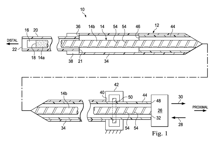
FIG. 1 is an enlarged scale longitudinally foreshortened schematic cross-
sectional
view through medical sensing catheter apparatus embodying principles of the
present
invention;
FIG. 2 is a longitudinally foreshortened schematic cross-sectional view
through a
telescope section of the catheter apparatus with an inner telescope portion of
the section being
in its fully retracted position; and
FIG. 3 is a view similar to that in FIG. 2 but with the inner telescope
portion of the
telescope section being in its fully extended position.
DETAILED DESCRIPTION
A catheter 10 embodying principles of the present invention is schematically
depicted
in FIGS. 1-3. By way of non-limiting example, the catheter apparatus 10 is a
medical sensing
catheter, and more specifically is an intravascular ultrasound (IVUS) imaging
catheter.
Catheter 10 includes an elongated flexible tubular assembly 12 that
circumscribes an
elongated flexible drive shaft or cable 14 having an ultrasound sensor 16 on
its distal end 18.
The tubular assembly 12 that circumscribes the drive cable 14 and the sensor
16
includes a sheath 20 having a proximal end 21, and a distal end 22 insertable
into the body of
a patient, and a telescope section 24 (see FIGS. 2 and 3) that facilitates
movement of the
drive cable 14 distally and proximally through the sheath 20 while it remains
stationary
within the patient's body. Selective rotation and translation of the drive
cable 14 relative to
the sheath 20 is effected by a conventional, schematically depicted
translational/rotational
drive mechanism 26 (FIG. 1) that may be selectively translated in distal and
proximal
directions as respectively illustrated by arrows 28,30 in FIG. 1. The drive
mechanism 26 is
operatively coupled to the proximal end 32 of the drive cable 14 and functions
in a
conventional manner to translate and rotate the drive cable 14.
Telescope section 24 includes an elongated flexible tubular outer catheter or
telescope
member 34 having a distal end 36 fixedly secured to an annular coupling 38
that
circumscribes and is fixedly secured to the proximal end of the sheath 20. The
proximal end
40 of the outer telescope member 34 is anchored to a schematically depicted
stationary
support structure 42 distally positioned relative to the drive mechanism 26.
The telescope
section 24 further includes an elongated flexible tubular inner catheter or
telescope member
44 which has distal and proximal ends 46,48 (see FIG. 1). Proximal end 48 is
secured to the
drive mechanism 26, and the inner telescope member 44 slidably extends through
an 0-ring
3
CA 02895170 2015-06-12
WO 2014/093465
PCT/US2013/074344
seal member 50 carried by the stationary support structure 42 which may be of
a conventional
construction and may be assembled around the 0-ring 50.
According to a feature of the present invention the 0-ring seal 50 is formed
of a self-
lubricating material, representatively a fluoroelastomeric material. The use
of a self-
lubricating seal member substantially facilitates and quickens the assembly of
the support
structure 42 by eliminating the necessity of lubricating the seal and one or
more of the
support structure parts prior to using the support structure 42.
As shown in FIGS. 1-3, the inner telescope member 44 is distally telescoped
into the
outer telescope member portion 34 of the overall tubular assembly 12 for
translation relative
thereto (by means of the drive mechanism 26) between a retracted position
shown in FIG. 2
(in which the sensor 16 is distally advanced within the sheath 20) and an
extended position
shown in FIG. 3 (in which the sensor is proximally retracted within the sheath
20).
According to a further feature of the present invention, the flexible drive
shaft 14 is
not of a uniform construction along its length. Instead, a first portion 14a
of the drive shaft
14 extending proximally away from the sensor 16 (see FIGS. 1 and 3) is of a
conventional
construction, representatively of a helically wound wire construction. Fixedly
and coaxially
secured to the proximal end of the drive shaft portion 14a (as, for example,
by an end weld 52
as shown in FIG. 3), and extending proximally away therefrom, is a second
representatively
metal drive shaft portion 14b. The relative lengths of the drive shaft
sections 14a,14b are
sized in a manner such that when the inner telescope member 44 is in its FIG.
3 extended
position the section 14b extends from the section 14a at least through
essentially the entire
interior length of the telescope section 24.
The flexible drive shaft section 14b has a stiffness sufficiently greater than
that of the
drive shaft portion 14a so as to be self-supporting during operation within
the telescope
section 24 when, as depicted in FIG. 3, the inner telescope member 44 is
proximally moved
away from its FIG. 2 retracted position toward or completely to its FIG. 3
extended position.
By way of non-limiting example, the flexible drive shaft section 14b may be a
tubular
helically cut metal beam member with the representatively illustrated helical
cut patterns 54
formed on its exterior surface. Although the illustrated embodiment shows the
drive shaft
section 14b as straight, it will be appreciated that the helical cuts 54 along
the tubular metal
beam allow the drive shaft section 14b to bend, if necessary, during operation
and still rotate
the sensor 16. However, the drive shaft section 14b could alternatively be of
a variety of
other materials and constructions without departing from principles of the
present invention.
4
CA 02895170 2015-06-12
WO 2014/093465
PCT/US2013/074344
For example, a sufficiently rigid polymer tube may be selected as the drive
shaft section 14b
that can be joined to the shaft section 14a via a mechanical coupling.
For purposes of manufacturing efficiency, the relatively stiffer self-
supporting flexible
drive shaft section 14b may, as schematically depicted in FIG. 1, extend from
its connection
52 at the drive shaft section 14a (see FIG. 3) to the drive mechanism 26.
Alternatively, the
length of the drive shaft section 14b may be somewhat shorter and connected at
its proximal
end to a terminal drive shaft section of a different construction such as, for
example, the
helically wound wire material used in the drive shaft section 14a, or a solid
metal material.
The unique incorporation in the catheter 10 of the self-supporting flexible
drive shaft
section 14b desirably eliminates the previous necessity of shielding and
supporting a drive
shaft portion exposed within the telescope section by providing and installing
a separate
protective structure within the telescope section.
While the catheter 10 has been representatively illustrated as being an IVUS
catheter,
it will be readily appreciated by those of ordinary skill in this particular
art that other types of
catheter structures with flexible internal drive shafts or cables and
associated telescope
sections may advantageously incorporate the above-described type of self-
supporting cable
structure without departing from principles of the present invention. Such
other types of
catheter structures and sensing elements include, for example, photo-acoustic,
optical
coherence tomography (OCT), phased array/multiple transducer, and
spectroscopic systems.
Still further, while the outer telescope member 34 is shown fixed to the
proximal end 21 of
the sheath 20, and the inner telescope member 44 is fixed to the drive
mechanism 26, these
fixation locations of the inner and outer telescope members 44,34 may be
reversed such that
the drive shaft 14 moves with the outer telescope member 34.
5