Note : Les descriptions sont présentées dans la langue officielle dans laquelle elles ont été soumises.
CA 02899992 2015-08-10
MARKING OF FLUOROSCOPE FIELD-OF-VIEW
FIELD OF THE INVENTION
The present invention relates generally to medical
imaging, and particularly to methods and systems for
visualization of fluoroscopic system Field-Of-View (FOV)
during medical procedures.
BACKGROUND OF THE INVENTION
Minimally invasive medical procedures commonly involve
real-time (RT) imaging such as fluoroscopic imaging,
sometimes in conjunction with other Three Dimensional (3D)
imaging modalities. Several publications deal with
registration of RT images with 3D models and 3D maps of
patient organs obtained by other modalities.
For example, U.S. Patent 8,515,527, whose disclosure is
incorporated herein by reference, describes a method and an
apparatus for registering 3D models of anatomical regions of
a heart and a tracking system with projection images of an
interventional fluoroscopic system.
U.S. Patent 7,327,872, whose disclosure is incorporated
herein by reference, describes a method and a system for
registering 3D models with projection images of anatomical
regions. A first image acquisition system of a first modality
employing a catheter at an anatomical region of a patient is
configured to produce a first image of the anatomical region
using fluoroscopy, the first image comprising a set of
fluoroscopy projection images. A second image acquisition
system of a second different modality is configured to
generate a 3D model of the anatomical region. An anatomical
reference system is common to both the first and second image
acquisition systems. A processing circuit is configured to
process executable instructions for registering the 3D model
with the fluoroscopy image in response to the common
1
CA 02899992 2015-08-10
, .
reference system and discernible parameters associated with
the catheter in both the first and second image acquisition
systems.
SUMMARY OF THE INVENTION
An embodiment of the present invention that is described
herein provides a method including registering a first
coordinate system of a fluoroscopic imaging system and a
second coordinate system of a magnetic position tracking
system. A three-dimensional (3D) map of an organ of a
patient, which is produced by the magnetic position tracking
system, is displayed. A 3D volume that would be irradiated by
the fluoroscopic imaging system is calculated using the
registered first and second coordinate systems, and the
calculated 3D volume is marked on the 3D map.
In some embodiments, marking the 3D volume includes
marking objects of the 3D map that fall inside the 3D volume.
In other embodiments, calculating and marking the 3D volume
are performed while the fluoroscopic imaging system does not
irradiate the patient. In yet other embodiments, the method
includes, in response to a change in a position of the
fluoroscopic imaging system relative to the organ,
recalculating the 3D volume, and re-marking the recalculated
3D volume on the 3D map.
There is additionally provided, in accordance with an
embodiment of the present invention, a system including an
interface and a processor. The interface is configured to
communicate with a fluoroscopic imaging system. The processor
is configured to register a first coordinate system of the
fluoroscopic imaging system and a second coordinate system of
a magnetic position tracking system, to display a three-
dimensional (3D) map of an organ of a patient, which is
produced by the magnetic position tracking system, to
calculate, using the registered first and second coordinate
2
CA 02899992 2015-08-10
,
systems, a 3D volume that would be irradiated by the
fluoroscopic imaging system, and to mark the calculated 3D
volume on the 3D map.
The present invention will be more fully understood from
the following detailed description of the embodiments
thereof, taken together with the drawings in which:
BRIEF DESCRIPTION OF THE DRAWINGS
Fig. 1 is a schematic pictorial illustration of a
fluoroscopic imaging system and a magnetic position tracking
system, in accordance with an embodiment of the present
invention;
Figs. 2A and 2B are schematic pictorial illustrations of
a simulated fluoroscopic system FOV overlaid on a 3D magnetic
position tracking map, in accordance with an embodiment of
the present invention; and
Fig. 3 is a flow chart that schematically illustrates a
method for visualizing a simulated fluoroscopic system FOV,
in accordance with an embodiment of the present invention.
DETAILED DESCRIPTION OF EMBODIMENTS
OVERVIEW
Catheterization processes are used in a variety of
therapeutic and diagnostic procedures. Catheter guidance
requires imaging capabilities, such as magnetic position
tracking. For example, Biosense-Webster, Inc. (Diamond Bar,
California) provides the CARTOTm system, used for navigating
a catheter in a patient heart.
In some scenarios, it is desirable to operate a
fluoroscopic system in parallel with the magnetic position
tracking system, in order to acquire a real-time image of the
organ in question. Fluoroscopic imaging, however, exposes the
patient and staff to potentially-hazardous doses of X-ray
radiation. In practice, the Field-Of-View (FOV) of the
3
CA 02899992 2015-08-10
fluoroscopic system is often narrow, and a considerable
portion of X-ray radiation is applied when attempting to
position the fluoroscopic system to image the desired area of
the organ.
Embodiments of the present invention that are described
herein provide improved methods and systems for operating a
fluoroscopic system and a magnetic position tracking system.
In some embodiments, a processor of the magnetic position
tracking system registers the coordinate systems of the
fluoroscopic system and the magnetic position tracking
system. Using the registration, the processor calculates a
volume (e.g., 3D funnel) that would be irradiated by the
fluoroscopic system, and marks this volume on a 3D map of the
organ produced by the magnetic position tracking system.
The disclosed techniques mark the position of the
fluoroscopic system 3D FOV to the physician, without having
to activate the fluoroscopic system. Using this technique,
the lengthy process of adjusting the fluoroscopic system FOV
can be performed without exposing the patient and staff to X-
ray radiation. The fluoroscopic system is typically activated
only after its FOV is positioned correctly.
Several example visualization techniques are described
herein. In some embodiments the processor is configured to
mark objects (e.g., anatomical features and medical
equipment) falling within the volume of the fluoroscopic
system FOV.
SYSTEM DESCRIPTION
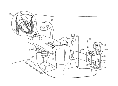
Fig. 1 is a schematic pictorial illustration of a
fluoroscopic imaging system 22 and a magnetic position
tracking system 20 during a minimally invasive cardiac
procedure, in accordance with an embodiment of the present
invention. Fluoroscopic imaging system 22 is connected to
magnetic position tracking system 20 via an interface 56.
4
CA 02899992 2015-08-10
Magnetic position tracking system 20 comprises a console 26,
and a catheter 24, which has a distal end 34 as shown in an
insert 32 of Fig. 1.
A cardiologist 42 (or any other user) navigates catheter
24 in a patient's heart 28, until distal end 34 reaches the
desired location in this organ, and then cardiologist 42
performs medical procedure using catheter 24. In other
embodiments, the disclosed techniques can be used with
procedures that are performed in any other organ, and instead
of cardiologist 42, any suitable user (such as a pertinent
physician, or an authorized technician) can operate the
system.
This method of position tracking is implemented, for
example, in the CARTOTM system, produced by Biosense Webster
Inc. (Diamond Bar, Calif.) and is described in detail in U.S.
Patents 5,391,199, 6,690,963, 6,484,118, 6,239,724, 6,618,612
and 6,332,089, in PCT Patent Publication WO 96/05768, and in
U.S. Patent Application Publications 2002/0065455 Al,
2003/0120150 Al and 2004/0068178 Al, whose disclosures are
all incorporated herein by reference.
Console 26 comprises a processor 58, a driver circuit
60, interface 56 to fluoroscopic imaging system 22, input
devices 46, and a display 40. Driver circuit 60 drives
magnetic field generators 36, which are placed at known
positions below a patient's 30 torso. In case a fluoroscopic
image is needed, cardiologist 42 uses input devices 46 and a
suitable Graphical User Interface (GUI) on display 40 to
request a fluoroscopic image in patient's heart 28.
Typically, processor 58 calculates and displays a 3D
volume (e.g., a funnel-shaped volume) that would be
irradiated by fluoroscopic imaging system 22. In other words,
the calculated volume marks the FOV of the fluoroscopic
system. The calculated 3D volume may have any suitable shape.
5
CA 02899992 2015-08-10
The description that follows refers mainly to a funnel-shaped
volume, for the sake of clarity, and the terms "3D volume"
and "3D funnel" are used interchangeably. The calculation can
be performed entirely without irradiating X-rays by
fluoroscopic imaging system 22.
In some embodiments, processor 58 calculates and
displays the 3D volume based on a-priori registration between
the coordinate systems of systems 20 and 22. Any suitable
registration process can be used for this purpose. In one
example process, one or more magnetic position sensors are
fitted on moving parts of fluoroscopic system 22. Position
tracking system 20 measures the positions of these sensors in
the coordinate system of system 20, and is thus able to
register the two coordinate systems. In another example
process, processor 58 identifies and correlates objects in
the 3D magnetic position map (produced by system 20) and in
the fluoroscopic images (produced by system 22), and uses the
correlation to register the coordinate systems of systems 20
and 22. Additional example registration processes are
described in the references cited in the Background section
of this application.
In some embodiments, processor 58 creates an overlaid
image of the 3D magnetic position tracking map with the
calculated fluoroscopic 3D funnel and displays this image on
display 40. The overlaid image comprises a marking of the
objects of the 3D position tracking map, which fall within
the calculated 3D funnel.
The configuration of system 20 shown in Fig. 1 is an
example configuration, which is chosen purely for the sake of
conceptual clarity. In alternative embodiments, any other
suitable configuration can be used for implementing the
system. Certain elements of system 20 can be implemented
using hardware, such as using one or more Application-
Specific Integrated Circuits (ASICs) or Field-Programmable
6
CA 02899992 2015-08-10
Gate Arrays (FPGAs) or other device types. Additionally or
alternatively, certain elements of system 20 can be
implemented using software, or using a combination of
hardware and software elements.
Processor 58 typically comprises a general-purpose
computer, which is programmed in software to carry out the
functions described herein. The software may be downloaded to
the computer in an electronic form, over a network, for
example, or it may, alternatively or additionally, be
provided and/or stored on non-transitory tangible media, such
as magnetic, optical, or electronic memory.
OVERLAY OF SIMULATED 3D FLUOROSCOPIC FUNNEL ON 3D MAP
In some embodiments, processor 58 of system 20 displays
a 3D map of patient's heart 28 comprising distal end 34, so
cardiologist 42 knows the exact location of distal end 34
with respect to the pertinent area in heart 28. During the
navigation and treatment process, cardiologist 42 may need
images of the pertinent organ around or near distal end 42.
The embodiments described herein fulfill the need for
minimizing X-ray irradiation while acquiring a 3D
fluoroscopic image.
In a typical flow, in case a fluoroscopic image is
needed in the vicinity of the catheter's distal end,
cardiologist 42 defines the desired area by positioning
fluoroscopic imaging system 22 to point to the desired
location. Processor 58 of system 20 calculates a simulated
volume (e.g., 3D funnel) that would be irradiated by
fluoroscopic imaging system 22 on the area in patient's heart
28 where fluoroscopic imaging system 22 is pointing, without
irradiating X-rays by fluoroscopic imaging system 22.
Processor 58 creates an overlaid image of the 3D
magnetic position tracking map with the calculated 3D funnel
and displays this image on display 40. In some embodiments,
7
CA 02899992 2015-08-10
the overlaid image comprises markers of the elements which
appear in the calculated 3D funnel and in the pertinent frame
of the 3D magnetic position tracking map. The marked elements
may comprise, for example, objects of patient's heart 28 or
other organ falling inside the simulated fluoroscopic 3D
funnel, and catheter's distal-end 34, if it falls into the
same 3D funnel.
In various embodiments, processor 58 may mark the
calculated 3D volume in various ways. For example, processor
58 may distinguish the 3D volume, and/or objects in the
volume, using different colors, different intensities,
different contrasts, or using any suitable visualization
means.
Cardiologist 42 examines the presented markers on
display 40. If the markers comprise the desired objects in
patient's heart 28, and distal-end 34, then fluoroscopic
imaging system 22 is positioned accurately and ready to
acquire a 3D fluoroscopic image. If the markers do not
comprise the desired objects in patient's heart 28 or distal-
end 34, fluoroscopic imaging system 22 is not positioned at
the desired location.
Typically, when cardiologist 42 concludes that
fluoroscopic imaging system 22 is positioned in the desired
location, he uses operating console 26 to request from
fluoroscopic imaging system 22 to acquire a fluoroscopic
image by irradiating the patient with ionizing X-rays. In
case of a positioning mismatch, cardiologist 42 moves patient
with respect to the irradiation head of fluoroscopic
imaging system 22, until the 3D funnel reaches the desired
30 location. Only then, cardiologist 42 (or another user) uses
console 26 to request fluoroscopic imaging system 22 to
acquire a fluoroscopic image and to collect the relevant
information required to continue the medical procedure.
8
CA 02899992 2015-08-10
In some embodiments cardiologist 42 may decide whether
the 3D funnel is located at the right position by looking at
the overlaid image with markers in screen 40. In alternative
embodiments, processor 58 may decide autonomously whether the
3D funnel is located in the desired location (e.g., if
distal-end 34 is in the center of the 3D funnel's FOV) and
recommend the medical staff to acquire a fluoroscopic image.
Depending on the Fluoroscopic system orientation, the
catheter can be centered in the funnel's FOV at various
angles, whereas the cardiologist may be interested in a
specific viewing angle. In some embodiments, cardiologist 42
specifies the required angle and imaging criteria. In
response, processor 58 calculates the new position,
illumination angle, and relative orientation required in
system 22, and instruct the system or the operator how to
operate system 22 to accomplish the new state.
Fig. 2A is a schematic pictorial illustration of a
simulated fluoroscopic 3D funnel 44, overlaid on a 3D
magnetic position tracking map 33, in accordance with an
embodiment of the present invention. An image of this sort is
displayed by processor 58 on display 40. Processor 58
calculates the location of simulated fluoroscopic 3D funnel
44 on 3D magnetic position tracking map 33, based on the
aligned coordinates of fluoroscopic imaging system 22 and
magnetic position tracking system 20. Processor 58 presents
the overlaid image, with marked elements in the 3D funnel's
Field of View (FOV), on display 40.
In the example that is presented in Fig. 2A, simulated
3D funnel 44 FOV is not positioned in the target location.
Distal-end 34 should be located at the center of the FOV of
simulated 3D funnel 44, and in this example, distal-end 34 is
not even within this FOV. In the example of Fig. 2A,
cardiologist 42 examines 3D funnel 44 overlaid on 3D map 33
of Fig. 2A and concludes that he/she should request to move
9
CA 02899992 2015-08-10
the 3D funnel's FOV up-and-right so distal-end 34 is located
in the center of the 3D funnel's FOV.
Fig. 2B is a schematic pictorial illustration of
simulated fluoroscopic 3D funnel 44, overlaid on 3D magnetic
position tracking map 33, in accordance with an embodiment of
the present invention. In this example, cardiologist 42 has
moved the FOV of 3D funnel 44 up-and-right from its location
in Fig. 2A, and positioned the simulated fluoroscopic 3D
funnel's 44 FOV in the desired location where distal-end 34
is in the center of the simulated fluoroscopic 3D funnel's
FOV, as shown in Fig. 2B.
In an embodiment, Fig. 2B is obtained by processor 58,
which calculates the location of simulated fluoroscopic 3D
funnel's 44 FOV and presents it on screen 40, overlaid on 3D
magnetic position tracking map 33, with markers of pertinent
elements falling within this FOV.
As shown in Fig. 2B, distal-end 34 is located in the
center of simulated fluoroscopic 3D funnel's 44 FOV and
pertinent objects are marked accordingly. In some embodiments
this accurate positioning of fluoroscopic imaging system 22
with respect to patient 30 and magnetic position tracking
system 20, is obtained based on the presented technique,
without exposing patient 30, cardiologist 42, and other
individuals in the operating room, to excess X-ray radiation.
Based on the image shown in Fig. 2B, which is created by
processor 58 and presented on display 40, cardiologist 42, or
any other suitable user, may proceed to use fluoroscopic
imaging system 22 and to acquire a fluoroscopic image the
desired location in patient's heart 28, which may comprise
distal-end 34 in the same FOV.
Fig. 3 is a flow chart that schematically illustrates a
method for minimizing irradiation of X-rays using markers of
a simulated fluoroscopic 3D funnel 44 on a position tracking
CA 02899992 2015-08-10
map 33, in accordance with an embodiment of the present
invention.
The method begins at a coordinate acquisition step 100,
where processor 58 acquires the coordinate systems of
fluoroscopic imaging system 22 and magnetic position tracking
system 20. At a coordinate alignment step 102, processor 58
aligns the coordinate systems of fluoroscopic imaging system
22 and magnetic position tracking system 20, in order to
match positions of a pertinent organ in patient 30 at both
systems.
At a position tracking presentation step 104, processor
58 presents 3D position tracking map 33 of a given organ of
patient 30. In an embodiment, the organ is heart 28, but may
be any pertinent organ of patient 30 in other embodiments. At
a 3D funnel calculation step 106, processor 58 receives the
planned irradiation setup parameters of fluoroscopic imaging
system 22 via interface 56, and calculates the 3D funnel that
would be irradiated by fluoroscopic imaging system 22 on the
pertinent organ of patient 30.
At an object marking step 108, processor 58 applies the
calculated 3D funnel obtained at 3D funnel calculation step
106 and the position of fluoroscopic imaging system 22 with
respect to magnetic position tracking system 20, to mark
objects falling inside simulated fluoroscopic 3D funnel 44,
on position tracking map 33. As a result, cardiologist 42 can
see on display 40 an overlaid image of position tracking map
33 with marked objects that would be obtained in case
cardiologist 42 applies fluoroscopic imaging system 22.
At a decision step 110, cardiologist 42 examines the
overlaid image comprising markers of simulated fluoroscopic
3D funnel 44 and decides whether fluoroscopic imaging system
22 is positioned at the desired location to acquire a 3D
fluoroscopic image. If cardiologist 42 decides that
fluoroscopic imaging system 22 is positioned at the desired
11
CA 02899992 2015-08-10
location, he/she uses input devices 46 and GUI on display 40
to command fluoroscopic imaging system 22 (via processor 58
and interface 56) to acquire a fluoroscopic image, at an
image acquisition step 114. Note that all the method steps
prior to step 114 are typically performed while fluoroscopic
system 22 does not emit X-ray radiation.
If cardiologist 42 decides that fluoroscopic imaging
system 22 is not positioned at the desired location, the
cardiologist repositions the fluoroscopic system relative to
the patient, at a repositioning step 112. At this point, in
various embodiments, the method may loop back to various
previous stages of the process.
In one embodiment, the flow loops back to 3D funnel
calculation step 106, in which processor 58 recalculates the
3D funnel that would be irradiated by fluoroscopic imaging
system 22 on the pertinent organ of patient 30.
In the description above, the process of recalculating
and visualizing the position of the fluoroscopic system FOV
is continuous and on-going. In alternative embodiments,
however, recalculation can be triggered by an event, e.g., in
response to detecting motion of the fluoroscopic system or in
response to a request from the user.
Although the embodiments described herein mainly address
cardiology applications, the methods and systems described
herein can also be used in other applications that involve
mapping registered with Fluoroscopic imaging.
It will thus be appreciated that the embodiments
described above are cited by way of example, and that the
present invention is not limited to what has been
particularly shown and described hereinabove. Rather, the
scope of the present invention includes both combinations and
sub-combinations of the various features described
hereinabove, as well as variations and modifications thereof
which would occur to persons skilled in the art upon reading
12
CA 02899992 2015-08-10
. .
the foregoing description and which are not disclosed in the
prior art. Documents incorporated by reference in the present
patent application are to be considered an integral part of
the application except that to the extent any terms are
defined in these incorporated documents in a manner that
conflicts with the definitions made explicitly or implicitly
in the present specification, only the definitions in the
present specification should be considered.
13