Note : Les descriptions sont présentées dans la langue officielle dans laquelle elles ont été soumises.
CA 02904623 2015-09-08
WO 2014/160507 PCT/US2014/026872
FUSION PROTEINS COMPRISING PDGF AND VEGF BINDING PORTIONS AND
METHODS OF USING THEREOF
CROSS REFERENCE TO RELATED APPLICATION
[0001] This application claims the benefit under 35 USC 119(e) of prior co-
pending U.S.
Provisional Patent Application No. 61/780,914, filed March 13, 2013, the
disclosure of which
is hereby incorporated by reference in its entirety.
SUBMISSION OF SEQUENCE LISTING ON ASCII TEXT FILE
[0002] The content of the following submission on ASCII text file is
incorporated herein
by reference in its entirety: a computer readable form (CRF) of the Sequence
Listing (file
name: 1597920095405EQLI5T.txt, date recorded: March 11, 2014, size: 113 KB).
FIELD OF THE INVENTION
[0003] The present invention relates to fusion proteins that inhibit the PDGF
pathway and
the VEGF pathway, compositions of these fusion proteins as well as methods for
producing
and using the same.
BACKGROUND OF THE INVENTION
[0004] Formation of new blood vessels, caused by the overproduction of growth
factors
such as vascular endothelial growth factor (VEGF), is a key component of
diseases like
tumor growth, age-related macular degeneration (AMD) and proliferative
diabetic
retinopathy (PDR) (Connolly et al., J Clin Invest., 1989, 84(5):1470-8;
Ferrara et al., Biochem
Biophys Res Commun., 1989, 161(2):851-9; and Ferrara et al., Nat Med., 1998,
4(3):336-40).
Wet AMD is the most severe form of AMD disease that is characterized by
abnormal
neovascularization beneath the retina and often leads to permanent vision
loss. Blocking of
VEGF with antibodies, soluble VEGF receptors, or inhibition of VEGF receptor
tyrosine
kinase activity are strategies that have shown promising preclinical and
clinical results in the
suppression of retinal neovascularization (Aiello et al., PNAS, 1995, 92:10457-
10461 and
Willet, et al., Nat Med., 2004, 10:145-147). However, recent clinical data
shows that new
vascular tissue typically does not regress with VEGF inhibition alone, because
pericytes,
which interact with endothelial cells and contribute to the establishment of
the blood-retinal
barrier, provide survival signals to neovascular endothelial cells and hence
make them
-1-
CA 02904623 2015-09-08
WO 2014/160507 PCT/US2014/026872
resistant to VEGF withdrawal (Benjamin et al., Development, 1998, 125(9)1591-8
and Patel
S., Retina, 2009, 29(6 Suppl):S45-8). Furthermore, platelet-derived growth
factor isoform B
(PDGF-B) and PDGF receptor-beta (PDGFRI3), found in proliferative retinal
membranes,
have important roles in recruitment of pericytes for stabilization of the
developing
vasculature (Robbins et al., Invest Opth Vis Sci., 1994, 35(10):3649-63;
Lindahl et al.,
Development, 1997, 124:3943-3953; and Hellstrom et al., Development, 1999,
126:3047-
3055).
[0005] The VEGF binding function of VEGFR1 (Flt-1) has been mapped to the
second
extracellular domain (ECD) (Davis-Smyth et al., EMBO J., 1996, 15:4919-4927;
Barleon et
al., J Biol Chem., 1997, 272:10382-10388; Wiesmann et al., Cell, 1997, 91:695-
704; and
Davis-Smyth et al., J Biol Chem., 1998, 273:3216-3222). A naturally occurring
alternatively
spliced form of high affinity VEGF-binding receptor, soluble VEGFR1 (sFlt1),
exists as a
secreted protein that functions primarily as a decoy receptor (Shibuya et al.,
Oncogene, 1990,
5:519-524 and Kendall et al., PNAS, 1993, 90:10705-10709). A soluble receptor,
VEGF-
Trap, engineered for therapeutic use, has the second domain of VEGFR1 fused to
the third
domain of VEGFR2 (KDR) and to human IgG1 Fc region (Holash et al. 2002). An
extracellular region of PDGFRI3 was previously shown to antagonize PDGF-B
stimulated
responses (Duan et al., J Biol Chem, 1991, 266(1)413-8 and Ueno et al.,
Science, 1991,
252(5007):844-8). Studies with PDGFRP-Fc chimera demonstrated that human
PDGFRI3
ECDs 1 to 3 are sufficient for high-affinity PDGF-B ligand binding (Heidaran
et al., FASEB
J., 1995, 9(1):140-5 and Lokker et al., J Biol Chem, 1997, 272(52):33037-44).
An effect of
predimerization on high-affinity PDGF-B ligand binding was also described when
PDGFRI3
ECDs 1 to 3 were fused to glutathione S-transferase (GST) domain (Leppanen et
al.,
Biochemistry, 2000, 39(9):2370-5).
[0006] Current eye treatments require monthly intravitreal injections for
years by a retinal
specialist. Therefore, there is a need for improved therapeutic agents and an
approach to
deliver such therapeutic agents to sites such as the eye.
BRIEF SUMMARY OF THE INVENTION
[0007] The invention provided herein discloses, inter alia, fusion proteins
that inhibit the
PDGF pathway and the VEGF pathway, compositions comprising these fusion
proteins and
compositions comprising viral particles comprising a nucleic acid encoding the
fusion
protein, as well as methods for producing and using these fusion proteins and
viral particles
-2-
CA 02904623 2015-09-08
WO 2014/160507 PCT/US2014/026872
for the treatment or prevention of a disease such as an ocular disease, an
inflammatory
disease, an autoimmune disease, or cancer.
[0008] Accordingly, in one aspect, the invention provides a fusion protein
comprising (a)
an extracellular portion of a PDGF receptor, (b) an extracellular portion of a
VEGF receptor,
and (c) a multimerization domain, wherein the fusion proteins binds to a PDGF
and a VEGF.
In some embodiments, the fusion protein is arranged from N-terminus to C-
terminus in the
following order: (a), (b) and (c). In some embodiments, the PDGF receptor is a
PDGFRI3. In
some embodiments herein, the extracellular portion of the PDGFR comprises the
Ig-like
domains D1-D3 of the PDGFR. In some embodiments herein, the extracellular
portion of the
PDGFR comprises the Ig-like domains D1-D4 of the PDGFR. In some embodiments
herein,
the extracellular portion of the PDGFR comprises the Ig-like domains D1-D5 of
the PDGFR.
In some embodiments herein, the extracellular portion of the PDGFR comprises
the amino
acid sequence SEQ ID NO:1, 2, or 3, or an amino acid sequence having at least
85% identity
to SEQ ID NO:1, 2, or 3. In some embodiments herein, the extracellular portion
of the
VEGF receptor comprises an Ig-like domain D2 of a VEGF receptor. In some
embodiments
herein, the extracellular portion of the VEGF receptor comprises an Ig-like
domain D2 of a
VEGFR1 (FLT-1). In some embodiments herein, the extracellular portion of the
VEGF
receptor comprises an Ig-like domain D2 of a VEGFR1 (FLT-1) and an Ig-like
domain D3 of
a VEGFR2. In some embodiments herein, the extracellular portion of the VEGF
receptor
comprises the Ig-like domains D1-D3 of a VEGFR1 (FLT-1). In some embodiments
herein,
the extracellular portion of the VEGF receptor comprises the amino acid
sequence of SEQ ID
NO:4 or 5, or an amino acid sequence having at least 85% identity to SEQ ID
NO:4 or 5. In
some embodiments herein, the fusion protein further comprises a linker peptide
between the
extracellular portion of the PDGF receptor and the extracellular portion of
the VEGF
receptor, and/or a peptide linker between the extracellular portion of the
VEGF receptor and
the multimerization domain. In a further embodiment, the peptide linker
comprises the
amino acid sequence selected from the group consisting of G1y9(SEQ ID NO:47),
G1u9(SEQ
ID NO:48), Ser9(SEQ ID NO:49), G1y5-Cys-Pro2-Cys (SEQ ID NO:50), (G1y4-Ser)3
(SEQ ID
NO: 51), Ser-Cys-Val-Pro-Leu-Met-Arg-Cys-Gly-Gly-Cys-Cys-Asn (SEQ ID NO: 52),
Pro-
Ser-Cys-Val-Pro-Leu-Met-Arg-Cys-Gly-Gly-Cys-Cys-Asn (SEQ ID NO: 53), Gly-Asp-
Leu-
Ile-Tyr-Arg-Asn-Gln-Lys (SEQ ID NO: 54), and G1y9-Pro-Ser-Cys-Val-Pro-Leu-Met-
Arg-
Cys-Gly-Gly-Cys-Cys-Asn (SEQ ID NO:55). In some embodiments herein, the
multimerization domain is a Fc region of an antibody. In a further embodiment,
the Fc region
comprises a CH3 region of IgG 1, IgG2, IgG3, or IgG4, or a CH2 and a CH3
region of IgG 1,
-3-
CA 02904623 2015-09-08
WO 2014/160507 PCT/US2014/026872
IgG2, IgG3, or IgG4. In some embodiments herein, the Fe region comprises the
amino acid
sequence of SEQ ID NO:6, or an amino acid sequence having at least 85%
identity to SEQ
ID NO:6. In some embodiments herein, the fusion protein comprises the amino
acid
sequence of SEQ ID NO:13 or 15, or an amino acid sequence having at least 85%
identity to
SEQ ID NO:13 or 15. In some embodiments herein, the fusion protein is in a
multimeric
form. In some of the embodiments herein, the fusion protein is in a dimeric
form.
[0009] In one aspect, the invention provides a fusion protein produced by
culturing a host
cell comprising a nucleic acid encoding any of the fusion proteins disclosed
herein under a
condition that produces the fusion protein, and recovering the fusion protein
produced by the
host cell.
[0010] In another aspect, the invention provides a dimeric fusion protein
comprising two
fusion proteins, wherein each fusion protein comprises any of the fusion
proteins disclosed
herein.
[0011] In yet another aspect, the invention provides a composition comprising
any of the
fusion proteins disclosed herein and a pharmaceutically acceptable carrier.
[0012] In still another aspect, the invention provides a nucleic acid encoding
any of the
fusion proteins disclosed herein.
[0013] In some aspects, the invention also provides a host cell comprising a
nucleotide
sequence encoding any of the fusion proteins disclosed herein.
[0014] In some aspects, the invention provides a method of producing a fusion
protein,
comprising culturing a host cell comprising a nucleic acid encoding any of the
fusion proteins
disclosed herein under a condition that produces the fusion protein, and
recovering the fusion
protein produced by the host cell. In further embodiments, the host cell is a
mammalian cell.
In further embodiments, the host cell is a bacterial cell. In further
embodiments, the host cell
is an Escherichia coli cell.
[0015] In another aspect, the invention provides a method of delivering a
fusion protein to
a subject comprising administering an effective amount of any of the fusion
proteins
disclosed herein to the subject. In some embodiments, the subject has macular
degeneration
or proliferative diabetic retinopathy. In a further embodiment, the macular
degeneration is
wet age-related macular degeneration or dry age-related macular degeneration.
In some
embodiments herein, the fusion protein is administered by intravitreal
injection to the subject.
In some embodiments, the subject has cancer. In some embodiments, the subject
has
rheumatoid arthritis, osteoarthritis, or asthma. In some embodiments, the
subject has uveitis
or corneal neovascularization.
-4-
CA 02904623 2015-09-08
WO 2014/160507 PCT/US2014/026872
[0016] In some aspects, the invention provides a vector comprising a
nucleotide sequence
encoding any of the fusion proteins disclosed herein. In some embodiments, the
vector is a
viral vector. In a further embodiment, the viral vector is a recombinant adeno-
associated
virus vector (rAAV). In further embodiments, the rAAV vector comprises an ITR
of AAV1,
AAV2, AAV3, AAV4, AAV5, AAV6, AAV7, AAV8, AAV9, AAVrh8, AAVrh8R, or
AAVrh10.
[0017] In one aspect, the invention also provides an rAAV particle comprising
a nucleic
acid encoding any of the fusion proteins disclosed herein. In some
embodiments, the rAAV
particle comprises capsid proteins of AAV1, AAV2, AAV3, AAV4, AAV5, AAV6,
AAV7,
AAV8, AAV9, AAVrh8, AAVrh8R, or AAVrh10. In some embodiments, the nucleic acid
comprises an ITR from a serotype different from the serotype of the capsid. In
a further
embodiment, the ITR is an ITR of AAV1, AAV2, AAV3, AAV4, AAV5, AAV6, AAV7,
AAV8, AAV9, AAVrh8, AAVrh8R, or AAVrh10.
[0018] In yet another aspect, the invention provides a method of producing an
rAAV
particle, comprising (a) culturing a host cell under a condition that rAAV
particles are
produced, wherein the host cell comprises (i) one or more AAV package genes,
wherein each
said AAV packaging gene encodes an AAV replication or encapsidation protein;
(ii) an
rAAV pro-vector comprising a nucleotide encoding any of the fusion proteins
disclosed
herein flanked by at least one AAV ITR, and (iii) an AAV helper function; and
(b) recovering
the rAAV particles produced by the host cell. In a further embodiment, the
rAAV particles
are purified.
[0019] In still another aspect, the invention provides a method of delivering
a viral vector
to a subject, comprising administering any of the rAAV particles disclosed
herein to the
subject, wherein the fusion protein encoded by the rAAV particle is expressed
in the subject.
In some embodiments, the invention provides a method of delivering a viral
vector to a
subject, comprising administering any of the rAAV particles disclosed herein
to the subject,
wherein an effective amount of the fusion protein encoded by the rAAV particle
is expressed
in the subject. In some embodiments, the subject has macular degeneration or
proliferative
diabetic retinopathy. In a further embodiment, the macular degeneration is wet
age-related
macular degeneration or dry age-related macular degeneration. In some
embodiments herein,
the rAAV particle is administered by intravitreal injection to the subject. In
some
embodiments, the subject has cancer. In some embodiments, the subject has
rheumatoid
arthritis, osteoarthritis, or asthma. In some embodiments, the subject has
uveitis or corneal
neovascularization.
-5-
CA 02904623 2015-09-08
WO 2014/160507 PCT/US2014/026872
[0020] The specification is considered to be sufficient to enable one skilled
in the art to
practice the invention. Various modifications of the invention in addition to
those shown and
described herein will become apparent to those skilled in the art from the
foregoing
description and fall within the scope of the appended claims. All
publications, patents, and
patent applications cited herein are hereby incorporated by reference in their
entirety for all
purposes.
BRIEF DESCRIPTION OF THE DRAWINGS
[0021] Figure 1 shows generation of truncated PDGFR-13 soluble receptors. A)
Schematic
of the PDGFR-13 IgG1 Fc-coupled dimerizing forms, PDGFR(D1-D5)9G-Fc, PDGFR(D1-
D2)9G-Fc, and PDGFR(D1-D3)9G-Fc as well as the PDGFR-13 monomeric receptor
forms,
PDGFR(D1-D4) and PDGFR(D1-D5). White blocks indicate PDGFR-13 sequences,
including the extracellular domains and the signal peptide (sp). Diagonal
shaded blocks
represent 9Gly linker and dark dotted blocks represent domains CH2 and CH3 of
human
IgG1 Fc region. B) Western blots of monomeric sPDGFR-13 and IgG1 Fc-coupled
dimerizing
soluble receptor forms under reducing (left panel) and non-reducing (right
panel) conditions.
Protein was detected with anti-PDGFR-13 antibody.
[0022] Figure 2 shows a volumetric PDGF BB binding assay of truncated PDGFR-13
soluble receptors. A) Monomeric PDGFR-13 soluble receptor forms PDGFR(D1-D4)
and
PDGFR(D1-D5) as compared to the full-size IgG1 Fc-coupled dimerizing form
PDGFR(D1-
D5)9G-Fc. B) Dimeric IgG1 Fc-coupled sPDGFR-13 soluble receptor forms PDGFR(D1-
D2)9G-Fc and PDGFR(D1-D3)9G-Fc as compared to the IgG1 Fc-coupled dimerizing
form
PDGFR(D1-D5)9G-Fc. Increasing volumes (0) of conditioned media (CM) containing
soluble receptors (x axis) from representative transfections were incubated
overnight with
human PDGF BB ligand and the amount of unbound ligand (y axis) was measured by
ELISA.
Data expressed as mean SD (n=3); all receptors were significantly different
in PDGF
binding affinities by 2-way ANOVA; Bonferroni Test; ***P<0.00 1.
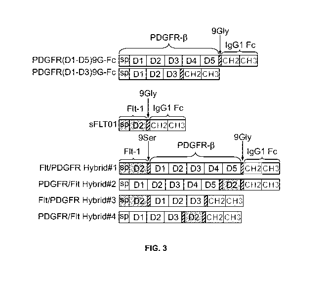
[0023] Figure 3 is a schematic of VEGFR1/ PDGFR-13 and PDGFR-13/VEGFR1 hybrid
proteins, Hybrids 1 to 4, and their parental constructs PDGFR(D1-D5)9G-Fc,
PDGFR(D1-
D3)9G-Fc and sFLT01. White blocks indicate PDGFR-13 sequences, grey blocks
indicate
VEGFR1 (Flt- 1) sequences, including their extracellular domains and signal
peptides (sp).
Diagonal shaded blocks represent 9Gly or 9Ser linkers and dark dotted blocks
represent
domains CH2 and CH3 of the human IgG1 Fc region.
-6-
CA 02904623 2015-09-08
WO 2014/160507 PCT/US2014/026872
[0024] Figure 4 is a Western blot of VEGFR1/ PDGFR-13 and PDGFR-13/VEGFR1
hybrid
proteins, Hybrids 1 to 4, as compared to full-size IgG1 Fe-coupled dimerizing
form
PDGFR(D1-D5)9G-Fc (shown as (D1-D5)9G-Fc) under reducing (left panel) and non-
reducing (right panel) conditions. Protein was detected with anti-PDGFR-13
antibody and
anti-Flt-1 antibody. Samples containing Hybrids 3 and 4 were duplicates from
individual
transfections.
[0025] Figure 5 shows graphs demonstrating inhibition of VEGF-induced and VEGF
+
PDGF-13-induced proliferation of human umbilical vein endothelial cells
(HUVECs) by
hybrid protein, Hybrids 1 to 4. A) HUVEC proliferation assay with VEGF only:
Inhibitory
effect of 5 pi conditioned media (CM) containing soluble receptors on HUVEC
proliferation
was compared in the presence of VEGF (10 ng/ml) only. B) HUVEC competitive
proliferation assay in the presence of VEGF (10 ng/ml) and PDGF (20 ng/ml):
Inhibitory
effect of 5 pi CM containing soluble receptors on HUVEC proliferation was
compared in the
presence of both ligands, VEGF and PDGF. Samples from three independent
transfections
(n=3) were evaluated in one assay. Data expressed as mean + SD. One-way ANOVA;
Tukey's Test; ***p<0.001 for difference between positive control VEGF+ alone
or VEGF+
in combination with PDGF BB+ versus other samples. Control = EGFP CM; VEGF+ =
EGFP CM + 10 ng/ml VEGF; BB+ Only = EGFP CM +20 ng/ml; VEGF+BB+ = EGFP CM
+ 10 ng/ml VEGF +20 ng/ml PDGF BB.
[0026] Figure 6 shows volumetric binding assays of hybrid proteins. A) PDGF BB
volumetric binding assay of Hybrid proteins 1 to 4 as compared to PDGFR(D1-
D5)9G-Fc.
B) VEGF volumetric binding assay of Hybrid proteins 1 to 4 as compared to
sFLT01.
Increasing conditioned media volumes containing soluble receptors (x axis)
from
representative transfections were incubated overnight with either human PDGF
BB or VEGF
ligands and the amount of unbound ligand (y axis) was measured by ELISA in
triplicates.
[0027] Figure 7 shows competitive VEGF and PDGF cell-free binding assays of
hybrid
proteins. A) Comparison of Hybrid 3 (Hyb#3), Hybrid 4 (Hyb #4), and PDGFR(D1-
D3)9G-
Fc in increasing conditioned media volumes (x-axis) and the amount of the
unbound PDGF
ligand (y axis) as measured by PDGF BB ELISA. B) Comparison of Hybrid 3
(Hyb#3),
Hybrid 4 (Hyb #4), and sFltT01 in increasing conditioned media volumes (x-
axis) and the
amount of the unbound VEGF ligand (y axis) as measured by VEGF ELISA.
[0028] Figure 8 is a graph showing in vivo efficacy of AAV2.Hybrid 4
intravitreal delivery
in a mouse choroidal neovascularization (CNV) laser model. Number of burns
without
neovascularization (NV) in the AAV2.Hybrid 4 (shown as Hybrid-4), AAV2.sFLT02
(shown
-7-
CA 02904623 2015-09-08
WO 2014/160507 PCT/US2014/026872
as sFLT02), AAV2.PDGFR (shown as PDGFR) treated (left) eye was compared to the
untreated contralateral (right; Naive) eye. Data from both eyes (n=20 eyes per
treatment) was
expressed as percentage of burns without CNV. The PDGFR portion used for
construction of
AAV2.PDGFR was PDGFR(D1-D3)9G-Fc.
DETAILED DESCRIPTION
[0029] The present invention provides, inter alia, fusion proteins, and
compositions
thereof, that inhibit the plasma-derived growth factor (PDGF) signaling
pathway and the
vascular endothelial growth factor (VEGF) signaling pathway. A fusion protein
of the
invention as described herein comprises an extracellular portion of a PDGF
receptor
(PDGFR), an extracellular portion of a VEGF receptor (VEGFR), and a
multimerization
domain, wherein the fusion protein binds to a PDGF and a VEGF for inhibition
of PDGF
activity and VEGF activity, respectively. Also provided herein are methods for
production of
the fusion proteins, methods of delivery of the fusion proteins, and methods
of using the
fusion proteins in the treatment of ocular diseases, autoimmune diseases,
inflammatory
diseases, and/or cancer.
I. General techniques
[0030] The techniques and procedures described or referenced herein are
generally well
understood and commonly employed using conventional methodology by those
skilled in the
art, such as, for example, the widely utilized methodologies described in
Molecular Cloning:
A Laboratory Manual (Sambrook et al., 4th ed., Cold Spring Harbor Laboratory
Press, Cold
Spring Harbor, N.Y., 2012); Current Protocols in Molecular Biology (F.M.
Ausubel, et al.
eds., 2003); the series Methods in Enzymology (Academic Press, Inc.); PCR 2: A
Practical
Approach (M.J. MacPherson, B.D. Hames and G.R. Taylor eds., 1995); Antibodies,
A
Laboratory Manual (Harlow and Lane, eds., 1988); Culture of Animal Cells: A
Manual of
Basic Technique and Specialized Applications (R.I. Freshney, 6" ed., J. Wiley
and Sons,
2010); Oligonucleotide Synthesis (M.J. Gait, ed., 1984); Methods in Molecular
Biology,
Humana Press; Cell Biology: A Laboratory Notebook (J.E. Cellis, ed., Academic
Press,
1998); Introduction to Cell and Tissue Culture (J.P. Mather and P.E. Roberts,
Plenum Press,
1998); Cell and Tissue Culture: Laboratory Procedures (A. Doyle, J.B.
Griffiths, and D.G.
Newell, eds., J. Wiley and Sons, 1993-8); Handbook of Experimental Immunology
(D.M.
Weir and C.C. Blackwell, eds., 1996); Gene Transfer Vectors for Mammalian
Cells (J.M.
Miller and M.P. Cabs, eds., 1987); PCR: The Polymerase Chain Reaction, (Mullis
et al.,
-8-
CA 02904623 2015-09-08
WO 2014/160507 PCT/US2014/026872
eds., 1994); Current Protocols in Immunology (J.E. Coligan et al., eds.,
1991); Short
Protocols in Molecular Biology (Ausubel et al., eds., J. Wiley and Sons,
2002);
Immunobiology (C.A. Janeway et al., 2004); Antibodies (P. Finch, 1997);
Antibodies: A
Practical Approach (D. Catty., ed., IRL Press, 1988-1989); Monoclonal
Antibodies: A
Practical Approach (P. Shepherd and C. Dean, eds., Oxford University Press,
2000); Using
Antibodies: A Laboratory Manual (E. Harlow and D. Lane, Cold Spring Harbor
Laboratory
Press, 1999); The Antibodies (M. Zanetti and J. D. Capra, eds., Harwood
Academic
Publishers, 1995); and Cancer: Principles and Practice of Oncology (V.T.
DeVita et al., eds.,
J.B. Lippincott Company, 2011).
II. Definitions
[0031] A "vector," as used herein, refers to a recombinant plasmid or virus
that comprises a
nucleic acid to be delivered into a host cell, either in vitro or in vivo.
[0032] The term "polynucleotide" or "nucleic acid" as used herein refers to a
polymeric
form of nucleotides of any length, either ribonucleotides or
deoxyribonucleotides. Thus, this
term includes, but is not limited to, single-, double- or multi-stranded DNA
or RNA, genomic
DNA, cDNA, DNA-RNA hybrids, or a polymer comprising purine and pyrimidine
bases, or
other natural, chemically or biochemically modified, non-natural, or
derivatized nucleotide
bases. The backbone of the polynucleotide can comprise sugars and phosphate
groups (as
may typically be found in RNA or DNA), or modified or substituted sugar or
phosphate
groups. Alternatively, the backbone of the polynucleotide can comprise a
polymer of
synthetic subunits such as phosphoramidates and thus can be a
oligodeoxynucleoside
phosphoramidate (P-NH2) or a mixed phosphoramidate- phosphodiester oligomer.
In
addition, a double-stranded polynucleotide can be obtained from the single
stranded
polynucleotide product of chemical synthesis either by synthesizing the
complementary
strand and annealing the strands under appropriate conditions, or by
synthesizing the
complementary strand de novo using a DNA polymerase with an appropriate
primer.
[0033] A "recombinant viral vector" refers to a recombinant polynucleotide
vector
comprising one or more heterologous sequences (i.e., nucleic acid sequence not
of viral
origin). In the case of recombinant AAV vectors, the recombinant nucleic acid
is flanked by
at least one, preferably two, inverted terminal repeat sequences (ITRs).
[0034] A "recombinant AAV vector (rAAV vector)" refers to a polynucleotide
vector
comprising one or more heterologous sequences (i.e., nucleic acid sequence not
of AAV
origin) that are flanked by at least one, preferably two, AAV inverted
terminal repeat
-9-
CA 02904623 2015-09-08
WO 2014/160507 PCT/US2014/026872
sequences (ITRs). Such rAAV vectors can be replicated and packaged into
infectious viral
particles when present in a host cell that has been infected with a suitable
helper virus (or that
is expressing suitable helper functions) and that is expressing AAV rep and
cap gene products
(i.e. AAV Rep and Cap proteins). When a rAAV vector is incorporated into a
larger
polynucleotide (e.g. in a chromosome or in another vector such as a plasmid
used for cloning
or transfection), then the rAAV vector may be referred to as a "pro-vector"
which can be
"rescued" by replication and encapsidation in the presence of AAV packaging
functions and
suitable helper functions. An rAAV can be in any of a number of forms,
including, but not
limited to, plasmids, linear artificial chromosomes, complexed with lips,
encapsulated within
liposomes, and, most preferable, encapsidated in a viral particle,
particularly AAV. A rAAV
vector can be packaged into an AAV virus capsid to generate a "recombinant
adeno-
associated virus particle (rAAV particle)".
[0035] "Heterologous" means derived from a genotypically distinct entity from
that of the
rest of the entity to which it is compared or into which it is introduced or
incorporated. For
example, a polynucleotide introduced by genetic engineering techniques into a
different cell
type is a heterologous polynucleotide (and, when expressed, can encode a
heterologous
polypeptide). Similarly, a cellular sequence (e.g., a gene or portion thereof)
that is
incorporated into a viral vector, is a heterologous nucleotide sequence with
respect to the
vector.
[0036] An "inverted terminal repeat" or "ITR" sequence is a term well
understood in the art
and refers to relatively short sequences found at the termini of viral genomes
which are in
opposite orientation.
[0037] An "AAV inverted terminal repeat (ITR)" sequence, a term well-
understood in the
art, is an approximately 145-nucleotide sequence that is present at both
termini of the native
single-stranded AAV genome. The outermost 125 nucleotides of the ITR can be
present in
either of two alternative orientations, leading to heterogeneity between
different AAV
genomes and between the two ends of a single AAV genome. The outermost 125
nucleotides
also contains several shorter regions of self-complementarity, allowing
intrastrand base-
pairing to occur within this portion of the ITR.
[0038] A "terminal resolution sequence" or "trs" is a sequence in the D region
of the AAV
ITR that is cleaved by AAV rep proteins during viral DNA replication. A mutant
terminal
resolution sequence is refractory to cleavage by AAV rep proteins.
[0039] The terms "genome particles (gp)," "genome equivalents," or "genome
copies" as
used in reference to a viral titer, refer to the number of virions containing
the recombinant
-10-
CA 02904623 2015-09-08
WO 2014/160507 PCT/US2014/026872
AAV DNA genome, regardless of infectivity or functionality. The number of
genome
particles in a particular vector preparation can be measured by procedures
such as described
in the Examples herein, or for example, in Clark et al. (1999) Hum. Gene
Ther., 10:1031-
1039; Veldwijk et al. (2002) Mol. Ther., 6:272-278.
[0040] The terms "infection unit (iu)," "infectious particle," or "replication
unit," as used in
reference to a viral titer, refer to the number of infectious and replication-
competent
recombinant AAV vector particles as measured by the infectious center assay,
also known as
replication center assay, as described, for example, in McLaughlin et al.
(1988) J. Virol.,
62:1963-1973.
[0041] The term "transducing unit (tu)" as used in reference to a viral titer,
refers to the
number of infectious recombinant AAV vector particles that result in the
production of a
functional transgene product as measured in functional assays such as
described in Examples
herein, or for example, in Xiao et al. (1997) Exp. Neurobiol., 144:113-124; or
in Fisher et al.
(1996) J. Virol., 70:520-532 (LFU assay).
[0042] A "helper virus" for AAV refers to a virus that allows AAV (which is a
defective
parvovirus) to be replicated and packaged by a host cell. A number of such
helper viruses
have been identified, including adenoviruses, herpesviruses and poxviruses
such as vaccinia.
The adenoviruses encompass a number of different subgroups, although
Adenovirus type 5 of
subgroup C (Ad5) is most commonly used. Numerous adenoviruses of human, non-
human
mammalian and avian origin are known and are available from depositories such
as the
ATCC. Viruses of the herpes family, which are also available from depositories
such as
ATCC, include, for example, herpes simplex viruses (HSV), Epstein-Ban viruses
(EBV),
cytomegaloviruses (CMV) and pseudorabies viruses (PRV).
[0043] A "fusion protein" refers to a protein having two or more portions
covalently linked
together, where each of the portions is derived from different proteins.
[0044] "Percent (%) sequence identity" with respect to a reference polypeptide
or nucleic
acid sequence is defined as the percentage of amino acid residues or
nucleotides in a
candidate sequence that are identical with the amino acid residues or
nucleotides in the
reference polypeptide or nucleic acid sequence, after aligning the sequences
and introducing
gaps, if necessary, to achieve the maximum percent sequence identity, and not
considering
any conservative substitutions as part of the sequence identity. Alignment for
purposes of
determining percent amino acid or nucleic acid sequence identity can be
achieved in various
ways that are within the skill in the art, for instance, using publicly
available computer
software programs, for example, those described in Current Protocols in
Molecular Biology
-11-
CA 02904623 2015-09-08
WO 2014/160507 PCT/US2014/026872
(Ausubel et al., eds., 1987), Supp. 30, section 7.7.18, Table 7.7.1, and
including BLAST,
BLAST-2, ALIGN or Megalign (DNASTAR) software. A preferred alignment program
is
ALIGN Plus (Scientific and Educational Software, Pennsylvania). Those skilled
in the art
can determine appropriate parameters for measuring alignment, including any
algorithms
needed to achieve maximal alignment over the full length of the sequences
being compared.
For purposes herein, the % amino acid sequence identity of a given amino acid
sequence A
to, with, or against a given amino acid sequence B (which can alternatively be
phrased as a
given amino acid sequence A that has or comprises a certain % amino acid
sequence identity
to, with, or against a given amino acid sequence B) is calculated as follows:
100 times the
fraction X/Y, where X is the number of amino acid residues scored as identical
matches by
the sequence alignment program in that program's alignment of A and B, and
where Y is the
total number of amino acid residues in B. It will be appreciated that where
the length of
amino acid sequence A is not equal to the length of amino acid sequence B, the
% amino acid
sequence identity of A to B will not equal the % amino acid sequence identity
of B to A. For
purposes herein, the % nucleic acid sequence identity of a given nucleic acid
sequence C to,
with, or against a given nucleic acid sequence D (which can alternatively be
phrased as a
given nucleic acid sequence C that has or comprises a certain % nucleic acid
sequence
identity to, with, or against a given nucleic acid sequence D) is calculated
as follows: 100
times the fraction W/Z, where W is the number of nucleotides scored as
identical matches by
the sequence alignment program in that program's alignment of C and D, and
where Z is the
total number of nucleotides in D. It will be appreciated that where the length
of nucleic acid
sequence C is not equal to the length of nucleic acid sequence D, the %
nucleic acid sequence
identity of C to D will not equal the % nucleic acid sequence identity of D to
C.
[0045] An "isolated" molecule (e.g., nucleic acid or protein) or cell means it
has been
identified and separated and/or recovered from a component of its natural
environment.
[0046] An "effective amount" is an amount sufficient to effect beneficial or
desired results,
including clinical results. An effective amount can be administered in one or
more
administrations. In terms of a disease state, an effective amount is an amount
sufficient to
ameliorate, stabilize, or delay development of a disease.
[0047] An "individual" or "subject" is a mammal. Mammals include, but are not
limited to,
domesticated animals (e.g., cows, sheep, cats, dogs, and horses), primates
(e.g., humans and
non-human primates such as monkeys), rabbits, and rodents (e.g., mice and
rats). In certain
embodiments, the individual or subject is a human.
-12-
CA 02904623 2015-09-08
WO 2014/160507 PCT/US2014/026872
[0048] As used herein, "treatment" is an approach for obtaining beneficial or
desired
clinical results. For purposes of this invention, beneficial or desired
clinical results include,
but are not limited to, alleviation of symptoms, diminishment of extent of
disease, stabilized
(i.e., not worsening) state of disease, preventing spread (i.e., metastasis)
of disease, delay or
slowing of disease progression, amelioration or palliation of the disease
state, and remission
(whether partial or total), whether detectable or undetectable. "Treatment"
can also mean
prolonging survival as compared to expected survival if not receiving
treatment.
[0049] Reference to "about" a value or parameter herein includes (and
describes)
embodiments that are directed to that value or parameter per se. For example,
description
referring to "about X" includes description of "X."
[0050] As used herein, the singular form of the articles "a," "an," and "the"
includes plural
references unless indicated otherwise. For example, the phrase "a rAAV
particle" includes
one or more rAAV particles.
[0051] It is understood that aspects and embodiments of the invention
described herein
include "comprising," "consisting," and/or "consisting essentially of" aspects
and
embodiments.
III. Fusion proteins and fusion protein components
Plasma-derived growth factor (PDGF) receptor
[0052] Plasma-derived growth factors (PDGFs) are involved in many biological
activities
and have been implicated in a number of diseases such as atherosclerosis,
glomerulonephritis,
vascular restenosis following angioplasty, and cancer. There are at least four
members of the
plasma-derived growth factor (PDGF) family of proteins that regulate the PDGF
signaling
pathway, specifically PDGF-A, PDGF-B, PDGF-C, and PDGF-D. These four PDGFs
assemble into disulfide-linked dimers via homo- or heterodimerization. At
least five different
dimeric isoforms of PDGF have been described to date and include PDGF-AA, PDGF-
BB,
PDGF-CC, PDGF-DD, and PDGF-AB, all of which bind to PDGF receptors (PDGFRs) to
activate the PDGF signaling pathway. There are at least two identified PDGFRs,
PDGFR-a
and PDGFR-13. Each PDGFR has an extracellular region, a transmembrane domain,
and an
intracellular region having intracellular tyrosine kinase activity. PDGFRs can
dimerize to
form the homodimers PDGFR-a/PDGFR-a or PDGFR-I3/PDGFR-13 and the heterodimer
PDGFR-a/PDGFR-13. Each of these PDGFR dimer forms recognize different dimeric
isoforms of PDGF. For example, PDGFR-a/PDGFR-a recognizes PDGF-AA, AB, BB and
CC ligands, PDGFR-a/PDGFR-13 recognizes PDGF-AB, BB, CC, and DD, and PDGFR-
-13-
CA 02904623 2015-09-08
WO 2014/160507 PCT/US2014/026872
13/PDGFR-13 recognizes PDGF-BB and DD. Deletion mutagenesis of the PDGF-AA and
-BB
binding sites have been mapped to amino acids 1-314 of PDGFR-a while the PDGF-
BB
binding sites have been mapped to amino acids 1-315 of PDGFR-13. The
extracellular region
of these PDGFRs, which mediate binding to PDGFs contain five immunoglobulin
(Ig)-like
domains, each ranging from about 88 to about 114 amino acids in length. See
Lokker et al., J
Biol Chem., 1997, 272(52):33037-44, Miyazawa et al., J Biol Chem., 1998,
273(39):25495-
502; and Mahadevan et al., J Biol Chem., 1995, 270(46):27595-600, which are
incorporated
herein by reference their entirety.
[0053] The present invention provides an extracellular portion of a PDGF
receptor that can
be a component of any fusion protein disclosed herein. Accordingly, in one
aspect, the
invention provides for an extracellular portion of a PDGFR that includes, but
is not limited
to, PDGFR-a and PDGFR-13. In some of the embodiments herein, the PDGFR is from
a
mammal, such as a human. There are five Ig-like domains numbered 1, 2, 3, 4,
and 5 starting
from the N-terminus to the C-terminus of a PDGFR extracellular region. As used
herein the
terms "extracellular portion of a PDGFR" refers to one or more of the five Ig-
like domains in
the PDGFR extracellular region. For example, "an extracellular portion of a
PDGFR" refers
to one or more of any of the five Ig-like domains found in the extracellular
region of a
PDGFR such as Ig-like domain D1, Ig-like domain D2, Ig-like domain D3, Ig-like
domain
D4, or Ig-like domain D5. As used herein, terms such as "Ig-like domain Dl" or
"extracellular domain (ECD) 1" of a PDGFR specifically refers to the first Ig-
like domain
found at the N-terminus of the extracellular region of PDGFR, "Ig-like domain
D2" or "ECD
1" of a PDGFR specifically refers to the second Ig-like domain from the N-
terminus of the
extracellular region of PDGFR, and so forth. In any of the aspects herein, an
extracellular
portion of a PDGFR comprises at least one Ig-like domain of one or more PDGFRs
selected
from the group consisting of PDGFR-a and PDGFR-13. In some aspects, an
extracellular
portion of a PDGFR comprises at least 1, 2, 3, 4, but no more than 5 Ig-like
domains of a
PDGFR (e.g., PDGFR-I3). In some aspects, an extracellular portion of a PDGFR
comprises 1
to 5, 1 to 4, 1 to 3, or 1 to 2 Ig-like domains of a PDGFR (e.g., PDGFR-I3).
For example, an
extracellular portion of a PDGFR can comprise an Ig-like domain D2 of a PDGFR.
In
another example, an extracellular portion of a PDGFR can comprise of Ig-like
domains D1 to
D2 of a PDGFR (e.g., PDGFR-I3). In yet another example, an extracellular
portion of a
PDGFR can comprise the Ig-like domains D1 to D3, the Ig-like domains D1 to D4,
or the Ig-
like domains D1 to D5 of a PDGFR (e.g., PDGFR-I3).
-14-
CA 02904623 2015-09-08
WO 2014/160507 PCT/US2014/026872
[0054] An extracellular portion comprising any combination of the five Ig-like
domains of
each PDGFR are contemplated herein. Accordingly, in one aspect, the present
invention
provides an extracellular portion of a PDGFR comprising at least one Ig-like
domain of two
PDGFRs. In some embodiments, an extracellular portion of a PDGFR comprises at
least one
Ig-like domain from two PDGFRs selected from the group consisting of PDGFR-a
and
PDGFR-13. For example, a fusion protein as described herein can comprise an
extracellular
portion of a PDGFR comprising at least one Ig-like domain of PDGFR-a and at
least one Ig-
like domain of PDGFR-13. In some aspects, an extracellular portion of a PDGFR
comprises at
least 1, 2, 3, 4, 5, 6, 7, 8, 9, but no more than 10 Ig-like domains of at
least two or more
PDGFRs. In a further aspect, an extracellular portion of a PDGFR comprises 1
to 10, 1 to 9,
1 to 8, 1 to 7, 1 to 6, 1 to 5, 1 to 4, 1 to 3, or 1 to 2 Ig-like domains of
at least two or more
PDGFRs. For a further description of Ig-like domains that can be used as part
of an
extracellular portion of a PDGFR, see U.S. Patent Number 5,686,572,
W02006113277, and
Lokker et al., J Biol Chem. 1997, 272(52):33037-44, all of which are
incorporated herein by
reference in their entirety.
[0055] In some aspects, an extracellular portion of a PDGFR comprises the
amino acid
sequence selected from the group consisting of SEQ ID NOs:1-3. For example, an
extracellular portion of a PDGFR comprising the amino acid sequence of SEQ ID
NO:1, SEQ
ID NO:2, or SEQ ID NO:3 can be a component of any fusion protein disclosed
herein. In
some embodiments, an extracellular portion of a PDGFR comprises the amino acid
sequence
selected from the group consisting of SEQ ID NOs:7 and 8.
[0056] Amino acid sequence variants of any extracellular portion of a PDGFR
provided
herein are also contemplated. For example, binding affinity and/or other
biological properties
of the extracellular portion of a PDGFR can be improved by altering the amino
acid sequence
encoding the protein. Amino acids sequence variants of an extracellular
portion of a PDGFR
can be prepared by introducing appropriate modifications into the nucleic acid
sequence
encoding the protein or by introducing the modification by peptide synthesis.
Such
modifications include, for example, deletions from, insertions into, and/or
substitutions
within the amino acid sequence of the extracellular portion of a PDGFR. Any
combination of
deletion, insertion, and substitution can be made to arrive at the final amino
acid construct of
the extracellular portion of a PDGFR provided that the final construct
possesses the desired
characteristics such as binding to a PDGF family protein and/or inhibiting
activation of the
PDGF pathway. Accordingly, provided herein are variants of an extracellular
portion of a
PDGFR that can be a component of any fusion protein disclosed herein. In some
-15-
CA 02904623 2015-09-08
WO 2014/160507 PCT/US2014/026872
embodiments, an extracellular portion of a PDGFR comprises an amino acid
sequence with at
least 85%, at least 86%, at least 87%, at least 88%, at least 89%, at least
90%, at least 91%, at
least 92%, at least 93%, at least 94%, at least 95%, at least 96%, at least
97%, at least 98%, or
at least 99% sequence identity to the amino acid sequence of any one of Ig-
like domains D1,
D2, D3, D4, or D5 of a PDGFR-a (e.g., human PDGFR-a). In some embodiments, an
extracellular portion of a PDGFR comprises an amino acid sequence with at
least 85%, at
least 86%, at least 87%, at least 88%, at least 89%, at least 90%, at least
91%, at least 92%, at
least 93%, at least 94%, at least 95%, at least 96%, at least 97%, at least
98%, or at least 99%
sequence identity to the amino acid sequence of any one of Ig-like domains D1,
D2, D3, D4,
or D5 of a PDGFR-I3 (e.g., human PDGFR-I3). In some embodiments, an
extracellular
portion of a PDGFR comprises an amino acid sequence with at least 85%, at
least 86%, at
least 87%, at least 88%, at least 89%, at least 90%, at least 91%, at least
92%, at least 93%, at
least 94%, at least 95%, at least 96%, at least 97%, at least 98%, or at least
99% sequence
identity to an amino acid sequence selected from the group consisting of SEQ
ID NOs:1-3.
In some embodiments, an extracellular portion of a PDGFR comprises an amino
acid
sequence with at least 85%, at least 86%, at least 87%, at least 88%, at least
89%, at least
90%, at least 91%, at least 92%, at least 93%, at least 94%, at least 95%, at
least 96%, at least
97%, at least 98%, or at least 99% sequence identity to an amino acid sequence
selected from
the group consisting of SEQ ID NOs:7 and 8.
[0057] Without being bound by theory, it is contemplated herein that an
extracellular
portion of a PDGFR inhibits activation of the PDGF pathway by binding to a
PDGF family
protein to block its interaction with a PDGFR. Without being bound by theory,
it is also
contemplated herein that an extracellular portion of a PDGFR can bind to a
PDGFR for
dominant negative inhibition of the PDGF signaling pathway. In some aspects,
an
extracellular portion of a PDGFR binds a PDGF family protein selected from the
group
consisting of PDGF-A, PDGF-B, PDGF-C, and PDGF-D. In some aspects, an
extracellular
portion of a PDGFR binds a PDGF family protein dimer selected from the group
consisting
of P PDGF-AA, PDGF-AB, PDGF-BB, PDGF-CC, and PDGF-DD. In some aspects, an
extracellular portion of a PDGFR binds a PDGFR selected from the group
consisting of
PDGFR-a and PDGFR-13.
[0058] An extracellular portion of a PDGFR may or may not comprise a signal
peptide that
serves as a signal sequence for secretion of the extracellular portion of a
PDGFR from a host
cell. The signal peptide can be operably linked to a nucleic acid encoding the
protein of
interest (e.g., an extracellular portion of a PDGFR). In some embodiments, an
extracellular
-16-
CA 02904623 2015-09-08
WO 2014/160507 PCT/US2014/026872
portion of a PDGFR comprises a signal peptide. In some embodiments, an
extracellular
portion of a PDGFR does not comprise a signal peptide.
Vascular endothelial growth factor (VEGF) receptor
[0059] There are at least five members of the VEGF family of proteins that
regulate the
VEGF signaling pathway: VEGF-A, VEGF-B, VEGF-C, VEGF-D, and placental growth
factor (P1GF). Furthermore, alternative splicing of mRNA that encodes VEGF-A,
VEGF-B,
and P1GF results in the generation of multiple isoforms of these proteins. For
example,
alternative splicing of VEGF-A yields nine different isoforms including
isoforms VEGF121,
VEGF165, VEGF189, and VEGF206. The VEGF family of proteins activate the VEGF
signaling pathway by binding to the extracellular region of transmembrane VEGF
receptors.
There are at least three identified VEGF receptors: VEGFR1 (also known as fms-
related
tyrosine kinase 1 (Flt-1)), VEGFR2 (also known as kinase insert domain
receptor (KDR)) and
VEGFR3 (also known as fms-like tyrosine kinase 4 (Flt-4)). VEGFRs each contain
an
extracellular region comprising seven immunoglobulin (Ig)-like domains, a
single
transmembrane domain segment, a juxtamembrane segment, and an intracellular
protein-
tyrosine kinase domain. The extracellular regions of VEGFRs bind to different
members of
the VEGF family of proteins. For example, VEGFR1 binds VEGF-A, VEGF-B, and
P1GF;
VEGFR2 binds all VEGF-A isoforms, VEGF-C, VEGF-D, and VEGF-E; and VEGFR3 binds
to VEGF-C and VEGF-D. See Roskoski, R et al., Crit Rev Oncol Hematol., 2007,
62(3):179-
213, which is incorporated herein by reference its entirety, for a review of
VEGF and
VEGFR mediated signaling.
[0060] The present invention provides an extracellular portion of a VEGF
receptor that can
be a component of any fusion protein disclosed herein. Accordingly, in one
aspect, the
invention provides for an extracellular portion of a VEGFR that includes, but
is not limited
to, VEGFR1, VEGFR2, and VEGFR3. In some of the embodiments herein, the VEGFR
is
from a mammal, such as a human. There are seven extracellular Ig-like domains
numbered 1,
2, 3, 4, 5, 6, and 7 starting from the N-terminus to the C-terminus of a VEGFR
extracellular
region. As used herein the terms "extracellular portion of a VEGFR" refers to
one or more of
the seven Ig-like domains in the VEGFR extracellular region. For example, "an
extracellular
portion of a VEGFR" refers to one or more of any of the seven Ig-like domains
found in the
extracellular region of a VEGFR such as Ig-like domain D1, Ig-like domain D2,
Ig-like
domain D3, Ig-like domain D4, Ig-like domain D5, Ig-like domain D6, or Ig-like
domain D7.
As used herein, terms such as "Ig-like domain Dl" or "extracellular domain
(ECD) 1" of a
VEGFR both specifically refer to the first Ig-like domain found at the N-
terminus of the
-17-
CA 02904623 2015-09-08
WO 2014/160507 PCT/US2014/026872
extracellular region of VEGFR, "Ig-like domain D2" or "ECD 2" of a VEGFR both
specifically refer to the second Ig-like domain from the N-terminus of the
extracellular region
of VEGFR, and so forth. In any of the aspects herein, an extracellular portion
of a VEGFR
comprises at least one Ig-like domain of one or more VEGFRs selected from the
group
consisting of VEGFR1, VEGFR2, and VEGFR3. In some aspects, an extracellular
portion of
a VEGFR comprises at least 1, 2, 3, 4, 5, 6, but no more than 7 Ig-like
domains of a VEGFR
(e.g., VEGFR1). In some aspects, an extracellular portion of a VEGFR comprises
1 to 7, 1 to
6, 1 to 5, 1 to 4, 1 to 3, or 1 to 2 Ig-like domains of a VEGFR (e.g.,
VEGFR1). For example,
an extracellular portion of a VEGFR can comprise an Ig-like domain D2 of a
VEGFR1. In
another example, an extracellular portion of a VEGFR can comprise of Ig-like
domains D1 to
D3 of a VEGR1. In yet another example, an extracellular portion of a VEGFR can
comprise
the Ig-like domains D2 to D3 of VEGFR1 or the Ig-like domains D1 to D3 of
VEGFR2.
[0061] An extracellular portion comprising any combination of the seven Ig-
like domains
of each VEGFR are contemplated herein. Accordingly, in one aspect, the present
invention
provides an extracellular portion of a VEGFR comprising at least one Ig-like
domain of two
or more VEGFRs. In some embodiments, an extracellular portion of a VEGFR
comprises at
least one Ig-like domain from two or more VEGFRs selected from the group
consisting of
VEGFR1, VEGFR2, and VEGFR3. For example, a fusion protein as described herein
can
comprise an extracellular portion of a VEGFR comprising at least one Ig-like
domain of
VEGFR1 and at least one Ig-like domain of VEGFR2. In another example, a fusion
protein
as described herein can comprise an extracellular portion of a VEGFR
comprising the Ig-like
domain D2 of VEGFR1 and the Ig-like domains D3 to D4 of VEGFR2. In another
example, a
fusion protein as described herein can comprise an extracellular portion of a
VEGFR
comprising the Ig-like domain D2 of VEGFR1 and the Ig-like domain D3 of
VEGFR3. In
some aspects, an extracellular portion of a VEGFR comprises at least 1, 2, 3,
4, 5, 6, 7, 8, 9,
10, 11, 12, 13, 14, 15, 16, 17, 18, 19, 20, but no more than 21 Ig-like
domains of at least two
or more VEGFRs. In a further aspect, an extracellular portion of a VEGFR
comprises 1 to
21, 1 to 20, 1 to 19, 1 to 18, 1 to 17, 1 to 16, 1 to 15, 1 to 14, 1 to 13, 1
to 12, 1 to 11, 1 to 10,
1 to 9, 1 to 8, 1 to 7, 1 to 6, 1 to 5, 1 to 4, 1 to 3, or 1 to 2 Ig-like
domains of at least two or
more VEGFRs. For a further description of Ig-like domains that can be used as
part of an
extracellular portion of a VEGFR, see U.S. Patent number 7,928,072,
W02006113277,
Davis-Smyth, T., et al., J Biol Chem, 1998, 273:3216-3222, Holash, J., et al.,
PNAS, 2002,
99(17):11393-11398, and Pechan, P., et al., Gene Ther, 2009, 16:10-16, all of
which are
incorporated in their entirety by reference.
-18-
CA 02904623 2015-09-08
WO 2014/160507 PCT/US2014/026872
[0062] In some aspects, an extracellular portion of a VEGFR comprises the
amino acid
sequence of SEQ ID NO:4. In some aspects, an extracellular portion of a VEGFR
comprises
the amino acid sequence of SEQ ID NO:5. For example, an extracellular portion
of a
VEGFR comprising the amino acid sequence of SEQ ID NO:4 or SEQ ID NO:5 can be
a
component of any fusion protein disclosed herein.
[0063] Amino acid sequence variants of any extracellular portion of a VEGFR
provided
herein are also contemplated. For example, binding affinity and/or other
biological properties
of the extracellular portion of a VEGFR can be improved by altering the amino
acid sequence
encoding the protein. Amino acids sequence variants of an extracellular
portion of a VEGFR
can be prepared by introducing appropriate modifications into the nucleic acid
sequence
encoding the protein or by introducing the modification by peptide synthesis.
Such
modifications include, for example, deletions from, insertions into, and/or
substitutions
within the amino acid sequence of the extracellular portion of a VEGFR. Any
combination
of deletion, insertion, and substitution can be made to arrive at the final
amino acid construct
of the extracellular portion of a VEGFR provided that the final construct
possesses the
desired characteristics such as binding to a VEGF family protein and/or
inhibiting activation
of the VEGF pathway. Accordingly, provided herein are variants of an
extracellular portion
of a VEGFR that can be a component of any fusion protein disclosed herein. In
some
embodiments, an extracellular portion of a VEGFR comprises an amino acid
sequence with at
least 85%, at least 86%, at least 87%, at least 88%, at least 89%, at least
90%, at least 91%, at
least 92%, at least 93%, at least 94%, at least 95%, at least 96%, at least
97%, at least 98%, or
at least 99% sequence identity to the amino acid sequence of any one of Ig-
like domains D1,
D2, D3, D4, D5, D6, or D7 of a VEGFR1 (e.g., human VEGFR1). In some
embodiments, an
extracellular portion of a VEGFR comprises an amino acid sequence with at
least 85%, at
least 86%, at least 87%, at least 88%, at least 89%, at least 90%, at least
91%, at least 92%, at
least 93%, at least 94%, at least 95%, at least 96%, at least 97%, at least
98%, or at least 99%
sequence identity to the amino acid sequence of any one of Ig-like domains D1,
D2, D3, D4,
D5, D6, or D7 of a VEGFR2 (e.g., human VEGFR2). In some embodiments, an
extracellular
portion of a VEGFR comprises an amino acid sequence with at least 85%, at
least 86%, at
least 87%, at least 88%, at least 89%, at least 90%, at least 91%, at least
92%, at least 93%, at
least 94%, at least 95%, at least 96%, at least 97%, at least 98%, or at least
99% sequence
identity to the amino acid sequence of any one of Ig-like domains D1, D2, D3,
D4, D5, D6,
or D7 of a VEGFR3 (e.g., human VEGFR3). In some embodiments, an extracellular
portion
of a VEGFR comprises an amino acid sequence with at least 85%, at least 86%,
at least 87%,
-19-
CA 02904623 2015-09-08
WO 2014/160507 PCT/US2014/026872
at least 88%, at least 89%, at least 90%, at least 91%, at least 92%, at least
93%, at least 94%,
at least 95%, at least 96%, at least 97%, at least 98%, or at least 99%
sequence identity to an
amino acid sequence selected from the group consisting of SEQ ID NOs:4 and 5.
[0064] Without being bound by theory, it is contemplated herein that an
extracellular
portion of a VEGFR inhibits activation of the VEGF pathway by binding to a
VEGF family
protein to block its interaction with a VEGFR. Without being bound by theory,
it is also
contemplated herein that an extracellular portion of a VEGFR can bind to a
VEGFR for
dominant negative inhibition of the VEGF signaling pathway. In some aspects,
an
extracellular portion of a VEGFR binds a VEGF family protein selected from the
group
consisting of VEGF-A, VEGF-B, VEGF-C, VEGF-D, and P1GF. In some aspects, an
extracellular portion of a VEGFR binds a VEGFR (e.g., VEGFR1, VEGFR2, and/or
VEGFR3).
[0065] An extracellular portion of a VEGFR may or may not comprise a signal
peptide that
serves as a signal sequence for secretion of the extracellular portion of a
VEGFR from a host
cell. The signal peptide can be operably linked to a nucleic acid encoding the
protein of
interest (e.g., an extracellular portion of a VEGFR). In some embodiments, an
extracellular
portion of a VEGFR comprises a signal peptide. In some embodiments, an
extracellular
portion of a VEGFR does not comprise a signal peptide.
Multimerization domain
[0066] The present invention provides a multimerization domain (e.g., an Fc
region of an
antibody) that can be a component of any fusion protein disclosed herein.
Multimerization
domains are those portions of multimeric proteins that promote the association
of subunits to
form, for example dimers, trimers, tetramers, and so forth. As used herein the
term
"multimerizing domain" may be used to refer to a dimerizing domain, a
trimerizing domain, a
tetramerizing domain, and so forth. Fusion proteins comprising a
multimerization domain
can interact with other fusion proteins comprising a multimerization domain to
produce
fusion protein multimers (e.g., fusion protein dimers). For example, an IgG Fc
region is a
dimerizing domain that can be fused to an extracellular portion of a PDGFR or
an
extracellular portion of VEGFR as disclosed herein. A fusion protein
comprising an
extracellular portion of a PDGFR and an IgG Fc region can dimerize with
another fusion
protein comprising an IgG Fc region to produce a fusion protein dimer with
multispecificity
to at least a PDGF. A multimerization domain can be any polypeptide that forms
a multimer
with another polypeptide. Multimerization domains that can be used are known
in the art.
See. U.S. Patent Number 7,928,072 and W02006/113277. For example, an Fc region
of an
-20-
CA 02904623 2015-09-08
WO 2014/160507 PCT/US2014/026872
IgG1 or IgG2 lambda heavy chain, such as the CH3 domain alone or both the CH2
and CH3
domains, can be used as a multimerization domain. Other Fc regions from
immunoglobulin
isotypes, such as IgA, IgM, IgD, or IgE can also be used as multimerization
domains. As
used herein the term "Fc region" is used to define a C-terminal region of an
immunoglobulin
heavy chain that contains at least a portion of the constant region. The term
includes native
sequence Fc regions and variant Fc regions. In one embodiment, a human IgG
heavy chain
Fc region extends from Cys226, or from Pro230, to the carboxyl-terminus of the
heavy chain.
However, the C-terminal lysine (Lys447) of the Fc region may or may not be
present. In one
embodiment, the multimerization domain is an Fc region of an antibody. In a
further
embodiment, the Fc region of an antibody is selected from the group consisting
of an IgG Fc
region, an IgA Fc region, an IgM Fc region, an IgD Fc region, and an IgE Fc
region. In
another further embodiment, the Fc region of an antibody is selected from the
group
consisting of an IgG1 Fc region, an IgG2 Fc region, an IgG3 Fc region, and an
IgG4 Fc
region. In some aspects, the Fc region comprises a CH3 region of IgGl, IgG2,
IgG3, or
IgG4. In some aspects, the Fc region comprises a CH2 and a CH3 region of IgG
1, IgG2,
IgG3, or IgG4. Amino acid sequences encoding immunoglobulins that comprise Fc
regions
are well known in the art. For example, the IgG1 lambda heavy chain amino acid
sequence
can be found under Genbank accession no. CAA75032. An Fc region of an
immunoglobulin
can be obtained by cleavage with the enzyme papain or by other means. In some
embodiments, the Fc region comprises the amino acid sequence of SEQ ID NO:6.
The
multimerization domain of a VEGF can also be used such as the multimerization
domain of
VEGF-A. VEGF-A is encoded by a nucleic acid shown at Genbank accession no.
NM003376. For example, the multimerization domain of VEGF-A is encoded by VEGF-
A
exon 3 and can be linked to any of the fusion protein components disclosed
herein such as the
extracellular portion of a PDGFR and/or the extracellular portion of a VEGFR.
[0067] In some embodiments, amino acid sequence variants of a multimerization
domain
are provided herein. For example, it may be desirable to improve the
biological properties
(e.g., multimerization properties) of the multimerization domain. Amino acids
sequence
variants of a multimerization domain can be prepared by introducing
appropriate
modifications into the nucleic acid sequence encoding the protein or by
introducing the
modification by peptide synthesis. Such modifications include, for example,
deletions from,
insertions into, and/or substitutions within the amino acid sequence of the
multimerization
domain. Any combination of deletion, insertion, and substitution can be made
to arrive at the
final amino acid construct of the multimerization provided that the final
construct possesses
-21-
CA 02904623 2015-09-08
WO 2014/160507 PCT/US2014/026872
the desired characteristics such as formation of multimer proteins.
Accordingly, provided
herein are variants of a multimerization domain (e.g., an Fc region of an
antibody) that can be
a component of any fusion protein disclosed herein. In some embodiments, an Fc
region
comprises an amino acid sequence with at least 85%, at least 86%, at least
87%, at least 88%,
at least 89%, at least 90%, at least 91%, at least 92%, at least 93%, at least
94%, at least 95%,
at least 96%, at least 97%, at least 98%, or at least 99% sequence identity to
the amino acid
sequence of a CH3 region of IgGl, IgG2, IgG3, or IgG4. In some embodiments, an
Fc region
comprises an amino acid sequence with at least 85%, at least 86%, at least
87%, at least 88%,
at least 89%, at least 90%, at least 91%, at least 92%, at least 93%, at least
94%, at least 95%,
at least 96%, at least 97%, at least 98%, or at least 99% sequence identity to
the amino acid
sequence of a CH2 and a CH3 region of IgGl, IgG2, IgG3, or IgG4. In some
embodiments,
an Fc region comprises an amino acid sequence with at least 85%, at least 86%,
at least 87%,
at least 88%, at least 89%, at least 90%, at least 91%, at least 92%, at least
93%, at least 94%,
at least 95%, at least 96%, at least 97%, at least 98%, or at least 99%
sequence identity to the
amino acid sequence of SEQ ID NO:6. Variants of multimerization domains are
well known
in the art. See for example U.S. Patent Application No. 2012/0251531, which is
incorporated
herein by reference in its entirety.
Linkers
[0068] Components of the fusion protein (e.g., the extracellular portion of a
PDGFR, the
extracellular portion of a VEGFR, or the multimerization domain) may be linked
by a linking
moiety such as a peptide linker. Preferably, the linker increases flexibility
of the fusion
protein components and does not interfere significantly with the structure of
each functional
component within the fusion protein. In some embodiments, the linker moiety is
a peptide
linker. In some embodiments, the peptide linker comprises 2 to 100 amino
acids. In some
embodiments, the peptide linker comprises 2, 3, 4, 5, 6, 7, 8, 9, 10, 11, 12,
13, 14, 15, 16, 17,
18, 19, 20, 21, 22, 23, 24, 25, 26, 27, 28, 29, 30, 31, 32, 33, 34, 35, 36,
37, 38, 39, 40, 41, 42,
43, 44, 45, 46, 47, 48, 49, 50, 51, 52, 53, 54, 55, 56, 57, 58, 59, 60, 61,
62, 63, 64, 65, 66, 67,
68, 69, 70, 71, 72, 73, 74, 75, 76, 77, 78, 79, 80, 81, 82, 83, 84, 85, 86,
87, 88, 89, 90, 91, 92,
93, 94, 95, 96, 97, 98, 99 but no greater than 100 amino acids. In some
embodiments, the
peptide linker is between 5 to 75, 5 to 50, 5 to 25, 5 to 20, 5 to 15, 5 to 10
or 5 to 9 amino
acids in length. Exemplary linkers include linear peptides having at least two
amino acid
residues such as Gly-Gly, Gly-Ala-Gly, Gly-Pro-Ala, Gly-Gly-Gly-Gly-Ser (SEQ
ID
NO:46). Suitable linear peptides include poly glycine, polyserine,
polyproline, polyalanine
and oligopeptides consisting of alanyl and/or serinyl and/or prolinyl and/or
glycyl amino acid
-22-
CA 02904623 2015-09-08
WO 2014/160507 PCT/US2014/026872
residues. In some embodiments, the peptide linker comprises the amino acid
sequence
selected from the group consisting of G1y9 (SEQ ID NO:47), G1u9 (SEQ ID
NO:48), Ser9
(SEQ ID NO:49), G1y5-Cys-Pro2-Cys (SEQ ID NO:50), (G1y4-Ser)3(SEQ ID NO:51),
Ser-
Cys-Val-Pro-Leu-Met-Arg-Cys-Gly-Gly-Cys-Cys-Asn (SEQ ID NO: 52), Pro-Ser-Cys-
Val-
Pro-Leu-Met-Arg-Cys-Gly-Gly-Cys-Cys-Asn (SEQ ID NO: 53), Gly-Asp-Leu-Ile-Tyr-
Arg-
Asn-Gln-Lys (SEQ ID NO: 54), and G1y9-Pro-Ser-Cys-Val-Pro-Leu-Met-Arg-Cys-Gly-
Gly-
Cys-Cys-Asn (SEQ ID NO:55).
[0069] Linker moieties can also be made from other polymers, such as
polyethylene glycol.
Such linkers can have from 10 to 1000, 10 to 500, 10 to 250, 10 to 100, or 10
to 50 ethylene
glycol monomer units. Suitable polymers should be of a size similar to the
size occupied by
the appropriate range of amino acid residues. A typical sized polymer would
provide a
spacing of from about 10-25 angstroms.
[0070] The linker moiety may be a protein multivalent linker that has branched
"arms" that
link multiple fusion protein components in a non-linear fashion. In some
embodiments, a
multivalent linker has about 3 to 40 amino acid residues, all or some of which
provide
attachment sites for conjugation with fusion protein components (e.g., the
extracellular
portion of a PDGFR, the extracellular portion of a VEGFR, or the
multimerization domain).
Alpha amino groups and alpha carboxylic acids can serve as attachment sites.
Exemplary
multivalent linkers include, but are not limited to, polylysines,
polyornithines, polycysteines,
polyglutamic acid and polyaspartic acid. Optionally, amino acid residues with
inert side
chains, e.g., glycine, alanine and valine, can be included in the amino acid
sequence. The
linkers may also be a non-peptide chemical entity such as a chemical linker
that is suitable for
administration (e.g., ocular administration) once attached to a fusion protein
component (e.g.,
the extracellular portion of a PDGFR, the extracellular portion of a VEGFR,
and/or the
multimerization domain). The chemical linker may be a bifunctional linker,
each of which
reacts with a fusion protein component (e.g., the extracellular portion of a
PDGFR, the
extracellular portion of a VEGFR, and/or the multimerization domain).
Alternatively, the
chemical linker may be a branched linker that has a multiplicity of
appropriately spaced
reactive groups, each of which can react with a functional group of a fusion
protein
component (e.g., the extracellular portion of a PDGFR, the extracellular
portion of a VEGFR,
and/or the multimerization domain). The fusion protein components (e.g., the
extracellular
portion of a PDGFR, the extracellular portion of a VEGFR, and/or the
multimerization
domain) are attached by way of reactive functional groups and are spaced such
that steric
hindrance does not substantially interfere with formation of covalent bonds
between some of
-23-
CA 02904623 2015-09-08
WO 2014/160507 PCT/US2014/026872
the reactive functional groups (e.g., amines, carboxylic acids, alcohols,
aldehydes and thiols)
and the peptide. Examples of linker moieties include, but are not limited to,
those disclosed in
Tarn, J.P., et al., J. of Immunol Methods, 1996, 196:17-32.
[0071] The linker moieties may be used to link any of the components of the
fusion
proteins disclosed herein. For example, a peptide linker (e.g., G1y9(SEQ ID
NO:47)) can be
used to link the C-terminus end of an extracellular portion of a PDGFR to the
N-terminus end
of an extracellular portion of a VEGFR and can be further used to link the C-
terminus end of
the extracellular portion of a VEGR to the N-terminus end of a multimerization
domain (e.g.,
an IgG1 Fc region). In some embodiments, a linker is used between an
extracellular portion
of a PDGFR and a multimerization domain. In some embodiments, a linker is used
between
an extracellular portion of a VEGFR and a multimerization domain. In some
embodiments, a
linker is used between an extracellular portion of a PDGFR and an
extracellular portion of a
VEGFR. In some embodiments, the fusion protein comprises a linker between an
extracellular portion of a PDGFR and an extracellular region of a VEGFR, and a
linker
between the extracellular region of the VEGFR and a multimerization domain
(e.g., Fc
region). In some embodiments, a fusion protein comprises at least one linker
but no more
than four linkers. For example, a fusion protein can comprise (a) an
extracellular portion of a
PDGFR, (b) an extracellular portion of a VEGFR, (c) a multimerization domain
(e.g., an
IgG1 Fc region), and at least one linker from the N-terminus to the C-terminus
in an order
selected from the group consisting of: (1) linker, a, linker, b, linker, c,
linker; (2) a, linker, b,
linker, c, linker; (3) linker, a, linker, b, linker, c; (4) a, linker, b,
linker, c; (5) a, linker, b, c;
and (6) a, b, linker, c. In another example, a fusion protein can comprise (a)
an extracellular
portion of a PDGFR, (b) a multimerization domain (e.g., an IgG1 Fc region),
and at least one
linker from the N-terminus to the C-terminus in an order selected from the
group consisting
of: (1) linker, a, linker, b, linker; (2) linker, a, linker, b; (3) a, b,
linker; (4) a, linker, b; (5)
linker, b, linker, a, linker; (6) linker, b, linker, a; (7) b, a, linker; and
(8) b, linker, a.
Fusion proteins
[0072] Provided herein are fusion proteins that have binding specificities to
at least two
different binding partners (e.g., PDGF and VEGF). In some embodiments, a
fusion protein
comprises a first binding specificity to a protein of the PDGF family (e.g.,
PDGF-A, PDGF-
B, PDGF-C, or PDGF-D) and a second binding specificity to a VEGF (e.g., VEGF-A
VEGF-
B, VEGF-C, VEGF-D, or P1GF). In some embodiments, a fusion protein comprises a
first
binding specificity to a protein dimer of the PDGF family (e.g., PDGF-AA, PDGF-
AB,
PDGF-BB, PDGF-CC, or PDGF-DD) and a second binding specificity to a VEGF
(e.g.,
-24-
CA 02904623 2015-09-08
WO 2014/160507 PCT/US2014/026872
VEGF-A VEGF-B, VEGF-C, VEGF-D, or P1GF). In some embodiments, a fusion protein
comprises a first binding specificity to a mammalian (e.g., human) PDGF and a
second
binding specificity to a mammalian (e.g., human) VEGF. In some embodiments, a
fusion
protein binds to the same PDGF as any of the PDGFRs described herein. In some
embodiments, a fusion protein binds to the same component of the PDGF pathway
as any one
of PDGFR-a or PDGFR-13. In some embodiments, a fusion protein binds to the
same PDGF
as any one of PDGFR-a/PDGFR-a, PDGFR-13/PDGFR-13, or PDGFR-a/PDGFR-13 dimers.
In
some embodiments, a fusion protein comprises at least one extracellular
portion of a PDGFR
of any of the PDGFRs described herein. For example, a fusion protein can
comprise at least
one extracellular portion of PDGFR-a and at least one extracellular portion of
PDGFR-13. In
another example, a fusion protein can comprise two extracellular portions of
PDGFR-13 such
as Ig-like domain D1-D3 and Ig-like domain D1-D5. In some aspects, a fusion
protein
comprises an extracellular portion of a PDGFR comprising the amino acid
sequence selected
from the group consisting of SEQ ID NOs:1-3. In some aspects, a fusion protein
comprises
an extracellular portion of a PDGFR comprising the amino acid sequence
selected from the
group consisting of SEQ ID NOs:7 and 8. In some embodiments, a fusion protein
binds to
the same component of the VEGF pathway as any of the VEGFRs described herein.
In some
embodiments, a fusion protein binds to the same component of the VEGF pathway
as any one
of VEGFR1, VEGFR2, or VEGFR3. In some embodiments, a fusion protein comprises
at
least one extracellular portion of a VEGFR of any of the VEGFRs described
herein. For
example, a fusion protein can comprise at least one extracellular portion of
VEGFR1 and at
least one extracellular portion of VEGFR2. In another example, a fusion
protein can
comprise two extracellular portions of VEGFR1 such as Ig-like domain D2 and Ig-
like
domain D1-D3. In some aspects, a fusion protein comprises an extracellular
portion of a
VEGFR comprising the amino acid sequence selected from the group consisting of
SEQ ID
NOs:4 and 5. Any of the fusion proteins disclosed herein comprising an
extracellular portion
of a PDGFR and an extracellular portion of a VEGFR can further comprise a
multimerization
domain. In some embodiments, the multimerization domain is an Fc region (e.g.,
an IgG1 Fc
region). In some embodiments, the Fc region comprises the amino acid sequence
of SEQ ID
NO:6. In some embodiments, the fusion protein comprising an extracellular
portion of a
PDGFR, an extracellular portion of VEGFR, and a multimerization domain
inhibits the
PDGF and VEGF signaling pathways (e.g., inhibition of PDGF and VEGF activity).
Any of
the fusion proteins disclosed herein comprising an extracellular portion of a
PDGFR, an
extracellular portion of VEGFR, and a multimerization domain can further
comprise a linker.
-25-
CA 02904623 2015-09-08
WO 2014/160507 PCT/US2014/026872
The linker can be any linker as disclosed herein. In some embodiments, the
linker is a
peptide linker. In some embodiments, the linker comprises the amino acid
sequence selected
from the group consisting of G1y9(SEQ ID NO:47), G1u9(SEQ ID NO:48), Ser9(SEQ
ID
NO:49), G1y5-Cys-Pro2-Cys (SEQ ID NO:50), (G1y4-Ser)3(SEQ ID NO:51), Ser-Cys-
Val-
Pro-Leu-Met-Arg-Cys-Gly-Gly-Cys-Cys-Asn (SEQ ID NO: 52), Pro-Ser-Cys-Val-Pro-
Leu-
Met-Arg-Cys-Gly-Gly-Cys-Cys-Asn (SEQ ID NO: 53), Gly-Asp-Leu-Ile-Tyr-Arg-Asn-
Gln-
Lys (SEQ ID NO: 54), and G1y9-Pro-Ser-Cys-Val-Pro-Leu-Met-Arg-Cys-Gly-Gly-Cys-
Cys-
Asn (SEQ ID NO:55). In some embodiments, the extracellular portion of a PDGFR
comprises an extracellular portion of a mammalian (e.g., human) PDGFR. In some
embodiments, the extracellular portion of a VEGFR comprises an extracellular
portion of a
mammalian (e.g., human) VEGFR. In some embodiments, a fusion protein comprises
an
extracellular portion of a human PDGFR (e.g., human PDGFR-13) and an
extracellular portion
of a human VEGFR (e.g., human VEGFR1).
[0073] In one aspect, the invention provides a fusion protein comprising: a)
an extracellular
portion of a PDGFR comprising the amino acid sequence of SEQ ID NO:1, 2, 3, 7,
or 8; b) an
extracellular portion of a VEGFR comprising the amino acid sequence of SEQ ID
NO:4 or 5;
and c) a multimerization domain comprising the amino acid sequence of SEQ ID
NO:6. In
some embodiments, the fusion protein comprises: a) an extracellular portion of
a PDGFR
comprising the amino acid sequence of SEQ ID NO:1; b) an extracellular portion
of a
VEGFR comprising the amino acid sequence of SEQ ID NO:4; and c) a
multimerization
domain comprising the amino acid sequence of SEQ ID NO:6. In some embodiments,
the
fusion protein comprises: a) an extracellular portion of a PDGFR comprising
the amino acid
sequence of SEQ ID NO:3; b) an extracellular portion of a VEGFR comprising the
amino
acid sequence of SEQ ID NO:4; and c) a multimerization domain comprising the
amino acid
sequence of SEQ ID NO:6.
[0074] Provided herein are fusion proteins comprising an extracellular portion
of a
PDGFR, an extracellular portion of a VEGFR, and a multimerization domain in a
specific
order. In some embodiments, the fusion protein comprises (a) an extracellular
portion of a
PDGFR, (b) an extracellular portion of a VEGFR, and (c) a Fc region arranged
from the N-
terminus to C-terminus in an order of a, b, c. In some of the embodiments, an
extracellular
portion of a PDGFR comprises the Ig-like domains D1-D3 of a PDGFR (e.g., PDGFR-
13). In
some embodiments, an extracellular portion of a PDGFR comprises the Ig-like
domains D1-
D4 of a PDGFR (e.g., PDGFR-13). In some embodiments, an extracellular portion
of a
PDGFR comprises the Ig-like domains Dl-D5 of a PDGFR (e.g., PDGFR-13). In some
-26-
CA 02904623 2015-09-08
WO 2014/160507 PCT/US2014/026872
embodiments, an extracellular portion of a VEGFR comprises the Ig-like domain
D2 of a
VEGFR (e.g., VEGFR1). In some embodiments, an extracellular portion of a VEGFR
comprises the Ig-like domains D1-D3 of a VEGFR (e.g., VEGFR1). In some
embodiments, a
multimerization domain comprises the Fe region of an IgG1 antibody.
[0075] In some embodiments, the fusion protein comprises the amino acid
sequence of
SEQ ID NO:12. In other embodiments, the fusion protein comprises the amino
acid sequence
of SEQ ID NO:13. In still other embodiments, the fusion protein comprises the
amino acid
sequence of SEQ ID NO:14. In yet other embodiments, the fusion protein
comprises the
amino acid sequence of SEQ ID NO:15.
[0076] Fusion proteins comprising at least two or more extracellular portions
of a PDGFR,
two or more extracellular portions of a VEGFR, and/or two or more
multimerization domains
are also contemplated. For example, a fusion protein may comprise (a) an
extracellular
portion of a PDGFR, (b) an extracellular portion of a VEGFR, and (c) a Fe
region arranged
from the N-terminus to C-terminus in an order of a, a, b, c or in an order of
a, b, b, c. Any
combination of at least one extracellular portion of a PDGFR, at least one
extracellular
portion of a VEGFR, and at least one multimerization domain is provided herein
as if each
combination had been expressly stated herein.
[0077] Fusion proteins comprising an extracellular portion of a PDGFR and a
multimerization domain are also contemplated. In some embodiments, the fusion
protein
comprises (a) an extracellular portion of a PDGFR and (b) a Fe region arranged
from the N-
terminus to C-terminus in an order of a and b. In some embodiments, the fusion
protein
comprises (a) an extracellular portion of a PDGFR and (b) a Fe region arranged
from the N-
terminus to C-terminus in an order of b and a. In some embodiments, an
extracellular portion
of a PDGFR comprises the Ig-like domains D1-D2 of a PDGFR (e.g., PDGFR-13). In
some
embodiments, an extracellular portion of a PDGFR comprises the Ig-like domains
D1-D3 of a
PDGFR (e.g., PDGFR-13). In some embodiments, an extracellular portion of a
PDGFR
comprises the Ig-like domains D1-D4 of a PDGFR (e.g., PDGFR-13). In some
embodiments,
an extracellular portion of a PDGFR comprises the Ig-like domains D1-D5 of a
PDGFR (e.g.,
PDGFR-13). In some embodiments, a multimerization domain comprises the Fe
region of an
IgG1 antibody. Any combination of at least one extracellular portion of a
PDGFR and at
least one multimerization domain is provided herein as if each combination had
been
expressly stated herein.
[0078] In some embodiments, the fusion protein comprises the amino acid
sequence of
SEQ ID NO:9. In some embodiments, the fusion protein comprises the amino acid
sequence
-27-
CA 02904623 2015-09-08
WO 2014/160507 PCT/US2014/026872
of SEQ ID NO:10. In some embodiments, the fusion protein comprises the amino
acid
sequence of SEQ ID NO:11.
[0079] The fusion proteins described in the present invention can comprise
modified forms
of the extracellular portion of a PDGFR, the extracellular portion of a VEGFR,
and/or the
multimerization domain. For example, the fusion protein components can have
post-
translational modifications, including for example, glycosylation,
sialylation, acetylation, and
phosphorylation.
[0080] In some embodiments, amino acid sequence variants of the fusion
proteins are
provided herein. For example, it may be desirable to improve the binding
affinity and/or
other biological properties of the extracellular portion of a PDGFR, the
extracellular portion
of a VEGFR, and/or the multimerization domain. Amino acid sequence variants of
the fusion
protein may be prepared by introducing appropriate modifications into the
nucleotide
sequence encoding the extracellular portion of a PDGFR, the extracellular
portion of a
VEGFR, and/or the multimerization domain, or by introduction through peptide
synthesis.
Such modifications include, for example, deletions from, insertions into,
and/or substitutions
of residues within the amino acid sequences of the extracellular portion of a
PDGFR, the
extracellular portion of a VEGFR, and/or the multimerization domain. Any
combination of
deletion, insertion, and substitution can be made to arrive at the final
construct, provided that
the final construct possesses the desired characteristics (e.g., binding to a
PDGF, binding to a
VEGF, inhibiting activation of a PDGF pathway, multimer formation, and/or
inhibiting
activation of a VEGF pathway). In some embodiments, the fusion protein
comprises at least
85%, at least 86%, at least 87%, at least 88%, at least 89%, at least 90%, at
least 91%, at least
92%, at least 93%, at least 94%, at least 95%, at least 96%, at least 97%, at
least 98%, at least
99% sequence identity to the amino acid sequence of a fusion protein
comprising any
extracellular portion of a PDGFR, extracellular portion of a VEGFR, and/or
multimerization
domain as disclosed herein. In some embodiments, a fusion protein variant
comprises at least
85%, at least 86%, at least 87%, at least 88%, at least 89%, at least 90%, at
least 91%, at least
92%, at least 93%, at least 94%, at least 95%, at least 96%, at least 97%, at
least 98%, or at
least 99% sequence identity to the amino acid sequence selected from the group
consisting of
SEQ ID NOs:12-15. In some embodiments, a fusion protein variant comprises at
least 85%,
at least 86%, at least 87%, at least 88%, at least 89%, at least 90%, at least
91%, at least 92%,
at least 93%, at least 94%, at least 95%, at least 96%, at least 97%, at least
98%, or at least
99% sequence identity to the amino acid sequence selected from the group
consisting of SEQ
ID NOs:9-11.
-28-
CA 02904623 2015-09-08
WO 2014/160507 PCT/US2014/026872
[0081] Amino acid residue substitutions disclosed herein also include
conservative
substitutions. Conservative substitutions are shown in the Table 1 below under
the heading
of "conservative substitutions". If such substitutions result in a change in
biological activity,
then more substantial changes, denominated "exemplary substitutions" in Table
1, or as
further described below in reference to amino acid classes, may be introduced
and the
products screened. Amino acid substitutions as shown in Table 1 or as
described below in
reference to the amino acid classes may be introduced into any of the fusion
proteins or
protein components (e.g., extracellular portion of a PDGFR, extracellular
portion of a
VEGFR, multimerization domain, etc.) provided herein.
Table 1. Potential amino acid substitutions
Original Exemplary Conservative
Residue Substitutions Substitutions
Ala (A) Val; Leu; Ile Val
Arg (R) Lys; Gln; Asn Lys
Asn (N) Gln; His; Asp, Lys; Arg Gln
Asp (D) Glu; Asn Glu
Cys (C) Ser; Ala Ser
Gln (Q) Asn; Glu Asn
Glu (E) Asp; Gln Asp
Gly (G) Ala Ala
His (H) Asn; Gln; Lys; Arg Arg
Ile (I) Leu; Val; Met; Ala; Phe; Norleucine Leu
Leu (L) Norleucine; Ile; Val; Met; Ala; Phe Ile
Lys (K) Arg; Gln; Asn Arg
Met (M) Leu; Phe; Ile Leu
Phe (F) Trp; Leu; Val; Ile; Ala; Tyr Tyr
Pro (P) Ala Ala
Ser (S) Thr Thr
Thr (T) Val; Ser Ser
Trp (W) Tyr; Phe Tyr
Tyr (Y) Trp; Phe; Thr; Ser Phe
Val (V) Ile; Leu; Met; Phe; Ala; Norleucine Leu
-29-
CA 02904623 2015-09-08
WO 2014/160507 PCT/US2014/026872
[0082] Substantial modifications in the biological properties of the proteins
or polypeptides
are accomplished by selecting substitutions that differ significantly in their
effect on
maintaining (a) the structure of the polypeptide backbone in the area of the
substitution, for
example, as a sheet or helical conformation, (b) the charge or hydrophobicity
of the molecule
at the target site, or (c) the bulk of the side chain. Amino acids may be
grouped according to
common side-chain properties:
(1) hydrophobic: Norleucine, Met, Ala, Val, Leu, Ile;
(2) neutral hydrophilic: Cys, Ser, Thr, Asn, Gln;
(3) acidic: Asp, Glu;
(4) basic: His, Lys, Arg;
(5) residues that influence chain orientation: Gly, Pro;
(6) aromatic: Trp, Tyr, Phe;
(7) large hydrophobic: Norleucine, Met, Val, Leu, Ile.
[0083] Non-conservative substitutions entail exchanging a member of one of
these classes
for another class.
[0084] A useful method for identification of certain residues or regions of
the fusion
protein that are preferred locations for mutagenesis is called "alanine
scanning mutagenesis"
as described by Cunningham and Wells in Science, 1989, 244:1081-1085. Here, a
residue or
group of target residues are identified (e.g., charged residues such as arg,
asp, his, lys, and
glu) and replaced by a neutral or negatively charged amino acid (most
preferably alanine or
polyalanine) to affect the interaction of the amino acids with the target
binding partner.
Those amino acid locations demonstrating functional sensitivity to the
substitutions then are
refined by introducing further or other variants at, or for, the sites of
substitution. Thus,
while the site for introducing an amino acid sequence variation is
predetermined, the nature
of the mutation per se need not be predetermined. For example, to analyze the
performance
of a mutation at a given site, ala scanning or random mutagenesis is conducted
at the target
codon or region and the expressed fusion polypeptide variants are screened for
the desired
activity.
[0085] Any cysteine residue not involved in maintaining the proper
conformation of the
fusion proteins or protein components (e.g., extracellular portion of a PDGFR,
extracellular
portion of a VEGFR, multimerization domain, etc.) also may be substituted,
generally with
serine, to improve the oxidative stability of the molecule and prevent
aberrant crosslinking.
Conversely, cysteine bond(s) may be added to the fusion protein or protein
components (e.g.,
-30-
CA 02904623 2015-09-08
WO 2014/160507 PCT/US2014/026872
extracellular portion of a PDGFR, extracellular portion of a VEGFR,
multimerization
domain, etc.) to improve its stability.
[0086] In further embodiments, proteins or peptides of the invention may
comprise one or
more non-naturally occurring or modified amino acids. A "non-naturally
occurring amino
acid residue" refers to a residue, other than those naturally occurring amino
acid residues
listed above, which is able to covalently bind adjacent amino acid residues(s)
in a polypeptide
chain. Non-natural amino acids include, but are not limited to homo-lysine,
homo-arginine,
homo-serine, azetidinecarboxylic acid, 2-aminoadipic acid, 3-aminoadipic acid,
beta-alanine,
aminopropionic acid, 2-aminobutyric acid, 4-aminobutyric acid, 6-aminocaproic
acid, 2-
aminoheptanoic acid, 2aminoisobutyric acid, 3-aminoisbutyric acid, 2-
aminopimelic acid,
tertiary-butylglycine, 2,4-diaminoisobutyric acid, desmosine, 2,2'-
diaminopimelic acid, 2,3-
diaminopropionic acid, N-ethylglycine, N-ethylasparagine, homoproline,
hydroxylysine, allo-
hydroxylysine, 3-hydroxyproline, 4-hydroxyproline, isodesmosine, allo-
isoleucine, N-
methylalanine, N-methylglycine, N-methylisoleucine, N-methylpentylglycine, N-
methylvaline, naphthalanine, norvaline, norleucine, ornithine, citrulline,
pentylglycine,
pipecolic acid and thioproline. Modified amino acids include natural and non-
natural amino
acids which are chemically blocked, reversibly or irreversibly, or modified on
their N-
terminal amino group or their side chain groups, as for example, N-methylated
D and L
amino acids, side chain functional groups that are chemically modified to
another functional
group. For example, modified amino acids include methionine sulfoxide;
methionine sulfone;
aspartic acid- (beta-methyl ester), a modified amino acid of aspartic acid; N-
ethylglycine, a
modified amino acid of glycine; or alanine carboxamide and a modified amino
acid of
alanine. Additional non-natural and modified amino acids, and methods of
incorporating
them into proteins and peptides, are known in the art (see, e.g., Sandberg et
al., (1998) J.
Med. Chem. 41: 2481-91; Xie and Schultz (2005) Curr. Opin. Chem. Biol. 9: 548-
554;
Hodgson and Sanderson (2004) Chem. Soc. Rev. 33: 422-430.
[0087] Amino acid sequence insertions include amino- ("N") and/or carboxy-
("C")
terminal fusions ranging in length from one residue to a hundred or more
residues, as well as
intrasequence insertions of single or multiple amino acid residues. Examples
of terminal
insertions include a fusion protein with an N-terminal methionyl residue or
the fusion protein
fused to a cytotoxic polypeptide. Other insertional variants of the fusion
protein molecule
include fusion to the N- or C-terminus of the fusion protein a polypeptide
that allows
formation of protein multimers.
-31-
CA 02904623 2015-09-08
WO 2014/160507 PCT/US2014/026872
[0088] The present invention provides a signal peptide, also referred herein
as a signal
sequence, which can be a component of any fusion protein provided herein. For
example, a
fusion protein comprising an extracellular portion of a PDGFR, an
extracellular portion of a
VEGFR, and a multimerization domain may further comprise a heterologous
peptide,
preferably a signal sequence or other peptide having a specific cleavage site
at the N-terminus
of the mature fusion protein. The heterologous signal sequence selected
preferably is one
that is recognized and processed (i.e., cleaved by a signal peptidase) by
eukaryotic host-cells.
For prokaryotic host-cells that do not recognize and process native mammalian
signal
sequences, the eukaryotic (i.e., mammalian) signal sequence is replaced by a
prokaryotic
signal sequence selected, for example, from the group consisting of leader
sequences from
alkaline phosphatase, penicillinase, lpp, or heat-stable enterotoxin II genes.
For yeast
secretion the native signal sequence may be substituted by, e.g., the yeast
invertase leader,
factor leader (including Saccharomyces and Kluyveromyces -factor leaders), or
acid
phosphatase leader, the C. albi cans glucoamylase leader, or the signal
described in WO
90/13646. In mammalian cell expression, mammalian signal sequences as well as
viral
secretory leaders, for example, the herpes simplex virus gD signal, are
available. A signal
peptide can be completely cleaved from the fusion protein as it is produced
from host cells or
it can be partially cleaved. A mixed population of fusion proteins can be
produced from a
host cell wherein fusion proteins comprise a completely cleaved signal
sequence (e.g., no
signal sequence), a partially cleaved signal sequence (e.g., portion of the
signal sequence)
and/or a non-cleaved signal sequence (e.g., complete signal sequence). For
example, a fusion
protein further comprising a signal peptide at the N-terminus can be cleaved
at the N-
terminus by any one of 1, 2, 3, 4, 5, 6, 7, 8, 9, 10, 11, 12, 13, 14, 15, 16,
17, 18, 19, 20, 21, or
22 amino acid residues. In some embodiments, any fusion protein described
herein comprises
a signal peptide for protein secretion from a cell. In some embodiments, any
fusion protein
described herein does not comprise a signal peptide for protein secretion from
a cell.
[0089] The present invention provides a dimeric fusion protein comprising two
fusion
proteins, wherein each fusion protein comprises any fusion protein disclosed
herein. In one
embodiment, the dimeric fusion protein comprises two identical fusion
proteins. In another
embodiment, the dimeric fusion protein comprises two different fusion
proteins. The fusion
proteins disclosed herein may form multimers of two or more fusion proteins.
Multimers
(e.g., dimers, trimers, tetramers, etc.) can form from identical fusion
proteins (e.g.,
homomultimer) or form heterologous fusion proteins (e.g., heteromultimer). In
another
embodiment, the multimeric fusion protein comprises at least one fusion
protein comprising
-32-
CA 02904623 2015-09-08
WO 2014/160507 PCT/US2014/026872
the amino acid sequence selected from the group consisting of SEQ ID NOs:12-
15, or an
amino acid sequence having at least 90% identity to the amino acid sequence
selected from
the group consisting of SEQ ID NOs:12-15. In another embodiment, the
multimeric fusion
protein comprises at least one fusion protein comprising the amino acid
sequence selected
from the group consisting of SEQ ID NOs:9-11, or an amino acid sequence having
at least
90% identity to the amino acid sequence selected from the group consisting of
SEQ ID
NOs:9-11. In an embodiment, the fusion protein is recovered as a protein
fusion multimer
from a host cell comprising a nucleic acid encoding said fusion protein. In
some
embodiments, the fusion proteins are glycosylated. For example, the fusion
protein may be
glycosylated after release from a host cell at the extracellular portion of a
PDGFR, the
extracellular portion of a VEGFR, and/or multimerization domain.
[0090] Also provided herein are pharmaceutical compositions comprising a
fusion protein
of the invention and a pharmaceutically acceptable carrier. The pharmaceutical
compositions
may be suitable for a variety of modes of administration described herein,
including for
example systemic or localized administration. The pharmaceutical compositions
can be in
the form of eye drops, injectable solutions, or in a form suitable for
inhalation (either through
the mouth or the nose) or oral administration. In some embodiments, the
pharmaceutical
compositions comprising a fusion protein described herein and a
pharmaceutically acceptable
carrier is suitable for administration to human. In some embodiments, the
pharmaceutical
compositions comprising a fusion protein described herein and a
pharmaceutically acceptable
carrier is suitable for intravitreal injection or topical application to the
eye. Such
pharmaceutically acceptable carriers can be sterile liquids, such as water and
oil, including
those of petroleum, animal, vegetable or synthetic origin, such as peanut oil,
soybean oil,
mineral oil, and the like. Saline solutions and aqueous dextrose, polyethylene
glycol (PEG)
and glycerol solutions can also be employed as liquid carriers, particularly
for injectable
solutions. The pharmaceutical composition may further comprise additional
ingredients, for
example preservatives, buffers, tonicity agents, antioxidants and stabilizers,
nonionic wetting
or clarifying agents, viscosity-increasing agents, and the like. The
pharmaceutical
compositions described herein can be packaged in single unit dosages or in
multidosage
forms. The compositions are generally formulated as sterile and substantially
isotonic
solution. Compositions can also be formulated to have osmotic values that are
compatible
with the aqueous humor of the eye and ophthalmic tissues. Such osmotic values
will
generally be in the range of from about 200 to about 400 milliosmoles per
kilogram of water
-33-
CA 02904623 2015-09-08
WO 2014/160507 PCT/US2014/026872
("mOsm/kg"), but will preferably be about 300 mOsm/kg. The retina is
considered to have
an osmotic value of ¨283 mOsm/kg.
IV. Nucleic acids, Vectors, and Host Cells
Nucleic acids
[0091] Provided herein are isolated nucleic acids encoding any of the fusion
proteins
components disclosed herein, such as an extracellular portion of a PDGFR, and
extracellular
portion of a VEGFR, and a multimerization domain. Nucleic acids encoding
mammalian
PDGFR have been described for both receptor types, PDGFR-a and PDGFR-13.
Exemplary
nucleic acid sequences can be found at, but are not limited to, Yarden et al.,
Nature, 1986,
323:226-232; Matsui et al., Science, 1989, 243: 800-803; U.S. Patent
Application Ser. No.
07/771,829 which is a continuation of U.S. Patent Application Ser. No.
07/309,332, now
abandoned, U.S. Patent Number 5,686,572, and W02006/113277. mRNA encoding
human
PDGFR-a and PDGFR-13 can be found at Genbank Accession Nos. NM_006206.4 and
NM_002609.3, respectively. In some embodiments, an isolated nucleic acid
encodes an
extracellular portion of a PDGFR comprising an amino acid sequence selected
from the
group consisting of SEQ ID NOs:1-3. In some embodiments, an isolated nucleic
acid
encodes an extracellular portion of a PDGFR comprising an amino acid sequence
with at
least 85%, at least 86%, at least 87%, at least 88%, at least 89%, at least
90%, at least 91%, at
least 92%, at least 93%, at least 94%, at least 95%, at least 96%, at least
97%, at least 98%, or
at least 99% sequence identity to the amino acid sequence selected from the
group consisting
of SEQ ID NOs:1-3. In some embodiments, the isolated nucleic acid encoding an
extracellular portion of a PDGFR is selected from the group consisting of SEQ
ID NOs:16
and 17. Also provided herein are isolated nucleic acids encoding an
extracellular portion of a
VEGFR. Nucleic acids encoding mammalian VEGFR have been described for all
receptor
types. Exemplary nucleic acid sequences can be found at, but are not limited
to, U.S. Patent
Number 7,928,072 and W02006/113277. mRNA encoding human VEGFR1 and VEGFR2
can be found at Genbank Accession Nos. NM_001159920.1 and NM_002253.2,
respectively.
mRNA encoding human VEGFR3 can be found at Genbank Accession Nos. NM_002020.7
and NM_182925.4. In some embodiments, an isolated nucleic acid encodes an
extracellular
portion of a VEGFR comprising an amino acid sequence selected from the group
consisting
of SEQ ID NOs:4 and 5. In some embodiments, an isolated nucleic acid encodes
an
extracellular portion of a VEGFR comprising an amino acid sequence with at
least 85%, at
least 86%, at least 87%, at least 88%, at least 89%, at least 90%, at least
91%, at least 92%, at
-34-
CA 02904623 2015-09-08
WO 2014/160507 PCT/US2014/026872
least 93%, at least 94%, at least 95%, at least 96%, at least 97%, at least
98%, or at least 99%
sequence identity to the amino acid sequence selected from the group
consisting of SEQ ID
NOs:4 and 5. Also provided herein are isolated nucleic acids encoding a
multimerization
domain (e.g., Fc region). In some embodiments, an isolated nucleic acid
encodes a
multimerization domain comprising an amino acid sequence of SEQ ID NO:6. In
some
embodiments, an isolated nucleic acid encodes a multimerization domain
comprising an
amino acid sequence with at least 85%, at least 86%, at least 87%, at least
88%, at least 89%,
at least 90%, at least 91%, at least 92%, at least 93%, at least 94%, at least
95%, at least 96%,
at least 97%, at least 98%, or at least 99% sequence identity to the amino
acid sequence
selected from the group consisting of SEQ ID NO:6.
[0092] Also provided are isolated nucleic acids encoding a fusion protein as
disclosed
herein. In some embodiments, an isolated nucleic acid encodes a fusion protein
comprising
an amino acid sequence selected from the group consisting of SEQ ID NOs:12-15.
In some
embodiments, an isolated nucleic acid encodes a fusion protein comprising an
amino acid
sequence selected from the group consisting of SEQ ID NOs:9-11. In some
embodiments, an
isolated nucleic acid encodes a fusion protein comprising an amino acid
sequence with at
least 85%, at least 86%, at least 87%, at least 88%, at least 89%, at least
90%, at least 91%, at
least 92%, at least 93%, at least 94%, at least 95%, at least 96%, at least
97%, at least 98%, or
at least 99% sequence identity to the amino acid sequence selected from the
group consisting
of SEQ ID NOs:12-15. In some embodiments, an isolated nucleic acid encodes a
fusion
protein comprising an amino acid sequence with at least 85%, at least 86%, at
least 87%, at
least 88%, at least 89%, at least 90%, at least 91%, at least 92%, at least
93%, at least 94%, at
least 95%, at least 96%, at least 97%, at least 98%, or at least 99% sequence
identity to the
amino acid sequence selected from the group consisting of SEQ ID NOs:9-11. In
some
embodiments, an isolated nucleic acid encoding a fusion protein comprises the
nucleic acid
sequence selected from the group consisting of SEQ ID NOs:21-24. In some
embodiments,
an isolated nucleic acid encoding a fusion protein comprises the nucleic acid
sequence
selected from the group consisting of SEQ ID NOs:18-20.
[0093] An isolated nucleic acid sequence encoding a fusion protein or a
component of a
fusion protein (e.g., the extracellular portion of a PDGFR, the extracellular
portion of a
VEGFR, or the multimerization domain) may further include a nucleic acid
sequence
encoding a linker. In some embodiments, a nucleic acid encodes a linker
selected from the
group consisting of G1y9(SEQ ID NO:47), G1u9(SEQ ID NO:48), Ser9(SEQ ID
NO:49),
G1y5-Cys-Pro2-Cys (SEQ ID NO:50), (G1y4-Ser)3 (SEQ ID NO:51), Ser-Cys-Val-Pro-
Leu-
-35-
CA 02904623 2015-09-08
WO 2014/160507 PCT/US2014/026872
Met-Arg-Cys-Gly-Gly-Cys-Cys-Asn (SEQ ID NO: 52), Pro-Ser-Cys-Val-Pro-Leu-Met-
Arg-
Cys-Gly-Gly-Cys-Cys-Asn (SEQ ID NO: 53), Gly-Asp-Leu-Ile-Tyr-Arg-Asn-Gln-Lys
(SEQ
ID NO: 54), and G1y9-Pro-Ser-Cys-Val-Pro-Leu-Met-Arg-Cys-Gly-Gly-Cys-Cys-Asn
(SEQ
ID NO:55).
[0094] Isolated nucleic acids may further include a sequence encoding a signal
peptide that
serves as a signal sequence to secrete the fusion protein from the host cells.
In some
embodiments, the isolated nucleic acid does not comprise a sequence encoding a
signal
peptide.
[0095] Isolated nucleic acid molecules encoding a fusion protein or a
component of a
fusion protein (e.g., the extracellular portion of a PDGFR, the extracellular
portion of a
VEGFR, or the multimerization domain) can be in the form of RNA, such as mRNA,
hnRNA, tRNA or any other form, or in the form of DNA, including, but not
limited to, cDNA
and genomic DNA obtained by cloning or produced synthetically, or any
combinations
thereof. The DNA can be triple-stranded, double-stranded or single-stranded,
or any
combination thereof. Any portion of at least one strand of the DNA or RNA can
be the
coding strand, also known as the sense strand, or it can be the non-coding
strand, also
referred to as the anti-sense strand. The isolated nucleic acids can be
obtained from
biological sources using any number of cloning methodologies known to those of
skill in the
art. The isolated nucleic acids can also be prepared by direct chemical
synthesis by known
methods. Nucleic acids encoding a fusion protein or fusion protein component
(e.g., the
extracellular portion of a PDGFR, the extracellular portion of a VEGFR, or the
multimerization domain) can be prepared by a variety of methods known in the
art including,
but not limited to, isolation from a natural source or preparation by
oligonucleotide-mediated
mutagenesis, site-directed mutagenesis, PCR mutagenesis, and cassette
mutagenesis of an
earlier prepared variant or a non-variant version of the fusion protein or
fusion protein
component. See Molecular Cloning: A Laboratory Manual (Sambrook et al., 4th
ed., Cold
Spring Harbor Laboratory Press, Cold Spring Harbor, N.Y., 2012) and Current
Protocols in
Molecular Biology (F.M. Ausubel, et al. eds., 2003).
Vectors
[0096] The present invention contemplates the use of a nucleic acid delivery
vehicle for
introduction of one or more nucleic acid sequences encoding for a fusion
protein or fusion
protein component into a cell for expression of said protein. Examples of
nucleic acid
delivery vehicles are liposomes, biocompatible polymers, including natural
polymers and
synthetic polymers; lipoproteins; polypeptides; polysaccharides;
lipopolysaccharides;
-36-
CA 02904623 2015-09-08
WO 2014/160507 PCT/US2014/026872
artificial viral envelopes; metal particles; and bacteria, viruses, such as
baculovirus,
adenovirus and retrovirus, bacteriophage, cosmid, plasmid, fungal vectors and
other
recombination vehicles typically used in the art which have been described for
expression in
a variety of eukaryotic and prokaryotic hosts. In some embodiments, the
nucleic acid
delivery vehicle is an expression vector such as a plasmid. The vector may
include any
element to establish a conventional function of an expression vector, for
example, a promoter,
ribosome binding element, terminator, enhancer, selection marker, and origin
of replication.
The promoter can be a constitutive, inducible or repressible promoter.
Exemplary promoters
include, but are not limited to, the cytomegalovirus (CMV) immediate early
promoter, the
RSV LTR, the MoMLV LTR, the phosphoglycerate kinase- 1 (PGK) promoter, a
simian
virus 40 (SV40) promoter and a CK6 promoter, a transthyretin promoter (TTR), a
TK
promoter, a tetracycline responsive promoter (TRE), an HBV promoter, an hAAT
promoter, a
LSP promoter, chimeric liver-specific promoters (LSPs), the E2F promoter, the
telomerase
(hTERT) promoter; the cytomegalovirus enhancer/chicken beta-actin/RabbitI3-
globin
promoter (CAG promoter; Niwa et al., Gene, 1991, 108(2):193-9) and the
elongation factor
1-alpha promoter (EF1-alpha) promoter (Kim et al., Gene, 1990, 91(2):217-23
and Guo et al.,
Gene Ther., 1996, 3(9):802-10). A number of expression vectors capable of
delivering
nucleic acids to a cell (e.g., bacterial cell, yeast cell, plant cell, or
mammalian cell) are known
in the art and may be used herein for production of a fusion protein or fusion
protein
component in the cell. For example, E. coli can be used to produce a fusion
protein if
transformed with a plasmid, such as pBR322 (Mandel et al., J. Mol. Biol.,
1970, 53:154),
engineered to comprise a nucleic acid encoding the fusion protein. Expressed
fusion proteins
or fusion protein components can be harvested from the cells and purified
according to
conventional techniques known in the art and as described herein.
Host cells
[0097] Provided herein are host cells comprising a nucleic acid encoding a
fusion protein
described herein. Nucleic acids encoding fusion proteins or fusion protein
components (e.g.,
an extracellular portion of a PDGFR, an extracellular portion of a VEGFR,
and/or a
multimerization domain) can be provided to a target cell by any means known in
the art. In
some embodiments, the nucleic acid encoding a protein of interest (e.g., a
fusion protein) is in
a viral vector and the vector has been packaged, then the virions can be used
to infect cells.
In some embodiments, the nucleic acid encoding a protein of interest (e.g., a
fusion protein)
is in an expression vector such as a plasmid. Transfection or transformation
procedures as
are appropriate for the particular cells can be used for introducing a nucleic
acid encoding a
-37-
CA 02904623 2015-09-08
WO 2014/160507 PCT/US2014/026872
protein of interest (e.g., fusion protein) into a target cell. Formulations
utilizing polymers,
liposomes, or nanospheres can be used for delivery of nucleic acids encoding a
protein of
interest (e.g., a fusion protein). Cells which can be transformed or
transfected with
recombinant constructs according to the invention may be any which are
convenient to one of
skill in the art. Exemplary cell types which may be used include bacteria,
yeast, fungi, insect,
plant, and mammalian cells. Exemplary mammalian cells which may be used
include, but are
not limited to, fibroblasts, hepatocytes, endothelial cells, stem cells,
hematopoietic cells,
epithelial cells, myocytes, neuronal cells, and keratinocytes. Additional
exemplary
mammalian cell lines that can be used include, but are not limited to, COS
cells, VERO cells,
HeLa cells, Chinese hamster ovary (CHO) cells, 293 cells, NSO cells, SP20
cells, 3T3
fibroblast cells, W138 cells, BHK cells, HEPG2 cells, DUX cells and MDCK
cells. These
cells can be used to produce and harvest the protein of interest. In some
embodiments,
transformed or transfected cells can be provided to a cell or mammalian host.
Suitable cells
for delivery to a cell or mammalian host include any mammalian cell type from
any organ,
tumor, or cell line. For example, human, murine, goat, ovine, bovine, dog,
cat, and porcine
cells can be used. In some embodiments, the host cell is a bacterial cell. In
further
embodiements, the host cell is an E. coli cell.
[0098] The term "host cell" includes a cell which has been or can be a
recipient for a
vector(s) of this invention and the progeny thereof. The progeny may not
necessarily be
completely identical (in morphology or in genomic of total DNA complement) to
the original
parent cell due to natural, accidental, or deliberate mutation. Host cells are
preferably
eukaryotic cells, preferably mammalian cells, most preferably human cells. In
some
embodiments, the host cell is a bacterial cell. In further embodiements, the
host cell is an E.
coli cell.
V. Methods of producing fusion proteins and fusion protein components
[0099] Provided herein are methods for producing fusion proteins or fusion
protein
components (e.g., an extracellular portion of a PDGFR, an extracellular
portion of a VEGFR,
and/or a multimerization domain) of the invention as disclosed herein. In some
aspects, a
method is provided for producing any fusion protein as disclosed herein
comprising culturing
a host cell comprising a nucleic acid encoding any of the fusion proteins
disclosed herein
under a condition that produces the fusion protein, and recovering the fusion
protein
produced by the host cell. In some embodiments, a nucleic acid encoding a
fusion protein is
selected from the group consisting of SEQ ID NOs:18-24.
-38-
CA 02904623 2015-09-08
WO 2014/160507 PCT/US2014/026872
(1) Culturing the host cells
[0100] Cells used to produce the fusion proteins or fusion protein components
(e.g., an
extracellular portion of a PDGFR, an extracellular portion of a VEGFR, and/or
a
multimerization domain) of the invention are grown in media known in the art
and suitable
for culture of the selected host cells. Examples of suitable media include
Ham's F10 (Sigma),
Minimal Essential Medium (MEM, Sigma), RPMI 1640 (Sigma), Dulbecco's Modified
Eagle's Medium (DMEM, Sigma), and Luria Broth (LB). In addition, any of the
media
described in Ham et al., Meth. Enz. 58:44 (1979), Barnes et al., Anal.
Biochem.102:255
(1980), U.S. Patent Nos. 4,767,704; 4,657,866; 4,927,762; 4,560,655; or
5,122,469; WIPO
Publication Nos. WO 90/03430; WO 87/00195; or U.S. Patent Re. 30,985 may be
used as
culture media for the cells. A given medium is generally supplemented as
necessary with
hormones and/or other growth factors (such as insulin, transferrin, or
epidermal growth
factor), DHFR, salts (such as sodium chloride, calcium, magnesium, and
phosphate), buffers
(such as HEPES), nucleosides (such as adenosine and thymidine), antibiotics,
trace elements,
and glucose or an equivalent energy source. Any other necessary supplements
may also be
included at appropriate concentrations that would be known to those skilled in
the art. The
culture conditions, such as temperature, pH, and the like, are those
previously used with the
cell selected for expression, and will be apparent to one of skill in the art.
For E. coli growth,
for example, the preferred temperature ranges from about 20 C to about 39 C,
more
preferably from about 25 C to about 37 C, even more preferably at about 30 C.
The pH of
the medium may be any pH ranging from about 5 to about 9, depending mainly on
the host
organism. For E. coli, the pH is preferably from about 6.8 to about 7.4, and
more preferably
about 7Ø If an inducible promoter is used in the expression vector, protein
expression is
induced under conditions suitable for the activation of the promoter. For
example, if a PhoA
promoter is used for controlling transcription, the transformed host cells may
be cultured in a
phosphate-limiting medium for induction. A variety of other inducers may be
used, according
to the vector construct employed, as is known in the art.
(2) Purification of fusion proteins or fusion protein components
[0101] When using recombinant techniques, the fusion proteins or fusion
protein
components (e.g., an extracellular portion of a PDGFR, an extracellular
portion of a VEGFR,
and/or a multimerization domain) described herein can be produced
intracellularly, in the
periplasmic space, or secreted directly into the medium. If the polypeptides
are produced
-39-
CA 02904623 2015-09-08
WO 2014/160507 PCT/US2014/026872
intracellularly, as a first step, protein recovery typically involves
disrupting the cell, generally
by such means as osmotic shock, sonication or lysis. Once cells are disrupted,
particulate
debris from either host cells or lysed fragments is removed, for example, by
centrifugation or
ultrafiltration. Where the polypeptides are secreted into the medium,
supernatants from such
expression systems are generally first filtered and concentrated using a
commercially
available protein concentration filter, for example, an Amicon or Millipore
Pellicon
ultrafiltration unit. A protease inhibitor such as PMSF may be included in any
of the
foregoing steps to inhibit proteolysis and antibiotics may be included to
prevent the growth of
adventitious contaminants.
[0102] Compositions of fusion proteins or fusion protein components (e.g., an
extracellular
portion of a PDGFR, an extracellular portion of a VEGFR, and/or a
multimerization domain)
prepared from such cells can be purified using, for example, hydroxylapatite
chromatography, gel electrophoresis, dialysis, and affinity chromatography. In
some
embodiments, protein A or protein G is used as an affinity ligand for use in
affinity
chromatography. The suitability of protein A as an affinity ligand depends on
the species and
isotype of any immunoglobulin Fc region that is present in the fusion proteins
(Lindmark et
al., J. Immunol. Meth. 62:1-13 (1983). In some embodiments, protein A is used
as an affinity
ligand for isolating and purifying fusion proteins or fusion protein
components (e.g., an
extracellular portion of a PDGFR, an extracellular portion of a VEGFR, and/or
a
multimerization domain) as described herein. In some embodiments, protein G is
used as an
affinity ligand for isolating and purifying fusion proteins or fusion protein
components (e.g.,
an extracellular portion of a PDGFR, an extracellular portion of a VEGFR,
and/or a
multimerization domain) as described herein. The matrix to which the affinity
ligand is
attached is most often agarose, but other matrices are available. Mechanically
stable matrices
such as controlled pore glass or poly(styrene-divinyl)benzene allow for faster
flow rates and
shorter processing times than can be achieved with agarose. Other techniques
for protein
purification, such as fractionation on an ion-exchange column, ethanol
precipitation, Reverse
Phase HPLC, chromatography on silica, heparin, SEPHAROSETM, or anion or cation
exchange resins (such as a polyaspartic acid column), as well as
chromatofocusing, SDS-
PAGE, and ammonium sulfate precipitation are also available depending on the
fusion
proteins or fusion protein components (e.g., an extracellular portion of a
PDGFR, an
extracellular portion of a VEGFR, and/or a multimerization domain) to be
recovered. In
some embodiments, the recovered fusion protein is substantially pure. In a
further
embodiment, the recovered fusion protein is at least any of 90%, 91%, 92%,
93%, 94%, 95%,
-40-
CA 02904623 2015-09-08
WO 2014/160507 PCT/US2014/026872
96%, 97%, 98%, or 99% pure. Following any preliminary purification step or
steps, the
mixture comprising the fusion proteins or fusion protein components (e.g., an
extracellular
portion of a PDGFR, an extracellular portion of a VEGFR, and/or a
multimerization domain)
of interest and contaminants may be subjected to low pH hydrophobic
interaction
chromatography using an elution buffer at a pH between about 2.5-4.5,
preferably performed
at low salt concentrations (e.g., from about 0-0.25 M salt).
[0103] In general, various methodologies for preparing fusion proteins or
fusion protein
components (e.g., an extracellular portion of a PDGFR, an extracellular
portion of a VEGFR,
and/or a multimerization domain) for use in research, testing, and clinical
applications are
well-established in the art, consistent with the above-described methodologies
and/or as
deemed appropriate by one skilled in the art for a particular fusion proteins
or fusion protein
components of interest.
(3) Biological activities of fusion proteins or fusion protein components
[0104] Proteins may be purified and identified using commonly known methods
such as
fractionation on immunoaffinity or ion-exchange columns; ethanol
precipitation; reverse
phase HPLC; chromatography on silica or on a cation exchange resin such as
DEAE;
chromatofocusing; SDS-PAGE; ammonium sulfate precipitation; gel filtration
using, for
example, Sephadex G-75; hydrophobic affinity resins, ligand affinity using a
suitable binding
partner immobilized on a matrix, centrifugation, ELISA, BIACore, Western blot
assay, amino
acid and nucleic acid sequencing, and biological activity.
[0105] The fusion proteins or fusion protein components disclosed herein may
be
characterized or assessed for biological activities including, but not limited
to, affinity to a
target binding partner (e.g., a PDGF and/or VEGF family protein), competitive
binding (e.g.,
blocking of target binding partner to PDGFR or VEGFR), inhibitory activity
(e.g., inhibition
of PDGF or VEGF pathway activation), inhibition of cell proliferation,
inhibition of tumor
growth, and inhibition of angiogenesis (e.g., choroidal neovascularization).
In some
embodiments, the fusion proteins or fusion protein components disclosed herein
can be
assessed for biological activity in vivo or in vitro. In any of the assays
described herein, the
assay is performed at a temperature of 4 C, 20-28 C (e.g., 25 C), or 37 C.
[0106] The fusion proteins or fusion protein components disclosed herein can
be assessed
for affinity to a binding partner such as a PDGF family protein (e.g., PDGF-A,
PDGF-B,
PDGF-C, or PDGF-D), a dimer of a PDGF family protein (e.g., PDGF-AA, PDGF-AB,
PDGF-BB, PDGF-CC, or PDGF-DD) or a VEGF family protein (e.g., VEGF-A VEGF-B,
-41-
CA 02904623 2015-09-08
WO 2014/160507 PCT/US2014/026872
VEGF-C, VEGF-D or P1GF). Many methods for assessing binding affinity are known
in the
art and can be used to identify the binding affinities of fusion proteins or
fusion protein
components to a binding partner. Binding affinities can be expressed as
dissociation constant
(Kd) values or half maximal effective concentration (EC50) values. Techniques
for
determining binding affinities (e.g., Kd values) are well known in the art
such as Enzyme-
Linked Immunosorbent Assay (ELISA) and BIAcore. See Harlow and Lane,
Antibodies: A
Laboratory Manual, CSH Publications, NY (1988); Ausubel et al., Current
Protocols in
Molecular Biology, John Wiley & Sons, New York, (2009); Altschuh et al.,
Biochem.,
31:6298 (1992); and the BIAcore method disclosed by Pharmacia Biosensor, all
of which are
incorporated herein by reference. For example, binding affinities of the
fusion proteins to a
binding partner can be determined using ELISA. In some embodiments, binding of
fusion
proteins to PDGF-BB is assayed using ELISA. In this exemplary assay, secreted
fusion
proteins were serially diluted, mixed with human PDGF BB ligand at a 20 pM
final
concentration and incubated overnight at room temperature on an orbital shaker
platform.
After incubation, the amount of unbound PDGF-BB is measured by a human PDGF-
specific
ELISA (Human PDGF-BB DuoSet Product #DY220, R&D Systems). Statistical
significance
in binding affinities is analyzed using Prism 5.0d (GraphPad Software, Inc)
and was
calculated using the 2-way ANOVA test followed by Bonferroni correction. In a
further
example, binding of a fusion protein to a VEGF family protein is assayed using
ELISA. In
an exemplary assay, secreted fusion proteins are serially diluted, mixed with
human VEGF at
a 20 pM final concentration and incubated overnight at room temperature on an
orbital shaker
platform. The amount of unbound VEGF is then measured by a human VEGF-specific
ELISA (Human VEGF Quantikine ELISA kit Cat# DVE00, R&D Systems).
[0107] In any of the embodiments herein, a fusion protein has an EC50 of <
li.tM, < 100
nM, < 10 nM, < 1 nM, < 0.1 nM, < 0.01 nM, or < 0.001 nM (e.g., 10-8M or less,
e.g., from
10-8M to 10-13M, e.g., from 10-9M to 10-13 M) for inhibition of an activity
(e.g., inhibition of
PDGF activity and/or VEGF activity). In any of the embodiments herein, a
fusion protein
has a Kd for a binding partner (e.g., PDGF and/or VEGF) of less than about any
of about 1.0
mM, 500 [t.M, 100 [tM, 50 M, 25 [tM, 10 [tM, 5 [tM, 1 [tM, 900 nM, 800 nM, 700
nM, 600
nM, 500 nM, 400 nM, 350 nM, 300 nM, 250 nM, 200 nM, 150 nM, 100 nM,95 nM, 90
nM,
85 nM, 80 nM, 75 nM, 70 nM, 65 nM, 60 nM, 55 nM, 50 nM, 45 nM, 40 nM, 35 nM,
30 nM,
25 nM, 20 nM, 15 nM, 10 nM, 5 nM, 1 nM, 900 pM, 800 pM, 700 pM, 600 pM, 500
pM, 400
pM, 300 pM, 200 pM, 100 pM, 50 pM, 25 pM, 12.5 pM, 6.25 pM, 5 pM, 4 pM, or
3pM,
inclusive, including any values in between these numbers. In some embodiments,
the fusion
-42-
CA 02904623 2015-09-08
WO 2014/160507 PCT/US2014/026872
protein variants described herein bind to a binding partner with a higher
affinity compared to
the binding of a wild-type fusion protein described herein. In some aspects,
the fusion
protein variant binds to a binding partner with at least any of 10, 20, 30,
40, 50, 60, 70, 80,
90, 100, 200, 300, 400, 500, 600, 700, 800, 900, 1000, 1500, 2000, 2500, 3000,
3500, 4000,
4500, 5000, 6000, 7000, 8000, 9000, or 10,000, inclusive, including any value
in between
these numbers, higher fold affinity compared to the binding of the binding
partner by a fusion
protein comprising the amino acid sequence selected from the group consisting
of SEQ ID
NOs:9-15.
[0108] In some embodiments, the fusion proteins disclosed herein can be
assessed for anti-
proliferative activities such as reduction of cell proliferation. Many methods
for assessing
anti-proliferative properties for a fusion protein are known in the art. In
one exemplary
assay, human umbilical vein endothelial cells (HUVECs) can be used to
demonstrate
inhibition of VEGF-dependent and/or PDGF-dependent cell proliferation by a
fusion protein
described herein. In this assay, the fusion protein is applied to HUVECs in
the presence of
VEGF and/or PDGF and cell proliferation is measure. For example, HUVECs (HUVEC-
Cambrex Bio Science Walkersville, Inc) are seeded in a 96 well plate at a
density of 2,000
cells/well in Media 199 (Invitrogen) supplemented with 5% Fetal Bovine Serum
(Invitrogen)
and settled overnight. After incubation, the media is replaced with Media 199
(Invitrogen)
supplemented with 5% Fetal Bovine Serum (Invitrogen) containing an equal
volume (50) of
harvested cell culture and recombinant hVEGF-165 ligand alone at a final
concentration of
1 Ong/ml (R&D Systems Cat# 293-VE), or in combination with PDGF-BB ligand at a
final
concentration of 2Ong/m1 (R&D Systems Cat# 220-BB) in a final volume of 100 pi
per well.
Cells are incubated at 37 C in 5% CO2 for three to four days. Cell Titer 96
AO
-,ueous One
Solution Reagent (Promega Cat# G3580) is added at 200/well and absorbance at
490nm is
taken four hours later to determine inhibition of cell proliferation by the
fusion protein. In
some embodiments, anti-angiogenic properties for a fusion protein are measured
using
techniques well known in the art. In an exemplary assay, an animal model of
wet age-related
macular degeneration is used to assay inhibition of neovascularization in the
eye by the
fusion protein. In this assay, the eyes of normal adult mouse is treated with
a single
intravitreal injection of a fusion protein or an rAAV particle comprising a
nucleic acid
encoding a fusion protein into the left eye (OS) on study day 0 while the
right eye (OD) is left
naïve to treatment. CNV is induced in both eyes using a laser (e.g., 3 burns
placed per eye.
200mW power, 50[tm spot, 100ms) on study day 28. Mice are perfused with FITC-
Dextran
and euthanized on study day 42. The eyes are collected, fixed in 10% neutral
buffered
-43-
CA 02904623 2015-09-08
WO 2014/160507 PCT/US2014/026872
formalin and choroidal flatmounts are subsequently prepared in order to
examine the extent
of neovascularization. The number of burns without CNV in the treated (OS) eye
is
compared to the contralateral (OD) eye to determine the efficacy of the fusion
protein. See,
e.g., Example 5.
VI. Viral particles and methods of producing viral particles
[0109] Also provided herein are viral particles comprising a nucleic acid
encoding a fusion
protein described herein. Viral vectors can be used for delivery of a nucleic
acid encoding a
fusion protein or fusion protein component for expression of the protein in a
target cell within
a particular target tissue (e.g., a diseased tissue). Many species of virus
are known, and many
have been studied for purposes of delivering nucleic acids to target cells.
The exogenous
nucleic acid can be inserted into a vector such as adenovirus, partially-
deleted adenovirus,
fully-deleted adenovirus, adeno-associated virus (AAV), retrovirus,
lentivirus, and so forth
for delivery to a cell. In some embodiments, the cell is in an individual and
the virus is
delivered via an intravenous, intramuscular, intraportal or other route of
administration. The
most commonly used viral vectors include those derived from adenoviruses,
adeno-associated
viruses (AAV) and retroviruses, including lentiviruses, such as human
immunodeficiency
virus (HIV). For exemplary viral vectors see U.S. Patent N. 7,928,072 and
W02006/113277,
both of which are incorporated herein by reference in their entirety.
[0110] In some embodiments, the viral particle is a recombinant AAV particle
comprising
a nucleic acid comprising one or two AAV ITRs and a sequence encoding a fusion
protein
described herein flanked by one or two ITRs. The nucleic acid is encapsidated
in the AAV
particle. The AAV particle also comprises capsid proteins. In some
embodiments, the
nucleic acid comprises operatively linked components in the direction of
transcription,
control sequences including transcription initiation and termination
sequences, and the
protein coding sequence(s) of interest (e.g., nucleic acid encoding a fusion
protein). These
components are flanked on the 5' and 3' end by functional AAV ITR sequences.
By
"functional AAV ITR sequences" it is meant that the ITR sequences function as
intended for
the rescue, replication and packaging of the AAV virion. See Davidson et al.,
PNAS, 2000,
97(7)3428-32; Passini et al., J. Virol., 2003, 77(12):7034-40; and Pechan et
al., Gene Ther.,
2009, 16:10-16, all of which are incorporated herein in their entirety by
reference. For
practicing some aspects of the invention, the recombinant vectors comprise at
least all of the
sequences of AAV essential for encapsidation and the physical structures for
infection by the
rAAV. AAV ITRs for use in the vectors of the invention need not have a wild-
type
-44-
CA 02904623 2015-09-08
WO 2014/160507 PCT/US2014/026872
nucleotide sequence (e.g., as described in Kotin, Hum. Gene Ther., 1994, 5:793-
801), and
may be altered by the insertion, deletion or substitution of nucleotides or
the AAV ITRs may
be derived from any of several AAV serotypes. More than 40 serotypes of AAV
are currently
known, and new serotypes and variants of existing serotypes continue to be
identified. See
Gao et al., PNAS, 2002, 99(18): 11854-6; Gao et al., PNAS, 2003, 100(10):6081-
6; and
Bossis et al., J. Virol., 2003, 77(12):6799-810. Use of any AAV serotype is
considered
within the scope of the present invention. In some embodiments, a rAAV vector
is a vector
derived from an AAV serotype, including without limitation, AAV1, AAV2, AAV3,
AAV4,
AAV5, AA6, AAV7, AAV8, AAV9, AAVrh.8, AAVrh8R, AAVrh.10, AAV11, or AAV12.
In some embodiments, the nucleic acid in the AAV comprises an ITR of AAV1,
AAV2,
AAV3, AAV4, AAV5, AA6, AAV7, AAV8, AAV9, AAVrh.8, AAVrh8R, AAVrh.10,
AAV11, or AAV12. In some embodiments, a nucleic acid encoding a fusion protein
selected
from the group consisting of SEQ ID NOs:12-15 is flanked by at least one AAV
ITR. In
some embodiments, the nucleic acid is selected from the group consisting of
SEQ ID Nos:21-
24. In further embodiments, the rAAV particle comprises capsid proteins of
AAV1, AAV2,
AAV3, AAV4, AAV5, AA6, AAV7, AAV8, AAV9, AAVrh.8, AAVrh8R, AAVrh.10,
AAV11, or AAV12. In further embodiments, the rAAV particle comprises capsid
proteins of
an AAV serotype from Clades A-F (Gao, et al. J. Virol. 2004, 78(12):6381).
[0111] Different AAV serotypes are used to optimize transduction of particular
target cells
or to target specific cell types within a particular target tissue (e.g., a
diseased tissue). A
rAAV particle can comprise viral proteins and viral nucleic acids of the same
serotype or a
mixed serotype. For example, a rAAV particle can comprise AAV2 capsid proteins
and at
least one AAV2 ITR or it can comprise AAV2 capsid proteins and at least one
AAV1 ITR. In
another example, a rAAV particle can comprise AAV1 capsid proteins and at
least one
AAV2 ITR. In yet another example, a rAAV particle can comprise capsid proteins
from both
AAV1 and AAV2, and further comprise at least one AAV2 ITR. Any combination of
AAV
serotypes for production of a rAAV particle is provided herein as if each
combination had
been expressly stated herein.
[0112] In some aspects, the invention provides viral particles comprising a
recombinant
self-complementing genome. AAV viral particles with self-complementing genomes
and
methods of use of self-complementing AAV genomes are described in US Patent
Nos.
6,596,535; 7,125,717; 7,765,583; 7,785,888; 7,790,154; 7,846,729; 8,093,054;
and
8,361,457; and Wang Z., et al., (2003) Gene Ther 10:2105-2111, each of which
are
incorporated herein by reference in its entirety. An rAAV comprising a self-
complementing
-45-
CA 02904623 2015-09-08
WO 2014/160507 PCT/US2014/026872
genome, will quickly form a double stranded DNA molecule by virtue of its
partially
complementing sequences (e.g., complementing coding and non-coding strands of
a
transgene). In some embodiments, the invention provides an AAV viral particle
comprising
an AAV genome, wherein the rAAV genome comprises a first heterologous
polynucleotide
sequence (e.g., a fusion protein coding strand) and a second heterologous
polynucleotide
sequence (e.g., a fusion protein noncoding or antisense strand) wherein the
first heterologous
polynucleotide sequence can form intrastrand base pairs with the second
polynucleotide
sequence along most or all of its length. In some embodiments, the first
heterologous
polynucleotide sequence and a second heterologous polynucleotide sequence are
linked by a
sequence that facilitates intrastrand basepairing; e.g., a hairpin DNA
structure. Hairpin
structures are known in the art, for example in siRNA molecules. In some
embodiments, the
first heterologous polynucleotide sequence and a second heterologous
polynucleotide
sequence are linked by a mutated ITR (e.g. ,the right ITR). In some
embodiments, the ITR
comprises the polynucleotide sequence 5'-
CACTCCCTCTCTGCGCGCTCGCTCGCTCACT
GAGGCCGGGCGACCAAAGGTCGCCCACGCCCGGGCTTTGCCCGGGCG ¨ 3'(SEQ
ID NO:41). The mutated ITR comprises a deletion of the D region comprising the
terminal
resolution sequence. As a result, on replicating an AAV viral genome, the rep
proteins will
not cleave the viral genome at the mutated ITR and as such, a recombinant
viral genome
comprising the following in 5' to 3' order will be packaged in a viral capsid:
an AAV ITR,
the first heterologous polynucleotide sequence including regulatory sequences,
the mutated
AAV ITR, the second heterologous polynucleotide in reverse orientation to the
first
heterologous polynucleotide and a third AAV ITR. In some embodiments, the
invention
provides AAV viral particles comprising a recombinant viral genome comprising
a functional
AAV2 ITR, a first polynucleotide sequence encoding a fusion protein, a mutated
AAV2 ITR
comprising a deletion of the D region and lacking a functional terminal
resolution sequence, a
second polynucleotide sequence comprising the complementary sequence to the
sequence
encoding the fusion protein of the first polynucleotide sequence and a
functional AAV2 ITR.
[0113] The rAAV particles can be produced using methods know in the art. See,
e.g., U.S.
Pat. Nos. 6,566,118, 6,989,264, 6,995,006. In practicing the invention, host
cells for
producing rAAV particles include mammalian cells, insect cells, plant cells,
microorganisms
and yeast. Host cells can also be packaging cells in which the AAV rep and cap
genes are
stably maintained in the host cell or producer cells in which the AAV vector
genome is stably
-46-
CA 02904623 2015-09-08
WO 2014/160507 PCT/US2014/026872
maintained. Exemplary packaging and producer cells are derived from 293, A549
or HeLa
cells. AAV vectors are purified and formulated using standard techniques known
in the art.
[0114] In some aspects, a method is provided for producing any rAAV particle
as disclosed
herein comprising (a) culturing a host cell under a condition that rAAV
particles are
produced, wherein the host cell comprises (i) one or more AAV package genes,
wherein each
said AAV packaging gene encodes an AAV replication or encapsidation protein;
(ii) an
rAAV pro-vector comprising a nucleic acid encoding any fusion protein
disclosed herein
flanked by at least one AAV ITR, and (iii) an AAV helper function; and (b)
recovering the
rAAV particles produced by the host cell. In some embodiments, a nucleic acid
encodes a
fusion protein selected from the group consisting of SEQ ID NOs:12-15. In some
embodiments, said at least one AAV ITR is selected from the group consisting
of AAV1,
AAV2, AAV3, AAV4, AAV5, AA6, AAV7, AAV8, AAV9, AAVrh.8, AAVrh8R, and
AAVrh.10 ITR. In some embodiments, said encapsidation protein is selected from
the group
consisting of AAV1, AAV2, AAV3, AAV4, AAV5, AA6, AAV7, AAV8, AAV9, AAVrh.8,
AAVrh8R, AAVrh10, AAV10, AAV11, AAV12 capsid protein and the like. In further
embodiments, the rAAV particle comprises capsid proteins of an AAV serotype
from Clades
A-F. In some embodiments, the rAAV particles comprise an AAV9 capsid and a
recombinant self-complementing genome comprising AAV2 ITRs, a mutant AAV2 ITR
and
a transgene encoding a fusion protein. In a further embodiment, the rAAV
particles are
purified. The term "purified" as used herein includes a preparation of rAAV
particles devoid
of at least some of the other components that may also be present where the
rAAV particles
naturally occur or are initially prepared from. Thus, for example, isolated
rAAV particles
may be prepared using a purification technique to enrich it from a source
mixture, such as a
culture lysate or production culture supernatant. Enrichment can be measured
in a variety of
ways, such as, for example, by the proportion of DNase-resistant particles
(DRPs) or genome
copies (gc) present in a solution, or by infectivity, or it can be measured in
relation to a
second, potentially interfering substance present in the source mixture, such
as contaminants,
including production culture contaminants or in-process contaminants,
including helper virus,
media components, and the like.
[0115] Also provided herein are pharmaceutical compositions comprising a rAAV
particle
comprising a nucleic acid encoding a fusion protein of the invention and a
pharmaceutically
acceptable carrier. The pharmaceutical compositions may be suitable for a
variety of modes
of administration described herein, including for example systemic or
localized
administration. A pharmaceutical composition of a rAAV comprising a nucleic
acid encoding
-47-
CA 02904623 2015-09-08
WO 2014/160507 PCT/US2014/026872
a fusion protein described herein can be introduced systemically, e.g., by
intravenous
injection, by catheter, see U.S. Patent No. 5,328,470, or by stereotactic
injection, Chen et al.,
1994, PNAS, 91: 3054-3057. The pharmaceutical compositions can be in the form
of eye
drops, injectable solutions, or in a form suitable for inhalation or oral
administration. In some
embodiments, the pharmaceutical compositions comprising a rAAV described
herein and a
pharmaceutically acceptable carrier is suitable for administration to human.
In some
embodiments, the pharmaceutical compositions comprising a rAAV described
herein and a
pharmaceutically acceptable carrier is suitable for intravitreal injection or
topical application
to the eye. Such pharmaceutically acceptable carriers can be sterile liquids,
such as water and
oil, including those of petroleum, animal, vegetable or synthetic origin, such
as peanut oil,
soybean oil, mineral oil, and the like. Saline solutions and aqueous dextrose,
polyethylene
glycol (PEG) and glycerol solutions can also be employed as liquid carriers,
particularly for
injectable solutions. The pharmaceutical composition may further comprise
additional
ingredients, for example preservatives, buffers, tonicity agents, antioxidants
and stabilizers,
nonionic wetting or clarifying agents, viscosity-increasing agents, and the
like. The
pharmaceutical compositions described herein can be packaged in single unit
dosages or in
multidosage forms. The compositions are generally formulated as sterile and
substantially
isotonic solution. Compositions can also be formulated to have osmotic values
that are
compatible with the aqueous humor of the eye and ophthalmic tissues. Such
osmotic values
will generally be in the range of from about 200 to about 400 mOsm/kg, but
will preferably
be about 300 mOsm/kg. Ophthalmic solutions useful for storing and/or
delivering expression
vectors or viral vectors have been disclosed, for example, in W003077796A2.
VII. Methods of treatment using fusion proteins and viral particles
[0116] The methods of the present invention use any fusion protein disclosed
herein. In
some embodiments, the fusion protein binds a PDGF protein or a VEGF protein.
In some
embodiments, the fusion protein binds a PDGFR protein and a VEGFR protein. The
fusion
proteins described herein may have one or more of the following
characteristics: (a) bind one
or more proteins of the PDGF family such as PDGF-A, PDGF-B, PDGF-C, or PDGF-D;
(b)
bind one or more proteins of the VEGF family such as VEGF-A, VEGF-B, VEGF-C,
VEGF-
D, or PIGF; (c) block binding of a PDGF family protein to a PDGF receptor; (d)
block
binding of a VEGF family protein to a VEGF receptor; (e) inhibit activation of
the PDGF
signaling pathway and/or VEGF signaling pathway; (f) treat and/or prevent a
disease such as
-48-
CA 02904623 2015-09-08
WO 2014/160507 PCT/US2014/026872
an ocular disease, autoimmune disease, inflammatory disease, or cancer. The
activities of
fusion proteins may be measured in vitro and/or in vivo.
[0117] The present invention provides methods of treating a disease (such as
an ocular
disease, an inflammatory disease, an autoimmune disease, or cancer) by
administering an
effective amount of any fusion protein described herein to an individual. In
some
embodiments, a method of treating a disease comprises administering an
effective amount of
a composition comprising the fusion protein to an individual. In some
embodiments, a
method of treating a disease comprises administering an effective amount of a
rAAV
comprising a nucleic acid encoding the fusion protein to an individual.
Methods of treating
or preventing one or more aspects or symptoms of a disease (such as an ocular
disease, an
inflammatory disease, an autoimmune disease, or cancer) by administering an
effective
amount of any fusion protein described herein to an individual are also
provided. In some
embodiments, a method of treating or preventing one or more aspects or
symptoms of a
disease comprises administering an effective amount of a composition
comprising the fusion
protein to an individual. In some embodiments, a method of treating or
preventing one or
more aspects or symptoms of a disease comprises administering an effective
amount of a
rAAV comprising a nucleic acid encoding the fusion protein to an individual.
[0118] The methods described herein can be used for the treatment of a variety
of diseases,
including, but not limited to, inflammatory disease, ocular disease,
autoimmune disease, or
cancer. In some embodiments, the disease to be treated includes, but is not
limited to,
rheumatoid arthritis, inflammatory arthritis, osteoarthritis, cancer, age-
related macular
degeneration (AMD) (such as wet AMD or dry AMD), ocular disease characterized
by
neovascularization (such as choroidal neovascularization), uveitis (such as
anterior uveitis or
posterior uveitis), retinitis pigmentosa, and diabetic retinopathy.
[0119] In certain embodiments, the methods and compositions of the invention
can be used
to treat an autoimmune disease. In some embodiments, the autoimmune disease is
rheumatoid arthritis, multiple sclerosis, or systemic lupus erythematosus.
Rheumatoid
arthritis (RA) is a chronic autoimmune disease that leads to inflammation of
the joints. While
RA principally affects synovial joints, it can affect surrounding tissues and
organs. The
pathology of RA involves an inflammatory process that can lead to the
destruction of
cartilage and the ankylosis (fusion) of joints. Other pathological
manifestations of RA
include vasculitis (inflammation of the blood vessels), which can affect
nearly any organ
system and can cause additional complications, including polyneuropathy,
cutaneous
ulceration, and visceral infarction. Pleuropulmonary manifestations include
pleuritis,
-49-
CA 02904623 2015-09-08
WO 2014/160507 PCT/US2014/026872
interstitial fibrosis, Caplan's syndrome, pleuropulmonary nodules,
pneumonitis, rheumatoid
lung disease and arteritis. Other manifestations include the development of
inflammatory
rheumatoid nodules on a variety of periarticular structures such as extensor
surfaces, as well
as on pleura and meninges. Weakness and atrophy of skeletal muscle are common.
[0120] In certain embodiments, the methods and compositions of the invention
can be used
to treat an inflammatory disease. In some embodiments, the inflammatory
disease is
inflammatory arthritis, osteoarthritis, psoriasis, chronic inflammation,
irritable bowel disease,
lung inflammation or asthma. Inflammatory arthritis refers to inflammation of
the joints that
can result from an autoimmune disease such as, e.g., ankylosing spondylitis,
juvenile
idiopathic arthritis, mixed connective tissue disease, psoriatic arthritis,
reactive arthritis,
scleroderma, Sjogren's Syndrome, Still's Disease, and systemic lupus
erythematosus.
Inflammatory arthritis can also be caused by certain types of bacteria (such
as with reactive
arthritis) or by deposits of crystalline structures in the joints (such as
with gout and
pseudogout). The characteristic symptoms of inflammatory arthritis are pain
and swelling of
one or more joints, which may be warmer than the other joints. Stiffness of
the joints
following prolonged inactivity (such as in the morning or after sitting for a
length of time) is
a very common symptom. Patients with inflammatory arthritis usually have
multiple joint
complaints. Osteoarthritis, also known as degenerative arthritis or
degenerative joint disease,
is a group of mechanical abnormalities involving degradation of joints,
including articular
cartilage and subchondral bone. Symptoms may include joint pain, tenderness,
stiffness,
locking, and sometimes an effusion (i.e., the presence of increased intra-
articular fluid). A
variety of causes, e.g., hereditary, developmental, metabolic, obesity-
related, and mechanical,
may initiate processes leading to loss of cartilage. As breakdown products
from the cartilage
are released into the synovial space, the cells lining the joint attempt to
remove them. New
bone outgrowths, or "spurs" can form. Often, when bone becomes less well
protected by
cartilage, bone may be exposed and damaged. These bone changes, in combination
with
inflammation of the joint, cause pain. As a result of decreased movement
secondary to pain,
regional muscles may atrophy, and ligaments may become more lax.
[0121] Persistent and unregulated angiogenesis occurs in a multiplicity of
disease states
such as cancer. In cancer, cells divide and grow uncontrollably, forming
malignant tumors,
which vascularize and invade nearby parts of the body. The cancer may also
spread
(metastasize) to more distant parts of the body through the lymphatic system
or bloodstream.
The causes of cancer can be environmental (due to exposure to chemicals,
radiation or due to
lifestyle), hereditary, or infectious. In some embodiments, the methods and
compositions of
-50-
CA 02904623 2015-09-08
WO 2014/160507 PCT/US2014/026872
the invention can be used to treat cancer. In some embodiments, the cancer is
prostate
cancer, breast cancer, lung cancer, esophageal cancer, colon cancer, rectal
cancer, liver
cancer, urinary tract cancer (e.g., bladder cancer), kidney cancer, lung
cancer (e.g., non-small
cell lung cancer), ovarian cancer, cervical cancer, endometrial cancer,
pancreatic cancer,
stomach cancer, thyroid cancer, skin cancer (e.g., melanoma), hematopoietic
cancers of
lymphoid or myeloid lineage, head and neck cancer, nasopharyngeal carcinoma
(NPC),
glioblastoma, teratocarcinoma, neuroblastoma, adenocarcinoma, cancers of
mesenchymal
origin such as a fibrosarcoma or rhabdomyosarcoma, soft tissue sarcoma and
carcinoma,
choriocarcinioma, hepatoblastoma, Karposi's sarcoma or Wilm's tumor.
[0122] Other diseases that are associated with angiogenesis can be treated
with the methods
and compositions disclosed herein. These diseases include atherosclerosis,
retrolentral
fibroplasia, thyroid hyperplasias (including grave's disease), nephrotic
syndrome,
preclampasia, ascites, pericardial effusion (such as associated with
pericarditis) and pleural
effusion.
[0123] In some embodiments, the methods and compositions of the invention can
be used
to treat an ocular disease. In some embodiments, the ocular disease is AMD
such as wet
AMD or dry AMD, uveitis, retinitis pigmentosa, neovascular glaucoma, diabetic
retinopathy,
and other eye diseases that involve a local inflammatory process. In some
embodiments, the
ocular disease is characterized by neovascularization, such as choroidal
neovascularization.
In some embodiments, the ocular disease is a result of corneal
transplantation. In some
embodiments, the invention provides methods of treating or preventing one or
more aspects
or symptoms of an ocular disease including, but not limited to, formation of
ocular drusen,
inflammation in the eye or eye tissue and loss of vision. In certain
embodiments, the
compositions and methods described herein can be used to detect and/or treat
uveitis, i.e.,
inflammation of the uvea, the middle layer of the eye beneath the sclera.
Uveitis is estimated
to be responsible for approximately 10%-20% of the blindness in the United
States. The uvea
is traditionally divided into 3 areas, from front to back, the iris, ciliary
body, and choroid.
The prime functions of the uvea are nutrition and gas exchange, light
absorption, and
secretion of the aqueous humour by the cilliary processes. Uveitis is
typically associated
with exposure to toxins, infection, and/or autoimmune disorders. However, in
many cases,
the cause is unknown. Uveitis can affect one or both eyes. Symptoms may
develop rapidly
and can include blurred vision, floating dark spots in the field of vision,
eye pain, eye
redness, and sensitivity to light. The most common form of uveitis is anterior
uveitis, or
iritis, which involves inflammation of the iris. Pars plantis refers to
inflammation of the uvea
-51-
CA 02904623 2015-09-08
WO 2014/160507 PCT/US2014/026872
in the middle of the eye, i.e., between the iris and the choroid. Posterior
uveitis affects the
back of the eye, i.e., the choroid. Inflammation associated with posterior
uveitis can also
affect the retina (retinitis) or the blood vessels at the back of the eye
(vasculitis).
[0124] In certain embodiments, the methods and compositions of the invention
can be used
to treat retinitis pigmentosa (RP). RP is a heritable eye disease that is
caused by
abnormalities of the photoreceptors (rods and cones) or the retinal pigment
epithelium of the
retina. The disease can lead to progressive sight loss and often blindness.
The symptoms of
RP include decreased vision at night or in low light, loss of side
(peripheral) vision, and, in
advanced cases, loss of central vision. The diagnosis of RP relies upon the
documentation of
progressive loss in photoreceptor cell function via visual field testing and
electroretinography. At least 35 genetic loci are known to cause "non-
syndromic retinitis
pigmentosa" (i.e., RP that is not the result of another disease or part of a
wider syndrome).
[0125] In certain embodiments, the methods and compositions of the invention
can be used
to treat diabetic retinopathy. Diabetic retinopathy refers to damage to the
retina caused by the
complications of diabetes. Specifically, vascular walls are compromised by
hyperglycemia,
changing the formation of the blood-retinal barrier and making the retinal
blood vessels more
permeable. The damaged blood vessels least fluid and lipids into the macula,
causing the
macular to swell (i.e., macular edema), which blurs vision. As the disease
progresses, it
enters a proliferative stage, in which blood vessels grow along the retina and
in the vitreous
humour that fills the eye. These blood vessels can bleed, cloud vision, and
e.g., destroy the
retina, cause retinal detachment, or cause neovascular glaucoma.
[0126] In certain embodiments, the methods and compositions of the invention
can be used
to treat age-related macular degeneration (AMD). AMD is characterized by
progressive loss
of central vision which occurs as a result of damage to the photoreceptor
cells in an area of
the retina called the macula. AMD has been broadly classified into two
clinical states: a wet
form and a dry form, with the dry form making up to 80-90% of total cases. Dry
AMD is
characterized by the formation of macular drusen, tiny yellow or white
accumulations of
extracellular material that builds up between Bruch's membrane and the retinal
pigment
epithelium of the eye. Wet AMD, which accounts for approximately 90% of
serious vision
loss, is associated with neovascularization, wherein blood vessels grow up
from the choroid
beneath the retina, and with the leakage of these new vessels. The
accumulation of blood and
fluid can cause retinal detachment followed by rapid photoreceptor
degeneration and loss of
vision in either form of AMD. It is generally accepted that the wet form of
AMD is preceded
by and arises from the dry form.
-52-
CA 02904623 2015-09-08
WO 2014/160507 PCT/US2014/026872
[0127] Methods of delivering an effective amount of a fusion protein to a
subject are
provided herein. The fusion protein can be delivered to a subject in a
composition. The
fusion protein can also be delivered to a subject by a rAAV comprising a
nucleic acid
encoding the fusion protein. Compositions comprising the fusion protein or the
rAAV
comprising a nucleic acid encoding the fusion protein are contemplated herein.
[0128] The compositions described herein can be administered to an individual
via any
route, including, but not limited to, intravenous (e.g., by infusion pumps),
intraperitoneal,
intraocular, intra-arterial, intrapulmonary, oral, inhalation, intravesicular,
intramuscular,
intra-tracheal, subcutaneous, intraocular, intrathecal, transdermal,
transpleural, intraarterial,
topical, inhalational (e.g., as mists of sprays), mucosal (such as via nasal
mucosa),
subcutaneous, transdermal, gastrointestinal, intraarticular, intracisternal,
intraventricular,
intracranial, intraurethral, intrahepatic, and intratumoral. In some
embodiments, the
compositions are administered intravascularly, such as intravenously (IV) or
intraarterially.
In some embodiments, the compositions are administered directly into arteries.
In some
embodiments, the compositions are administered systemically (for example by
intravenous
injection). In some embodiments, the compositions are administered locally
(for example by
intraarterial or intraocular injection).
[0129] In some embodiments, the compositions are administered directly to the
eye or the
eye tissue. In some embodiments, the compositions are administered topically
to the eye, for
example, in eye drops. In some embodiments, the compositions are administered
by injection
to the eye (intraocular injection) or to the tissues associated with the eye.
The compositions
can be administered, for example, by intraocular injection, periocular
injection, subretinal
injection, intravitreal injection, trans-septal injection, subscleral
injection, intrachoroidal
injection, intracameral injection, subconjunctival injection, sub-Tenon's
injection, retrobulbar
injection, peribulbar injection, or posterior juxtascleral delivery. These
methods are known
in the art. For example, for a description of exemplary periocular routes for
retinal drug
delivery, see Raghava et al., Expert Opin. Drug Deliv., 2004, 1(1):99-114. The
compositions
may be administered, for example, to the vitreous, aqueous humor, sclera,
conjunctiva, the
area between the sclera and conjunctiva, the retina choroids tissues, macula,
or other area in
or proximate to the eye of an individual. The compositions can also be
administered to the
individual as an implant. Preferred implants are biocompatible and/or
biodegradable
sustained release formulations which gradually release the compounds over a
period of time.
Ocular implants for drug delivery are well-known in the art. See, e.g., U.S.
Pat. No.
5,501,856, 5,476,511, and 6,331,313. The compositions can also be administered
to the
-53-
CA 02904623 2015-09-08
WO 2014/160507 PCT/US2014/026872
individual using iontophoresis, including, but are not limited to, the
ionophoretic methods
described in U.S. Pat. No. 4,454,151 and U.S. Pat. App. Pub. No. 2003/0181531
and 2004/
0058313.
[0130] The optimal effective amount of the compositions can be determined
empirically
and will depend on the type and severity of the disease, route of
administration, disease
progression and health, mass and body area of the individual. Such
determinations are within
the skill of one in the art. For example, when administered intraocularly, the
amount of a
rAAV comprising a nucleic acid encoding a fusion protein described herein can
be
administered to an individual as a DNAse particle resistant (drps) titer of
about 104 to about
1014 drps per dose. In some embodiments, the amount of a rAAV comprising a
nucleic acid
encoding fusion protein can be administered to an individual at about 105 to
about 1013, about
106 to about 1012, about 107 to about 1011, about 108 to about 1010, about 109
to about 1010
,
about 1010 to about 1011, or about 1011 to about 1012 drps per dose.
[0131] Compositions comprising a fusion protein may be administered in a
single daily
dose, or the total daily dose may be administered in divided dosages of two,
three, or four
times daily. Compositions comprising a fusion protein can also be administered
six times a
week, five times a week, four times a week, three times a week, twice a week,
once a week,
once every two weeks, once every three weeks, once a month, once every two
months, once
every three months, once every six months, once every nine months, or once
every year.
Compositions comprising a rAAV comprising a nucleic acid encoding a fusion
protein can be
administered less frequently, for example, once every three months, every four
months, once
every five months, once every six months, once every seven months, once every
eight
months, once every nine months, once every ten months, once every eleven
months, or once
every year. In some embodiments, a single dose of a composition comprising a
rAAV
comprising a nucleic acid encoding a fusion protein described herein is
administered once a
year. The compositions may also be administered in a sustained release
formulation, such as
in an implant which gradually releases the composition for use over a period
of time, and
which allows for the composition to be administered less frequently, such as
once a month,
once every 2-6 months, once every year, or even a single administration. The
sustained
release devices (such as pellets, nanoparticles, microparticles, nanospheres,
microspheres,
and the like) may be administered by injection or surgical implanted in
various locations in
the eye or tissue associated with the eye, such as intraocular, intravitreal,
subretinal,
periocular, subconjunctival, or sub-Tenons.
-54-
CA 02904623 2015-09-08
WO 2014/160507 PCT/US2014/026872
[0132] Compositions of the invention (e.g., a fusion protein or a rAAV
comprising a
nucleic acid encoding a fusion protein) can be used either alone or in
combination with one or
more additional therapeutic agents. For example, the compositions of the
invention can be
administered alone or in combination with other therapeutic agents known to
have a
beneficial effect on age-related macular degeneration (AMD), retinal
attachment or damaged
retinal tissue. Exemplary therapeutic agents include complement inhibitors,
anti-angiogenics,
anti-VEGF agents (including, but not limited to Macugen (pegaptanib sodium),
Eylea (VEGF
Trap-Eye), and anti-VEGF antibody, such as Lucentis or Avastin ), and anti-
PDGF agents
(such as FostivaTm). The compositions of the invention can be administered in
combination
with nutritional supplements shown to be beneficial in lowering the risk of
macular
degeneration progressing to advanced stages, e.g., vitamin C, vitamin E, beta
carotene, zinc
oxide, and copper. Other useful cofactors include symptom-alleviating
cofactors, including
antiseptics, antibiotics, antiviral and antifungal agents, and analgesics and
anesthetics. In
some embodiments, a combination is provided as a simultaneous administration,
wherein a
fusion protein or a rAAV comprising a nucleic acid encoding a fusion protein
and at least one
therapeutic agent is administered together in the same composition or
administered
simultaneously in different compositions. In some embodiments, a combination
is provided
as a separate administration, wherein the administration of a fusion protein
or a rAAV
comprising a nucleic acid encoding a fusion protein can occur prior to,
simultaneously,
and/or following administration of at least one therapeutic agent. The
interval between
sequential administration can be in terms of at least (or, alternatively, less
than) minutes,
hours, or days.
[0133] The compositions described herein can also be used in conjunction with
other AMD
therapies, such as photodynamic therapy. Photodynamic therapy entails the
intravenous
administration of Visudyne (verteporfin), after which light of a specific
wavelength is applied
to the abnormal blood vessels. The light activates the Visudyne and
obliterates the vessels.
Alternatively, the compositions described herein can be used in conjunction
with laser
therapy, which entails using a high-energy laser beam to destroy abnormal
blood vessels
under the macula.
VIII. Articles of Manufacture and Kits
[0134] Also provided are kits or articles of manufacture comprising the
compositions
described herein (e.g., fusion proteins or rAAV particles) in suitable
packaging. Suitable
packaging for compositions (such as ophthalmic compositions) described herein
are known in
-55-
CA 02904623 2015-09-08
WO 2014/160507 PCT/US2014/026872
the art, and include, for example, vials (such as sealed vials), vessels,
ampules, bottles, jars,
flexible packaging (e.g., sealed Mylar or plastic bags), and the like. These
articles of
manufacture may further be sterilized and/or sealed.
[0135] The present invention also provides kits comprising compositions
described herein
and may further comprise instruction(s) on methods of using the composition,
such as uses
described herein. The kits described herein may further include other
materials desirable
from a commercial and user standpoint, including other buffers, diluents,
filters, needles,
syringes, and package inserts with instructions for performing any methods
described herein.
For example, in some embodiments, the kit comprises a fusion protein described
herein and/
or a rAAV encoding a fusion protein described herein, a pharmaceutically
acceptable carrier
suitable for intraocular injection, and one or more of: a buffer, a diluent, a
filter, a needle, a
syringe, and a package insert with instructions for performing intraocular
injection.
EXAMPLES
Example 1: Production of sPDGFR-I3/Fc fusion proteins.
[0136] The PDGF-beta receptor (PDGFR-I3) ectodomain contains 5 extracellular
domains
(ECD) numbered 1 to 5 from N-terminus to C-terminus of the protein. The full
length
PDGFR-I3 ectodomain was used to generate several truncated soluble PDGFR-I3
(PDGFR-I3)
monomeric and dimeric proteins (Fig. 1A).
[0137] Two PDGFR-I3 monomeric constructs were made that contained a PDGFR-I3
signal
peptide (SP) at the N-terminus of the full length PDGFR-I3 ectodomain,
PDGFR(D1-D5)
(SEQ ID NO:7), or at the N-terminus of a PDGFR-I3 ectodomain containing the
first four
ECDs, PDGFR(D1-D4) (SEQ ID NO:8). Three PDGFR-I3 dimeric constructs were
produced
by fusing all five, first three, or first three domains of the PDGFR-I3
ectodomain (ECD) to the
N-terminus of human immunoglobulin G1 heavy-chain fragment (IgG1 Fc) via a
peptide
linker consisting of nine glycine residues (9Gly) and were termed PDGFR(D1-
D5)9G-Fc
(SEQ ID NO:9), PDGFR(D1-D3)9G-Fc (SEQ ID NO:10) and PDGFR(D1-D2)9G-Fc (SEQ
ID NO:11), respectively. Similar to the monomeric constructs, all dimeric
constructs
contained an SP at the N-terminus of the fused full-length or truncated PDGFR-
I3
ectodomains. For construction of PDGFR(D1-D2)9G-Fc, the plasmid pCMV6-XL5-
PDGFRB (Cat.# 5C309979; Origene, Rockville, MD) was used as a template
together with
primers that introduced the restriction sites SpeI (PDGFRBPR6SpeI F: 5'-
GACTAGTATGCGGCTTCCGGGTG (SEQ ID NO:25) and AgeI (PDGFRBPR7AgeI R:
5'-ACCGGTGGATGACACCTGGAGTCTG (SEQ ID NO:26) into the template.
-56-
CA 02904623 2015-09-08
WO 2014/160507 PCT/US2014/026872
Amplification of the PCR products was achieved with the following cycling
parameters: 1
cycle at 50 C for 2 min, 1 cycle at 95 C for 10 min; 40 cycles of 95 C for 15
sec, and 60 C
for 60 sec. The PCR product was inserted into pCR-Blunt II-TOPO plasmid using
TOPO
Cloning Kit (Invitrogen) and the sequence of the PCR product insert was
verified by
sequencing before subcloning into SpeI and AgeI sites of plasmid pCMV/K-D2-
9Gly-Fc (See
Pechan P., et al. Gene Ther. (2009), 16:10-16 for a description of the pCMV/K-
D2-9Gly-Fc)
to generate plasmid pCMV-PDGFR-S-(D1-D2)-9Gly-Fc with the open reading frame
of
PDGFR(D1-D2)9G-Fc (SEQ ID NO:20) under control of CMV promoter and 5V40
polyadenylation sequence. For construction of PDGFR(D1-D5)9G-Fc, the plasmid
pCMV6-
XL5-PDGFRB (Cat.# 5C309979; Origene, Rockville, MD) was used as a template
together
with primers that introduced the restriction sites AccI (PDGFRB-PR1-Acc F: 5'-
CTATGTCTACAGACTCCAGGTGTC (SEQ ID NO:27) and AgeI (D5-PR9-AgeI-Rev R:
5'-ACCGGTAAAGGGCAAGGAGTGTGGC (SEQ ID NO:28) into the template.
Amplification of the PCR products was achieved with the following cycling
parameters:
lcycle at 50 C for 2 min, 1 cycle at 95 C for 10 min; 40 cycles of 95 C for 15
sec, and 60 C
for 60 sec. The PCR product was then inserted into pCR-Blunt II-TOPO plasmid
using
TOPO Cloning Kit (Invitrogen) and the sequence of PCR product insert in the
pTOPO-
PDGFRB (D3-D5) was verified by sequencing. The 622 base pair (bp) SpeI-AccI
fragment
from plasmid pCMV-PDGFR-S-(D1-D2)-9Gly-Fc was inserted into SpeI and AccI
sites of
plasmid pTOPO-PDGFRB (D3-5) to generate plasmid pTOPO-PDGFR(D1-D5). The 1,596
bp SpeI-AgeI fragment from plasmid pTOPO-PDGFR(D1-D5) was then inserted into
the
SpeI and AgeI sites of plasmid pCMV-PDGFR-S-(D1-D2)-9Gly-Fc to generate the
plasmid
pCMV-PDGFR-(D1-D5)-9Gly-Fc with the open reading frame of PDGFR(D1-D5)9G-Fc
(SEQ ID NO:18) under control of the CMV promoter and 5V40 polyadenylation
sequence.
For construction of PDGFR(D1-D3)9G-Fc, the plasmid pCMV-PDGFR (D1-5)9G-Fc was
used as a template together with primers that introduced the restriction sites
SpeI (PDGF02
F: 5'- CCTCCACCGGTGTAGCCGCTCTCAACCACGGT (SEQ ID NO:29) and AgeI
(PDGF03 R: 5'- CCCGGGACTAGTATGCGGCTTCCGGGTG (SEQ ID NO:30) into the
template. Amplification of the PCR product was achieved with the following
cycling
parameters: 1 cycle at 95 C for 1 min; 35 cycles of 95 C for 30 sec, 60 C for
30 sec, and
72 C for 1 min. The PCR product was inserted into the Spe I and Age I sites of
plasmid
pCMV-PDGFR (D1-5)9G- Fc to complete the open reading frame of PDGFR (D1-3)9G-
Fc (SEQ ID NO:19). For construction of PDGFR(D1-D5), the 5307 bp AgeI-EagI
fragment
-57-
CA 02904623 2015-09-08
WO 2014/160507 PCT/US2014/026872
of plasmid pCMV-sPDGFR(D1-D5)-9G-Fc was ligated to an annealed oligonucleotide
fragment consisting of oligonucleotides D5-SV40 F: CCGGTTAGGGA (SEQ ID NO:31)
and D5-5V40 B-2: GGCCTCCCTAA (SEQ ID NO:32) to generate plasmid
pCMVPDGFRB (D1-D5) with the open reading frame of PDGFR(D1-D5) (SEQ ID NO:16)
under control of the CMV promoter and 5V40 polyadenylation sequence. For the
construct
PDGFR(D1-D4), a 4924 bp BbvCI-EagI fragment of plasmid pCMV-sPDGFR(D1-D5)-9G-
Fc was ligated to an annealed oligonucleotide fragment consisting of
oligonucleotides D4-
5V40 F:
TGAGGTCCAGCTCTCCTTCCAGCTACAGATCAATGTCCCTGTCCGAGTGCTGGAG
TAGC (SEQ ID NO:33) and D4-5V40 B:
GGCCGCTACTCCAGCACTCGGACAGGGACATTGATCTGTAGCTGGAAGGAGAGC
TGGACC (SEQ ID NO:34) to generate plasmid pCMV-PDGFRB (D1-D4) with the open
reading frame of PDGFR(D1-D4) (SEQ ID NO:17) under control of the CMV promoter
and
5V40 polyadenylation sequence.
[0138] The predicted molecular weights for the mature proteins, excluding the
SP region,
was 56.2 kDa for PDGFR(D1-D5) and 43.6 kDa for PDGFR(D1-D4). The predicted
molecular weights for the mature proteins as monomers, excluding the SP
region, were 82.7
kDa for PDGFR(D1-D5)9G-Fc, 46.7 kDa for PDGFR(D1-D2)9G-Fc, and 58.2 kDa for
PDGFR(D1-D3)9G-Fc. The plasmids encoding these protein constructs were used
for
transfection of 293 cells. Media from the cells was collected 72 hours post-
transfection and
crude conditioned media (CM) was used for analysis of secreted PDGFR(D1-D5),
PDGFR(D1-D4), PDGFR(D1-D5)9G-Fc, PDGFR(D1-D2)9G-Fc and PDGFR(D1-D3)9G-Fc
proteins. Production of PDGFR(D1-D5)9G-Fc, PDGFR(D1-D2)9G-Fc and PDGFR(D1-
D3)9G-Fc protein homodimers by cells transfected with their respective
constructs was
confirmed by Western blot analysis. Briefly, secreted proteins purified from
cell culture
media was loaded into reducing or non-reducing polyacrylamide gel
electrophoresis (PAGE)
gels. Enhanced Green Fluorescent protein (EGFP) purified from cell culture
media of cells
transfected with an EGFP construct was also loaded on the gels and used as a
control. After
separating the proteins on the SDS-PAGE gels (NuPAGE Novex 4-12% Bis-Tris,
Invitrogen), the proteins were transferred to nitrocellulose membranes. The
membranes were
probed with a biotinylated goat anti-human PDGFR-13 antibody (R&D Systems),
followed by
labeling with Streptavidin conjugated to horseradish peroxidase (R&D Systems)
and
developed with chemiluminescence reagent (ThermoScientific Pierce) prior to
imaging. The
mobility of PDGFR(D1-D5)9G-Fc, PDGFR(D1-D2)9G-Fc and PDGFR(D1-D3)9G-Fc
-58-
CA 02904623 2015-09-08
WO 2014/160507 PCT/US2014/026872
proteins changed under reducing and non-reducing conditions while the mobility
of the
PDGFR(D1-D5) and PDGFR(D1-D4) monomer proteins remained unchanged indicating
that
the PDGFR-13/Fc fusion proteins formed homodimers (Fig. 1B).
[0139] The relative binding affinity between PDGF BB ligand and the PDGFR-13
monomeric and dimeric proteins was determined using a cell-free volumetric
PDGF binding
assay system (Fig. 2). For production of PDGFR-13 monomeric and dimeric
proteins, 293
cells were transfected with plasmids encoding PDGFR(D1-D5), PDGFR(D1-D4),
PDGFR(D1-D5)9G-Fc, PDGFR(D1-D2)9G-Fc, or PDGFR(D1-D3)9G-Fc proteins and cell
culture media was harvested 72 hours post-transfection. The presence of
secreted PDGFR-13
monomeric and dimeric proteins was confirmed by ELISA and Western blot
analysis prior to
binding affinity analysis. Secreted proteins were serially diluted, mixed with
human PDGF
BB ligand (20 pM final concentration) and incubated overnight at room
temperature on an
orbital shaker platform. The amount of unbound PDGF BB was then measured by a
human
PDGF-specific ELISA (Human PDGF-BB DuoSet Product #DY220, R&D Systems).
Statistical significance in binding affinities was analyzed using Prism 5.0d
(GraphPad
Software, Inc) and was calculated using the 2-way ANOVA test followed by
Bonferroni
correction. Binding affinity analysis showed that monomeric PDGFR(D1-D4)
protein bound
PDGF with significantly (***P<0.001) higher affinity than monomeric PDGFR(D1-
D5)
protein that contained all 5 ECDs (Fig. 2A). However, the dimeric full length
PDGFR(D1-
D5)9G-Fc protein, that served as a positive control for PDGF binding was a
significantly
(***13<0.001) better PDGF binder than both monomeric PDGFR(D1-D4) and PDGFR(D1-
D5) (Fig. 2A). Out of three dimeric IgG1 Fc-coupled PDGFR-13 constructs
generated, the
construct with first three ECDs, PDGFR(D1-D3)9G-Fc, was a significantly
(***P<0.001)
better PDGF binder than the full-size PDGFR(D1-D5)9G-Fc protein, while the
construct with
the first two ECDs, PDGFR(D1-D2)9G-Fc showed no PDGF-binding affinity (Fig.
2B).
Example 2: Generation of hybrid VEGFR1/ PDGFR-I3 and PDGFR-I3/VEGFR-1
proteins.
[0140] The Flt-1 receptor (VEGFR-1) ectodomain contains 7 extracellular
domains (ECD)
numbered 1 to 7 from N-terminus to C-terminus of the protein. In order to
block both PDGF
BB and VEGF ligands, fusion proteins comprising ECDs of PDGFR-13 and VEGFR1
were
generated and termed hybrid proteins (Fig. 3).
[0141] A previously generated VEGF-binding protein, sFLT01, consisting of ECD
2 of
human VEGFR1 linked to a human immunoglobulin G1 heavy-chain fragment (IgG1
Fc) was
-59-
CA 02904623 2015-09-08
WO 2014/160507 PCT/US2014/026872
used to generate DNA constructs encoding the VEGFR1/ PDGFR-13 or PDGFR-
13/VEGFR1
hybrid proteins. See Pechan P., et al. Gene Ther. (2009), 16:10-16 for a
description of the
sFLT01 protein, which is incorporated herein by reference in its entirety.
VEGFR1/ PDGFR-
0 Hybrid 1 (SEQ ID NO:12) was constructed by linking a fragment from sFLT01
containing
the VEGFR1 signal peptide (SP) and VEGFR1 ECD 2 to the N-terminus of PDGFR-13
ECD
1-5 via a peptide linker consisting of nine serine residues (9Ser) which was
further linked to
IgG1 Fc via a peptide linker consisting of nine glycine residues (9Gly). PDGFR-
13/VEGFR1
Hybrid 2 (SEQ ID NO:13) was constructed by linking PDGFR-13 ECD 1-5 and the
PDGFR-13
SP to the N-terminus of VEGFR1 ECD 2 via a 9Ser peptide linker which was
further linked
to IgG1 Fc via a 9Gly peptide linker. VEGFR1/ PDGFR-13 Hybrid 3 (SEQ ID NO:14)
and
PDGFR-13/VEGFR1 Hybrid 4 (SEQ ID NO:15) were composed similarly to Hybrids 1
and 2,
respectively, with the exception that PDGFR-13 ECD 1-3 was used instead of
PDGFR-13 ECD
1-5. For construction of VEGFR1/PDGFR-13 Hybrid 1, a 372 bp SpeI-XhoI fragment
Kozak-
SP-D2-9Ser encoding a VEGFR1 signal peptide fused to a VEGFR1 domain D2
comprising
a 9-Serine (9Ser) linker (SEQ ID NO:35) was inserted into SpeI and XhoI sites
of plasmid
pTOPO-PDGFR(D1-D5) to generate plasmid pTOPO-(D2-9Ser)-PDGFR(D1-D5). The SpeI-
AgeI fragment from plasmid pTOPO-(D2-9Ser)-PDGFR(D1-D5) was inserted into SpeI-
AgeI sites of plasmid pCMV-PDGFR-(D1-D5)-9Gly-Fc to create pCMV-F(D2-95)-P(D1-
D5)-9G-Fc or pCMV-Hybrid 1 with the open reading frame of VEGFR1/PDGFR-13
Hybrid 1
(SEQ ID NO:21) under control of CMV promoter and 5V40 polyadenylation
sequence. For
construction of PDGFR-13/VEGFR1 Hybrid 2, a 678 bp BstBI-BmgXI fragment
containing
synthetic DNA (GenScript) (SEQ ID NO:36) was ligated with 5626 bp BstBI-BmgXI
fragment of plasmid pCMV-5PDGFR(D1-D5)-9G-Fc to create pCMV- P(D1-D5)-95-
F(D2)-
9G-Fc or pCMV-Hybrid 2 with an open reading frame of PDGFR-13/VEGFR-1 Hybrid 2
(SEQ ID NO:22) under control of CMV promoter and 5V40 polyadenylation
sequence. For
construction of VEGFR1/PDGFR-13 Hybrid 3, a 452 bp BmgBI- PshAI fragment
containing
synthetic DNA (GenScript) (SEQ ID NO:37) was ligated with 5334 bp BmgBI- PshAI
fragment of plasmid pCMV- P(D1-D5)-95- F(D2)-9G-Fc or pCMV-Hybrid 2 to create
pCMV-F(D2-95)-P(D1-D3)-9G-Fc or pCMV-Hybrid 3 with open reading frame of
VEGFR1/PDGFR-13 Hybrid 3 (SEQ ID NO:23) under control of CMV promoter and 5V40
polyadenylation sequence. For construction of PDGFR-13/VEGFR1 Hybrid 4, a 758
bp
BmgBI- PshAI fragment containing synthetic DNA (GenScript) (SEQ ID NO:38) was
ligated
with 5021 bp BmgBI- PshAI fragment of plasmid pCMV- P(D1-D5)-95- F(D2)-9G-Fc
or
pCMV-Hybrid 2 to create pCMV- P(D1-D3)-95- F(D2)-9G-Fc or pCMV-Hybrid 4 with
-60-
CA 02904623 2015-09-08
WO 2014/160507 PCT/US2014/026872
open reading frame of PDGFR-13/VEGFR1 Hybrid 4 (SEQ ID NO:24) under control of
CMV
promoter and 5V40 polyadenylation sequence.
[0142] Production of Hybrid 1, Hybrid 2, Hybrid 3, and Hybrid 4 protein
homodimers by
cells transfected with their respective constructs was confirmed by Western
blot analysis.
Briefly, secreted proteins from cell culture media were loaded into reducing
or non-reducing
polyacrylamide gel electrophoresis (PAGE) gels. PDGFR(D1-D5)9G-Fc protein was
also
loaded on the gels and used as a control. After separating the proteins on the
SDS-PAGE
gels, (NuPAGE Novex 4-12% Bis-Tris, Invitrogen), the proteins were transferred
to
nitrocellulose membranes. The membranes were probed with a biotinylated goat
anti-human
PDGFR-13 antibody (R&D Systems), followed by labeling with Streptavidin
conjugated to
horseradish peroxidase (R&D Systems) and developed with chemiluminescence
reagent
(ThermoScientific Pierce) prior to imaging. The protein mobility of Hybrids 1,
2, 3, and 4
under non-reducing conditions as compared to reducing conditions confirmed
that the hybrid
proteins dimerized (Fig. 4). PDGFR(D1-D5)9G-Fc and Hybrids 1 and 2, which all
contain
five PDGFR-13 ECDs, showed two PDGFR-positive bands under reducing conditions,
suggesting a possible proteolytic cleavage of these proteins in the area of
the fifth PDGFR-13
ECD (Fig. 4, left panel). Hybrids 3 and 4, which contain only the first three
PDGFR-13
ECDs, do not appear to be cleaved indicating that they do not contain the
proteolytic cleavage
site seen in Hybrids 1 and 2 (Fig. 4, left panel).
Example 3: Inhibition of HUVEC proliferation by PDGFR-I3NEGFR1 hybrid
proteins.
[0143] Hybrid PDGFR-13/ VEGFR1 proteins were tested for their ability to
inhibit VEGF-
and/or PDGFR-13-induced proliferation of human umbilical vein endothelial
cells (HUVECs).
For production of hybrid proteins, 293 cells were transfected with constructs
encoding
Hybrid 1, Hybrid 2, Hybrid 3, or Hybrid 4 and the cell culture media
containing the secreted
hybrid proteins was harvested 72 hours after transfection. The harvested cell
culture was
applied to HUVECs in the presence of VEGF ligand. HUVECs (HUVEC- Cambrex Bio
Science Walkersville, Inc) were seeded in a 96 well plate at a density of
2,000 cells/well in
Media 199 (Invitrogen) supplemented with 5% Fetal Bovine Serum (Invitrogen)
and settled
overnight. After incubation, the media was replaced with Media 199
(Invitrogen)
supplemented with 5% Fetal Bovine Serum (Invitrogen) containing an equal
volume (50) of
harvested cell culture generated from three independent receptor or control
transfections
together with recombinant hVEGF-165 ligand alone at a final concentration of
1Ong/m1
(R&D Systems Cat# 293-VE), or in combination with PDGF-BB ligand at a final
-61-
CA 02904623 2015-09-08
WO 2014/160507 PCT/US2014/026872
concentration of 20ng/m1 (R&D Systems Cat# 220-BB) in a final volume of 100 pi
per well.
Negative controls consisted of an equal volume (50) of harvested cell culture
from cells
transfected with an EGFP construct at a 100 [t.L final volume per well.
Positive controls
included harvested cell culture of an equal volume (50) from cells transfected
with an EGFP
construct in the presence of VEGF ligand or VEGF and PDGF BB ligand 100 [t.L
final
volume per well. Cells were incubated at 37 C in 5% CO2 for three to four
days. Cell Titer
96 AQueous One Solution Reagent (Promega Cat# G3580) was added at 200/well and
absorbance at 490nm was taken four hours later. In the VEGF-dependent HUVEC
proliferation assay, Hybrid 2, Hybrid 3, and Hybrid 4 significantly blocked
HUVEC
proliferation with Hybrids 2 and 4 having a similar potency as sFLT01 (Fig.
5A). In
comparison to Hybrids 2, 3, and 4, Hybrid 1 did not block VEGF-induced HUVEC
proliferation and had similar levels of anti-proliferative activity as
PDGFR(D1-D5)9G-Fc
protein that lacks VEGFR1 ECDs (Fig. 5A). The proteolytic excision of the
dimerizing
IgGl-Fc sequence in Hybrid 1 probably eliminated its VEGF binding ability,
because
dimerization is a limiting factor for VEGFR1 D2 mediated VEGF binding when
other ECDs
are not present (Pechan P., et al. Gene Ther. (2009), 16:10-16). In Hybrid 2,
however,
proteolytic cleavage separated the molecule into PDGFR-0 ECDs and sFLT01-
containing
units that were still able to bind VEGF (Fig. 5A). The harvested hybrid PDGFR-
13/VEGFR1
proteins were also tested in a HUVEC competitive proliferation assay. In this
assay,
harvested cell culture was applied to HUVECs in the presence of both VEGF
ligand and
PDGF BB ligand as described above. Results from this assay were similar to the
previous
assay in that Hybrid 2 and Hybrid 4 significantly blocked HUVEC proliferation
with Hybrids
2 and 4 having a similar potency as sFLT01 (Fig. 5B). Hybrid 1 also did not
block HUVEC
proliferation in the presence of both ligands. In contrast to the last assay,
Hybrid 3 had
weaker ant-proliferative potency in the presence of both ligands and was
comparable to
activity of the PDGFR(D1-D5)9G-Fc protein that lacks VEGFR1 ECDs (Fig. 5B).
Statistical
significance for both HUVEC proliferation assays was analyzed using Prism 5.0d
(GraphPadSoftware Inc) and calculated using the one-way ANOVA test followed by
Tukey's
Test.
Example 4: Binding affinities for PDGFR-13/VEGFR1 hybrid proteins.
[0144] The relative binding affinities between both VEGF and PDGF BB ligands
and the
PDGFR-13/VEGFR1 hybrid proteins was determined using a cell-free volumetric
PDGF or
VEGF binding assay system (Fig. 6). For production of hybrid PDGFR-13/VEGFR1
proteins,
-62-
CA 02904623 2015-09-08
WO 2014/160507 PCT/US2014/026872
293 cells were transfected with plasmids encoding Hybrid 1, Hybrid 2, Hybrid
3, or Hybrid 4.
Cells were also transfected with plasmids encoding PDGFR(D1-D5)9G-Fc or
sFLTOlproteins for use as binding controls. Cell culture media was harvested
72 hours post-
transfection and the presence of secreted proteins was confirmed by ELISA and
Western blot
analysis prior to binding affinity analysis. Secreted proteins were serially
diluted, mixed with
human VEGFR1 ligand (20 pM final concentration) or human PDGF BB ligand (80 pm
final
concentration) and incubated overnight at room temperature on an orbital
shaker platform.
The amount of unbound PDGF BB was then measured by a human VEGF-specific ELISA
(Human VEGF Quantikine ELISA kit Cat# DVE00, R&D Systems) or a human PDGF-
specific ELISA (Human PDGF-BB DuoSet, R&D Systems). Comparison of all four
hybrids
in the VEGF binding assay showed that Hybrid 1 was the weakest VEGF binder
(Fig. 6A).
VEGF binding comparison of Hybrid 3, Hybrid 4 and sFLT01 in conditioned media
harvested from three individual transfections in one assay showed that Hybrid
4 bound VEGF
similarly to sFLT01 and was a stronger VEGF binder than Hybrid 3 (Fig. 6A).
Comparison
of all four hybrids in the PDGF binding assay demonstrated that Hybrid 1 was
also the
weakest PDGF binder while Hybrid 4 demonstrated the best binding out of all
four hybrids
(Fig. 6B).
[0145] Competitive VEGF and PDGF cell-free binding assays were conducted using
conditioned media harvested from 293 cells transfected with constructs
encoding Hybrid 3,
Hybrid 4, PDGFR(D1-D3)9G-Fc, or sFLT01 proteins. Cell culture media was
harvested 72
hours post-transfection and the presence of secreted proteins was confirmed by
ELISA and
Western blot analysis prior to binding affinity analysis. Secreted proteins
were serially
diluted, mixed with both human PDGF BB ligand (20 pM final concentration) and
human
VEGFR1 ligand (20 pM final concentration) and incubated overnight at room
temperature on
an orbital shaker platform. The amount of unbound PDGF-BB and VEGF ligands was
subsequently measured by a human VEGF-specific ELISA (Human VEGF Quantikine
ELISA kit Cat# DVE00, R&D Systems) or a human PDGF-specific ELISA (Human PDGF-
BB DuoSet, R&D Systems). Comparison of Hybrid 3 and Hybrid 4 to the PDGF BB
binding
control (PDGFR(D1-D3)9G-Fc) and VEGF binding control (sFLT01) showed that
Hybrid 3
and Hybrid 4 both bound to PDGF BB (Fig. 7A) and VEGF (Fig. 7B) ligands, with
Hybrid 4
demonstrating a higher binding affinity to both ligands. PDGF binding
comparison using cell
culture media from cells expressing Hybrid 3, Hybrid 4, PDGFR(D1-D5)9G-Fc, or
PDGFR(D1-D3)9G-Fc demonstrated that Hybrid 4 had a similar affinity to the
best PDGF
-63-
CA 02904623 2015-09-08
WO 2014/160507 PCT/US2014/026872
binder, PDGFR(D1-D3)9G-Fc (Fig. 7A) while PDGFR(D1-D5)9G-Fc was a
significantly
weaker PDGF binder than all of Hybrids 1 to 4 (Fig. 6B) or PDGFR(D1-D3)9G-Fc
(Fig. 2B).
Example 5: Inhibition of laser-induced CNV in mice by hybrid PDGFR-13/VEGFR1
proteins.
[0146] Adeno-associated virus (AAV) vectors are attractive tools for
intraocular gene
delivery because of their nonpathogenic nature, minimal toxicity and
immunogenicity, their
ability to transduce nondividing cells, and their potential for a life-time
expression of a
therapeutic protein (Ali et al. 1996; Ali et al. 1997; Ali et al., 1998; Lai
et al. 2005).
[0147] For adeno-associated virus-mediated delivery of Hybrid 4, the CMV
promoter was
replaced by a chicken beta-actin promoter-CMV intron/enhancer and the an
expression
cassette comprising the promoter and the fragment encoding Hybrid 4 was
inserted into the
RsrII and MluI sites of a previral plasmid vector pAAVSP70. See Ziegler et al.
Mol Ther.,
2004; 9: 231-240. The total size of the resulting AAV genome in plasmid
sp7O.BR/Hybrid 4
was 4.6 kb. The recombinant vector AAV2.Hybrid 4 was produced by triple
transfection of
293 cells using helper plasmids p5rep-D-CMVcap and pHelper (Stratagene, La
Jolla, CA,
USA), and purified according to the manufacture's protocol using an iodixanol
step gradient
and HiTrap Heparin column (GE Healthcare Life Sciences, Piscataway, NJ, USA)
on an A-
KTA FPLC system (GE Healthcare Life Sciences, Piscataway, NJ). See Vincent at
al., J
Virol., 1997; 71: 1897-1905. The AAV2.Hybrid 4 viral preparation had a titer
of 2.2E12 drps
(DNase resistant particles) per ml. Viral titers were determined using a real-
time TaqMan
PCR assay (ABI Prism 7700; Applied Biosystems, Foster City, CA, USA).
AAV2.sFLT02
was constructed as previously described in U.S. Patent Number 7,928,072 using
a nucleic
acid (SEQ ID NO:40) encoding for the VEGFR1 D2-9Gly-CH3 protein (SEQ ID
NO:39).
AAV2.Hybrid 4, AAV2.sFLT02, or AAV2.PDGFR (PDGFR = PDGFR(D1-D3)9G-Fc) was
administered by intravitreal delivery in a mouse choroidal neovascularization
(CNV) laser
model to assess the in vivo efficacy of Hybrid 4 in the inhibition of CNV.
Briefly, the eyes of
normal adult C57BL/6 mice were treated with a single intravitreal injection of
1 E9 drps of
AAV2.Hybrid 4, AAV2.sFLT02, or AAV2.PDGFR into the left eye (OS) on study day
0
while the right eye (OD) was left naïve to treatment. CNV was induced in both
eyes using a
laser (3 burns placed per eye. 200mW power, 50[tm spot, 100ms) on study day
28. Mice
were perfused with 5mg/mL of 2.0 x 10⁶ molecular weight FITC-Dextran and
euthanized on
study day 42. The eyes were collected, fixed in 10% neutral buffered formalin
and choroidal
flatmounts were subsequently prepared in order to examine the extent of
neovascularization.
-64-
CA 02904623 2015-09-08
WO 2014/160507 PCT/US2014/026872
The number of burns without CNV in the treated (OS) eye was compared to the
contralateral
(OD) eye. Analysis of in vivo efficacy demonstrated that a single intravitreal
injection of
AAV2.Hybrid 4 was more effective than AAV2.sFLT02 in the inhibition of retinal
neovascularization (Fig. 8). Furthermore, AAV2.PDGFR did not inhibit retinal
neovascularization in the murine laser-induced CNV model (Fig. 8).
-65-
CA 02904623 2015-09-08
WO 2014/160507 PCT/US2014/026872
SEQUENCES
PDGFR extracellular region D1-D3 amino acid sequence
LVVTPPGPELVLNVS S TFVLTC S GS APVVWERM S QEPPQEMAKAQDGTFSSVLTLTN
LTGLDTGEYFCTHND SRGLETDERKRLYIFVPDPTVGFLPNDAEELFIFLTEITEITIPC
RVTDPQLVVTLHEKKGDVALPVPYDHQRGFFGIFEDRSYICKTTIGDREVDSDAYYV
YRLQVSSINVSVNAVQTVVRQGENITLMCIVIGNEVVNFEWTYPRKESGRLVEPVTD
FLLDMPYHIRSILHIPSAELEDSGTYTCNVTESVNDHQDEKAINITVVESGY (SEQ ID
NO:1)
PDGFR extracellular region D1-D4 amino acid sequence
LVVTPPGPELVLNVS S TFVLTC S GS APVVWERM S QEPPQEMAKAQDGTFSSVLTLTN
LTGLDTGEYFCTHND SRGLETDERKRLYIFVPDPTVGFLPNDAEELFIFLTEITEITIPC
RVTDPQLVVTLHEKKGDVALPVPYDHQRGFFGIFEDRSYICKTTIGDREVDSDAYYV
YRLQVSSINVSVNAVQTVVRQGENITLMCIVIGNEVVNFEWTYPRKESGRLVEPVTD
FLLDMPYHIRSILHIPS AELEDSGTYTCNVTESVNDHQDEKAINITVVESGYVRLLGEV
GTLQFAELHRSRTLQVVFEAYPPPTVLWFKDNRTLGDS SAGEIALS TRNVSETRYVSE
LTLVRVKVAEAGHYTMRAFHEDAEVQLSFQLQINVPVRVLE (SEQ ID NO:2)
PDGFR extracellular region D1-D5 amino acid sequence
LVVTPPGPELVLNVS S TFVLTC S GS APVVWERM S QEPPQEMAKAQDGTFSSVLTLTN
LTGLDTGEYFCTHND SRGLETDERKRLYIFVPDPTVGFLPNDAEELFIFLTEITEITIPC
RVTDPQLVVTLHEKKGDVALPVPYDHQRGFFGIFEDRSYICKTTIGDREVDSDAYYV
YRLQVSSINVSVNAVQTVVRQGENITLMCIVIGNEVVNFEWTYPRKESGRLVEPVTD
FLLDMPYHIRSILHIPS AELEDSGTYTCNVTESVNDHQDEKAINITVVESGYVRLLGEV
GTLQFAELHRSRTLQVVFEAYPPPTVLWFKDNRTLGDS SAGEIALS TRNVSETRYVSE
LTLVRVKVAEAGHYTMRAFHEDAEVQLSFQLQINVPVRVLELSESHPDSGEQTVRCR
GRGMPQPNIIVVSACRDLKRCPRELPPTLLGNSSEEES QLETNVTYWEEEQEFEVVSTL
RLQHVDRPLSVRCTLRNAVGQDTQEVIVVPHSLPFK (SEQ ID NO:3)
VEGFR1 extracellular region D2 amino acid sequence
RPFVEMYSEIPEIIHMTEGRELVIPCRVTSPNITVTLKKFPLDTLIPDGKRIIVVDSRKGFII
SNATYKEIGLLTCEATVNGHLYKTNYLTHRQT (SEQ ID NO:4)
-66-
CA 02904623 2015-09-08
WO 2014/160507 PCT/US2014/026872
VEGFR1 extracellular region Dl-D3 amino acid sequence
PELS LKGTQHIMQAGQTLHLQCRGEAAHKWSLPEMVSKESERLSITKSACGRNGKQF
CS TLTLNTAQANHTGFYS C KYLAVPTS KKKETES AIYIFIS DTGRPFVEMYS EIPEIIHM
TEGRELVIPCRVTSPNITVTLKKFPLDTLIPDGKRIIVVDSRKGFIISNATYKEIGLLTCEA
TVNGHLYKTNYLTHRQTNTIIDVQISTPRPVKLLRGHTLVLNCTATTPLNTRVQMTW
S YPDEKNKRAS VRRRID QS NS HANIFYS VLTID KMQNKD KGLYTCRVRS GPS FKS VN
TSVHIYDK (SEQ ID NO:5)
IgG1 Fc region amino acid sequence
PKS CD KTHTCPPCPAPELLGGPS VFLFPPKPKDTLM IS RTPEVTCVVVDVS HEDPEVK
FNWYVDGVEVHNAKTKPREEQYNSTYRVVSVLTVLHQDWLNGKEYKCKVSNKAL
PAPIEKTISKAKGQPREPQVYTLPPSRDELTKNQVS LTCLVKGFYPSDIAVEWESNGQ
PENNYKTTPPVLD S DGS FFLYS KLTVD KS RWQQGNVFS C S VMHEALHNHYTQKS LS
LSPGK (SEQ ID NO:6)
PDGFR(D1-D5) amino acid sequence with secretory peptide (underlined)
MRLPGAMPALALKGELLLLSLLLLLEPQIS QGLVVTPPGPELVLNVS S TFVLTCS GS A
PVVWERMS QEPPQEMAKAQDGTFS SVLTLTNLTGLDTGEYFCTHNDSRGLETDERK
RLYIFVPDPTVGFLPNDAEELFIFLTEITEITIPCRVTDPQLVVTLHEKKGDVALPVPYD
HQRGFFGIFEDRSYICKTTIGDREVDSDAYYVYRLQVS SINVSVNAVQTVVRQGENIT
LMCIVIGNEVVNFEWTYPRKESGRLVEPVTDFLLDMPYHIRSILHIPSAELEDSGTYTC
NVTES VNDHQDEKAINITVVES GYVRLLGEVGTLQFAELHRS RTLQVVFEAYPPPTV
LWFKDNRTLGDS SAGEIALSTRNVSETRYVSELTLVRVKVAEAGHYTMRAFHEDAE
VQLSFQLQINVPVRVLELSESHPDSGEQTVRCRGRGMPQPNIIVVS ACRDLKRCPRELP
PTLLGNS SEEES QLETNVTYWEEEQEFEVVSTLRLQHVDRPLSVRCTLRNAVGQDTQ
EVIVVPHSLPFK (SEQ ID NO:7)
PDGFR(D1-D4) amino acid sequence with secretory peptide (underlined)
MRLPGAMPALALKGELLLLSLLLLLEPQIS QGLVVTPPGPELVLNVS S TFVLTCS GS A
PVVWERMS QEPPQEMAKAQDGTFS SVLTLTNLTGLDTGEYFCTHNDSRGLETDERK
RLYIFVPDPTVGFLPNDAEELFIFLTEITEITIPCRVTDPQLVVTLHEKKGDVALPVPYD
HQRGFFGIFEDRSYICKTTIGDREVDSDAYYVYRLQVS SINVSVNAVQTVVRQGENIT
LMCIVIGNEVVNFEWTYPRKESGRLVEPVTDFLLDMPYHIRSILHIPSAELEDSGTYTC
NVTES VNDHQDEKAINITVVES GYVRLLGEVGTLQFAELHRS RTLQVVFEAYPPPTV
-67-
CA 02904623 2015-09-08
WO 2014/160507 PCT/US2014/026872
LWFKDNRTLGDS SAGEIALSTRNVSETRYVSELTLVRVKVAEAGHYTMRAFHEDAE
VQLSFQLQINVPVRVLE (SEQ ID NO:8)
PDGFR(D1-D5)9G-Fc amino acid sequence with secretory peptide (underlined)
MRLPGAMPALALKGELLLLSLLLLLEPQIS QGLVVTPPGPELVLNVS S TFVLTCS GS A
PVVWERMS QEPPQEMAKAQDGTFSSVLTLTNLTGLDTGEYFCTHNDSRGLETDERK
RLYIFVPDPTVGFLPNDAEELFIFLTEITEITIPCRVTDPQLVVTLHEKKGDVALPVPYD
HQRGFFGIFEDRSYICKTTIGDREVDSDAYYVYRLQVS S INVSVNAVQTVVRQGENIT
LMCIVIGNEVVNFEWTYPRKESGRLVEPVTDFLLDMPYHIRSILHIPSAELEDSGTYTC
NVTES VNDHQDEKAINITVVES GYVRLLGEVGTLQFAELHRS RTLQVVFEAYPPPTV
LWFKDNRTLGDS SAGEIALSTRNVSETRYVSELTLVRVKVAEAGHYTMRAFHEDAE
VQLSFQLQINVPVRVLELSESHPDSGEQTVRCRGRGMPQPNIIVVS ACRDLKRCPRELP
PTLLGNSSEEES QLETNVTYWEEEQEFEVVSTLRLQHVDRPLSVRCTLRNAVGQDTQ
EVIVVPHS LPFKGGGGGGGGGPKS CD KTHTCPPCPAPELLGGPS VFLFPPKPKDTLMI
S RTPEVTCVVVD VS HEDPEVKFNWYVD GVEVHNAKTKPREEQYN S TYRVVS VLTV
LHQDWLNGKEYKCKVS NKALPAPIEKTIS KAKGQPREPQVYTLPP S RDELTKNQVS L
TCLVKGFYPS DIAVEWES NGQPENNYKTTPPVLD S DGS FFLYS KLTVD KS RWQQGN
VFSCSVMHEALHNHYTQKSLSLSPGK (SEQ ID NO:9)
PDGFR(D1-D3)9G-Fc amino acid sequence with secretory peptide (underlined)
MRLPGAMPALALKGELLLLSLLLLLEPQIS QGLVVTPPGPELVLNVS S TFVLTCS GS A
PVVWERMS QEPPQEMAKAQDGTFSSVLTLTNLTGLDTGEYFCTHNDSRGLETDERK
RLYIFVPDPTVGFLPNDAEELFIFLTEITEITIPCRVTDPQLVVTLHEKKGDVALPVPYD
HQRGFFGIFEDRSYICKTTIGDREVDSDAYYVYRLQVS S INVSVNAVQTVVRQGENIT
LMCIVIGNEVVNFEWTYPRKESGRLVEPVTDFLLDMPYHIRSILHIPSAELEDSGTYTC
NVTES VNDHQDEKAINITVVES GYGGGGGGGGGPKS CD KTHTCPPCPAPELLGGPS V
FLFPPKPKDTLMISRTPEVTCVVVDVSHEDPEVKFNWYVDGVEVHNAKTKPREEQY
NS TYRVVS VLTVLHQDWLNGKEYKC KVS NKALPAPIEKTIS KAKG QPREPQVYTLPP
S RDELTKNQVS LTCLVKGFYPS DIAVEWES NGQPENNYKTTPPVLD S DGS FFLYS KL
TVDKSRWQQGNVFSCSVMHEALHNHYTQKS LS LSPGK (SEQ ID NO:10)
PDGFR(D1-D2)9G-Fc amino acid sequence with secretory peptide (underlined)
MRLPGAMPALALKGELLLLSLLLLLEPQIS QGLVVTPPGPELVLNVS S TFVLTCS GS A
PVVWERMS QEPPQEMAKAQDGTFSSVLTLTNLTGLDTGEYFCTHNDSRGLETDERK
-68-
CA 02904623 2015-09-08
WO 2014/160507 PCT/US2014/026872
RLYIFVPDPTVGFLPNDAEELFIFLTEITEITIPCRVTDPQLVVTLHEKKGDVALPVPYD
HQRGFFGIFEDRSYICKTTIGDREVDSDAYYVYRLQVS S GGGGGGGGGPKS CD KTHT
CPPCPAPELLGGPS VFLFPPKPKDTLMIS RTPEVTCVVVDVS HEDPEVKFNWYVD GV
EVHNAKTKPREEQYNS TYRVVS VLTVLH QDWLNGKEY KCKVS NKALPAPIEKTIS K
AKGQPREPQVYTLPP S RDELTKNQVS LTCLVKGFYPS DIAVEWES NGQPENNYKTTP
PVLD S DGS FFLYS KLTVD KS RWQQGNVFS CS VMHEALHNHYTQKS LS LS PGK (SEQ
ID NO:11)
Hybrid 1 amino acid sequence
MVSYWDTGVLLCALLSCLLLTGSGRPFVEMYSEIPEIIHMTEGRELVIPCRVTSPNITV
TLKKFPLDTLIPDGKRIIVVDSRKGFIISNATYKEIGLLTCEATVNGHLYKTNYLTHRQT
S S SS SSSSS QIS QGLVVTPPGPELVLNVSSTFVLTCSGSAPVVWERMSQEPPQEMAKA
QDGTFS S VLTLTNLTGLDTGEYFCTHND S RGLETDERKRLYIFVPD PTVGFLPNDAEE
LFIFLTEITEITIPCRVTDPQLVVTLHEKKGDVALPVPYDHQRGFFGIFEDRSYICKTTIG
DREVDSDAYYVYRLQVS S INVSVNAVQTVVRQGENITLMCIVIGNEVVNFEWTYPR
KESGRLVEPVTDFLLDMPYHIRS ILHIPSAELEDSGTYTCNVTESVNDHQDEKAINITV
VESGYVRLLGEVGTLQFAELHRSRTLQVVFEAYPPPTVLWFKDNRTLGDS SAGEIAL
STRNVSETRYVSELTLVRVKVAEAGHYTMRAFHEDAEVQLSFQLQINVPVRVLELSE
SHPDSGEQTVRCRGRGMPQPNIIVVSACRDLKRCPRELPPTLLGNSSEEES QLETNVTY
WEEEQEFEVVSTLRLQHVDRPLSVRCTLRNAVGQDTQEVIVVPHS LPFTGGGGGGG
GGPKS CD KTHTCPPCPAPELLGGPS VFLFPPKPKDTLM IS RTPEVTCVVVDVS HEDPE
VKFNWYVDGVEVHNAKTKPREEQYNSTYRVVSVLTVLHQDWLNGKEYKCKVSNK
ALPAPIEKTISKAKGQPREPQVYTLPPSRDELTKNQVSLTCLVKGFYPSDIAVEWESN
GQPENNYKTTPPVLD S DGS FFLYS KLTVD KS RWQQGNVFSCSVMHEALHNHYTQKS
LSLSPGK (SEQ ID NO:12)
Hybrid 2 amino acid sequence
MRLPGAMPALALKGELLLLSLLLLLEPQIS QGLVVTPPGPELVLNVS S TFVLTCS GS A
PVVWERMS QEPPQEMAKAQDGTFSSVLTLTNLTGLDTGEYFCTHNDSRGLETDERK
RLYIFVPDPTVGFLPNDAEELFIFLTEITEITIPCRVTDPQLVVTLHEKKGDVALPVPYD
HQRGFFGIFEDRSYICKTTIGDREVDSDAYYVYRLQVS S INVSVNAVQTVVRQGENIT
LMCIVIGNEVVNFEWTYPRKESGRLVEPVTDFLLDMPYHIRSILHIPSAELEDSGTYTC
NVTES VNDHQDEKAINITVVES GYVRLLGEVGTLQFAELHRS RTLQVVFEAYPPPTV
LWFKDNRTLGDS SAGEIALSTRNVSETRYVSELTLVRVKVAEAGHYTMRAFHEDAE
-69-
CA 02904623 2015-09-08
WO 2014/160507 PCT/US2014/026872
VQLSFQLQINVPVRVLELSESHPDSGEQTVRCRGRGMPQPNIIVVSACRDLKRCPRELP
PTLLGNSSEEES QLETNVTYWEEEQEFEVVSTLRLQHVDRPLSVRCTLRNAVGQDTQ
EVIVVPHSLPFSSSSSSSSSRPFVEMYSEIPEIIHMTEGRELVIPCRVTSPNITVTLKKFPL
DTLIPDGKRIIVVDSRKGFIISNATYKEIGLLTCEATVNGHLYKTNYLTHRQTGGGGGG
GGGPKS CD KTHTCPPCPAPELLGGPS VFLFPPKPKDTLMIS RTPEVTCVVVDVS HEDP
EVKFNWYVDGVEVHNAKTKPREEQYNSTYRVVSVLTVLHQDWLNGKEYKCKVSN
KALPAPIEKTISKAKGQPREPQVYTLPPSRDELTKNQVSLTCLVKGFYPSDIAVEWES
NGQPENNYKTTPPVLD S DGS FFLYS KLTVD KS RWQQGNVFS C S VMHEALHNHYTQ
KSLSLSPGK (SEQ ID NO:13)
Hybrid 3 amino acid sequence
MVSYWDTGVLLCALLSCLLLTGSGRPFVEMYSEIPEIIHMTEGRELVIPCRVTSPNITV
TLKKFPLDTLIPDGKRIIVVDSRKGFIISNATYKEIGLLTCEATVNGHLYKTNYLTHRQT
S S SS SSSSS QIS QGLVVTPPGPELVLNVSSTFVLTCSGSAPVVWERMSQEPPQEMAKA
QDGTFS S VLTLTNLTGLDTGEYFCTHND S RGLETDERKRLYIFVPD PTVGFLPNDAEE
LFIFLTEITEITIPCRVTDPQLVVTLHEKKGDVALPVPYDHQRGFFGIFEDRSYICKTTIG
DREVDSDAYYVYRLQVS SINVSVNAVQTVVRQGENITLMCIVIGNEVVNFEWTYPR
KESGRLVEPVTDFLLDMPYHIRSILHIPSAELEDSGTYTCNVTESVNDHQDEKAINITV
VES GYTGGGGGGGGGPKS CD KTHTCPPCPAPELLGGPS VFLFPPKPKDTLMIS RTPEV
TCVVVDVSHEDPEVKFNWYVDGVEVHNAKTKPREEQYNSTYRVVSVLTVLHQDW
LNGKEYKCKVS NKALPAPIEKTIS KAKGQPREPQVYTLPPS RDELTKNQVS LTCLVK
GFYPS D IAVEWES NG QPENNYKTTPPVLD S DGS FFLYS KLTVD KS RWQQGNVFS CS V
MHEALHNHYTQKSLSLSPGK (SEQ ID NO:14)
Hybrid 4 amino acid sequence
MRLPGAMPALALKGELLLLSLLLLLEPQIS QGLVVTPPGPELVLNVS S TFVLTCS GS A
PVVWERMS QEPPQEMAKAQDGTFSSVLTLTNLTGLDTGEYFCTHNDSRGLETDERK
RLYIFVPDPTVGFLPNDAEELFIFLTEITEITIPCRVTDPQLVVTLHEKKGDVALPVPYD
HQRGFFGIFEDRSYICKTTIGDREVDSDAYYVYRLQVS SINVSVNAVQTVVRQGENIT
LMCIVIGNEVVNFEWTYPRKESGRLVEPVTDFLLDMPYHIRSILHIPSAELEDSGTYTC
NVTESVNDHQDEKAINITVVESGYS SSSSSS SSRPFVEMYSEIPEIIHMTEGRELVIPCR
VTSPNITVTLKKFPLDTLIPDGKRIIVVDSRKGFIISNATYKEIGLLTCEATVNGHLYKTN
YLTHRQTGGGGGGGGGPKS CD KTHTCPPCPAPELLGGPS VFLFPPKPKDTLM IS RTPE
VTCVVVDVS HEDPEVKFNWYVDGVEVHNAKTKPREEQYNS TYRV VS VLTVLHQD
-70-
CA 02904623 2015-09-08
WO 2014/160507 PCT/US2014/026872
WLNGKEYKCKVSNKALPAPIEKTIS KAKGQPREPQVYTLPPSRDELTKNQVSLTCLV
KGFYPSDIAVEWESNGQPENNYKTTPPVLDSDGSFFLYS KLTVD KS RWQQGNVFSCS
VMHEALHNHYTQKSLSLSPGK (SEQ ID NO:15)
PDGFR(D1-D5) open reading frame
ATGCGGCTTCCGGGTGCGATGCCAGCTCTGGCCCTCAAAGGCGAGCTGCTGTTGC
TGTCTCTCCTGTTACTTCTGGAACCACAGATCTCTCAGGGCCTGGTCGTCACACCC
CCGGGGCCAGAGCTTGTCCTCAATGTCTCCAGCACCTTCGTTCTGACCTGCTCGG
GTTCAGCTCCGGTGGTGTGGGAACGGATGTCCCAGGAGCCCCCACAGGAAATGG
CCAAGGCCCAGGATGGCACCTTCTCCAGCGTGCTCACACTGACCAACCTCACTGG
GCTAGACACGGGAGAATACTTTTGCACCCACAATGACTCCCGTGGACTGGAGAC
CGATGAGCGGAAACGGCTCTACATCTTTGTGCCAGATCCCACCGTGGGCTTCCTC
CCTAATGATGCCGAGGAACTATTCATCTTTCTCACGGAAATAACTGAGATCACCA
TTCCATGCCGAGTAACAGACCCACAGCTGGTGGTGACACTGCACGAGAAGAAAG
GGGACGTTGCACTGCCTGTCCCCTATGATCACCAACGTGGCTTTTTTGGTATCTTT
GAGGACAGAAGCTACATCTGCAAAACCACCATTGGGGACAGGGAGGTGGATTCT
GATGCCTACTATGTCTACAGACTCCAGGTGTCATCCATCAACGTCTCTGTGAACG
CAGTGCAGACTGTGGTCCGCCAGGGTGAGAACATCACCCTCATGTGCATTGTGAT
CGGGAATGAGGTGGTCAACTTCGAGTGGACATACCCCCGCAAAGAAAGTGGGCG
GCTGGTGGAGCCGGTGACTGACTTCCTCTTGGATATGCCTTACCACATCCGCTCC
ATCCTGCACATCCCCAGTGCCGAGTTAGAAGACTCGGGGACCTACACCTGCAAT
GTGACGGAGAGTGTGAATGACCATCAGGATGAAAAGGCCATCAACATCACCGTG
GTTGAGAGCGGCTACGTGCGGCTCCTGGGAGAGGTGGGCACACTACAATTTGCT
GAGCTGCATCGGAGCCGGACACTGCAGGTAGTGTTCGAGGCCTACCCACCGCCC
ACTGTCCTGTGGTTCAAAGACAACCGCACCCTGGGCGACTCCAGCGCTGGCGAA
ATCGCCCTGTCCACGCGCAACGTGTCGGAGACCCGGTATGTGTCAGAGCTGACA
CTGGTTCGCGTGAAGGTGGCAGAGGCTGGCCACTACACCATGCGGGCCTTCCATG
AGGATGCTGAGGTCCAGCTCTCCTTCCAGCTACAGATCAATGTCCCTGTCCGAGT
GCTGGAGCTAAGTGAGAGCCACCCTGACAGTGGGGAACAGACAGTCCGCTGTCG
TGGCCGGGGCATGCCCCAGCCGAACATCATCTGGTCTGCCTGCAGAGACCTCAA
AAGGTGTCCACGTGAGCTGCCGCCCACGCTGCTGGGGAACAGTTCCGAAGAGGA
GAGCCAGCTGGAGACTAACGTGACGTACTGGGAGGAGGAGCAGGAGTTTGAGGT
GGTGAGCACACTGCGTCTGCAGCACGTGGATCGGCCACTGTCGGTGCGCTGCAC
-71-
CA 02904623 2015-09-08
WO 2014/160507 PCT/US2014/026872
GCTGCGCAACGCTGTGGGCCAGGACACGCAGGAGGTCATCGTGGTGCCACACTC
CTTGCCCTTTTAA (SEQ ID NO:16)
PDGFR(D1-D4) open reading frame
ATGCGGCTTCCGGGTGCGATGCCAGCTCTGGCCCTCAAAGGCGAGCTGCTGTTGC
TGTCTCTCCTGTTACTTCTGGAACCACAGATCTCTCAGGGCCTGGTCGTCACACCC
CCGGGGCCAGAGCTTGTCCTCAATGTCTCCAGCACCTTCGTTCTGACCTGCTCGG
GTTCAGCTCCGGTGGTGTGGGAACGGATGTCCCAGGAGCCCCCACAGGAAATGG
CCAAGGCCCAGGATGGCACCTTCTCCAGCGTGCTCACACTGACCAACCTCACTGG
GCTAGACACGGGAGAATACTTTTGCACCCACAATGACTCCCGTGGACTGGAGAC
CGATGAGCGGAAACGGCTCTACATCTTTGTGCCAGATCCCACCGTGGGCTTCCTC
CCTAATGATGCCGAGGAACTATTCATCTTTCTCACGGAAATAACTGAGATCACCA
TTCCATGCCGAGTAACAGACCCACAGCTGGTGGTGACACTGCACGAGAAGAAAG
GGGACGTTGCACTGCCTGTCCCCTATGATCACCAACGTGGCTTTTTTGGTATCTTT
GAGGACAGAAGCTACATCTGCAAAACCACCATTGGGGACAGGGAGGTGGATTCT
GATGCCTACTATGTCTACAGACTCCAGGTGTCATCCATCAACGTCTCTGTGAACG
CAGTGCAGACTGTGGTCCGCCAGGGTGAGAACATCACCCTCATGTGCATTGTGAT
CGGGAATGAGGTGGTCAACTTCGAGTGGACATACCCCCGCAAAGAAAGTGGGCG
GCTGGTGGAGCCGGTGACTGACTTCCTCTTGGATATGCCTTACCACATCCGCTCC
ATCCTGCACATCCCCAGTGCCGAGTTAGAAGACTCGGGGACCTACACCTGCAAT
GTGACGGAGAGTGTGAATGACCATCAGGATGAAAAGGCCATCAACATCACCGTG
GTTGAGAGCGGCTACGTGCGGCTCCTGGGAGAGGTGGGCACACTACAATTTGCT
GAGCTGCATCGGAGCCGGACACTGCAGGTAGTGTTCGAGGCCTACCCACCGCCC
ACTGTCCTGTGGTTCAAAGACAACCGCACCCTGGGCGACTCCAGCGCTGGCGAA
ATCGCCCTGTCCACGCGCAACGTGTCGGAGACCCGGTATGTGTCAGAGCTGACA
CTGGTTCGCGTGAAGGTGGCAGAGGCTGGCCACTACACCATGCGGGCCTTCCATG
AGGATGCTGAGGTCCAGCTCTCCTTCCAGCTACAGATCAATGTCCCTGTCCGAGT
GCTGGAGTAG (SEQ ID NO:17)
PDGFR(D1-D5)9G-Fc open reading frame
ATGCGGCTTCCGGGTGCGATGCCAGCTCTGGCCCTCAAAGGCGAGCTGCTGTTGC
TGTCTCTCCTGTTACTTCTGGAACCACAGATCTCTCAGGGCCTGGTCGTCACACCC
CCGGGGCCAGAGCTTGTCCTCAATGTCTCCAGCACCTTCGTTCTGACCTGCTCGG
GTTCAGCTCCGGTGGTGTGGGAACGGATGTCCCAGGAGCCCCCACAGGAAATGG
-72-
CA 02904623 2015-09-08
WO 2014/160507 PCT/US2014/026872
CCAAGGCCCAGGATGGCACCTTCTCCAGCGTGCTCACACTGACCAACCTCACTGG
GCTAGACACGGGAGAATACTTTTGCACCCACAATGACTCCCGTGGACTGGAGAC
CGATGAGCGGAAACGGCTCTACATCTTTGTGCCAGATCCCACCGTGGGCTTCCTC
CCTAATGATGCCGAGGAACTATTCATCTTTCTCACGGAAATAACTGAGATCACCA
TTCCATGCCGAGTAACAGACCCACAGCTGGTGGTGACACTGCACGAGAAGAAAG
GGGACGTTGCACTGCCTGTCCCCTATGATCACCAACGTGGCTTTTTTGGTATCTTT
GAGGACAGAAGCTACATCTGCAAAACCACCATTGGGGACAGGGAGGTGGATTCT
GATGCCTACTATGTCTACAGACTCCAGGTGTCATCCATCAACGTCTCTGTGAACG
CAGTGCAGACTGTGGTCCGCCAGGGTGAGAACATCACCCTCATGTGCATTGTGAT
CGGGAATGAGGTGGTCAACTTCGAGTGGACATACCCCCGCAAAGAAAGTGGGCG
GCTGGTGGAGCCGGTGACTGACTTCCTCTTGGATATGCCTTACCACATCCGCTCC
ATCCTGCACATCCCCAGTGCCGAGTTAGAAGACTCGGGGACCTACACCTGCAAT
GTGACGGAGAGTGTGAATGACCATCAGGATGAAAAGGCCATCAACATCACCGTG
GTTGAGAGCGGCTACGTGCGGCTCCTGGGAGAGGTGGGCACACTACAATTTGCT
GAGCTGCATCGGAGCCGGACACTGCAGGTAGTGTTCGAGGCCTACCCACCGCCC
ACTGTCCTGTGGTTCAAAGACAACCGCACCCTGGGCGACTCCAGCGCTGGCGAA
ATCGCCCTGTCCACGCGCAACGTGTCGGAGACCCGGTATGTGTCAGAGCTGACA
CTGGTTCGCGTGAAGGTGGCAGAGGCTGGCCACTACACCATGCGGGCCTTCCATG
AGGATGCTGAGGTCCAGCTCTCCTTCCAGCTACAGATCAATGTCCCTGTCCGAGT
GCTGGAGCTAAGTGAGAGCCACCCTGACAGTGGGGAACAGACAGTCCGCTGTCG
TGGCCGGGGCATGCCCCAGCCGAACATCATCTGGTCTGCCTGCAGAGACCTCAA
AAGGTGTCCACGTGAGCTGCCGCCCACGCTGCTGGGGAACAGTTCCGAAGAGGA
GAGCCAGCTGGAGACTAACGTGACGTACTGGGAGGAGGAGCAGGAGTTTGAGGT
GGTGAGCACACTGCGTCTGCAGCACGTGGATCGGCCACTGTCGGTGCGCTGCAC
GCTGCGCAACGCTGTGGGCCAGGACACGCAGGAGGTCATCGTGGTGCCACACTC
CTTGCCCTTTACCGGTGGAGGTGGAGGTGGAGGTGGAGGTCCTAAATCTTGTGAC
AAAACTCACACATGCCCACCGTGCCCAGCACCTGAACTCCTGGGGGGACCGTCA
GTCTTCCTCTTCCCCCCAAAACCCAAGGACACCCTCATGATCTCCCGGACCCCTG
AGGTCACATGCGTGGTGGTGGACGTGAGCCACGAAGACCCTGAGGTCAAGTTCA
ACTGGTACGTGGACGGCGTGGAGGTGCATAATGCCAAGACAAAGCCGCGGGAGG
AGCAGTACAACAGCACGTACCGTGTGGTCAGCGTCCTCACCGTCCTGCACCAGG
ACTGGCTGAATGGCAAGGAGTACAAGTGCAAGGTCTCCAACAAAGCCCTCCCAG
CCCCCATCGAGAAAACCATCTCCAAAGCCAAAGGGCAGCCCCGAGAACCACAGG
TGTACACCCTGCCCCCATCCCGGGATGAGCTGACCAAGAACCAGGTCAGCCTGA
-73-
CA 02904623 2015-09-08
WO 2014/160507 PCT/US2014/026872
CCTGCCTGGTCAAAGGCTTCTATCCCAGCGACATCGCCGTGGAGTGGGAGAGCA
ATGGGCAGCCGGAGAACAACTACAAGACCACGCCTCCCGTGCTGGACTCCGACG
GCTCCTTCTTCCTCTACAGCAAGCTCACCGTGGACAAGAGCAGGTGGCAGCAGG
GGAACGTCTTCTCATGCTCCGTGATGCATGAGGCTCTGCACAACCACTACACGCA
GAAGAGCCTCTCCCTGTCTCCGGGTAAATAG (SEQ ID NO:18)
PDGFR(D1-D3)9G-Fc open reading frame
ATGCGGCTTCCGGGTGCGATGCCAGCTCTGGCCCTCAAAGGCGAGCTGCTGTTGC
TGTCTCTCCTGTTACTTCTGGAACCACAGATCTCTCAGGGCCTGGTCGTCACACCC
CCGGGGCCAGAGCTTGTCCTCAATGTCTCCAGCACCTTCGTTCTGACCTGCTCGG
GTTCAGCTCCGGTGGTGTGGGAACGGATGTCCCAGGAGCCCCCACAGGAAATGG
CCAAGGCCCAGGATGGCACCTTCTCCAGCGTGCTCACACTGACCAACCTCACTGG
GCTAGACACGGGAGAATACTTTTGCACCCACAATGACTCCCGTGGACTGGAGAC
CGATGAGCGGAAACGGCTCTACATCTTTGTGCCAGATCCCACCGTGGGCTTCCTC
CCTAATGATGCCGAGGAACTATTCATCTTTCTCACGGAAATAACTGAGATCACCA
TTCCATGCCGAGTAACAGACCCACAGCTGGTGGTGACACTGCACGAGAAGAAAG
GGGACGTTGCACTGCCTGTCCCCTATGATCACCAACGTGGCTTTTTTGGTATCTTT
GAGGACAGAAGCTACATCTGCAAAACCACCATTGGGGACAGGGAGGTGGATTCT
GATGCCTACTATGTCTACAGACTCCAGGTGTCATCCATCAACGTCTCTGTGAACG
CAGTGCAGACTGTGGTCCGCCAGGGTGAGAACATCACCCTCATGTGCATTGTGAT
CGGGAATGAGGTGGTCAACTTCGAGTGGACATACCCCCGCAAAGAAAGTGGGCG
GCTGGTGGAGCCGGTGACTGACTTCCTCTTGGATATGCCTTACCACATCCGCTCC
ATCCTGCACATCCCCAGTGCCGAGTTAGAAGACTCGGGGACCTACACCTGCAAT
GTGACGGAGAGTGTGAATGACCATCAGGATGAAAAGGCCATCAACATCACCGTG
GTTGAGAGCGGCTACACCGGTGGAGGTGGAGGTGGAGGTGGAGGTCCTAAATCT
TGTGACAAAACTCACACATGCCCACCGTGCCCAGCACCTGAACTCCTGGGGGGA
CCGTCAGTCTTCCTCTTCCCCCCAAAACCCAAGGACACCCTCATGATCTCCCGGA
CCCCTGAGGTCACATGCGTGGTGGTGGACGTGAGCCACGAAGACCCTGAGGTCA
AGTTCAACTGGTACGTGGACGGCGTGGAGGTGCATAATGCCAAGACAAAGCCGC
GGGAGGAGCAGTACAACAGCACGTACCGTGTGGTCAGCGTCCTCACCGTCCTGC
ACCAGGACTGGCTGAATGGCAAGGAGTACAAGTGCAAGGTCTCCAACAAAGCCC
TCCCAGCCCCCATCGAGAAAACCATCTCCAAAGCCAAAGGGCAGCCCCGAGAAC
CACAGGTGTACACCCTGCCCCCATCCCGGGATGAGCTGACCAAGAACCAGGTCA
GCCTGACCTGCCTGGTCAAAGGCTTCTATCCCAGCGACATCGCCGTGGAGTGGGA
-74-
CA 02904623 2015-09-08
WO 2014/160507 PCT/US2014/026872
GAGCAATGGGCAGCCGGAGAACAACTACAAGACCACGCCTCCCGTGCTGGACTC
CGACGGCTCCTTCTTCCTCTACAGCAAGCTCACCGTGGACAAGAGCAGGTGGCA
GCAGGGGAACGTCTTCTCATGCTCCGTGATGCATGAGGCTCTGCACAACCACTAC
ACGCAGAAGAGCCTCTCCCTGTCTCCGGGTAAATAG (SEQ ID NO:19)
PDGFR(D1-D2)9G-Fc open reading frame
ATGCGGCTTCCGGGTGCGATGCCAGCTCTGGCCCTCAAAGGCGAGCTGCTGTTGC
TGTCTCTCCTGTTACTTCTGGAACCACAGATCTCTCAGGGCCTGGTCGTCACACCC
CCGGGGCCAGAGCTTGTCCTCAATGTCTCCAGCACCTTCGTTCTGACCTGCTCGG
GTTCAGCTCCGGTGGTGTGGGAACGGATGTCCCAGGAGCCCCCACAGGAAATGG
CCAAGGCCCAGGATGGCACCTTCTCCAGCGTGCTCACACTGACCAACCTCACTGG
GCTAGACACGGGAGAATACTTTTGCACCCACAATGACTCCCGTGGACTGGAGAC
CGATGAGCGGAAACGGCTCTACATCTTTGTGCCAGATCCCACCGTGGGCTTCCTC
CCTAATGATGCCGAGGAACTATTCATCTTTCTCACGGAAATAACTGAGATCACCA
TTCCATGCCGAGTAACAGACCCACAGCTGGTGGTGACACTGCACGAGAAGAAAG
GGGACGTTGCACTGCCTGTCCCCTATGATCACCAACGTGGCTTTTTTGGTATCTTT
GAGGACAGAAGCTACATCTGCAAAACCACCATTGGGGACAGGGAGGTGGATTCT
GATGCCTACTATGTCTACAGACTCCAGGTGTCATCCACCGGTGGAGGTGGAGGTG
GAGGTGGAGGTCCTAAATCTTGTGACAAAACTCACACATGCCCACCGTGCCCAG
CACCTGAACTCCTGGGGGGACCGTCAGTCTTCCTCTTCCCCCCAAAACCCAAGGA
CACCCTCATGATCTCCCGGACCCCTGAGGTCACATGCGTGGTGGTGGACGTGAGC
CACGAAGACCCTGAGGTCAAGTTCAACTGGTACGTGGACGGCGTGGAGGTGCAT
AATGCCAAGACAAAGCCGCGGGAGGAGCAGTACAACAGCACGTACCGTGTGGTC
AGCGTCCTCACCGTCCTGCACCAGGACTGGCTGAATGGCAAGGAGTACAAGTGC
AAGGTCTCCAACAAAGCCCTCCCAGCCCCCATCGAGAAAACCATCTCCAAAGCC
AAAGGGCAGCCCCGAGAACCACAGGTGTACACCCTGCCCCCATCCCGGGATGAG
CTGACCAAGAACCAGGTCAGCCTGACCTGCCTGGTCAAAGGCTTCTATCCCAGCG
ACATCGCCGTGGAGTGGGAGAGCAATGGGCAGCCGGAGAACAACTACAAGACC
ACGCCTCCCGTGCTGGACTCCGACGGCTCCTTCTTCCTCTACAGCAAGCTCACCG
TGGACAAGAGCAGGTGGCAGCAGGGGAACGTCTTCTCATGCTCCGTGATGCATG
AGGCTCTGCACAACCACTACACGCAGAAGAGCCTCTCCCTGTCTCCGGGTAAATA
G (SEQ ID NO:20)
-75-
CA 02904623 2015-09-08
WO 2014/160507 PCT/US2014/026872
Hybrid 1 open reading frame
ATGGTCAGCTACTGGGACACCGGGGTCCTGCTGTGCGCGCTGCTCAGCTGTCTGC
TTCTCACAGGATCTGGTAGACCTTTCGTAGAGATGTACAGTGAAATCCCCGAAAT
TATACACATGACTGAAGGAAGGGAGCTCGTCATTCCCTGCCGGGTTACGTCACCT
AACATCACTGTTACTTTAAAAAAGTTTCCACTTGACACTTTGATCCCTGATGGAA
AACGCATAATCTGGGACAGTAGAAAGGGCTTCATCATATCAAATGCAACGTACA
AAGAAATAGGGCTTCTGACCTGTGAAGCAACAGTCAATGGGCATTTGTATAAGA
CAAACTATCTCACACATCGACAAACCTCGAGTTCCAGCTCCTCTTCCTCAAGCCA
GATCTCTCAGGGCCTGGTCGTCACACCCCCGGGGCCAGAGCTTGTCCTCAATGTC
TCCAGCACCTTCGTTCTGACCTGCTCGGGTTCAGCTCCGGTGGTGTGGGAACGGA
TGTCCCAGGAGCCCCCACAGGAAATGGCCAAGGCCCAGGATGGCACCTTCTCCA
GCGTGCTCACACTGACCAACCTCACTGGGCTAGACACGGGAGAATACTTTTGCAC
CCACAATGACTCCCGTGGACTGGAGACCGATGAGCGGAAACGGCTCTACATCTTT
GTGCCAGATCCCACCGTGGGCTTCCTCCCTAATGATGCCGAGGAACTATTCATCT
TTCTCACGGAAATAACTGAGATCACCATTCCATGCCGAGTAACAGACCCACAGCT
GGTGGTGACACTGCACGAGAAGAAAGGGGACGTTGCACTGCCTGTCCCCTATGA
TCACCAACGTGGCTTTTTTGGTATCTTTGAGGACAGAAGCTACATCTGCAAAACC
ACCATTGGGGACAGGGAGGTGGATTCTGATGCCTACTATGTCTACAGACTCCAGG
TGTCATCCATCAACGTCTCTGTGAACGCAGTGCAGACTGTGGTCCGCCAGGGTGA
GAACATCACCCTCATGTGCATTGTGATCGGGAATGAGGTGGTCAACTTCGAGTGG
ACATACCCCCGCAAAGAAAGTGGGCGGCTGGTGGAGCCGGTGACTGACTTCCTC
TTGGATATGCCTTACCACATCCGCTCCATCCTGCACATCCCCAGTGCCGAGTTAG
AAGACTCGGGGACCTACACCTGCAATGTGACGGAGAGTGTGAATGACCATCAGG
ATGAAAAGGCCATCAACATCACCGTGGTTGAGAGCGGCTACGTGCGGCTCCTGG
GAGAGGTGGGCACACTACAATTTGCTGAGCTGCATCGGAGCCGGACACTGCAGG
TAGTGTTCGAGGCCTACCCACCGCCCACTGTCCTGTGGTTCAAAGACAACCGCAC
CCTGGGCGACTCCAGCGCTGGCGAAATCGCCCTGTCCACGCGCAACGTGTCGGA
GACCCGGTATGTGTCAGAGCTGACACTGGTTCGCGTGAAGGTGGCAGAGGCTGG
CCACTACACCATGCGGGCCTTCCATGAGGATGCTGAGGTCCAGCTCTCCTTCCAG
CTACAGATCAATGTCCCTGTCCGAGTGCTGGAGCTAAGTGAGAGCCACCCTGACA
GTGGGGAACAGACAGTCCGCTGTCGTGGCCGGGGCATGCCCCAGCCGAACATCA
TCTGGTCTGCCTGCAGAGACCTCAAAAGGTGTCCACGTGAGCTGCCGCCCACGCT
GCTGGGGAACAGTTCCGAAGAGGAGAGCCAGCTGGAGACTAACGTGACGTACTG
GGAGGAGGAGCAGGAGTTTGAGGTGGTGAGCACACTGCGTCTGCAGCACGTGGA
-76-
CA 02904623 2015-09-08
WO 2014/160507 PCT/US2014/026872
TCGGCCACTGTCGGTGCGCTGCACGCTGCGCAACGCTGTGGGCCAGGACACGCA
GGAGGTCATCGTGGTGCCACACTCCTTGCCCTTTACCGGTGGAGGTGGAGGTGGA
GGTGGAGGTCCTAAATCTTGTGACAAAACTCACACATGCCCACCGTGCCCAGCA
CCTGAACTCCTGGGGGGACCGTCAGTCTTCCTCTTCCCCCCAAAACCCAAGGACA
CCCTCATGATCTCCCGGACCCCTGAGGTCACATGCGTGGTGGTGGACGTGAGCCA
CGAAGACCCTGAGGTCAAGTTCAACTGGTACGTGGACGGCGTGGAGGTGCATAA
TGCCAAGACAAAGCCGCGGGAGGAGCAGTACAACAGCACGTACCGTGTGGTCAG
CGTCCTCACCGTCCTGCACCAGGACTGGCTGAATGGCAAGGAGTACAAGTGCAA
GGTCTCCAACAAAGCCCTCCCAGCCCCCATCGAGAAAACCATCTCCAAAGCCAA
AGGGCAGCCCCGAGAACCACAGGTGTACACCCTGCCCCCATCCCGGGATGAGCT
GACCAAGAACCAGGTCAGCCTGACCTGCCTGGTCAAAGGCTTCTATCCCAGCGA
CATCGCCGTGGAGTGGGAGAGCAATGGGCAGCCGGAGAACAACTACAAGACCA
CGCCTCCCGTGCTGGACTCCGACGGCTCCTTCTTCCTCTACAGCAAGCTCACCGT
GGACAAGAGCAGGTGGCAGCAGGGGAACGTCTTCTCATGCTCCGTGATGCATGA
GGCTCTGCACAACCACTACACGCAGAAGAGCCTCTCCCTGTCTCCGGGTAAATAG
(SEQ ID NO:21)
Hybrid 2 open reading frame
ATGCGGCTTCCGGGTGCGATGCCAGCTCTGGCCCTCAAAGGCGAGCTGCTGTTGC
TGTCTCTCCTGTTACTTCTGGAACCACAGATCTCTCAGGGCCTGGTCGTCACACCC
CCGGGGCCAGAGCTTGTCCTCAATGTCTCCAGCACCTTCGTTCTGACCTGCTCGG
GTTCAGCTCCGGTGGTGTGGGAACGGATGTCCCAGGAGCCCCCACAGGAAATGG
CCAAGGCCCAGGATGGCACCTTCTCCAGCGTGCTCACACTGACCAACCTCACTGG
GCTAGACACGGGAGAATACTTTTGCACCCACAATGACTCCCGTGGACTGGAGAC
CGATGAGCGGAAACGGCTCTACATCTTTGTGCCAGATCCCACCGTGGGCTTCCTC
CCTAATGATGCCGAGGAACTATTCATCTTTCTCACGGAAATAACTGAGATCACCA
TTCCATGCCGAGTAACAGACCCACAGCTGGTGGTGACACTGCACGAGAAGAAAG
GGGACGTTGCACTGCCTGTCCCCTATGATCACCAACGTGGCTTTTTTGGTATCTTT
GAGGACAGAAGCTACATCTGCAAAACCACCATTGGGGACAGGGAGGTGGATTCT
GATGCCTACTATGTCTACAGACTCCAGGTGTCATCCATCAACGTCTCTGTGAACG
CAGTGCAGACTGTGGTCCGCCAGGGTGAGAACATCACCCTCATGTGCATTGTGAT
CGGGAATGAGGTGGTCAACTTCGAGTGGACATACCCCCGCAAAGAAAGTGGGCG
GCTGGTGGAGCCGGTGACTGACTTCCTCTTGGATATGCCTTACCACATCCGCTCC
ATCCTGCACATCCCCAGTGCCGAGTTAGAAGACTCGGGGACCTACACCTGCAAT
-77-
CA 02904623 2015-09-08
WO 2014/160507 PCT/US2014/026872
GTGACGGAGAGTGTGAATGACCATCAGGATGAAAAGGCCATCAACATCACCGTG
GTTGAGAGCGGCTACGTGCGGCTCCTGGGAGAGGTGGGCACACTACAATTTGCT
GAGCTGCATCGGAGCCGGACACTGCAGGTAGTGTTCGAGGCCTACCCACCGCCC
ACTGTCCTGTGGTTCAAAGACAACCGCACCCTGGGCGACTCCAGCGCTGGCGAA
ATCGCCCTGTCCACGCGCAACGTGTCGGAGACCCGGTATGTGTCAGAGCTGACA
CTGGTTCGCGTGAAGGTGGCAGAGGCTGGCCACTACACCATGCGGGCCTTCCATG
AGGATGCTGAGGTCCAGCTCTCCTTCCAGCTACAGATCAATGTCCCTGTCCGAGT
GCTGGAGCTAAGTGAGAGCCACCCTGACAGTGGGGAACAGACAGTCCGCTGTCG
TGGCCGGGGCATGCCCCAGCCGAACATCATCTGGTCTGCCTGCAGAGACCTCAA
AAGGTGTCCACGTGAGCTGCCGCCCACGCTGCTGGGGAACAGTTCCGAAGAGGA
GAGCCAGCTGGAGACTAACGTGACGTACTGGGAGGAGGAGCAGGAGTTTGAGGT
GGTGAGCACACTGCGTCTGCAGCACGTGGATCGGCCACTGTCGGTGCGCTGCAC
GCTGCGCAACGCTGTGGGCCAGGACACGCAGGAGGTCATCGTGGTGCCACACTC
CTTGCCCTTTAGTTCCAGCTCCTCTTCCTCAAGCTCGCCTTTCGTAGAGATGTACA
GTGAAATCCCCGAAATTATACACATGACTGAAGGAAGGGAGCTCGTCATTCCCT
GCCGGGTTACGTCACCTAACATCACTGTTACTTTAAAAAAGTTTCCACTTGACAC
TTTGATCCCTGATGGAAAACGCATAATCTGGGACAGTAGAAAGGGCTTCATCATA
TCAAATGCAACGTACAAAGAAATAGGGCTTCTGACCTGTGAAGCAACAGTCAAT
GGGCATTTGTATAAGACAAACTATCTCACACATCGACAAACCGGTGGAGGTGGA
GGTGGAGGTGGAGGTCCTAAATCTTGTGACAAAACTCACACATGCCCACCGTGC
CCAGCACCTGAACTCCTGGGGGGACCGTCAGTCTTCCTCTTCCCCCCAAAACCCA
AGGACACCCTCATGATCTCCCGGACCCCTGAGGTCACATGCGTGGTGGTGGACGT
GAGCCACGAAGACCCTGAGGTCAAGTTCAACTGGTACGTGGACGGCGTGGAGGT
GCATAATGCCAAGACAAAGCCGCGGGAGGAGCAGTACAACAGCACGTACCGTGT
GGTCAGCGTCCTCACCGTCCTGCACCAGGACTGGCTGAATGGCAAGGAGTACAA
GTGCAAGGTCTCCAACAAAGCCCTCCCAGCCCCCATCGAGAAAACCATCTCCAA
AGCCAAAGGGCAGCCCCGAGAACCACAGGTGTACACCCTGCCCCCATCCCGGGA
TGAGCTGACCAAGAACCAGGTCAGCCTGACCTGCCTGGTCAAAGGCTTCTATCCC
AGCGACATCGCCGTGGAGTGGGAGAGCAATGGGCAGCCGGAGAACAACTACAA
GACCACGCCTCCCGTGCTGGACTCCGACGGCTCCTTCTTCCTCTACAGCAAGCTC
ACCGTGGACAAGAGCAGGTGGCAGCAGGGGAACGTCTTCTCATGCTCCGTGATG
CATGAGGCTCTGCACAACCACTACACGCAGAAGAGCCTCTCCCTGTCTCCGGGTA
AATAG (SEQ ID NO:22)
-78-
CA 02904623 2015-09-08
WO 2014/160507 PCT/US2014/026872
Hybrid 3 open reading frame
ATGGTCAGCTACTGGGACACCGGGGTCCTGCTGTGCGCGCTGCTCAGCTGTCTGC
TTCTCACAGGATCTGGTAGACCTTTCGTAGAGATGTACAGTGAAATCCCCGAAAT
TATACACATGACTGAAGGAAGGGAGCTCGTCATTCCCTGCCGGGTTACGTCACCT
AACATCACTGTTACTTTAAAAAAGTTTCCACTTGACACTTTGATCCCTGATGGAA
AACGCATAATCTGGGACAGTAGAAAGGGCTTCATCATATCAAATGCAACGTACA
AAGAAATAGGGCTTCTGACCTGTGAAGCAACAGTCAATGGGCATTTGTATAAGA
CAAACTATCTCACACATCGACAAACCTCGAGTTCCAGCTCCTCTTCCTCAAGCCA
GATCTCTCAGGGCCTGGTCGTCACACCCCCGGGGCCAGAGCTTGTCCTCAATGTC
TCCAGCACCTTCGTTCTGACCTGCTCGGGTTCAGCTCCGGTGGTGTGGGAACGGA
TGTCCCAGGAGCCCCCACAGGAAATGGCCAAGGCCCAGGATGGCACCTTCTCCA
GCGTGCTCACACTGACCAACCTCACTGGGCTAGACACGGGAGAATACTTTTGCAC
CCACAATGACTCCCGTGGACTGGAGACCGATGAGCGGAAACGGCTCTACATCTTT
GTGCCAGATCCCACCGTGGGCTTCCTCCCTAATGATGCCGAGGAACTATTCATCT
TTCTCACGGAAATAACTGAGATCACCATTCCATGCCGAGTAACAGACCCACAGCT
GGTGGTGACACTGCACGAGAAGAAAGGGGACGTTGCACTGCCTGTCCCCTATGA
TCACCAACGTGGCTTTTTTGGTATCTTTGAGGACAGAAGCTACATCTGCAAAACC
ACCATTGGGGACAGGGAGGTGGATTCTGATGCCTACTATGTCTACAGACTCCAGG
TGTCATCCATCAACGTCTCTGTGAACGCAGTGCAGACTGTGGTCCGCCAGGGTGA
GAACATCACCCTCATGTGCATTGTGATCGGGAATGAGGTGGTCAACTTCGAGTGG
ACATACCCCCGCAAAGAAAGTGGGCGGCTGGTGGAGCCGGTGACTGACTTCCTC
TTGGATATGCCTTACCACATCCGCTCCATCCTGCACATCCCCAGTGCCGAGTTAG
AAGACTCGGGGACCTACACCTGCAATGTGACGGAGAGTGTGAATGACCATCAGG
ATGAAAAGGCCATCAACATCACCGTGGTTGAGAGCGGCTACACCGGTGGAGGTG
GAGGTGGAGGTGGAGGTCCTAAATCTTGTGACAAAACTCACACATGCCCACCGT
GCCCAGCACCTGAACTCCTGGGGGGACCGTCAGTCTTCCTCTTCCCCCCAAAACC
CAAGGACACCCTCATGATCTCCCGGACCCCTGAGGTCACATGCGTGGTGGTGGA
CGTGAGCCACGAAGACCCTGAGGTCAAGTTCAACTGGTACGTGGACGGCGTGGA
GGTGCATAATGCCAAGACAAAGCCGCGGGAGGAGCAGTACAACAGCACGTACC
GTGTGGTCAGCGTCCTCACCGTCCTGCACCAGGACTGGCTGAATGGCAAGGAGT
ACAAGTGCAAGGTCTCCAACAAAGCCCTCCCAGCCCCCATCGAGAAAACCATCT
CCAAAGCCAAAGGGCAGCCCCGAGAACCACAGGTGTACACCCTGCCCCCATCCC
GGGATGAGCTGACCAAGAACCAGGTCAGCCTGACCTGCCTGGTCAAAGGCTTCT
ATCCCAGCGACATCGCCGTGGAGTGGGAGAGCAATGGGCAGCCGGAGAACAACT
-79-
CA 02904623 2015-09-08
WO 2014/160507 PCT/US2014/026872
ACAAGACCACGCCTCCCGTGCTGGACTCCGACGGCTCCTTCTTCCTCTACAGCAA
GCTCACCGTGGACAAGAGCAGGTGGCAGCAGGGGAACGTCTTCTCATGCTCCGT
GATGCATGAGGCTCTGCACAACCACTACACGCAGAAGAGCCTCTCCCTGTCTCCG
GGTAAATAG (SEQ ID NO:23)
Hybrid 4 open reading frame
ATGCGGCTTCCGGGTGCGATGCCAGCTCTGGCCCTCAAAGGCGAGCTGCTGTTGC
TGTCTCTCCTGTTACTTCTGGAACCACAGATCTCTCAGGGCCTGGTCGTCACACCC
CCGGGGCCAGAGCTTGTCCTCAATGTCTCCAGCACCTTCGTTCTGACCTGCTCGG
GTTCAGCTCCGGTGGTGTGGGAACGGATGTCCCAGGAGCCCCCACAGGAAATGG
CCAAGGCCCAGGATGGCACCTTCTCCAGCGTGCTCACACTGACCAACCTCACTGG
GCTAGACACGGGAGAATACTTTTGCACCCACAATGACTCCCGTGGACTGGAGAC
CGATGAGCGGAAACGGCTCTACATCTTTGTGCCAGATCCCACCGTGGGCTTCCTC
CCTAATGATGCCGAGGAACTATTCATCTTTCTCACGGAAATAACTGAGATCACCA
TTCCATGCCGAGTAACAGACCCACAGCTGGTGGTGACACTGCACGAGAAGAAAG
GGGACGTTGCACTGCCTGTCCCCTATGATCACCAACGTGGCTTTTTTGGTATCTTT
GAGGACAGAAGCTACATCTGCAAAACCACCATTGGGGACAGGGAGGTGGATTCT
GATGCCTACTATGTCTACAGACTCCAGGTGTCATCCATCAACGTCTCTGTGAACG
CAGTGCAGACTGTGGTCCGCCAGGGTGAGAACATCACCCTCATGTGCATTGTGAT
CGGGAATGAGGTGGTCAACTTCGAGTGGACATACCCCCGCAAAGAAAGTGGGCG
GCTGGTGGAGCCGGTGACTGACTTCCTCTTGGATATGCCTTACCACATCCGCTCC
ATCCTGCACATCCCCAGTGCCGAGTTAGAAGACTCGGGGACCTACACCTGCAAT
GTGACGGAGAGTGTGAATGACCATCAGGATGAAAAGGCCATCAACATCACCGTG
GTTGAGAGCGGCTACAGTTCCAGCTCCTCTTCCTCAAGCTCGAGACCTTTCGTAG
AGATGTACAGTGAAATCCCCGAAATTATACACATGACTGAAGGAAGGGAGCTCG
TCATTCCCTGCCGGGTTACGTCACCTAACATCACTGTTACTTTAAAAAAGTTTCCA
CTTGACACTTTGATCCCTGATGGAAAACGCATAATCTGGGACAGTAGAAAGGGC
TTCATCATATCAAATGCAACGTACAAAGAAATAGGGCTTCTGACCTGTGAAGCA
ACAGTCAATGGGCATTTGTATAAGACAAACTATCTCACACATCGACAAACCGGT
GGAGGTGGAGGTGGAGGTGGAGGTCCTAAATCTTGTGACAAAACTCACACATGC
CCACCGTGCCCAGCACCTGAACTCCTGGGGGGACCGTCAGTCTTCCTCTTCCCCC
CAAAACCCAAGGACACCCTCATGATCTCCCGGACCCCTGAGGTCACATGCGTGG
TGGTGGACGTGAGCCACGAAGACCCTGAGGTCAAGTTCAACTGGTACGTGGACG
GCGTGGAGGTGCATAATGCCAAGACAAAGCCGCGGGAGGAGCAGTACAACAGC
-80-
CA 02904623 2015-09-08
WO 2014/160507 PCT/US2014/026872
ACGTACCGTGTGGTCAGCGTCCTCACCGTCCTGCACCAGGACTGGCTGAATGGCA
AGGAGTACAAGTGCAAGGTCTCCAACAAAGCCCTCCCAGCCCCCATCGAGAAAA
CCATCTCCAAAGCCAAAGGGCAGCCCCGAGAACCACAGGTGTACACCCTGCCCC
CATCCCGGGATGAGCTGACCAAGAACCAGGTCAGCCTGACCTGCCTGGTCAAAG
GCTTCTATCCCAGCGACATCGCCGTGGAGTGGGAGAGCAATGGGCAGCCGGAGA
ACAACTACAAGACCACGCCTCCCGTGCTGGACTCCGACGGCTCCTTCTTCCTCTA
CAGCAAGCTCACCGTGGACAAGAGCAGGTGGCAGCAGGGGAACGTCTTCTCATG
CTCCGTGATGCATGAGGCTCTGCACAACCACTACACGCAGAAGAGCCTCTCCCTG
TCTCCGGGTAAATAG (SEQ ID NO:24)
PDGFRBPR6SpeI F nucleic acid primer
GACTAGTATGCGGCTTCCGGGTG (SEQ ID NO:25)
PDGFRBPR7AgeI R nucleic acid primer
ACCGGTGGATGACACCTGGAGTCTG (SEQ ID NO:26)
PDGFRB-PR1-Acc F nucleic acid primer
CTATGTCTACAGACTCCAGGTGTC (SEQ ID NO:27)
D5-PR9-AgeI-Rev R nucleic acid primer
ACCGGTAAAGGGCAAGGAGTGTGGC (SEQ ID NO:28)
PDGF02 F nucleic acid primer
CCTCCACCGGTGTAGCCGCTCTCAACCACGGT (SEQ ID NO:29)
PDGF03 R nucleic acid primer
CCCGGGACTAGTATGCGGCTTCCGGGTG (SEQ ID NO:30)
D5-5V40 F nucleic acid primer
CCGGTTAGGGA (SEQ ID NO:31)
D5-5V40 B-2 nucleic acid primer
GGCCTCCCTAA (SEQ ID NO:32)
-81-
CA 02904623 2015-09-08
WO 2014/160507 PCT/US2014/026872
D4-SV40 F nucleic acid primer
TGAGGTCCAGCTCTCCTTCCAGCTACAGATCAATGTCCCTGTCCGAGTGCTGGAG
TAGC (SEQ ID NO:33)
D4-5V40 B nucleic acid primer
GGCCGCTACTCCAGCACTCGGACAGGGACATTGATCTGTAGCTGGAAGGAGAGC
TGGACC (SEQ ID NO:34)
Kozak-SP-D2-9Ser synthetic nucleic acid fragment
ACTAGTGGCGGCCGCCACCATGGTCAGCTACTGGGACACCGGGGTCCTGCTGTG
CGCGCTGCTCAGCTGTCTGCTTCTCACAGGATCTGGTAGACCTTTCGTAGAGATG
TACAGTGAAATCCCCGAAATTATACACATGACTGAAGGAAGGGAGCTCGTCATT
CCCTGCCGGGTTACGTCACCTAACATCACTGTTACTTTAAAAAAGTTTCCACTTG
ACACTTTGATCCCTGATGGAAAACGCATAATCTGGGACAGTAGAAAGGGCTTCA
TCATATCAAATGCAACGTACAAAGAAATAGGGCTTCTGACCTGTGAAGCAACAG
TCAATGGGCATTTGTATAAGACAAACTATCTCACACATCGACAAACCTCGAGTTC
CAGCTCCTCTTCCTCAAGCCAGATCT (SEQ ID NO:35)
D2-2220-2908 synthetic nucleic acid fragment
CCACGCTGCTGGGGAACAGTTCCGAAGAGGAGAGCCAGCTGGAGACTAACGTGA
CGTACTGGGAGGAGGAGCAGGAGTTTGAGGTGGTGAGCACACTGCGTCTGCAGC
ACGTGGATCGGCCACTGTCGGTGCGCTGCACGCTGCGCAACGCTGTGGGCCAGG
ACACGCAGGAGGTCATCGTGGTGCCACACTCCTTGCCCTTTAGTTCCAGCTCCTC
TTCCTCAAGCTCGAGACCTTTCGTAGAGATGTACAGTGAAATCCCCGAAATTATA
CACATGACTGAAGGAAGGGAGCTCGTCATTCCCTGCCGGGTTACGTCACCTAACA
TCACTGTTACTTTAAAAAAGTTTCCACTTGACACTTTGATCCCTGATGGAAAACG
CATAATCTGGGACAGTAGAAAGGGCTTCATCATATCAAATGCAACGTACAAAGA
AATAGGGCTTCTGACCTGTGAAGCAACAGTCAATGGGCATTTGTATAAGACAAA
CTATCTCACACATCGACAAACCGGTGGAGGTGGAGGTGGAGGTGGAGGTCCTAA
ATCTTGTGACAAAACTCACACATGCCCACCGTGCCCAGCACCTGAACTCCTGGGG
GGACCGTCAGTCTTCCTCTTCCCCCCAAAACCCAAGGACACCCTCATGATCTCCC
GGACCCCTGAGGTCACATGCGTGGTGGTGGACGTG (SEQ ID NO:36)
-82-
CA 02904623 2015-09-08
WO 2014/160507 PCT/US2014/026872
D3-Fc synthetic nucleic acid fragment
GACTGTGGTCCGCCAGGGTGAGAACATCACCCTCATGTGCATTGTGATCGGGAAT
GAGGTGGTCAACTTCGAGTGGACATACCCCCGCAAAGAAAGTGGGCGGCTGGTG
GAGCCGGTGACTGACTTCCTCTTGGATATGCCTTACCACATCCGCTCCATCCTGC
ACATCCCCAGTGCCGAGTTAGAAGACTCGGGGACCTACACCTGCAATGTGACGG
AGAGTGTGAATGACCATCAGGATGAAAAGGCCATCAACATCACCGTGGTTGAGA
GCGGCTACACCGGTGGAGGTGGAGGTGGAGGTGGAGGTCCTAAATCTTGTGACA
AAACTCACACATGCCCACCGTGCCCAGCACCTGAACTCCTGGGGGGACCGTCAG
TCTTCCTCTTCCCCCCAAAACCCAAGGACACCCTCATGATCTCCCGGACCCCTGA
GGTCACATGCGTGGTGGTGGACGTG (SEQ ID NO:37)
D3-F(D2) synthetic nucleic acid fragment
GACTGTGGTCCGCCAGGGTGAGAACATCACCCTCATGTGCATTGTGATCGGGAAT
GAGGTGGTCAACTTCGAGTGGACATACCCCCGCAAAGAAAGTGGGCGGCTGGTG
GAGCCGGTGACTGACTTCCTCTTGGATATGCCTTACCACATCCGCTCCATCCTGC
ACATCCCCAGTGCCGAGTTAGAAGACTCGGGGACCTACACCTGCAATGTGACGG
AGAGTGTGAATGACCATCAGGATGAAAAGGCCATCAACATCACCGTGGTTGAGA
GCGGCTACAGTTCCAGCTCCTCTTCCTCAAGCTCGAGACCTTTCGTAGAGATGTA
CAGTGAAATCCCCGAAATTATACACATGACTGAAGGAAGGGAGCTCGTCATTCC
CTGCCGGGTTACGTCACCTAACATCACTGTTACTTTAAAAAAGTTTCCACTTGAC
ACTTTGATCCCTGATGGAAAACGCATAATCTGGGACAGTAGAAAGGGCTTCATC
ATATCAAATGCAACGTACAAAGAAATAGGGCTTCTGACCTGTGAAGCAACAGTC
AATGGGCATTTGTATAAGACAAACTATCTCACACATCGACAAACCGGTGGAGGT
GGAGGTGGAGGTGGAGGTCCTAAATCTTGTGACAAAACTCACACATGCCCACCG
TGCCCAGCACCTGAACTCCTGGGGGGACCGTCAGTCTTCCTCTTCCCCCCAAAAC
CCAAGGACACCCTCATGATCTCCCGGACCCCTGAGGTCACATGCGTGGTGGTGG
ACGTG (SEQ ID NO:38)
sFLT02 (D2-9Gly-CH3) amino acid sequence
MVSYWDTGVLLCALLSCLLLTGSGRPFVEMYSEIPEIIHMTEGRELVIPCRVTSPNITV
TLKKFPLDTLIPDGKRIIVVDSRKGFIISNATYKEIGLLTCEATVNGHLYKTNYLTHRQT
GGGGGGGGGQPREPQVYTLPPSRDELTKNQVS LTCLVKGFYPSDIAVEWESNGQPE
NNYKTTPPVLDSDGS FFLYS KLTVD KS RWQQGNVFS CS VMHEALHNHYTQKS LS LS
PGK (SEQ ID NO:39)
-83-
CA 02904623 2015-09-08
WO 2014/160507 PCT/US2014/026872
sFLT02 (D2-9Gly-CH3) open reading frame
ATGGTCAGCTACTGGGACACCGGGGTCCTGCTGTGCGCGCTGCTCAGCTGTCTGC
TTCTCACAGGATCTGGTAGACCTTTCGTAGAGATGTACAGTGAAATCCCCGAAAT
TATACACATGACTGAAGGAAGGGAGCTCGTCATTCCCTGCCGGGTTACGTCACCT
AACATCACTGTTACTTTAAAAAAGTTTCCACTTGACACTTTGATCCCTGATGGAA
AACGCATAATCTGGGACAGTAGAAAGGGCTTCATCATATCAAATGCAACGTACA
AAGAAATAGGGCTTCTGACCTGTGAAGCAACAGTCAATGGGCATTTGTATAAGA
CAAACTATCTCACACATCGACAAACCGGTGGAGGTGGAGGTGGAGGTGGAGGTC
AGCCCCGAGAACCACAGGTGTACACCCTGCCCCCATCCCGGGATGAGCTGACCA
AGAACCAGGTCAGCCTGACCTGCCTGGTCAAAGGCTTCTATCCCAGCGACATCGC
CGTGGAGTGGGAGAGCAATGGGCAGCCGGAGAACAACTACAAGACCACGCCTC
CCGTGCTGGACTCCGACGGCTCCTTCTTCCTCTACAGCAAGCTCACCGTGGACAA
GAGCAGGTGGCAGCAGGGGAACGTCTTCTCATGCTCCGTGATGCATGAGGCTCT
GCACAACCACTACACGCAGAAGAGCCTCTCCCTGTCTCCGGGTAAATAG (SEQ ID
NO:40)
Mutated ITR nucleic acid sequence
CACTCCCTCTCTGCGCGCTCGCTCGCTCACTGAGGCCGGGCGACCAAAGGTCGCC
CACGCCCGGGCTTTGCCCGGGCG (SEQ ID NO:41)
Hybrid 1 amino acid sequence without secretory peptide
RPFVEMYSEIPEIIHMTEGRELVIPCRVTSPNITVTLKKFPLDTLIPDGKRIIVVDSRKGFII
SNATYKEIGLLTCEATVNGHLYKTNYLTHRQTS SSSSS SS S QIS QGLVVTPPGPELVLN
VS S TFVLTCS GS APVVWERMS QEPPQEMAKAQDGTFSSVLTLTNLTGLDTGEYFCTH
NDSRGLETDERKRLYIFVPDPTVGFLPNDAEELFIFLTEITEITIPCRVTDPQLVVTLHE
KKGDVALPVPYDHQRGFFGIFEDRSYICKTTIGDREVDSDAYYVYRLQVSSINVSVN
AVQTVVRQGENITLMCIVIGNEVVNFEWTYPRKESGRLVEPVTDFLLDMPYHIRSILH
IPS AELED S GTYTCNVTES VNDHQDEKAINITVVES GYVRLLGEVGTLQFAELHRS RT
LQVVFEAYPPPTVLWFKDNRTLGDS SAGEIALSTRNVSETRYVSELTLVRVKVAEAG
HYTMRAFHEDAEVQLSFQLQINVPVRVLELSESHPDSGEQTVRCRGRGMPQPNIIVVS
ACRDLKRCPRELPPTLLGNSSEEES QLETNVTYWEEEQEFEVVSTLRLQHVDRPLSVR
CTLRNAVGQDTQEVIVVPHS LPFTGGGGGGGGGPKS CD KTHTCPPCPAPELLGGPS V
FLFPPKPKDTLMISRTPEVTCVVVDVSHEDPEVKFNWYVDGVEVHNAKTKPREEQY
NS TYRVVS VLTVLHQDWLNGKEYKC KVS NKALPAPIEKTIS KAKGQPREPQVYTLPP
-84-
CA 02904623 2015-09-08
WO 2014/160507 PCT/US2014/026872
S RDELTKNQVS LTCLVKGFYPS DIAVEWES NGQPENNYKTTPPVLD S DGS FFLYS KL
TVDKSRWQQGNVFSCS VMHEALHNHYTQKS LS LSPGK (SEQ ID NO:42)
Hybrid 2 amino acid sequence without secretory peptide
LVVTPPGPELVLNVS S TFVLTC S GS APVVWERM S QEPPQEMAKAQDGTFSSVLTLTN
LTGLDTGEYFCTHND SRGLETDERKRLYIFVPDPTVGFLPNDAEELFIFLTEITEITIPC
RVTDPQLVVTLHEKKGDVALPVPYDHQRGFFGIFEDRSYICKTTIGDREVDSDAYYV
YRLQVSSINVSVNAVQTVVRQGENITLMCIVIGNEVVNFEWTYPRKESGRLVEPVTD
FLLDMPYHIRSILHIPS AELEDSGTYTCNVTESVNDHQDEKAINITVVESGYVRLLGEV
GTLQFAELHRSRTLQVVFEAYPPPTVLWFKDNRTLGDS SAGEIALS TRNVSETRYVSE
LTLVRVKVAEAGHYTMRAFHEDAEVQLSFQLQINVPVRVLELSESHPDSGEQTVRCR
GRGMPQPNIIVVSACRDLKRCPRELPPTLLGNSSEEES QLETNVTYWEEEQEFEVVSTL
RLQHVDRPLSVRCTLRNAVGQDTQEVIVVPHSLPFS SSSSSSS SRPFVEMYSEIPEIIHM
TEGRELVIPCRVTSPNITVTLKKFPLDTLIPDGKRIIVVDSRKGFIISNATYKEIGLLTCEA
TVNGHLYKTNYLTHRQTGGGGGGGGGPKS CD KTHTCPPCPAPELLGGPS VFLFPPKP
KDTLMISRTPEVTCVVVDVSHEDPEVKFNWYVDGVEVHNAKTKPREEQYNSTYRV
VS VLTVLHQDWLNG KEYKC KVS NKALPAPIEKTIS KAKGQPREPQVYTLPPS RDELT
KNQVS LTCLVKGFYPS D IAVEWES NGQPENNYKTTPPVLD S DGS FFLYS KLTVD KS R
WQQGNVFSCSVMHEALHNHYTQKSLSLSPGK (SEQ ID NO:43)
Hybrid 3 amino acid sequence without secretory peptide
RPFVEMYSEIPEIIHMTEGRELVIPCRVTSPNITVTLKKFPLDTLIPDGKRIIVVDSRKGFII
SNATYKEIGLLTCEATVNGHLYKTNYLTHRQTS SSSSS SS S QIS QGLVVTPPGPELVLN
VS S TFVLTCS GS APVVWERMS QEPPQEMAKAQDGTFSSVLTLTNLTGLDTGEYFCTH
NDSRGLETDERKRLYIFVPDPTVGFLPNDAEELFIFLTEITEITIPCRVTDPQLVVTLHE
KKGDVALPVPYDHQRGFFGIFEDRSYICKTTIGDREVDSDAYYVYRLQVSSINVSVN
AVQTVVRQGENITLMCIVIGNEVVNFEWTYPRKESGRLVEPVTDFLLDMPYHIRSILH
IPS AELED S GTYTCNVTES VNDHQDEKAINITVVES GYTGGGGGGGGGPKS CD KTHT
CPPCPAPELLGGPS VFLFPPKPKDTLMIS RTPEVTCVVVDVS HEDPEVKFNWYVD GV
EVHNAKTKPREEQYNS TYRVVS VLTVLH QDWLNGKEY KCKVS NKALPAPIEKTIS K
AKGQPREPQVYTLPP S RDELTKNQVS LTCLVKGFYPS DIAVEWES NGQPENNYKTTP
PVLD S DGS FFLYS KLTVD KS RWQQGNVFS CS VMHEALHNHYTQKS LS LS PGK (SEQ
ID NO:44)
-85-
CA 02904623 2015-09-08
WO 2014/160507 PCT/US2014/026872
Hybrid 4 amino acid sequence without secretory peptide
LVVTPPGPELVLNVS S TFVLTC S GS APVVWERM S QEPPQEMAKAQDGTFSSVLTLTN
LTGLDTGEYFCTHND SRGLETDERKRLYIFVPDPTVGFLPNDAEELFIFLTEITEITIPC
RVTDPQLVVTLHEKKGDVALPVPYDHQRGFFGIFEDRSYICKTTIGDREVDSDAYYV
YRLQVSSINVSVNAVQTVVRQGENITLMCIVIGNEVVNFEWTYPRKESGRLVEPVTD
FLLDMPYHIRSILHIPS AELEDSGTYTCNVTESVNDHQDEKAINITVVESGYS SSSSS SS
SRPFVEMYSEIPEIIHMTEGRELVIPCRVTSPNITVTLKKFPLDTLIPDGKRIIVVDSRKGF
IISNATYKEIGLLTCEATVNGHLYKTNYLTHRQTGGGGGGGGGPKSCDKTHTCPPCP
APELLGGPS VFLFPPKPKDTLMIS RTPEVTCVVVDVS HEDPEVKFNWYVDGVEVHNA
KTKPREEQYNS TYRVVS VLTVLHQDWLNGKEYKCKVS NKALPAPIEKTIS KAKGQP
REPQVYTLPPSRDELTKNQVS LTCLVKGFYPSDIAVEWESNGQPENNYKTTPPVLDS
DGSFFLYSKLTVDKSRWQQGNVFSCSVMHEALHNHYTQKSLSLSPGK (SEQ ID
NO:45)
-86-