Note : Les descriptions sont présentées dans la langue officielle dans laquelle elles ont été soumises.
1
ANTIBODIES AGAINST CANINE PD-1
10
FIELD OF THE INVENTION
The present invention relates to murine antibodies to canine PD-1 that have
specific sequences
and a high binding affinity for canine PD-1. The invention also relates to use
of the antibodies of
the present invention in the treatment of cancer in dogs.
BACKGROUND OF THE INVENTION
An immunoinhibitory receptor that is primarily expressed on activated T and B
cells,
Programmed Cell Death Receptor 1, also referred to as Programmed Death
Receptor 1 (PD-1),
is a member of the immunoglobulin superfamily related to CD28 and CTLA-4. PD-1
and like
family members are type I transmembrane glycoproteins containing an
extracellular Ig Variable-
type (V-type) domain that binds its ligands and a cytoplasmic tail that binds
signaling molecules.
The cytoplasmic tail of PD-1 contains two tyrosine-based signaling motifs, an
IT1M
(immunorcceptor tyrosine-based inhibition motif) and an ITSM (immunoreccptor
tyrosine-based
switch motif).
PD-1 attenuates T-cell responses when bound to Programmed Cell Death Ligand 1,
also referred
to as Programmed Death Ligand 1 (PD-L1), and/or Programmed Cell Death Ligand
2, also
referred to as Programmed Death Ligand 2 (PD-L2). The binding of either of
these ligands to
PD-1 negatively regulates antigen receptor signaling. Blocking the binding of
PD-Li to PD-1
enhances tumor-specific CD8 T-cell immunity, while aiding the clearance of
tumor cells by the
immune system. The three-dimensional structure of murinc PD-1, as well as the
co-crystal
Date recue / Date received 2021-12-16
CA 02932519 2016-06-02
WO 2015/091911 PCT/EP2014/078655
2
structure of mouse PD-1 with human PD-Li have been reported [Zhang et al.,
Immunity 20: 337-
347 (2004); Lin et al., Proc. Natl. Acad. Sci. USA 105: 3011-3016 (2008)].
PD-Li and PD-L2 are type I transmembrane ligands that contain both IgV- and
IgC-like domains
in the extracellular region along with short cytoplasmic regions with no known
signaling motifs.
Both PD-L1 and PD-L2 are either constitutively expressed or can be induced in
a variety of cell
types, including non-hematopoietic tissues as well as various tumor types. PD-
Li is not only
expressed on B, T, myeloid and dendritic cells (DCs), but also on peripheral
cells, such as
microvascular endothelial cells and non-lymphoid organs e.g., heart or lung.
In contrast, PD-L2
is only found on macrophages and DCs. The expression pattern of PD-1 ligands
suggests that
PD-1 plays a role in maintaining peripheral tolerance and may further serve to
regulate self-
reactive T- and B-cell responses in the periphery.
In any case, it is now abundantly clear that PD-1 plays a critical role in at
least certain human
cancers, presumably by mediating immune evasion. Accordingly, PD-Li has been
shown to be
expressed on a number of mouse and human tumors and is inducible by 1FN gamma
in the
majority of PD-Li negative tumor cell lines [Iwai et al., Proc. Natl. Acad.
Sci. U.S.A. 99: 12293-
12297 (2002); Strome et al., Cancer Res., 63: 6501-6505 (2003)]. Furthermore,
the expression
of PD-I on tumor infiltrating lymphocytes and/or PD-L I on tumor cells has
been identified in a
number of primary human tumor biopsies. Such tumor tissues include cancers of
the lung, liver,
ovary, cervix, skin, colon, glioma, bladder, breast, kidney, esophagus,
stomach, oral squamous
cell, urothelial cell, and pancreas, as well as tumors of the head and neck
[Brown et al., J.
Immunol. 170: 1257-1266 (2003); Dong et al., Nat. Med. 8: 793-800 (2002);
Wintterle etal.,
Cancer Res. 63: 7462-7467 (2003); Strome etal., Cancer Res., 63: 6501-6505
(2003);
Thompson et al., Cancer Res. 66: 3381-5 (2006); Thompson et al., Clin. Cancer
Res. 13: 1757-
1761 (2007); Nomi et al., Clin.Cancer Res. 13: 2151-2157. (2007)]. More
strikingly, PD-ligand
expression on tumor cells has been correlated to poor prognosis of human
cancer patients across
multiple tumor types [reviewed in Okazaki and Honjo, Int. Immunol. 19: 813-824
(2007)].
CA 02932519 2016-06-02
WO 2015/091911 PCT/EP2014/078655
3
Moreover, Nomi et al. [Clin. Cancer Res. 13: 2151-2157 (2007)] demonstrated
the therapeutic
efficacy of blocking the binding of PD-L1 to PD-1 in a murine model of
aggressive pancreatic
cancer through administering either PD-1 or PD-Li directed antibody. These
antibodies
effectively promoted tumor reactive CD84 T cell infiltration into the tumor
resulting in the up-
regulation of anti-tumor effectors including IFN gamma, granzyme B, and
perforin. Similarly,
the use of antibodies to block the binding of PD-Li and PD-1 significantly
inhibited tumor
growth in a model of mouse squamous cell carcinoma [Tsushima et al., Oral
Oncol. 42: 268-274
(2006)].
In other studies, transfection of a murine mastocytoma line with PD-Li led to
decreased lysis of
the tumor cells when co-cultured with a tumor-specific CTL clone. Lysis was
restored when
anti-PD-L1 monoclonal antibody was added [Iwai etal., Proc. Natl. Acad. Sci.
U.S.A. 99: 12293-
12297 (2002)] In vivo, blocking the PD1/PD-I,1 interaction was shown to
increase the efficacy
of adoptive T cell transfer therapy in a mouse tumor model [Strome et al.,
Cancer Res. 63: 6501-
6505 (2003)]. Further evidence for the role of PD-1 in cancer treatment comes
from
experiments performed with PD-1 knockout mice in which PD-Li expressing
myeloma cells
grew only in wild-type animals (resulting in tumor growth and associated
animal death), but not
in PD-1 deficient mice [Iwai Y. et al., Proc. Natl. Acad. Sci. U.S.A. 99:
12293-12297 (2002)].
More recently, antibodies against PD-1 (including humanized murine monoclonal
antibodies
against human PD-1) have shown at least initial success in cancer therapy in
humans [see e.g.,
US 8,354,509 B2, US 8,008,449 B2, and US 7,595,048 B2].
Anti-PD-1 antibodies may also be useful in chronic viral infection. Memory CD8
T cells
generated after an acute viral infection are highly functional and constitute
an important
component of protective immunity. In contrast, chronic infections are often
characterized by
varying degrees of functional impairment (exhaustion) of virus-specific T-cell
responses, and
this defect is a principal reason for the inability of the host to eliminate
the persisting pathogen.
Although functional effector T cells arc initially generated during the early
stages of infection,
they gradually lose function during the course of a chronic infection. Barber
etal. [Nature 439:
682-687 (2006)] showed that mice infected with a laboratory strain of LCMV
developed chronic
CA 02932519 2016-06-02
WO 2015/091911 PCT/EP2014/078655
4
infection resulted in high levels of virus in the blood and other tissues.
These mice initially
developed a robust T cell response, but eventually succumbed to the infection
upon T cell
exhaustion. Barber et al. found that the decline in number and function of the
effector T cells in
chronically infected mice could be reversed by injecting an antibody that
blocked the interaction
between PD-1 and PD-Li.
The citation of any reference herein should not be construed as an admission
that such reference
is available as "prior art" to the instant application.
SUMMARY OF THE INVENTION
The present invention relates to anti-canine PD-1 antibodies that have a high
binding affinity to
canine PD-1, as well as have the ability to block the binding of canine PD-1
to canine PD-I,1 In
particular embodiments such anti-canine PD-1 antibodies are murine anti-canine
PD-1
antibodies. In particular embodiments the anti-canine PD-1 antibodies have a
high binding
affinity to canine PD-1, as well as have the ability to also block the binding
of canine PD-1 to
canine PD-L2.
Moreover, the present invention relates to the complementary determining
regions (CDRs)
comprised by these antibodies and the combination of these CDRs (e.g.,
obtained from murine
anti-canine PD-1 antibodies) into canine frames to form caninized anti-canine
PD-1 antibodies.
The present invention also relates to use of such antibodies in the treatment
of disease such as
cancer and/or those due to infections.
Accordingly, the present invention provides unique sets of CDRs from seven
exemplified murine
anti-canine PD-1 antibodies. Although each of the seven exemplified murine
anti-canine PD-1
antibodies have a unique set of CDRs, i.e., three light chain CDRs: CDR light
1 (CDRL1), CDR
light 2 (CDRL2), and CDR light 3 (CDRL3) and three heavy chain CDRs CDR heavy
1
(CDRH1), CDR heavy 2 (CDRH2) and CDR heavy 3 (CDRH3), as detailed below, there
is
substantial sequence homology within each group of CDRs, e.g., the set of
CDRL1s. Therefore,
CA 02932519 2016-06-02
WO 2015/091911 PCT/EP2014/078655
the present invention not only provides the amino acid sequences of the six
CDRs from seven
exemplified murine anti-canine PD-1 antibodies, but further provides
conservatively modified
variants of those CDRs, as well as variants that comprise (e.g., share) the
same canonical
structure and/or bind to one or more (e.g., 1 to 4, or even all) amino acid
residues of canine PD-1
5 that are comprised by an epitope of canine PD-1.
Therefore, the present invention provides an antibody or antigen binding
fragment thereof that
binds canine Programmed Death Receptor 1 (canine PD-1) with specificity
comprising a light
chain complementary determining region 1 (VL CDR1) that comprises the amino
acid sequence
of SEQ ID NO. 13, SEQ ID NO. 14, or SEQ ID NO. 15, and/or a light chain
complementary
determining region 2 (VL CDR2) comprising the amino acid sequence of SEQ ID
NO: 16, SEQ
ID NO: 17, SEQ ID NO: 18, SEQ ID NO: 19, SEQ ID NO: 20, or SEQ ID NO: 21,
and/or a light
chain complementary determining region 3 (VI, CDR) comprising the amino acid
sequence of
SEQ ID NO: 22, SEQ ID NO: 23, SEQ ID NO: 24, SEQ ID NO: 25, or SEQ ID NO: 26,
and/or a
heavy chain complementary determining region 1 (VH CDR1) in which the CDRH1
comprises
the amino acid sequence of SEQ ID NO: 27, SEQ ID NO: 28, SEQ ID NO: 29, or SEQ
ID
NO: 30, and/or a heavy chain complementary determining region 2 (VH CDR2)
comprising the
amino acid sequence of SEQ ID NO: 31, SEQ ID NO: 32, SEQ ID NO: 33, SEQ ID NO:
34, or
SEQ ID NO: 35, and/or a heavy chain complementary determining region 3 (VH
CDR3)
comprising the amino acid sequence of SEQ ID NO: 36, SEQ ID NO: 37, SEQ ID NO:
38, or
SEQ ID NO: 114. In particular embodiments the antibody is a mammalian
antibody. In more
particular embodiments the antibody is a caninized antibody.
Accordingly, a caninized antibody of the present invention or antigen binding
fragment thereof
comprises one or more of the heavy chain complementary determining region 1
(VH CDR1)
with an amino acid sequence of SEQ ID NO: 27, SEQ ID NO: 28, SEQ ID NO: 29, or
SEQ ID
NO: 30. In another embodiment, the heavy chain complementary determining
region 2 (VH
CDR2) comprises an amino acid sequence of SEQ ID NO: 31, SEQ ID NO: 32, SEQ ID
NO: 33,
SEQ ID NO: 34, or SEQ ID NO: 35. In still another embodiment the heavy chain
complementary determining region 3 (VH CDR3) comprises an amino acid sequence
of SEQ ID
CA 02932519 2016-06-02
WO 2015/091911 PCT/EP2014/078655
6
NO: 36, SEQ ID NO: 37, SEQ ID NO: 38, or SEQ ID NO: 114. In a particular
embodiment of
this type, the caninized antibody or antigen binding fragment comprises both a
VH CDR1
comprising an amino acid sequence of SEQ ID NO: 27, SEQ ID NO: 28, SEQ ID NO:
29, or
SEQ ID NO: 30 and a VH CDR2 comprising an amino acid sequence of SEQ ID NO:
31, SEQ
ID NO: 32, SEQ ID NO: 33, SEQ ID NO: 34, or SEQ ID NO: 35. In another such
embodiment,
the caninized antibody or antigen binding fragment comprises both a VH CDR1
comprising an
amino acid sequence of SEQ ID NO: 27, SEQ ID NO: 28, SEQ ID NO: 29, or SEQ ID
NO: 30
and a VH CDR3 comprising an amino acid sequence of SEQ ID NO: 36, SEQ ID NO:
37, SEQ
ID NO: 38, or SEQ ID NO: 114. In yet another such embodiment, the caninized
antibody or
antigen binding fragment comprises both a VH CDR2 comprising an amino acid
sequence of
SEQ ID NO: 31, SEQ ID NO: 32, SEQ ID NO: 33, SEQ ID NO: 34, or SEQ ID NO: 35
and a
VH CDR3 comprising an amino acid sequence of SEQ ID NO: 36, SEQ ID NO: 37, SEQ
ID
NO. 38, or SEQ ID NO. 114 In still another such embodiment, the caninized
antibody or
antigen binding fragment comprises a VH CDR1 comprising an amino acid sequence
of SEQ ID
NO: 27, SEQ ID NO: 28, SEQ ID NO: 29, or SEQ ID NO: 30, a VH CDR2 comprising
an amino
acid sequence of SEQ ID NO: 31, SEQ ID NO: 32, SEQ ID NO: 33, SEQ ID NO: 34,
or SEQ ID
NO: 35, and a VH CDR3 comprising an amino acid sequence of SEQ ID NO: 36, SEQ
ID
NO: 37, SEQ ID NO: 38, or SEQ ID NO: 114.
In particular embodiments, the caninized antibody or antigen binding fragment
also comprises a
light chain complementary determining region 1 (VL CDR1) comprising an amino
acid sequence
of SEQ ID NO: 13, SEQ ID NO: 14, or SEQ ID NO: 15. In related embodiments the
light chain
complementary determining region 2 (VL CDR2) comprises an amino acid sequence
of SEQ ID
NO: 16, SEQ ID NO: 17, SEQ ID NO: 18, SEQ ID NO: 19, SEQ ID NO: 20, or SEQ ID
NO: 21.
In still another embodiment the light chain complementary determining region 3
(VL CDR3)
comprises an amino acid sequence of SEQ ID NO: 22, SEQ ID NO: 23, SEQ ID NO:
24, SEQ
ID NO: 25, or SEQ ID NO: 26. In a particular embodiment of this type, the
caninized antibody
or antigen binding fragment comprises both a VL CDR1 comprising an amino acid
sequence of
SEQ ID NO: 13, SEQ ID NO: 14, or SEQ ID NO: 15 and a VL CDR2 comprising an
amino acid
CA 02932519 2016-06-02
WO 2015/091911 PCT/EP2014/078655
7
sequence of SEQ ID NO: 16, SEQ ID NO: 17, SEQ ID NO: 18, SEQ ID NO: 19, SEQ ID
NO: 20, or SEQ ID NO: 21.
In other such embodiments, the caninized antibody or antigen binding fragment
comprises both a
VL CDR1 comprising an amino acid sequence of SEQ ID NO: 13, SEQ ID NO: 14, or
SEQ ID
NO: 15 and a VL CDR3 comprising an amino acid sequence of SEQ ID NO: 22, SEQ
ID
NO: 23, SEQ ID NO: 24, SEQ ID NO: 25, or SEQ ID NO: 26. In yet another such
embodiments, the caninized antibody or antigen binding fragment comprises both
a VL CDR2
comprising an amino acid sequence of SEQ ID NO: 16, SEQ ID NO: 17, SEQ ID NO:
18, SEQ
ID NO. 19, SEQ ID NO. 20, or SEQ ID NO. 21 and a VL CDR3 comprising an amino
acid
sequence of SEQ ID NO: 22, SEQ ID NO: 23, SEQ ID NO: 24, SEQ ID NO: 25, or SEQ
ID
NO: 26. In still other such embodiments, the caninized antibody or antigen
binding fragment
comprises a VI, CDR1 comprising an amino acid sequence of SEQ ID NO. 11, SEQ
ID NO. 14,
or SEQ ID NO: 15, a VL CDR2 comprising an amino acid sequence of SEQ ID NO:
16, SEQ ID
NO: 17, SEQ ID NO: 18, SEQ ID NO: 19, SEQ ID NO: 20, or SEQ ID NO: 21, and a
VL CDR3
comprising an amino acid sequence of SEQ ID NO: 22, SEQ ID NO: 23, SEQ ID NO:
24, SEQ
ID NO: 25, or SEQ ID NO: 26.
In particular embodiments the caninized anti-canine PD-1 antibody further
comprises
complementary determining regions (CDRs) in which the CDRs have canonical
structures of:
H1-1, H2-1, and H3-6, respectively for CDR1, CDR2, and CDR3 of the heavy
chain, i.e., CDR1
of the heavy chain has the canonical structure class 1, CDR2 of the heavy
chain has the canonical
structure class 1, and CDR3 of the heavy chain has the canonical structure
class 6. In even more
particular embodiments, the CDRs for the corresponding light chains have
canonical structures
of: L1-3, L2-1, and L3-1, respectively for CDR1, CDR2, and CDR3 of the light
chain. In other
embodiments the caninized anti-canine PD-1 antibody further comprises
complementary
determining regions (CDRs) in which the CDRs have canonical structures of: H1-
1, H2-1, and
H3-11, respectively for CDR1, CDR2, and CDR3 of the heavy chain. In even more
particular
embodiments of this type, the CDRs for the corresponding light chains have
canonical structures
of: LI -2A, L2-1, and L3-1, respectively for CDR1, CDR2, and CDR3 of the light
chain. In still
CA 02932519 2016-06-02
WO 2015/091911 PCT/EP2014/078655
8
other embodiments the caninized anti-canine PD-1 antibody further comprises
complementary
determining regions (CDRs) in which the CDRs have canonical structures of: H1-
1, H2-2A, and
H3-11, respectively for CDR1, CDR2, and CDR3 of the heavy chain. In even more
particular
embodiments of this type, the CDRs for the corresponding light chains have
canonical structures
of: LI-2A, L2-1, and L3-1, respectively for CDR1, CDR2, and CDR3 of the light
chain. In yet
other embodiments the caninized anti-canine PD-1 antibody further comprises
complementary
determining regions (CDRs) in which the CDRs have canonical structures of: H1-
1, H2-2A, and
H3-13, respectively for CDR1, CDR2, and CDR3 of the heavy chain. In even more
particular
embodiments of this type, the CDRs for the corresponding light chains have
canonical structures
of. L1-4, L2-1, and L3-1, respectively for CDR1, CDR2, and CDR3 of the light
chain.
Furthermore, the present invention provides antibodies to canine PD-1, e.g.,
monoclonal
antibodies, that comprise variants of the CDRs of the present invention that
have the
corresponding canonical structures provided herein and that bind to the amino
acid sequence of
SEQ ID NO: 103. In particular embodiments of this type, the dissociation
constant (Kd) for
antibody-canine PD-1 binding is 1 X 10⁻⁵ to 1 X 10-12M. In more particular
embodiments the
antibodies to canine PD-1, comprise variants of the CDRs of the present
invention that have the
corresponding canonical structures provided herein and bind to the amino acid
sequence of SEQ
ID NO: 104.
The present invention also provides an isolated caninized antibody or antigen
binding fragment
thereof that specifically binds Programmed Death Receptor 1 (PD-1) comprising
a canine IgG
heavy chain and a canine kappa or lambda light chain. In particular
embodimenents of this type,
the canine kappa or lambda light chain that comprises three light chain
complementary
determining regions (CDRs): CDR light 1 (CDRL1), CDR light 2 (CDRL2), and CDR
light 3
(CDRL3); and the canine IgG heavy chain comprises three heavy chain CDRs: CDR
heavy 1
(CDRH1), CDR heavy 2 (CDRH2) and CDR heavy 3 (CDRH3) is obtained from the
murine
anti-canine PD-1 antibodies. Particular embodiments of the caninized
antibodies and antigen
binding fragments thereof of the present invention bind canine PD-1 and/or
block the binding of
canine PD-1 to canine Programmed Death Ligand 1 (PD-L1).
CA 02932519 2016-06-02
WO 2015/091911 PCT/EP2014/078655
9
In specific embodiments, the present invention provides an isolated mammalian
antibody or
antigen binding fragment thereof that binds canine Programmed Death Receptor 1
(canine PD-1)
with specificity comprising three light chain complementary determining
regions (CDRs): CDR
light 1 (CDRL1), CDR light 2 (CDRL2), and CDR light 3 (CDRL3); and three heavy
chain
.. CDRs: CDR heavy 1 (CDRH1), CDR heavy 2 (CDRH2) and CDR heavy 3 (CDRH3). In
certain embodiments the CDRL1 comprises the amino acid sequence of SEQ ID NO:
13, a
variant of SEQ ID NO: 13, a conservatively modified variant of SEQ ID NO: 13,
a variant of
SEQ ID NO: 13 that comprises the canonical structure class of 3, SEQ ID NO:
15, a variant of
SEQ ID NO: 15, a conservatively modified variant of SEQ ID NO: 15, or a
variant of SEQ ID
NO. 15 that comprises the canonical structure class of 2A, the CDRL2 comprises
the amino acid
sequence of SEQ ID NO: 16, a variant of SEQ ID NO: 16, a conservatively
modified variant of
SEQ ID NO: 16, a variant of SEQ ID NO: 16 that comprises the canonical
structure class of 1,
SEQ ID NO. 18, a variant of SEQ ID NO. 18, a conservatively modified variant
of SEQ ID
NO: 18, a variant of SEQ ID NO: 18 that comprises the canonical structure
class oft, SEQ ID
.. NO: 19, a variant of SEQ ID NO: 19, a conservatively modified variant of
SEQ ID NO: 19, a
variant of SEQ ID NO: 19 that comprises the canonical structure class of 1,
SEQ ID NO: 20, a
variant of SEQ ID NO: 20, a conservatively modified variant of SEQ ID NO: 20,
a variant of
SEQ ID NO: 20 that comprises the canonical structure class of 1, SEQ ID NO:
21, a variant of
SEQ ID NO: 21, a conservatively modified variant of SEQ ID NO: 21, or a
variant of SEQ ID
NO: 21 that comprises the canonical structure class of 1, the CDRL3 comprises
the amino acid
sequence of SEQ ID NO: 22, a variant of SEQ ID NO: 22, a conservatively
modified variant of
SEQ ID NO: 22, or a variant of SEQ ID NO: 22 that comprises the canonical
structure class of 1,
SEQ ID NO: 24, a variant of SEQ ID NO: 24, a conservatively modified variant
of SEQ ID
NO: 24, a variant of SEQ ID NO: 24 that comprises the canonical structure
class oft, SEQ ID
NO: 25, a variant of SEQ ID NO: 25, a conservatively modified variant of SEQ
ID NO: 25, a
variant of SEQ ID NO: 25 that comprises the canonical structure class of 1,
SEQ ID NO: 26, a
variant of SEQ ID NO: 26, a conservatively modified variant of SEQ ID NO: 26,
or a variant of
SEQ ID NO: 26 that comprises the canonical structure class of 1, the CDRH1
comprises the
amino acid sequence of SEQ ID NO: 27, a variant of SEQ ID NO: 27, a
conservatively modified
variant of SEQ ID NO: 27, a variant of SEQ ID NO: 27 that comprises the
canonical structure
CA 02932519 2016-06-02
WO 2015/091911 PCT/EP2014/078655
class of 1, SEQ ID NO: 29, a variant of SEQ ID NO: 29, a conservatively
modified variant of
SEQ ID NO: 29, a variant of SEQ ID NO: 29 that comprises the canonical
structure class of 1,
SEQ ID NO: 30, a variant of SEQ ID NO: 30, a conservatively modified variant
of SEQ ID
NO: 30, or a variant of SEQ ID NO: 30 that comprises the canonical structure
class of 1, the
5 CDRH2 comprises the amino acid sequence of SEQ ID NO: 31, a variant of
SEQ ID NO: 31, a
conservatively modified variant of SEQ ID NO: 31, or a variant of SEQ ID NO:
31 that
comprises the canonical structure class of 1, SEQ ID NO: 33, a variant of SEQ
ID NO: 33, a
conservatively modified variant of SEQ ID NO: 33, a variant of SEQ ID NO: 33
that comprises
the canonical structure class of 2A, SEQ ID NO: 34, a variant of SEQ ID NO:
34, a
10 conservatively modified valiant of SEQ ID NO. 34, a valiant of SEQ ID
NO. 34 that comprises
the canonical structure class of 1, SEQ ID NO: 35, a variant of SEQ ID NO: 35,
a conservatively
modified variant of SEQ ID NO: 35, or a variant of SEQ ID NO: 35 that
comprises the canonical
structure class of 1, the CDRH1 comprises the amino acid sequence of SEQ ID
NO. 16, a variant
of SEQ ID NO: 36, a conservatively modified variant of SEQ ID NO: 36, a
variant of SEQ ID
NO: 35 that comprises the canonical structure class of 6, SEQ ID NO: 38, a
variant of SEQ ID
NO: 38, a conservatively modified variant of SEQ ID NO: 38, et' a variant of
SEQ ID NO: 38
that comprises the canonical structure class of 11, SEQ ID NO: 114, a variant
of SEQ ID
NO: 114, a conservatively modified variant of SEQ ID NO: 114, or a variant of
SEQ ID NO: 114
that comprises the canonical structure class of 11. In particular embodiments
the antibody and
antigen binding fragment thereof bind canine PD-1 and block the binding of
canine PD-1 to
canine Programmed Death Ligand 1 (PD-L1). In related embodiments the antibody
also blocks
the binding of canine PD-1 to canine Programmed Death Ligand 2 (PD-L2). In
particular
embodiments the isolated mammalian antibody is a caninized antibody. In more
particular
embodiments when bound to canine PD-1, the antibody or antigen binding
fragment thereof
binds to at least one amino acid residue within one or more amino acid
sequences of the
following: SEQ ID NO: 83, SEQ ID NO: 84, SEQ ID NO: 99, SEQ ID NO: 100, of SEQ
ID
NO: 101, SEQ ID NO: 102, SEQ ID NO: 103, and/or SEQ ID NO: 104.
In other embodiments the CDRL1 comprises the amino acid sequence of SEQ ID NO:
13, a
variant of SEQ ID NO: 13, a conservatively modified variant of SEQ ID NO: 13,
or a variant of
CA 02932519 2016-06-02
WO 2015/091911 PCT/EP2014/078655
11
SEQ ID NO: 13 that comprises the canonical structure class of 3; the CDRL2
comprises the
amino acid sequence of SEQ ID NO: 16, a variant of SEQ ID NO: 16, a
conservatively modified
variant of SEQ ID NO: 16, or a variant of SEQ ID NO: 16 that comprises the
canonical structure
class of 1; the CDRL3 comprises the amino acid sequence of SEQ ID NO: 22, a
variant of SEQ
ID NO: 22, a conservatively modified variant of SEQ ID NO: 22, or a variant of
SEQ ID NO: 22
that comprises the canonical structure class of 1, the CDRH1 comprises the
amino acid sequence
of SEQ ID NO: 27, a variant of SEQ ID NO: 27, a conservatively modified
variant of SEQ ID
NO: 27, or a variant of SEQ ID NO: 27 that comprises the canonical structure
class of 1; the
CDRH2 comprises the amino acid sequence of SEQ ID NO: 31, a variant of SEQ ID
NO: 31, a
conservatively modified valiant of SEQ ID NO. 31, and a variant of SEQ ID NO.
31 that
comprises the canonical structure class of 1,
the CDRH3 comprises the amino acid sequence of SEQ ID NO: 36, a variant of SEQ
ID NO: 36,
a conservatively modified variant of SEQ NO. 16, or a variant of SEQ ID NO. 36
that
comprises the canonical structure class of 6. In particular embodiments when
bound to canine
.. PD-1, the antibody or antigen binding fragment thereof binds to at least
one amino acid residue
within one or more amino acid sequences of the following: SEQ ID NO: 83, SEQ
ID NO: 84,
SEQ ID NO: 99, SEQ ID NO: 100, of SEQ ID NO: 101, SEQ ID NO: 102, SEQ ID NO:
103,
and/or SEQ ID NO: 104. In more particular embodiments when bound to canine PD-
1, the
antibody or antigen binding fragment thereof binds to at least one amino acid
residue within SEQ
ID NO: 102.
In yet other embodiments the CDRL1 comprises the amino acid sequence of SEQ ID
NO: 13, a
variant of SEQ ID NO: 13, a conservatively modified variant of SEQ ID NO: 13,
or a variant of
SEQ ID NO: 13 that comprises the canonical structure class of 3; the CDRL2
comprises the
.. amino acid sequence of SEQ ID NO: 19, a variant of SEQ ID NO: 19, a
conservatively modified
variant of SEQ ID NO: 19, or a variant of SEQ ID NO: 19 that comprises the
canonical structure
class of 1; the CDRL3 comprises the amino acid sequence of SEQ ID NO: 25, a
variant of SEQ
ID NO: 25, a conservatively modified variant of SEQ ID NO: 25, or a variant of
SEQ ID NO: 25
that comprises the canonical structure class of 1, the CDRH1 comprises the
amino acid sequence
.. of SEQ ID NO: 27, a variant of SEQ ID NO: 27, a conservatively modified
variant of SEQ ID
CA 02932519 2016-06-02
WO 2015/091911 PCT/EP2014/078655
12
NO: 27, or a variant of SEQ ID NO: 27 that comprises the canonical structure
class of 1; the
CDRH2 comprises the amino acid sequence of SEQ ID NO: 31, a variant of SEQ ID
NO: 31, a
conservatively modified variant of SEQ ID NO: 31, and a variant of SEQ ID NO:
31 that
comprises the canonical structure class of 1, the CDRH3 comprises the amino
acid sequence of
SEQ ID NO: 36, a variant of SEQ ID NO: 36, a conservatively modified variant
of SEQ ID
NO: 36, or a variant of SEQ ID NO: 36 that comprises the canonical structure
class of 6. In
particular embodiments when bound to canine PD-1, the antibody or antigen
binding fragment
thereof binds to at least one amino acid residue within one or more amino acid
sequences of the
following: SEQ ID NO: 83, SEQ ID NO: 84, SEQ ID NO: 99, SEQ ID NO: 100, of SEQ
ID
NO. 101, SEQ ID NO. 102, SEQ ID NO. 103, and/or SEQ ID NO. 104. In more
specific
embodiments when bound to canine PD-1, the antibodies or antigen binding
fragments thereof
bind to one or both amino acid residues R75, and R90 of SEQ ID NO: 2
In still other embodiments the CDRL1 comprises the amino acid sequence of SEQ
ID NO: 13, a
variant of SEQ ID NO: 13, a conservatively modified variant of SEQ 1D NO: 13,
or a variant of
SEQ ID NO: 13 that comprises the canonical structure class of 3; the CDRL2
comprises the
amino acid sequence of SEQ ID NO: 20, a variant of SEQ ID NO: 20, a
conservatively modified
variant of SEQ ID NO: 20, or a variant of SEQ ID NO: 20 that comprises the
canonical structure
class of 1; the CDRL3 comprises the amino acid sequence of SEQ ID NO: 25, a
variant of SEQ
ID NO: 25, a conservatively modified variant of SEQ ID NO: 25, or a variant of
SEQ ID NO: 25
that comprises the canonical structure class of 1, the CDRH1 comprises the
amino acid sequence
of SEQ ID NO: 27, a variant of SEQ ID NO: 27, a conservatively modified
variant of SEQ ID
NO: 27, or a variant of SEQ ID NO: 27 that comprises the canonical structure
class of 1; the
CDRH2 comprises the amino acid sequence of SEQ ID NO: 34, a variant of SEQ ID
NO: 34, a
conservatively modified variant of SEQ ID NO: 34, and a variant of SEQ ID NO:
34 that
comprises the canonical structure class of 1, the CDRH3 comprises the amino
acid sequence of
SEQ ID NO: 36, a variant of SEQ ID NO: 36, a conservatively modified variant
of SEQ ID
NO: 36, or a variant of SEQ ID NO: 36 that comprises the canonical structure
class of 6. In
particular embodiments when bound to canine PD-1, the antibody or antigen
binding fragment
thereof binds to at least one amino acid residue within one or more amino acid
sequences of the
CA 02932519 2016-06-02
WO 2015/091911 PCT/EP2014/078655
13
following: SEQ ID NO: 83, SEQ ID NO: 84, SEQ ID NO: 99, SEQ ID NO: 100, of SEQ
ID
NO: 101, SEQ ID NO: 102, SEQ ID NO: 103, and/or SEQ ID NO: 104.
In yet other embodiments the CDRL1 comprises the amino acid sequence of SEQ ID
NO: 13, a
variant of SEQ ID NO: 13, a conservatively modified variant of SEQ ID NO: 13,
or a variant of
SEQ ID NO: 13 that comprises the canonical structure class of 3; the CDRL2
comprises the
amino acid sequence of SEQ ID NO: 16, a variant of SEQ ID NO: 16, a
conservatively modified
variant of SEQ ID NO: 16, or a variant of SEQ ID NO: 16 that comprises the
canonical structure
class of 1; the CDRL3 comprises the amino acid sequence of SEQ ID NO: 22, a
variant of SEQ
ID NO. 22, a conservatively modified variant of SEQ ID NO. 22, or a variant of
SEQ ID NO. 22
that comprises the canonical structure class of 1, the CDRHI comprises the
amino acid sequence
of SEQ ID NO: 30, a variant of SEQ ID NO: 30, a conservatively modified
variant of SEQ ID
NO. 30, or a variant of SEQ ID NO. 10 that comprises the canonical structure
class of]; the
CDRH2 comprises the amino acid sequence of SEQ ID NO: 31, a variant of SEQ ID
NO: 31, a
conservatively modified variant of SEQ ID NO: 31, and a variant of SEQ ID NO:
31 that
comprises the canonical structure class of 1, the CDRH3 comprises the amino
acid sequence of
SEQ ID NO: 36, a variant of SEQ ID NO: 36, a conservatively modified variant
of SEQ ID
NO: 36, or a variant of SEQ ID NO: 36 that comprises the canonical structure
class of 6. In
particular embodiments when bound to canine PD-1, the antibody binds to at
least one amino
acid residue within one or more amino acid sequences of the following: SEQ ID
NO: 83, SEQ ID
NO: 84, SEQ ID NO: 99, SEQ ID NO: 100, of SEQ ID NO: 101, SEQ ID NO: 102, SEQ
ID
NO: 103, and/or SEQ ID NO: 104.
In still other embodiments the CDRL1 comprises the amino acid sequence of SEQ
ID NO: 15, a
variant of SEQ ID NO: 15, a conservatively modified variant of SEQ ID NO: 15,
or a variant of
SEQ ID NO: 15 that comprises the canonical structure class of 2A; the CDRL2
comprises the
amino acid sequence of SEQ ID NO: 18, a variant of SEQ ID NO: 18, a
conservatively modified
variant of SEQ ID NO: 18, or a variant of SEQ ID NO: 18 that comprises the
canonical structure
class of 1; the CDRL3 comprises the amino acid sequence of SEQ ID NO: 24, a
variant of SEQ
ID NO: 24, a conservatively modified variant of SEQ ID NO: 24, or a variant of
SEQ ID NO: 24
CA 02932519 2016-06-02
WO 2015/091911 PCT/EP2014/078655
14
that comprises the canonical structure class of 1, the CDRH1 comprises the
amino acid sequence
of SEQ ID NO: 29, a variant of SEQ ID NO: 29, a conservatively modified
variant of SEQ ID
NO: 29, or a variant of SEQ ID NO: 29 that comprises the canonical structure
class of 1; the
CDRH2 comprises the amino acid sequence of SEQ ID NO: 33, a variant of SEQ ID
NO: 33, a
conservatively modified variant of SEQ ID NO: 33, and a variant of SEQ ID NO:
33 that
comprises the canonical structure class of 1, the CDRH3 comprises the amino
acid sequence of
SEQ ID NO: 38, a variant of SEQ ID NO: 38, a conservatively modified variant
of SEQ ID
NO: 38, or a variant of SEQ ID NO: 38 that comprises the canonical structure
class of 11. In
particular embodiments when bound to canine PD-1, the antibody or antigen
binding fragment
thereof binds to at least one amino acid residue within one or more amino acid
sequences of the
following: SEQ ID NO: 83, SEQ ID NO: 84, SEQ ID NO: 99, SEQ ID NO: 100, of SEQ
ID
NO: 101, SEQ ID NO: 102, SEQ ID NO: 103, and/or SEQ ID NO: 104. In more
particular
embodiments when bound to canine PD-1, the antibody or antigen binding
fragment thereof
binds to at least one amino acid residue within SEQ ID NO: 84.
In yet other embodiments the CDRL1 comprises the amino acid sequence of SEQ ID
NO: 15, a
variant of SEQ ID NO: 15, a conservatively modified variant of SEQ ID NO: 15,
or a variant of
SEQ ID NO: 15 that comprises the canonical structure class of 2A; the CDRL2
comprises the
amino acid sequence of SEQ ID NO: 21, a variant of SEQ ID NO: 21, a
conservatively modified
variant of SEQ ID NO: 21, or a variant of SEQ ID NO: 21 that comprises the
canonical structure
class of 1; the CDRL3 comprises the amino acid sequence of SEQ ID NO: 26, a
variant of SEQ
ID NO: 26, a conservatively modified variant of SEQ ID NO: 26, or a variant of
SEQ ID NO: 26
that comprises the canonical structure class of 1, the CDRH1 comprises the
amino acid sequence
of SEQ ID NO: 29, a variant of SEQ ID NO: 29, a conservatively modified
variant of SEQ ID
NO: 29, or a variant of SEQ ID NO: 29 that comprises the canonical structure
class of 1; the
CDRH2 comprises the amino acid sequence of SEQ ID NO: 35, a variant of SEQ ID
NO: 35, a
conservatively modified variant of SEQ ID NO: 35, and a variant of SEQ ID NO:
35 that
comprises the canonical structure class of 1, the CDRH3 comprises the amino
acid sequence of
SEQ ID NO: 114, a variant of SEQ ID NO: 114, a conservatively modified variant
of SEQ ID
NO: 114, or a variant of SEQ ID NO: 114 that comprises the canonical structure
class of 11. In
CA 02932519 2016-06-02
WO 2015/091911 PCT/EP2014/078655
particular embodiments when bound to canine PD-1, the antibody or antigen
binding fragment
thereof binds to at least one amino acid residue within one or more amino acid
sequences of the
following: SEQ ID NO: 83, SEQ ID NO: 84, SEQ ID NO: 99, SEQ ID NO: 100, of SEQ
ID
NO: 101, SEQ ID NO: 102, SEQ ID NO: 103, and/or SEQ ID NO: 104.
5
In still other embodiments the CDRL1 comprises the amino acid sequence of SEQ
ID NO: 14, a
variant of SEQ ID NO: 14, a conservatively modified variant of SEQ ID NO: 14,
or a variant of
SEQ ID NO: 14 that comprises the canonical structure class of 4; the CDRL2
comprises the
amino acid sequence of SEQ ID NO: 17, a variant of SEQ ID NO: 17, a
conservatively modified
10 variant of SEQ ID NO. 17, or a variant of SEQ ID NO. 17 that comprises
the canonical structure
class of 1; the CDRL3 comprises the amino acid sequence of SEQ ID NO: 23, a
variant of SEQ
ID NO: 23, a conservatively modified variant of SEQ ID NO: 23, or a variant of
SEQ ID NO: 23
that comprises the canonical stnicture class of 1, the CDRH1 comprises the
amino acid sequence
of SEQ ID NO: 28, a variant of SEQ ID NO: 28, a conservatively modified
variant of SEQ ID
15 NO: 28, or a variant of SEQ ID NO: 28 that comprises the canonical
structure class of 1; the
CDRH2 comprises the amino acid sequence of SEQ ID NO: 32, a variant of SEQ ID
NO: 32, a
conservatively modified variant of SEQ ID NO: 32, and a variant of SEQ ID NO:
32 that
comprises the canonical structure class of 2A, the CDRH3 comprises the amino
acid sequence of
SEQ ID NO: 37, a variant of SEQ ID NO: 37, a conservatively modified variant
of SEQ ID
NO: 37, or a variant of SEQ ID NO: 37 that comprises the canonical structure
class of 13. In
particular embodiments when bound to canine PD-1, the antibody or antigen
binding fragment
thereof binds to at least one amino acid residue within one or more amino acid
sequences of the
following: SEQ ID NO: 83, SEQ ID NO: 84, SEQ ID NO: 99, SEQ ID NO: 100, of SEQ
ID
NO: 101, SEQ ID NO: 102, SEQ ID NO: 103, and/or SEQ ID NO: 104. In more
particular
embodiments when bound to canine PD-1, the antibody or antigen binding
fragment thereof
binds to at least one amino acid residue within SEQ ID NO: 83, SEQ ID NO: 84
and/or SEQ ID
NO: 100. In more specific embodiments when bound to canine PD-1, the
antibodies or antigen
binding fragments thereof bind to one or more amino acid residues of the
following argininc
residues: R62, R69, R72, and R75 of SEQ ID NO: 2.
CA 02932519 2016-06-02
WO 2015/091911 PCT/EP2014/078655
16
The present invention includes antibodies and antigen binding fragments
thereof that bind canine
Programmed Death Receptor 1 (canine PD-1) with specificity, that when they are
bound to
canine PD-1, the antibody binds to at least one amino acid residue within SEQ
ID NO: 103. In
particular embodiments of this type, the antibodies and antigen binding
fragments thereof bind
canine PD-1 and block the binding of canine PD-1 to canine Programmed Death
Ligand 1 (PD-
L1). In more particular embodiments the antibodies and antigen binding
fragments thereof bind
canine PD-1 and also block the binding of canine PD-1 to canine Programmed
Death Ligand 2
(PD-L2),
Accordingly, in particular embodiments when bound to canine PD-1, the antibody
(including the
antibodies with one or more variant CDR, e.g., a variant including a
conservatively modified
variant and/or a variant that comprises a defined canonical structure class)
binds to at least one
amino acid residue within one or more amino acid sequences of the following.
SEQ ID NO. 83,
SEQ ID NO: 84, SEQ ID NO: 99, SEQ ID NO: 100, of SEQ ID NO: 101, SEQ ID NO:
102,
and/or SEQ ID NO: 104. In even more particular embodiments vv-hen bound to
canine PD-1, the
antibodies or antigen binding fragments thereof bind to one or more amino acid
residues of the
following arginine residues: R62, R69, R72, R75, and R90 of SEQ ID NO: 2. In
specific
embodiments when bound to canine PD-1, the antibodies or antigen binding
fragments thereof
bind to at least one amino acid residue within SEQ ID NO: 104. In more
specific embodiments
when bound to canine PD-1, the antibodies or antigen binding fragments thereof
bind to one or
more amino acid residues of the following arginine residues: R62, R69, R72,
and R75 of SEQ ID
NO: 2. In even more specific embodiments when bound to canine PD-1, the
antibodies or
antigen binding fragments thereof bind to R75 of SEQ ID NO: 2.
The present invention further provides mammalian antibodies or antigen binding
fragments
thereof that bind to canine PD-1 with a dissociation constant (Kd) that is
lower (e.g., 1 X 1 0-13 M,
or lower) than 1 X 1 0-12 M. In particular embodiments the mammalian
antibodies or antigen
binding fragments thereof bind to canine PD-1 with a dissociation constant of
1 X 1 0-5 M to 1 X 1 0-12 M. In more particular embodiments the mammalian
antibodies or antigen
binding fragments thereof bind to canine PD-1 with a dissociation constant of
CA 02932519 2016-06-02
WO 2015/091911 PCT/EP2014/078655
17
1 X 10⁻⁷M to 1 X 10¹¹ M. In still more particular embodiments the mammalian
antibodies or
antigen binding fragments thereof bind to canine PD-1 with a dissociation
constant of
1 X 10⁻⁸M to 1 X 10-11M. In yet more particular embodiments the mammalian
antibodies or
antigen binding fragments thereof bind to canine PD-1 with a dissociation
constant of
1X 10⁻⁸M to 1X 10⁻¹⁰M.
The present invention also provides mammalian antibodies or antigen binding
fragments thereof
that bind to canine PD-1 with an on rate (lcon) that is greater than 1 X 10⁷M-
1s-1. In particular
embodiments the mammalian antibodies or antigen binding fragments thereof bind
to canine PD-
1 with an on rate of 1 X 10²M-1s-Ito 1 X 10⁷M-1s-1. In more particular
embodiments the
mammalian antibodies or antigen binding fragments thereof bind to canine PD-1
with an on rate
of 1 X 10³M-1s-lto 1 X 10⁶M-ls-1. In still more particular embodiments the
mammalian
antibodies or antigen binding fragments thereof bind to canine PD-1 with an on
rate of 1 X
103M-1s-Ito 1 X 10⁵M-is-1. In yet more particular embodiments the mammalian
antibodies or
antigen binding fragments thereof bind to canine PD-1 on rate of
1 X 10⁴M-1s-1to 1 X 10⁵M-1s-1.
The present invention further provides mammalian antibodies or antigen binding
fragments
thereof that bind to canine PD-1 with an off rate (koff) slower than 1 X 10⁻⁷
s-1. In particular
embodiments the mammalian antibodies or antigen binding fragments thereof bind
to canine PD-
1 with an off rate of 1 X 101' s-I to 1 X 10 s. In more particular embodiments
the mammalian
antibodies or antigen binding fragments thereof bind to canine PD-1 with an
off rate of 1 X 10⁴
s-1 to 1 X 10⁻⁷s-1. In still more particular embodiments the mammalian
antibodies or antigen
binding fragments thereof bind to canine PD-1 with an off rate of
1 X 10⁻⁵s-1 to 1 X 10⁻⁷s-1.
In related embodiments, the mammalian antibodies or antigen binding fragments
thereof
stimulate antigen-specific memory responses to a tumor or pathogen. In
particular embodiments,
the mammalian antibodies or antigen binding fragments thereof stimulate an
antibody response
in vivo. In other particular embodiments, the mammalian antibodies or antigen
binding
CA 02932519 2016-06-02
WO 2015/091911 PCT/EP2014/078655
18
fragments thereof stimulate an immune response in an animal subject. In more
specific
embodiments the animal subject is a canine. In a related embodiment, the
animal subject is a
feline.
Accordingly, any of the antibodies of the present invention can exhibit one,
two, three, four, five,
or all these properties, i.e., the aforesaid dissociation constants with
canine PD-1, the aforesaid
on rates for binding with canine PD-1, the aforesaid off rates for
dissociating from from the
antibody-canine PD-1 binding complex, stimulating an antigen-specific memory
responses to a
tumor or pathogen, stimulating an antibody response in vivo, and/or
stimulating an immune
response in an animal subject
As indicated above, the antibodies (and antigen binding fragments thereof) of
the present
invention, including the aforesaid antibodies (and antigen binding fragments
thereof), can he
monoclonal antibodies (and antigen binding fragments thereof), mammalian
antibodies (and
antigen binding fragments thereof), e.g., murinc (mouse) antibodies (and
antigen binding
fragments thereof), caninized antibodies (and antigen binding fragments
thereof) including
eaninized murine antibodies (and antigen binding fragments thereof), and in
certain
embodiments the antibodies (and antigen binding fragments thereof) are
isolated.
The present invention further provides nucleic acids (including isolated
nucleic acids) that
encode any one of the light chains of the caninized antibody of the present
invention. Similarly,
the present invention provides isolated nucleic acids that encode any one of
the heavy chains of
the caninized antibody of the present invention. Examples of specific
nucleotide sequences are
provided herein.
The present invention further provides expression vectors that comprise one or
more of the
nucleic acids (including isolated nucleic acids) of the present invention. The
present invention
further provides host cells that comprise one or more expression vectors of
the present invention.
CA 02932519 2016-06-02
WO 2015/091911 PCT/EP2014/078655
19
In particular embodiments, the antibody is a recombinant antibody or an
antigen binding
fragment thereof In related embodiments, the variable heavy chain domain and
variable light
chain domain are connected by a flexible linker to form a single-chain
antibody.
In particular embodiments, the antibody or antigen binding fragment is a Fab
fragment.
In other embodiments, the antibody or antigen binding fragment is a Fab'
fragment. In other
embodiments, the antibody or antigen binding fragment is a (Fab')2 fragment.
In still other
embodiments, the antibody or antigen binding fragment is a diabody. In
particular embodiments,
the antibody or antigen binding fragment is a domain antibody. In particular
embodiments, the
antibody or antigen binding fragment is a camelized single domain antibody.
In particular embodiments, a caninized murine anti-canine PD-1 antibody or
antigen binding
fragment increases the immune response of the canine subject being treated
The present invention further provides isolated nucleic acids that encode
caninized murinc anti-
canine PD-1 antibodies or portions thereof. In related embodiments such
antibodies or antigen
binding fragments can be used for the preparation of a medicament to treat
cancer in a canine
subject. Alternatively, or in conjunction, the present invention provides for
the use of any of the
antibodies or antibody fragments of the present invention for diagnostic use.
In yet additional
embodiments, a kit is provided comprising any of the caninized antibodies or
antigen binding
fragments disclosed herein.
In yet additional embodiments, an expression vector is provided comprising an
isolated nucleic
acid encoding any of the caninized murine anti-canine PD-1 antibodies or
antigen binding
.. fragments of the invention. The invention also relates to a host cell
comprising any of the
expression vectors described herein. In particular embodiments, these nucleic
acids, expression
vectors or polypeptides of the invention are useful in methods of making an
antibody.
The present invention further provides antigenic peptides (including isolated
antigenic peptides)
.. that consist of 80 or fewer amino acid residues that comprise the amino
acid sequence of SEQ ID
CA 02932519 2016-06-02
WO 2015/091911 PCT/EP2014/078655
NO: 103, andior SEQ ID NO: 83, and/or SEQ ID NO: 84, and/or SEQ ID NO: 99,
and/or SEQ
ID NO: 100, and/or SEQ ID NO: 101, and/or SEQ ID NO: 102, and/or SEQ ID NO:
104. In
related embodiments, the antigenic peptides (including isolated peptides)
consist of 60 or fewer
amino acid residues that comprise the amino acid sequence of SEQ ID NO: 103,
and/or SEQ ID
5 NO: 83, and/or SEQ ID NO: 84, and/or SEQ ID NO: 99, and/or SEQ ID NO:
100, and/or SEQ
ID NO: 101, and/or SEQ ID NO: 102, and/or SEQ ID NO: 104. In other embodiments
the
antigenic peptides consist of 10 to 44 amino acid residues from the amino acid
sequence of SEQ
ID NO: 103. In still other embodiments the antigenic peptides consist of 15 to
45 amino acid
residues from the amino acid sequence of SEQ ID NO: 103.
The present invention further provides antigenic peptides (including isolated
peptides) that
consist of 80 or fewer amino acid residues that comprise an amino acid
sequence that is 80%,
85%, 90%, 95% or 100% identical with SEQ ID NO. 101, and/or SEQ ID NO. 83,
and/or SEQ
ID NO: 84, and/or SEQ ID NO: 99, and/or SEQ ID NO: 100, and/or SEQ ID NO: 101,
and/or
SEQ ID NO: 102, and/or SEQ ID NO: 104 and binds to an isolated mammalian
antibody or
antigen binding fragment thereof of the present invention. In related
embodiments, the antigenic
peptides (including isolated antigenic peptides) consist of 60 or fewer amino
acid residues that
comprise an amino acid sequence that is 80%, 85%, 90%, 95% or 100% identical
with SEQ ID
NO: 103 and/or SEQ ID NO: 83, and/or SEQ ID NO: 84, and/or SEQ ID NO: 9,
and/or SEQ ID
NO: 100, and/or SEQ ID NO: 101, and/or SEQ ID NO: 102, and/or SEQ ID NO: 104
and binds
to an isolated mammalian antibody or antigen binding fragment thereof. In
other embodiments
the peptides consist of 10 to 44 amino acid residues from an amino acid
sequence that is 80%,
85%, 90%, 95% or 100% identical with SEQ ID NO: 103 and/or SEQ ID NO: 83,
and/or SEQ
ID NO: 84, and/or SEQ ID NO: 99, and/or SEQ ID NO: 100, and/or SEQ ID NO: 101,
and/or
SEQ ID NO: 102, and/or SEQ ID NO: 104 and binds to an isolated mammalian
antibody or
antigen binding fragment thereof In particular embodiments the antibody is
IBS. In other
embodiments the antibody is 3B6. In other particular embodiments the antibody
is 2H9. In still
other embodiments, the antibody is 2G9. In yet other embodiments the antibody
is 1A1. In still
other embodiments, the antibody is 1E4.
CA 02932519 2016-06-02
WO 2015/091911 PCT/EP2014/078655
21
The present invention further provides fusion proteins that comprise any of
the aforesaid
antigenic peptides. In a particular embodiment, the fusion protein comprises
such an antigenic
peptide and an Fe region of a non-canine mammalian IgG antibody. In a more
particular
embodiment the fusion protein comprises an Fe region of a non-canine mammalian
IgG
antibody. In certain embodiments the non-canine mammalian IgG antibody is a
murine IgG. In
alternative embodiments the non-canine mammalian IgG antibody is a human IgG.
In other
embodiments the non-canine mammalian IgG antibody is an equine IgG. In still
other
embodiments the non-canine mammalian IgG antibody is a porcine IgG. In yet
other
embodiments the non-canine mammalian IgG antibody is a bovine IgG.
In particular embodiments the non-canine mammalian IgG antibody is an IgGI. In
other
embodiments the non-canine mammalian IgG antibody is an IgG2a. In still other
embodiments
the non-canine mammalian IgC1 antibody is an IgG? In yet other embodiments the
non-canine
mammalian IgG antibody is an IgG4.
In other embodiments the fusion protein comprises any of the aforesaid
antigenic peptides and
maltose-binding protein. In yet other embodiments, the fusion protein
comprises any of the
aforesaid antigenic peptides and beta-galactosidase. In still other
embodiments the fusion
protein comprises any of the aforesaid antigenic peptides and glutathione S-
transferase. In yet
other embodiments, the fusion protein comprises any of the aforesaid antigenic
peptides and
thioredoxin. In still other embodiments the fusion protein comprises any of
the aforesaid
antigenic peptides and Gro EL. In yet other embodiments the fusion protein
comprises any of
the aforesaid antigenic peptides and NusA.
The present invention further provides nucleic acids (including isolated
nucleic acids) that
encode the antigenic peptides and the corresponding fusion proteins of the
present invention.
The present invention also provides expression vectors that comprise these
nucleic acids.
In addition, the present invention includes pharmaceutical compositions
comprising anti-canine
PD-1 antibodies or antigen binding fragments thereof of the present invention,
antigenic peptides
CA 02932519 2016-06-02
WO 2015/091911 PCT/EP2014/078655
22
(including isolated antigenic peptides) from canine PD-1, fusion proteins
comprising the
antigenic peptides from canine PD-1 of the present invention, nucleic acids
(including isolated
nucleic acids) encoding the antigenic fragments and/or fusion proteins of the
present invention,
the expression vectors comprising such nucleic acids, or any combination
thereof, and a
pharmaceutically acceptable carrier or diluent.
In addition, the present invention provides methods of increasing the activity
of an immune cell,
comprising administering to a subject in need thereof a therapeutically
effective amount of such
pharmaceutical compositions. In certain embodiments the method is used for the
treatment of
cancer. In other embodiments, the method is used in the treatment of an
infection or infectious
disease. In still other embodiments, a caninized antibody of the present
invention or antigen
binding fragment thereof is used as a vaccine adjuvant.
These and other aspects of the present invention will be better appreciated by
reference to the
following Brief Description of the Drawings and the Detailed Description.
BRIEF DESCRIPTION OF THE DRAWINGS
Figure 1 shows the reactivity of mouse mAbs against extracellular domain of
canine PD-1.
Various mouse mAbs were tested for their binding to extracellular domain of
canine PD-1 by
ELISA. Tested mAbs are designated as = 3B6, = 5F3, ¨ 5G5, X 4D12, * 2H9, =
2C12,
2G9, A unrelated mAb.
Figure 2 shows the reactivity of mouse mAbs against cell surface-expressed
canine PD-1.
Various mouse mAbs were tested for their binding to canine PD-1 expressed on
CHO cells by
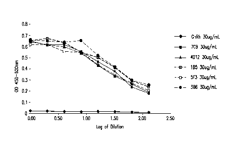
CELISA. Antibodies are designated as: ¨410¨Gn11 LC ugi ;7C9 CuWnL ;
D123OugrL ; =111...1B5 30iimL ; = -7. f3ugf i-- I_ and .. B5 3111
Figure 3A shows ligand blockade with mouse mAbs against canine PD-1. Various
mouse mAbs
were tested for their ability to inhibit binding of PD-1 expressed on CHO
cells to PD-Li.
CA 02932519 2016-06-02
WO 2015/091911 PCT/EP2014/078655
23
Antibodies arc designated as: i,GnRh 3OugimL ; =535.3OugiernL ; 10.1A1
3Ougi L;
5A8ug.L and .q1== 4912 L
Figure 3B shows ligand blockade with mouse mAbs against canine PD-1. Various
mouse mAbs
were tested for their ability to inhibit binding of PD-1 expressed on CHO
cells to PD-Li.
Antibodies are designated as: ¨410¨GnRh Cii 1- L ; ¨M¨ 585 S3.Jg,)-
1 ; '..r14 6,8.4 ug= L ;
52.4 ugini L 283. Li L and ¨111,2G9 5L.8UrL
Figure 3C shows ligand blockade with mouse mAbs against canine PD-1. Various
mouse mAbs
were tested for their ability to inhibit binding of PD-1 expressed on CHO
cells to PD-Li.
Antibodies are designated as: GnRh ugj rL; SOug,r, L ;
:1'719 2S.2 ug: I- and ¨m¨L 9 28. 8 ugimL
Figure 4 shows the binding of mouse mAbs to canine PD-1 on CD + T cells in
PBMC from
healthy dogs. Various mouse mAbs were tested for their ability to bind to
canine PD-1
expressed on CD + T cells from PBMC from healthy dogs. Antibodies were tested
at 2 fold
dilutions covering starting with 0.156-20 Orli range.
Figure 5 shows the binding of mouse mAbs to canine PD-1 on CD8-' T cells in
PBMC from
dogs with cancer. Indicated mouse mAbs were tested for their ability to bind
to canine PD-1
expressed on CD8+T cells from dogs with cancer (sarcoma). Antibodies were
tested at 2.5 and 5
ug/ml.
Figure 6 shows the cytokinc secretion induced by mouse mAbs to canine PD-1.
Various mouse
mAbs were tested for their ability to induce cytokine secretion from PBMC from
healthy dogs.
Figure 7 shows the cytokine secretion induced by mouse mAbs to canine PD-1.
Various mouse
mAbs were tested for their ability to induce cytokine secretion from PBMC from
dogs with
cancer (hemangiosarcoma).
CA 02932519 2016-06-02
WO 2015/091911 PCT/EP2014/078655
24
Figure 8 provides the alignment of canine IgGB constant heavy chains (CHs)
lacking ADCC
function. The canine wild type IgB [cIgGB wt], Canine IgGB(+)A-hinge [cIgGB(--
) A-hinge],
Canine IgGB(+) D-hinge [cIgGB(+) D-hinge], and Canine IgGB (-)ADCC [cIgGB(-)
ADCC]
are depicted. The (+) A-hinge is the replacement with IgG-A hinge plus a
lysine and asparagine
amino acid replacement as shown; the (+) D-hinge is the replacement with IgG-D
hinge plus a
lysine and the asparagine amino acid replacement as shown. The (-)ADCC is the
lysine and
asparagine amino acid replacement.
Figure 9A shows the characterization of the interface between canine PD-1 and
the caninized
antibody 2G9 The amino acid positions are with respect to the PD-1 amino acid
sequence
without the signal sequence, i.e., SEQ ID NO: 2. The determination was
performed by chemical
cross-linking, High-Mass MAI,DI mass spectrometry and nI,C-Orbitrap mass
spectrometry
Figure 9B shows the characterization of the interface between canine PD-1 and
the caninized
antibody 3B6. The amino acid positions arc with respect to the PD-1 amino acid
sequence
without the signal sequence, i.e., SEQ ID NO: 2. The determination was
performed by chemical
cross-linking, High-Mass MALDI mass spectrometry and nLC-Orbitrap mass
spectrometry.
DETAILED DESCRIPTION
Abbreviations
Throughout the detailed description and examples of the invention the
following abbreviations
will be used:
ADCC Antibody-dependent cellular cytotoxicity
CDC Complement-dependent cyotoxicity
CDR Complementarity determining region in the immunoglobul in
variable
regions, defined using the Kabat numbering system
CHO Chinese hamster ovary
EC50 concentration resulting in 50% efficacy or binding
ELISA Enzyme-linked immunosorbant assay
CA 02932519 2016-06-02
WO 2015/091911 PCT/EP2014/078655
FR Antibody framework region: the immunoglobulin variable
regions
excluding the CDR regions.
HRP Horseradish peroxidase
IFN interferon
5 IC50 concentration resulting in 50% inhibition
IgG Immunoglobulin G
Kabat An immunoglobulin alignment and numbering system
pioneered by Elvin
A. Kabat [Sequences of Proteins of Immunological Interest, 5th Ed. Public
Health Service, National Institutes of Health, Bethesda, Md. (1991)]
10 mAb Monoclonal antibody (also Mab or MAb)
MES 2-(N-morpholino)ethanesu1fonic acid
MOA Mechanism of action
NHS Normal human serum
PCR Polymerase chain reaction
15 PK Pharmacokinctics
SEB Staphylococcus Entcrotoxin B
TT Tetanus toxoid
V region The segment of IgG chains which is variable in sequence
between
different antibodies. It extends to Kabat residue 109 in the light
chain and 113 in
20 the heavy chain.
VH Immunoglobulin heavy chain variable region
VK Immunoglobulin kappa light chain variable region
DEFINITIONS
25 So that the invention may be more readily understood, certain technical
and scientific terms are
specifically defined below. Unless specifically defined elsewhere in this
document, all other
technical and scientific terms used herein have the meaning commonly
understood by one of
ordinary skill in the art to which this invention belongs.
CA 02932519 2016-06-02
WO 2015/091911 PCT/EP2014/078655
26
As used herein, including the appended claims, the singular forms of words
such as "a," "an,"
and "the," include their corresponding plural references unless the context
clearly dictates
otherwise.
"Activation" as it applies to cells or to receptors refers to the activation
or treatment of a cell or
receptor with a ligand, unless indicated otherwise by the context or
explicitly. "Ligand"
encompasses natural and synthetic ligands, e.g., cytokines, cytokine variants,
analogues, muteins,
and binding compounds derived from antibodies. "Ligand" also encompasses small
molecules,
e.g., peptide mimetics of cytokines and peptide mimetics of antibodies.
"Activation" can refer to
cell activation as regulated by internal mechanisms as well as by external or
environmental
factors.
"Activity" of a molecule may describe or refer to the binding of th e molecule
to a ligand or to a
receptor, to catalytic activity; to the ability to stimulate gene expression
or cell signaling,
differentiation, or maturation; to antigenic activity, to the modulation of
activities of other
molecules, and the like. "Activity" of a molecule may also refer to activity
in modulating or
maintaining cell-to-cell interactions, e.g., adhesion, or activity in
maintaining a structure of a
cell, e.g., cell membranes or cytoskeleton. "Activity" can also mean specific
activity, e.g.,
[catalytic activity]/[mg protein], or [immunological activity]/[mg protein],
concentration in a
biological compartment, or the like. "Activity" may refer to modulation of
components of the
innate or the adaptive immune systems.
"Administration" and "treatment," as it applies to an animal, e.g., a canine
experimental subject,
cell, tissue, organ, or biological fluid, refers to contact of an exogenous
pharmaceutical,
therapeutic, diagnostic agent, or composition to the animal e.g., a canine
subject, cell, tissue,
organ, or biological fluid. Treatment of a cell encompasses contact of a
reagent to the cell, as
well as contact of a reagent to a fluid, where the fluid is in contact with
the cell.
"Administration" and "treatment" also means in vitro and ex vivo treatments,
e.g., of a cell, by a
reagent, diagnostic, binding compound, or by another cell. The term "subject"
includes any
CA 02932519 2016-06-02
WO 2015/091911 PCT/EP2014/078655
27
organism, preferably an animal, more preferably a mammal (e.g., canine,
feline, or human) and
most preferably a canine.
As used herein, a "substitution of an amino acid residue" with another amino
acid residue in an
amino acid sequence of an antibody for example, is equivalent to "replacing an
amino acid
residue" with another amino acid residue and denotes that a particular amino
acid residue at a
specific position in the amino acid sequence has been replaced by (or
substituted for) by a
different amino acid residue. Such substitutions can be particularly designed
i.e., purposefully
replacing an alanine with a serine at a specific position in the amino acid
sequence by e.g.,
recombinant DNA technology. Alternatively, a particular amino acid residue or
string of amino
acid residues of an antibody can be replaced by one or more amino acid
residues through more
natural selection processes e.g., based on the ability of the antibody
produced by a cell to bind to
a given region on that antigen, e g , one containing an epitope or a portion
thereof, and/or for the
antibody to comprise a particular CDR that retains the same canonical
structure as the CDR it is
replacing. Such substitutions/replacements can lead to "variant" CDRs and/or
variant antibodies.
"Treat" or "treating" means to administer a therapeutic agent, such as a
composition containing
any of the antibodies or antigen binding fragments of the present invention,
internally or
externally to a canine subject or patient having one or more disease symptoms,
or being
suspected of having a disease, for which the agent has therapeutic activity.
Typically, the agent is administered in an amount effective to alleviate
and/or ameliorate one or
more disease symptoms in the treated subject or population, whether by
inducing the regression
of or inhibiting the progression of such symptom(s) by any clinically
measurable degree. The
amount of a therapeutic agent that is effective to alleviate any particular
disease symptom (also
referred to as the "therapeutically effective amount") may vary according to
factors such as the
disease state, age, and weight of the patient (e.g., canine), and the ability
of the pharmaceutical
composition to elicit a desired response in the subject. Whether a disease
symptom has been
alleviated or ameliorated can be assessed by any clinical measurement
typically used by
veteranarians or other skilled healthcare providers to assess the severity or
progression status of
CA 02932519 2016-06-02
WO 2015/091911 PCT/EP2014/078655
28
that symptom. While an embodiment of the present invention (e.g., a treatment
method or article
of manufacture) may not be effective in alleviating the target disease
symptom(s) in every
subject, it should alleviate the target disease symptom(s) in a statistically
significant number of
subjects as determined by any statistical test known in the art such as the
Student's t-test, the
ehi2-test, the U-test according to Mann and Whitney, the Kruskal-Wallis test
(H-test),
Jonckheere-Terpstra-test and the Wilcoxon-test.
"Treatment," as it applies to a human, veterinary (e.g., canine) or research
subject, refers to
therapeutic treatment, as well as research and diagnostic applications.
'Treatment" as it applies
to a human, veterinary (e.g., canine), or research subject, or cell, tissue,
or organ, encompasses
contact of the antibodies or antigen binding fragments of the present
invention to a canine or
other animal subject, a cell, tissue, physiological compartment, or
physiological fluid.
As used herein, the term "canine" includes all domestic dogs, Canis lupus
familiaris or Canis
familiaris, unless otherwise indicated.
As used herein, the term "feline" refers to any member of the Felidae family.
Members of this
family include wild, zoo, and domestic members, such as any member of the
subfamilies
Felinae, e.g., cats, lions, tigers, pumas, jaguars, leopards, snow leopards,
panthers, North
American mountain lions, cheetahs, lynx, bobcats, caracals or any cross breeds
thereof Cats
also include domestic cats, pure-bred and/or mongrel companion cats, show
cats, laboratory cats,
cloned cats, and wild or feral cats.
As used herein the term "canine frame" refers to the amino acid sequence of
the heavy chain and
light chain of a canine antibody other than the hypervariable region residues
defined herein as
CDR residues. With regard to a caninized antibody, in the majority of
embodiments the amino
acid sequences of the native canine CDRs are replaced with the corresponding
foreign CDRs
(e.g., those from a mouse antibody) in both chains. Optionally the heavy
and/or light chains of
the canine antibody may contain some foreign non-CDR residues, e.g., so as to
preserve the
CA 02932519 2016-06-02
WO 2015/091911 PCT/EP2014/078655
29
conformation of the foreign CDRs within the canine antibody, and/or to modify
the Fe function,
as exemplified below.
Canine PD-1 has been found to comprise the amino acid sequence of SEQ ID NO:
2. In a
specific embodiment canine PD-1 is encoded by a nucleic acid that comprises
the nucleotide
sequence of SEQ ID NO: 1. Canine PD-1 sequences may differ by having, for
example,
conserved variations in non-conserved regions, but the canine PD-1 will have
substantially the
same biological function as the canine PD-1 comprising the amino acid sequence
of SEQ ID
NO: 2. For example, a biological function of PD-1 is to attenuate T-cell
responses when bound
to PD-L1 and/or PD-L2. That is, PD-1 may be considered a negative regulator.
Notably, the
cytoplasmic tail of PD-1 contains two tyrosine-based signaling motifs. an ITIM
(immunoreceptor
tyrosine-based inhibition motif) and an ITSM (immunoreceptor tyrosine-based
switch motif). In
addition, a biological fiinction of canine PD-1 may be 'having, for example,
an epitope in the
extracellular domain that is specifically bound by an antibody of the instant
disclosure.
Canine PD-L I has been found to comprise the amino acid sequence of SEQ ID NO:
8. In a
specific embodiment canine PD-L1 is encoded by a nucleotide sequence
comprising SEQ ID
NO: 7. Canine PD-Li sequences may differ by having, for example, conserved
variations in
non-conserved regions, but the canine PD-Li will have substantially the same
biological
function as the canine PD-Li comprising the amino acid sequence of SEQ ID NO:
8. For
example, one biological function of PD-L1 is to attenuate T-cell responses
when bound to PD-1.
A particular canine PD-1 or PD-Li amino acid sequence respectively, will
generally be at least
90% identical to the canine PD-1 comprising the amino acid sequence of SEQ ID
NO: 2, or
canine PD-Li comprising the amino acid sequence of SEQ ID NO: 8, respectively.
In certain
cases, a canine PD-1 or PD-L1 respectively, may be at least 95%, or even at
least 96%, 97%,
98% or 99% identical to the canine PD-1 comprising the amino acid sequence of
SEQ ID NO: 2,
or the canine PD-Li comprising the amino acid sequence of SEQ ID NO: 8,
respectively. In
certain embodiments, a canine PD-1 or a PD-Li amino acid sequence
respectively, will display
no more than 10 amino acid differences from the canine PD-1 comprising the
amino acid
CA 02932519 2016-06-02
WO 2015/091911 PCT/EP2014/078655
sequence of SEQ ID NO: 2, or the canine PD-L1 comprising the amino acid
sequence of SEQ ID
NO: 8, respectively. In certain embodiments, the canine PD-1 or the PD-L1
amino acid
sequence respectively, may display no more than 5, or even no more than 4, 3,
2, or 1 amino acid
difference from the canine PD-1 comprising the amino acid sequence of SEQ ID
NO: 2, or the
5 canine PD-Li comprising the amino acid sequence of SEQ ID NO: 8,
respectively. Percent
identity can be determined as described herein below.
The term "immune response" refers to the action of, for example, lymphocytes,
antigen
presenting cells, phagocytic cells, granulocytes, and soluble macromolecules
produced by the
10 above cells or the liver (including antibodies, cytokines, and
complement) that results in
selective damage to, destruction of, or elimination from the mammalian body
(e.g., canine body)
of cancerous cells, cells or tissues infected with pathogens, or invading
pathogens.
Anti-canine PD-1. antibodies
15 The present invention provides isolated antibodies (particularly murine
anti-canine PD-1
antibodies and caninizcd antibodies thereof) or antigen binding fragments
thereof that bind
canine PD-1 and uses of such antibodies or fragments thereof In specific
embodiments murine
anti-canine PD-1 CDRs from murine anti-canine PD-1 antibodies are provided
that have been
shown to both bind canine PD-1 and to block the binding of canine PD-1 to its
ligand, canine
20 PD-Li. These CDRs can be inserted into a modified canine frame of a
canine antibody to
generate a caninized murine anti-canine PD-1 antibody.
As used herein, an "anti-canine PD-1 antibody- refers to an antibody that was
raised against
canine PD-1 (e.g., in a mammal such as a mouse or rabbit) and that
specifically binds to canine
25 PD-1. An antibody that "specifically binds to canine PD-1," and in
particular canine PD-1, or an
antibody that "specifically binds to a polypeptide comprising the amino acid
sequence of canine
PD-1", is an antibody that exhibits preferential binding to canine PD-1 as
compared to other
antigens, but this specificity does not require absolute binding specificity.
An anti-canine PD-1
antibody is considered "specific" for canine PD-1 if its binding is
determinative of the presence
30 of canine PD-1 in a sample, or if it is capable of altering the activity
of canine PD-1 without
CA 02932519 2016-06-02
WO 2015/091911 PCT/EP2014/078655
31
unduly interfering with the activity of other molecules in a canine sample,
e.g. without producing
undesired results such as false positives in a diagnostic context or side
effects in a therapeutic
context. The degree of specificity necessary for an anti-canine PD-1 antibody
may depend on
the intended use of the antibody, and at any rate is defined by its
suitability for use for an
intended purpose. The antibody, or binding compound derived from the antigen-
binding site of
an antibody, of the contemplated method binds to its antigen, or a variant or
mutein thereof, with
an affinity that is at least two-fold greater, preferably at least ten-times
greater, more preferably
at least 20-times greater, and most preferably at least 100-times greater than
the affinity with any
other antigen.
As used herein, an antibody is said to bind specifically to a polypeptide
comprising a given
antigen sequence (in this case a portion of the amino acid sequence of canine
PD-1) if it binds to
polypeptides comprising the portion of the amino acid sequence of canine PD-1,
but does not
bind to other canine proteins lacking that portion of the sequence of canine
PD-1. For example,
an antibody that specifically binds to a polypcptide comprising canine PD-1
may bind to a
FLAG -tagged form of canine PD-1, but will not bind to other FLAW-tagged
canine proteins.
An antibody, or binding compound derived from the antigen-binding site of an
antibody, binds to
its canine antigen, or a variant or mutein thereof, "with specificity" when it
has an affinity for
that canine antigen or a variant or mutein thereof which is at least ten-times
greater, more
preferably at least 20-times greater, and even more preferably at least 100-
times greater than its
affinity for any other canine antigen tested.
As used herein, the term "antibody" refers to any form of antibody that
exhibits the desired
biological activity. Thus, it is used in the broadest sense and specifically
covers, but is not
limited to, monoclonal antibodies (including full length monoclonal
antibodies), polyclonal
antibodies, multispecific antibodies (e.g., bispecific antibodies), canonized
antibodies, fully
canine antibodies, chimeric antibodies and camelized single domain antibodies.
"Parental
antibodies" arc antibodies obtained by exposure of an immune system to an
antigen prior to
modification of the antibodies for an intended use, such as caninization of an
antibody for use as
.. a canine therapeutic antibody.
CA 02932519 2016-06-02
WO 2015/091911 PCT/EP2014/078655
32
As used herein, unless otherwise indicated, "antibody fragment" or "antigen
binding fragment"
refers to antigen binding fragments of antibodies, i.e. antibody fragments
that retain the ability to
bind specifically to the antigen bound by the full-length antibody, e.g.
fragments that retain one
or more CDR regions. Examples of antigen binding fragments include, but are
not limited to,
Fab, Fab', F(ab')), and Fv fragments; diabodies; linear antibodies; single-
chain antibody
molecules, e.g., sc-Fv; nanobodies and multispecific antibodies formed from
antibody fragments.
A "Fab fragment" is comprised of one light chain and the CHI and variable
regions of one heavy
chain. The heavy chain of a Fab molecule cannot form a disulfide bond with
another heavy chain
molecule. A ''Fab fragment" can be the product of papain cleavage of an
antibody.
A "fragment crystallizable" ("Fe") region contains two heavy chain fragments
comprising the
CH1 and C112 domains of an antibody. The two heavy chain fragments are held
together by two
or more disulfide bonds and by hydrophobic interactions of the CH3 domains.
A "Fab' fragment" contains one light chain and a portion or fragment of one
heavy chain that
contains the VH domain and the C H1 domain and also the region between the CHI
and CH2
domains, such that an interchain disulfide bond can be formed between the two
heavy chains of
two Fab' fragments to form a F(ab') 2 molecule.
A "F(ab)2 fragment" contains two light chains and two heavy chains containing
a portion of the
constant region between the CH1 and C112 domains, such that an interchain
disulfide bond is
formed between the two heavy chains. A F(ab') 2 fragment thus is composed of
two Fab'
fragments that are held together by a disulfide bond between the two heavy
chains. An "F(ab)2
fragment" can be the product of pepsin cleavage of an antibody.
The "Fv region" comprises the variable regions from both the heavy and light
chains, but lacks
the constant regions.
CA 02932519 2016-06-02
WO 2015/091911 PCT/EP2014/078655
33
The term "single-chain Fv" or "scFv" antibody refers to antibody fragments
comprising the VH
and VT domains of an antibody, wherein these domains are present in a single
polypeptide chain.
Generally, the Fv polypeptide further comprises a polypeptide linker between
the VTT and VT
domains which enables the scFv to form the desired structure for antigen
binding. [See,
Pluckthun, THE PHARMACOLOGY OF MONOCLONAL ANTIBODIES, vol. 113 Rosenburg and
Moore
eds., Springer-Verlag, New York, pp. 269-315 (1994); WO 88/01649; and U.S.
4,946,778 and
U.S. 5,260,203.]
As used herein, the term "canonical structure" refers to the local
conformation that can be
adopted by each of the hypervariable regions of the heavy and light chain of
an antibody within
the framework that they reside. For each hypervariable region, there are a
small number of
canonical structures (generally denoted by simple integers such as 1 or 2
etc.), which can be
predicted with great accuracy from the amino acid sequences of the
corresponding hypervariable
region (particularly within the context of the amino acid sequence of its
framework, as provided
below for the corresponding anti-canine PD-1 variable domains). These
canonical structures can
be determinative regarding whether a modification of the amino acid sequence
of a given CDR
will result in the retention or loss of the ability to bind to its antigen
binding partner [See,
Chothia and Lesk, Canonical Structures for the hypervariable regions of
immunoglobulins, J.
Biol. 196:901-917(1987); Chothia et al., Conformation of immunoglobulin
hypervaribale
regions, Nature, 34:877-883(1989); and Al-Lazikani et al., Standard
Conformations for the
canonical structures of immunoglobulins, J. Mol. Biol. 273:927-948 (1997)].
A "domain antibody" is an immunologically functional immunoglobulin fragment
containing
only the variable region of a heavy chain or the variable region of a light
chain. In some
instances, two or more VH regions are covalently joined with a peptide linker
to create a bivalent
domain antibody. The two VH regions of a bivalent domain antibody may target
the same or
different antigens.
CA 02932519 2016-06-02
WO 2015/091911 PCT/EP2014/078655
34
A "bivalent antibody" comprises two antigen binding sites. In some instances,
the two binding
sites have the same antigen specificities. However, bivalent antibodies may be
bispecific (see
below).
In certain embodiments, monoclonal antibodies herein also include camelized
single domain
antibodies. [See, e.g., Muyldermans et al., Trends Biochetn. Sci. 26:230
(2001); Reichmann et
al., J. Invnunol. Methods 231:25 (1999); WO 94/04678; WO 94/25591; U.S.
6,005,079]. In one
embodiment, the present invention provides single domain antibodies comprising
two VH
domains with modifications such that single domain antibodies are formed.
As used herein, the term "diabodies" refers to small antibody fragments with
two antigen-binding
sites, which fragments comprise a heavy chain variable domain (VH) connected
to a light chain
variable domain (VL) in the same polypeptide chain (VH-VL or VL-VH) By using a
linker that is
too short to allow pairing between the two domains on the same chain, the
domains are forced to
pair with the complementary domains of another chain and create two antigen-
binding sites.
[See, EP 0 404 097 Bl; WO 93/11161; and Holliger etal., Proc. Natl. Acad. Sci.
USA 90: 6444-
6448 (1993)]. For a review of engineered antibody variants [generally see
Holliger and Hudson
Nat. Biotechnol. 23:1126-1136 (2005)].
Typically, an antibody or antigen binding fragment of the invention retains at
least 10% of its
canine PD-1 binding activity (when compared to the parental antibody) when
that activity is
expressed on a molar basis. Preferably, an antibody or antigen binding
fragment of the invention
retains at least 20%, 50%, 70%, 80%, 90%, 95% or 100% or more of the canine PD-
1 binding
affinity as the parental antibody. It is also intended that an an antibody or
antigen binding
fragment of the invention can include conservative or non-conservative amino
acid substitutions
(referred to as "conservative variants" or "function conserved variants" of
the antibody) that do
not substantially alter its biologic activity.
"Isolated antibody" refers to the purification status and in such context
means the molecule is
substantially free of other biological molecules such as nucleic acids,
proteins, lipids,
CA 02932519 2016-06-02
WO 2015/091911 PCT/EP2014/078655
carbohydrates, or other material such as cellular debris and growth media.
Generally, the term
"isolated" is not intended to refer to a complete absence of such material or
to an absence of
water, buffers, or salts, unless they are present in amounts that
substantially interfere with
experimental or therapeutic use of the binding compound as described herein.
5
As used herein, a "chimeric antibody" is an antibody having the variable
domain from a first
antibody and the constant domain from a second antibody, where the first and
second antibodies
are from different species. [U.S. 4,816,567; and Morrison et al., Proc. Natl.
Acad. Sei. USA 81:
6851-6855 (1984)]. Typically the variable domains are obtained from an
antibody from an
10 experimental animal (the "parental antibody"), such as a rodent, and the
constant domain
sequences are obtained from the animal subject antibodies, e.g., human or
canine so that the
resulting chimeric antibody will be less likely to elicit an adverse immune
response in a canine or
human subject respectively, than the parental (to g , rodent) antibody
As used herein, the term "caninized antibody" refers to forms of antibodies
that contain
15 sequences from both canine and non-canine (e.g., murinc) antibodies. In
general, the caninized
antibody will comprise substantially all of at least one or more typically,
two variable domains in
which all or substantially all of the hypervariable loops correspond to those
of a non-canine
immunoglobulin (e.g., comprising 6 murine anti-canine PD-1 CDRs as exemplified
below), and
all or substantially all o f the framework (FR) regions (and typically all or
substantially all of the
20 remaining frame) are those of a canine immunoglobulin sequence. As
exemplified herein, a
caninized antibody comprises both the three heavy chain CDRs and the three
light chain CDRS
from a murine anti-canine PD-1 antibody together with a canine frame or a
modified canine
frame. A modified canine frame comprises one or more amino acids changes as
exemplified
herein that further optimize the effectiveness of the caninized antibody,
e.g., to increase its
25 binding to canine PD-1 and/or its ability to block the binding of canine
PD-1 to canine PD-Ll.
The term "fully canine antibody" refers to an antibody that comprises canine
immunoglobulin
protein sequences only. A fully canine antibody may contain murine
carbohydrate chains if
produced in a mouse, in a mouse cell, or in a hybridoma derived from a mouse
cell. Similarly,
30 "mouse antibody" refers to an antibody that comprises mouse
immunoglobulin sequences only.
CA 02932519 2016-06-02
WO 2015/091911 PCT/EP2014/078655
36
Alternatively, a fully canine antibody may contain rat carbohydrate chains if
produced in a rat, in
a rat cell, or in a hybridoma derived from a rat cell. Similarly, "rat
antibody" refers to an
antibody that comprises rat immunoglobulin sequences only.
There are four known IgG heavy chain subtypes of dog IgG and they are referred
to as IgG-A,
IgG-B, IgG-C, and IgG-D. The two known light chain subtypes are referred to as
lambda and
kappa.
The variable regions of each light/heavy chain pair form the antibody binding
site. Thus, in
general, an intact antibody has two binding sites. Except in bifunctional or
bispecific antibodies,
the two binding sites are, in general, the same.
Typically, the variable domains of both the heavy and light chains comprise
three hypervariable
regions, also called complementarily determining regions (CDRs), located
within relatively
conserved framework regions (FR). The CDRs are usually aligned by the
framework regions,
enabling binding to a specific epitope. In general, from N-terminal to C-
terminal, both light and
heavy chains variable domains comprise FRI. CDR1, FR2 , CDR2, FR3, CDR3 and
FR4. The
assignment of amino acids to each domain is, generally, in accordance with the
definitions of
Sequences of Proteins of Immunological Interest, Kabat, et al.; National
Institutes of Health,
Bethesda, Md. ; 5th ed.; NIH Publ. No. 91-3242 (1991); Kabat, Adv. Prot. Chem.
32:1-75
(1978); Kabat, et al., J. Biol. Chem. 252:6609-6616 (1977); Chothia, et al.,
J. Mol. Biol.
196:901-917 (1987) or Chothia, et al., Nature 342:878-883 (1989)].
As used herein, the term "hypervariable region" refers to the amino acid
residues of an antibody
that are responsible for antigen-binding. The hypervariable region comprises
amino acid
residues from a "complementarity determining region" or "CDR" (i.e. CDRL1,
CDRL2 and
CDRL3 in the light chain variable domain and CDRHI , CDRH2 and CDRH3 in the
heavy chain
variable domain). [See Kabat et al. Sequences of Proteins of Immunological
Interest, 5th Ed.
Public Health Service, National Institutes of Health, Bethesda, Md. (1991),
definining the CDR
regions of an antibody by sequence; see also Chothia and Lesk, J. Mol. Biol.
196: 901-917
CA 02932519 2016-06-02
WO 2015/091911 PCT/EP2014/078655
37
(1987) defining the CDR regions of an antibody by structure]. As used herein,
the term
"framework" or "FR" residues refers to those variable domain residues other
than the
hypervariable region residues defined herein as CDR residues.
Besides binding and activating of canine immune cells, a canine or caninized
antibody against
PD-1 optimally has two attributes:
1. Lack of effector functions such as antibody-dependent cytotoxicity
(ADCC) and
complement-dependent cytotoxicity (CDC), and
2. be readily purified on a large scale using industry standard
technologies such as
that based on protein A chromatography.
None of the naturally occurring canine IgG isotypes satisfy both criteria For
example, TgG-B
can be purified using protein A, but has high level of ADCC activity. On the
other hand, IgG-A
binds weakly to protein A, but displays undesirable ADCC activity. Moreover,
neither IgG-C
nor IgG-D can be purified on protein A columns, although IgG-D display no ADCC
activity.
(IgG-C has considerable ADCC activity). The present invention overcomes this
difficulty by
providing mutant canine IgG-B antibodies specific to PD-1; such antibodies
lack effector
functions such as ADCC and can be easily of purified using industry standard
protein A
chromatography.
"Homology" refers to sequence similarity between two polynucleotide sequences
or between two
polypeptide sequences when they are optimally aligned. When a position in both
of the two
compared sequences is occupied by the same base or amino acid monomer subunit,
e.g., if a
position in each of two DNA molecules is occupied by adenine, then the
molecules are
homologous at that position. The percent of homology is the number of
homologous positions
shared by the two sequences divided by the total number of positions compared
x100. For
example, if 6 of 10 of the positions in two sequences arc matched or
homologous when the
sequences arc optimally aligned then the two sequences are 60% homologous.
Generally, the
comparison is made when two sequences arc aligned to give maximum percent
homology.
CA 02932519 2016-06-02
WO 2015/091911 PCT/EP2014/078655
38
"Isolated nucleic acid molecule" means a DNA or RNA of genomic, mRNA, cDNA, or
synthetic
origin or some combination thereof which is not associated with all or a
portion of a
polynucleotide in which the isolated polynucleotide is found in nature. or is
linked to a
polynucleotide to which it is not linked in nature. For purposes of this
disclosure, it should be
understood that "a nucleic acid molecule comprising" a particular nucleotide
sequence does not
encompass intact chromosomes. Isolated nucleic acid molecules "comprising"
specified nucleic
acid sequences may include, in addition to the specified sequences, coding
sequences for up to
ten or even up to twenty or more other proteins or portions or fragments
thereof, or may include
operably linked regulatory sequences that control expression of the coding
region of the recited
nucleic acid sequences, and/or may include vector sequences.
The phrase "control sequences" refers to DNA sequences necessary for the
expression of an
operably linked coding sequence in a particular host organism. The control
sequences that are
suitable for prokaryotes, for example, include a promoter, optionally an
operator sequence, and a
ribosome binding site. Eukaryotic cells arc known to use promoters,
polyadenylation signals,
and enhancers.
A nucleic acid is "operably linked" when it is placed into a functional
relationship with another
nucleic acid sequence. For example, DNA for a presequence or secretory leader
is operably
linked to DNA for a polypeptide if it is expressed as a preprotein that
participates in the secretion
of the polypeptide; a promoter or enhancer is operably linked to a coding
sequence if it affects
the transcription of the sequence; or a ribosome binding site is operably
linked to a coding
sequence if it is positioned so as to facilitate translation. Generally,
"operably linked" means that
.. the DNA sequences being linked are contiguous, and, in the case of a
secretory leader,
contiguous and in reading phase. However, enhancers do not have to be
contiguous Linking is
accomplished by ligation at convenient restriction sites. If such sites do not
exist, the synthetic
oligonucleotide adaptors or linkers arc used in accordance with conventional
practice.
CA 02932519 2016-06-02
WO 2015/091911 PCT/EP2014/078655
39
As used herein, the expressions "cell," "cell line," and "cell culture" are
used interchangeably and
all such designations include progeny. Thus, the words "transformants" and
"transformed cells"
include the primary subject cell and cultures derived therefrom without regard
for the number of
transfers. It is also understood that not all progeny will have precisely
identical DNA content,
due to deliberate or inadvertent mutations. Mutant progeny that have the same
function or
biological activity as screened for in the originally transformed cell are
included. Where distinct
designations are intended, it will be clear from the context.
As used herein, "germline sequence" refers to a sequence of unrearranged
immunoglobulin DNA
sequences. Any suitable source of unrearranged immunoglobulin sequences may be
used.
Human gerrnline sequences may be obtained, for example, from JOINSOLVER
germline
databases on the website for the National Institute of Arthritis and
Musculoskeletal and Skin
Diseases of the united States National Institutes of Health Mouse germline
sequences may be
obtained, for example, as described in Giudicelli et al. [Nucleic Acids Res.
33:D256-D261
(2005)].
Properties of Murine Anti-Canine PD-1 and
Caninized Murine Anti-Canine PD-1 Antibodies
The present invention provides isolated murine anti-canine PD-1 antibodies and
caninizcd
antibodies thereof; methods of use of the antibodies or antigen binding
fragments thereof in the
treatment of disease e.g., the treatment of cancer in canines. In canine,
there are four IgG heavy
chains referred to as A, B, C, and D. These heavy chains represent four
different subclasses of
dog IgG, which are referred to as IgGA, IgGB, IgGC and IgGD. The DNA and amino
acid
sequences of these four heavy chains were first identified by Tang et al.
[Vet. Immunol.
Immunopathol. 80: 259-270 (2001)1. The amino acid and DNA sequences for these
heavy chains
are also available from the GenBank data bases. For example, the amino acid
sequence of IgGA
heavy chain has accession number AAL35301.1, IgGB has accession number
AAL35302.1,
IgGC has accession number AAL35303.1, and IgGD has accession number
(AAL35304.1).
Canine antibodies also contain two types of light chains, kappa and lambda.
The DNA and
CA 02932519 2016-06-02
WO 2015/091911 PCT/EP2014/078655
amino acid sequence of these light chains can be obtained from GenBank
Databases. For
example the kappa light chain amino acid sequence has accession number ABY
57289.1 and the
lambda light chain has accession number ABY 55569.1. In the present invention,
the amino acid
sequence for each of the four canine IgG Fe fragments is based on the
identified boundary of
5 CH1 and CH2 domains as determined by Tang et al, supra. Caninized murine
anti-canine PD-1
antibodies that bind canine PD-1 include, but are not limited to: antibodies
that comprise canine
IgG-A, IgG-B, and IgG-D heavy chains and/or canine kappa light chains together
with murine
anti-canine PD-1 CDRs. Accordingly, the present invention provides isolated
murine anti-canine
PD-1 and/or caninized murine anti-canine PD-1 antibodies or antigen binding
fragments thereof
10 .. that bind to canine PD-1 and block the binding of canine PD-1 to canine
PD-Li.
The present invention further provides full length canine heavy chains that
can be matched with
corresponding light chains to make a caninized antibody Accordingly, the
present invention
further provides caninized murine anti-canine antigen antibodies (including
isolated caninized
15 murinc anti-canine PD-1 antibodies) and methods of usc of the antibodies
or antigen binding
fragments thereof in the treatment of disease e.g., the treatment of cancer in
canines.
The isolated antibody or antigen binding fragment thereof that binds canine PD-
1 can comprise
one, two, three, four, five, or six of the complementarity determining regions
(CDRs) of the
20 murine anti-canine antibody as described herein. The one, two, three,
four, five, or six CDRs
may be independently selected from the CDR sequences of those provided below.
In a further
embodiment, the isolated antibody or antigen-binding fragment thereof that
binds canine PD-1
comprises a canine antibody kappa light chain comprising a murine light chain
CDR-1, CDR-2
and/or CDR-3 and a canine antibody heavy chain IgG comprising a murine heavy
chain CDR-1,
25 CDR-2 and/or CDR-3.
In other embodiments, the invention provides antibodies or antigen binding
fragments thereof
that specifically binds PD-1 and have canine antibody kappa light chains
comprising one to six
different CDRs comprising at least 80%, 85%, 90%, 95%, 98% or 99% sequence
identity with
30 the amino acid sequences of SEQ ID NOs: 13, 14, 15, 16, 17, 18, 19, 20,
21, 22, 23,24, 25,
CA 02932519 2016-06-02
WO 2015/091911 PCT/EP2014/078655
41
and/or 26 and canine antibody heavy chain IgG comprising one to six different
CDRs comprising
at least 80%, 85%, 90%, 95%, 98% or 99% sequence identity with the amino acid
sequences of
SEQ ID NOs: 27, 28, 29, 30, 31, 32, 33, 34, 35, 36, 37, 38, and/or 114, while
still exhibiting the
desired binding and functional properties. In another embodiment the antibody
or antigen
binding fragment of the present invention comprises a canine framecomprising
of a combination
of IgG heavy chain sequence with a kappa light chain having one or more of the
above-
mentioned CDR amino acid sequences with 0, 1, 2, 3, 4, or 5 conservative or
non-conservative
amino acid substitutions, while still exhibiting the desired binding and
functional properties.
Sequence identity refers to the degree to which the amino acids of two
polypeptides are the same
at equivalent positions when the two sequences are optimally aligned. As used
herein one amino
acid sequence is 100% "identical" to a second amino acid sequence when the
amino acid residues
of both sequences are identical Accordingly, an amino acid sequence is 50%
"identical" to a
second amino acid sequence when 50% of the amino acid residues of the two
amino acid
sequences arc identical. The sequence comparison is performed over a
contiguous block of
amino acid residues comprised by a given protein, e.g., a protein, or a
portion of the polypeptidc
being compared. In a particular embodiment, selected deletions or insertions
that could
otherwise alter the correspondence between the two amino acid sequences are
taken into account.
.. Sequence similarity includes identical residues and nonidentical,
biochemically related amino
acids. Biochemically related amino acids that share similar properties and may
be
interchangeable are discussed
"Conservatively modified variants" or "conservative substitution" refers to
substitutions of
amino acids in a protein with other amino acids having similar characteristics
(e.g. charge, side-
chain size, hydrophobicity/hydrophilicity, backbone conformation and rigidity,
etc.), such that
the changes can frequently be made without altering the biological activity of
the protein. Those
of skill in this art recognize that, in general, single amino acid
substitutions in non-essential
regions of a polypeptide do not substantially alter biological activity [see,
e.g., Watson et al.,
Molecular Biology of the Gene, The Benjamin/Cummings Pub. Co., p. 224 (4th
Ed.; 1987)]. In
CA 02932519 2016-06-02
WO 2015/091911 PCT/EP2014/078655
42
addition, substitutions of structurally or functionally similar amino acids
are less likely to disrupt
biological activity. Exemplary conservative substitutions are set forth in
Table 3 directly below.
TABLE 3
Exemplary Conservative Amino Acid Substitutions
Original residue Conservative substitution
Ala (A) Gly; Ser;
Arg (R) Lys; His
Asn (N) Gln; His
Asp (D) Glu; Asn
Cys (C) Ser; Ala
Gln (Q) Asn
Glu (E) Asp; Gin
Gly (G) _Ala
His (H) Asn; Gln
Ile (I) Leu; Val
Leu (L) Ile; Val
Lys (K) Arg; His
Met (M) Leu; Ile; Tyr
Phe (F) Tyr; Met; Leu
Pro (P) Ala; Gly
Ser (S) Thr
Thr (T) Ser
Trp (W) Tyr; Phe
Tyr (Y) Trp; Phe
Val (V) Ile; Leu
Function-conservative variants of the antibodies of the invention are also
contemplated by the
present invention. "Function-conservative variants," as used herein, refers to
antibodies or
fragments in which one or more amino acid residues have been changed without
altering a
desired property, such an antigen affinity and/or speficity. Such variants
include, but are not
limited to, replacement of an amino acid with one having similar properties,
such as the
conservative amino acid substitutions of Table 3 above.
CA 02932519 2016-06-02
WO 2015/091911 PCT/EP2014/078655
43
Nucleic Acids
The present invention further comprises the nucleic acids encoding the
immunoglobulin chains
of murine anti-canine PD-1 and/or caninized murine anti-canine PD-1 antibodies
and antigen
binding fragments thereof disclosed herein (see Examples below).
Also included in the present invention are nucleic acids that encode
immunoglobulin
polypeptides comprising amino acid sequences that are at least about 70%
identical, preferably at
least about 80% identical, more preferably at least about 90% identical and
most preferably at
least about 95% identical (e.g., 95%, 96%, 97%, 98%, 99%, 100%) to the amino
acid sequences
of the CDRs and antibodies provided herein when the comparison is performed by
a BLAST
algorithm wherein the parameters of the algorithm are selected to give the
largest match between
the respective sequences over the entire length of the respective reference
sequences. The
present invention further provides nucleic acids that encode immunoglobulin
polypeptides
comprising amino acid sequences that are at least about 70% similar,
preferably at least about
80% similar, more preferably at least about 90% similar and most preferably at
least about 95%
similar (e.g., 95%, 96%, 97%, 98%, 99%, 100%) to any of the reference amino
acid sequences
when the comparison is performed with a BLAST algorithm, wherein the
parameters of the
algorithm are selected to give the largest match between the respective
sequences over the entire
length of the respective reference sequences, are also included in the present
invention.
As used herein, nucleotide and amino acid sequence percent identity can be
determined using C,
MacVector (MacVector, Inc. Cary, NC 27519), Vector NTI (Informax, Inc. MD),
Oxford
Molecular Group PLC (1996) and the Clustal W algorithm with the alignment
default
parameters, and default parameters for identity. These commercially available
programs can also
be used to determine sequence similarity using the same or analogous default
parameters.
Alternatively, an Advanced Blast search under the default filter conditions
can be used, e.g.,
using the GCG (Genetics Computer Group, Program Manual for the GCG Package,
Version 7,
Madison, Wisconsin) pileup program using the default parameters.
CA 02932519 2016-06-02
WO 2015/091911 PCT/EP2014/078655
44
The following references relate to BLAST algorithms often used for sequence
analysis: BLAST
ALGORITHMS: Altschul, S.F., etal., J. Mol. Biol. 215:403-410 (1990); Gish, W.,
et al.,Nature
Genet. 3:266-272 (1993); Madden, T.L., et al., Meth. Enzyrnol. 266:131-
141(1996); Altschul,
S.F., et al., Nucleic Acids Res. 25:3389-3402 (1997); Zhang, J., etal., Genome
Res. 7:649-656
(1997); Wootton, J.C., etal., Comput. Chem. 17:149-163 (1993); Hancock, J.M.
etal., Comput.
App!. Biosci. 10:67-70 (1994); ALIGNMENT SCORING SYSTEMS: Dayhoff, M.O., et
al., "A
model of evolutionary change in proteins." in Atlas of Protein Sequence and
Structure, vol. 5,
suppl. 3. M.O. Dayhoff (ed.), pp. 345-352, (1978); Natl. Biomed. Res. Found.,
Washington, DC;
Schwartz, R.M., et al., "Matrices for detecting distant relationships." in
Atlas of Protein
Sequence and Structure, vol. 5, suppl. 3." (1978), M.O. Dayhoff (ed.), pp. 353-
358 (1978), Natl.
Biomed. Res. Found., Washington, DC; Altschul, S.F., J. Mol. Biol. 219:555-565
(1991); States,
D.J., et al., Methods 3:66-70(1991); Henikoff, S., et al.,Proc. Natl. Acad.
Sci. USA 89:10915-
10919 (1992); Altschul, SE, et al ,.I Evnl 16.290-100 (1991); ALIGNMENT
STATISTICS: Karlin, S., etal., Proc. Natl. Acad. Sci. USA 87:2264-2268 (1990);
Karlin, S., et
al., Proc. Nat!. Acad. Sci. USA 90:5873-5877 (1993); Dembo, A., et al., Ann.
Prob. 22:2022-
2039 (1994); and Altschul, S.F. "Evaluating the statistical significance of
multiple distinct local
alignments." in Theoretical and Computational Methods in Genorne Research (S.
Suhai, ed.), pp.
1-14, Plenum, New York (1997).
This present invention also provides expression vectors comprising the
isolated nucleic acids of
the invention, wherein the nucleic acid is operably linked to control
sequences that are
recognized by a host cell when the host cell is transfected with the vector.
Also provided are
host cells comprising an expression vector of the present invention and
methods for producing
the antibody or antigen binding fragment thereof disclosed herein comprising
culturing a host
cell harboring an expression vector encoding the antibody or antigen binding
fragment in culture
medium, and isolating the antigen or antigen binding fragment thereof from the
host cell or
culture medium.
CA 02932519 2016-06-02
WO 2015/091911 PCT/EP2014/078655
Epitope Binding and Binding Affinity
The present invention further provides antibodies or antigen binding fragments
thereof that bind
to amino acid residues of the same epitope of canine PD-1 as the murine anti-
canine PD-1
antibodies disclosed herein. In particular embodiments the murine anti-canine
PD-1 antibodies
5 or antigen binding fragments thereof are also capable of
inhibiting/blocking the binding of
canine PD-1 to canine PD-Li.
A caninized murine anti-canine PD-1 antibody can be produced recombinantly by
methods that
are known in the field. Mammalian cell lines available as hosts for expression
of the antibodies
10 or fragments disclosed herein are well known in the art and include many
immortalized cell lines
available from the American Type Culture Collection (ATCC). These include,
inter alia,
Chinese hamster ovary (CHO) cells, NSO, SP2 cells, HeLa cells, baby hamster
kidney (BHK)
cells, monkey kidney cells (COS), human hepatocellular carcinoma cells (e g ,
Hep (12), A549
cells, 3T3 cells, HEK-293 cells and a number of other cell lines. Mammalian
host cells include
15 human, mouse, rat, dog, monkey, pig, goat, bovine, horse and hamster
cells. Cell lines of
particular preference are selected through determining which cell lines have
high expression
levels. Other cell lines that may be used are insect cell lines, such as SP9
cells, amphibian cells,
bacterial cells, plant cells and fungal cells. When recombinant expression
vectors encoding the
heavy chain or antigen-binding portion or fragment thereof, the light chain
and/or antigen-
20 binding fragment thereof are introduced into mammalian host cells, the
antibodies are produced
by culturing the host cells for a period of time sufficient to allow for
expression of the antibody
in the host cells or, more preferably, secretion of the antibody into the
culture medium in which
the host cells are grown.
25 Antibodies can be recovered from the culture medium using standard
protein purification
methods. Further, expression of antibodies of the invention (or other moieties
therefrom) from
production cell lines can be enhanced using a number of known techniques. For
example, the
glutamine synthetase gene expression system (the GS system) is a common
approach for
enhancing expression under certain conditions. The GS system is discussed in
whole or part in
CA 02932519 2016-06-02
WO 2015/091911 PCT/EP2014/078655
46
connection with European Patent Nos. 0 216 846, 0 256 055, and 0 323 997 and
European Patent
Application No. 89303964.4.
In general, glycoproteins produced in a particular cell line or transgenic
animal will have a
glycosylation pattern that is characteristic for glycoproteins produced in the
cell line or
transgenic animal. Therefore, the particular glycosylation pattern of an
antibody will depend on
the particular cell line or transgenic animal used to produce the antibody.
However, all
antibodies encoded by the nucleic acid molecules provided herein, or
comprising the amino acid
sequences provided herein, comprise the instant invention, independent of the
glycosylation
pattern that the antibodies may have. Similarly, in particular embodiments,
antibodies with a
glycosylation pattern comprising only non-fucosylated N-glycans may be
advantageous, because
these antibodies have been shown to typically exhibit more potent efficacy
than their fucosylated
counterparts both in vitro and in vivo [See for example, Shinlcawa et al ,.1
Rini Chem 278.
3466-3473 (2003); U.S. Patent Nos. 6,946,292 and 7,214,775].
The present invention further includes antibody fragments of the murinc anti-
canine PD-1
antibodies disclosed herein. The antibody fragments include F(ab)2 fragments,
which may be
produced by enzymatic cleavage of an IgG by, for example, pepsin. Fab
fragments may be
produced by, for example, reduction of F(ab)2 with dithiothreitol or
mercaptoethylamine. A Fab
fragment is a VL-CL chain appended to a VH-Cm chain by a disulfide bridge. A
F(ab)2 fragment
is two Fab fragments which, in turn, are appended by two disulfide bridges.
The Fab portion of
an F(ab)2 molecule includes a portion of the F, region between which disulfide
bridges are
located. An Fv fragment is a VL or Vii region.
In one embodiment, the antibody or antigen binding fragment comprises a heavy
chain constant
region, e.g., a canine constant region, such as IgG-A, IgG-B, IgG-C and IgG-D
canine heavy
chain constant region or a variant thereof In another embodiment, the antibody
or antigen
binding fragment comprises a light chain constant region, e.g., a canine light
chain constant
region, such as lambda or kappa canine light chain region or variant thereof.
By way of
CA 02932519 2016-06-02
WO 2015/091911 PCT/EP2014/078655
47
example, and not limitation, the canine heavy chain constant region can be
from IgG-B and the
canine light chain constant region can be from kappa.
Antibody Engineering
Caninized murine anti-canine PD-1 antibodies of the present invention can be
engineered to
include modifications to canine framework and/or canine frame residues within
the variable
domains of a parental (i.e., canine) monoclonal antibody, e.g. to improve the
properties of the
antibody.
Experimental and diagnostic uses
Murine anti-canine PD-1 and/or caninized murine anti-canine PD-1 antibodies or
antigen-
binding fragments thereof of the present invention may also be useful in
diagnostic assays for
canine PD-1 protein, e g , detecting its expression in specific tumor cells,
tissues, or serum Such
diagnostic methods may be useful in various disease diagnoses, particularly
certain cancers in
canines.
For example, such a method comprises the following steps:
(a) coat a substrate (e.g., surface of a microtiter plate well, e.g., a
plastic plate)
with a murine anti-canine PD-1 antibody or an antigen-binding fragment thereof
(b) apply a sample to be tested for the presence of canine PD-1 to the
substrate;
(c) wash the plate, so that unbound material in the sample is removed;
(d) apply detectably labeled antibodies (e.g., enzyme-linked antibodies) which
are
also specific to the PD-1 antigen;
(e) wash the substrate, so that the unbound, labeled antibodies are removed;
(f) if the labeled antibodies are enzyme linked, apply a chemical which is
converted by the enzyme into a fluorescent signal; and
(g) detect the presence of the labeled antibody.
In a further embodiment, the labeled antibody is labeled with peroxidase which
react with ABTS
[e.g., 2,2'-azino-bis(3-ethylbenzthiazoline-6-sulphonic acid)] or 3,3',5,5'-
Tetramethylbenzidine
CA 02932519 2016-06-02
WO 2015/091911 PCT/EP2014/078655
48
to produce a color change which is detectable. Alternatively, the labeled
antibody is labeled with
a detectable radioisotope (e.g., 3H) which can be detected by scintillation
counter in the presence
of a scintillant. Murine anti-canine PD-1 antibodies of the invention may be
used in a Western
blot or immuno protein blot procedure.
Such a procedure forms part of the present invention and includes for example:
(i) contacting a membrane or other solid substrate to be tested for the
presence of
bound canine PD-1 or a fragment thereof with a murine anti-canine PD-1
antibody or antigen-
binding fragment thereof ofthe present invention. Such a membrane may take the
form of a
nitrocellulose or vinyl-based [e.g., polyvinylidene fluoride (PVDF)] membrane
to which the
proteins to be tested for the presence of canine PD-1 in a non-denaturing PAGE
(polyacrylamide
gel electrophoresis) gel or SDS-PAGE (sodium dodecyl sulfate polyacrylamide
gel
electrophoresis) gel have been transferred (e g , following el ectrophoretic
separation in the gel)
Before contact of membrane with the murine anti-canine PD-1 antibody or
antigen-binding
fragment thereof, the membrane is optionally blocked, e.g., with non-fat dry
milk or the like so
as to bind non-specific protein binding sites on the membrane.
(ii) washing the membrane one or more times to remove unbound murine anti-
canine PD-1 antibody or an antigen-binding fragment thereof and other unbound
substances; and
(iii) detecting the bound murine anti-canine PD-1 antibody or antigen-binding
fragment thereof
Detection of the bound antibody or antigen-binding fragment may be by binding
the antibody or
antigen-binding fragment with a secondary antibody (an anti-immunoglobulin
antibody) which is
detectably labeled and, then, detecting the presence of the secondary
antibody.
The murine anti-canine PD-1 antibodies and antigen-binding fragments thereof
disclosed herein
may also be used for immunobistochemistry. Such a method forms part of the
present invention
and comprises, e.g., (1) contacting a cell to be tested for the presence of
canine PD-1 with a
murinc anti-canine PD-1 antibody or antigen-binding fragment thereof of the
present invention;
and (2) detecting the antibody or fragment on or in the cell. If the antibody
or antigen-binding
CA 02932519 2016-06-02
WO 2015/091911 PCT/EP2014/078655
49
fragment itself is delectably labeled, it can be detected directly.
Alternatively, the antibody or
antigen-binding fragment may be bound by a detectably labeled secondary
antibody which is
detected.
Certain murine anti-canine PD-1 antibodies and antigen-binding fragments
thereof disclosed
herein may also be used for in vivo tumor imaging. Such a method may include
injection of a
radiolabeled murine anti-canine PD-1 antibodies or antigen-binding fragment
thereof into the
body of a canine to be tested for the presence of a tumor associated with
canine PD-1 expression
followed by nuclear imaging of the body of the patient to detect the presence
of the labeled
antibody or antigen-binding fragment e.g., at loci comprising a high
concentration of the
antibody or antigen-binding fragment which are bound to the tumor.
Imaging techniques include SPECT imaging (single photon emission computed
tomography) or
PET imaging (positron emission tomography). Labels include e.g., iodine-123
(1231) and
technetium-99m (99mTc), e.g., in conjunction with SPECT imaging or "C, 13N,
150 or 18F, e.g., in
conjunction with PET imaging or Indium-111 [See e.g., Gordon et al.,
International Rev.
Neurobiol. 67:385-440 (2005)].
Cross-Blocking Antibodies
Furthermore, an anti-canine PD-1 antibody or antigen-binding fragment thereof
of the present
invention includes any antibody or antigen-binding fragment thereof that binds
to the same
epitope in canine PD-1 to which the antibodies and fragments discussed herein
bind and any
antibody or antigen-binding fragment that cross-blocks (partially or fully) or
is cross-blocked
(partially or fully) by an antibody or fragment discussed herein for canine PD-
1 binding; as well
as any variant thereof.
The cross-blocking antibodies and antigen-binding fragments thereof discussed
herein can be
identified based on their ability to cross-compete with any of IB5, 3B6, 4D12,
7C9, 2149, 5G5,
and/or 2G9 in standard binding assays (e.g., BlACore , EL1SA, as exemplified
below, or flow
cytometry). For example, standard ELISA assays can be used in which a
recombinant canine
CA 02932519 2016-06-02
WO 2015/091911 PCT/EP2014/078655
PD-1 protein is immobilized on the plate, one of the antibodies is
fluorescently labeled and the
ability of non-labeled antibodies to compete off the binding of the labeled
antibody is evaluated.
Additionally or alternatively, BIAcore- analysis can be used to assess the
ability of the
antibodies to cross-compete. The ability of a test antibody to inhibit the
binding of, for example,
5 IBS, 3B6, 4D12, 7C9, 2H9, 5G5, and/or 2G9, to canine PD-1 demonstrates
that the test antibody
can compete with IBS, 3B6, 4D12, 7C9, 2H9, 5G5, and/or 2G9 for binding to
canine PD-1 and
thus, may, in some cases, bind to the same epitope on canine PD-1 as IB5, 3B6,
4D12, 7C9, 2H9,
5G5, and/or 2G9. As stated above, antibodies and fragments that bind to the
same epitope as any
of the anti-canine PD-1 antibodies or fragments of the present invention also
form part of the
10 present invention.
Pharmaceutical Compositions and Administration
To prepare pharmaceutical or sterile compositions of a caninized murine anti-
canine PD-1
antibody or antigen binding fragment thereof it can be admixed with a
pharmaceutically
15 .. acceptable carrier or excipient. [See, e.g., Remington 's Pharmaceutical
Sciences and U.S.
Pharmacopeia: National Formulary, Mack Publishing Company, Easton, PA (1984)].
Formulations of therapeutic and diagnostic agents may be prepared by mixing
with acceptable
carriers, excipients, or stabilizers in the form of, e.g., lyophilized
powders, slurries, aqueous
20 solutions or suspensions [sec, e.g., lIardman, et at. (2001) Goodman and
Gilman 's The
Pharmacological Basis of Therapeutics, McGraw-Hill, New York, NY; Gennaro
(2000)
Remington: The Science and Practice of Pharmacy, Lippincott, Williams, and
Wilkins, New
York, NY; Avis, et al. (eds.) (1993) Pharmaceutical Dosage Forms: Parenteral
Medications,
Marcel Dekker, NY; Lieberman, et al. (eds.) (1990) Pharmaceutical Dosage
Forms: Tablets,
25 Marcel Dekker, NY; Lieberman, et al. (eds.) (1990) Pharmaceutical Dosage
Forms: Disperse
Systems, Marcel Dekker, NY; Weiner and Kotkoskie (2000) Excipient Toxicity and
Safety,
Marcel Dekker, Inc., New York, NY]. In one embodiment, anti-PD-1 antibodies of
the present
invention are diluted to an appropriate concentration in a sodium acetate
solution pH 5-6, and
NaCl or sucrose is added for tonicity. Additional agents, such as polysorbate
20 or polysorbate
30 80, may be added to enhance stability.
CA 02932519 2016-06-02
WO 2015/091911 PCT/EP2014/078655
51
Toxicity and therapeutic efficacy of the antibody compositions, administered
alone or in
combination with another agent, can be determined by standard pharmaceutical
procedures in
cell cultures or experimental animals, e.g., for determining the LD50 (the
dose lethal to 50% of
the population) and the ED50 (the dose therapeutically effective in 50 A of
the population). The
dose ratio between toxic and therapeutic effects is the therapeutic index
(LD501 ED50). In
particular aspects, antibodies exhibiting high therapeutic indices are
desirable. The data obtained
from these cell culture assays and animal studies can be used in formulating a
range of dosage
for use in canines. The dosage of such compounds lies preferably within a
range of circulating
concentrations that include the ED50 with tittle or no toxicity. The dosage
may vary within this
range depending upon the dosage form employed and the route of administration.
The mode of administration can vary Suitable routes of administration include
oral, rectal,
transmucosal, intestinal, parenteral; intramuscular, subcutaneous,
intradermal, intramedullary,
intrathccal, direct intraventricular, intravenous, intraperitoneal,
intranasal, intraocular, inhalation,
insufflation, topical, cutaneous, transdermal, or intra-arterial.
In particular embodiments, the murine anti-canine PD-1 antibody or antigen
binding fragment
thereof can be administered by an invasive route such as by injection. In
further embodiments of
the invention, a murine anti-canine PD-1 antibody or antigen binding fragment
thereof, or
pharmaceutical composition thereof, is administered intravenously,
subcutaneously,
intramuscularly, intraarterially, intratumorally, or by inhalation, aerosol
delivery. Administration
by non-invasive routes (e.g., orally; for example, in a pill, capsule or
tablet) is also within the
scope of the present invention.
Compositions can be administered with medical devices known in the art. For
example, a
pharmaceutical composition of the invention can be administered by injection
with a hypodermic
needle, including, e.g., a prefilled syringe or autoinjector. The
pharmaceutical compositions
disclosed herein may also be administered with a needleless hypodermic
injection device; such
as the devices disclosed in U.S. Patent Nos.: 6,620,135; 6,096,002; 5,399,163;
5,383,851;
5,312,335; 5,064,413; 4,941,880; 4,790,824 or 4,596,556.
CA 02932519 2016-06-02
WO 2015/091911 PCT/EP2014/078655
52
The pharmaceutical compositions disclosed herein may also be administered by
infusion.
Examples of well-known implants and modules form administering pharmaceutical
compositions
include: U.S. Patent No. 4,487,603, which discloses an implantable micro-
infusion pump for
dispensing medication at a controlled rate; U.S. Patent No. 4,447,233, which
discloses a
medication infusion pump for delivering medication at a precise infusion rate;
U.S. Patent No.
4,447,224, which discloses a variable flow implantable infusion apparatus for
continuous drug
delivery; U.S. Patent. No. 4,439,196, which discloses an osmotic drug delivery
system having
multi-chamber compartments. Many other such implants, delivery systems, and
modules are
.. well known to those skilled in the art.
Alternately, one may administer a murine anti-canine or a caninized murine
anti-canine PD-1
antibody in a local rather than systemic manner, for example, via injection of
the antibody
directly into an arthritic joint or pathogen-induced lesion characterized by
immunopathology,
.. often in a depot or sustained release formulation. Furthermore, one may
administer the antibody
in a targeted drug delivery system, for example, in a liposomc coated with a
tissue-specific
antibody, targeting, for example, arthritic joint or pathogen-induced lesion
characterized by
immunopathology. The liposomes will be targeted to and taken up selectively by
the afflicted
tissue.
The administration regimen depends on several factors, including the serum or
tissue turnover
rate of the therapeutic antibody, the level of symptoms, the immunogenicity
ofthe therapeutic
antibody, and the accessibility of the target cells in the biological matrix.
Preferably, the
administration regimen delivers sufficient therapeutic antibody to effect
improvement in the
target disease state, while simultaneously minimizing undesired side effects.
Accordingly, the
amount of biologic delivered depends in part on the particular therapeutic
antibody and the
severity of the condition being treated. Guidance in selecting appropriate
doses of therapeutic
antibodies is available [see, e.g., Wawrzynczak Antibody Therapy, Bios
Scientific Pub. Ltd,
Oxfordshire, UK (1996); Krcsina (ed.) Monoclonal Antibodies, Cytokines and
Arthritis, Marcel
Dekker, New York, NY (1991); Bach (ed.) Monoclonal Antibodies and Peptide
Therapy in
CA 02932519 2016-06-02
WO 2015/091911 PCT/EP2014/078655
53
Autoimmune Diseases, Marcel Dekker, New York, NY (1993); Baert, et al. New
Engl. J. Med.
348:601-608 (2003); Milgrom et al. New Engl. J. Med. 341:1966-1973 (1999);
Slamon et al.
New Engl. J. Med. 344:783-792 (2001); Beniaminovitz et al. New Engl. J. Med.
342:613-619
(2000); Ghosh et al. New Engl. J. Med. 348:24-32 (2003); Lipsky et al. New
Engl. I Med.
343:1594-1602 (2000)1.
Determination of the appropriate dose is made by the veteranarian, e.g., using
parameters or
factors known or suspected in the art to affect treatment. Generally, the dose
begins with an
amount somewhat less than the optimum dose and it is increased by small
increments thereafter
until the desired or optimum effect is achieved relative to any negative side
effects. Important
diagnostic measures include those of symptoms of, e.g., the inflammation or
level of
inflammatory cytokines produced.
Antibodies or antigen binding fragments thereof disclosed herein may be
provided by continuous
.. infusion, or by doses administered, e.g., daily, 1-7 times per week,
weekly, bi-weekly, monthly,
bimonthly, quarterly, semiannually, annually ctc. Doses may be provided, e.g.,
intravenously,
subcutaneously, topically, orally, nasally, rectally, intramuscular,
intracerebrally, intraspinally, or
by inhalation. A total weekly dose is generally at least 0.05 jig/kg body
weight, more generally
at least 0.2 jig/kg, 0.5 jig/kg, 1 jig/kg, 1014/kg, 100 jig/kg, 0.25 mg/kg,
1.0 mg/kg, 2.0 mg/kg,
5.0 mg/ml, 10 mg/kg, 25 mg/kg, 50 mg/kg or more [see, e.g., Yang, et al. New
Engl. J. Med.
349:427-434 (2003); Herold, et al. New Engl. J. Med. 346:1692-1698 (2002);
Liu, et al. J.
Neurol. Neurosurg. Psych. 67:451-456 (1999); Portielji, et al. Cancer Immunol.
Immunother.
52:133-144 (2003)]. Doses may also be provided to achieve a pre-determined
target
concentration of a caninized murine anti-canine PD-1 antibody in the subject's
serum, such as
0.1, 0.3, 1, 3, 10, 30, 100, 300 lag/m1 or more. In other embodiments, a
caninized murine anti-
canine PD-1 antibody of the present invention is administered subcutaneously
or intravenously,
on a weekly, biweekly, "every 4 weeks," monthly, bimonthly, or quarterly basis
at 10, 20, 50, 80,
100, 200, 500, 1000 or 2500 mg/subject.
CA 02932519 2016-06-02
WO 2015/091911 PCT/EP2014/078655
54
The antigenic peptides recognized by anti-canine PD-1 and PDL-1 mAbs also may
be used as
vaccines to elicit antibodies that block the binding of PD-1 to PDL-1 and
result in T cell
activation and enhancement of the immune response. Such vaccines may be useful
as
therapeutic vaccines for diseases such as cancer or to act as enhancers of the
immune response to
other vaccines. In order to use these antigenic peptides as vaccines, one or
more of these peptides
may be coupled chemically or through the techniques of recombinant DNA
technology to
another carrier protein in order to enhance the immunogenicity of these
peptides and elicit
peptide-specific antibodies. Techniques for coupling peptides to carrier
proteins are known to
those skilled in the art. Peptide vaccines may be used to vaccinate animals by
IM, S/C, oral,
spray or in ova routes. Peptide vaccines may be used as subunit proteins
expressed from
bacterial, viral, yeast or baculovirus virus systems. Alternatively such
peptide vaccines may be
delivered following administration of a variety of viral or bacterial vectors
that express such
peptide vaccines as can be practiced by methods known to those skilled in the
art The peptide
vaccines may be administered in doses from 1-1000 lug and may optionally
contain an adjuvant
and an acceptable pharmaceutical carrier.
As used herein, "inhibit" or "treat" or "treatment" includes a postponement of
development of the
symptoms associated with a disorder and/or a reduction in the severity of the
symptoms of such
disorder. The terms further include ameliorating existing uncontrolled or
unwanted symptoms,
preventing additional symptoms, and ameliorating or preventing the underlying
causes of such
symptoms. Thus, the terms denote that a beneficial result has been conferred
on a vertebrate
subject with a disorder, disease or symptom, or with the potential to develop
such a disorder,
disease or symptom.
As used herein, the terms "therapeutically effective amount", "therapeutically
effective dose" and
"effective amount" refer to an amount of a caninized murine anti-canine PD-1
antibody or
antigen binding fragment thereof of the present invention that, when
administered alone or in
combination with an additional therapeutic agent to a cell, tissue, or
subject, is effective to cause
a measurable improvement in one or more symptoms of a disease or condition or
the progression
of such disease or condition. A therapeutically effective dose further refers
to that amount of the
CA 02932519 2016-06-02
WO 2015/091911 PCT/EP2014/078655
binding compound sufficient to result in at least partial amelioration of
symptoms, e.g.,
treatment, healing, prevention or amelioration of the relevant medical
condition, or an increase in
rate of treatment, healing, prevention or amelioration of such conditions.
When applied to an
individual active ingredient administered alone, a therapeutically effective
dose refers to that
5 ingredient alone. When applied to a combination, a therapeutically
effective dose refers to
combined amounts of the active ingredients that result in the therapeutic
effect, whether
administered in combination, serially or simultaneously. An effective amount
of a therapeutic
will result in an improvement of a diagnostic measure or parameter by at least
10%; usually by at
least 20%; preferably at least about 30%; more preferably at least 40%, and
most preferably by at
10 least 50%. An effective amount can also result in an improvement in a
subjective measure in
cases where subjective measures are used to assess disease severity.
Other Combination Therapies
As previously described, a caninized murine anti-canine PD-1 antibody or
antigen binding
15 .. fragment thereof and/or an antigenic peptide of the present invention
may be coadministered
with one or other more therapeutic agents (such as a chemotherapeutic agent).
The antibody may
be linked to the agent (as an immunocomplex) or can be administered separately
from the agent.
In the latter case (separate administration), the antibody can be administered
before, after or
concurrently with the agent or can be co-administered with other known
therapies.
Kits
Further provided are kits comprising one or more components that include, but
are not limited to,
an antibody or antigen binding fragment, as discussed herein, which
specifically binds PD-1
(e.g., a caninized murine anti-canine PD-1 antibody or antigen binding
fragment thereof) in
association with one or more additional components including, but not limited
to a
pharmaceutically acceptable carrier and/or a chemotherapeutic agent, as
discussed herein. The
binding composition and/or the chemotherapeutic agent can be formulated as a
pure composition
or in combination with a pharmaceutically acceptable carrier, in a
pharmaceutical composition.
CA 02932519 2016-06-02
WO 2015/091911 PCT/EP2014/078655
56
In one embodiment, the kit includes a binding composition of the present
invention (e.g., a
caninized murine anti-canine PD-1 or a pharmaceutical composition thereof in
one container
(e.g., in a sterile glass or plastic vial) and a pharmaceutical composition
thereof and/or a
chemotherapeutic agent in another container (e.g., in a sterile glass or
plastic vial).
If the kit includes a pharmaceutical composition for parenteral administration
to a subject, the kit
can also include a device for performing such administration. For example, the
kit can include
one or more hypodermic needles or other injection devices as discussed above.
The kit can also
include a package insert including information concerning the pharmaceutical
compositions and
dosage forms in the kit. Generally, such information aids pet owners and
veteranarians in using
the enclosed pharmaceutical compositions and dosage forms effectively and
safely. For
example, the following information regarding a combination of the invention
may be supplied in
the insert. pharmacokineties, phamiacodynamics, clinical studies, efficacy
parameters,
indications and usage, contraindications, warnings, precautions, adverse
reactions, overdosage,
proper dosage and administration, how supplied, proper storage conditions,
references,
manufacturer/distributor information and patent information.
As a matter of convenience, an antibody or specific binding agent disclosed
herein can be
provided in a kit, i.e., a packaged combination of reagents in predetermined
amounts with
instructions for performing the diagnostic or detection assay. Where the
antibody is labeled with
an enzyme, the kit will include substrates and cofactors required by the
enzyme (e.g., a substrate
precursor which provides the detectable chromophore or fluorophore). In
addition, other
additives may be included such as stabilizers, buffers (e.g., a block buffer
or lysis buffer) and the
like. The relative amounts of the various reagents may be varied widely to
provide for
concentrations in solution of the reagents which substantially optimize the
sensitivity of the
assay. Particularly, the reagents may be provided as dry powders, usually
lyophilized, including
excipients which on dissolution will provide a reagent solution having the
appropriate
concentration.
CA 02932519 2016-06-02
WO 2015/091911 PCT/EP2014/078655
57
EXAMPLES
EXAMPLE 1
CANINE PD-1 AND PD-Li
Identification and Cloning of Canine PD-1:
A nucleic acid encoding a full length canine PD-1 (cPD-1) was identified
through a search of the
NCBI gene bank data bases (accession number XM_543338.4, SEQ ID NO: 1). The
translated
amino acid sequence SEQ ID NO: 2 (accession number XP-543338.3) corresponds to
putative
canine PD-1 protein which was further identified through searching the gene
bank (NCBI)
protein databases and aligning the identified amino acid sequence with murine,
feline, and
human PD-1 amino acid sequences. The DNA sequence corresponding to the full
length canine
PD-1 gene that was codon optimized for CHO cells was synthesized and cloned
into a plasmid
designated p96791 Comparison of DNA and protein sequences of predicted canine
PD-1 with
known PD-1 DNA and protein sequences led to the identification of the DNA
sequences
encoding the extra-cellular domain (ECD) of canine PD-1 (SEQ ID NO: 3) and the
amino acid
sequence of the ECD of canine PD-1 (SEQ ID NO: 4).
A DNA sequence encoding the ECD of canine PD-1 in addition to a GT linker and
8 histidine
residues was synthesized and cloned into a plasmid designated LPD2726. A
nucleic acid
sequence (SEQ ID NO: 5) corresponding to the canine PD-1 ECD plus a GT linker
and the Fc
part of human IgG1 Fe gene was chemically synthesized and cloned into a
plasmid designated
LPD2727. Canine PD-1 ECD and the Fe part of human IgG1 Fe comprises the amino
acid
sequence of SEQ ID NO: 6.
Identification and Cloning of Canine PD-Li:
A nucleic acid encoding a full length canine PD-Li was identified through a
search of the NCBI
gene bank data bases (accession number XM_54I302.4; SEQ Ill NO: 7) . The
translated amino
acid sequence (accession number XP-541302.4; SEQ ID NO: 8) corresponding to
the putative
canine PD-L1 protein was identified by searching the gene bank (NCBI) protein
databases and
alignment of the identified sequence with known PD-Li mouse and human
sequences.
CA 02932519 2016-06-02
WO 2015/091911 PCT/EP2014/078655
58
Comparison of DNA encoding canine PD-Ll with known PD-Ll sequences identified
the DNA
sequence corresponding to the ECD domain of canine PD-Ll (SEQ ID NO: 9; which
was codon
optimized for CHO cells). The predicted amino acid sequence of the ECD of
canine PD-Li is
SEQ ID NO: 10. DNA encoding PD-Li ECD plus GT linker and 8 histidine residues
was
synthesized and cloned into a plasmid designated LPD2695.
A DNA sequence encoding the amino acid sequence of canine PD-Li ECD plus GT
linker and
the Fe part of human IgG1 Fe (SEQ ID NO: 11) was chemically synthesized and
cloned into a
plasmid designated LPD2697. Canine PD-L1 ECD plus GT linker and the Fe part of
human
IgG1 comprises the amino acid sequence of SEQ ID NO: 12. Table 4 contains a
description of
the expression plasmids mentioned above.
TABLE 4
PLASMIDS COMPRISING DNA ENCODING PD-1 or PD-Li
PLASMID EXPRESSED GENE
NAME
P96793 Canine PD-1
LPD2726 Canine PD-1 ECD-8HIS
LPD2727 Canine PD-1 ECD-/Human IgG1 Fe
LPD2695 Canine PD-L1 ECD-8HIS
LPD2697 _ Canine PD-Li ECD-/Human IgG1 Fe
Expression of PD-1 and PD-L1 proteins:
Expression plasmids encoding the PD-1ECD-HIS, PD-1ECD-Fc, PDL-1 ECD-HIS, and
PD-LlECD-Fc proteins were transfected into HEK 293 cells and the proteins were
purified from
the supernatant of transfected cells using Protein A for Fe fusion proteins or
Nickel (Ni2-')
column chromatography for HIS-tagged proteins. Purified proteins were used
for: ELISA or
binding assays as detailed below. Expressed proteins were analyzed by SDS-PAGE
gels.
CA 02932519 2016-06-02
WO 2015/091911
PCT/EP2014/078655
59
Full length canine PD-1 DNA sequence: signal sequence underlined and in bold
SEQ ID NO: 1 is without the signal sequence; SEQ ID NO: 105 is with the signal
sequence.
a tggggagccggegggggcc ctggccgc tcgtctgggccgtgc tgcagc tgggctggtggcc aggatggc
tcct aga
ctoccctgacaggccctggagcccgct cacct t ct ccccggcgcagct
cacggtgcaggagggagagaacgccacgt
tcacctgcagcctggccgacatccccgacagcttcgtgctcaactggt accgcctgagcccccgcaaccagacggac
aagctggccgccttccaggaggaccgcat cgagccgggccgggacaggcgctt
ccgcgtcatgcggctgcccaacgg
gcgggact tccacatgagcatcgtcgctgcgcgcctcaacgacagcggcat
ctacctgtgcggggccatctacctgc
ccoccaacacacagatcaacgagagtccccgcgcagagctct ccgtgacggagagaaccctggagccccccacacag
agccccagccccccacccagactcagcggccagtt gcaggggctggtcatcggcgtcacgagcgtgctggtgggtgt
cctgctactgctgctgctgacctgggt cctggccgctgt ctt
ccccagggccacccgaggtgcctgtgtgtgcggga
gcgaggacgagcct ctgaaggagggccccgatgcagcgcccgtctt ca ccctggacta cggggagct gga ct
t ccag
tggcgagagaagacgccggagcccccggcgcc ctgtgcc ccggagcagaccgagt atgccacc at cgtct tc
ccggg
caggccggcgt cccogggccgcagggcct cggccagcagcct gcagggagcccagcct
ccgagccccgaggacggac
ccggcctgtggcccctctga
Full length canine PD-1 Amino acid sequence: signal sequence underlined and in
bold
SEQ ID NO: 2 is without the signal sequence; SEQ ID NO: 106 is with the signal
sequence.
MGSRRGDWPLVWAVLQLGWWPGWLLDSPDRPir7SPLTFSPAQLTVQEGENATFTCSLADIPDSFVLNWYRL
SPRNQTD
KLAAFQEDRIEPGRDRRFRVMRLPNGRDEHMS IVAARLNDSGIYLCGAIYLPPNTQINESPRAELSV7ERTLEPPTQ
S PS PPPRL
SGQLQGLVIGVTSVLVGVLIN,LLLTWVLAAVFPRATRGACVCGSEDEPLKEGPDAAPVFI'LDYGELDFQ
WREKTPEPPAPCAPEQTEYATIVFPGRPASPGRRASASSLQGAQPPSPEDGPGLWPL
Canine PD-1 extracellular domain DNA sequence: SEQ ID NO: 3 (Codon optimized
for
expression in CHO cells)
ctggattcccccgacagaccctggagccct ct cacct tctcccctgcccagctgaccgtccaggaaggcgagaa
tgc
caccttcacctgcagcctcgccgacat ccccgacagctt
cgtgctgaactggtacagactgagccccaggaaccaga
ccgacaagctggccgctttccaggaggacaggatcgaacccggcagggacaggaggtt tagggt cat
gaggctgccc
aacggcagggacttccacatgtccatcgtggccgccagactgaacgactccggcatct acctgtgcggcgct at
ct a
ectgccoccca acacccaga tcaacgagagccccagggccga actgagcgt gacagagaga accctggaa
cct ccca
cccagagccct tcccctcct cctagactgagcggacagctgcagggcctggtg
Canine PD-1 extracellular domain: SEQ ID NO: 4:
LDS PDRPWSPLEFS PAQLTVQEGENATFTC SLADI
PDSFVLNAIYRLSPRNQTDKLAAFQEDRIEPGRDRRFRVMRLP
NGRDFHMS IVAARLNDSGIYLCGAIYLPPNTQINESPRAELSVTERTLEPPTQSPSPPPRLSGQLQGLV
Canine PD-1 extracellular domain ¨ human IgG1 Fc DNA sequence: SEQ ID NO: 5
(Codon
optimized for expression in HEK-293 cells)
ctggattcccccgacagaccctggagccct ctcaccttctcccctgcccagctgaccgtccaggaaggcgagaatgc
caccttcacctgcagcctcgccgacatccccgacagcttcgtgctgaactggtacagactgagccccaggaaccaga
ccgacaagctggccgct t tccaggaggacaggatcgaacccggcagggacaggaggtt tagggt cat
gaggctgccc
aacggcagggacttccacatgtccatcgtggccgccagactgaacgactccggcatct acc tgtgcggcgct at
ct a
cctgcccccca acacccagatc aacgagagccccagggccga actgagcgt gacagagagaaccctggaa cct
ccca
cccagagccct toccctcctcctagactgagcggacagctgcagggcctggtgggtaccgacaaaactcacacatgc
ccaccgtgcccagcacctgaactectggggggaccgt cagt ctt cctct tccccccaa aacccaaggaca
ccct cat
gat ct cccgga cccctgaggtca catgcgtggtggtgga cgt gagcca cga agaccctgaggt
caagttcaactggt
acgtggacggcgtggaggtgcataatgccaagacaaagccgcgggaggagcagtacaacagcacgtaccgtgtggtc
agcgtcctcaccgtcctgcaccaggactggctgaatggcaaggagtacaagtgcaaggtctccaacaaagccctccc
agccccca tcgagaaaa coat ctccaa agccaaagggcagccccgaga
accacaggtgtacaccctgcccccat ccc
CA 02932519 2016-06-02
WO 2015/091911 PCT/EP2014/078655
gggatgagctgaccaagaaccaggtcagcctgacctgcctggtcaaaggcttctatcccagcgacatcgccgtggag
tgggagagcaatgggcagccggagaacaactacaagaccacgcctcccgtgctggactccgacggctccttcttcct
ctacagcaagctcaccgtggacaagagcaggtggcagcaggggaacgtcttctcatgctccgtgatgcatgaggctc
tgcacaaccactacacgcagaagagcctctccctgtctccgggtaaatga
5
Canine PD-1 extracellular domain¨human IgG1 Fe fusion protein: signal sequence
underlined
and in bold:
SEQ ID NO: 6 is without the signal sequence; SEQ ID NO: 113 is with the signal
sequence
MNFLLSTATVHWSLALLLYLHHAKWSQALDS PDRPWSPL TFS PAQL TVQEGENAT FTC SLADI PD
SFVLNWYRL SPRNQ
10 TDKLAAFQEDRIEPGRDRRFRVMRLPNGRDFHMS IVAARLNDSGIYLCGAI YLPPNTQ
INESPRAELSVTERTLEPP
TQSPSPPPRLSGQLQGLVGTDKTHTCP PC PAPELLGGPSVFL FP PKPKDTLMI
SRTREVTCVVVDVSHEDPEVKFNW
YVDGVEVHNAKTKPREEQYNSTYRVVSVL TVLHQDWLNGKEYKCKVSNKAL PAP IEKT I SKAKGQ PRE
PQVY TL P PS
RDELTKNQVSL TCLVKGFYP S D IAVEWESNGQPENNYKT TP PVLDSDG S FFLY
SKLTVDKSRWQQGNVFS CSVMHEA
LHNHYTQK SL S LS PGK
Full length canine PD-L1 DNA sequence: signal sequence underlined and in bold
SEQ ID NO: 7 is without the signal sequence; SEQ ID NO: 107 is with the signal
sequence.
atgagaatgfttagtgtctttacattcatggcctactgccatftgetaaaagcatttacgatcacagffictaaggacc
tgtatgtggtagagtatg
gtggcaatgtgacaatggaatgcaaattcccggtggaaaaacagttaaacttgtttgcactaatcgtctactgggaaat
ggaggataaaaaaa
ttatacaatttgtgaatggaaaggaagacctgaaagttcagcacagcagetacagccagagggctcagetattgaagga
ccagctcttatg
gggaaggctgcgettcagatcacagatgtgagattgcaggatgcaggggtttactgctgcttgateggctatggeggtg
ctgactacaagc
ggattactttgaaagttcatgccc cgtac cgcaacatc ag cc aaagaatttctgtggatc
ctgtcacctctgaacatgaactaatgtgtcaggct
gagggttaccctgaggctgaagtcatctggacaagcagtgaccaccgagtcctgagtggcaaaaccaccatcactaatt
ccaatagggaa
gagaagcttttcaatgtgacc agc acgctgaac atcaatgcaac agctaatgagattttctactgc acttttc
aaagatcag gtcctg ag gaaa
acaatactgccg agttggtc atcc c agaacg actg cc cgttc cagc aagtgagagg
actcatttcatgattctgg gacattectgttgcttctt
ggtgtagtcctggc agtc actttctgtctaaaaaaacatgggagaatgatgg atgtgg aaaaatgttgcaccc
gagatagg aactc aaagaa
acgaaatgatatacaatttgaagagacataa
Full length canine PD-Li: signal sequence underlined and in bold
SEQ ID NO: 8 is without the signal sequence; SEQ ID NO: 108 is with the signal
sequence.
MRMFSVFTFMAYCHLLKAFT I TVSKDLYVVEYGGNVTMECKFPVEKQLNLFAL IVYWEMEDKK I I QFVNGKE
DLKVQ
HS SY SQRAQLLi<DQLFLGKAALQI TDVRLQDAGVYCCL I GYGGADYKR I TLKVHAPYRNI SQRI
SVDPVT SEHELMC
QAEGYPEAEVIWTSSDHRVL SGFTT I TNSNREEKLFITVT STLNINATANEI
FYCTFQRSGPEENNTAELVIPERL PV
PASERTHEMILGPFLLLLGVVLAVTFCLKKHGRNMDVEKCCTRDRNSKKRNDIQFEET
Canine PD-Ll extracellular domain DNA sequence: SEQ ID NO: 9 (Codon optimized
for
expression in CHO cells)
tttaccatcaccgtgtccaaggacctgtacgtggtcgagtacggcggcaatgtgaccatggagtgcaagttccccgt
ggagaagcagctgaacctgttcgccctcatcgtgtactgggagatggaggacaagaagat cat
ccagttcgtgaacg
gcaaggaggacctgaaggtgcagcactccagctactcccagagagcccagctgctgaaggaccagctgttcctgggc
aaggccgccctgcagatcaccgacgtgagactgcaggacgccggcgtgtattgctgcctgatcggctacggaggcgc
cgactacaagaggatcaccctgaaggtgcatgcaccctacaggaacatcagccagaggatcagcgtcgatcccgtga
ccagcgagcacgagctgatgtgccaagccgagggctatcccgaggccgaagtgatctggaccagcagcgaccacagg
gtcctgagcggcaagaccaccatcaccaacagcaacagggaggagaagctgttcaacgtgaccagcaccctcaacat
caacgccaccgccaacgagatct tctactgcaccttccagaggagcggccccgaagagaacaacaccgccgagctgg
tgatccccgagagactgcctgtgcctgccagcgagaggacccac
CA 02932519 2016-06-02
WO 2015/091911
PCT/EP2014/078655
61
Canine PD-Li extracellular domain protein: SEQ ID NO: 10
FT I TVSKDLYV7EYGGNVTMECKF PVEKQLNLFAL IVYWENIE DKKI
IQFVNGKEDLKVQHSSYSQRAQLLKDQLFLG
KAALQ I TDVRLQDAGVYCCL IGYGGADYKRIT LKVHAPYRN I SQRI
SVDPVTSEHELMCQAEGYPEAEVIWT SSDHR
VLSGKTTI TNSNREEKL FNVTS T LNINATANE IFYCTFQRSGPEENNTAELVI PERLPVPASERTH
Canine PD-L1 extracellular domain ¨ human IgG1 Fc DNA sequence: SEQ ID NO: 11
(Codon
optimized for expression in HEK-293 cells)
tttaccatcaccgtgtccaaggacctgtacgtggtcgagtacggcggcaatgtgaccatggagtgcaagttccccgt
ggagaagcagctgaacctgttcgccctcatcgtgtactgggagatggaggacaagaagatcatccagttcgtgaacg
gcaaggaggacctgaaggtgcagcactccagctactcccagagagcccagctgctgaaggaccagctgttcctcggc
aaggccgccctgcagatcaccgacgtgagactgcaggacgccggcgtgtattgctgcctgatcggctacggaggcgc
cgactacaagaggatcaccctgaaggtgcatgcaccctacaggaacatcagccagaggatcagcgtcgatcccgtga
ccagcgagcacgagctgatgtgccaagccgagggctatcccgaggccgaagtgatctggaccagcagcgaccacagg
gtoctgagcggcaagaccaccatcaccaacagcaacagggaggagaagctgttcaacgtgaccagcaccctcaacat
caacgccaccgccaacgagatcttctactgcaccttccagaggagcggccccgaagagaacaacaccgccgagctgg
tgatccccgagagactgcctgtgectgccagcgagaggacccacggtaccgacaaaactcacacatgcccaccgtgc
ccagcacctgaactcctggggggaccgtcagtcttcctcttcccoccaaaacccaaggacaccctcatgatctcccg
gaccectgaggtcacatgcgtggtggtggacgtgagccacgaagaccctgaggtcaagttcaactggtacgtggacg
gcgtggaggtgeataatgccaagacaaagccgcgggaggagcagtacaacagcacgtaccgtgtggtcagcgtcctc
accgtcctgcaccaggactggctgaatggcaaggagtacaagtgcaaggtctccaacaaagccctcccagcccccat
cgagaaaaccatctccaaagccaaagggcagccccgagaaccacaggtgtacaccctgcccccatcccgggatgagc
tgaccaagaaccaggtcagcctgacctgcctggtcaaaggcttctatcccagcgacatcgccgtggagtgggagagc
aatgggcagccggagaacaactacaagaccacgcctoccgtgctggactccgacggctccttcttcctctacagcaa
gctcaccgtggacaagagcaggtggcagcaggggaacgtcttctcatgctccgtgatgcatgaggctctgcacaacc
actacacgcagaagagcctctccctgtctccgggtaaatga
Canine PD-Li extracellular domain ¨ human IgG1 Fc fusion protein: SEQ ID NO:
12
FT I TVSKDLYVVEYGGNVTMECKFPVEKQLNLFALIVYWEMEDKKI
IQFVNGEEDLKVQHSSYSQRAQLLKDQLFLG
KAALQ I TDVRLQDAGVYCCL IGYGGADYKRIT LKVHAPYRN I SQRI
SVDPVTSEHELMCQAEGYPEAEVIWT SSDHR
VLSGKTTITNSNREEKLFNVTSTLNINATANEIFYCTFQRSGPEENNTAELVI PERLPVPASERTHGITKTHTCP
PC
PAPELLGGPSVELEPPKPKDTLMI SRT
PEVTCVVVDVSHEDPEVKFNWYVDGVEVHNAKTKPREEQYNSTYRVVSVL
TVLHQDWLNGKEYKCKVSNKAL PAP IEKT I SKAKGQPRE PQVYTLP PS RDE LTKNQVS LTC LVKGFY
P SD IAVEWE S
NGQPENNYKTTPPVLDSDGSFFLYSKLTVDKSRLIQQGNVESCSVIAHEALHNHYTQKSLSL SPGK
EXAMPLE 2
ANTI-CANINE PD-1 ANTIBODIES
Generation of anti-Canine PD] monoclonal antibodies:
A total of three Balbic mice were immunized multiple times (with 10 ug each
time) over a 17
day period. The immunizing antigen was the canine PD-1 ECD-Fc fusion protein.
Following
immunization, serum was collected from each mouse and tested for reactivity
with canine PD-1
ECD-HIS tagged protein. The spleen cells of the mouse with the highest serum
anti-PD-1 ECD-
HIS titer were fused to the myeloma P3X63Ag8.653 cell line. Approximately 2
weeks
following fusion, supernatant from putative hybridoma cells were tested by
ELISA for their
62
reactivity to the PD-1 ECD-HIS tagged protein. Hybridomas producing strong
positive signals
in the ELISA were subcloned by limiting dilution and tested again for
reactivity to canine PD-1
ECD-HIS tagged protein.
Confirmation of monoclonal murine antibodies reactivity against canine PD-1:
The reactivity of antibodies secreted by hybridomas to ECD of canine PD-1 was
confirmed by
ELISA. Hybridoma cells were cultured using CELLine bioreactors (Integra-
biosciences) for 10-
30 days. Cells were initially maintained in DMEM supplemented with 4 mM L-
glutamine and
10% Ultra Low IgG fetal bovine serum (FBS) from Gibco. Hybridoma cells were
seeded in
CELLine bioreactor cell chambers at a cell density of approximately 2x10⁶
cells/mL in 15 mL of
the same medium with the FBS concentration increased to 20%. The outer chamber
was filled
with 1 L of nutrient medium (DMEM with 4mM L-glutamine and 2% standard FBS).
Hybridoma cells in the cell chamber were expanded to approximately 2 5)(107
cells/mi. over 1-7
days. Then, 10 mL of cell suspension was harvested from the cell chamber and
replaced with
fresh media to allow for re-expansion of cells and subsequent harvests. This
procedure was
repeated as necessary to obtain adequate amounts of mAb from each hybridoma
clone.
Harvested cell suspensions were centrifuged and the supernatants were filtered
through 0.2
micron filter membranes. For antibody purification, each clone's supernatant
was purified using
a Protein G Sepharose 4 Fast flow 5 mL column (GE Healthcare) by gravity flow.
After washing
with Tris-EDTA (TE) buffer pH 8.0, bound antibodies were eluted using 0.1 M
glycine buffer,
pH 2.7, followed by pH neutralization using 1 M Tris, pH 8Ø Antibodies were
concentrated
and buffer exchanged into phosphate-buffered saline (PBS) using Centriprep YM-
10,10 kDa
NMWL centrifugal filter units (Millipore). Antibody concentrations were
quantified using
spectrophotometry.
Purified anti-canine PD-1 mAbs were tested for reactivity with the HIS-tagged
ECD domain of
canine PD-1 by ELISA as follows: HIS-tagged canine PD-1 ECD protein is diluted
to 10ittg/mL
in coating buffer (Carbonate/Bicarbonate pH 9.0) and dispensed at 100 pi/well
in 96-well flat
bottomed ELISA plates (NUNC). The plates are incubated at 4 C overnight. The
plates are then
TM
washed three times with phosphate buffered saline containing 0.05% Twcen-20
(PBST). Next,
Date Recue/Date Received 2021-01-29
CA 02932519 2016-06-02
WO 2015/091911 PCT/EP2014/078655
63
200 Jul of blocking buffer (5% skim milk in PBST) is added to each well and
the plates are
incubated at 37 C for 60 minutes. The plates are then washed three times with
PBST. Next, 100
ttl of test mAbs diluted in blocking buffer is added to the first wells of the
appropriate columns.
Test mAbs are then diluted two-fold to the appropriate plate position.
Following incubation of
the plates at 37 C for 60 minutes, the plates are washed three times with
PBST. Next, 100 ,u1 per
well of a 1:2,000 dilution of a horseradish peroxidase conjugated goat anti-
mouse IgG (KPL) is
added to the plates, which are then incubated at 37 C for 60 minutes. Then the
plates are washed
three times with PBST, and 100 iitl/well of 3,3',5,5' tetramethyl benzidine,
(TMB) substrate
(from KPL) is added to the plates. The color reaction is allowed to develop
for 5-20 minutes at
37 C prior to measuring absorbance at 650nm.
CHO cells expressing canine PD-1 protein:
The fill length canine PD-1 gene was cloned into plasrnid p96791 In this
plasrnid the
expression of the PD-1 protein is driven by an hCMV promoter. CHO DXB11 cells
(dhfr-) were
maintained in MEM-alpha (Gibco) supplemented with 10% fetal bovine scrum.
Transfcction of
CHO cells with plasmid p96793 was carried out in 75 cm2 flasks containing
approximately 6x10⁶
cells by liposome-mediated gene delivery using Lipofectamine (Invitrogen).
After 48 hours,
cells were passaged into MEM-alpha medium without nucleosides, supplemented
with 10% FBS
and 400 g/mL hygromycin B (selective medium). Limited-dilution cloning was
performed on
the pool of dhfr+, hygromycin resistant cells. Clones were assessed for
expression of canine PD-
1 by immunofluorescence assay. Briefly, cell monolayers were fixed in 96 well
plates with 80%
acetone. Fixed and dried cell monolayers were then incubated for 1 hour with a
polyclonal goat
anti-human PD-1 antibody (R&D Systems). Plates were washed with PBS, then
incubated for 1
hour with a fluorescein-labeled rabbit anti-goat IgG antibody (KPL). Plates
were washed with
PBS. Clones exhibiting fluorescence were expanded and cell stocks were
established.
Reactivity of mouse mAbs against Canine PD-1 proteins expressed on CHO cells:
The reactivity of mouse anti-canine PD-1 mAbs with canine PD-1 on CHO cells
was determined
by a cell-based assay using CHO cells that express PD-1. Briefly, the CHO
cells expressing
canine PD-1 were cultured to 80-100% conflucncy in 50 Jul media (DMEM/HAM's
F12, 10%
64
FBS). Next, 50 tl of media containing various concentrations of purified mAbs
were added for
1 hour at 37 C. Following three washes with PBS-Tween, 100 pl of goat anti-
mouse horse
raddish peroxidase (HRP) diluted 1:1000 in culture media was added for one
hour at 37 C.
After three additional washes with PBS-Tweerim, bound mAbs were visualized
with a perioxidase
substrate (TMB). The absorbance increase due to perioxidase activity at 450 nm
was measured
in a microplate reader.
Binding studies of mouse anti-canine PD-1 mAbs and
caninized mouse anti-canine PD-1 mAbs with canine PD-1
Approximately 70 resonance units (RU) of the canine PD-1 antigen was
immobilized directly by
amine coupling. Affinity measuments were made via label-free surface plasmon
resonance
based technology (e.g., Biacore T200) with an association time of 300
seconds, a dissociation
time of 1200 seconds, and at concentrations of 50, 100, 200 (x2) 400, and 800
nanomolar (nM)
A fitting model of 1:1 binding was used. The antigen (canine PD-1) was
immobilized on the
sensor chip through amine coupling and the four antibodies as indicated in
Table 5 below, were
used as analytes that flowed through the antigen surface. The results
demonstrated that the
binding affinities of the anti-canine PD-1 antibodies of the present invention
for the canine PD-1
antigen were strong, having nanomolar and even subnanomolar dissociation
constants (Kd).
Moreover, the mouse anti-canine PD-1 monoclonal antibody and the corresponding
caninized
mouse anti-canine PD-1 monoclonal antibody from the same clone yielded
strikingly similar Kd
values (see Table 5 below).
TABLE 5
Binding Constant Deteminations
Antibody k.. (k1) kw (0) Kd Chi2 Rmax
M's' s1 M (RU2) (RU)
Murine 2H9 2.3 x10⁴ <5 x10-64 <2.0 x10⁻¹ # 0.19 25.6
Caninized 2 H9 1.0 x10⁴ 5.9 x10⁻⁶ 5.9 x10⁻¹ 0.10 27.7
Murine 3B6 1.8 x10⁴ 3.4x10⁵ 2.0x10⁹ 0.13 48.7
Caninized 3B6 1.6 x10⁴ 4.7 x10⁻⁵ 2.9 x10⁻⁹ 0.07 49.9
4 The off-rate was so slow that it was below the detection limit of the
instrument used.
Date Recue/Date Received 2021-01-29
65
Ligand blockade by mouse anti-canine PDI mAbs:
A cell-based ELISA (CELISA) assay based on the CHO cell line expressing canine
PD-1 was
used for mouse mAbs which react with canine PD-1 (cPD-1). Ligand blockade was
confirmed
using this assay in conjunction with biotinylated cPD-Ll/Fc protein. Briefly,
seed cPD-1 CHO
cells were placed in 96-well plates at 4x10⁴ cells per well and the cells were
incubated at 37 C
for 18-24 hours till they are 95-100% confluent. The cell culture media was
aspirated off, and
TM
the plates were washed 3x with PBS + 0.05% Tween20 and lx CHO media. Three-
fold serial
dilutions were made of anti-cPD1 mAbs in CHO media, starting at 30 ag/mL, and
50 pt/well of
each antibody dilution were added down the plate. The incubation was performed
at 37 C, 5%
CO2 with shaking for 30 min. 50 L/well of cPD-Li-Fc ¨biotin (2 jugiml in CHO
media stock)
was added and the incubation at 37 C, 5% CO2 was continued with shaking for
45min. The
plates were washed 6 times with PBS + 0.05% Tween 20. 100u1Avell of
Streptavidin-HRP
(1:2000) in CHO media was added followed by a incubation for 30-60min at 37
C/5% CO2. The
plates were washed 5 times with PBS + 0.05% Tween20 and then 100 al/well of
TMB color
developing substrate was added. The color development was stopped by adding 50
al/well of
1M phosphoric acid. The optical density (0.D.) at A450 ¨ A620 was measured
using an ELISA
plate reader.
Reactivity of mouse mAbs with PD-1 expressed on PBMC from healthy and cancer-
ridden dogs:
PBMC were prepared from EDTA blood samples obtained from healthy dogs and dogs
with
cancer, using Ficoll separation. PBMC were resuspended in FACS buffer (PBS, 1%
FBS, and
0.1% sodium azide) added at a concentration of 2.5 X 10⁵ cells per well, and
incubated with test
monocloncal antibodies (mAb) at various concentrations. Cells were incubated
for 30 min at
room temp, then washed twice. Cells were then resuspended and incubated with
Alexa-488
conjugated donkey anti-mouse IgG (H+L chain) for 30 min at room temp, then
washed twice.
Cells were then incubated with PB and PE conjugated antibodies to canine CD4
and CD8 for 30
mm, and then washed. Cells were then resuspended in FACS buffer and analyzed
by flow
cytometry to determine the percentage of CD4 or CD8 T cells positive for
binding of the PD-1
Date Recue/Date Received 2021-01-29
CA 02932519 2016-06-02
WO 2015/091911 PCT/EP2014/078655
66
mAbs. Controls included cells incubated with secondary antibody only, or with
irrelevant
isotype matched mAbs.
Cytokine release from PBMC obtained from healthy and cancer-ridden dogs:
PBMC were prepared from EDTA blood samples obtained from healthy dogs and dogs
with
cancer, using Ficoll separation. Cells were washed 3 times, and resuspended in
complete tissue
culture medium at a concentration of 2.5 X 10⁵ cells per well in triplicate
wells in 96-well plates.
Cells were activated with concanavalin A at 1 jug/ml. Test antibodies were
added at various
concentrations and the cultures were incubated for 96 hours. Controls included
cells incubated
with conA and no antibody, or conA and irrelevant isotype-matched antibodies.
After 96 hours
in culture, supernatants were collected and assayed for IFN-gamma release,
using a commercial
canine IFN-gamma ELISA kit (R & D Systems).
Cloning and identification of DNA sequences corresponding to mouse mAbs
variable regions:
The DNA sequence of mouse VH and VL chains and the DNA sequences encoding
their CDRs
are identified following isolation of mRNA from each hybridoma using standard
molecular
biology methods. The SEQ ID NOs. of predicted amino acid sequences of the CDRs
from
these hybridomas are listed below:
Notably, there is substantial homology between the amino acid sequences of the
CDRs for each
of the seven mouse anti-Canine PD-1 antibodies exemplified.
CDR AMINO ACID SEQUENCES
VL CDRI SEQ ID NO.
1135 Lys Ser Ser Gin Ser Leu Leu Asn Ser Val Asn Gin Lys Asn Tyr Len Ala
13
2G9 Arg Ser Ser Gin Asn Ile Val His Ser Asn Gly Asn Thr Tyr Leu Glu
14
2H9 His Ala Ser Gin Asn Ile Asn Val Trp Leu Ser 15
3B6 Lys Ser Ser Gin Ser Leu Leu Asn Ser Val Asn Gin Lys Asn Tyr Leu Ala
13
4D12 Lys Ser Ser Gin Ser Leu Leu Asn Ser Val Asn Gin Lys Asn Tyr Leu Ala 13
5G5 His Ala Ser Gin Asn Ile Asn Val Trp Leu Ser 15
7C9 Lys Ser Ser Gin Ser Leu Leu Asn Ser Val Asn Gin Lys Asn Tyr Leu Ala
13
aN 02932519 2016-06-02
WO 2015/091911
PCT/EP2014/078655
67
VL CDR2
1B5 Phe Ala Ser The Arg Val Ser 16
2G9 Lys Val Ser Asn Arg Phe Ser 17
2H9 Lys Ala Ser His Leu His Thr 18
3B6 Phe Ala Ser
Ala Arg Val Ser 19
4D12 Phe Ala Ser Thr Arg Ile Ser 20
5G5 Lys Ala Ser Asn Leu His Thr 21
7C9 Phe Ala Ser Thr Arg Val Ser 16
VL CDR3
1B5 Gin Gin Tvr Phe Ser Thr Pro Leu Thr 22
2G9 Phe Gin Gly Ser His Val Pro Tyr Thr 23
2H9 Gin Gin Gly Gin Ser Trp Pro Lau Thr 24
3B6 Gin Gin Tyr Phe Ser Thr Pro Leu Thr 25
4012 Gin Gin Tyr Phe Ser Thr Pro Leu Thr 25
5G5 Gin Gin Gly Gin Ser Tyr Pro Leu Thr 26
7C9 Gin Gin Tvr Phe Ser Thr Pro Leu Thr 22
VH CDR1
1B5 Gly Tyr Thr Phe Thr Thr Tyr Gly Met Ser 27
2G9 Gly Tyr Thr Phe Thr Arg Tyr Asn Met His 28
2H9 Gly Phe Asn Ile Lys An Thr Tyr Met His 29
3B6 Gly Tyr Thr Phe Thr Thr Tyr Gly Met Her 27
4012 Gly Tyr Thr Phe Thr Thr Tyr Gly Met Ser 27
5G5 Gly Phe Asn Ile Lys Asn Thr Tyr Met His 29
7C9 Gly Phe Ser Leu Thr Ser Tyr Gly Val His 30
VH CDR2
1B5 Trp Ile Asn Ile Tyr Ser Gly Ile Pro Thr Tyr Ala Asp Asp Phe Lys Gly
31
2G9 The lie Tyr Pro Gly Tyr Gly Asp Thr Ser Tyr Asn Gin Lys Phe Lys Gly
32
2H9 Arg Ile Ala Pro Ala Asn Val Asp Thr Lys Tyr Ala Pro Lys Phe Gin Gly 33
3B6 Trp Ile Asn Ile Tyr Ser Gly Ile Pro Thr Tyr Ala Asp Asp Phe Lys Gly
31
4012 Trp Tie Asn Tie Tyr Ser Gly Met Pro Thr Tyr Ala Asp Asp Phe Lys Gly 34
5G5 Arg Ile Asp Pro Ala Asn Val Asn Thr Lys Tyr Ala Pro Lys Phe Gin Gly 35
7C9 Trp Ile Asn Ile Tyr Ser Gly Ile Pro Thr Tyr Ala Asp Asp Phe Lys Gly
31
VHCDR3
105 Phe Asp Gly Pro Asp Tyr 36
2G9 Glu Phe Ala Asp Asp Tyr Pro Ile Pro Pro Phe Asp Tyr 37
2H9 Ile Tyr Tyr Asp Tyr Asp Gly Asp Ile Asp Val 38
3B6 Phe Asp Gly Pro Asp Tyr 36
4012 Phe Asp Gly Pro Asp Tyr 36
5G5 Ile Phe Tyr Asp Tyr Asp Gly Asp Ile Asp Val
114
7C9 Phe Asp Gly Pro Asp Tyr 36
CA 02932519 2016-06-02
WO 2015/091911
PCT/EP2014/078655
68
Canonical structures (classes) for VH chain CDRs
mAbs: 4D12, 3B6, 7C9, and 1B5: CDR: H1-1; CDR2: H2-1; CDR3: H3-6
mAb: 5G5: CDR: H1-1; CDR2: H2-1; CDR3: H3-11
mAb: 2H9 CDR: H1-1; CDR2: H2-2A; CDR3: H3-11
mAb: 2G9 CDR: H1-1; CDR2: H2-2A; CDR3: H3-13
Canonical structures (classes) for VL chain CDRs
mAbs: 4D12, 3B6, 7C9, 1B5: CDRL: Li-3; CDR2: L2-1; CDR3: L3-1
mAb: 5G5: CDR: Ll -2A; CDR2: L2-1; CDR3:L3-1
mAb: 2H9 CDR: L1-2A; CDR2: L2-1; CDR3:L3-1
mAb: 2G9 CDR: L1-4; CDR2: L2-1; CDR3:L3-1
EXAMPLE 3
MUTANT CANINE IgG-B ANTIBODIES SPECIFIC TO PD-1
There are four known IgG heavy chain subtypes of dog IgG and they are referred
to as IgG-A,
IgG-B, IgG-C, and IgG-D. The two known light chain subtypes are referred to as
lambda and
kappa. however, besides binding and activating of canine immune cells, a
canine or caninizcd
antibody against PD-1 optimally has two attributes:
1. lack of effector functions such as antibody-dependent cytotoxicity
(ADCC) and
complement-dependent cytotoxicity (CDC), and
2. be readily purified on a large scale using industry standard
technologies such as
that based on protein A chromatography.
None of the naturally occurring canine IgG isotypes satisfy both criteria. For
example, IgG-B
can be purified using protein A, but has a high level of ADCC activity. IgG-C
also has
considerable ADCC activity. On the other hand, IgG-A binds weakly to protein
A, but displays
CA 02932519 2016-06-02
WO 2015/091911 PCT/EP2014/078655
69
undesirable ADCC activity. Moreover, neither IgG-C nor IgG-D can be purified
on protein A
columns, although IgG-D display no ADCC activity. The present invention
overcomes this
difficulty by providing mutant canine IgG-B antibodies specific to PD-1; such
antibodies lack
effector functions such as ADCC and can be easily of purified using industry
standard protein A
chromatography. The exact modifications are shown in Figure 8.
The IgG-B variants with reduced effector functions described encompass a first
IgG-B variant in
which a lysine (D 277) and an asparagine (N 325) residue is each mutated to an
alanine residue
[cIgGB(-) ADCC], a second variant in which the hinge region of IgG-B is
replaced by the hinge
region of IgG-D [eIgGB(+) D-hinge], and a third variant in which the hinge
region of IgG-B is
replaced with the hinge region of IgG-A [cIgGB(+) A-hinge]. Additionally, the
second and third
variants also include replacement of the same lysine and asparagine residues
of the first variant
with an alanine residue_ The numbering attic lysine and asparagine residues
mutated in this
invention is based on the numbering scheme described for canine IgG heavy
chains in Tang et
al., [Vet Immunol and Immunopathol, 80:259-270 (2001)].
Canine IgGB wt
SASTTAFSVFPLAPSCGSTSGSTVALACLVSGYFFEPVTVSWNSGSLTSGVHTFFSVLQSSGLYSLSSMVIVPSSRW
PSETFTCNVAHPASKTKVDKPVPKRENGRVPRPPDCPKCPAPEMLGGPSVFIFPPKPKDTLLIARTPEVTCVVVDLD
PEDPEVQISWFVDGKQMQTAKTQPREEQFNGTYPVVSVLPIGHQDWLKGKQFTCKVNNKALPSPIERI'ISKARGQAH
QPSVYVLPPSREELSKNTVSLTCLIKDFFPPDIDVEWQSNGQQEPESKYRTTPPQLDEDGSYFLYSKLSVDKSRWQR
GDTFICAVMHEALHNHYTQESLSHSPGK SEQ ID NO:39
Canine IgGB(+)A-hinge
SASTTAPSVFPLAPSCGSTSGSTVALACLVSGYPPEPVTVSWNSGSLTSGVHTFPSVLQSSGLYSLSSMVTVPSSRW
PSETFTCNVAHPASKTKVDKPVFNECRCIDTPPCPAPEMLGGPSVFIFPPKPKAILLIARTPEVTCVVVDLDPEDPE
VQISWFVDGEQMQTAKTQPREEQFAGTYRVVSVIPIGHQDWLEGKQFTCKVNNKALPSPIERTISKARGQAHQPSVY
VLPPSREELSKNTVSLICLIKDFFPPDIDVEWQSNGQQEPESKYRTTPPQLDEDGSYFLYSKLSVDKSRWQRGDTFI
CAvmHEALHNHYTQESLSHSPGE SEQ ID NO:40
Canine IgGB(+)D-hinge
SASTTAPSVFPLAPSCGSTSGSTVALACLVSGYFPEPVTVSWNSGSLTSGVHTFPSVLQSSGLYSLSSMVTVPSSRW
PSETFTCNVAHPASKTEVDKPVPKESTCKCISPCPAPEMLGGPSVFIFPPKPKATLLIARTPEVTCVVVDLDPEDPE
VQISWFVDGKQMQTAKTQPREEQFAGTYRVVSVIPIGHQDWLKGKQFTCKVNNKALPSPIERTISKARGQAHQPSVY
VLPPSREELSKNTVSLICLIKDFFPPDIDVEWQSNGQQEPESKYRTTPPQLDEDGSYFLYSKLSVDKSRWQRGDTFI
CAVMHEALHNHYTQESLSHSPGK SEQ ID NO: 41
Canine IgGB(-)ADCC
SASTTAPSVFPLAPSCGSTSGSTVALACLVSGYFPEPVTVSWNSGSLTSGVHTFPSVLQSSGLYSLSSMVTVPSSRW
PSETFTCNVAHPASKTKVDKPVPKRENGRVPRPPDCPKCPAPEMLGGPSVFIFPPKPKATLLIARTPEVTCVVVDLD
PEDPEVQISWFVDGKQMQTAKTQPREEQFAGTYPVVSVLPIGHQDWLKGKQFTCKVNNKALPSPIER7ISKARGQAH
CA 02932519 2016-06-02
WO 2015/091911
PCT/EP2014/078655
QPSVYVLPPSREELSKNTVSLICLIKDFFPPDIDVEWQSNGQQEPESKYRTTPPQLDEDGSYFLYSKLSVDKSRWQR
GDTFICAVMHEALHNHYTQESLSHSPGK SEQ ID NO: 42
EXAMPLE 4
5 ANTIBODY SEQUENCE INFORMATION
(from EXAMPLE 2 above)
The Leader sequence is underlined; the CDR sequences are in bold; and the
Framework
sequence are neither underlined nor in bold.
10 mAb 1B5: Heavy chain variable region, DNA (SEQ ID NO: 43)
FRI -CDR1 -FR2-CDR2-FR3-CDR3 -FR4
cagatccagttggtacagtctggacctgaactgaagaagcctggagagacagtcaagatctcctgcaaggcttctgg
gtataCCttCaCaaCetatggaatgagetgggtgaaacaggctccaggaaagggtttaaagtggatgggctggatta
15
atatctactctggaatcccaacatatgctgatgacttcaagggacggtttgccttctctttggaaacctctgccagc
actgcctatttgcagatcgacaacctcaaaaatgaggacacggctacatatttctgtgcaagatttgatggtcccga
CtaCtggggccaaggcaccactCtCaCCgt ctcccca
mAb 1B5: Heavy chain variable region, protein (SEQ ID NO: 44)
FRI -CDR1 -FR2-CDR2-FR3-CDR3 -FR4
QIQLVQSGPEL:<KPGETVKISCKASGYTFTTYGMSWVKQAPGKGLKWMGWINIYSGIPTYADDFKGRFAFSLETSAS
TAYLQIDNLKNEDTATYFCARFDGPDYWGQGTTLTVSP
Gin lie Gin Leu Val Gin Ser Gly Pro Giu Leu Lys Lys Pro Gly
Glu Thr Val Lys Ile Ser Cys Lys Ala Ser Gly Tyr Thr Phe Thr
Thr Tyr Gly Met Ser Trp Val Lys Gin Ala Pro Gly Lys Glv Leu
Lys Trp Met Gly Trp Ile Asn Ile Tyr Ser Gly Ile Pro Thr Tyr
Ala Asp Asp Phe Lys Gly Arg Phe Ala Phe Ser Leu Glu Thr Ser
Ala Ser Thr Ala Tyr Leu Gin Ile Asp Asn Leu Lys Asn Glu Asp
Thr Ala Thr Tyr Phe Cys Ala Arg Phe Asp Gly Pro Asp Tyr Trp
Gly Gin Gly Thr Thr Leu Thr Val Ser Pro
mAb 1B5: Light chain variable region, DNA (SEQ ID NO: 45)
FR1-CDR1-FR2-CDR2-FR3-CDR3 -FR4
gacattgtgatgacacagtctccatcctocctggctatgtcagtaggacagaaggtcactatgagctgcaagtccag
tcagagccttttaaatagtgtcaatcaaaagaactatttggcctggtaccagcagaaaccaggacagtctcctaaag
ttctggtatactttgCatCCaCtagggtatCtggggt ccctgatcgcttcataggcagtggatctgggacagatttc
actcttaccatcaccagtgtgcaggctgaagacctgacaacttacctctgtCagCaatattttageaCteCtCtCaC
gttcggtgctgggaccaagctggaaataaaa
mAb 1B5: Light chain variable region, protein (SEQ ID NO: 46)
FRI -CDR1 -FR2-CDR2-FR3 -CDR3 -FR4
DIVMTQSPSSLAMSVGQKVTMSCKSSQSLLNSVINIQKNYLAWYQQKPGQSPKVLVYFASTRITSGVPDRFIGSGSGTD
F
TLTITSVQAEDLTTYLCQQYFSTPLTFGAGTKLEIK
CA 02932519 2016-06-02
WO 2015/091911
PCT/EP2014/078655
71
Asp Ile Val Met The Gin Ser Pro Ser Ser Leu Ala Met Ser Val
Gly Gin Lys Val The Met Ser Cys Lys Ser Ser Gin Ser Leu Leu
Asn Ser Val Asn Gin Lys Asn Tyr Leu Ala Trp Tyr Gin Gin Lys
Pro Gly Gin Ser Pro Lys Val Leu Val Tyr Phe Ala Ser Thr Arg
Val Ser Gly Val Pro Asp Arg Phe Ile Gly Ser Gly Ser Glv The
Asp Phe Tnr Leu The Ile Thr Ser Val Gin Ala Glu Asp Leu The
Thr Tyr Leu Cys Gin Gin Tyr Phe Ser Thr Pro Leu Thr Phe Gly
Ala Gly Tnr Lys Leu Glu Ile Lys
mAb 2G9: Heavy chain variable region, DNA (SEQ ID NO: 47)
Leader sequence-FRI -CDR1-FR2-CDR2-FR3-CDR3-FR4
atgggattcagcaggatetttctottectectgtcagtaactacaggtgtccacteccaggettatotacagcagtc
tggggctgagctggtgaggcctggggcctcagtgaagatctcctgcaaggcttctggctacacatttaccagataca
atatgcactgggtaaagcagacacctagacagggcctggaatggattggaactatttatcccggatatggtgatact
tettacaatcagaaattcaagggcaaggccacactgactgtagacatatectccagcacagcctacatgcagctcac
cagcctgacatctgaggactctgoggtctatttctgttcaagggagtttgccgatgattaccccattcceccetttg
actactggggccaaggoaccactotcacagtotcctca
mAb 2G9: Heavy chain variable region, protein (SEQ ID NO: 48)
Leader sequence-FRI-CDR1-FR2-CDR2-FR3-CDR3-FR4
MGESRIFLELL SVTTGVHSQAYLQQSGAELVRPGASVKI
SCKASGYTFTRYNMIDNI<QTPRQGLEWIGTIYPGYGDT
SYNQKFKGKATLTVDISSSTAYMQLTSLTSEDSAVYFCSREFADDYPIPPFDYWGQGTTLTVSS
Met Gly Phe Ser Arg Ile Phe Leu Phe Leu Leu Ser Val Thr The Gly Val His Ser
Gin Ala Tyr Leu Gin Gin Ser Gly Ala Glu Leu Val Arg Pro Gly Ala Ser Val Lys
Ile Ser Cys Lys Ala Ser Gly Tyr Thr Phe Thr Arg Tyr Asn Met His Trp Val Lys
Gin Thr Pro Arg Gin Glv Leu Glu Trp Ile Gly Thr Ile Tyr Pro Gly Tyr Gly Asp
Thr Ser Tyr Asn Gin Lys Phe Lys Gly Lys Ala Thr Leu Thr Val Asp Ile Ser Ser
Ser The Ala Tyr MeL Gin Leu The See J.Ju The See Glu Asp See Ala Val Tye Fq].
Cys Ser Arg Glu Phe Ala Asp Asp Tyr Pro Ile Pro Pro Phe Asp Tyr Trp Gly Gin
Gly Thr Thr Leu Thr Val Ser Ser
mAb 2G9: Light chain variable region, DNA (SEQ ID NO: 49)
Leader sequence-FRI -CDR1-FR2-CDR2-FR3-CDR3-FR4
atgaagttgoctgttaggctgttggtgctgatgttotggattoctgettccagcagtgatgttttgatgacccaaac
tccactotccctgcctgtcagtcttggagatcaagcctccatctottgtagatctagtcagaacattgtacatagta
atggaaacacctacttagaatggtacctgcagaaaccaggccagtctccaaagctcctgatctacaaagtttccaac
cgattttctggggteccagacaggttcagtggcagtggatcagggacagatttcacactcaagatcagcagagtgga
ggctgaggatctgggaatttattactgctttcaaggttcacatgttccgtacacgttcggaggggggaccaagctgg
aaataaaa
mAb 2G9: Light chain variable region, protein (SEQ ID NO: 50)
Leader sequence-FR1-CDR1-FR2-CDR2-FR3-CDR3-FR4
CA 02932519 2016-06-02
WO 2015/091911
PCT/EP2014/078655
72
MKLPVRLLVLMFWIPASSSDVLMTQTPLSLPVSLGDQASISCRSSQNIVHSNGNTYLEWYLQKPGQSPKLLIYKVSN
RFSGVPDRFSGSGSGTDFTLKISRVEAEDLGIYYCFQGSHVPYTFGGGTELEIK
Met Lys Leu Pro Val Arg Leu Leu Val Leu Met Phe Trp Ile Pro Ala Ser Ser Ser
Asp Val Leu Met Thr Gin Thr Pro Leu Ser Leu Pro Val Ser Leu Gly Asp Gin Ala
Ser Ile Ser Cys Arg Ser Ser Gin Asn Ile Val His Ser Asn Gly Asn Thr Tyr Leu
Glu Trp Tyr Leu Gin Lys Pro Gly Gin Ser Pro Lys Leu Leu Ile Tyr Lys Val Ser
Asn Arg Phe Ser Gly Val Pro Asp Arg Phe Ser Gly Ser Gly Ser Gly Thr Asp Phe
Thr Leu Lys Ile Ser Arg Val Glu Ala Glu Asp Leu Gly Ile Tyr Tyr Cys Phe Gin
Gly Ser His Val Pro Tyr Thr Phe Gly Gly Gly Thr Lys Leu Glu Ile Lys
mAb 2H9: Heavy chain variable region, DNA (SEQ ID NO: 51)
Leader sequence-FRI-CDR1-FR2-CDR2-FR3-CDR3-FR4
atgaaattcagctgggtcatcttottcctgatggcagtggttacaggggtcaattcagaggttcagotgcagcagto
tgtggcagagcttgtgaggccaggggcctcagtcaagttgtcctgcacagcttctggcttcaacattaaaaacacct
atatgcactggataaaacagaggcctgaacagggcctggagtggattggaaggattgctcctgcgaatgttgatact
aaatatgccccgaagttccagggcaaggccactataactgcagacacatcctccaacacagcctacatgcagctcag
caccctgacatcggaggacactgccatctattactgtgtcctgatctactatgattacgacggggacatcgatgtct
ggggcacagggaccacggtcaccgtctected
mAb 2H9: Heavy chain variable region, protein (SEQ ID NO: 52)
Leader sequence-FR1-CDR1-FR2-CDR2-FR3-CDR3-FR4
MKFSWVIFFLMAVVTGVNSEVQLQQSVAELVRPGASVKLSCTASGFNIKNTYMHWIKQRPEQGLEWIGRIAPANVDT
KYAPKFQGKATITADTSSNTAYMQLSTLTSEDTAIYYCVLIYYDYDGDIDVWGTGTTVTVSS
Met Lys Phe Ser Trp Val Ile Phe Phe Leu Met Ala Val Val Thr Gly Val Asn Ser
Glu Val Gin Leu Gin Gin Ser Val Ala Glu Leu Val Arg Pro Gly Ala Ser Val Lys
Leu Ser Cys Thr Ala Ser Gly Phe Asn Ile Lys Asn Thr Tyr Met His Trp Ile Lys
Gin Arg Pro Glu Gin Gly Leu Glu Trp Ile Gly Arg Ile Ala Pro Ala Asn Val Asp
Thr Lys Tyr Ala Pro Lys Phe Gin Gly Lys Ala Thr Ile Thr Ala Asp Thr Ser Ser
Asn Thr Ala Tyr Met Gin Leu Ser Thr Leu Thr Ser Glu Asp Thr Ala Ile Tyr Tyr
Cys Val Leu Ile Tyr Tyr Asp Tyr Asp Gly Asp Ile Asp Val Trp Gly Thr Gly Thr
Thr Val Thr Val Ser Ser
mAb 2H9: Light chain variable region, DNA (SEQ ID NO: 53)
Leader sequence-FRI -CDR1-FR2-CDR2-FR3-CDR3-FR4
atgagggtecttgctgagetcctggggctgotgctgttctgetttttaggtgtgagatgtgacatccagatgaacca
gtotccatccagtctgtctgcatccottggagacacaattaccatcacttgccatgccagtcagaacattaatgttt
ggttaagttggtaccagcagagaccaggaaatattcctaaactattgatctataaggcttctcacttacacacaggc
gtoccatcaaggtttagtggcagtggatctggaacaggtttcacattaaccatcagcagcctgcagcctgaagacat
tgccacttactactgtcaacagggtcaaagttggccgctcacgttcggtgctgggaccaaactggagctgaaa
mAb 2H9: Light chain variable region, protein (SEQ ID NO: 54)
Leader sequence-FR I -CDR1 -FR2 -CDR2-FR3-CDR3-FR4
MRVLAELLGLLLFCFLGVRCDIQMNQSPSSLSASLGDTITITCHASQNINVWLSWYQQRPGNIPKLLIYKASHLHTG
VPSRFSGSGSGTGFTLTISSLQPEDIATYYCQQGQSWPLTFGAGTKLELE
CA 02932519 2016-06-02
WO 2015/091911 PCT/EP2014/078655
73
Met Arg Val Leu Ala Glu Leu Leu Gly Leu Leu Leu Phe Cys Phe Leu Gly Val Arg
Cys Asp Ile Gin Met Asn Gin Ser Pro Ser Ser Leu Ser Ala Ser Leu Gly Asp Thr
Ile Thr Ile Thr Cys His Ala Ser Gin Asn Ile Asn Val Trp Leu Ser Trp Tyr Gin
Gin Arg Pro Gly Asn Ile Pro Lys Leu Leu Ile Tyr Lys Ala Ser His Leu His Thr
Gly Val Pro Ser Arg Phe Ser Gly Ser Gly Ser Gly Thr Gly Phe Thr Leu Thr Ile
Ser Ser Leu Gin Pro Glu Asp Ile Ala Thr Tyr Tyr Cys Gin Gin Gly Gin Ser Trp
Pro Leu Thr Phe Gly Ala Gly Thr Lys Leu Glu Leu Lys
mAb 3B6: Heavy chain variable region, DNA (SEQ ID NO: 55)
Leader s equence-FR1-CDR1 -FR2 -CDR2-FR3 -CDR3-FR4
atgggttggctgtggaacttgctattectgatggcagctgcccaaagtgcccaaacacagatccagttggtacagtc
tggacctgaactgaagaagcctggagagacagtcaagatctcctgcaaggcttctgggtataccttcacaacctatg
gaatgagctgggtgaaacaggctccaggaaagggtttaaagtggatgggctggattaatatctactctggaatccca
acatatgctgatgacttcaagggacgatttgccttctotttggaaacctotgccagcactgcctatttgcagatcga
caacctcaaaaatgaggacacggctacatatttctgtgcaagatttgatggteccgactactggggccaaggcacca
ctctcacagtctcctca
mAb 3B6: Heavy chain variable region, protein (SEQ ID NO: 56)
MGWLWNLLFLMAAAQSAQTQIQLVQSGPELKKPGETVKISCKASGYTFTTYGMSWVKQAPGKGLKWMGWINIYSGIP
TYADDFKGRFAFSLETSASTAYLQIDNLKNEDTATYFCARFDGPDYWGQGTTLTVSS
Met Gly Trp Leu Trp Asn Leu Leu Phe Leu Met Ala Ala Ala Gin
Ser Ala an Thr Gin Ile Gin Leu Val Gin Ser Gly Pro Glu Leu
Lys Lys Pro Gly Glu Thr Val Lys Ile Ser Cys Lys Ala Ser Gly
Tyr Thr Phe Thr Thr Tyr Gly Met Ser Trp Val Lys Gin Ala Pro
Gly Lys Gly Leu Lys Trp Met Gly Trp Ile Asn Ile Tyr Ser Gly
Ile Pro Thr Tyr Ala Asp Asp Phe Lys Gly Arg Phe Ala Phe Ser
Leu Glu Thr Ser Ala Ser Thr Ala Tyr Leu Gin Ile Asp Asn Leu
Lys Asn Glu Asp Thr Ala Thr Tyr Phe Cys Ala Arg Phe Asp Gly
Pro Asp Tyr Trp Gly Gin Gly Thr Thr Leu Thr Val Ser Ser
mAb 3B6: Light chain variable region, DNA (SEQ ID NO: 57)
Leader sequence-FR1-CDR1-FR2-CDR2-FR3-CDR3-FR4
atggaatcacagacccaggtectcatgtttcttctgctctgggtatctggtgcctgtgcagacattgtgatgacaca
gtotccatcctocctggctgtgtcagtaggacggaaggtcactatgagctgcaagtccagtcagagccttttaaata
gtgtcaatcaaaagaactatttggcctggtaccagcagaaaccaggacagtotcctaaagttctggtatactttgca
tccgctagggtatctggggtecctgatcgcttcataggcagtggatctgggacagatttcactottgecatcagcag
tgtgcaggctgaagacctgacaacttacttctgtcagcaatattttagcactcctctcacgtteggtgctgggacca
agctggaactgaaa
mAb 3B6: Light chain variable region, protein (SEQ ID NO: 58)
Leader sequence-FR1 -CORI -FR2 -CDR2-FR3-CDR3-FR4
MESQTQVLMELLLWVSGACADIVMTQSPSSLAVSVGRKVTMSCKSSQSLLNSVNQKNYLAWYQQKPGQSPKVLVYFA
SARVSGVPDRFIGSGSGTDFTLAISSVQAEDLTTYFCQQYFSTPLTFGAGTKLELK
Met Glu Ser Gin Thr Gin Val Leu Met Phe Leu Leu Leu Trp Val
Ser Gly Ala Cys Ala Asp Ile Val Met Thr Gin Ser Pro Ser Ser
Leu Ala Val Ser Val Gly Arg Lys Val Thr Met Ser Cys Lys Ser
Ser Gin Ser Leu Leu Asn Ser Val Asn Gin Lys Asn Tyr Leu Ala
CA 02932519 2016-06-02
WO 2015/091911
PCT/EP2014/078655
74
Trp Tyr Gin Gin Lys Pro Gly Gin Ser Pro Lys Val Leu Val Tyr
Phe Ala Ser Ala Arg Val Ser Gly Val Pro Asp Arg Phe Ile Gly
Ser Gly Ser Gly Thr Asp Phe Thr Leu Ala Ile Ser Ser Va_ Gin
Ala Glu Asp Leu Thr Thr Tyr Phe Cvs Gln Gin Tyr Phe Ser Thr
Pro Leu Thr Phe Gly Ala Gly Thr Lys Leu Glu Leu Lys
mAb 4D12: Heavy chain variable region, DNA (SEQ ID NO: 59)
Leader sequence-FRI -CDR1 -FR2 -CDR2-FR3-CDR3-FR4
atgggttggctgtggaacttgctattcctgatggcagctgcccaaagtgcccaagcacagatccagttggtacagtc
tggacctgaactgaagaagcctggagagacagtcaagatctcctgcaaggcttctgggtataccttcacaacctatg
gaatgagotgggtgaaacaggcgccaggaaagggtttaaagtggatgggctggataaatatotactetggaatgcca
acatatgctgatgacttcaagggacggtttgccttctctttggaaacctctgtcagcactgcctatttgcagatcaa
caacctcaaaaatgaggacacggctacatatttctgtgcaagatttgatggtcccgactactggggccaaggcacca
etctcacagtctcctca
mAb 4D12: Heavy chain variable region, protein (SEQ ID NO: 60)
Leader s equence-FR1 -CDR1 -FR2 -CDR2 -FR3 -CDR3 -FR4
MGWLWNLLFLMAAAQSAQAQIQLVQSGPELEKPGETVKISCKASGYTFTTYGMSKVKQAPGEGLKWMGWINIYSGMP
TYADDFKGRFAFSLETSVSTAYLQINNLKNEDTATYFCARFDGPDYWGQGTTLTVSS
Met Gly Trp Leu Trp Asn Leu Leu Phe Leu Met Ala Ala Ala Gin
Ser Ala Gin Ala Gin Ile Gin Leu Val Gin Ser Gly Pro Glu Leu
Lys Lys Pro Gly Glu Thr Val Lys Ile Ser Cys Lys Ala Ser Gly
Tyr Thr Phe Thr Thr Tyr Gly Met Ser Trp Val Lys Gin Ala Pro
Gly Lys Gly Leu Lys Trp Met Gly Trp Ile Asn Ile Tyr Ser Gly
met Pro Thr Tyr Ala Asp Asp Phe Lys Gly Arg Phe Ala Phe her
Leu Glu Thar Ser Val Ser Thr Ala Tyr Leu Gin Ile Asn Asn Leu
Lys Asn Glu Asp Thr Ala Thr Tyr Phe Cys Ala Arg Phe Asp Gly
Pro Asp Tyr Trp Gly Gin Gly Thr Thr Leu Thr Val Ser Ser
mAb 4D12: Light chain variable region, DNA (SEQ ID NO: 61)
Leader s equence-FR1 -CDR1 -FR2 -CDR2 -FR3 -CDR3 -FR4
atggaatcacagacccaggtcctcatgtttottctgctctgggtatctggtgcctgtgcagacattgtgatgacaca
gtctccatcctccctggctatgtcagtaggacagaaggtcactatgagctgcaagtccagtcagagccttttaaata
gtgtcaatcaaaagaactatttggcctggtaccagcagaaaccaggacagtctectaaagttctggtatactttgca
tocactaggatatotggggtccctgatcgcttcataggcagtggatctgggacagatttcactcttaccatcagcag
tgtgcaggctgaagacctggcagattacttctgtcagcaatattttagcactectctcacgtteggtgctgggacca
agctggagctgaaa
mAb 4D12: Light chain variable region, protein (SEQ ID NO: 62)
Leader s equence-FR1 -CDR1 -FR2 -CDR2 -FR3 -CDR3 -FR4
MESQTQVLMELLLWVSGACADIVMTQSPSSLAMSVGQKVTMSCKSSQSLLNSVNQKNYLAWYQQKPGQSPKVLVYFA
STRISGVPDRFIGSGSGTDFTLTISSVQAEDLADYFCQQYFSTPLTFGAGTKLELK
Met Glu Ser Gin Thr Gin Val Leu Met Phe Leu Leu Leu Trp Val
Ser Gly Ala Cys Ala Asp Ile Val Met Thr Gin Ser Pro Ser Ser
Leu Ala Met Ser Val Gly Gin Lys Val Thr Met Ser Cys Lys Ser
CA 02932519 2016-06-02
WO 2015/091911 PCT/EP2014/078655
Ser Gin Ser Leu Leu Asn Ser Val Asn Gin Lys Asn Tyr Leu Ala
Trp Tyr Gin Gin Lys Pro Gly Gin Ser Pro Lys Val Leu Val Tyr
Phe Ala Ser Thr Arg Ile Ser Gly Val Pro Asp Arg Phe Ile Gly
Ser Giy Ser Gly Thr Asp Phe Thr Leu Thr Ile Ser Ser Va_ Gin
Ala Glu Asp Leo Ala Asp Tyr Phe Cvs Gin Gin Tyr Phe Ser Thr
Pro Leu Thr Phe Gly Ala Gly Thr Lys Leu Glu Leu Lys
mAb 5G5: Heavy chain variable region, DNA (SEQ ID NO: 63)
Leader s equence-FRI -CDR1 -FR2 -CDR2-FR3-CDR3-FR4
atgaaattodgctgggtcatottuttootgatggcagtggitacaggggtcaattcagaggttuagotgoagcagto
tgtggcagagcttgtgaggccaggggcct cagtcaagttgtcctgcacagt
ttctggcttcaacattaaaaacacct
atatgcactgggtgaagcagaggcctgaa cagggcctggagtggattggaagaattgatcctgcgaatgttaatact
aaatatgccccgaagttccagggcaaggccacta taactacagacaca tcctccaacacagcctacatgcagc
tcag
cagcctgacat cggaggacactgccatct att actgtgt
cctgattttctatgattacgacggggacatcgatgtct
ggggcacagggaccaaggtcaccgtctcctca
mAb 5G5: Heavy chain variable region, protein (SEQ ID NO: 64)
Leader sequence-FRI -CDR1 -FR2 -CDR2-FR3 -CDR3-FR4
MKESWVIFFLMAVVTGVNSEVOLQQSVAELVRPGASVKL
SCTVSGFNIKNTYMIDTVHQRPEQGLEWIGRIDPANVNT
KYAPKFQCKz\ TIT TDT SNTAYMQLS ,9'LT ,9'EDTZ_I YYDVLIFYDYDDDIDVWC TCTKVTV,9'S
Met Lys Phe Ser Trp Val Ile Phe Phe Leu Met Ala Val Val Thr
Gly Val Asn Ser Glu Val Gin Leu Gin Gin Ser Val Ala Glu Leu
Val Arg Pro Gly Ala Ser Val Lys Leu Ser Cys Thr Val Ser Gly
Phe Asn Ile Lys Asn Thr Tyr Met His Trp Val Lys Gin Arg Pro
Glu Gin Gly Leu Glu Trp Ile Gly Arg Ile Asp Pro Ala Asn Val
Asn Thr Lys Tyr Ala Pro Lys Phe Gin Gly Lys Ala Thr Ile Thr
Thr Asp Thr Ser Ser Asn Thr Ala Tyr Met Gin Leu Ser Ser Leu
Thr Ser Glu Asp Thr Ala Ile Tyr Tyr Cys Val Lau Ile Phe Tyr
Asp Tyr Asp Gly Asp Ile Asp Val Trp Gly Thr Gly Thr Lys Val
Thr Val Ser Ser
mAb 5G5: Light chain variable region, DNA (SEQ ID NO: 65)
Leader s equence-FRI -CDR1 -FR2 -CDR2-FR3 -CDR3-FR4
atgagggtccttgctgagctcctggggctgetgctgttctgctttttaggtgtgagatgtgacatccagatgaacca
gtc tccatccagtc tgtctgcatccc ttggagacacaattaccatcac
ttgccatgccagtcagaaeattaatgttt
ggttaagctggtaccagcagaaaccaggaaatat tcctaaactattgatct
ataaggcttccaacttacacacaggc
gtoccatcaaggtttagtggcagtggatctggaacagatttcacattaaccatcagcagcctgcagcctgaagacat
tgccactt act actgtcaacagggtcaaagttatccgctcacgttcggtgctgggaccaagctggagctgaaa
mAb 5G5: Light chain variable region, protein (SEQ ID NO: 66)
Leader sequence-FR1-CDR1-FR2-CDR2-FR3-CDR3-FR4
MRVLAELLGLLLFCFLGVRCDIQMNQSPSSLSASLGDTITITCHASQNINVWLSWYQQKPGNIPKLLIYKASNLHTG
VPSRFSGSGSGTDFTLT I SS LQPEDIATYYCQQGQSYPLTFGAGTKLELK
Met Arg Val Leu Ala Glu Leu Leu Gly Leu Leu Leu Phe Cys Phe
Leu Gly Val Arg Cys Asp Ile Gin Met Asn Gin Ser Pro Ser Ser
Leu Ser Ala Ser Leu Gly Asp Thr Ile Thr Ile Thr Cys His Ala
CA 02932519 2016-06-02
WO 2015/091911 PCT/EP2014/078655
76
Ser Gin Asn Ile Asn Val Trp Leu Ser Trp Tyr Gin Gin Lys Pro
Gly Asn lie Pro Lys Leu Leu Ile Tyr Lys Ala Ser Asn Leu His
Thr Gly Val Pro Ser Arg Phe Ser Gly Ser Gly Ser Gly Thr Asp
Phe Thr Leu Thr Ile Ser Ser Leu Gin Pro Giu Asp Ile Ala Thr
Tyr Tyr Cys Gin Gin Gly Gin Ser Tyr Pro Leu 'Thr Phe Gly Ala
Gly Thr Lys Lau Glu Leu Lys
mAb 7C9: Heavy chain variable region, DNA (SEQ ID NO: 67)
FR1 -CDR1 -FR2-CDR2-FR3 -CDR3 -FR4
uagyLgcagcAgadgcag LcaggdccLygccLdg LgcagcccLcacdgagccLyLccaLaaccLgcacag Lc Lc
Lgg
tttctcattaactagetatggtgtacactgggttcgccagtctccaggaaagggt ttaaagtggatgggctggatta
atatctactotggaatcccaacatatgctgatgacttcaagggacggtt tgccttctctttggaaacctctgccagc
ac tgcctat t tgcagatogacaacctoaaaaatgaggacacggctaca ta tt
tctgtgcaagatttgatggtcccga
ctactggggccaaggcatcactctcactgtctccgca
mAb 7C9: Heavy chain variable region, protein (SEQ 11) NO: 68)
FR 1 -CDR1-FR2-CDR2-FR3-CDR3-FR4
QVQLKQSGPGL7QPSQSLSI TCTVSGFSLTSYGVHWVRQS PGKGLKWMGWINIYSGIPTYADDFKGRFAFSLET
SAS
TAYLQ I DWLKNEDTATYFCARFDGPDYWGQG I TLTVSA
Gin Val Gin Lou Lys Gin Ser Gly Pro Gly Leu Val Gin Pro Ser
Gin Ser Leu Ser Ile Thr Cys Thr Val Ser Gly Phe Ser Leu Thr
Ser Tyr Gly Val His Trp Val Arg Gin Ser Pro Gly Lys Glv Leu
Lys Trp Met Gly Trp Ile Asn Ile Tyr Ser Gly Ile Pro Thr Tyr
Ala Asp Asp Phe Lys Gly Arg Phe Ala Phe Ser Lou Glu Thr Ser
Ala Ser Thr Ala Tyr Leu Gin Ile Asp Asn Leu Lys Asn Glu Asp
Thr Ala Thr Tyr Phe Cys Ala Arg Phe Asp Gly Pro Asp Tyr Trp
Gly Gin Gly Ile Thr Leu Thr Val Ser Ala
mAb 7C9: Light chain variable region, DNA (SEQ ID NO: 69)
FR1 -CDR1 -FR2-CDR2-FR3 -CDR3 -FR4
gacattgtgatgacacagtetccatectecctggctatgtcagtaggacagaaggtcactatgagctgcaagtccag
tcagagecttttaaatagtgtcaatcaaaagaactatt tggcctggtaccagcagaaaccaggacagtctcctaaag
ttctggtatactttgcatccactagggtatctggggt ccctgat cgcttcataggcagtggat
ctgggacagatttc
actettaccatcaccagtgtgcaggctgaagacctgacaacttacttctgtcagcaatattttagcactectctcac
gt tcggtgctgggaccaagctggaac tgaaa
mAb 7C9: Light chain variable region, protein (SEQ ID NO: 70)
FR1 -CDR1-FR2-CDR2-FR3-CDR3 -FR4
DIVMTQSP SSLAMSVGQKVTMSCKSSQSLLNSVNQKNYLAWYQQKPGQSPKVLVYFASTRVSGVPDRFIGSGSGTDF
TLT I T SVQAEDLTTYFCQQYFSTPLTFGAGTKLELK
Asp Ile Val Met Thr Gin Ser Pro Ser Ser Leu Ala Met Ser Val
Gly Gin Lys Vol Thr Met Ser Cys Lys Ser Ser Gin Ser Leu Leu
Asn Ser Val Asn Gin Lys Asn Tyr Leu Ala Trp Tyr Gin Gin Lys
Pro Gly Gin Ser Pro Lys Val Leu Val Tyr Phe Ala Ser Thr Arg
Val Ser Gly Val Pro Asp Arg Phe Ile Gly Ser Gly Ser Glv Thr
CA 02932519 2016-06-02
WO 2015/091911
PCT/EP2014/078655
77
Asp Phe Thr Leu Thr Ile Thr Ser Val Gin Ala Glu Asp Leu Thr
Thr Tyr Phe Cys Gin Gin Tyr Phe Ser Thr Pro Leu Thr Phe Gly
Ala Gly Thr Lys Leu Glu Leu Lys
mAb 1E4:Heavy chain variable region, DNA (SEQ ID NO: 109)
Leader sequence-FR1-CDR1-FR2-CDR2-FR3-CDR3-FR4
atgggatt cagcaggatctt tctcttcct cctgtcagtaactacaggtgtccactcccaggcttatc
tacagcagtctggggctgagctggtgaggcctggggcctcagtgaagatgtectgcaaggettttgg
ctacacatttaccagttacaatatgcactqqqtqaagcaqacacctaqacaqqqcctqqaatqqatt
ggaaccatttatccaggagatggtgacgcttectacaatcagaaattccaggacaaggccacactga
c tgttgacaaatcctccagcacagcc tacatgcagc tcagcagcctgacatctgaagactctgcggt
ct at t t ct gt t caagggagtttgccgatgcttaccccattcccccctttgactactggggccaaggc
accactctcacagtctcctca
mAb 1E4:Iteavy chain variable region, protein (SEQ ID NO: 110)
Leader sequence-FR1-CDR1-FR2-CDR2-FR3-CDR3-FR4
MGFSR I FL FLL SVTTGVHSQAYLQQSGAELVRPGASVKMSCKAFGYTFTSYNMHWV,<QTPKGLEWI
GTIYPGDGDASYNQKFQDKATLTVDKSSSTAYMQLSSLTSEDSAVYFCSREFADAYPIPPFDYWGQG
TTLTVSS
mAb 1E4: Light chain variable region, DNA (SEQ ID NO: 111)
Leader sequence-FR1-CDR1-FR2-CDR2-FR3-CDR3-FR4
atgaagttgcctgttaggctgttggtgctgattttctggattcctgcttccagtagtgatgttttga
tgacccaaactccactctocctggttgtcagtottggagatcaggcctccatctcttgcagatctag
tcagagcattgtatatagtaatggaaacacctatttagaatggtacctgcaaaaaccaggccagtct
ccaaagctcctgatttacaaagtttccaaccgattttctggggtoccagacaggtteagtggcagtg
gatcagggacagatttcacactcaagatcagcagagtggaggctgaggatctgggagtttattactg
ctttcaaggttcacatgttccgtacacgtt cggaggggggaccaagctggaaataaaa
mAb 1E4: Light chain variable region, protein (SEQ ID NO: 112)
Leader sequence-FR1-CDR1-FR2-CDR2-FR3-CDR3-FR4
MKLPVRLLVL I FWI PASSSDVLMTQTPLSLVVSLGDQAS I SCRSSQSIVYSNGNTYLEWYLQKPGQS
PKLL IYKVSNRFSGVPDRFSGSGSGTDFTLKI SRVEAEDLGVYYCFQGSHVPYTEGGGTKLEIK
EXAMPLE 5
EPITOPE MAPPING OF ANTI-CANINE PD-1 ANTIBODIES
Introduction
The interaction of antibodies with their cognate protein antigens is mediated
through the binding
of specific amino acids (paratopes) of the antibodies with specific amino
acids (epitopes) of
CA 02932519 2016-06-02
WO 2015/091911 PCT/EP2014/078655
78
target antigens. An epitope is an antigenic determinant that causes a specific
reaction by an
immunoglobulin. It consists of a group of amino acids on the surface of the
antigen.
A protein of interest may contain several epitopes that are recognized by
different antibodies.
The epitopes recognized by antibodies are classified as linear or
conformational epitopes. Linear
epitopes are formed by a stretch of continuous sequence of amino acids in a
protein, while
conformational epitopes are composed of amino acids that are discontinuous
(e.g, far apart) in
the primary amino acid sequence, but are brought together upon three-
dimensional protein
folding.
Epitope mapping refers to the process of identifying the amino acid sequences
(i.e., epitopes)
that are recognized by antibodies on their target antigens. Identification of
epitopes recognized
by monoclonal antibodies (mAbs) on target antigens has important applications.
For example, it
can aid in the development of new therapeutics, diagnostics, and vaccines
Epitope mapping can
also aid in the selection of optimized therapeutic mAbs and help elucidate
their mechanisms of
action. Epitope information can also elucidate unique cancer epitopes and
define the protective
or pathogenic effects of vaccines.
Epitope mapping can be carried out using polyclonal or monoclonal antibodies
and several
methods are employed for epitope identification depending on the suspected
nature of the
epitope (i.e., linear versus conformational). Mapping linear epitopes is more
straightforward and
relatively easy to perform. For this purpose, commercial services for linear
epitope mapping
often employ peptide scanning. In this case, an overlapping set of short
peptide sequences of the
target protein are chemically synthesized and tested for their ability to bind
antibodies of interest.
The strategy is rapid, high-throughput, and relatively inexpensive to perform.
On the other hand,
mapping of discontinuous epitope is more technically challenging and requires
more specialized
techniques such as x-ray co-crystallography of a monoclonal antibody together
with its target
protein, Hydrogen-Deuterium (HID) exchange, and/or Mass Spectroscopy coupled
with
enzymatic digestion.
CA 02932519 2016-06-02
WO 2015/091911 PCT/EP2014/078655
79
Mapping of PD-1 epitopes using a ProImmune MicroArray:
In order to identify the amino acids that form the epitopes for anti-PD1 mAbs,
a total of 28
peptides that are 15 amino acids long and overlapping by 10 amino acids were
chemically
synthesized. This library of overlapping peptides was designed to cover the
full length canine
PD-1 protein. The sequences of these peptides are listed in Table 6 below. The
determination of
peptide-antibody binding was performed by attachment of antibody samples to
the ProArray
Ultra') peptide microarray, followed by incubation with a fluorescent-labelled
secondary
antibody. All peptides are synthesized separately, and then bound to the
ProArray Ultra slide
surface alongside Prolmmune murine IgG controls. This optimized process
ensures that
peptides are presented on the array in such a manner as to closely mimic the
properties of the
corresponding protein region, circumventing the inherent physiochemical
variation of the free
peptides themselves and making a compatible, combined peptide and protein
array platform.
The test analytes (peptides) are dispensed onto the ProArray ultra slide in
discrete spots and
appropriate gal-files enable exact alignment of the resulting array features
back to the analyte
deposited. ProArray Ultra slides were blocked using a validated blocking
buffer to reduce non-
specific binding of the mAbs. They were then incubated with the mAb samples,
followed by
incubation with a specific fluorescent-labelled secondary antibody. After
several washing steps,
the ProArray Ultra arrays were dried and scanned using a high-resolution
fluorescence
microarray scanning system. After scanning the fluorescent labelled ProArray
Ultra slides, the
scanner recorded an image which was evaluated using image analysis software ¨
enabling
interpretation and quantification of the levels of fluorescent intensities
associated with each
fluorescent spot on the scanned microarray slide. The results of this
experiment indicated some
of the canine PD-1 peptides were recognized by some of the mAbs evaluated. The
identity of the
mAbs and the amino acid sequence recognized by these mAbs are listed in Table
7. This study
indicates that mAb 2H9 recognizes an epitope located in the extracellular
domain of canine PD-1
comprised of the amino acid sequence represented by SEQ ID NO: 84 and that mAb
1A1
recognizes an epitope comprising the amino acid sequence represented by SEQ ID
NO: 84 and
the overlapping amino acid sequence represented by the amino acid sequence
represented by
SEQ ID NO: 83.
CA 02932519 2016-06-02
WO 2015/091911 PCT/EP2014/078655
Mapping of PD-1 epitopes using Mass spectroscopy:
In order to identify potentially discontinuous epitopes recognized by anti-
canine PD-1 a method
based on chemical crosslinking and mass spectrometry detection was used
(CovaiX Instrument
Incorporated). The application of this technology to epitope mapping of canine
PD-1 resulted in
identification of at least portions of epitopes recognized by the indicated
mAbs which are listed
in Table 8. As can be seen from Table 8, mAb 3B6 recognizes at least a portion
of an epitope
located in the extracellular domain of canine PD-1 within the amino acid
sequence represented
by SEQ ID NO: 99 and that mAb 2G9 recognizes at least a portion of an epitope
within the
amino acid sequence represented by SEQ ID NO: 100. On the other hand, mAb 1E4
and mAb
1B5 recognize at least a portion of an epitope within the amino acid sequence
represented by
SEQ ID NO: 101 and acid sequence represented by SEQ ID NO: 102, respectively.
As depicted in Figure 9A a determination performed by chemical cross-linking,
High-Mass
MALDI mass spectrometry and nLC-Orbitrap mass spectrometry shows that the
epitope on
canine PD-1 recognized by caninized antibody 2G9 comprises R62, R69, R72, and
R75 of SEQ ID
NO: 2. The analogous determination for the epitope on canine PD-1 recognized
by caninized
antibody 3B6 comprises R75 and R9Oof SEQ ID NO: 2. Accordingly, R75 appears to
be a
particularly important amino acid residue in one or more epitopes of canine PD-
1. Interestingly,
after performing these analyses, the amino acid sequence for the CDRs of 1A1
were found to be
identical to that of 2G9. The consistency between the region on PD-1 that 2G9
binds with that
found for 1A1, which were obtained by these two very different methodologies,
indicates that
this region contains amino acid residues comprised by a PD-1 epitope that is
recognized by the
anti-caninine PD-1 antibodies (see, Tables 7 and 8 below).
Moreover, the region of the amino acid sequence of PD-1 that is recognized by
the blocking
antibodies of the present invention tested is within the extracellular domain
of canine PD-1. The
region recognized is comprised by the following peptide (see, Tables 7 and 8
below).
CA 02932519 2016-06-02
WO 2015/091911 PCT/EP2014/078655
81
NQTDKLAAFQEDRIEPGRDRRFRVM*RLPNGRDEHMSIVAARLNDS (SEQ ID NO: 103)
Within this peptide, is a shorter peptide that is in bold. This shorter
peptide was recognized with
the ProImmune MicroArray (see, Table 7).
DRIEPGRDRRFRVM*RLPNGR (SEQ ID NO:104)
_
Notably, R62, R69, R72, and R75 of SEQ ID NO: 2 are all comprised by both the
longer peptide
(SEQ ID NO:103) and the shorter peptide (SEQ ID NO: 104), whereas R90 of SEQ
ID NO: 2 is
in the longer peptide. These five arginine residues appear to be important
amino acid residues in
one or more epitopes of canine PD-1. As indicated in the Tables 6-8, the
starred methionine
residue (*) has also been reported as being a threonine residue.
TABLE 6
PEPTIDES USED FOR EPITOPE MAPPING BY PROIMMUNE MICROARRAY
SEQ ANTIGEN PEPTIDE SEQ ID ANTIGEN PEPTIDE
ID NO:
NO:
71 LDSPDRPWSPLTFSP 85 FRVM*RLPNGRDEHMS
72 RPWSPLTFSPAQLTV 86 LPNGRDFHMSIVAAR
73 LTFSPAQLTVQEGEN 87 DFHMSIVAARLNDSG
74 AQLTVQEGENATFTC 88 IVAARLNDSGIYLCG
75 QEGENATFTCSLADI 89 LNDSGIYLCGAIYLP
76 ATFTCSLADIPDSFV 90 IYLCGAIYLPPNTQI
77 SLADIPDSFVLNWYR 91 AIYLPPNTQINESPR
78 PDSFVLNWYRLSPRN 92 PNTQINESPRAELSV
79 LNWYRLSPRNQTDKL 93 NESPRAELSVTERTL
80 LSPRNQTDKLAAFQE 94 AELSVTERTLEPPTQ
81 QTDKLAAFQEDRIEP 95 TERTLEPPTQSPSPP
82 AAFQEDRIEPGRDRR 96 EPPTQSPSPPPRLSG
83 DRIEPGRDRRFRVM*R 97 SPSPPPRLSGQLQGL
84 GRDRRERVM*RLPNGR 98 PSPPPRLSGQLQGLV
* This methionine residue has also been reported as being a threonine residue.
CA 02932519 2016-06-02
WO 2015/091911
PCT/EP2014/078655
82
TABLE 7
PD-1 EPITOPES RECOGNIZED BY ANTI-CANINE PD-1 MAABS
USING PROIMMUNE MICROARRAY
ANTIBODY ANTIGEN PEPTIDE SEQ ID
NO:
2H9 GRDRRFRVM*RLPNGR 84
1A1/4 DRIEPGRDRRFRVM*R 83
1A1 GRDRRFRVM*RLPNGR 84
* This methionine residue has also been reported as being a threonine residue.
The CDRs of 1A1 are identical to those of 2G9.
TABLE 8
PD-1 EPITOPES RECOGNIZED BY ANTI-CANINE PD-1 MAABS
USING MASS SPECTROMETRY
ANTIBODY PEPTIDE ANTIGEN SEQ ID
NO:
3B6 RFRVM*RLPNGRDFHMSIVAARLNDS 99
2 G9 LAAFQEDR1EPGRDRRFRVM*RLPNGR 100
1E4 EDRIEPGRDRRFRVM*RLPNGRDFHMSIVAAR 101
1B 5 NQTDKLAAF QED R1EPGRDRRFRVM*RLPNGR 102
* This methionine residue has also been reported as being a threonine residue.
83
TABLE 9
SEQUENCE LISTING FOR CANINE PD-1 AND PD-Li
SE0 N.A A.A. Description SEQ N.A. A.A. Description
ID ID
1 Ai Canine PD-1 Canine PD-L1
7 Ai
Full Length Full Length
2 Ai Canine PD-1 Canine PD-L1
Aii
Full Length 8 Full Length
Canine PD-1 Canine PD-Li
3 Ai 9 Ai
ECD ECD
4 Ai Canine PD-1
I Canine PD-Li
ECD 10 N
ECD
Canine PD-1 - 11 Canine PD-Ll -
Ai Ai
Human IgG1 Human IgG1
6 Ai Canine PD-1 -
12 Ai Canine PD-Li -
Human IgG1 Human IgG1
105 .\/ Full Length Canine
107 Ai Full Length
Canine
PD-1H-signal sequence PD-Ll+signal sequence
106 Ai Pull Length Canine
108 Ai Pull Length
Canine
PD-1¨signal sequence PD-L I +signal sequence
113 .\I Canine PD-1 -
Hum. IgGl+sig.seq
TABLE 10
SEQUENCE LISTING TABLE FOR CANINE IgGB MODIFICATIONS
ID N.A. A.A. Description ID N.A. A.A. Description
39 Ai cIgGB wt 41 '\/ cIgGB(+)D-
hinge
40 .\I cIgGB(+)A-hinge 42 Ai cIgGB(-
)ADCC
Date Recue/Date Received 2021-01-29
84
Citation of the references herein is not intended
as an admission that the reference is pertinent prior art, nor does it
constitute any admission as to
the contents or date of these publications or documents.
The present invention is not to be limited in scope by the specific
embodiments described herein.
Indeed, various modifications of the invention in addition to those described
herein will become
apparent to those skilled in the art from the foregoing description and the
accompanying figures.
Such modifications are intended to fall within the scope of the appended
claims
The foregoing written specification is considered to be sufficient to enable
one skilled in the art
to practice the invention. Various modifications of the invention in addition
to those shown and
described herein will become apparent to those skilled in the art from the
foregoing description
and fall within the scope of the appended claims.
Date Recue/Date Received 2021-01-29