Note : Les descriptions sont présentées dans la langue officielle dans laquelle elles ont été soumises.
CA 02943351 2016-09-20
WO 2015/143388
PCT/US2015/021858
UTEROTUBAL IRRIGATION TECHNIQUE AND DEVICE
RELATED APPLICATIONS
[0001] This application claims priority benefit of US Provisional
Application Serial Number
61/968,226 filed March 20, 2014; the contents of which are hereby incorporated
by reference.
FIELD OF THE INVENTION
[0002] The present invention in general relates to medical devices and
in particular to a device
and method to serially irrigate fluid followed by evacuation of the fluid in
the uterus, with extension
of the fluid into the Fallopian tubes, for the purpose of collecting cells
from the Fallopian tubes.
BACKGROUND OF THE INVENTION
[0003] Ovarian cancer is a cancer that begins in an ovary, and is the
result of the development
of abnormal cells that have the ability to invade or spread to other parts of
the body. In 2012,
ovarian cancer occurred in 239,000 women and resulted in 152,000 deaths
worldwide, which made
ovarian cancer the seventh most common cancer and the eighth most common cause
of death from
cancer in women. Ovarian cancer is disproportionately deadly because this type
of cancer lacks any
clear early detection or screening test, meaning that most cases of ovarian
cancer are not diagnosed
until they have reached advanced stages. Thus, ovarian cancer screening is of
high clinical interest
because the disease is not typically detectable at its early stages, when it
is the most curable.
[0004] Occasionally, ovarian tumor cells may migrate into the uterus.
Thus, it would be useful
to have a device that may irrigate a portion of the Fallopian tubes on both
sides, and collect the
irrigation fluid for cell analysis in the search for an ovarian malignancy.
Furthermore, ovarian
cancer cells may proceed in a retrograde direction from the ovary into the
Fallopian tube. It is also
thought that some ovarian cancers have their origins in the Fallopian tube.
Therefore, the ability to
flush fluid into the Fallopian tube and to collect this fluid is desirable
from a diagnostic standpoint.
[0005] Introduction of fluid into the uterus is commonly performed for a
hysterosalpingogram
(HSG), a diagnostic radiologic procedure involving introduction of contrast
material under pressure
into the uterus, to cause the contrast to flow into the Fallopian tubes for
visualization of the uterus
and Fallopian tubes. However, retrieval of injected fluid is extremely
difficult or impossible to
1
CA 02943351 2016-09-20
WO 2015/143388
PCT/US2015/021858
perform. The uterus is a muscular organ with a tiny intraluminal volume
(approximately 3-5 cc)
with a collapsible structure, and the Fallopian tube has a small diameter
(approximately 1 mm at its
proximal portion). At the junction of the uterus and the Fallopian tube is the
uterotubal junction,
where the lumen is 0.3 to 0.5 mm in diameter. Thus, irrigation requires
significant pressure to cause
injected fluid to track from the uterus into the tube, and attempts to
retrieve the injected fluid are
generally unsuccessful. When a vacuum is drawn on an intrauterine catheter,
the uterus collapses
around the catheter and prevents withdrawal of injected fluid.
[0006] Infusion catheters are used for hysterosalpingography. During
hysterosalpingography,
infusion catheters are advanced into the uterus, while an enlarged portion of
the catheter seals
against the cervical os to allow fluid pressure to be developed in the uterus.
The cervical sealing
portion of the catheter may be a balloon, a solid dilated structure on the
catheter body, or a foam
stopper. Infusion catheters are designed to inject fluid, and fluid retrieval
is not contemplated or
performed with these catheters. Thus, there exists a need for a device and
method to serially irrigate
fluid, followed by evacuation of the fluid in the uterus, with extension of
the fluid into the Fallopian
tubes, for the purpose of collecting cells from the Fallopian tubes for
examination and analysis,
while also avoiding the pain and discomfort experienced by patients during the
diagnostic
procedure.
SUMMARY OF THE INVENTION
[0007] An uterotubal irrigation system is provided that includes a cannula
with an external
sheath that has a larger inner diameter than an external diameter of an
irrigation tube positioned
within the sheath so as to form an evacuation channel between the external
sheath and the irrigation
tube along a length of the cannula, and where a distal end of the sheath is
connected to a second
distal end of the irrigation tube; a syringe in fluid communication via an
irrigation port with the
irrigation tube and a fluid reservoir, the said syringe having a primary
vacuum port connected to a
primary vacuum line connected to a vacuum source; an evacuation port
connecting the cannula to
the syringe; a second vacuum line that is smaller then the primary vacuum line
in fluid
communication with the evacuation channel and a collection tube, the
collection tube for storing a
fluid evacuated from a patient's uterus following injection of the fluid that
has been previously
stored in the fluid reservoir; and two or more slits formed in a distal end of
the sheath, the two or
2
CA 02943351 2016-09-20
WO 2015/143388
PCT/US2015/021858
more slits expanding outward with the retraction of the irrigation tube to
form an evacuation basket
to support uterine walls of the patient's uterus under an applied vacuum
during fluid evacuation
from the uterus. The syringe further includes a plunger having a plunger seal,
where the plunger is
biased with a spring so that the plunger seal is positioned to block the
primary vacuum port into the
syringe, and a vacuum produced by the vacuum source is pulled through the
second vacuum line to
evacuate the injected fluid via the evacuation basket and the evacuation
channel.
[0008] A process of using the uterotubal irrigation system is provided
that includes inserting a
cannula into the patient's uterus; expanding the evacuation basket by
retracing the irrigation tube;
injecting a fluid into the patients uterus; evacuating the fluid from the
patient's uterus and retrieving
and collecting the fluid in the collection tube; and wherein the injecting and
evacuating are
controlled with the depression of the syringe plunger to modulate the degree
of vacuum. During the
process of using the uterotubal irrigation system, the injecting and
evacuating are staggered to
serially and repetitively inject and retrieve multiple fluid aliquots to
provide a sufficient fluid
volume and number of sample cells for evaluation. In a specific embodiment the
process is a
hystero salp ingo gram (HSG) procedure.
[0009] An uterotubal irrigation system is provided that includes a
catheter with two opposing
outlet openings on a distal tip of the catheter that injects an irrigation
fluid in two opposing jets that
splay out laterally toward the openings of a patient's Fallopian tubes when
the catheter is inserted in
the uterus of the patient, where the two opposing outlets are angled toward
the openings to the
patient's Fallopian tubes, an occlusion balloon or a plug that is situated on
a wall of the catheter at a
distance between 1.5 to 2.5 centimeters proximal to the distal tip of the
catheter that is inflated to
seal the patient's cervical os prior to insertion of the catheter distal tip
into the patient's uterus; and a
collection inlet proximal to the distal catheter tip for collecting the
injected irrigation fluid.
[0010] A process of using the uterotubal irrigation catheter system is
provided that includes
inflating the occlusion balloon; inserting the catheter into a patient's
uterus; injecting a fluid into the
uterus; and evacuating the fluid from the patient's uterus and retrieving and
collecting the fluid at
the collection port. The process of injecting and evacuating are staggered to
serially and repetitively
inject and retrieve multiple fluid aliquots to provide a sufficient fluid
volume and number of sample
cells for evaluation. In a specific embodiment the process is a
hysterosalpingogram (HSG)
procedure.
3
CA 02943351 2016-09-20
WO 2015/143388
PCT/US2015/021858
BRIEF DESCRIPTION OF THE DRAWINGS
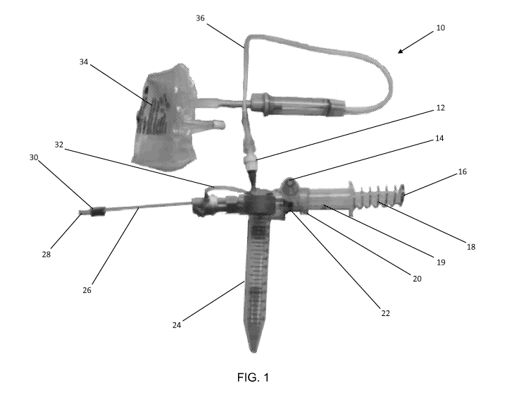
[0011] FIG.1 is a picture of a uterotubal irrigation system according
to an embodiment of the
invention;
[0012] FIG. 2 is a close-up picture of an irrigation cannula of the
uterotubal irrigation system of
FIG. 1 according to an embodiment of the invention;
[0013] FIG. 3 is a close-up picture of an evacuation basket on the tip
of the irrigation cannula of
the uterotubal irrigation system of FIG. 1 according to an embodiment of the
invention;
[0014] FIG. 4 is a schematic block diagram of the uterotubal irrigation
system of FIG. 1 in an
injection mode according to an embodiment of the invention;
[0015] FIG. 5 is a schematic block diagram of the uterotubal irrigation
system of FIG. 1 in a
collection mode according to an embodiment of the invention;
[0016] FIGs. 6A and 6B are schematic block diagrams of irrigation
cannula of FIG. 1 showing
the retraction of the irrigation tube to expand the distal evacuation basket
according to an
embodiment of the invention;
[0017] FIG. 7A is a schematic block diagram of a cell collection irrigation
catheter according to
an embodiment of the invention;
[0018] FIG. 7B is a cross-sectional view along line A-A of the cell
collection irrigation catheter
of FIG. 7A;
[0019] FIG. 8 is a schematic block diagram of the cell collection
irrigation catheter of FIGs. 7A
and 7B showing fluid flow paths in an irrigation and collection mode when
inserted in a uterus
according to an embodiment of the invention; and
[0020] FIG. 9 is schematic block diagram of a pressure limiting
injection device for use with
the cell collection irrigation catheter of FIG. lA and FIG. 7A according to an
embodiment of the
invention.
DESCRIPTION OF THE INVENTION
[0021] The present invention has utility as an uterotubal irrigation
system and process for
implanting hysterosalpingogram (HSG) procedures. Embodiments of the inventive
uterotubal
irrigation system serially irrigate fluid followed by evacuation of the fluid
in the uterus, with
extension of the fluid into the Fallopian tubes, for the purpose of collecting
cells from the Fallopian
4
CA 02943351 2016-09-20
WO 2015/143388
PCT/US2015/021858
tubes. Due to the tiny volume of the uterus, a single injection of several
cc's of fluid followed by
evacuation will yield a minimal amount of fluid for analysis. It is thus
necessary to repetitively
inject and retrieve multiple fluid aliquots to provide sufficient fluid volume
and sample cells for
evaluation. It is also important to stagger the steps of fluid injection and
fluid retrieval, otherwise a
concomitant injection and evacuation will prevent fluid from ever entering the
Fallopian tube.
[0022] It is to be understood that in instances where a range of values
are provided that the
range is intended to encompass not only the end point values of the range but
also intermediate
values of the range as explicitly being included within the range and varying
by the last significant
figure of the range. By way of example, a recited range from 1 to 4 is
intended to include 1-2, 1-3,
2-4, 3-4, and 1-4.
[0023] Embodiments of the uterotubal irrigation system provide a
partially flexible cannula that
is introduced through the cervical os into the uterine cavity. The inventive
cannula has an enlarged
external plug of approximately 2 cm that is proximal to the distal tip of the
cannula, to occlude the
os and permit infusion of fluid into the uterus. The inventive cannula has an
internal tube for
irrigation, and an external sheath with multiple slits near the distal end of
the cannula. The distal tip
of the external sheath is attached to the distal end of the irrigation tube.
In specific embodiments,
the inner diameter of the external sheath is approximately 0.5 mm greater than
the outer diameter of
the irrigation tube. The irrigation tube passes through a sliding seal on the
proximal end of the
external sheath. An irrigation port on the proximal end of the irrigation tube
permits fluid infusion,
while an evacuation port on the proximal end of the external sheath allows
evacuation of fluid via
the space between the outer diameter of the irrigation tube and the inner
diameter of the external
sheath. When the irrigation tube is retracted relative to the external sheath,
a series of slits on the
sheath at the distal tip of the cannula expand outward to form a basket to
maintain the uterine cavity
and prevent the uterine cavity from collapse due to a vacuum draw during
evacuation of fluid for
cell analysis.
[0024] Embodiments of the inventive uterotubal irrigation system
provide serial fluid injection
followed by fluid evacuation. The injection and evacuation modes of
embodiments of the inventive
system are controlled with the depression of a syringe plunger to modulate the
degree of vacuum
exhibited in the evacuation mode of the irrigation cannula. The system
utilizes a port in the side of
the syringe body, where upon full retraction of the syringe plunger against a
stop set at a
5
CA 02943351 2016-09-20
WO 2015/143388
PCT/US2015/021858
predetermined volume (e.g., 5 cc), the plunger seal covers and seals the port.
Parallel vacuum lines
are present in the system, where one vacuum line is a small diameter
(approximately 0.5 mm) line
that connects to the evacuation port on the external sheath of the irrigation
cannula, and the other
vacuum line is a large diameter (approximately 5-10 mm along the majority of
its length) line that
extends to the vacuum source. A collection tube is positioned in-line between
the evacuation port
on the irrigation cannula and the large diameter pressure line. When the
spring loaded syringe
plunger is completely retracted, during refilling of the syringe from a fluid
source, the plunger seal
closes off the vacuum port into the syringe from the large vacuum line, and a
vacuum is pulled
through the small diameter line to evacuate fluid via the expanded basket on
the irrigation cannula.
When the syringe plunger is depressed to inject fluid, the port on the side of
the syringe connected
to the large diameter vacuum line is opened. During plunger depression, the
majority of vacuum
flow is derived from the large diameter line, with its low fluid resistance,
and a minimal vacuum is
experienced in the small diameter line to drain the fluid as the fluid is
being injected by the syringe.
The flow rate and negative pressure provided by the vacuum source is also
maintained at a moderate
level to render the system functional. A one-way valve is present at the fluid
irrigation source, so
that fluid may only be introduced into the syringe and out of the irrigation
cannula upon syringe
depression and retraction.
[0025] An inventive embodiment of the uterotubal irrigation system is
provided as a catheter
for cell sampling that has two outlet openings on the catheter distal tip that
inject irrigation fluid in
two opposing jet streams that splay out laterally toward the os or openings of
both Fallopian tubes.
The two separate opposing outlets on the distal tip of the catheter are angled
toward the openings to
the Fallopian tubes, where the irrigation channels within the catheter bend
outward toward the outlet
openings. In operation an occlusion balloon or a plug that is situated at a
distance between 1.5 to
2.5 centimeters proximal to the tip of the catheter is inflated to seal the
cervical os prior to insertion
of the catheter tip into the uterus. In a specific embodiment, the occlusion
balloon or a plug is
situated at a distance of 2.0 centimeters proximal to the tip of the catheter.
The occlusion balloon
seals the cervical os during the irrigation and fluid collection process. The
injected irrigation fluid
proceeds a distance into both Fallopian tubes, and the fluid then circulates
back into the uterine
cavity, where the fluid exits via a collection port in the catheter. In an
inventive embodiment, the
6
CA 02943351 2016-09-20
WO 2015/143388
PCT/US2015/021858
collection inlet is approximately 1 cm proximal to the distal catheter tip.
The retrieved irrigation
fluid undergoes cytologic examination to detect the presence of malignant
cells.
[0026] Embodiments of the catheter based uterotubal irrigation system
provide serial fluid
injection followed by fluid evacuation. An inventive fluid injection device is
provided that may be
used in conjunction with the inventive cell collection irrigation catheter to
limit the amount of
pressure used for injection. The use of embodiments of the inventive fluid
injection device may be
used to avoid the pain and discomfort experienced by the patient during the
diagnostic procedure.
Embodiments of the pressure limiting fluid injection device have a syringe
plunger that is composed
of a compression spring connected to the distal sealing plunger face. A
threaded plunger advances
to compress the compression spring. The threaded plunger contains a groove
along its length that is
keyed by a pin that extends through the syringe body into the groove. A drive
disc is rotatably fixed
on the proximal end of the syringe body, and the drive disc contains internal
threads that mate with
the threaded plunger. When the drive disc is rotated, the threaded plunger
moves forward to
compress the fluid inside the syringe. The maximal pressure that may be
developed by the syringe
is determined by a clutch disc that lies coaxially outside the drive disc. At
a predetermined amount
of torque, the clutch disc slips relative to the drive disc. The torque
setting may be set by adjusting
the friction that exists between the clutch disc and the drive disc. In a
specific embodiment, one or
more clutch adjustment screws extending through the clutch disc may be
tightened down on the
drive disc, so that the torque exerted on the threaded plunger will be limited
to a given level. This in
turn limits the degree of compression exerted by the spring, thus limiting the
injection pressure. The
pressure limiting injection device incorporates the compression spring for
energy storage, such that
continuous rotation of the clutch disc is unnecessary for fluid injection.
Rather, the clutch disc is
rotated to bring the syringe to the desired injection pressure level, and then
rotated at intervals as
necessary to re-pressurize the system. In a specific embodiment the full
retraction of the syringe
plunger provides a predetermined volume of irrigation fluid (e.g., 5 cc).
[0027] Referring now to the figures, FIG.1 is a picture of an
uterotubal irrigation system 10
according to an embodiment of the invention. A syringe 19 with a user
controlled plunger 16 is
outwardly biased by spring 18, and a plunger seal 22 rests against a plunger
stop 20 positioned so
that the plunger seal 22 blocks the primary vacuum port 14 on the side of the
syringe 19 to the
primary vacuum line 46 (see FIG. 4) that is connected to a vacuum source (not
shown). A source of
7
CA 02943351 2016-09-20
WO 2015/143388
PCT/US2015/021858
injection fluid, such as saline, is stored or held in a fluid reservoir 34,
and supplied to the syringe 19
via a supply line 36 and a one way valve 12. A collection tube 24 stores
fluids that are evacuated
from the uterus via small diameter vacuum line 32 via the irrigation cannula
26. The irrigation
cannula 26 is shown in greater detail in FIG. 2 and FIG. 3. At the distal end
of the irrigation
cannula 26, a cervical plug 30 forms a seal at the cervix os upon insertion of
the cannula 26 into the
uterus, and allows fluid pressure to be developed in the uterus. The cervical
plug may be a balloon,
a solid dilated structure on the cannula body, or a foam stopper. FIG. 3 shows
an evacuation basket
on the distal tip 28 of the irrigation cannula 26. The evacuation basket forms
when two or more slits
38 in an external sheath 44 expand outward with the retraction of the
irrigation tube 40. The
irrigation arrow (I) illustrates the direction of fluid injected via the
irrigation tube 40, and evacuation
arrow (E) illustrates the direction and entry of fluid withdrawn from the
uterus that travels in the
space between the external sheath 44 and the irrigation tube 40.
[0028] FIG. 4 is a schematic block diagram of the uterotubal irrigation
system of FIG. 1 in
injection mode according to an embodiment of the invention. With the plunger
16 depressed, the
plunger seal 22 is removed from the primary vacuum port 14 on the side of the
syringe 19 to the
primary vacuum line 46 that is connected to a vacuum source (not shown). In
the injection mode,
the large diameter vacuum line 46 is open to flow, and there is a resultant
minimal vacuum
exhibited by the irrigation cannula 26 during fluid injection through the
irrigation port 50. FIG. 5 is
a schematic block diagram of the uterotubal irrigation system of FIG. 1 in
fluid and cell collection
mode according to an embodiment of the invention. With the syringe plunger 16
retracted and the
large diameter vacuum line 46 is blocked, and vacuum is pulled through the
small diameter line 32
to evacuate fluid via the expanded basket formed from the two or more slits 38
on the irrigation
cannula 26. The fluid is drawn through the evacuation port 52 and the small
diameter vacuum line
32 to the collection tube 24
[0029] FIGs. 6A and 6B are schematic block diagrams of irrigation cannula
26 of FIG. 1
showing the retraction of the irrigation tube 40 relative to the external
sheath 44 to expand the distal
evacuation basket formed by the two or more slits 38 according to an
embodiment of the invention.
The irrigation tube 40 slides on the sliding seal 48 of the evacuation port
52.
[0030] FIG. 7A is a schematic block diagram of a cell collection
irrigation catheter 60
according to an embodiment of the invention. The catheter 62 for cell sampling
contains two outlet
8
CA 02943351 2016-09-20
WO 2015/143388
PCT/US2015/021858
openings (70R, 70L) on the catheter distal tip 62D that inject irrigation
fluid in two opposing jets 92
that splay out laterally toward the os or openings 90 of both Fallopian tubes
as shown in FIG. 8.
The two separate opposing outlets (70R, 70L) on the distal tip 62D of the
catheter 62 are angled
toward the openings 90 to the Fallopian tubes, where the irrigation channels
(66CR, 66CL) within
the catheter 62 have a bend 68 that angle the irrigation channels (66CR, 66CL)
outward toward the
outlet openings (70R, 70L). The irrigation channels (66CR, 66CL) are in fluid
communication with
irrigation fluid supply lines (66L, 66R), respectively that split off from
irrigation fluid supply line
66 that terminates with irrigation port 64. A source of injection fluid, such
as saline, is stored or
held in a fluid reservoir (see FIG. 9) that connects with the irrigation port
64.
[0031] In operation as shown in FIG. 8 an occlusion balloon 84 or a plug
that is situated at a
distance between 1.5 to 2.5 centimeters proximal to the tip 62D of the
catheter 62 is inflated to a full
state 841 (as shown by the dotted lines) to seal the cervical os 86 prior to
insertion of the catheter tip
62D into the uterine cavity 88. The inflated balloon 841 also serves as a
tactile stop to indicate to
the physician when to cease the applied inward pressure when inserting the
catheter 62 into the
uterine cavity 88. The occlusion balloon 84 is inflated with air or gas via
supplied inflation outlet
opening 82 positioned on the wall of the catheter 62. An inflation channel 80C
runs internally along
the length of catheter 62 and terminates at the inflation outlet opening 82.
The inflation channel
80C is in fluid communication with gas supply line 80 that terminates in
balloon inflation port 78.
In a specific embodiment, the occlusion balloon 84 or a plug is situated at a
distance of 2.0
centimeters proximal to the tip 62D of the catheter 62. The occlusion balloon
84 seals the cervical
OS 86 during the irrigation and fluid collection process. The injected
irrigation fluid, represented by
the arrows 92 proceeds a distance into both Fallopian tubes 90, and the fluid
then circulates back
into the uterine cavity 88, where the fluid exits via a collection inlet 76 in
the catheter 62. In an
inventive embodiment, the collection port 76 is approximately 1 cm proximal to
the distal catheter
tip 62D. The collection inlet 76 provides an opening to the collection channel
74C that runs along
the inside of the catheter 62. The collection channel 74C is in fluid
communication with an external
collection line 74 that terminates in a collection port 72. The retrieved
irrigation fluid collected at
the collection port 72 undergoes cytologic examination to detect the presence
of malignant cells.
FIG. 7B is a cross-sectional view along line A-A of the cell collection
irrigation catheter 62 of FIG.
9
CA 02943351 2016-09-20
WO 2015/143388
PCT/US2015/021858
7A that shows the irrigation channels (66CR, 66CL), the collection channel
74C, and inflation
channel 80C within the catheter 62.
[0032] FIG. 9 is schematic block diagram of an inventive embodiment of
a pressure limiting
fluid injection device 100 for use with the cell collection irrigation
catheter system 60 of FIGs. 7A,
7B, and 8. It is noted that the pressure limiting fluid injection device 100
may also be used with
uterotubal irrigation system 10 that was described in FIGs. 1-6. The inventive
fluid injection device
100 is provided that may be used in conjunction with the inventive cell
collection irrigation catheter
62 to limit the amount of pressure used for injection. The use of embodiments
of the inventive fluid
injection device 100 may be used to avoid the pain and discomfort experienced
by the patient during
a diagnostic procedure.
[0033] Embodiments of the pressure limiting fluid injection device 100
of FIG. 9 have a
syringe plunger body 102 that is composed of a compression spring 104
connected to the distal
sealing plunger face 106. A threaded plunger 108 advances to compress the
compression spring
104. The threaded plunger 108 contains a groove 110 along its length that is
keyed by a pin 112
that extends through the syringe plunger body 102 into the groove 110. A drive
disc 114 is
rotatably fixed on the proximal end of the syringe plunger body 102, and the
drive disc 114 contains
internal threads 116 that mate with the threaded plunger 108. When the drive
disc 114 is rotated,
the threaded plunger 108 moves forward to compress the fluid inside the
syringe plunger body 102.
The maximal pressure that may be developed by the syringe is determined by a
clutch disc 118 that
lies coaxially outside the drive disc 114. At a predetermined amount of
torque, the clutch disc 118
slips relative to the drive disc 114. The torque setting may be set by
adjusting the friction that exists
between the clutch disc 118 and the drive disc 114. In a specific embodiment,
one or more clutch
adjustment screws 120 extending through the clutch disc 118 may be tightened
down on the drive
disc 114, so that the torque exerted on the threaded plunger 108 will be
limited to a given level.
This in turn limits the degree of compression exerted by the compression
spring 104, thus limiting
the injection pressure. The pressure limiting injection device 100
incorporates the compression
spring 114 for energy storage, such that continuous rotation of the clutch
disc 118 is unnecessary for
fluid injection. Rather, the clutch disc 118 is rotated to bring the pressure
limiting fluid injection
device 100 to the desired injection pressure level, and then rotated at
intervals as necessary to re-
pressurize the system. In a specific embodiment the full retraction of the
syringe plunger provides a
CA 02943351 2016-09-20
WO 2015/143388
PCT/US2015/021858
predetermined volume of irrigation fluid (e.g., 5 cc). The output of the
pressure limiting injection
device 100 is coupled to irrigation port 64 that is in fluid communication
with irrigation fluid supply
line 66, and a source of injection fluid, such as saline, that is stored or
held in a fluid reservoir 72.
The irrigation port 64 may act as a check valve which allows fluid to be
released from the reservoir
72 on the retraction of the plunger face 56 inside the syringe plunger body
102 which draws in fluid,
and closes off the reservoir 72 on a forward stroke of the plunger face 106
that pushes the fluid into
the supply line 66 via the irrigation port 64.
[0034] The foregoing description is illustrative of particular
embodiments of the invention, but
is not meant to be a limitation upon the practice thereof. The following
claims, including all
equivalents thereof, are intended to define the scope of the invention.
11