Note : Les descriptions sont présentées dans la langue officielle dans laquelle elles ont été soumises.
HUMANIZED ANTIBODIES THAT BIND LGR5
RELATED APPLICATIONS
[0001] This application claims the benefit of U.S. Provisional App.
No.
62/081497 filed November 18, 2014, and U.S. Provisional App. No. 61/975589
filed April 4,
2014.
FIELD OF THE INVENTION
[0002] The present invention relates generally to the field of cancer
biology.
More particularly, embodiments are drawn to humanized antibodies against LGR5
and uses
of such antibodies. Several embodiments relate to monoclonal, humanized, or
fully human
antibodies against LGR5, hybridomas or other cell lines expressing such
antibodies, nucleic
acids and vectors comprising nucleic acids encoding for such antibodies, and
methods of
blocking cancer stem cell growth with such antibodies.
REFERENCE TO SEQUENCE LISTING
[0003] The present application is being filed along with a Sequence
Listing in
electronic format. The Sequence Listing is provided as a file entitled
BION010W0 SEQLISTING.TXT, created April 2, 2015 which is approximately 40 Kb
in
size.
BACKGROUND OF THE INVENTION
[0004] Leucine-rich repeat containing G-protein-coupled receptor 5
(LGR5), also
known as GPR49/HG38/FEX, belongs to the leucine-rich repeat containing G-
protein-
coupled receptor (LGR) / G-Protein-coupled Receptor (GPR) protein family of
receptor
proteins that are structurally similar to glycoprotein hormone receptors. LGRs
are divided
into three subgroups: (1) glycoprotein hormone receptors including thyroid-
stimulating
hormone (TSH) receptor, follicle-stimulating hormone (FSH) receptor, and
luteinizing
hormone (LH) receptor; (2) relaxin receptors LGR7 and LGR8; and (3) LRG4,
LGR5, and
LGR6. LGR5 is expressed in several tissues including the intestine, skeletal
muscle, placenta,
brain, and spinal cord.
SUMMARY OF THE INVENTION
[00051 Some embodiments of the compositions, methods and kits
provided herein
include a humanized or human monoclonal antibody that binds LGR5. In some
-1 -
CA 2944649 2017-07-11
CA 02944649 2016-09-30
WO 2015/153916 PCMJS2015/024162
embodiments, the antibody comprises a heavy chain CDR1 comprising SEQ ID NO:23
or
conservative variations thereof. In some embodiments, the antibody comprises a
heavy chain
CDR2 comprising SEQ ID NO:2 or conservative variations thereof. In some
embodiments,
the antibody comprises a heavy chain CDR3 comprising SEQ ID NO:3 or
conservative
variations thereof In some embodiments, the antibody comprises a light chain
CDRI
comprising SEQ ID NO:4 or conservative variations thereof In some embodiments,
the
antibody comprises a light chain CDR2 having amino acids LTS or conservative
variations
thereof. In some embodiments, the antibody comprises a light chain CDR3
comprising SEQ
ID NO:33 or conservative variations thereof. In some embodiments, the antibody
comprises a
heavy chain variable domain comprising SEQ ID NOs:19 or 48. In some
embodiments, the
antibody comprises a light chain variable domain comprising SEQ ID NOs: 21 or
49. In
some embodiments, the antibody binds an epitope within amino acids T175, E176,
Q180,
R183, S186, A187, Q189, D247, E248, T251, R254, S257, N258. K260 of LGR5 (SEQ
ID
NO:47). In some embodiments, the antibody binds an epitope within leucine rich
repeats 6-9
of LGR5 (SEQ ID NO:47). In some embodiments, the antibody binds an epitope on
the
convex surface of LGR5. In some embodiments, the antibody does not bind a RSPO-
LGR5
binding site. In some embodiments, the antibody does not disrupt LGR5-RSPO
binding. In
some embodiments, the antibody does not disrupt LGR5-RSPO signaling. In some
embodiments, the RSPO is selected from the group consisting of RSPOI, R5P02,
RSP03,
and RSP04. In some embodiments, the antibody does disrupt formation of a
complex such as
LGR5-RSPO-RNF43, LGR5-RSPO-ZNRF3, LGR5-RSPO-LRP6, LGR5-NORRIN-RNF43,
LGR5- NORRIN-ZNRF3, LGR5-NORRIN-LRP6. In some embodiments, the antibody
disrupts LGR5 signaling through Wnt/[3 -catenin pathway. In some embodiments,
the
antibody induces expression of differentiation markers in a tumor. In some
embodiments, the
antibody is capable of inducing cells in a tumor to differentiate. In some
embodiments, the
antibody which inhibits tumor growth. In some embodiments, the antibody
reduces the
frequency of cancer stem cells in a tumor.
[0006] Some embodiments of the compositions, methods and kits provided
herein
include an isolated polynucleotide molecule comprising a polynucleotide that
encodes any
one of the foregoing antibodies. Some embodiments of the compositions, methods
and kits
provided herein include a vector comprising any one of the foregoing
polynucleotides. Some
-2-
CA 02944649 2016-09-30
WO 2015/153916 PCMJS2015/024162
embodiments of the compositions, methods and kits provided herein include a
host cell
comprising any one of the foregoing vectors. Some embodiments of the
compositions,
methods and kits provided herein include a method of producing an antibody
comprising
culturing any one of the foregoing host cells so that the antibody is
produced.
[0007] Some embodiments of the compositions, methods and kits provided
herein
include a pharmaceutical composition comprising any one of the foregoing
antibodies and a
pharmaceutically acceptable carrier.
[0008] Some embodiments of the compositions, methods and kits provided
herein
include a method of treating a subject having a cancer comprising
administering any one of
the foregoing antibodies to the subject. Some embodiments also include
administering a
chemotherapeutic agent in combination with the antibody. In some embodiments,
the
chemotherapeutic agent is selected from the group consisting of folinic acid,
fluorouracil,
irinotecan, gemcitabine and Abraxane. In some embodiments, the folinic acid,
fluorouracil,
and irinotecan are administered in combination with the antibody to the
subject.
[0009] In some embodiments, the treatment increases the likelihood of
survival of
the subject for a period of at least 3 months after the treatment compared to
the likelihood of
survival of a subject not treated with the antibody. In some embodiments, the
likelihood of
survival of the subject is increased for a period of at least 6 months. In
some embodiments,
the likelihood of survival of the subject is increased for a period of at
least 12 months.
[0010] In some embodiments, the treatment reduces the risk of recurrence
of the
cancer in the subject compared to the risk of recurrence of the cancer in a
subject not treated
with the antibody.
[0011] In some embodiments, the treatment reduces the level of tumor
cells in the
peripheral blood of the subject compared to the level of tumor cells in the
peripheral blood of
a subject not treated with the antibody.
[0012] In some embodiments, the cancer is selected from the group
consisting of
colon cancer, colorectal cancer, pancreatic cancer, breast cancer, and lung
cancer. In some
embodiments, the cancer is selected from the group consisting of colon cancer
comprising an
APC mutation, colon cancer comprising an KRAS mutation, metastatic colorectal
cancer,
metastatic pancreatic cancer, triple-negative breast cancer, and small cell
lung cancer.
-3-
CA 02944649 2016-09-30
WO 2015/153916 PCMJS2015/024162
[0013] In some embodiments, the subject is mammalian. In some
embodiments,
the subject is human.
[0014] Some embodiments of the compositions, methods and kits provided
herein
include a method for reducing the risk of developing a cancer, preventing the
recurrence of a
cancer, or preventing a cancer in a subject predisposed to the cancer
comprising
administering any one of the foregoing antibodies to the subject.
[0015] Some embodiments of the compositions, methods and kits provided
herein
include a method of increasing the likelihood of survival of a subject having
a cancer
comprising administering any one of the foregoing antibodies to the subject.
In some
embodiments, the likelihood of survival of the subject is increased for a
period of at least 3
months after the treatment compared to the likelihood of survival of a subject
not treated with
the antibody. In some embodiments, the likelihood of survival of the subject
is increased for
a period of at least 6 months. In some embodiments, the likelihood of survival
of the subject
is increased for a period of at least 12 months.
[0016] Some embodiments of the compositions, methods and kits provided
herein
include a method of reducing the risk of recurrence of a cancer in a subject
comprising
administering any one of the foregoing antibodies to the subject.
[0017] Some embodiments of the compositions, methods and kits provided
herein
include a method of reducing the level of tumor cells of a cancer in the
peripheral blood of a
subject comprising administering any one of the foregoing antibodies to the
subject.
[0018] Some embodiments also include administering a chemotherapeutic
agent
in combination with the antibody. In some embodiments, the chemotherapeutic
agent is
selected from the group consisting of folinic acid, fluorouracil, irinotecan,
gemcitabine and
Abraxane. In some embodiments, the folinic acid, fluorouracil, and irinotecan
are
administered in combination with the antibody to the subject.
[0019] In some embodiments, the subject is determined to be predisposed
to the
cancer by a predictive clinical test, a genetic analysis, or a family history
analysis.
[0020] In some embodiments, the cancer is selected from the group
consisting of
colon cancer, colorectal cancer, pancreatic cancer, breast cancer, and lung
cancer. In some
embodiments, the cancer is selected from the group consisting of colon cancer
comprising an
-4-
CA 02944649 2016-09-30
WO 2015/153916 PCMJS2015/024162
APC mutation, colon cancer comprising an KRAS mutation, metastatic colorectal
cancer,
metastatic pancreatic cancer, triple-negative breast cancer, and small cell
lung cancer.
[0021] In some embodiments, the subject is mammalian. In some
embodiments,
the subject is human.
[0022] Some embodiments of the compositions, methods and kits provided
herein
include a method of selecting a treatment for a subject having a tumor
comprising: (a)
administering a chemotherapeutic agent to the subject; (b) identifying an
increased level of a
LGR5 polypeptide or a nucleic acid encoding LGR5 in the tumor; and (c)
administering any
one of the foregoing antibodies to the subject having the increased level of
LGR5
polypeptide or a nucleic acid encoding LGR5 in the tumor. In some embodiments,
the
chemotherapeutic agent is selected from the group consisting of folinic acid,
fluorouracil,
irinotecan, gemcitabine and Abraxane. In some embodiments, the tumor is
selected from the
group consisting of colon cancer tumor, colorectal cancer tumor, pancreatic
cancer tumor,
breast cancer tumor, and lung cancer tumor. In some embodiments, the tumor is
selected
from the group consisting of colon cancer tumor comprising an APC mutation,
colon cancer
tumor comprising an KRAS mutation, metastatic colorectal cancer tumor,
metastatic
pancreatic cancer tumor, triple-negative breast cancer tumor, and small cell
lung cancer
tumor.
[0023] Some embodiments of the compositions, methods and kits provided
herein
include a method of assessing the efficacy of a treatment with any one of the
foregoing
antibodies comprising measuring the level of a biomarker in a tumor treated
with the
antibody. In some embodiments, the biomarker is a nucleic acid or a
polypeptide encoded by
the nucleic acid, wherein the biomarker selected from the group consisting of
WNT6, FZD8,
FOSL1, WT11, NFATC1, FZD5, FZD2, FRZB, PRICKLE], FZDB, FZD7, WNT7B,
FBXW11, FZD1, DVL], CSNK2A1, ANGPT2, AKAP12, ADM, CTNNB], ALDOC,
CDH5, ITGA2, DAB1, MIR655, NKX1-2, ZBTB11. ITPKA, PSMC3IP and BAK]. In some
embodiments, a decrease in the level of the biomarker compared to a the level
of the
biomarker in a tumor not treated with the tumor is indicative of an effective
treatment. In
some embodiments, the biomarker is selected from the group consisting of WNT6,
EZD8,
FOSL1, WT11, NFATC1, FZD5, FZD2, FRZB, PRICKLEL FZDB, FZD7, WNT7B,
FBXW11, FZD1, DVL], CSNK2A1, ANGPT2, AKAP12, ADM, CTNNB1, ALDOC,
-5-
CDH5, and ITGA2. In some embodiments, an increase in the level of the
biomarker
compared to a the level of the biomarker in a tumor not treated with the tumor
is indicative of an
effective treatment. In some embodiments, the biomarker is selected form the
group consisting of
DAB1, MIR655, NKX1-2, ZBTB11, ITPKA, PSMC3IP and BAK1. In some embodiments,
the
tumor is selected from the group consisting of colon cancer tumor, colorectal
cancer tumor,
pancreatic cancer tumor, breast cancer tumor, and lung cancer tumor. In some
embodiments, the
tumor is selected from the group consisting of colon cancer tumor comprising
an APC mutation,
colon cancer tumor comprising an KRAS mutation, metastatic colorectal cancer
tumor, metastatic
pancreatic cancer tumor, triple-negative breast cancer tumor, and small cell
lung cancer tumor.
[0023a] In accordance with an aspect of the invention is a humanized
monoclonal
antibody that specifically binds Leucine-rich repeat containing G-protein-
coupled receptor 5
(LGR5) and comprises a heavy chain variable domain comprising an amino acid
sequence shown
in SEQ ID NO: 19 and a light chain variable domain comprising an amino acid
sequence shown
in SEQ ID NO: 21.
BRIEF DESCRIPTION OF THE DRAWINGS
[0024] FIG. 1 is a graph showing direct FACS binding of humanized
monoclonal
antibody 18G7H6A3 to human LGR5 (CHO).
[0025] FIG. 2 is a graph showing the effect of FOLFIRI, alone and in
combination with
18G7H6A3, on CT3 CRC tumor volume.
[0026] FIG. 3 is a graph showing 18G7H6A3 treatment significantly
reduced MDA-
MB-231-LM3 primary tumor volume.
[0027] FIG. 4 depicts graphs showing FolFiri treatment in mice bearing
CT1, or CT3
tumors results in upregulation of LGR5.
[0028] FIG. 5 is a bar chart showing chemotherapy results in
upregulation of LGR5
(more than 4-fold) in JH109 tumors.
[0029] FIG. 6 is a graph showing significant activity of 18G7H6A3
observed when
administered in combination with chemotherapy (gemcitabine).
[0030] FIG. 7 is a point plot showing that antibody 18G7H6A3 reduces
the number of
live events in a CT1 cancer stem cell population.
Date Recue/Date Received 2021-04-12
-6-
[0031] FIG. 8 is a line graph showing cells isolated from mice treated
with anti-LGR5
antibody 18G7H6A3 in combination with FOLFIRI had greatly decreased
tumorigenicity as
compared to cells isolated from mice treated with FOLFIRI alone.
[0032] FIG. 9 is a line graph showing that re-implanted cells from the
18G7H6A3
FOLFIRI combination had a significantly delayed time to progression.
Date Recue/Date Received 2021-04-12
-6a-
CA 02944649 2016-09-30
WO 2015/153916 PCMJS2015/024162
[0033] FIG. 10 is a line graph showing significant activity of humanized
antibody
18G7H6A3 is observed when administered prophylactically in combination with
chemotherapy (FOLFIRI).
[0034] FIG. 11 is a point plot showing that antibody l 8G7H6A3 is able
to inhibit
Wnt signaling in tumor cells in vivo as indicated by phospho-Thr41/Ser45-P-
catenin
immunoassays.
[0035] FIG. 12 is a bar chart showing that increasing concentrations of
soluble
antibody 18G7H6A3 did not affect the induction of TCF/LEF promoter driven GFP
expression by the combination of Wnt3a plus RSP02, demonstrating that the anti-
LGR5
antibody 18G7H6A3 does not block RSPO-driven TCF/LEF promoter activation. A
positive
control antibody C12 is shown to inhibit Wnt3a/RSPO2 driven TCF/LEF promoter
activitation.
[0036] FIG. 13 is a line graph showing that R-spondin does not block
antibody
18G7H6A3 binding to LGR5.
[0037] FIG. 14 is a bar chart showing that antibody I 8G7H6A3 binding to
LGR5
inhibits formation of ternary complex.
[0038] FIG. 15 depicts levels of LGR5 expression in treated samples.
[0039] FIG. 16 depicts levels of CTNNB1 expression, and p-f3-Catenin
expression in treated samples.
[0040] FIG. 17 depicts differentially expressed transcripts in various
treated
samples.
[0041] FIG. 18 depicts differentially expressed genes in 18G7H6A3-
(BNC101)
treated tumors.
[0042] FIG. 19 depicts differentially expressed genes in FOLFIRI treated
tumors.
[0043] FIG. 20 depicts differentially expressed genes in combination-
treated
tumors
[0044] FIG. 21 depicts levels of LGR5 in circulating HI,A+ cells.
[0045] FIG. 22A and FIG. 22B depict levels of LGR5 in circulating IILA+
cells.
[0046] HG. 23 is a graph showing animal survival of mice treated with
Gemcitabine/Abraxane or with Gemcitabine/Abraxane and 18G7H6A3.
-7-
CA 02944649 2016-09-30
WO 2015/153916 PCMJS2015/024162
DETAILED DESCRIPTION
[0047] Several embodiments of the present application are drawn to
humanized
antibodies that specifically bind to LRG5 and methods of inhibiting cancer
stem cell growth
with such antibodies. In some embodiments, the antibodies specifically bind
LGR5 but do
not inhibit R-Spo binding to LGR5. Other embodiments include antibodies that
bind LGR5
without inhibiting R-Spo signaling through LGR5. Still other embodiments
include
antibodies that bind LGR5 but do not inhibit both R-Spo binding or signaling
through LGR5.
[0048] Another embodiment is antibodies that bind LGR5 and also inhibit
LGR5
signaling through the Wnt pathway. In some embodiments, these antibodies may
inhibit
LGR5 signaling through the Wnt pathway, and be independent of RSpo signaling.
[0049] Other embodiments include methods of using the antibodies
described
above to inhibiting LGR5 or R-Spo signaling in a cell or tissue.
[0050] LGR5 was identified through lineage tracing studies as a highly
specific
marker of normal stem cells and tumor-initiating cells in the gut. Previously
about 150 genes
were identified whose expression was quenched following abrogation of Wnt
expression. A
comprehensive characterization of these `Wnt target genes' found LGR5 to be
selectively
expressed on a population of 10-14 proliferating wedge-shaped cells at the
crypt base. These
crypt-based columnar cells were previously proposed to be a candidate stem
cell population.
Using in vivo lineage tracing with a heritable lacZ ¨LGR5 reporter gene, it
has been
confirmed that LGR5 intestinal stem cells are a multi-potent, self-renewing
population of
adult intestinal stem cells that give rise to uninterrupted ribbons of lacZ+
progeny cells
initiating from the crypt base and extending to the villus tips.
[0051] The specific expression of LGR5 on CSCs provides an opportunity
to
target CSCs selectively and effectively. LGR5 is highly over expressed in CRC,
pancreatic
and most other solid tumors, compared to normal tissues, thereby providing a
wide
therapeutic window to target CSCs in CRC, pancreatic, breast, ovarian, lung,
gastric and liver
cancer.
[0052] LGR5 itself is a facultative component of the 'Wnt-Fzd-LRP
receptor
complex that binds secreted R-spondin ligands to selectively amplify and
enhance Wnt
signals on LGR5 positive cells. There is also evidence that LGR5 can signal in
a Wnt-
independent manner. In addition, the related transmembrane RING-type E3
ubiquitin ligase
-8-
CA 02944649 2016-09-30
WO 2015/153916 PCMJS2015/024162
ZNRF3 (zinc and RING finger 3) or RNF43 (RING finger 43), are uniquely
expressed in
LGR5+ stem cells and reduce Wnt signals by selectively ubiquitinating frizzled
receptors,
thereby targeting these Wnt receptors for degradation. The R-spondin ligands
interact with
LGR5, to form a ternary complex with the transmembrane ZNRF3 or RNF43.
Formation of
these ternary complexes sequester ZNRF3 or RNF43 from the Wnt-Fzd-LRP complex
and
stabilize canonical and noncanonical Wnt signaling. Finally, Norrin has been
identified as an
additional ligand for the LGR family with unknown associated biology.
[0053] The gate keeping mutation in CRC is loss of adenomatous polyposis
coli
(APC), resulting in the aberrant activation of Wnt signaling, which normally
acts to regulate
the balance between stem cell self-renewal and differentiation in the colon
crypt.
Dysregulated Wnt signaling in intestinal stem cells leads to the formation of
adenomatous
polyps in the colon that are the precursor to malignant CRC. LGR5 stem cells
were
confirmed to be the source or root of these mouse intestinal tumors, using a
strategy that
crossed inducible APC gene knockout mice with mice whose LGR5 stein cells were
specifically and randomly labeled with one of four (GFP/YFP/ECFP/RFP)
fluorescent
genetic markers. The appearance of single colored tumors (i e . all GFP or all
RFP) 4 weeks
after induction of APC deletion confirmed that these tumors arose from a
single LGR5 stem
cell. Furthermore, this model also allowed for the fluorescent genetic tag in
the LGR5 stem
cells to be flipped to a different color, so that an RFP+ LGR5 cancer stem
cell generating a
red tumor could be transformed midstream into a ECFP+ LGR5 cancer stem cell,
that was
still seeding the tumor but now giving rise to blue tumor cells invading the
previously all red
GFP+ tumor mass. This flipping experiment not only provided further
confirmation that
LGR5 CSCs are the origin of intestinal tumors, able to initiate and seed the
growth of
intestinal tumors, but also that they continuously maintain tumor formation
(i.e., have long-
term repopulating ability).
[0054] A functional role of LGR5 in cancer has been validated through
ribonucleic acid interference (RNAi) knockdown studies. Knockdown of LGR5 in a
panel of
CRC tumor cell lines significantly inhibited the growth of soft agar colonies
in vitro, and also
the growth of HC 1116 colon tumor xenografts in vivo. LGR5 RNAi knockdown was
subsequently shown to also reduce the growth of CSC colonies from patient-
derived CRC
tumor cells in vitro (data not shown). Finally, sorted LGR5+ PATIENT DERIVED
-9-
BION0.010W0
PATENT
XENOGRAFT CRC tumor cells were found to be highly tumorigenic in vivo compared
to
control LGR5- cells.
[0055]
CSCs are believed to responsible for the high incidence of tumor
recurrence in many cancer patients treated with surgery and standard of care
chemotherapy.
For example, CD44+ CSCs from breast cancer patients were found to be enriched
following
chemotherapy, and that high levels of CSCs correlated with poor clinical
response to
chemotherapy. Similarly, in metastatic CRC, LGR5 expression was upregulated in
damaged
liver following chemotherapy, suggesting that increased LGR5 CSCs in response
to
chemotherapy initiate and/or acerbate metastatic disease. Indeed, it has been
found that
LGR5 expression is significantly greater in metastatic sites compared to
primary CRC
tumors.
Anti-LGR5 Antibodies
[0056] As
used herein, the term "antibody" includes, but is not limited to,
synthetic antibodies, monoclonal antibodies, recombinantly produced
antibodies, intrabodies,
multispecific antibodies (including bi-specific antibodies), human antibodies,
humanized
antibodies, chimeric antibodies, synthetic antibodies, single-chain Fvs
(scFv), Fab fragments,
F(ab') fragments, disulfide-linked Fvs (sdFv) (including bi-specific sdFvs),
and anti-idiotypic
(anti-Id) antibodies, and epitope-binding fragments of any of the above. The
antibodies of
several embodiments provided herein may be monospecific, bispecific,
trispecific or of
greater multispecificity. Multispecific antibodies may be specific for
different epitopes of a
polypeptide or may be specific for both a polypeptide as well as for a
heterologous epitope,
such as a heterologous polypeptide or solid support material. See, e.g., PCT
publications WO
93/17715; WO 92/08802; W091/00360; WO 92/05793; Tutt, et al., J. Immunol.
147:60-69
(1991); U.S. Pat. Nos. 4,474,893; 4,714,681; 4,925,648; 5,573,920; 5,601,819;
Kostelny et
al., J. Immunol. 148:1547-1553 (1992).
100571 As
used herein, LGR5 includes, but is not limited to, human LGR5
including the polypeptide of NCBI Accession No. NP_003658.1, or fragments
thereof, which
is encoded by the coding nucleotide sequence within NM_003667.2, or fragments
thereof.
The amino acid sequence and entire entry of NCBI Accession No, NP 003658.1 and
nucleotide sequence and entire entry of NM 003667.2. Examples of LGR5
fragments
Date Recue/Date Received 2021-04-12
-10-
BION0.010W0
PATENT
contemplated herein include the LGR5 ectodomain, transmembrane domain, or
intracellular
domain and portions thereof.
[0058]
Several embodiments relate to a hybridoma that produces the light chain
and/or the heavy chain of an anti-LGR5 antibody, including the anti-LGR5
antibodies
designated as 18G7H6A3 and 18G7H6A1 produced and described in the Examples
below. In
one aspect, the hybridoma produces the light chain and/or the heavy chain of a
humanized or
fully human monoclonal antibody such as that of 18G7H6A3 and 18G7H6A1 produced
and
described in the Examples below.
[0059]
Some embodiments are drawn to a nucleic acid molecule encoding the
light chain or the heavy chain of an anti-LGR5 antibody, including any one of
the anti-LGR5
antibodies designated as 18G7H6A3 and 18G7H6A1 produced and described in the
Examples below. In some aspects, a nucleic acid molecule encodes the light
chain or the
heavy chain of a humanized or fully human monoclonal, such as antibody
18G7116A3 and
18G7H6A1 produced and described in the Examples below.
[0060]
Various embodiments are directed to a vector comprising a nucleic acid
molecule or molecules encoding a light chain and/or a heavy chain of an anti-
LGR5
antibody, including any one of the anti-LGR5 antibodies designated as 18G7H6A3
and
18G7H6A1 produced and described in the Examples below.
[0061] In
various embodiments, the glycosylation of the antibodies can be
modified. For example, an aglycosylated antibody can be made (i.e., the
antibody lacks
glycosylation). Glycosylation can be altered to, for example, increase the
affinity of the
antibody for a target antigen. Such carbohydrate modifications can be
accomplished by, for
example, altering one or more sites of glycosylation within the antibody
sequence. For
example, one or more amino acid substitutions can be made that result in
elimination of one
or more variable region framework glycosylation sites to thereby eliminate
glycosylation at
that site. Such aglycosylation may increase the affinity of the antibody for
antigen. Such an
approach is described in further detail in U.S. Pat. Nos. 5,714,350 and
6,350,861.
[0062] In
several embodiments, the antibodies specifically bind a polypeptide
comprising or consisting of a LGR5 polypeptide having at least 60% identity,
or at least 70%
identity, or at least 80% identity, at least 85% identity, at least 90%
identity, at least 95%
Date Recue/Date Received 2021-04-12
-11-
CA 02944649 2016-09-30
WO 2015/153916 PCMJS2015/024162
identity, or at least at least 97% identity, or at least 99% identity, or 100%
identity to the
human LGR5 polypeptide of NCBI Accession Nos. NP_003658.1 (SEQ ID NO: 47) or
fragments thereof. Such fragments can, for example, be at least about 5, 10,
15, 20, 25, 50,
75, 100, 150, 200, 250, 300, 350, 400, 450, 500, 550, 600, 650, 700, 750, 800,
850, or 900
contiguous or non-contiguous amino acids of the LGR5 polypeptide, or any
number of
contiguous or non-contiguous amino acids in between any of the aforementioned
lengths.
[0063] In several embodiments, the antibody is antibody 18G7H6A3 and
comprises a heavy chain amino acid sequence of SEQ ID NO: 13 and a DNA
sequence of
SEQ ID NO: 11. In some embodiments, the antibody is antibody 18G7H6A3 and has
a
heavy chain variable domain comprises SEQ ID NO: 19. In several embodiments,
the
antibody is antibody 18G7H6A3 and comprises a light chain sequence of SEQ ID
NO: 14.
In other embodiments, the antibody is antibody 18G7H6A3 and comprises a light
chain
variable domain of SEQ ID NO: 21.
[0064] In some embodiments the antibodies comprise a sequence that is
80%,
81%. 82%, 83%, 84%, 85%, 86%, 87%, 88%, 89%, 90%, 91%, 92%, 93%, 94%, 95%, 96%
97%, 98%, 99%, or 100% identical to the sequence of the above sequences. In
some
embodiments the antibodies comprise a sequence that is 100% identical to the
above
antibody sequences over a span of 29, 30, 31, 32, 33, 34, 35, 36, 37, 38, 39,
40, 41, 42, 43,
44, 45, 46, 47, 48, 49, 50, 51, 52, 53, 54, 55, 56, 57, 58, 59, 60, 61, 62,
63, 64, 65, 66, 67, 68,
69, 70, 70, 71, 72, 73, 74, 75, 76, 77, 78, 79, 80, 81, 82, 83, 84, 85, 86,
87, 88, 89, 90, 91, 92,
93, 94, 95, 96, 97, 98, 99, 100, 101, 102, 103, 104, 105, 106, 107, 108, 109,
110, 111, 112,
113, 114, 115, 116, 117, or 118 residues of the heavy chain, light chain, or
variable domains
of the above sequences.
[0065] In some embodiments the antibodies comprise a sequence that is
80%,
81%, 82%, 83%, 84%, 85%, 86%, 87%, 88%, 89%, 90%, 91%, 92%, 93%, 94%, 95%, 96%
97%, 98%, 99%, or 100% identical to the antibody sequences. In some
embodiments the
antibodies comprise a sequence that is 84%, 85%, 86%, 87%, 88%, 89%, 90%, 91%,
92%,
93%, 94%, 95%, 96% 97%, 98%, 99%, or 100% identical to the antibody sequences.
In some
embodiments the antibodies comprise a sequence that is 100% identical to the
antibody
sequences of over a span of 33, 34, 35, 36, 37, 38, 39, 40, 41, 42, 43, 44,
45, 46, 47. 48, 49,
50, 51, 52, 53, 54, 55, 56, 57, 58, 59, 60, 61, 62, 63, 64, 65, 66, 67, 68,
69, 70, 70, 71, 72, 73,
-12-
CA 02944649 2016-09-30
WO 2015/153916 PCMJS2015/024162
74, 75, 76, 77, 78, 79, 80, 81, 82, 83, 84, 85, 86, 87, 88, 89, 90, 91, 92,
93, 94, 95, 96, 97, 98,
99, 100, 101, 102, 103, 104, 105, 106, 107, 108, 109, 110, or 111 residues.
[0066] In some embodiments, an anti-LGR5 antibody provided herein
comprises
a heavy chain CDRI comprising GYSFTAYW (SEQ ID NO:23), a heavy chain CDR2
comprising ILPGSDST (SEQ ID NO:2), and a heavy chain CDR3 comprising
ARSGYYGSSQY (SEQ ID NO:3). In some embodiments, an anti-LGR5 antibody provided
herein comprises a light chain CDR1 comprising ESVDSYGNSF (SEQ ID NO:4), a
light
chain CDR2 comprising LTS, and a light chain CDR3 comprising QQNAEDPRT (SEQ ID
NO:33).
[0067] In some embodiments, an anti-LGR5 antibody provided herein
comprises:
(a) a heavy chain CDR1 comprising variants of the above sequences having 1, 2,
3, or 4
amino acid substitutions. The antibody may also have a heavy chain CDR2 having
a variant
comprising 1, 2, 3, or 4 amino acid substitutions. The antibody may also have
a heavy chain
CDR3 having a variant comprising 1, 2, 3, or 4 amino acid substitutions. In
addition to these
modifications of the heavy chain, the antibody may also have a light chain
CDR1 having a
variant comprising 1, 2, 3, or 4 amino acid substitutions. The antibody may
also have a light
chain CDR2 having a variant comprising 1, 2, 3, or 4 amino acid substitutions.
The antibody
may also have a light chain CDR3 having 1, 2, 3, or 4 amino acid
substitutions. In some
embodiments, the amino acid substitutions are conservative amino acid
substitutions.
[0068] In some embodiments, an anti-LGR5 antibody provided herein
comprises
an antibody which comprises a heavy chain variable region having at least 80%
or 90%
sequence identity to the sequences described herein in the attached sequence
listing. The
antibody may also have a light chain variable region having at least 80% or
90% sequence
identity to the antibody sequences described herein.
[0069] The percent identity of two amino acid sequences (or two nucleic
acid
sequences) can be determined, for example, by aligning the sequences for
optimal
comparison purposes (e.g., gaps can be introduced in the sequence of a first
sequence). The
amino acids or nucleotides at corresponding positions are then compared, and
the percent
identity between the two sequences is a function of the number of identical
positions shared
by the sequences (i.e., % identity = # of identical positions/total # of
positions x100). The
actual comparison of the two sequences can be accomplished by well-known
methods, for
-13-
BION0.010W0
PATENT
example, using a mathematical algorithm. A specific, non-limiting example of
such a
mathematical algorithm is described in Karlin et al., Proc. Natl. Acad. Sci.
USA, 90:5873-
5877 (1993). Such an algorithm is incorporated into the BLASTN and BLASTX
programs
(version 2.2) as described in Schaffer et al., Nucleic Acids Res., 29:2994-
3005 (2001). When
utilizing BLAST and Gapped BLAST programs, the default parameters of the
respective
programs (e.g., BLASTN) can be used. See http://www.ncbi.nlm.nih.gov, as
available on
Apr. 10, 2002. In one embodiment, the database searched is a non-redundant
(NR) database,
and parameters for sequence comparison can be set at: no filters; Expect value
of 10; Word
Size of 3; the Matrix is BLOSUM62; and Gap Costs have an Existence of 11 and
an
Extension of 1.
[0070]
Several embodiments also encompass variants of the above described
antibodies, including any one of the anti-LGR5 antibodies designated as
18G7H6A3 and
18G7H6A1 produced and described in the Examples below, comprising one or more
amino
acid residue substitutions in the variable light (VL ) domain and/or variable
heavy (VH )
domain. Several also encompass variants of the above described antibodies with
one or more
additional amino acid residue substitutions in one or more VL CDRs and/or one
or more Vn
CDRs. The antibody generated by introducing substitutions in the VH domain, VH
CDRs, VL
domain and/or VL CDRs of the above described antibodies can be tested in vitro
and in vivo,
for example, for its ability to bind to LGR5 (by, e.g., immunoassays
including, but not
limited to ELISAs and BIAcore).
[0071]
Various embodiments include antibodies that specifically bind to LGR5
comprising derivatives of the VH domains, VH CDRs, VL domains, or VL CDRs of
anti-LGR5
antibodies, such as any one of the anti-LGR5 antibodies designated as 18G7H6A3
and
18G7H6A1 produced and described in the Examples below, that specifically bind
to LGR5.
Standard techniques known to those of skill in the art can be used to
introduce mutations
(e.g., additions, deletions, and/or substitutions) in the nucleotide sequence
encoding an
antibody, including, for example, site-directed mutagenesis and PCR-mediated
mutagenesis
are routinely used to generate amino acid substitutions. In one embodiment,
the VH and/or VL
CDRs derivatives include less than 25 amino acid substitutions, less than 20
amino acid
substitutions, less than 15 amino acid substitutions, less than 10 amino acid
substitutions, less
Date Recue/Date Received 2021-04-12
-14-
CA 02944649 2016-09-30
WO 2015/153916 PCMJS2015/024162
than 5 amino acid substitutions, less than 4 amino acid substitutions, less
than 3 amino acid
substitutions, or less than 2 amino acid substitutions relative to the
original VH and/or VL
CDRs. In another embodiment, the VH and/or VL CDRs derivatives have
conservative amino
acid substitutions (e.g. supra) made at one or more predicted non-essential
amino acid
residues (i.e., amino acid residues which are not critical for the antibody to
specifically bind
to LGR5). Alternatively, mutations can be introduced randomly along all or
part of the VH
and/or VL CDR coding sequence, such as by saturation mutagenesis, and the
resultant
mutants can be screened for biological activity to identify mutants that
retain activity.
Following mutagenesis. the encoded antibody can be expressed and the activity
of the
antibody can be determined.
[0072] Several embodiments also encompass antibodies that specifically
bind to
LGR5 or a fragment thereof, the antibodies comprising an amino acid sequence
of a variable
heavy chain and/or variable light chain that is at least 45%, at least 50%, at
least 55%, at least
60%, at least 65%, at least 70%, at least 75%, at least 80%, at least 85%, at
least 90%, at least
95%. or at least 99% identical to the amino acid sequence of the variable
heavy chain and/or
light chain of any of the antibodies described herein including any one of the
anti-LGR5
antibodies including those designated as 18G7116A3 and 18G7116A1 produced and
described
in the Examples below.
[0073] Another embodiment includes the introduction of conservative
amino acid
substitutions in any portion of an anti-LGR5 antibody, such as any one of the
anti-LGR5
antibodies designated as 18G7116A3 and 18G7H6A1 produced and described in the
Examples below. It is well known in the art that "conservative amino acid
substitution" refers
to amino acid substitutions that substitute functionally-equivalent amino
acids. Conservative
amino acid changes result in silent changes in the amino acid sequence of the
resulting
peptide. For example, one or more amino acids of a similar polarity act as
functional
equivalents and result in a silent alteration within the amino acid sequence
of the peptide.
Substitutions that are charge neutral and which replace a residue with a
smaller residue may
also be considered "conservative substitutions" even if the residues are in
different groups
(e.g., replacement of phenylalanine with the smaller isoleucine). Families of
amino acid
residues having similar side chains have been defined in the art. Several
families of
conservative amino acid substitutions are shown in Table 1.
-15-
CA 02944649 2016-09-30
WO 2015/153916 PCMJS2015/024162
TABLE 1
Family Amino Acids
non-polar Trp, Phe, Met, Leu, Ile, Val, Ala, Pro
uncharged polar Gly, Ser, Thr, Asn, Gin, Tyr, Cys
acidic/negatively charged Asp. Glu
basic/positively charged Arg, Lys, His
Beta-branched Thr, Val. Ile
residues that influence chain orientation Gly, Pro
aromatic Trp, Tyr. Phe, His
Blocking Cancer Stem Cell Growth with Anti-LGR5 Antibodies
[0074] Several embodiments are drawn to blocking cancer stem cell growth
in
vitro and in vivo with anti-LGR5 antibodies. In some embodiments, a method of
blocking
cancer stem cell growth comprises administering an effective amount of an anti-
LGR5
antibody to cancer stem cells, wherein the effective amount of the anti-LGR5
antibody is
sufficient to reduce growth of the cancer stem cells.
[0075] In some embodiments, a method of blocking cancer stem cell growth
comprises administering an effective amount of an anti-LGR5 antibody to cancer
stem cells,
wherein the effective amount of the anti-LGR5 antibody is sufficient to reduce
or block
proliferation, or reduce or block the growth, of the cancer stem cells.
[0076] In some aspects, an effective amount of an anti-I.GR5 antibody is
administered to cancer stem cells in vitro. In other aspects, an effective
amount of an anti-
LGR5 antibody is administered to cancer stem cells in a patient in need of
treatment thereof,
in vivo.
[0077] In several embodiments, antibodies against LGR5 are used in
methods of
inhibiting LGR5 signaling without inhibiting R-Spo binding to LGR5. In several
embodiments, antibodies against LGR5 are used in methods of inhibiting LGR5
signaling
without inhibiting R-Spo signaling through LGR5. In several embodiments,
antibodies
against LGR5 are used in methods of inhibiting LGR5 signaling without
inhibiting R-Spo
binding to LGR5 or signaling through LGR5. In several embodiments, antibodies
against
LGR5 are used in methods of inhibiting LGR5 signaling through Wnt. In several
-16-
CA 02944649 2016-09-30
WO 2015/153916 PCMJS2015/024162
embodiments, antibodies against LGR5 are used in methods of inhibiting LGR5
signaling
through Wnt that is independent of RSpo signaling.
[0078] As used herein, the term "cancer stem cell(s)" refers to a cell
that can
proliferate extensively or indefinitely and give rise to a large proportion of
cancer cells in a
cancer. In some aspects, the large proportion of cancer cells represents a
majority of the
cancer cells in a given cancer. For illustration, but not limitation, a cancer
stem cell(s) can be
a founder of a tumor or a progenitor of the cancer cells that comprise the
majority of a
cancer's mass. In some aspects, cancer stem cells refer to cells that divide
to form one or
more tumors when implanted into an immunocompromised individual, in the
absence of any
additional mutation to the cells or introduction of exogenous cell
proliferation-inducing or
carcinogenic agents. In some aspects cancer stem cells divide to yield
additional cancer stem
cells as well as terminally differentiated cancer cells or cancer tissue.
[0079] In some embodiments cancer stem cell growth, proliferation, or
viability is
blocked without interfering with LGR5-RSpo binding or signaling. In some
embodiments
cancer stem cell growth, proliferation, or viability is blocked without
interfering with LGR5-
RSpo binding or signaling through blocking or inhibiting LGR5 signaling
through Wnt.
[0080] As used with respect to blocking cancer stem cell growth, the
term
-effective amount" refers to an amount of anti-LGR5 antibody sufficient to
reduce the
growth of cancer stem cells by any degree. Any assay known in the art can be
used to
measure cancer stem cell growth. For example, cancer stem cell growth can be
measured by
colony count, total cell count, or volume/size of a cell population or colony.
In several
embodiments, cancer stem cell growth can be measured by the tumor sphere
growth assay
described below in Example 1.
[0081] In certain embodiments, an effective amount of an anti-LGR5
antibody
can block cancer stem cell growth as measured by at least a 5%, 10%, 15%, 20%,
30%, 40%,
50%, 60%, 70%, 80%, 90%, 95%, 96%, 97%, 98%, 99%, or 100% reduction in the
cancer
stem cell population or tumorsphere growth, or any percentage in between any
of the
aforementioned numbers. In some aspects, the anti-LGR5 antibody is any one or
combination
of the anti-LGR5 antibodies designated as 18G7H6A3 and 18G7H6A I produced and
described in the Examples below.
-17-
CA 02944649 2016-09-30
WO 2015/153916 PCMJS2015/024162
[0082] For example, in some embodiments, an effective amount of an anti-
LGR5
antibody can block cancer stem cell growth as measured by at least about 5%-
99%, a 5%-
80%. a 5 to 40%, a 10 A to 99%, a 10 to 80%, a 10-60%, a 10%-40%, a 20 to 99%,
a 20%-
80%. a 20%-60%, a 20%-40%. a 50%-98%, 50%-80%, or a 60%-99% reduction in the
cancer stem cell population or tumorsphere growth. In some aspects, the anti-
LGR5 antibody
is any one or combination of the anti-LGR5 antibodies designated as 18G7H6A3
and
18G7H6A1 produced and described in the Examples below.
[0083] In other embodiments, the effective amount of an anti-LGR5
antibody can
block cancer stem cell growth as measured by at least about a 1.1, 1.2, 1.3,
1.4, 1.5, 1.6, 1.7,
1.8, 1.9, 2.0, 2.1, 2.2, 2.3, 2.4, 2.5, 2.6, 2.7, 2.8, 2.9, 3.0, 3.5, 4.0,
4.5, 5.0, 10, 25, 50, 75, 100,
200, or 1000-fold reduction in the cancer stem cell population or tumorsphere
growth, or any
fold-reduction in between any of the aforementioned numbers. In some aspects,
the anti-
LGR5 antibody is any one or combination of the anti-LGR5 antibodies designated
as
18G7H6A3 and 18G7H6A1 produced and described in the Examples below.
[0084] In some embodiments, the effective amount of an anti-LGR5
antibody
sufficient to block cancer stem cell growth by any degree described above is
in a
concentration of about 1 nM, 50 nM, 75 nM, 100 nM, 150 nM, 200 nM, 250 nM. 300
nM,
350 nM, 400 nM, 500 nM, 550 nM, 600 nM, 700 nM, 800 nM, 900 nM, 1 M, 50 M,
75
M, 100 M, 150 M, 200 JIM, 250 M, 300 M, 350 M, 400 M, 500 M, 550 M,
600
M, 700 M, 800 M, 900 M, 1 mM, 5 mM, 10 mM, 15 mM, 20 mM, 25 mM, 30 mM, 35
mM, 40 mM, 45 mM, 50 mM, 75 mM, 100 mM, 200 mM, 300 mM, 400 mM, 500 mM, 600
mM, 700 mM, 800 mM, 900 mM, 1000 mM, 1 M, 5 M, 10 M, 15 M, 20 M, 25 M, 30 M,
35
M, 40 M, 45 M, 50 M, 75 M, 100 M, or any number in between any two of the
aforementioned concentrations. In some aspects, an anti-LGR5 antibody
composition may
comprise both of antibodies designated as 18G7H6A3 and 18G7H6A1 produced and
described in the Examples below.
[0085] In some embodiments, an anti-I,GR5 antibody provided herein binds
human LGR5 with a KD of less than about 200 nM, less than about 100 nM, less
than about
80 nM, less than about 50 nM, less than about 20 nM, less than about 10 nM,
less than about
1 nM, and a range between any of the foregoing values. In some embodiments, an
anti-LGR5
antibody provided herein binds LGR5 with an affinity less than about 10 nM, 5
nM, 4 nM, 3
-18-
BION0.010W0
PATENT
nM, 2 nM, 1 nM, and within a range of any of the foregoing values. In some
embodiments,
an anti-LGR5 antibody provided herein binds LGR5 with an affinity greater than
about
0.0001 nM, 0.001 nM, 0.01 nM, and within a range of any of the foregoing
values.
[0086] In
some embodiments, an anti-LGR5 antibody provided herein binds to
an epitope comprising or consisting of or within amino acids T175, E176, Q180,
R183, S186,
A187, Q189, D247, E248, T251, R254, S257, N258, K260 of SEQ ID NO: 47. In some
embodiments, an anti-LGR5 antibody provided herein binds to an epitope
comprising or
consisting of or within leucine rich repeats 6-9 (See e.g., Chen et al. Genes
Dev. 27(12):1345-
50). In
some embodiments, an anti-LGR5 antibody provided herein binds to
an epitope comprising or consisting of or within the convex surface of the
LGR5 ecto domain
(See e.g., Chen et al. Genes Dev. 27(12):1345-50).
[0087] In
some embodiments, an anti-LGR5 antibody provided herein does not
significantly disrupt the binding of R-spondin (RSPO) proteins to LGR5. In
some
embodiments, an anti-LGR5 antibody provided herein does not bind a RSPO-LGR5
binding
site. In some embodiments, an anti-LGR5 antibody provided herein does not
compete with
RSPO for binding to LGR5. In some embodiments, an anti-LGR5 antibody provided
herein
does not significantly disrupt RSPO activation of Wnt signaling. In some
embodiments, an
anti-LGR5 antibody provided herein can disrupt LGR5-RSPO-RNF43 complex
formation. In
some embodiments, an anti-LGR5 antibody provided herein can disrupt LGR5-RSPO-
ZNRF3 complex formation. In some embodiments, an anti-LGR5 antibody provided
herein
can disrupt LGR5-RSPO-LRP6 complex formation. In some embodiments, the RSPO
can
include R-spondin-1 (RSP01), R-spondin-2 (RSP02), R-spondin-3 (RSP03), and R-
spondin-4 (RSP04). In some embodiments, an anti-LGR5 antibody provided herein
can
disrupt LGR5-NORRIN-RNF43 complex formation. In some embodiments, an anti-LGR5
antibody provided herein can disrupt LGR5- NORRIN-ZNRF3 complex formation. In
some
embodiments, an anti-LGR5 antibody provided herein can disrupt LGR5-NORRIN-
LRP6
complex formation.
[0088]
Some embodiments include methods of inhibiting Wnt/f3-catenin signaling
in a cell. More embodiments include methods of inhibiting NF-KB signaling in a
cell. Some
of the foregoing methods can include contacting the cell with an effective
amount of an anti-
Date Recue/Date Received 2021-04-12
-19-
CA 02944649 2016-09-30
WO 2015/153916 PCMJS2015/024162
LGR5 antibody provided herein. In some embodiments, the cell is a tumor cell.
In some
embodiments, the cell can include a colorectal tumor cell, breast cancer cell,
lung cancer cell,
or a pancreatic tumor cell. In some embodiments, the tumor cell can express
elevated levels
of LGR5 protein. In some embodiments, the anti-LGR5 antibody provided herein
inhibits
growth of the tumor cell, for example, by reducing the number and/or frequency
of cancer
stem cells.
[0089] Some embodiments include methods of treating cancer comprising
administering a therapeutically effective amount of an anti-LGR5 antibody
provided herein
to a subject in need thereof. In some embodiments, the cancer is selected from
pancreatic
cancer, colorectal cancer, lung cancer, pancreatic cancer, and breast cancer,
such as triple
negative breast cancer. In some embodiments, the colorectal cancer comprises
an inactivating
mutation in the adenomatous polyposis coli (APC) gene, does not comprise an
inactivating
mutation in the APC gene, or comprises a wild-type APC gene. In some
embodiments, the
cancer is. In some embodiments, the cancer comprises elevated levels of LGR5
protein. In
some embodiments, the cancer is colon cancer that expresses elevated levels of
LGR5. In
some embodiments, the cancer is a pancreatic cancer that expresses elevated
levels of LGR5,
In some embodiments, the cancer is a breast cancer that expresses elevated
levels of LGR5.
[0090] Some embodiments include methods of treating a disease in a
subject
wherein the disease is associated with activation of 13-catenin, and/or
aberrant 13-catenin
signaling. Some embodiments include administering a therapeutically effective
amount of an
anti-LGR5 antibody provided herein to a subject in need thereof
[0091] Some embodiments include methods of treating a disease comprising
administering a therapeutically effective amount of an anti-LGR5 antibody
provided herein
to a subject in need thereof in combination with at least one additional
therapeutic agent. In
some embodiments, the additional therapeutic agent comprises a
chemotherapeutic agent. . In
some embodiments, the additional therapeutic agent comprises a biologic agent.
Some
embodiments include administering an anti-I,GR5 antibody provided herein in
combination
with a chemotherapeutic agent and a biologic agent. In some embodiments,
administering an
anti-LGR5 antibody provided herein in combination with a chemotherapeutic
agent can
increase the expression level of LGR5 in a cancer, such as a tumor. Some
embodiments of
-20-
BION0.010W0
PATENT
the methods provided herein include determining the level of LGR5 protein
expression in a
tumor or cancer.
[0092]
Some embodiments of the methods provided herein include identifying a
subject for treatment with an anti-LGR5 antibody provided herein. Some
embodiments
include determining if the subject has a tumor comprising an elevated
expression level of
LGR5 as compared to the expression of the same LGR5 protein in normal tissue.
Some
embodiments include selecting a subject for treatment if the tumor has an
elevated level
of LGR5 expression. Some embodiments also include determining if the subject
has a tumor
that comprises an inactivating mutation in the APC gene. Some embodiments also
include
selecting a subject for treatment if the tumor comprises an inactivating
mutation in the APC
gene.
[0093]
Methods, compositions and related disclosure relevant to the above are
provided in, for example, PCT Publication No. WO 2013/067055, published May
10, 2013,
as well as for example, PCT Publication No. WO 2013/067054, published May 10,
2013, as
well as for example, PCT Publication No. WO 2013/067057, published May 10,
2013, as
well as for example, PCT Publication No. WO 2013/067060, published May 10,
2013.
Kits
[0094]
Some embodiments provided herein include kits. In some embodiments, a
kit can include a humanized antibody provided herein. In some embodiments, the
antibody is
lyophilized. In some embodiments, the antibody is in aqueous solution. In some
embodiments, the kit includes a pharmaceutical carrier for administration of
the antibody. In
some embodiments, the kit also includes a chemotherapeutic agent. In some
embodiments,
the chemotherapeutic agent is selected from folinic acid, fluorouracil,
irinotecan, gemcitabine
and Abraxane.
[0095]
While the present embodiments have been described in some detail for
purposes of clarity and understanding, one skilled in the art will appreciate
that various
Date Recue/Date Received 2021-04-12
-21-
CA 02944649 2016-09-30
WO 2015/153916 PCMJS2015/024162
changes in form and detail can be made without departing from the true scope
of the
invention.
EXAMPLES
[0096] Having generally described embodiments drawn to antibodies
against
LGR5, hybridomas or other cell lines expressing such antibodies, nucleic acids
and vectors
comprising nucleic acids encoding for such antibodies, and methods of blocking
cancer stem
cell growth with such antibodies, a further understanding can be obtained by
reference to
certain specific examples which are provided for purposes of illustration only
and are not
intended to be limiting.
Example 1 ¨Humanization of LGR5 antibody
[0097] Human germline sequences were used as the acceptor frameworks
for humanizing the murine antibody 18G7.1. To find the closest germline
sequences. the
most similar expressed light chain and the most similar heavy chain were
identified in a
database of germline sequences by NCI IgBLAST (ncbi.nlm.nih.gov/igblast/). In
this search
the CDR sequences of 18G7.1 were masked. The selection of the most suitable
expressed
sequence included checking for sequence identity of the canonical and
interface residues, and
checking for the similarity in CDR loop lengths.
[0098] In order to identify potential structural conflicts in key
structural
framework residues between the candidate humanized sequence and the parent
murine
monoclonal antibody 18G7.1, a three-dimensional model was generated. A
composite of
antibody structures was used to create a homology model with grafted candidate
humanized
sequences followed by molecular energy minimization. Structural analysis using
computer
software Pymol, was used to identify residues that could potentially
negatively impact proper
folding.
[0099] From this analysis, six candidate VH chains were constructed that
included: 1) a functional human framework containing selected substitutions
within the
candidate humanized framework region based on analysis of likely impact on
folding and ii)
the parental 18G7.1 murine antibody CDRs (SEQ ID NOs: 1, 2, and 3). fused in-
frame to the
human IgG1 constant region are chemically synthesized.
[0100] Similarly, two candidate VL chains were constructed that
included: 1) a
functional human framework containing selected substitutions within the
candidate
-22-
CA 02944649 2016-09-30
WO 2015/153916 PCMJS2015/024162
humanized framework region based on analysis of likely impact on folding and
ii) the
parental 18G7.1 murine antibody CDRs (SEQ ID NOs: 4, 5, and 6). The candidate
VL chain
and the candidate VH chain fused in-frame to the human IgG1 constant region
were
chemically synthesized.
[0101] Selected candidate variant humanized heavy and light chain
combinations
were tested for functionality by co-transfection into mammalian cells. Each of
the six
candidate humanized 18G7.1 heavy chains described above were co-transfected
with one of
the candidate 18G7.1 light chains into HEK 293 cells, and conditioned media
was assayed
for LGR5 antigen binding activity by flow cytometry. In addition, three
candidate humanized
18G7.1 heavy chains described above were co-transfected with the second
candidate 18G7.1
light chain into HEK 293 cells, and conditioned media was assayed for LGR5
antigen
binding activity by flow cytometry. The 18G7.1 candidate heavy chain/light
chain
combination (humanization variant) known as 18G7H6, and which exhibited the
most robust
binding was selected for affinity maturation.
Example 2 ¨ Humanized LGR5 Antibody Affinity Maturation
[0102] In order to increase the affinity of the selected humanized
variant
18G7116, a combination of alanine scanning mutagenesis and saturation
mutagenesis was
employed. Residues in heavy chain CDR1 and light chain CDR1 and CDR3 were
mutated to
alanine. transfected into HEK 293 cells, and the resultant conditioned media
was assayed for
LGR5 antigen binding activity by flow cytometry. Saturation mutagenesis was
performed on
heavy chain CDR3, in which every residue in CDR3 was mutated to each of the 19
naturally
occurring amino acids except the original amino acid identity at that
position. Each of the
mutants were transfected into HEK 293 cells, and the resultant conditioned
media was
assayed for LGR5 antigen binding activity by flow cytometry.
[0103] These mutations were incorporated at increasing number into 3
constructs.
These three constructs were then transfected into HEX 293 cells, and the
resultant
conditioned media was assayed for I,GR5 antigen binding activity by flow
cytometry. Two
constructs 18G7116A1 and 18G7116A3 were selected for further characterization.
TABLE
IA lists certain sequences of the antibodies.
-23-
CA 02944649 2016-09-30
WO 2015/153916
PCMJS2015/024162
TABLE 1A
Description SEQ ID NO:
18G7.1 Heavy Chain CDRI Amino Acid 1
18G7.1 Heavy Chain CDR2 Amino Acid 2
18G7.1 Heavy Chain CDR3 Amino Acid 3
18G7.1 Light Chain CDR1 Amino Acid 4
18G7.1 Light Chain CDR2 Amino Acid 5
18G7.1 Light Chain CDR3 Amino Acid 6
18G7H6A1 Heavy Chain DNA 7
18G7H6A1 Light Chain DNA 8
18G7H6A1 Heavy Chain Amino Acid 9
18G7H6A1 Light Chain Amino Acid 10
18G7H6A3 Heavy Chain DNA 11
18G7H6A3 Light Chain DNA 12
18G7H6A3 Heavy Chain Amino Acid 13
18G7H6A3 Light Chain Amino Acid 14
18G7Ch Heavy Chain DNA 15
18G7Ch Light Chain DNA 16
18G7Ch Heavy Chain Amino Acid 17
18G7ch Light Chain Amino Acid 18
18G7H6A3 Heavy Chain Variable Domain Amino Acid 19
18G7H6A3 Heavy Chain Variable Domain DNA 20
18G7H6A3 Light Chain Variable Domain 21
18G7H6A3 Light Chain Variable Domain DNA 22
18G7H6A3 Heavy Chain CDR1 Amino Acid 23
18G7H6A3 Heavy Chain CDR1 DNA 24
18G7H6A3 Heavy Chain CDR2 Amino Acid 25
18G7H6A3 Heavy Chain CDR2 DNA 26
18G7H6A3 Heavy Chain CDR3 Amino Acid 27
18G7H6A3 Heavy Chain CDR3 DNA 28
18G7H6A3 Light Chain CDR1 Amino Acid 29
18G7H6A3 Light Chain CDR] DNA 30
18G7H6A3 Light Chain CDR2 Amino Acid 31
18G7H6A3 Light Chain CDR2 DNA 32
18G7H6A3 Light Chain CDR3 Amino Acid 33
18G7H6A3 Light Chain CDR3 DNA 34
18G7H6A1 Heavy Chain CDR1 Amino Acid 35
-24-
CA 02944649 2016-09-30
WO 2015/153916 PCMJS2015/024162
Description SEQ ID NO:
18G7H6A1 Heavy Chain CDR1 DNA 36
18G7116A1 heavy Chain CDR2 Amino Acid 37
18G7H6A1 Heavy Chain CDR2 DNA 38
18G7H6A1 Heavy Chain CDR3 Amino Acid 39
18G7H6A1 Heavy Chain CDR3 DNA 40
18G7H6A1 Light Chain CDR1 Amino Acid 41
18G7H6A1 Light Chain CDR1 DNA 42
18G7H6A1 Light Chain CDR2 Amino Acid 43
18G7H6A1 Light Chain CDR2 DNA 44
18G7H6A1 Light Chain CDR3 Amino Acid 45
18G7H6A1 Light Chain CDR3 DNA 46
LGR5 Amino Acid Sequence 47
18G7H6A1 Heavy Chain Variable Amino acid 48
18G7116A1 Light Chain Variable Amino acid 49
Example 3 ¨ Production of humanized LGR5 Antibodies
[0104] GS single gene vectors for 18G7H6A1, 18G7H6A3 and a chimeric
18G7.1 (murine Fab from 18G7.1 fused to human IgG1 Fe), named 18G7Ch were
constructed, amplified and transiently co-transfected into Chinese Hamster
Ovary cells
(CHOK1SV GS-KO) using transient transfection for expression evaluation at a
volume of
200 ml. Large scale transient transfection of CHOK1SV GS-KO cells at a final
volume of 5
litres for 18G7CH and 2.5 litres for both 18G7H6A1 and 18G7H6A3 was then
initiated.
Clarified culture supernatant was purified using one-step Protein A
chromatography. Product
quality analysis in the form of SE-HPLC, SDS-PAGE and endotoxin measurement
was
carried out using purified material at a concentration of 1 mg/ml including an
in-house
human antibody as a control sample. Results showed high purity of product
recovered
(>95.7%).
Example 4 ¨ Construction of the Cell Line for a humanized LGR5 Antibody
[0105] Stable GS-CHO transfectant pools, expressing the 18G7H6A3
antibody
were created by transfection of CHOK1SV GS-K0 host cells with the expression
vector
p18G7H6A3/DGV. The DGV containing the gene encoding the antibody was
constructed,
transfected and resultant clonal cell lines were subsequently generated by
single cell sorting
of the transfectant pools using a FACS method. The 96-well plates generated
during cloning
-25-
CA 02944649 2016-09-30
WO 2015/153916 PCMJS2015/024162
were screened weekly for the presence of single colonies. After approximately
2 weeks,
supernatant from up 1000 colonies were screened for antibody production using
an Octet
System method. Of the 1000 colonies screened, 991 produced detectable levels
of antibody.
The Octet data were ranked and the highest producing colonies were selected
for progression.
[0106] The highest ranked colonies were progressed to suspension culture
in 96-
deep well plates in CD CHO medium and were subsequently adapted to subculture
medium.
Productivity of the selected cell lines were performed using a feed regime
which mimicked,
as closely as possible, the bioreactor process. The cultures were harvested on
day 12 and
assayed for antibody concentration using an Octet System method. Antibody
concentrations at harvest ranged from <20 mg/L to 3000 mg/L. Twenty cell lines
were
selected for further evaluation based upon rank position in the productivity
screen, the
parental pool from which the cell line was derived and evidence that each cell
line arose from
a single colony. The cultures of the 20 selected cell lines were expanded by
serial subculture
from 96 deep well plates to shake-flasks. Based upon rank position in the
'abridged' fed-
batch suspension culture productivity screen and having acceptable growth
characteristics
during routine subculture in shake-flask cultures (consistently >I x 106
viable cells per mL
at routine subculture), the lead cell line selected for evaluation in two 10 L
laboratory-scale
stirred-tank bioreactors. This lead cell line demonstrated consistently high
growth and
viability during routine subculture and has >2000mg/L titers at harvest. This
cell line was
used for creation of the Master Cell Bank (MCB) and for evaluation in 10 L
laboratory-scale
bioreactors
Example 5 ¨ Humanized LGR5 antibody binds to human LGR5
[0107] A FACS-based assay was used to measure the binding of purified
18G7H6A1 and 18G7H6A3 to recombinant human LGR5 overexpressed on the surface
of
CHO cells. CHO and CHO-LGR5 cells were stained with serial dilutions of
18G7H6A1 or
18G7H6A3 at 4 C, surface staining was detected with PE-conjugated anti-human
IgG
secondary antibodies and analyzed on the FACScalibur. The EC50 of 18G7H6A1 and
18G7116A3 for human LGR5 binding was < 10 nM. An antibody control (MOPC) was
used
as a negative control in this experiment as well as wild-type CHO without
LGR5.
18G7H6A3 showed no binding to the wild-type CHO and the isotype control did
not show
any measurable binding to human LGR5.
-26-
CA 02944649 2016-09-30
WO 2015/153916 PCMJS2015/024162
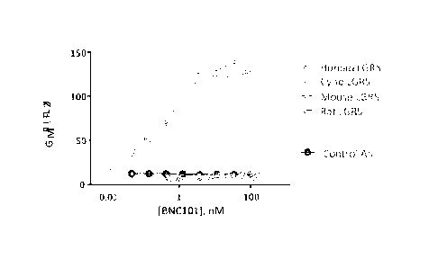
[0108] To identify potential animal model species for investigating the
therapeutic efficacy and safety of 18G7H6A3, the cross-reactivity of 18G7H6A3
to LGR5
expressed by species homologues was determined in a series of in vitro binding
studies. See
FIG. 1. As shown, antibody 18G7H6A3 (BNC101) was found to strongly bind human
and
cyno LGR5, but not bind to rat or mouse LGR5.
Example 6 ¨ Binding of a Humanized LGR5 antibody to plate-bound recombinant,
human
LGR5 ectodomain
[0109] Binding of 18G7H6A1 and 18G7H6A3 to human LGR5 was assessed in
vitro using an ELISA-based plate binding assay. The assay measured antibody
binding to
ELISA plate-bound purified recombinant, LGR5 ectodomain- IgG-Fc fusion, with
detection
of LGR5-bound antibody with horseradish peroxidase-conjugated anti-human IgG-
CH1
secondary antibody. The EC50 of 18G7H6A3 for human LGR5-Fc was found to be 300
pM.
Example 7 ¨ Binding Characteristics of a Humanized LGR5 antibody on Tumor
Cells
[0110] The binding characteristics of 18G7H6A3 to human cancer cell
lines
expressing different levels of LGR5, were analyzed by flow cytometry to define
the potential
targeting properties of 18G7116A3 on heterogeneous tumor populations. The
expression
levels of LGR5 in multiple tumor cell lines were quantified by flow cytometry.
[0111] Human tumor cell lines analyzed in these studies included colon
carcinoma cancer cell lines (CT1 (Bionomics), CT3 (Bionomics), DLD1 (ATCC),
Ls174T
(ATCC), LoVo (ATCC), SW48 (ATCC). SW480 (ATCC), SW620 (ATCC) and HCT116
(ATCC)), triple negative breast cancer cell lines (Hs578T (ATCC) and MDA-MB-
231
(ATCC)), pancreatic cancer cell lines (AsPC-1 (ATCC), BxPC3 (ATCC), Capan2
(ATCC),
HPAFII (ATCC), SW1990 (ATCC), CFPAC (ATCC), Panc10.05 (ATCC) and PANC-1
(ATCC)), cisplatin-sensitive ovarian cancer cell lines (OVCAR3 (ATCC) and SK-
OV-3
(ATCC)), cisplatin-resistant ovarian cancer cell lines (SK-OV-3/CP, OVCAR8/CP,
Igrovl /
CP and A2780/CP (TGEN)) and lung adenocarcinoma cell line H0P62 (ATCC).
[0112] Cells grown near confluence were lifted with TrypI,E cell
dissociation
buffer (Life Technologies), counted and plated in 96-well V-bottom plates at
1x10⁵ cells per
well. 18G7H6A3 was tested at a starting concentration of 100nM with serial
dilutions in
staining buffer (PBS/0.8% bovine serum albumin). Samples were incubated on ice
for 30
minutes, then centrifuged at 1800 rpm for 2 minutes at 4 C and washed 3 times
with staining
-27-
CA 02944649 2016-09-30
WO 2015/153916 PCMJS2015/024162
buffer. Fifty pl of secondary antibody goat anti-human IgG-PE conjugate at
1:250 dilution
(Southern Biotech) was added to each corresponding well in staining buffer.
Samples were
incubated for an additional 15 minutes on ice, and then washed as described
above and
resuspended in 100 al staining buffer containing propidium iodide (PI) (Life
Technologies)
for dead cell exclusion. Samples were analyzed on the FACScalibur flow
cytometer using
CellQuest (Becton Dickinson) and FlowJo (TreeStar, Inc) software.
[0113] The cell surface expression levels of LGR5 in multiple tumor cell
lines
were quantified by flow cytometry. CT1 colorectal tumor cells and pancreatic
cancer cell
lines Panc-1, Capan2 and CFPAC were among the highest LGR5 expressors.
Moderate
expression levels were observed in pancreatic cancer cell lines (AsPC-1,
SW1990, HPAFII),
cisplatin-resistant ovarian cancer cell lines (OVCAR8/ CP, A2780/CP and
Igrovl/CP) as
well as colon, breast and ovarian cancer cell lines (SW48, Hs578T and OVCAR3).
Low but
detectable levels of LGR5 cell surface expression were observed in colon
(5W480, LoVo)
and breast cancer cell lines (MDA-MB-231). Table 2 summarizes the data for
18G7H6A3
FACS binding to Tumor cell lines.
TABLE 2
Tumor Cell line 18G7H6A3 (18G7.1) IgG
CRC
CT I
CT3
DLD1 +/-
Ls174T +/-
LoVo +/-
SW48
S W480 +/-
SW620 +/-
IICT116 +/-
Breast
MDA-MB-231 +/-
MDA-MB-231 LM2 +/-
Hs578T
CN34 +/-
-28-
CA 02944649 2016-09-30
WO 2015/153916 PCMJS2015/024162
Tumor Cell line 18G7H6A3 (18G7.1) IgG
CN34 LM1 +/-
Prostate
PC-3 +/-
PCSD1 +7-
Ovarian
OVCAR-3
SK-OV-3 +7-
SK-OV-3/CP +/-
OVCAR8/CP
Igrovl /CP
A2780/CP
Lung
HOP-62 +/-
Pancreatic
AsPC-1
Capan2 ++
HPAFII
Sw1990*
CFPAC ++
PANC-1 ++
Example 8 ¨ Inhibition of Cachectic Colorectal Tumor Growth In Vivo by a
Humanized
Anti-LGR5 Antibody
[0114] The CT1 primary CRC xenograft model was derived from a patient
with
stage IV metastatic colon cancer. DNA sequencing of this tumor identified
common colon
cancer mutations in multiple genes including K-Ras. P13 K, PTEN, p53 and APC.
Low
passage CT1 tumorspheres maintained in culture under serum-free conditions
were injected
into SCID/Bg mice in Matrigel subcutaneously on day 0, and monitored twice
weekly for
tumor size and body weight. At day 25 CT1 subcutaneous tumors were randomized
into
groups of 10 mice when tumors reached 120 mm3. Mice were treated with either
PBS,
antibody control MOPC, 18G7116A1, 18G7116A3 or human]murine chimeric 18G7Ch.
Mice
were dosed BIW at 15 mg/kg for 2.5 weeks (5 doses total).
-29-
CA 02944649 2016-09-30
WO 2015/153916 PCMJS2015/024162
[0115] Antibody 18G7H6A3 showed significant anti-tumor activity in vivo
compared to PBS and MOPC antibody controls during the course of 4 doses
(15mg/kg, twice
weekly). While antibody 18G7H6A1 showed anti-tumor activity, monoclonal
18G7H6A3
showed superior activity to both 18G7H6A1 and the parental murine chimeric
18G7Ch
antibody. Table 3 shows percent CT1 tumor volume reduction (group vs MOPC)
after 1 - 4
doses of Lgr5+ Abs.
TABLE 3
H of Doses: 1 2 3 4
18G7Ch 9.2% 30.6% 19.5% 29.0%
18G7116A1 17.5% 19.1% 14.2% 19.0%
18G7H6A3 38.8% 42.0% 28.9% 35.4%
Example 9 ¨ Inhibition of Colorectal Tumor Growth In Vivo by a Humanized Anti-
LGR5
Antibody
[0116] The CT3 primary CRC xenograft model was derived from a patient
with
stage III mCRC with mutations in K-Ras, H-Ras, APC, PI3K, PTEN, STK11, RBI,
TP53,
FGFR2, VANGL2, and ISCO. Low passage cryopreserved CT3 primary xenograft tumor
fragments were implanted into 5 SCID/Bg mice. Tumors averaging 4150 mm3 pooled
from
five CT3 primary xenograft-bearing SCID mice were removed at day 41 post-
implant,
dissociated and re-implanted into CB.17 SCID mice in Matrigel subcutaneously,
and
monitored twice weekly for tumor size and body weight. When tumors reached an
average of
130mm3, mice were randomized (34 days post implant). Mice were treated with
either PBS,
antibody control MOPC, 18G7H6A3, 18G7H6A1 or human/murinc chimeric 18G7Ch.
Mice
were dosed BIW at 15 mg/kg for 2.5 weeks (5 doses), starting on day 34. All
mice were
monitored twice weekly for body weight and tumor size, as well as overall
health and
appearance, until termination.
[0117] While antibody 18G7H6A1 showed anti-tumor activity, monoclonal
18G7H6A3 showed significant anti-tumor activity compared to PBS and MOPC
antibody
controls after 4 doses (15mg/kg, twice weekly). 18G7H6A3 showed superior
activity to the
parental murine chimeric 18G7Ch antibody and equivalent activity to 18G7H6A1.
Table 4
shows percent CT3 tumor volume reduction (group vs MOPC) after n dose of test
Abs.
-30-
CA 02944649 2016-09-30
WO 2015/153916 PCMJS2015/024162
TABLE 4
ti of Ab Doses: 1 2 3 4
18G7Ch 22.6% 8.9% 17.0% 13.8%
18G7H6A1 18.3% 12.6% 28.8% 28.7%
18G71-16A3 34.2% 38.1% 23.4% 28.2%
Example 10 ¨ Inhibition of Colorectal Tumor Growth In Vivo by a Humanized Anti-
LGR5
Antibody in combination with FOI,FIRI
[0118] CB.17 SCID mice were implanted with CT3 cells grown under CSC
conditions. At day 40 post-implantation, when tumors reached ¨160 mm3, mice
were
randomized into treatment groups including i) PBS, ii) FolFiri (5FU 30 mg/kg,
lcucovorin 90
mg/kg and Irinotecan 24 mg/kg), given every 5 days for for 15 days (3 doses
total), and iii)
Combination of FolFiri (as in ii.) and 18G7H6A3 (15 mg/kg twice per week).
Analyses of
tumor volume showed that combination of 18G7H6A3 and FolFiri reduced growth of
CT3
tumors compared to FolFiri regimen. Combination treatment reduced tumor volume
at days
61, 65, 68, 71 and 75 by about 58%, 53%, 45%, 33% and 37% respectively (FIG.
2).
Example 11 ¨ Inhibition of Pancreatic Cancer Tumor Growth In Vivo by a
Humanized Anti-
LGR5 Antibody
[0119] To assess efficacy of 18G7H6A3 as single agent or in combination
with
standard of care, a pancreatic cancer xenograft model was tested. CB17.SCID
mice were
implanted with AsPC-1 cells (in matrigel+RPM1 in a 1:1 ratio). Tumors were
randomized at
day 20 post implantation into 5 groups: i) PBS, ii) MOPC (15 mg/kg, twice per
week, ip), iii)
18G7H6A3 (15 mg/kg, twice per week, ip), iv) gemcitabine (90 mg/kg, twice per
week, ip)
and v) concurrent combination of gemcitabine and 18G7H6A3 at the above doses.
[0120] It was discovered that 18G7H6A3 as single agent inhibited tumor
growth
compared to saline and/or control IgG up to nearly 40% at day 41 post
implantation. In
addition, the combination of 18G7H6A3 and gemcitabine significantly inhibited
tumor
growth in AsPC-1 model (up to 36% at day 61 post implantation) compared to
gemcitabine
alone. 18G7H6A3 as single agent also provided some inhibition in tumor growth
compared
to PBS and control IgG up to day 65.
-31-
CA 02944649 2016-09-30
WO 2015/153916 PCMJS2015/024162
Example 12 ¨ Inhibition of Triple Negative Breast Cancer Tumor Growth In Vivo
by a
Humanized Anti-LGR5 Antibody
[0121] This in vivo study was performed using low passage triple
negative breast
cancer cells (ER-, PR-, no HER2 overexpression). MDA-MB-231-LM3 cells were
maintained in adherent culture with DMEM/ 10% FBS/ anti-anti medium. CB.17
SCID mice
were injected on day 0 with MDA-MB-231-LM3 cells in RPMI:Matrigel (1:1) into
the 4th
mammary fat pad and monitored twice weekly for tumor size and body weight. At
day 27,
MDA-MB-231-LM3 tumors were randomized into 4 groups of 10 mice when tumors
reached
¨155mm3. Mice were treated with PBS, antibody control MOPC, or 18G7H6A3. Mice
were
dosed BIW at 15 mg/kg for 3.5 weeks (7 doses). It was discovered that antibody
18G7H6A3
showed significant anti-tumor activity compared to PBS (60.7% tumor growth
inhibition) or
MOPC antibody (49.3% tumor growth inhibition) controls (FIG. 3).
Example 13 ¨ Induction of expression of LGR5 in colorectal cancer cells
treated with a SN38
or a PI3K/mTOR inhibitor
[0122] A panel of CRC cell lines including DLD I, HCT116, LS174t, LoVo,
5W48, SW480 and SW620 were treated with a PI3K/mTOR dual inhibitor (NVP) or 2
different cytotoxic agents including 5N38 (active metabolite of Irinotecan) or
5FU (5
fluorouracil). Cells were treated with the above agents at I um and were
harvested after 72
hrs. Cells were then stained with anti-LGR5 Mab conjugated to Alexa Fluor647
and the data
were analyzed by flow cytometry using a FACScalibur.
[0123] Flow cytometry analyses of CRC cell lines showed greater
expression of
LGR5 in LoVo, HCT116, LS174t, SW48, SW480 and 5W620 cells when treated with a
PI3K/mTOR inhibitor. Additionally, treatment with SN38 promoted LGR5
expression in
HCT116, LS174t, 5W48, SW480 and especially 5W620 cells. 5FU treatment,
however, did
not induce LGR5 expression in any of these lines suggesting that underlying
mechanisms
governing LGR5 expression are distinct in these lines. These data indicate
that LGR5+ cells
are more resistant to treatment with the above agents as treatments have
mostly targeted the
LGR5 negative non-cancer stem cell population. To understand if treatment with
these agents
upregulate LGR5 expression on these cells, we analyzed LGR5 cell surface
expression by
flow cytometry in all the cell lines. Upon treatment with PI3K/mTOR inhibitor,
LGR5
expression was significantly upregulated in LoVo. These data indicate that
treatment with
-32-
CA 02944649 2016-09-30
WO 2015/153916 PCMJS2015/024162
small molecule inhibitors or cytotoxic agents target LGR5neg cells and causes
increased
expression of LGR5 in these cells.
Example 14¨ LGR5 expression is promoted in pancreatic cancer cell lines
treated with small
molecule inhibitors or cytotoxic agents
[0124] In addition to CRC cell lines to further expand the above
findings,
expression of LGR5 was investigated in a series of pancreatic cell lines
treated with relevant
standard of care including nab-paclitaxel, gemcitabine and taxol and also
small molecule
inhibitors targeting most relevant pathways in pancreatic cancer such as
inhibitors of PI3K,
MEK and GSK313. The pancreatic cell lines that were tested include: AsPcl,
HPAFII,
PANCI, BxPC3, CFPAC, PANC10.05, Capan2 and SW1990. Treatment with nab-
paclitaxel
results in LGR5 upregulation in PANC1, BxPc3 and PANC10.05 as assessed by flow
cytometry. Gemcitabine treatment upregulates LGR5 in PANCI and taxol treatment
results
in increased LGR5 expression in HPAFII. The PI3K/mTOR treatment results in
upreulation
of LGR5 in CFPAC and the MEK inhibitor upregulates LGR5 in HPAFII and SW1990.
Example 15 ¨ LGR5 is ITpregulated in Colorectal Cancer Tumors Treated with
FOLFIRI
regimen (5FU, Leucovorin and Irinotecan)
[0125] To investigate if chemo treatment alters LGR5 expression in
colorectal
tumors, mice were treated every 5 days with 5FU (30 mg/kg i.p), leucovorin (90
mg/kg) and
2 different doses of irinotecan (24 mg/kg or 8 mg/kg). The result of those
studies showed that
while CT3 tumors were sensitive to the chemo regimen, CT1 tumors did not full
regress and
showed some resistance to the regimen (FIG. 4). To examine the effect of
FOLFIRI
treatment of LGR5 expression, total mRNA was extracted from CT1 and CT3
patient derived
tumors and expression of LGR5 and was determined by ciRT-PCR and was analyzed
by
subtracting the Ct value (cycle threshold) of LGR5 in each sample from its
corresponding
GAPDH transcript to generate DCT (delta Ct) values. Data are presented as 2 to
the power of
DCT. Analyses of abundance of LGR5 showed that the LGR5 transcript is
increased in both
CT1 (for about 2 folds) and CT3 tumors (approximately 3.5 folds) compared to
corresponding saline treated tumors.
-33-
CA 02944649 2016-09-30
WO 2015/153916 PCMJS2015/024162
Example 16 ¨ LGR5 is Upregulated in Pancreatic Cancer Tumors Treated with
Gemcitabine
alone and in combination of nab-Paclitaxel
[0126] To investigate if standard of care chemotherapy treatment for
pancreatic
cancer alters LGR5 expression in pancreatic tumors, mice were treated twice
per week with
combination of gemcitabine and nab-paclitaxel (in JH109 primary xenografts).
At terminal
analysis, qRT-PCR data using tumor cDNA showed a remarkable increase in the
expression
of LGR5 in chemotherapy treated tumors compared to corresponding saline-
treated tumors
indicating that treatment with standard of care results in upregulation of
LGR5 in tumor cells.
[0127] LGR5 expression in JH109 model which is a patient derived
xenograft
model of pancreatic tumor. Mice were implanted with tumor chunks that were
continuously
passaged in the recipient but were never exposed to in vitro culture
condition. Treatment of
tumor-bearing mice with a chemotherapy regimen (combination of gemcitabine and
nab-
paclitaxel) resulted in a significant inhibition in tumor growth. Consistent
with the colon
cancer models, chemotherapy resulted in upregulation of LGR5 (more than 4-
fold) in JH109
tumors, further suggesting enrichment of the cancer stem cell population upon
treatment with
chemotherapy. See, for example, FIG. 5.
Example 17 ¨ Inhibition of Pancreatic Tumor Growth In Vivo by a Humanized Anti-
LGR5
Antibody
[0128] Efficacy of 18G7H6A3 was also investigated in a pancreatic cancer
xenograft model. CB.17 SCID mice were implanted with PANC1 cells (1E6/mouse
s.c in
matrigel+RPMI 1:1 ratio), and randomized at day 41 post implantation into
treatment groups:
i) PBS, ii) IgG control (15 mg/kg, twice per week, ip), iii) 18G7H6A3 (15
ma/kg, twice per
week, ip), iv) gemcitabine (90 mg/kg, twice per week, ip) and v) concurrent
combination of
gemcitabine and 18G7H6A3 (15 mg/kg, twice per week, ip). Gemcitabine was
administered
in assigned group for 3 weeks to inhibit tumor growth. All mice were monitored
twice
weekly for body weight and tumor size, as well as overall health and
appearance.
[0129] Analysis of tumor volume showed that while there is a trend in
favor of
18G7116A3 as single agent (up to 30% at day 70 post implantation) to inhibit
tumor growth,
combination of 18G71-16A3 and gemcitabine significantly inhibited growth of
PANC1
tumors (up to 52% at day 80 post implantation) compared to gemcitabine alone
group. See
FIG. 6.
-34-
CA 02944649 2016-09-30
WO 2015/153916 PCMJS2015/024162
[0130] In this example, the significant activity of 18G7H6A3 observed
when
administered in combination with chemotherapy (gemcitabine) can be attributed
to the
increased expressed of the target antigen LGR5 in response to gemcitabine
treatment.
Example l 8 ¨ Inhibition of Pre-treated Pancreatic Tumor Growth In Vivo by a
Humanized
Anti-LGR5 Antibody
[0131] In addition to cell lines, we also investigated the efficacy of
18G7H6A3 as
single agent or in combination with standard of care in the JH109 primary
patient derived
xenograft model of pancreatic cancer. The JH109 xenograft model is from a
patient that had
received four treatment regimens including 5-FU. Gemcitabine, Erbitux and
radiotherapy.
The original patient tumor has been passaged in immune-deficient mice
continuously without
any exposure to in vitro culture. To test efficacy of 18G7H6A3 in JH109 model,
tumor
bearing mice (n=7) were treated with control IgG (15 mg/kg i.p twice/week),
18G7H6A3 (15
mg/kg i.p twice/week) single agent, standard of care chemo (combination of
gemcitabine (50
mg/kg i.p once week: and nab-paclitaxel 30 mg/kg, i.v once a week),
combination of chemo
and control IgG, and combination of chemo and 18G7H6A3. While single 18G7H6A3
mAb
did not affect tumor growth, combination of 18G7116A3 with Nab-paclitaxel and
gemcitabine chemotherapy led to a significantly greater degree of tumor
inhibition compared
to chemotherapy alone. 18G7H6A3 combined with chemotherapy led to 77% greater
tumor
growth inhibition compared to chemotherapy alone. Three mice treated with the
18G7H7A3
chemotherapy combination had complete eradication of their tumor (no
measureable tumor
detected). The 18G7116A3 chemotherapy combination group continued to suppress
tumor
growth even after discontinuation of treatment and one mouse was still devoid
of any
measurable tumors three months after cessation of chemotherapy. In this
example, the
significant activity of 18G7H6A3 observed when administered in combination
with
chemotherapy (gemcitabine plus nab-paclitaxel) can be attributed to the
increased expressed
of the target antigen I,GR5 in response to gemcitabine nab-paclitaxel
treatment and is a
demonstration of prevention of re-growth or recurrence of a primary tumor in
vivo after
chemotherapy treatment to eradicate the primary tumor bulk.
Example 19¨ Humanized LGR5 Antibody Treatment Reduces Cancer Stem Cell
Populations
[0132] For flow cytometric analysis, cells from 5 individual tumors were
stained
with a variety of antibodies against stem cell specific markers CD44, and
CD166. Tumors
-35-
CA 02944649 2016-09-30
WO 2015/153916 PCMJS2015/024162
were dissociated, depleted for mouse cells and then counted for viable cells.
Dissociated cells
were used for analysis of cell surface stem cell marker expression by flow
cytometry.
[0133] There was a decrease in cancer stem cell population as defined by
CD I 66+/ CD44+, I.GR5+/ CD166+. or I,GR5+/ CD166+/ CD44+ subpopulations (FIG.
7).
Example 20 ¨ Humanized LGR5 Antibody Treatment Reduces Colon Cancer Tumor
Recurrence and Cancer Stem Cell Frequency In Vivo
[0134] The effects of 18G7H6A3 in combination with FolFiri were tested
in
colon cancer CT3 model (Example 10). The results of this primary tumor
efficacy study
showed that 18G7H6A3 in combination with a 3 cycle FOLFIRI regiment was more
effective than FolFiri alone in reducing tumor growth. To determine if the
18G7H6A3
FOLFIRI combination regimen was also effective in reducing cancer stem cell
(CSC)
frequency, tumors from day 78 were harvested, dissociated, pooled and re-
implanted in a
limiting dilution assay at 10, 30, 100 cells/flank into a new cohort of tumor
naïve CB I7.Scid
mice. The mice were then monitored 2x per week for tumor growth, and tumors
allowed to
grow with no further treatment.
[0135] Cells isolated from mice treated with anti-LGR5 antibody
18G7II6A3 in
combination with FOLFIRI had greatly decreased tumorigenicity as compared to
cells
isolated from mice treated with FOLFIRI alone (FIG. 8). In addition, the re-
implanted cells
from the I8G7H6A3 FOLFIRI combination had a significantly slower tumor growth
profile
and a delayed time to progression (FIG. 9) compared to FOLFIRI alone. Finally,
the
18G7116A3 treatment reduced cancer stem cell frequency by a linear regression
analysis by a
factor of 6 at day 40 (1/856.3 18G7H6A3/FOLFIRI vs 1/138.6 for FolFiri). These
data
indicate that 18G7H6A3 in combination with FOLFIRI effectively targets the
tumor
initiating or cancer stem cell population. Day 68 was the last day for the 30
cells/animal data.
The data are significant at p=0.0039.
Example 21 ¨ Humanized I,GR5 Antibody Treatment Reduces Pancreatic Cancer
Tumor
Recurrence and Cancer Stern Cell Frequency In Vivo
[0136] The effects of 18G7116A3 in combination with gemcitabine were
tested in
pancreatic cancer PANC1 model. This study showed that 18G7H6A3 in combination
with
gemcitabine significantly inhibited tumor growth in PANC1 model compared to
gemcitabine
alone. Tumors cells from these treatment groups were harvested, dissociated,
pooled and re-
-36-
CA 02944649 2016-09-30
WO 2015/153916 PCMJS2015/024162
implanted in a limiting dilution assay (500, 1500, 4500 or 13500 cells/animal)
into a new
cohort of CB.17 SCID mice and allowed to grow with no treatment.
[0137] Cells isolated from mice treated with anti-LGR5 antibody 18G7H6A3
in
combination with gemcitabine had greatly decreased tumorigenicity in the
limiting dilution
assay re-implant as compared to cells isolated from mice treated with
gemcitabine alone. Re-
implanted PANCI tumors treated with combination of gemcitabine and 18G7H6A3
showed
reduction in the frequency of engraftment in mice implanted with 4500 cells
(40% in
gemcitabine vs. 20% in combination) and also in mice implanted with 13500
cells (100% in
gemcitabine vs. 70% in combination). Using linear regression, frequency of
cancer stem cell
in gemcitabine implanted tumors was about 1.5 fold higher in gemcitabine
compared to
combination group (1 in 14883 vs. 1 in 21336). These data indicate that
18G7H6A3 in
combination with gemcitabine effectively targets the tumor initiating or
cancer stem cell
population.
[0138] In addition to PANC I tumors, we also analyzed percentage of
engraftment
and cancer stem cell frequency in an limiting dilution experiment (using 500,
1500, 4500 and
13500 cells) in mice bearing AsPC-1 tumors treated with gemcitabine as single
agent or in
combination with 18G7116A3. Tumor volume measurement at day 40 post treatment
showed
a reduction in percentage of tumor bearing mice in gemcitabine vs. combination
in mice
implanted with 4500 or 13500 cells (40% and 80% vs. 30% and 50%,
respectively).
Frequency of cancer stem cells was also greater by more than 1.5 fold in
gemcitabine vs.
combination group further indicating that 18G7H6A3 in combination with
gemcitabine is
targeting cancer stem cell population in pancreatic cancer.
Example 22 ¨ Humanized LGR5 Antibody Treatment Reduces Triple Negative Breast
Cancer Tumor Recurrence and Cancer Stem Cell Frequency In Vivo
[0139] The effects of 18G7H6A3 in combination with paclitaxel were
tested in
the triple negative breast cancer MDA-MB-231-I,M3 model (Example 12). This
study
showed that 18G7H6A3 in combination with paclitaxel had minimal additive
inhibition in
tumor growth compared to paclitaxel alone. These tumors were harvested,
dissociated,
pooled and re-implanted in a limiting dilution assay at 10, 30, 100
cells/flank into a new
cohort of CB.17 SCID mice and allowed to grow with no treatment.
-37-
CA 02944649 2016-09-30
WO 2015/153916 PCMJS2015/024162
[0140] Cells isolated from mice treated with anti-LGR5 antibody 18G7H6A3
in
combination with paclitaxel had greatly decreased tumorigenicity as compared
to cells
isolated from mice treated with paclitaxel alone. In addition, the re-
implanted cells from the
18G7H6A3 plus paclitaxel tumors had a significantly slower tumor growth
profile and a
delayed time to progress compared to paclitaxel alone. Finally. the 18G7H6A3
plus
paclitaxel treatment reduced cancer stem cell frequency by a linear regression
analysis. These
data indicate that 18G7H6A3 in combination with paclitaxel effectively targets
the tumor
initiating or cancer stem cell population.
Example 23 ¨ Inhibition of Metastatic Colorectal Cancer Growth In Vivo by
Prophylactic
Treatment with Humanized Anti-LGR5 Antibody and Chemotherapy
[0141] The in vivo study was performed using low passage colorectal
cancer cells
(BMCRC086) derived from a liver met of a patient with colorectal cancer. On
Day 0,
BMCRC086 cells were thawed, suspended in RPMI:Matrigel (1:1) and injected
subcutaneously into the rear flank of CB.17 SCID mice. Animals were monitored
twice
weekly for tumor size and body weight. At day 7, mice were treated with PBS.
18G7H6A3,
FOLFIRI or FOLFIRI in combination with 18G7116A3. Mice were dosed with PBS and
18G7116A3, BIW at 15 mg/kg for 7.5 weeks (16 doses). Mice were dosed with
FOLFIRI (30
mg/kg Fluorouracil and 90 mg/kg Leucovorin on days 7, 12, 17, 22, 27 and 32;
24 mg/kg
Irinotecan on days 8, 13, 18, 23, 28 and 33) for 4 weeks (6 doses). 18G7H6A3
in
combination with FOLFIRI showed significant anti-tumor activity compared to
FOLFIRI
alone (FIG. 10).
Example 24 ¨ Humanized LGR5 Antibody Treatment Inhibits Wnt Signaling Pathways
[0142] 18G7H6A3 treated tumors from colon cancer CT1 (Example 8) and CT3
(Example 9) in vivo tumor efficacy studies were characterized by western blot
analysis.
Tumor samples from each treated mouse (n=5 to 10 mice per group) were resected
after
sacrificing, immediately frozen in a liquid nitrogen cooled mortar, ground-up
pestle
(cryopulverization), flash frozen in liquid nitrogen and stored at -80 C until
used.
Cryopulverized tumors were lysed with ice cold lysis buffer (reducing RIPA
buffer
containing phosphatase and protease inhibitors) for 30 minutes on ice with
occasional
vortexing. Supernatants containing tumor lysate protein were run on a SDS-PAGE
gel
followed by western blotting for a number of Wnt-signal proteins (and their
phosphorylated
-38-
CA 02944649 2016-09-30
WO 2015/153916 PCMJS2015/024162
forms). A number of significant differences between treatment groups were
observed in
western blots of CT1 and CT3 tumors. In FIG. 11, phospho-Thr41/Ser45-13-
catenin (a Wnt-
signal protein) is a marker of inactive, and subsequently degraded, form of
the protein
demonstrating 18G7H6A3 is able to inhibit LGR5 signaling in tumor cells in
vivo.
Example 25 ¨ Humanized LGR5 Antibody Treatment Does Not Inhibit In Vitro Wnt-
Signaling Pathway
[0143] Parental HEK-293T cells and HEK-293T cell stably expressing LGR5
were transduced with a TCF-LEF reporter vector-containing lentivirus (GFP
Cigna',
QIAGEN) and selected for stable expression of the reporter. Parental and LGR5
expressing
stable reporter lines were plated at 25,000/well in a 96 well plate, attached
overnight, serum
starved and treated with antibodies or vehicle for 6h, then treated with
recombinant human
Wnt3a (3nM) and recombinant human R-spondins for 18h. Two concentrations for
each R-
spondins1-3 and one concentration of R-spo4 were tested (100pM, 300pM, 1nM,3nM
or
lOnM) based on our analysis of the activity of the different R-spondins in
activation of the
TCF/LEF reporter cell lines. The reporter driven GFP signal was measured on a
plate reader.
All experiments shown are pooled data from three independent experiments (each
experiment performed in duplicate) for each R-spondin tested (data are means +
SD).
[0144] As shown in FIG. 12, increasing concentrations of soluble
18G7H6A3 did
not affect the induction of TCF/LEF promoter driven GFP expression by the
combination of
Wnt3a plus RSP01, RSPO2 or RSP03. A positive control antibody 76C12. which has
been
shown to inhibit the induction of signaling activity through both LGR4 and
LGR5 in the
presence of RSPO and Wnt, is also shown. This data demonstrates that the anti-
LGR5
antibody 18G7H6A3 does not block RSPO-driven TCF/LEF promoter activation.
Example 26 ¨ Humanized LGR5 Antibody Targets Tumor Cells via ADCC (antibody
dependent cell cytotoxicity) mechanism
[0145] CHO-LGR5 cells were grown to confluent and were spun down,
resuspended in PBS and were counted. An aliquot of cells (approximately 100k)
were added
to another tube containing 100 M pre-warmed (37 C) CFSE (Carboxyfluorescein
succinimidyl ester) and the mixture was incubated in the cell incubator for 15
min. The final
CFSE concentration was about 1 p.M. Next, cells were washed and resuspended in
pre-
warmed medium and were placed in the incubator for another 30 minutes followed
by
-39-
CA 02944649 2016-09-30
WO 2015/153916 PCMJS2015/024162
washing with PBS. The stained cells were then stained with 18G7H6A3 (100 M).
To ensure
binding of the antibody to CHO-LGR5 cells, in some studies an aliquot of cells
was also
stained with a secondary goat anti-human PE conjugated antibody and was
analyzed on the
calibur machine ill the laboratory. The U937 cells were stained with DDAO-SE
(DDAO
succinimidyl ester; 2 uM of dye for 100K cells) for 15 minutes and in a light
protected place
in the laboratory and at room temperature. Cells were then 1 ml of FIB (fetal
bovine serum)
followed by incubation in a light protected place for 5 minutes. Next, cells
were washed with
PBS supplemented with FBS (10%) and were resuspended in RPMI supplemented with
FBS
(2.5%). Both CHO-LGR5-18G7H6A3 and U937-DDAO-SE labeled cells were co-
incubated
in the cell incubator for 5 hrs and were analyzed in the calibur machine in
the laboratory. As
a negative control, an aliquot of CHO-LGR5-CFSE cells (no 18G7H6A3 staining)
was also
co-incubated with U937 and was analyzed on the calibur machine.
[0146] Analysis of flow cytometry data showed that majority of CHO-LGR5
cells
stained with CFSE and 18G7H6A3 are viable and detectable in the calibur
machine.
Additionally, both 11937 (11937 (human monocyte cell line; effector cells) and
CHO-LGR5
cells were detectable when stained and were acquired individually. Finally co-
incubation of
U937-DDAO-SE and C110-LGR5-CFSE-18G7116A3 identified a double positive
population
of cells, however, co-incubation of U937 and CHO-LGR5-CFSE which lacks
18G7H6A3 did
not generate the double positive population. The presence of the double
positive population is
indicative of a cross binding of U937 (which express FcR) to CHO-LGR5-18G7H6A3
(which express Fc portion) and further suggests that ADCC is one of the
mechanisms of anti-
tumor activity of 18G7H6A3.
Example 27 ¨ Humanized LGR5 Antibody Internalizes LGR5
[0147] Internalization of 18G7H6A3 was examined on CHO cell
overexpressing
LGR5. Cells were stained with 100nM antibody for 30min-2hrs at 4 C, excess Ab
was
washed off and stained cells were incubated at either 4 C or 37 C. Cells were
stained with
AlexaFluor488-conjugated secondary antibodies at various time points to
monitor
internalization of cell surface-bound antibodies. Upon incubation at 37 C, the
internalized
rate had a measured t1/2 value for surface localization of 6.703 1.282
minutes.
Internalization was largely blocked by incubation at 4 C although some
decrease in surface-
bound antibody was observed.
-40-
CA 02944649 2016-09-30
WO 2015/153916 PCMJS2015/024162
Example 28 ¨ Humanized LGR5 Antibody Does Not Competitively Block Binding of
Soluble RSPOs to LGR5
[0148] Interaction of biotin-18G7H6A3 with hLGR5-Fc in the presence of
human
R-spondin 1/2/3/4 proteins was examined using competition ELISA format. LGR5-
Fc was
coated on a 96-well high binding ELISA plate at 2 ug/mL, and incubated
overnight at 4C.
The plate was blocked with PBS + 1% BSA. Biotin-18G7H6A3 was diluted in
binding buffer
to 1 lag/mL. The concentration was chosen from previous direct binding ELISA
between
LGR5-Fc and biotin-18G7H6A3 to give robust signal above EC50 concentration.
Competitor
proteins were added to the ELISA plate at the same time as biotin-18G7H6A3 at
varying
concentrations. A dilution of 1:1,000 of streptavidin-HRP (R&D Systems, cat #
890803) was
used for detection. Plate was developed with TMB (Thermo), and data were
collected on
SpectraMax Plus 384 plate reader at 450nm. Data analysis was done using
GraphPad Prism 6
program. The ELISA was repeated three times with some modifications of biotin-
mAb and
competitor concentrations.
[0149] As a positive control, LGR5-Fc was competed with the binding of
biotin-
18G7II6A3 to hLGR5-Fc on the plate. R-spondins 1/2/3/4 were tested for the
ability to block
binding of biotin-18G7116A3 to LGR5-Fc coated on the plate. The proteins were
purchased
from R&D Systems, and are full length constructs expressed in mammalian cells.
At the
highest concentration of R-spondin proteins, complete blocking of antibody
binding to LGR5
was not observed (FIG. 13).
Example 29 ¨ Humanized LGR5 Antibody Does Not Competitively Block Binding of
Soluble RSPOs to LGR5
[0150] Binding of ligand alone (RSPO or Norrin) to LGR5 is not
sufficient to
induce LGR5 signaling. Instead, LGR5 forms ternary complexes with multiple co-
receptors
to drive signaling. To examine the effects of 18G7H6A3 on the formation of
LGR5 ternary
complexes, the binding of LGR5 to RNF43, ZNRF3, and 1,RP6 in the presence of R-
spondin
1/2/3/4 and Norrin was examined using an LUSA format. RNF43-Fc, ZNRF3-Fc, and
LRP6-Fc were coated on a 96-well high binding plate at 4 ug/mL in lx PBS. The
plate was
incubated overnight at 4 C and blocked with PBS + 1% BSA. LGR5-Fc was diluted
in
primary buffer to 1 ug/mL, all in the presence or absence of 1 g/mL of R-
spondin 1/2/3/4 or
0.5 ug/mL of Norrin. R-spondin 1/2/3/4 or Norrin were preincubated together
with hLGR5-
-41-
CA 02944649 2016-09-30
WO 2015/153916 PCMJS2015/024162
Fc before being added to the ELISA plate. Triplicate wells were used for each
condition was
tested in triplicate. 1:2,000 anti-FLAG mAb (Cell Signaling) was used to
detect bound
hLGR5-Fc.1:10,000 dilution of anti-mouse IgG HRP (JIR) was used for detection.
Plate was
developed with TMB (Thermo), and data were collected on SpectraMax Plus 384
plate
reader at 450nm. Data analysis was done using GraphPad Prism 6 program.
Formation of a
ternary complex with LGR5, ligands RSPO or Norrin, and co-receptor (RNF43-Fc,
ZNRF3-
Fc, and LRP6-Fc) was observed.
[0151] Next, 18G7H6A3 was added in addition to the ELISA plate in the
presence of LGR5-Fc and RSPO or Norrin. 18G7H6A3 significantly reduced the
formation
of LGR5 ternary complexes with both RSPO and Norrin ligands as well as all
three co-
receptors (RNF43, ZNRF3, and LRP6). See FIG. 14. As 18G7H6A3 does not directly
or
competitively compete with ligand binding, this data is evidence of an
allosteric model of
inhibition.
Example 30¨ Epitope Mapping of Anti-LGR5 Antibody 18G7H6A3
[0152] To further characterize the specific region(s) of LGR5 that
antibody
18G7116A3 binds, an epitope mapping experiment was performed using hydrogen
deuterium
exchange mass spectrometry. Prior to conducting the hydrogen-deuterium
exchange
experiments, test digests prepared with undeuterated buffer in varying
concentrations of
guanidine hydrochloride (GdnHC1) were made to optimize proteolysis conditions
for the best
peptide coverage of LGR5 alone. For pepsin digestion for DXMS, a sample was
thawed at
C and then immediately digested over a protease column filled with porcine
pepsin
(Sigma) at a flow rate of 100 ill/min with 0.05% trifluoroacetic acid. Peptic
fragments were
collected on a C18 trap column and separated on a C18 reversed phase column
(Vydac) with
a linear acetonitrile gradient from 6 to 38%. The column effluent was
electrosprayed directly
into an LCQ Classic (Thermo Finnigan, Inc.) or Q-TOF mass spectrometer
(Micromass).
Determination of pepsin-generated peptides from MS/MS data sets was
facilitated through
the use of SEQUEST (Thermo Finnigan, Inc.). This set of peptides was then
further verified
by DXMS Explorer (Sierra Analytics Inc., Modesto, CA). The peptide coverage
maps for the
different concentrations of GdnHC1 were compared, and the condition with the
best coverage
map for each individual protein or protein complex was used for subsequent
deuterium
exchange experiments. All steps were performed at 0 C as described previously.
-42-
BION0.010W0
PATENT
[0153]
Exchange experiments were initiated by mixing LGR5-Fc in protein
buffer, or LGR5-Fc preincubated with 18G7H6A3 with D20 buffer to a final
concentration
of 50% D20. The mixtures were incubated at 0 C for 10, 30, 100, 300, 1,000,
3,000, or
10,000 s and then the exchange reaction was quenched by adding ice-cold quench
solution
(0.96% formic acid, 0-0.8 M guanidine hydrochloride) resulting in samples with
final
concentrations of 0.58% formic acid and 0-0.5 M guanidine hydrochloride, pH
2.5. The
samples were then immediately frozen on dry ice and stored at ¨80 C. Data
processing of
DXMS experiments utilized specialized software as previously described (DXMS
Explorer,
Sierra Analytics Inc.).
[0154] The
hydrogen/deuterium (HID)-exchange data provide details regarding
changes in solvent exposure due to binding of 18G7H6A3 and the burying of
surface
exposed residues upon binding of antibody to antigen. The HD exchange data
analysis
indicates that 18G7H6A3 binds to amino acids T175, E176, Q180, R183, S186,
A187, Q189,
D247, E248, T251, R254, S257, N258, K260 of SEQ ID NO: 47 within the convex
surface of
leucine rich repeats 6-9, on the opposite of the face of the R-spondin binding
site as
identified by X-ray crystallographic studies. (See e.g., Chen et al. Genes
Dev. 27(12):1345-
50). These data show that the residues involved in binding of LGR5 to the R-
spondins are not
involved in binding 18G7H6A3. These preliminary results do not preclude that
fact that other
structural elements in LGR5 may be involved in the binding site of 18G7H6A3.
Example 31 ¨ Administration of 18G7H6A3 to a human patient suffering from
colon cancer
[0155] A
population of human patients suffering from colon cancer is treated with
chemotherapy and tumor volume is monitored. It is observed that average tumor
volume
ceases to expand and in fact decreases upon initiation of chemotherapy.
Following an
extended duration of time, the tumor volume stabilizes and eventually begins
to increase.
[0156] A
second human patient population suffering from colon cancer is treated
with chemotherapy co-administered with 18G7H6A3. Again, average tumor volume
is
monitored. It is observed that tumor volume ceases to expand and in fact
decreases upon
initiation of chemotherapy. It is observed that tumor volume decreases to a
minimum volume
that is substantially lower than that of the first population. It is also
found that tumor size
remains low for a substantially extended period of time relative to the first
population.
Date Recue/Date Received 2021-04-12
-43-
CA 02944649 2016-09-30
WO 2015/153916 PCMJS2015/024162
Example 32 ¨ Administration of 18G7H6A1 to a human patient suffering from
colon cancer
[0157] A population of human patients suffering from colon cancer is
treated with
chemotherapy and tumor volume is monitored. It is observed that average tumor
volume
ceases to expand and in fact decreases upon initiation of chemotherapy.
Following an
extended duration of time, the tumor volume stabilizes and eventually begins
to increase.
[0158] A second human patient population suffering from colon cancer is
treated
with chemotherapy co-administered with 18G7H6A1. Again, average tumor volume
is
monitored. It is observed that tumor volume ceases to expand and in fact
decreases upon
initiation of chemotherapy. It is observed that tumor volume decreases to a
minimum volume
that is substantially lower than that of the first population. It is also
found that tumor size
remains low for a substantially extended period of time relative to the first
population.
Example 33 ¨ Administration of 18G7H6A3 to a human patient suffering from
colon cancer
[0159] A first population of human patients suffering from colon cancer
is
administered chemotherapy alone. A second population of human patients
suffering from
colon cancer is administered chemotherapy in combination with 18G7H6A3.
[0160] The first population demonstrates a temporary reduction in tumor
size and
growth, after which tumor growth resumes and symptoms return. Tumor growth
after
chemotherapy treatment is recalcitrant to subsequent chemotherapy treatments.
[0161] The second population demonstrates reduction in tumor size to a
basal
level and cessation of tumor growth. Tumor growth does not resume during or
upon
completion of a treatment regimen. After completion of the regimen, growth
does not return
and symptoms of the cancer are no longer present in the second population.
Example 34 ¨ Administration of 18G7H6A1 to a human patient suffering from
colon cancer
[0162] A first population of human patients suffering from colon cancer
is
administered chemotherapy alone. A second population of human patients
suffering from
colon cancer is administered chemotherapy in combination with 1867H6A1.
[0163] The first population demonstrates a temporary reduction in tumor
size and
growth, after which tumor growth resumes and symptoms return. Tumor growth
after
chemotherapy treatment is recalcitrant to subsequent chemotherapy treatments.
[0164] The second population demonstrates reduction in tumor size to a
basal
level and cessation of tumor growth. Tumor growth does not resume during or
upon
-44-
CA 02944649 2016-09-30
WO 2015/153916 PCMJS2015/024162
completion of a treatment regimen. After completion of the regimen, growth
does not return
and symptoms of the cancer are no longer present in the second population.
Example 35 ¨ Administration of I8G7H6A3 to a human patient suffering from
colon cancer
increases survival
[0165] A first population of human patients suffering from colon cancer
is
administered chemotherapy alone. A second population of human patients
suffering from
colon cancer is administered chemotherapy in combination with 18G7H6A3.
[0166] Patient survival at a set duration after treatment (1 year) is
monitored. It is
observed that patient survival in the second population is substantially
higher than patient
survival in the first population. That is, a significantly higher proportion
of the second
population survives past the first year after treatment as compared to the
survival rate of the
first population.
[0167] Similar observations are made at later intervals, and it is
observed that
among survivors at the first interval, members of the second group are
significantly more
likely to survive to a second interval (2 years after treatment) that are
members of the first
group alive at 1 year post treatment.
Example 36 ¨ Administration of 18G7116A1 to a human patient suffering from
colon cancer
increases survival
[0168] A first population of human patients suffering from colon cancer
is
administered chemotherapy alone. A second population of human patients
suffering from
colon cancer is administered chemotherapy in combination with 18G7116A1.
[0169] Patient survival at a set duration after treatment (1 year) is
monitored. It is
observed that patient survival in the second population is substantially
higher than patient
survival in the first population. That is, a significantly higher proportion
of the second
population survives past the first year after treatment as compared to the
survival rate of the
first population.
[0170] Similar observations are made at later intervals, and it is
observed that
among survivors at the first interval, members of the second group are
significantly more
likely to survive to a second interval (2 years after treatment) that are
members of the first
group alive at 1 year post treatment.
-45-
CA 02944649 2016-09-30
WO 2015/153916 PCMJS2015/024162
Example 37 ¨ Administration of 18G7H6A3 to a human patient suffering from
breast cancer
[0171] A population of human patients suffering from breast cancer is
treated
with chemotherapy and tumor volume is monitored. It is observed that average
tumor volume
ceases to expand and in fact decreases upon initiation of chemotherapy.
Following an
extended duration of time, the tumor volume stabilizes and eventually begins
to increase.
[0172] A second human patient population suffering from breast cancer is
treated
with chemotherapy co-administered with 18G7H6A3. Again, average tumor volume
is
monitored. It is observed that tumor volume ceases to expand and in fact
decreases upon
initiation of chemotherapy. It is observed that tumor volume decreases to a
minimum volume
that is substantially lower than that of the first population. It is also
found that tumor size
remains low for a substantially extended period of time relative to the first
population.
Example 38 ¨ Administration of 18G7H6A1 to a human patient suffering from
breast cancer
[0173] A population of human patients suffering from breast cancer is
treated
with chemotherapy and tumor volume is monitored. It is observed that average
tumor volume
ceases to expand and in fact decreases upon initiation of chemotherapy.
Following an
extended duration of time, the tumor volume stabilizes and eventually begins
to increase.
[0174] A second human patient population suffering from breast cancer is
treated
with chemotherapy co-administered with 18G7H6A1. Again, average tumor volume
is
monitored. It is observed that tumor volume ceases to expand and in fact
decreases upon
initiation of chemotherapy. It is observed that tumor volume decreases to a
minimum volume
that is substantially lower than that of the first population. It is also
found that tumor size
remains low for a substantially extended period of time relative to the first
population.
Example 39 ¨ Administration of 18G7H6A3 to a human patient suffering from
breast cancer
[0175] A first population of human patients suffering from breast cancer
is
administered chemotherapy alone. A second population of human patients
suffering from
breast cancer is administered chemotherapy in combination with 18G7H6A3.
[0176] The first population demonstrates a temporary reduction in tumor
size and
growth, after which tumor growth resumes and symptoms return. Tumor growth
after
chemotherapy treatment is recalcitrant to subsequent chemotherapy treatments.
[0177] The second population demonstrates reduction in tumor size to a
basal
level and cessation of tumor growth. Tumor growth does not resume during or
upon
-46-
CA 02944649 2016-09-30
WO 2015/153916 PCMJS2015/024162
completion of a treatment regimen. After completion of the regimen, growth
does not return
and symptoms of the cancer are no longer present in the second population.
Example 40¨ Administration of 18G7H6A1 to a human patient suffering from
breast cancer
[0178] A first population of human patients suffering from breast cancer
is
administered chemotherapy alone. A second population of human patients
suffering from
breast cancer is administered chemotherapy in combination with 18G7H6A .
[0179] The first population demonstrates a temporary reduction in tumor
size and
growth, after which tumor growth resumes and symptoms return. Tumor growth
after
chemotherapy treatment is recalcitrant to subsequent chemotherapy treatments.
[0180] The second population demonstrates reduction in tumor size to a
basal
level and cessation of tumor growth. Tumor growth does not resume during or
upon
completion of a treatment regimen. After completion of the regimen, growth
does not return
and symptoms of the cancer are no longer present in the second population.
Example 41 ¨ Administration of 18G7H6A3 to a human patient suffering from
breast cancer
increases survival
[0181] A first population of human patients suffering from breast cancer
is
administered chemotherapy alone. A second population of human patients
suffering from
breast cancer is administered chemotherapy in combination with l8G7H6A3.
[0182] Patient survival at a set duration after treatment (1 year) is
monitored. It is
observed that patient survival in the second population is substantially
higher than patient
survival in the first population. That is, a significantly higher proportion
of the second
population survives past the first year after treatment as compared to the
survival rate of the
first population.
[0183] Similar observations are made at later intervals, and it is
observed that
among survivors at the first interval, members of the second group are
significantly more
likely to survive to a second interval (2 years after treatment) that are
members of the first
group alive at 1 year pot treatment.
-47-
CA 02944649 2016-09-30
WO 2015/153916 PCMJS2015/024162
Example 42 ¨ Administration of 18G7H6A1 to a human patient suffering from
breast cancer
increases survival
[0184] A first population of human patients suffering from breast cancer
is
administered chemotherapy alone. A second population of human patients
suffering from
breast cancer is administered chemotherapy in combination with 18G7H6A1.
[0185] Patient survival at a set duration after treatment (I year) is
monitored. It is
observed that patient survival in the second population is substantially
higher than patient
survival in the first population. That is, a significantly higher proportion
of the second
population survives past the first year after treatment as compared to the
survival rate of the
first population.
[0186] Similar observations are made at later intervals, and it is
observed that
among survivors at the first interval, members of the second group are
significantly more
likely to survive to a second interval (2 years after treatment) that are
members of the first
group alive at 1 year pot treatment.
Example 43 ¨ Administration of 18G7H6A3 to a human patient suffering from
colon cancer
decreases side effects
[0187] A first population of human patients suffering from colon cancer
is
administered chemotherapy and an anti-LGR5 antibody that blocks LGR5-RSPO
binding and
signaling. A second population of human patients suffering from colon cancer
is
administered chemotherapy and 18G7H6A3.
[0188] The first population demonstrates non-therapeutic side effects
associated
with the interference of RSPO I signaling through LGR5. These side-effects are
detrimental
to patient health.
[0189] The second population, administered 18G7H6A3 in combination with
chemotherapy, does not demonstrate non-therapeutic side effects associated
with the
interference of RSPO I signaling through LGR5.
Example 44 ¨ LGR5 expression in advanced CRC tumors.
[0190] LGR5 transcript expression was investigated using RNAscope
technology
with LGR5 specific probes. LGR5 transcript was detectable in tissues including
colon,
intestine, cerebellum and pancreas. LGR5 transcript was also detectable in
patient derived
xenograft (PDX) tissues including CTI CRC and JH109 pancreatic tumors. LGR5
expression
-48-
CA 02944649 2016-09-30
WO 2015/153916 PCMJS2015/024162
was investigated in CRC patient samples isolated at different stages of
tumorigenesis
including early (Grade-I) vs. advanced (Metastatic) lesions. LGR5 transcript
was expressed
in CRC Grade I, II and II lesions, and was highly expressed in CRC metastatic
lesions.
Example 45 ¨1,GR54 expression in metastatic pancreatic patient derived
xenografts
[0191] LGR5 expression in metastatic pancreatic patient derived
xenografts was
investigated using the quantitative polymerase chain reaction (QPCR). A sample
of tumor
tissue was flash frozen or added to a cryovial containing RNAlater (Qiagen,
CA), and
transferred to -70 C after incubation at 4 C for several hours. Total RNA was
extracted using
a Qiagen RNeasy extraction kit (Qiagen, CA), and cDNA was synthesized using a
SuperScriptIII kit (Life Technologies, CA) and protocols provided by the
manufacturer.
Human LGR5 transcript abundance was measured using human specific LGR5 and
GAPDH
primers and the following thermal condition in the StepOne Thermocycler (Life
Technologies, CA): 50 C (2 min); 90 C (2 min) and 40 cycles of 90 C (15 sec)
and 60 C (1
min) and melt curve assessment (from 65 C-95 C). LGR5 abundance was quantified
using
2^6Ct equation.
[0192] LGR5 was highly expressed in metastatic pancreatic patient
derived
xenografts. Treatment with chemotherapy resulted in increased LGR5 expression
in
pancreatic tumors. Using human specific primers, LGR5 transcript was
measurable using
QPCR in a series of pancreatic patient derived xenografts. While LGR5 was
detectable in
most tumors there was a trend for increased LGR5 expression in metastatic
tumors further
suggesting a role for LGR5 in advanced tumorigenesis.
[0193] LGR5 expression was investigated in a series of pancreatic tumors
including JH109, ASPC1 and PANC1. Treatment with a standard of care treatment
(SOC)
(Gemzar and Abraxane in JH109 and Gemzar alone in PANC1 and ASPC1) resulted in
an
induction in LGR5 expression in each of the foregoing tumors (FIG. 15).
Notably, LGR5
expression was reduced to levels comparable to controls (saline or MOPC) in
tumors treated
with combination of 18G7H6A3 and SOC. These data further indicate that LGR5
expression
can serve as a biomarker of response to combination therapy (18G7116A3+SOC) in
PANC
tumors.
-49-
CA 02944649 2016-09-30
WO 2015/153916 PCMJS2015/024162
Example 46 ¨ CTNNB1 is one of the 18G7H6A3 target genes in CRC and pancreatic
tumors.
[0194] Potential targets in the Wnt pathway for I 8G7H6A3 were
investigated.
Wnt QPCR plates (Qiagen, CA) were prepared with primers for about 80 Wnt
pathway genes
ill a 96 well PCR plate. cDNA from 18G7H6A3 or MOPC (control) treated tumors
was
pooled and QPCR in the Wnt plate was performed. Data in each plate was
normalized to
corresponding GAPDH and the abundance of each gene was measured using an 2^5Ct
equation. To measure fold differences, data in each 18G7H6A3 treated tumor was
divided by
the corresponding value from MOPC treated group. Values above 1 or below 1
were
indicative of upregulation or downregulation in 18G7H6A3 treated group,
respectively.
Preliminary assessment of the number of genes that were up- or down- -
regulated showed
that in both tumor models (CT1 and CT3) there were more downregulated genes
than
upregulated genes, suggesting 18G7H6A3 has an inhibitory effect on gene
expression.
Detailed analyses identified several differentially expressed genes including
FZDB, FZD7,
WNT7B, FBXW11, FZD1, DVL1. CSNK2A1 and CTNNB1.
[0195] In cervical cancer, there may be a close correlation between LGR5
expression and CTNNB1. In other studies, over-expression (using LGR5
recombinant vector)
or dowregulation of LGR5 (using shRNA) resulted in upregulation or
downregulation of
CTNNB1, respectively (Chen Q. Cao HZ, Zheng PS. 2014. Oncotarget 5: 9092-105).
Additionally, analysis of immunohistochemical slides from cervical cancer
patients showed a
significant correlation between LGR5 and CTNNB1 expression. In this study,
CTNNB1
expression was investigated further using QPCR (to measure transcript level)
and Western
Blotting (to assess protein expression). Using human specific primers, CTNNB1
expression
was investigated in pancreatic and CRC tumors. Similar to LGR5 expression
explained in
Example 45, treatment with SOC increased CTNNB1 expression and the combination
of
18G7H6A3 and SOC resulted in a reduction in CTNNB I expression. Additionally,
CTNNB1
expression was reduced about 35% in CT1 tumors treated with 18G7H6A3. Thus,
treatment
with 18G7H6A3 inhibits CTNNB1. Expression of 13-catenin and phospho-f3-catenin
(indicative of lack of activity in Wnt pathway) was investigated by western
blot analysis.
Western blot data in ASPC1 tumors confirmed QPCR data in which 18G7H6A3 as
single
agent or in combination with SOC upregulated p13-catenin suggesting inhibition
of Wnt
pathway activity in these tumors (FIG. 16).
-50-
CA 02944649 2016-09-30
WO 2015/153916 PCMJS2015/024162
[0196] Other components of the Wnt pathway including p-f3-catenin, GSK-
313
(total and phospho), and LRP6 were investigated in a series of CRC, pancreatic
and breast
tumors. Quantification of Western blot data showed significant inhibition of
Wnt pathway
signaling in ASPC1 and PANC1 tumors but also revealed some trends in favor of
Wnt
pathway downregulation in other models. BMCRC086 tumors that were not
responsive to
treatment with 18G7H6A3 were also negative for the expression of LGR5 and Wnt
signaling
pathway components, further supporting that the mechanism of action for
18G7H6A3 was
specifically targeting LGR5 and inhibiting Wnt signaling.
[0197] Expression of Wnt pathway genes in pancreatic tumors including
ASPC1,
PANC1 and JH109 was investigated. Based on in vivo data, in both PANC1 and
ASPC1
there was a difference in tumor volume between 18G7H6A3- vs. PBS- -treated
tumors. In
contrast, JH109 tumors did not respond to a standard treatment regimen with
either
18G7H6A3 single agent or SOC chemo combination. Differences in Wnt gene
expression in
responsive cells (PANC1 and ASPC1) and non-responsive cells (JH109) were
investigated.
In combo treated groups, Wnt6, FZD8, FOSLI, Wntl 1, NFATC and FZD5 were
downregulated in both ASPC1 and PANC1 combo-treated tumors, are were
upregulated in
111109 tumors. In both the pancreatic and CRC data, genes including WNT11,
WNT6, FlkZB
and PRICKEL were downregulated in PANC1, ASPC1, CTI and CT3 cells, but not in
JH109
cells.
[0198] Gene Tree analysis identified potential genes co-regulated in
pancreatic
tumors treated with 18G7H6A3 that included Wnt11, FRAT1, LEF1, GSK3B, FZD8 and
LRP6. Analysis of differentially expressed transcripts in each treatment also
identified genes
that were up/down regulated more than 2 fold in pancreatic tumors (FIG. 17).
Some genes,
such as Wnt7A, were common between all the tumors in 18G7H6A3 vs. control
treated
tumors.
Example 47 ¨ 18G7H6A3 inhibits transcription in CT1 tumors
[0199] Expression of 18G7H6A3-targeted genes were investigated in early
vs.
late tumorigenesis. Mice were implanted mice with CT1, and tumors were
harvested from
control, 18G7H6A3, I-OLFIRI or combo groups at days 3, 10 and 17. Total RNA
from each
tumor at day-3 was harvested and prepared for gene array hybridization using
Illumina
human chips. Overall analysis of differentially expressed genes (more than 1.5
or 2 folds,
-51-
CA 02944649 2016-09-30
WO 2015/153916 PCMJS2015/024162
p<0.05) showed that in tumors treated with 18G7H6A3 (as single agent or in
combination
with FOLFIRI) there are more downregulated genes than upregulated ones. This
suggested
that treatment with 18G7H6A3 may have had a more suppressive impact on overall
cellular
transcriptional machinery. PCA (Principal Component Analyses) also showed a
proximity in
overall gene expression in 18G7H6A3 and control treated tumors. However, when
18G7H6A3 was added to FOLFIRI (i.e. combo group) there was clear separation
between
combo vs. FOLFIRI suggesting that targeting LGR5 may have significantly
changed gene
expression in FOLFIRI-treated tumors.
[0200] Analysis of differentially expressed genes in 18G7H6A3 vs.
Vehicle
identified several tumor promoters such as ANGPT2, AKAP12 and ADM that were
downregulated in 18G7H6A3 treated tumors, and also several tumor suppressors
such as
DAB 1, MIR655, NKX I -2 that were upregulated in 18G7H6A3 treated tumors (FIG.
18).
Conversely FOLFIRI treatment appears to upregulate tumor promoters (FBN2,
HKDCL
ABCB1. FGF2) and also some tumor suppressors such as TRIB3, ATF3 and TIMP3
(FIG.
19). Combination of FOLFIRI and 18G7H6A3 resulted in downregulation of more
tumor
promoters such as ALDOC, CDII5, ITGA2 and also upregulation of more tumor
suppressors
such as ZBTB11, ITPKA, PSMC3IP and BAKI (FIG. 20).
Example 48 ¨ 18G7H6A3 treatment significantly reduces human CTCs in peripheral
blood in
orthotopic models of pancreatic patient derived xenografts
[0201] To investigate the role of I 8G7H6A3 in inhibition of primary
tumor
growth and metastasis, LGR5 expression was examined in a series of pancreatic
patient
derived xenograft samples, and PANC1424 cells and PANC1427 cells.
[0202] Tumor samples were subcutaneously implanted in NOD/SCID (non-
obese
diabetic severe combined immunodeficient) mice and subsequently implanted into
the
pancreas in recipients designated for in vivo studies. Tumor volume was
measured weekly in
ultrasound and mice with tumors ¨100 mm3 were enrolled into the efficacy study
and were
treated with the followings: 1- MOPC isotype (15 mg/kg twice/week; ip); 2-
1867H6A3(15
mg/kg twice/week; ip); 3- SOC (Gemzar 50 mg/kg; ip twice per week and Abraxane
30
mg/kg iv twice per week): 4- Combination of 18G7H6A3 and SOC at the above
doses. At the
end of the study, peripheral blood from each tumor bearing mouse was collected
for CTC
(using flow cytometry) and circulating DNA assessments. For flow cytometry,
blood samples
-52-
CA 02944649 2016-09-30
WO 2015/153916 PCMJS2015/024162
were treated with RBC lysis buffer (ACK buffer, Life Tech, CA) using
manufacturer
protocol and were stained with human HLA-FITC (eBiosciences, CA) and human
LGR5-
AF647 (BD Pharmingen, CA) for 30 min at 4 C. Cells were washed with staining
buffer
(PBS-F1353%) twice and 7AAD (7-aminoactinomycin) prior to acquisition in the
FACS
calibur machine in the laboratory and the data were analyzed using rcs Express
software
(De Novo. CA).
[0203] LGR5 was expressed in various pancreatic patient derived
xenograft
samples. Human CTCs were detected in the peripheral blood. While percentage of
HLA+
cells did not significantly change in MOPC vs. 18G7H6A3, the percentage of
circulating
HLA+LGR5+ cells was significantly reduced in 18G7H6A3 treated mice (FIG. 21).
[0204] The percentage of HLA+ cells did not significantly change in
chemo vs.
combo treated mice, however, combination of 18G7H6A3 and SOC almost completely
ablated HLA+LGR5+ cells in both concurrent and debulk settings (FIG. 22A, and
FIG. 22B).
18G7H6A3 treatment (as single agent or in combination with SOC) significantly
reduces
human CTCs in peripheral blood in orthotopic models of pancreatic patient
derives
xenografts.
Example 49 ¨ LGR5 expression in other models
[0205] LGF5 expression was investigated in skin samples from Cynomolgus
macaques (Cynos) using flow cytometry and RNAscope. Skin samples from Cynos
were
treated with vehicle or various doses of 18G7H6A3 (G2:10 mg/kg; G3:50 mg/kg;
and G4:
150 mg/kg) at day 0, 7, 14 and 21. At study termination, skin samples were
provided in
DMEM supplemented with antibiotic (penicillin and streptomycin) and
antimycotic solution
(Anti-Anti 100X, Life Technologies, CA). Skin samples were digested using a
cocktail of
collagenases and thermolysin (Liberase, Roch Inc, CA). Skin progenitors (SPs)
were isolated
after overnight incubation with Liberase and mechanical disruption. SPs were
stained with
Rat anti-human LGR5 (AF647, BD Pharmingen, CA) and were analyzed in a calibur
machine in the laboratory. Data analyses using FCS Express (Denovo Software,
CA) showed
that LGR5 was detectable in Cynos SPs, however, there was no significant
difference in
LGR5 frequency between 18G7H6A3 (at different doses) vs. vehicle treated
group. Using
RNAscope, LGR5 was detectable in skin areas especially in hair follicles and
to a much
-53-
CA 02944649 2016-09-30
WO 2015/153916 PCMJS2015/024162
lesser extent in skin epithelial cells. There was no significant difference in
LGR5 positive
area in vehicle vs. 18G7H6A3 treated samples.
[0206] Gene
expression peripheral blood monocytes isolated from the Cynos was
investigated. Total RNA was extracted using Qiagen RNeasy kit and cDNA was
synthesized
using Superscript cDNA Synthesis Kit (Life Technologies, CA). The cDNA from
each
treatment was pooled and was added to RT- Sybergreen qPCR master mix
(SABiosciences,
MA). The final mixture was added to each well of a 96-well plate containing
Cyno QPCR
primers for chemokines or inflammatory cytokines. PCR thermal profile
included: 95 C for
min and 40 cycles of 95 C 15 sec and 60 C 1 min followed by melt curve stage.
Data (Ct
values) in each plate was normalized by subtracting from the corresponding
GAPDH and the
abundance of each transcript was calculated using 2^DCT equation. Analyses of
the number
of transcripts differentially expressed (more than 2 folds) between any of the
18G7H6A3
group vs. vehicle treated group showed that, consistent with gene array data,
there are much
more downregulated genes than the unregulated ones. With dose escalation there
were less
unregulated genes and more downregulated genes. The G4-recovery (G4R) group in
which
Cynos did not receive any treatment for 4 weeks after the last dose of
18G71116A3 showed
almost similar number of up- down- -regulated genes. Detailed analysis
identified
differentially expressed genes (CCL 1 I, IL3, SPP I, CCL I3, CXCL6 and TNFRSF
I I b) whose
expression was inversely correlated with 18G7H6A3 dose i.e. highest in 10
mg/kg and
lowest in 150 mg/kg.
[0207] Genes that
were commonly downregulated between the treatments
included CCL1, CCR8, IL2,
IL3 and IL4, some of which are enriched in Ml or M2
macrophages.
Example 50 ¨ Inhibition of Small Cell Lung Cancer Tumor Growth In Vivo by a
Humanized
Anti-LGR5 Antibody
[0208] Patient
derived small cell lung cancer xenograft model. Dissociated tumor
cells from BI,G293 tumors were implanted into CB.17 SCID mice in Matrigel
subcutaneously, and monitored twice weekly for tumor size and body weight.
When tumors
reached an average of 130mm3, mice were randomized. Mice were treated with
either PBS,
antibody control MOPC. or 18G7H6A3. Mice were dosed BIW at 15 mg/kg for. All
mice
-54-
BION0.010W0
PATENT
were monitored twice weekly for body weight and tumor size, as well as overall
health and
appearance, until termination.
[0209]
18G7H6A3 showed significant anti-tumor activity compared to PBS
(24.9% tumor growth inhibition) and MOPC antibody (24.7% tumor growth
inhibition)
controls.
Example 51 - 18G7H6A3 increases survival in mice with pancreatic tumors that
relapse
following debulk chemotherapy therapy
[0210]
Panc1427 (UCSD1427) tumors were completely debulked (regressed) by
treatment with chemotherapy (Gemcitabine/Abraxane) and 18G7H6A3. When tumors
were
regressed, chemotherapy was removed and mice were treated with either 18G7H6A3
or no
treatment. Animals treated with 18G7H6A3 were noticeably more healthy compared
to the
control animals, where several mice had to be euthanized due to severe health
observations
such as lameness or body weight loss. At day 150, 7/8 mice treated with
18G7H6A3 and
chemotherapy were alive, versus 4/8 mice treated with chemotherapy alone. FIG.
23
summarizes the results.
[0211] The
term "comprising" as used herein is synonymous with "including,"
"containing," or "characterized by," and is inclusive or open-ended and does
not exclude
additional, unrecited elements or method steps.
[0212] The
above description discloses several methods and materials of the
present invention. This invention is susceptible to modifications in the
methods and
materials, as well as alterations in the fabrication methods and equipment.
Such
modifications will become apparent to those skilled in the art from a
consideration of this
disclosure or practice of the invention disclosed herein. Consequently, it is
not intended that
this invention be limited to the specific embodiments disclosed herein, but
that it cover all
modifications and alternatives coming within the true scope and spirit of the
invention.
[0213]
Date Recue/Date Received 2021-04-12
-55-