Note : Les descriptions sont présentées dans la langue officielle dans laquelle elles ont été soumises.
CA 2945889
1
METHODS, KITS AND APPARATUS FOR EXPANDING A POPULATION OF CELLS
CROSS-REFERENCE TO RELATED APPLICATIONS
[0001] The present invention claims priority to US patent application
61/980,506
"Methods, Kits and Apparatus for Expanding A Population Of Cells" filed with
the US Patent
and Trademark Office on 16 April 2014.
FIELD OF THE INVENTION
[0002] The present invention relates to the expansion (proliferation) of a
population of cells
such as a population of lymphocytes. The invention in general provides novel
methods and
reagents for the expansion (proliferation) of cell populations that require
binding of a receptor
binding molecule (such as a first agent as described herein) to a receptor
molecule on the surface
of the cells, thereby providing a primary activation signal to the cells. The
invention employs a
multimerization reagent that has immobilized thereon (bound thereto) a first
agent that provides
a primary activation signal to the cells. This primary activation signal may
as such be sufficient
to activate the cells to expand/proliferate. This first agent can either be
bound reversibly or also
irreversibly to the multimerization reagent. The multimerization reagent may
have immobilized
thereon (bound thereto) also a second agent that stimulates an accessory
molecule on the surface
of the cells. The second agent, when binding to the accessory molecule on the
surface on the
surface of the cells, may thereby stimulate the activated cells to expand.
Also this second agent
can either be bound reversibly or also irreversibly to the multimerization
reagent. The
multimerization agent may either be immobilized on a solid support or soluble.
In one aspect, the
method disclosed herein is a serial expansion of a population of cells in
which a complete
population of lymphocytes is stimulated/expanded, the reagents necessary for
the expansion are
then removed by chromatography on a suitable stationary phase and the
expanded/stimulated
cells are optionally transfected with e.g. a T cell receptor or a chimeric
antigen receptor (CAR)
and subjected to a second stimulation expansion with a different stimulatory
molecule that binds
to the introduced T cell receptor or the chimeric antigen receptor. The
invention also relates to an
.. apparatus for the expansion of the selected cell population.
Date Recue/Date Received 2020-04-16
CA 02945889 2016-10-1.4
WO 2015/158868 PCT/EP2015/058339
2
BACKGROUND OF THE INVENTION
[0003] The development of techniques for propagating T cell populations in
vitro has
been crucial to many of the advances in the understanding of T cell
recognition of antigen and
T cell activation. The development of culture methods for the generation of
human antigen-
specific T cell clones has been useful in defining antigens expressed by
pathogens and tumors
that are recognized by T cells to establish methods of immunotherapy to treat
a variety of
human diseases. Antigen-specific T cells can be expanded in vitro for use in
adoptive cellular
immunotherapy or cancer therapy in which infusions of such T cells have been
shown to have
anti-tumor reactivity in a tumor-bearing host. In addition, adoptive
immunotherapy has also
been used to treat viral infections in immunocompromised individuals.
[0004] A method of expanding human T cells in vitro in the absence of
exogenous
growth factor and accessory cells that has been established in the recent
years is described in
US Patent 6,352,694 B1 and European Patent EP 0 700 430 Bl. Disclosed in these
patents is
an in vitro method for inducing a population of T cells to proliferate. The
method comprises
contacting a population of T cells with a solid phase surface having directly
immobilized
thereon: (a) a first agent which provides a primary activation signal to the T
cells, thereby
activating the T cells; and (b) a second agent which stimulates an accessory
molecule on the
surface of the T cells, thereby stimulating the activated T cells. The binding
of the first agent
and the second agent to the T cells induces the T cells to proliferate/to
expand. The preferred
first agent described in US Patent 6,352,694 BI and European Patent EP 0 700
430 B1 is a
monoclonal anti-CD3 antibody which binds to the TCRJ2D3 (TCR = T Cell
Receptor)
complex and thereby stimulates the TCR/CD3 complex-associated signal in the T
cells. The
preferred second agent according to these two patents is a monoclonal anti-
CD28 antibody
which binds the accessory molecule CD28 that is present on T cells. Binding of
this second
agent to the CD28 accessory molecule provides the necessary co-stimulus that
is necessary for
expansion/proliferation of activated T cells. Meanwhile, Dynabeads CD3/CD28
(lnvitrogen)
are commercially available for T cell expansion. Dynabeads CD3CD28 CTSTm are
uniform,
4.5 i.on superparamagnetic, sterile, non-pyrogenic polystyrene beads coated
with a mixture of
affinity purified monoclonal antibodies against the CD3 and CD28 cell surface
molecules on =
human T cells.
100051 However, such magnetic beads are, for example, difficult to integrate
into a
method to expand cells under conditions required for clinical trials or
therapeutic purposes
CA 02945889 2016-10-1.4
WO 2015/158868 PCT/EP2015/058339
3
since it has to be made sure that these magnetic beads are completely removed
before
administering the expanded T cells to a patient. Thus, the present invention
aims to provide an
alternative method for expanding cell populations such as regulatory T cells
or central memory
T-cells for research, diagnostic and especially therapeutic purposes. Ideally,
this new method
should also be compatible with integration into an automatized process which
can be used for
rapid and easy expansion of the desired cells population for thcrapcutic
applications.
[0006] This object is solved by the subject matter of the independent claims,
inter alia
the methods, kits, arrangements and apParatuses as recited in the independent
claims.
SUMMARY OF THE INVENTION
[0007] The present invention provides methods, kits, arrangements, and
apparatus for the
in vitro expansion of a desired cell population, having a receptor molecule on
its surface which
can provide upon binding of a suitable agent a primary activation signal to
the population of
= cells and thereby activating the population of cells for expansion
(proliferation). Thus, the
methods of the invention are also used for inducing a population of cells to
proliferate,
[0008] According to a first aspect, the invention provides an in vitro-mcthod
of expanding
a population of cells, comprising contacting a sample comprising the
population of cells with a
multimerization reagent,
wherein the multimerization reagent has reversibly immobilized thereon (bound
thereto)
a first agent that provides a primary activation signal to the cells;
wherein the muhimerisation reagent comprises at least one binding site Z1 for
the
reversible binding of the first agent,
wherein the first agent comprises at least one binding partner Cl, wherein the
binding
partner Cl is able of reversibly binding to the binding site Z1 of the
multimerization reagent,
wherein the first agent is bound to the multimerization reagent via the
reversible bond fiprmed
between the binding partner CI and the binding site Zl , and
wherein the first agent binds to a receptor molecule on the surface of the
cells, thereby
providing a primary activation signal to the cells and thereby activating the
cells.
100091 According to a second aspect the invention provides an in vitro-method
of
expanding a population of cells, comprising contacting a sample comprising the
population of
cells with a multimerization reagent,
wherein the multimerization reagent is in a soluble form and has immobilized
thereon
(bound thereto) a first agent that provides a primary activation signal to the
cells;
CA 02945889 2016-10-1.4
WO 2015/158868 PCT/EP2015/058339
4
wherein the multimerisation reagent comprises at least one binding site Z1 for
the binding
of the first agent,
wherein the first agent comprises at least one binding partner Cl, wherein the
binding
partner Cl is able of binding to the binding site Z1 of the multimerization
reagent, wherein the
first agent is bound to the multimerization reagent via the bond formed
between the binding
partner Cl and the binding site Zl, and
wherein the first agent binds to a receptor molecule on the surface of the
cells, thereby
providing a primary activation signal to the cells and thereby activating the
cells.
100101 According to a third aspect the invention provides a reagent kit for
expanding a
population of cells, the kit comprising
(i) a multimerization reagent,
wherein the multimerisation reagent comprises at least one binding site Z for
the reversible
binding of a first agent,
(ii) a first agent that binds to a receptor molecule on the surface of the
cells, thereby
providing a primary activation signal to the cells and thereby activating the
cells,
wherein the first agent comprises at least one binding partner CI, wherein the
binding
partner Cl is able of reversibly binding to a binding site Z1 of the
multimerization reagent,
wherein the first agent is bound to the multimerization reagent via the
reversible bond formed
between the binding partner Cl and the binding site Z1, and
(iii) a second agent that stimulates an accessory molecule on the surface of
the cells,
wherein the second agent comprises a binding partner C2, wherein the binding
partner
C2 is able of reversibly binding to a binding site Z2 of the multimerization
reagent, wherein
the second agent is bound to the multimerization reagent via the bond formed
between the
binding partner C2 and the binding site Z2,
wherein the second agent binds to the accessory molecule on the surface on the
surface
of the cells, thereby stimulating the activated cells.
[0011] According to a fourth aspect the invention provides a reagent kit for
expanding a
population of cells, the kit comprising
(i) a mu Itimerization reagent,
wherein the multimerisation reagent is in soluble form and comprises at least
one binding
site Z for the reversible binding of a first agent,
CA 02945889 2016-10-1.4
WO 2015/158868 PCT/EP2015/058339
(ii) a first agent that binds to a receptor molecule on the surface of the
cells, thereby
providing a primary activation signal to the cells and thereby activating the
cells,
wherein the first agent comprises at least one binding partner Cl, wherein the
binding
partner Cl is able of binding to a binding site Z1 of the multimerization
reagent, wherein the
5 first agent
is bound to the multimerization reagent via the reversible bond formed between
the
binding partner Cl and the binding site Z1.
[0012] According to a fifth aspect the invention provides an in vitro-method
of serially
= expanding a population of lymphocytes, wherein the population of
lymphocytes comprises T
cells, the method comprising
contacting a sample comprising the T cell comprising population of lymphocytes
with a
multimerization reagent,
wherein the multimerization reagent is in a soluble form and has reversibly
immobilized
thereon (i) a first agent that provides a primary activation signal to the T
cells and (ii) a
second agent which stimulates an accessory molecule on the surface of the T
cells,
wherein the multimerisation reagent comprises at least one binding site Z1 for
the
reversible binding of the first agent,
wherein the first agent comprises at least one binding partner Cl, wherein the
binding
partner Cl is able of reversibly binding to the binding site Z1 of the
multimerization reagent,
wherein the first agent is bound to the multimerization reagent via the
reversible bond formed
between the binding partner Cl and the binding site Z1,
wherein the multimerisation reagent comprises at least one binding site Z2 for
the
reversible binding of the second agent,
wherein the second agent comprises at least one binding partner C2, wherein
the binding
partner C2 is able of reversibly binding to the binding site Z2 of the
multimerization reagent,
wherein the first agent is bound to the multimerization reagent via the
reversible bond formed
between the binding partner C2 and the binding site Z2,
wherein the first agent binds to a receptor molecule on the surface of the T
cells,
thereby providing a primary activation signal to the cells and thereby
activating the T cells,
wherein the second agent binds to the accessory molecule on the surface of the
T cells,
CA 02945889 2016-10-1.4
WO 2015/158868 PCT/EP201.5/05/039
6
thereby stimulating the activated cells, the first agent and the second agent
thereby together
inducing the T cells to expand.
[0013] According to a sixth aspect the invention provides an arrangement of a
bioreactor
and a stationary phase for chromatography,
wherein the bioreactor is suitable for the expansion of cells,
wherein the stationary phase is suitable for cell separation and removal of
reagents, the
stationary phase being a gel filtration matrix and/or affinity chromatography
matrix, wherein
the gel filtration and/or affinity chromatography matrix comprises an affinity
reagent, wherein
the affinity reagent comprises a binding site Zl specifically binding to a
binding partner Cl
comprised in a first agent and/or the affinity reagent comprises a binding
site Z2 specifically
binding to a binding partner C2 comprised in a second agent, thereby being
suitable of
immobilizing on the stationary phase the first agent and/or the second agent,
the first binding
partner Cl and/or the free second binding partner C2,
wherein the bioreactor and the stationary phase are fluidly connected.
[0014] According to a seventh aspect the invention provides an apparatus for
purification
and expansion of a population of cells, the apparatus comprising at least one
arrangement of a
bioreactor and a stationary phase for chromatography according to the sixth
aspect.
[0015] According to an eight aspect, the invention provides a multimerization
reagent
capable of expanding a population of cells,
whcrcin the multimcrisation reagent is in soluble form and comprises at least
one binding
site Z1 for the reversible binding of a first agent that provides a primary
activation signal
to the cells,
wherein the multimerization reagent has reversibly immobilized thereon (bound
thereto) said
first agent that provides a primary activation signal to the cells;
wherein the first agent comprises at least one binding partner Cl, wherein the
binding partner
CI is able of reversibly binding to the at least one binding site Z1 of the
multimerization
reagent,
wherein the first agent is bound to the multimerization reagent via the
reversible bond formed
between the binding partner Cl and the binding site Z1,
CA 2945889
7
100161 According to a ninth aspect, the invention provides a composition
capable of
expanding a population of cells, the composition comprising
(i) a first multimerization reagent,
wherein the first multimerisation reagent is in soluble form and comprises at
least one binding
site Z1 for the reversible binding of a first agent that provides a primary
activation signal to
the cells,
wherein the first multimerization reagent has reversibly immobilized thereon
(bound thereto)
said first agent that provides a primary activation signal to the cells;
wherein the first agent comprises at least one binding partner Cl, wherein the
binding partner
Cl is able of reversibly binding to the at least one binding site Z1 of the
multimerization
reagent, wherein the first agent is bound to the multimerization reagent via
the reversible
bond formed between the binding partner Cl and the binding site Z1, and
(ii) a second multimerization reagent,
wherein the second multimerization reagent is in soluble form and comprises at
least one
binding site Z2 for the reversible binding of a second agent that stimulates
an accessory
molecule on the surface of the cells,
wherein the multimerization reagent has reversibly immobilized thereon (bound
thereto)
said second agent that stimulates an accessory molecule on the surface of the
cells,
wherein the second agent comprises a binding partner C2, wherein the binding
partner C2 is
able of binding to the at least one binding site Z2 of the multimerization
reagent, wherein the
second agent is bound to the multimerization reagent via the bond formed
between the
binding partner C2 and the binding site Z2.
[0016A] Aspects of the disclosure relate to an in vitro-method of expanding a
population of
cells, comprising contacting a sample comprising a population of cells with a
multimerization
reagent reversibly bound to a first agent, wherein the first agent comprises a
first binding partner
reversibly bound to a first binding site of the multimerization reagent, and
wherein the first agent
binds to a receptor molecule on the surface of a cell in the population to
stimulate a signal to the
cell, thereby stimulating cells in the population.
Date Recue/Date Received 2021-08-24
CA 2945889
7a
[0016B] Aspects of the disclosure also relate to an in vitro-method of
expanding a population of
cells, comprising: contacting a sample comprising a population of cells with a
multimerization reagent,
wherein the multimerization reagent is in a soluble form and bound to a first
agent, and wherein the first
agent comprises a first binding partner bound to a first binding site of the
multimerization reagent, and
wherein the first agent binds to a receptor molecule on the surface of a cell
in the population to stimulate
a signal to the cell, thereby stimulating cells in the population.
[0016C] Aspects of the disclosure also relate to a reagent kit for expanding a
population of cells,
the kit comprising: (i) a multimerization reagent, wherein the multimerization
reagent comprises a first
binding site for the reversible binding of a first agent and a second binding
site for the reversible binding
of a second agent, (ii) a first agent that comprises a first binding partner
able of reversibly binding to the
first binding site of the multimerization reagent, wherein the first agent
binds to a receptor molecule on
the surface of a cell in the population to stimulate a signal to the cell, and
(iii) a second agent that
comprises a second binding partner able of reversibly binding to the second
binding site of the
multimerization reagent, wherein the second agent binds to an accessory
molecule on the surface of the
cell to provide a co-stimulatory signal to the cell, thereby stimulating cells
in the population.
[0016D] Aspects of the disclosure also relate to a reagent kit for expanding a
population of cells,
the kit comprising: (i) a multimerization reagent, wherein the multimerization
reagent is in a soluble
form and comprises a first binding site for the binding of a first agent, (ii)
a first agent that comprises a
first binding partner able of binding to the first binding site of the
multimerization reagent, wherein the
first agent binds to a receptor molecule on the surface of a cell in the
population to stimulate a signal to
the cell, thereby stimulating cells in the population.
[0016E] Aspects of the disclosure also relate to an in vitro-method of
expanding a population of
lymphocytes, wherein the population of lymphocytes comprises T cells, the
method comprising:
contacting a sample comprising a population of lymphocytes comprising T cells
with a multimerization
reagent, wherein the multimerization reagent is in a soluble form and
reversibly bound to a first agent
and a second agent, wherein the first agent comprises a first binding partner
reversibly bound to a first
binding site of the multimerization reagent, wherein the first agent binds to
a receptor molecule on the
surface of a T cell in the population to stimulate a signal to the T cell,
wherein the second agent
comprises a second binding partner reversibly bound to a second binding site
of the multimerization
reagent, and wherein the second agent binds to an accessory molecule on the
surface of the T cell to
provide a co-stimulatory signal to the T cell, thereby stimulating T cells in
the population.
Date Recue/Date Received 2021-08-24
CA 2945889
7b
[0016F] Aspects of the disclosure also relate to an arrangement of a
bioreactor and a first stationary
phase for chromatography, wherein the bioreactor is suitable for the expansion
of cells, wherein the first
stationary phase is suitable for cell separation and removal of reagents, the
first stationary phase
comprising a first affinity reagent, wherein the first affmity reagent
comprises a first binding site capable
of specifically binding to a first binding partner comprised in a first agent
and/or the first affinity reagent
comprises a second binding site capable of specifically binding to a second
binding partner comprised in
a second agent, thereby being suitable of immobilizing on the first stationary
phase the first agent and/or
the second agent, the first binding partner and/or the second binding partner,
and wherein the bioreactor
and the fist stationary phase are fluidly connected.
[0016G] Aspects of the disclosure also relate to a multimerization reagent
capable of expanding a
population of cells, wherein the multimerization reagent is in a soluble form
and reversibly bound to a
first agent, wherein the first agent comprises a first binding partner
reversibly bound to a first binding site
of the multimerization reagent, and wherein the first agent binds to a
receptor molecule on the surface of
a cell in the population to stimulate a signal to the cell, thereby
stimulating cells in the population.
[001611] Aspects of the disclosure also relate to a composition capable of
expanding a population of
cells, the composition comprising: (i) a first multimerization reagent in a
soluble form and reversibly
bound to a first agent, wherein the first agent comprises a first binding
partner reversibly bound to a first
binding site of the multimerization reagent, and wherein the first agent binds
to a receptor molecule on
the surface of a cell in the population to stimulate a signal to the cell; and
(ii) a second multimerization
reagent in a soluble form and reversibly bound to a second agent, wherein the
second agent comprises a
second binding partner reversibly bound to a second binding site of the
multimerization reagent, and
wherein the second agent binds to an accessory molecule on the surface of the
cell to provide a co-
stimulatory signal to the cell, thereby stimulating cells in the population.
[00161] Aspects of the disclosure also relate to an in vitro-method of
stimulating a population of
cells, comprising contacting a sample comprising a population of cells with a
multimerization reagent,
wherein the multimerization reagent is in a soluble form and is reversibly
bound to a first agent and a
second agent, wherein the first agent comprises a first binding partner
reversibly bound to a first binding
site of the multimerization reagent, wherein the second agent comprises a
second binding partner
reversibly bound to a second binding site of the multimerization reagent,
wherein the first agent binds to
a receptor molecule on the surface of a cell in the population to provide a
primary activation signal to the
Date Regue/Date Received 2022-09-09
CA 2945889
7c
cell, and wherein the second agent binds to an accessory molecule on the
surface of the cell to provide a
co-stimulatory signal to the cell, thereby stimulating cells in the
population.
10016J1 Aspects of the disclosure also relate to a reagent kit for stimulating
a population of cells,
the kit comprising: (i) a multimerization reagent, wherein the multimerization
reagent is in a soluble
form and comprises a first binding site for the reversible binding of a first
agent and a second binding
site for the reversible binding of a second agent, (ii) a first agent that
comprises a first binding partner
capable of reversibly binding to the first binding site of the multimerization
reagent, wherein the first
agent binds to a receptor molecule on the surface of a cell in the population
to provide a primary
activation signal to the cell, and (iii) a second agent that comprises a
second binding partner capable of
reversibly binding to the second binding site of the multimerization reagent,
wherein the second agent
binds to an accessory molecule on the surface of the cell to provide a co-
stimulatory signal to the cell,
thereby stimulating cells in the population.
10016K] Aspects of the disclosure also relate to a multimerization reagent
capable of stimulating a
population of cells, wherein the multimerization reagent is in a soluble form
and reversibly bound to a
first agent and a second agent, wherein the first agent comprises a first
binding partner reversibly bound
to a first binding site of the multimerization reagent, wherein the second
agent comprises a second
binding partner reversibly bound to a second binding site of the
multimerization reagent, wherein the
first agent binds to a receptor molecule on the surface of a cell in the
population to provide a primary
activation signal to the cell, and wherein the second agent binds to an
accessory molecule on the surface
of the cell to provide a co-stimulatory signal to the cell, thereby
stimulating cells in the population.
100161,1 Various embodiments of the claimed invention relate to an in vitro-
method of stimulating
a population of lymphocytes, comprising contacting a sample comprising a
population of lymphocytes
with a multimerization reagent, wherein the multimerization reagent is in a
soluble form and is
reversibly bound to a first agent and a second agent, wherein the first agent
comprises a first binding
partner reversibly bound to a first binding site of the multimerization
reagent, wherein the second agent
comprises a second binding partner reversibly bound to a second binding site
of the multimerization
reagent, wherein the first agent binds to a receptor molecule on the surface
of a lymphocyte in the
population to provide a primary activation signal to the lymphocyte, and
wherein the second agent binds
to an accessory molecule on the surface of the lymphocyte to provide a co-
stimulatory signal to the
lymphocyte.
Date Regue/Date Received 2023-01-10
CA 2945889
7d
10016114] Various embodiments of the claimed invention also relate to a
reagent kit for stimulating a
population of lymphocytes, the kit comprising: (i) a multimerization reagent,
wherein the
multimerization reagent is in a soluble form and comprises a first binding
site for the reversible binding
of a first agent and a second binding site for the reversible binding of a
second agent, (ii) a first agent
that comprises a first binding partner capable of reversibly binding to the
first binding site of the
multimerization reagent, wherein the first agent binds to a receptor molecule
on the surface of a
lymphocyte in the population to provide a primary activation signal to the
lymphocyte, and (iii) a
second agent that comprises a second binding partner capable of reversibly
binding to the second
binding site of the multimerization reagent, wherein the second agent binds to
an accessory molecule on
the surface of the lymphocyte to provide a co-stimulatory signal to the
lymphocyte.
[0016N] Various embodiments of the claimed invention also relate to a reagent
kit for stimulating a
population of lymphocytes, the kit comprising: (i) a multimerization reagent,
wherein the
multimerization reagent is in a soluble form and comprises a first binding
site for the reversible binding
of a first agent and a second binding site for the reversible binding of a
second agent, (ii) a first agent
that comprises a first binding partner capable of reversibly binding to the
first binding site of the
multimerization reagent, wherein the first agent binds to a receptor molecule
on the surface of a
lymphocyte in the population to provide a primary activation signal to the
lymphocyte, and (iii) a
second agent that comprises a second binding partner capable of reversibly
binding to the second
binding site of the multimerization reagent, wherein the second agent binds to
an accessory molecule on
the surface of the lymphocyte to provide a co-stimulatory signal to the
lymphocyte.
DESCRIPTION OF THE DRAWINGS
[0017] The invention will be better understood with reference to the detailed
description when
considered in conjunction with the non-limiting examples and the accompanying
drawings. The figures
illustrate embodiments of methods of the invention. Without wishing to
Date Regue/Date Received 2023-01-10
CA 02945889 2016-10-1.4
WO 2015/158868 PCT/EP2015/058339
8
be bound by theory, the figures include conclusions with regard to the
underlying expansion
mechanism. The conclusions are given for illustrative purposes only and merely
serve in
allowing a visualization of the expansion method is achievable on a molecular
level.
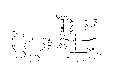
[0018] Figure 1 depicts an embodiment of an in vitro-method of expanding of
expanding a population of cells that has a cell surface receptor the binding
of which by a first
agent can provide an activation signal for the cells to expand,
[0019] As shown in Fig. la a sample that comprises the population of cells (2)
that carry
a surface receptor molecule (30) is contacted with a multimerization reagent
(4). The
population of cells (2) is in mixture with other cell populations (22) that
lack the surface
receptor molecule (30). The multimerization reagent (4) has reversibly
immobilized thereon
(bound thereto) a first agent (6) that provides a primary activation signal to
the cells. The
multimerization reagent (4) comprises at least one binding site Z1 (42) for
the reversible
binding of the first agent (6) and the first agent (6) comprises at least one
binding partner Cl
(6a), wherein the binding partner Cl (6a) is able of reversibly binding to the
binding site Z1
(44) of the multimerization reagent. Thus, for immobilization, the first agent
(6) is bound to
the multimerization reagent (4) via the reversible bond formed between the
binding partner Cl
(6a) and the binding site Zl (42). In the example shown in Fig. 1 the
multimerization reagent
(4) has a second binding site Z2 (44) which is not used in this example. The
multimerization
reagent (4) is itself immobilized on a solid support (10) such as a magnetic
bead, a polymeric
bead of a surface of a cell culture plate or reactor. The population of cells
(2) can, for example
be, a lymphocyte cell population such as &population of B cells that can be
activated via the
CD40 receptor (see, for example, Carpenter et at, Journal of Translational
Medicine 2009,
7:93 "Activation of human B cells by the agonist CD40 antibody CP-870,893 and
augmentation with simultaneous toll-like receptor 9 stimulation), In this
case, the cell surface
molecule (30) is CD40 and the first reagent (6) can be any CD40 binding
molecule that
provides the desired activation signal, for example, the monoclonal antibody
CP-870,893 or an
antibody binding fragment thereof such an a monovalent Fab fragment. The
binding partner
Cl of the first agent (6) may, for example, be any affinity peptide that is
fused or conjugated
to, for example, the C-terminus of one the two polypcptide chains (heavy or
light chain) of the
antibody molecule. The binding partner Cl (6a) may, for example, be a
streptavidin-binding
peptide such as the peptide Trp-Ser-His-Pro-Gln-Pbe-Glu-Lys (SEQ ID NO: 01),
also known
as the "Strep-tag ") that is described in US patent 5,506,121, for example, or
streptavidin
= binding peptides having a sequential arrangement of two or more
individual binding modules
CA 02945889 2016-10-1.4
WO 2015/158868 PCT/EP2015/058339
9
as described in International Patent Publication WO 02/077018 or US patent
7,981,632. When
using a streptavidin binding peptide as binding partner CI, the
multimerization reagent (4) be
any streptavidin mutein to which the streptavidin peptide (= first binding
partner CI (6a))
reversibly binds via its (biotin) binding sites Z1 (42) schematically shown in
Fig. 1. Such a
multimerization reagent may be a streptavidin mutein (analog) that comprises
the amino acid
sequence Va144-Thr45-Ala46-Arg47 (SEQ ID NO: 02) at sequence positions 44 to
47 of wild
type streptavidin or a streptavidin mutein (analog) that comprises the amino
acid sequence
11e44-Gly45-Ala46-Are (SEQ ID NO: 03) at sequence positions 44 to 47 of wild
type
streptavidin, both of which are described in US patent 6,103,493, for example,
and are
commercially available under the trademark Strep-Tactin . In the Example of
Fig. 1, the
multimerization reagent (4) might further include multimeric calmodulin or
glutathione-S-
transferase, both of which form reversible bonds with calmodulin binding
peptides or
glutathione. Thus, the binding site Z2 (44) can be formed by calmodulin or
glutathione-S-
transferase. Such a protein conjugate of for example, calmodulin with a
streptavidin mutein
can be made by standard protein chemistry, for example, by using bifunctional
linkers.
[0020] As shown in Fig. lb, after contacting the cell population (2) with the
multimerisation reagent (4) and usually incubating the cell population with
the multimerization
reagent (4), the population of cells (2) forms complexes/is bound to the
multimerization agent
via the first agent (6). The first agent binds specifically to the cell
surface receptor molecule
such as CD40 in this Example and provides the activation signal for cell
expansion, of for
example B cells. The other cell populations (22) contained in the initial
sample that lack the
specific cell surface molecule (30) do not bind to the multimerization
reagent. In this respect, it
is noted that the cell population (2) usually has multiple copies of the cell
surface molecule
(30) on its surface and binding of these multiple copies is typically needed
for activation.
Thus, the multimerization agent (4) provide typically more than one binding
site Z1 so that
multiple first agents (6) can be reversibly bound to achieve "multimerization"
of the first
agent, meaning to present the first agent in a sufficient density to the
population of cells (2)
(not shown in the scheme of Fig. I). In this respect, it is noted that a
multimerization agent as
used herein can as such have multiple binding sites Z I, for example, a
streptavidin mutein
(being a homo-tetrarner) in its native state has four such binding sites Zl.
it is however also
possible that the multimerization reagent is based on a compound that has as
such only one
binding site Z1 for the reversible binding of a binding partner Cl. Such an
example is
multimeric calmodulin. Calmodulin as such has only one binding site for
calmodulin binding
peptides. However, calmodulin can be biotinylated and then reacted with
streptavidin-
CA 02945889 2016-10-1.4
WO 2015/158868 PCT/EP2015/058339
oligomers (see also below), thereby providing a multimerization reagent in
which multiple
calmodulin molecules are presented in high density on a "scaffold", thereby
providing
multimeric calmodulin.
[0021] As shown in Fig.lc, after incubation (which is usually carried out over
a period
5 of time suitable to achieve expansion of the desired cell population) the
binding between the
binding partner Cl(6a) of the first agent (6) and the binding site Z1 of the
multimerization
reagent (4) is disrupted by disrupting the respective reversible bond. The
disruption may be
achieved by adding a competitor to the incubation/reaction mixture containing
the population
of cells (2) being bound to the multimerization reagent. For competitive
disruption (which can
10 be understood as being a competitive elution) of the reversible bond
between the binding
partner Cl (6a) of the first agent and the binding site Z1 (22) of the
multimerization reagent,
the incubation mixture/population of cells can be contacted with a free first
binding partner Cl
(20) or an analog of said first binding partner C that is capable of
disrupting the bond between
the first binding partner Cl (6a) and the binding site ZI (22). In the example
of the binding
partner Cl being a streptavidin binding peptide that binds to biotin binding
site of streptavidin,
the first free partner Cl (20) may be the corresponding free streptavidin
binding peptide or an
analogue that binds competitively. Such an analogue can, for example, be
biotin or a biotin
derivate such as desthiobiotin.
100221 As shown in Fig. id, addition of the first free partner (20) or the
analogue thereof
results in displacement of the binding partner Cl (6a) from the
multimerization reagent (4) and
thus, since the binding partner Cl is comprised in the first agent (6),
displacement of the first
agent (6) from the multimerization reagent (4). This displacement of the first
agent (6) in turn
results in a dissociation of the first agent (6) from the cell surface
receptor (30), in particular if
the binding affinity of the bond between the first agent and the cell surface
receptor (30) has a
dissociation constant (Kd) in the range of 10-2 M to 10-'3 M and is thus also
reversible. Due to
this dissociation, the stimulation of the cell population (2) is also
terminated. Thus, the present
invention provides the advantage that the time period of the stimulation or
expansion of the
cell population can be exactly controlled and thus also the functional status
of the cell
population can be closely controlled. In this context, it is noted that the
binding affinity of
antibody molecules towards their antigen, including for example, a cell
surface receptor
molecule such as CD40 in this Example, is usually in the affinity range of the
IQ of le M to
10-13 M. Thus, conventional monoclonal antibodies can be used as first agent
(and also of
course second agent as explained below) in the present invention. In order to
avoid any
CA 02945889 2016-10-1.4
WO 2015/158868 PCT/EP2015/058339
=
11
unwanted avidity effects that lead to a stronger binding, monoclonal
antibodies can also be
uscd in form of their monovalent antibody fragments such as Fab-fragments or
single chain Fv
fragments.
[0023] In addition, due to the dissociation of the first agent from the cell
surface
molecule (30), the present invention has the added advantage that the
stimulated cell
population is free of stimulating agents at the end of the stimulation period
and that all other
reagents used in the method, namely the first agent (6) as well as the free
first partner (20) of
the binding partner CI or the analogue thereof can be easily removed from the
stimulated cell
population (2) via a "removal cartridge" described in International patent
application WO
2013/124474 while the multimerization reagent (4) being immobilized on a solid
support such
as a bioreactor surface or a magnetic bead is being held back. Thus, reverting
to the removal of =
the free agent (6) and the free first partner (20), in accordance with the
description of the
"removal cartridge" in WO 2013/124474 (see with reference to Fig. 4 thereof,
for example),
the elution sample obtained in Fig. id here can be loaded onto the second
chromatography
column of WO 2013/124474. This chromatography column has a suitable stationary
phase that
is both an affinity chromatography matrix and, at the same time, can act as
gel permeation
matrix. This affinity chromatography matrix has an affinity reagent
inunobilized thereon. The
affinity reagent may, in the case of the current Example, for instance, be
streptavidin, a
streptavidin mutein, avidin, an avidin rnutein or a mixture thereof. The first
agent (6), the free
first partner (20) of the binding partner Cl (which is also called
"competition reagent" herein)
bind to the affinity reagent, thereby being immobilized on the chromatography
matrix. As a
result the elution sample containing the isolated and expanded cell population
(2) is being
depleted of the first agent (6) and the competition reagent (20). The expanded
cell population
(2), being freed of any reactants, is now in a condition for further use, for
example, for
diagnostic applications (for example, flirther FACSTM sorting) or for any cell
based therapeutic
application.
[0024] Fig.2 shows a further embodiment of an expansion method of the
invention. As
shown in Fig. 2a a sample comprises a population of cells (2) that carry two
specific cell
surface molecules (30) and (32). The cell surface molecule (30) is involved in
a primary
activation signal to the cell population, while the cell surface molecule (32)
is an accessory
molecule on the cell surface that is involved in providing a stimulus to the
cells. The
population of cells may, for example, be a T cell population in which the cell
surface molecule
(30) is a TCR/CD3 complex and the cell surface molecule (32) is the accessory
molecule
CA 02945889 2016-10-1.4
WO 2015/158868 PCT/EP2015/058339
12
CD28. Binding of both the TCR/CD3 complex as the primary activation signal and
CD28 as
co-stimulant are necessary for expansion/proliferation of T cells. The
population of T cells (2)
is in mixture with other cell populations (22) that lack the surface receptor
molecules (30) and
(32). ALso in this embodiment, the cell population (2) is contacted with a
multimerization
reagent (4). The multimerization reagent (4) has reversibly immobilized
thereon (bound
thereto) a first agent (6) that provides a primary activation signal to the
cells. In addition, the
multimerization agent has reversibly immobilized thereon (bound thereto) a
second agent (8)
that stimulates CD28 as accessory molecule on the surface of the cells.
=
[002,51 The multimerization reagent (4) comprises at least one binding site Z1
(42) for
the reversible binding of the first agent (6) and the first agent (6)
comprises at least one
binding partner Cl (6a), wherein the binding partner Cl (6a) is able of
reversibly binding to
the binding site Z1 (44) of the multimerization reagent. Thus, for
immobilization, the first
agent (6) is bound to the multimerization reagent (4) via the reversible bond
formed between
the binding partner Cl (6a) and the binding site Z1 (42). In addition, in the
Example illustrated
in Fig. 2, the second agent (8) comprises a binding partner C2 (8a), wherein
the binding
partner C2 is able of being reversibly bound to a binding site Z2 (44) of the
multimerization
reagent (4). The second agent (8) is bound to the multimerization reagent (4)
via the reversible
bond formed between the binding partner C2 (8a) and the binding site Z2 (44).
In this
Example, the first agent (6) might be a monoclonal anti-CD3-antibody or an
antigen binding
fragment thereof such as a Fab fragment. The second agent (8) Might be a
monoclonal anti-
CD28 antibody or an antigen binding fragment thereof such as Fab fragment. The
first binding
partner (6a) might be a streptavidin binding peptide (6a) that is fused or
conjugated to the anti-
CD3 antibody or the anti-CD3 antibody fragment. The second binding partner
(8a) might be
calmodulin binding peptide that is also conjugated or fused to the CD28
antibody or the CD28
binding antibody fragment. In this context, it is noted that monoclonal
antibodies against, for
example, CD3 or CD28 are well-known (see, for example, US Patent 6,352,694 B
or European
Patent EP 0 700 430 B1 discussed above) and arc commercially available from
numerous
suppliers such as Santa Cruz Biotechnology (Santa Cruz, CA, USA), Life
Technologies,
(Carlsbad, CA, USA), BD Bioseiences (San Jose, CA, USA), Biolegend (San Diego,
CA,
USA) or Miltenyi Biotec (Bergisch Gladbach, Germany) to name only a few.
Accordingly,
such monoclonal antibodies can be used as first and second agent and can, for
example, be
chemically coupled (conjugated) with a binding partner Cl or C2.
Alternatively, it is also
possible to either clone the genes of the variable domains from the hydridoma
cell line or use
CA 02945889 2016-10-1.4
WO 2015/158868 PCT/EP2015/058339
13
an antibody of which the amino acid sequence is known and produce a respective
antibody
fragment such as a Fab fragment or a Fv recombinantly. When using such an
approach as
described herein in the Example section for both the hybridoma cell line OKT3
(ATCC CRL-
8001 TM described in US Patent 4,361,549) that produces a monoclonal anti-CD3
antibody) and
the anti-CD28 antibody 28.3 described by Vanhove et al, BLOOD, 15 July 2003,
Vol. 102,
No. 2, pages 564-570 and GenBank accession number AF451974.1, the binding
partners Cl
and C2 arc conveniently provided by the respective expression vector used for
the recombinant
production so that the antibody fragment carries the binding partner Cl or C2
as a fusion
peptide as the C-terminus of either the light or the heavy chain (In this
context, the amino acid
sequence of the variable domain of the heavy chain and of the variable domain
of the light
chain of the antibody OKT3 that are described in Arakawa et al J. Biochem.
120, 657-662
(1996) are shown for illustration purposes as SEQ ID NOS 17 and 18 and in the
accompanying
Sequence Listings, while the amino acid sequence of the variable domain of the
anti-CD28
antibody 28.3 described by Vanhovc et at, supra, is shown as SEQ ID NOS 19
(VH) and 20
(VL) in the accompanying Sequence Listings). Also this methodology of cloning
the variable
domains of an antibody molecule and recombinantly producing a respective
antibody fragment
is well known to the person skilled in the art, see for example, Skerra, A.
(1994) A general
vector, pASK84, for cloning, bacterial production, and single-step
purification of antibody Fab
fragments. Gene 141, 79-84, or Skerra, A. (1993) Bacterial expression of
immunoglobulin
fragments. Curr Opin Itnnzunol. 5, 256-562). Finally, it is also possible to
generate antibody
molecules of artificial binding molecules with antibody like properties
against a given target
such as CD3 or CD28 as in the Example of Fig. 2 by well-lcnown evolutive
methods such as
phage display (reviewed, e.g., in Kay, B.K. et al. (1996) Phage Display of
Peptides and
Proteins ¨ A Laboratory Manual, 11 Ed., Academic Press, New York NY; Lowman.,
H.B.
.. (1997) Annu. Rev. Biophys. Biomol. Struct. 26, 401-424, or Rodi, D.J., and
Makowski, L.
(1999) Curr. Opin. Biotechnol. 10, 87-93), ribosome display (reviewed in
Amstutz, P. et al.
(2001) CWT. Opin. Biotechnol. 12, 400-405) or mRNA display as reported in
Wilson, D.S. et
aL (2001) Proc. Natl. Acad. Sci. USA 98, 3750-3755.
100261 In the case of the Example shown in Fig. 2, the multimerization reagent
(4) has
two different binding sites Z1 (42) and Z2 (44). With the binding partner Cl
(6a) being a
streptavidin binding peptide, the binding site Z1 (42) of the multimerization
reagent (4) is
provided by a suitable streptavidin mutein to which the streptavidin peptide
(6a) reversibly
binds. Since the binding C2 is a calmodulin binding peptide, the binding site
Z2 (44) of the
CA 02945889 2016-10-1.4
WO 2015/158868 PCT/EP2015/058339
14
multimerization reagent (4) is provided by multimeric calmodulin. The
multimerization
reagent (4) can be a single molecule, for example a conjugate of multimeric
calmodulin with
streptavidin (this alternative would be usually used in case of a soluble
multimerization) or can
also consist of two independent molecules. The latter option is preferred when
the
multimerization reagent (44) is immobilized on a solid support as shown in
Fig.2 In this case,
a mixture of a streptavidin mutein and calmodulin can be coated (immobilized)
on the solid
support, for example, in a 1:1 molar ratio with respect to the binding sites
Z1 and Z2. In this
context, it is noted that, due to the immobilisation of calmodulin on the
surface of the solid
support, there is no need to prepare multimeric calmodulin as explained above
but
immobilization of the calmodulin on the surface is sufficient to present
calmodulin (that, as
mentioned above, has only a single binding site for calmodulin binding
peptides, in a
sufficiently high density to ensure binding of the cell population (2). For
example, in this case,
a bivalent antibody fragment that has two binding sites against CD28 or an
intact antibody that
has per se two identical binding sites could be used as second reagent (8).
[0027] As shown in Fig. 2b, after contacting the T ccll population (2) with
the
multimerisation reagent (4) and usually incubating the cell population with
the multimerization
reagent (4), the population of T cells (2) forms complexes/is bound to the
multimerization
agent via the first agent (6) and the second agent (8). The first agent (6)
and the second agent
(8) bind specifically to the TCR/CD3 complex and the accessory molecule CD28,
thereby
inducing the T cells to proliferate/expand.
= [0028] As shown in Fig. 2c, after incubation (which is usually carried
out over a period
of time suitable to achieve expansion of the desired cell population) the
binding between the
binding partner Cl (6a) of the first agent (6) and the binding site Z1 of the
multimerization
reagent (4) is disrupted by disrupting the respective reversible bond.
Likewise, the binding
between the binding partner C2 (8a) of the second agent (8) and the binding
site Z2 of the
multimerization reagent (4) is disrupted by disrupting the respective
reversible bond. The
reversible bond between the binding partner Cl (6a) of the first agent (6) and
the binding site
Zl can be disrupted by biotin (which acts as an analogue (20) of the free
first partner) while
the reversible bond between the binding partner C2 (8a) of the first agent (8)
and the binding
site Z2 can be disrupted by the addition of a metal chelator (calcium
chelator) such as EDTA
or EGTA (that acts an analogue (20) of the free second partner) since the
binding of
calmodulin to calmodulin binding peptides is calcium ion (Ca2') dependent).
This of course
means that the contacting of the cell population (2) is carried out in a Calf
containing buffer.
CA 2945889
[0029] As shown in Fig. 2d, addition of the analogue (20) of the first free
partner and the second
free partner, respectively results in displacement of the binding partners Cl
(6a) and C2 (8a) from the
multimerization reagent (4) and thus in displacement of the first agent (6)
and the second agent (8) from
the multimerization reagent (4). This displacement of the first agent (6) and
second agent (8) in turn
5 .. results in a dissociation of the first agent (6) and the second agent (8)
from the TCR/CD3 complex and
the accessory molecule CD28, thereby terminating the stimulation/expansion of
the cell population (2).
Thus, as said above, the present invention provides the advantage that the
time period of the stimulation
or expansion of a T cell population can be exactly controlled and therefore
also the functional status of
the population of T cells can be closely controlled. After the elution of the
cells as illustrated in Fig. id,
10 the first agent (6), the second reagent (8) as well as the analogue (20)
of free first partner of the binding
partner Cl and the second free partner of the binding partner C2 can be easily
removed from the
stimulated cell population (2) via a -removal cartridge" described in
International patent application
WO 2013/124474. In addition, and importantly, in case the initial sample was a
population of
lymphocytes, for example, in form of PMBCs obtained from a FicollTM gradient,
the T cell population
15 (2) is now available for serial expansion as defined here. Since the
expanded cell population (e.g. by an
initial stimulation via CD3/CD28) can be transfected during expansion e.g.
with a T cell receptor (TCR)
or a chimeric antigen receptor (CAR, also known as artificial T cell
receptor), the genetically modified
cells can then be liberated from the initial stimulus and subsequently be
stimulated with a second type of
stimulus e.g. via the de novo introduced receptor. These second stimuli may
comprise an antigenic
.. stimulus in form of a peptide/MHC molecule, the cognate (cross-linking)
ligand of the genetically
introduced receptor (e.g. natural ligand of a CAR) or any ligand (such as an
antibody) that directly binds
within the framework of the new receptor (e.g. by recognizing constant regions
within the receptor).
Thus, the T cell population obtained from this serial expansion can be used
for adoptive cell transfer.
100301 Fig. 3 shows a further embodiment of an expansion method of the
invention. Also the
sample used in this Example comprises a population of T cells (2) that carry
two specific cell surface
molecules (30) and (32), with the cell surface molecule (30) being a TCR/CD3
complex and the cell
surface molecule (32) being the accessory molecule CD28. In Fig. 3a the
population of T cells (2) is
shown after being contacted with a multimerization reagent (4). Also in this
Example, the
multimerization reagent (4) has reversibly immobilized thereon (bound thereto)
as first agent (6) an anti-
CD3 antibody or an antigen binding fragment thereof that provides a primary
activation signal to the T
cells and as second agent (8) an anti-CD28
Date Recue/Date Received 2021-08-24
CA 02945889 2016-10-1.4
WO 2015/158868 PCT/EP2015/058339
16
antibody or an antigen binding fragment thereof that stimulates CD28 as
accessory molecule.
[0031] The multimerization reagent (4) shown in the Example of Fig. 3
comprises only
one type binding site Z1 (42) for the reversible binding of both the first
agent (6) and the
second agent (8). Both the first agent (6) and the second agent (8) comprise
at least one
binding partner Cl (6a, 8a), wherein both the binding partner Cl (6a) and the
binding partner
(8a) are able of reversibly binding to the binding site Zl (44) of the
multimerization reagent.
Thus, for immobilization, the first agent (6) and the second agent (8),
respectively are bound to
the multimerization reagent (4) via the reversible bond formed between the
binding partner Cl
(6a) and the binding partner C2 and the binding site Z1 (42). The binding
partners Cl and C2
can either be different or identical. For example, the binding partner Cl can
be a streptavidin
binding peptide of the sequences Trp-Ser-His-Pro-Gln-Phe-Glu-Lys ((SEQ ID NO:
01), the
"Strep-tag ") while the binding partner C2 can be the strcptavidin binding
peptide of the
sequence Trp-Scr-His-Pro-Ght-Phe-Glu-Lys-(GlyGlyGlySer)3-Trp-Ser-His-Pro-Gln-
Phe-Glu-
Lys ((SEQ ID NO: 04), also known as "di-tag3")) or of the sequence Trp-Ser-His-
Pro-Gln-
Phe-Glu-Lys-(GlyGlyGlySer)2-Trp-Ser-His-Pro-Gln-Phe-Ght-Lys ((SEQ ID NO: 05),
also
known as "the di-tag2"), described by Junttila et al., Proteomics 5 (2005),
1199-1203 or US
Patent 7,981,632). All these streptavidin binding peptides bind to the same
binding site,
namely the biotin binding site of streptavidin. If one or more of such
streptavidin binding
peptides is used as binding partners CI and C2, the multimerization reagent
(4) is a
streptavidin mutein. As shown in Fig. 3, a soluble multimerization reagent (4)
is used. In the
case of a streptavidin mutein, this soluble multimerization reagent may, for
example, be an
oligomer or a polymer of streptavidin or avidin or of any mutein (analog) of
streptavidin or
avidin. The oligomer may comprise three or more monomers of streptavidin,
avidin or a
mutein therof. The oligomer or polymer may be crosslinked by a polysaccharide.
Such
oligomers or polymers of streptavidin or of avidin or of muteins of
streptavidin or of avidin
can in a first step be prepared by the introduction of carboxyl residues into
a polysaccharide, e.
g. dextran, essentially as described in "Noguchi, A., Takahashi, T.,
Yamaguchi, T., Kitamura,
K., Takakura, Y., Hashida, M. & Sezaki, H. (1992). Preparation and properties
of the
immunoconjugatc composed of anti-human colon cancer monoclonal antibody and
mitomycin
C dextran conjugate. Bioconjugate Chemistry 3,132-137". In a second step,
streptavidin or
avidin or muteins thereof are coupled via primary amino groups of internal
lysine residue
and/or the free N-terminus to the carboxyl groups in the dextran backbone
using conventional
carbodiimide chemistry. Alternatively, cross-linked oligomers or polymers of
streptavidin or
avidin or of any muten of strcptavidin or avidin may also be obtained by
crosslinking via
CA 02945889 2016-10-1.4
WO 2015/158868 PCT/EP2015/058339
17
bifunctional linkers such as glutardialdehyde or by other methods described in
the literature.
[0032] Using as binding partners Cl and C2, moieties that bind to the same
binding site
(42) of the multimerization agent has the advantage that, as shown in Fig. 3b,
the same free
partner (of the first binding partner Cl and also of the second binding
partner C2) or analogue
thereof can be used to terminate the expansion of the population of T cells
(2) and to release
this population of T cells (2) from the multimerization agent. In the Example
of Fig. 3, an
analogue of the first and second partner Cl and C2 such as biotin or a biotin
derivate
(iminobiotin or desthiobiotin) can be conveniently used for the termination of
the expansion
and the elution of the population of T cells (2).
[0033] As shown in Fig. 3c, after the elution of the cells as illustrated in
Fig. id, the first
agent (6), the second reagent (8) as well as biotin as the analogue (20) of
free first partner of
the binding partner Cl and the second free partner of the binding partner C2
can be easily
removed from the stimulated cell population (2) via a "iemoval cartridge"
described in
International patent application WO 2013/124474. In addition, the embodiment
of using a
soluble multimcrization reagent (4) has the further advantage of being able to
avoid any solid
support such as magnetic beads. This means there is no risk of contamination
of the activated
T cells by such magnetic beads. This also means that a process that is
compliant with GMP
standards can be much easier established compared to the known method such as
the use of
Dynabeads in which additional measures have to be taken to ensure that the
final expanded T
cell population is free of magnetic beads. Furthermore, the use of a soluble
multimerisation
agent makes it much easier to remove the same from the activated cell
population (T cells, B
cells or also natural killer cells) since the cells can be simple sedimented
by centrifugation and
the supernatant including the soluble multimerization agent can be discarded.
Alternatively,
the soluble multimerization agent can be removed from the expanded cell
population in a gel
permeations matrix of the removal cartridge of International patent
application WO
2013/124474. Since no solid phase (e.g. magnetic beads) arc present, the
present invention
also provides for an automated closed 'system for expansion of the cells that
can be integrated
into known cell expansion systems such as the Xuri Cell Expansion System W25
and WAVE
Bioreactor 2/10 System, available from GE Healthcare (Little Chalfont,
Buckinghamshire,
United Kingdom) or the Quantum Cell Expansion System, available from
TerumoBCT Inc,
(Lakewood, CO, USA).
[00341 Fig. 4 shows the results of an experiment in which CD3+ T responder
cells were
proliferated after being stimulated in vitro with aCD3 and aCD28 Fab fragments
that were
CA 02945889 2016-10-1.4
WO 2015/158868 PCT1EP2015/058339
18
reversibly immobilized on beads coated with the streptavidin mutcin Strep-
tactin . Fig. 4A in
a histogram showing size-distribution (forward scatter) of stimulated cells,
Fig. 4B depicts
histograms representing the degree of proliferation according to the number of
cells per cell
division that are indicated on top of Fig. 4B (0 represents undivided cells; 5
represents cells
that have gone through at least 5 divisions), and Fig. 4C shows a picture of
the culture dish
after 4 days of stimulation.
[00351 Fig. 5 shows the results of the differential intracellular calcium
mobilization in
Jurkat cells that are either labelled with the aCD3 antibody OKT3 or with Fab
fragments of
OKT3 being multimerized with Strep-tactin (also referred to as Fab multimers
herein). Fig.
5A: Jurkat cells were loaded with the calcium-sensitive dye Indo-1 -AM and
calcium release
was triggered by injection of either aCD3 mAb (black squares) or aCD3 OKT3 Fab
multimcrs
(derived from the parental cell line OKT3) with or without prior D-biotin
disniption (dark grey
triangles and light grey circles respectively) compared to injection of PBS
(inverted white
triangles). Application of ionomycine served as positive control. Time-
resolved changes in
intracellular Ca2 concentration were monitored by flow-cytometry based on the
change in
FL6/FL7 ratio. Fig. 5B: Indo-l-AM-labeled Jurkat cells were activated by
different aCD3
stimuli as described in Fig 4a; OKT3: upper graph and aCD3 Fab-multimer:
middle graph)
followed by subsequent (1-140s) D-biotin mediated disruption of aCD3 Fab-
multimer
signaling. Injection of PBS (lower graph) and ionomycine served as negative or
positive
.. control. Data arc representative of three different experiments.
[0036] Fig. 6 shows the result of the reversible staining of cells by anti CD3
OKT3 Fab-
multimers. Freshly isolated PBMCs were stained with either a monoclonal
antibody (left dot
plot, parental clone for the Fab-multimers) or cognate PE-labeled Fab-
multimers and analyzed
either before (second left column) or after treatment with D-biotin (middle
column).
Remaining Fab monomers were then detected after subsequent washing steps using
fresh PE-
labeled Strep-Tactin (second right column). Secondary Fab-multimer staining
of reversibly
stained cells served as control (right column). Only live (Pregmive) cells are
shown. Numbers in
dot plots indicate the percentage of cells within gates.
[00371 Fig. 7 shows the isolation of cells by reversible binding of anti-CD28
Fab
fragments multimcrized with Strep-Tactin labeled with phycoerythrine as a
fluorescent label.
CD28+ cells were selected/isolation by Fab-multimer magnetic cell selection
from freshly
isolated PMI3Cs as described in International Patent Application
W02013/011011. Before
selection cells were control stained with either the cognate fluorescent aCD28-
multimers (left
CA 02945889 2016-10-1.4
WO 2015/158868 PCT/EP2015/058339
19
dot plot) or with an antibody directed against the immunoglobulin kappa light
chain (second
left dot plot, a-Ig kappa mAb). After selection, cells were treated with D-
biotin and
subsequently washed to remove magnetic beads and Fab-monomers. Liberated CD28+
cells
were subsequently (re-)stained either with CD28 Fab-multimers (second right
dot plot) or with
the a-Ig kappa mAb (right dot plot) to detect potentially remaining Fab-
monomers. Only live
(Pregath') CD3+ cells are shown. Numbers in dot plots indicate the percentage
of cells within
gates.
[0038] Fig. 8 shows the results of an experiment in which CD3+ T responder
cells were
proliferated after being stimulated in vitro with reversible aCD3/aCD28 Fab
fragments that
were reversibly immobilized on soluble oligomeric Strep-tactin acting a
soluble
multimerization reagent. For the experiments the results of which are shown in
Fig. 8,
300.000 CD3+ responder T cells (Tresp) were labeled with 21.1M
Carboxyfluorescein
suecinimidyl ester (CFSE) and stimulated with varying amounts of a preparation
of soluble
Streptactin oligomers on which a combination of a.CD3 Fab fragment and aCD28
Fab both
carrying a Strep-tag as streptavidin binding peptide at the heavy chain were
immobilized.
("lx" corresponds to 31.1g multimerized Strep-tactin fiinctionalized with 0.5
g aCD3- and
0.5ug aCD28 Fab; numbers indicate fold amount of "le). Tresp cells either left
unstimulated
or were stimulated with blank Strep-tactin multimers (no Fab) served as
negative control.
Tresp cells were seeded in duplicates in 48-well plates along with 300.000 CD3
negative
autologous feeder cells (irradiated with 30Gy) in lml cell culture medium
supplemented with
20U/m1 interleukin 2 (IL-2). Cells were incubated at 37 C without media
exchange and
proliferation was analyzed according to CFSE dilution after 5 days by FACS
analysis (Fig.
8B). Fig. 8A shows size distribution of cells after 5 days in culture.
Histograms show live
CO3+ cells, while Fig. SC shows cells after culture that were liberated by
stimulation reagents
after treated with 1mM D-biotin and washed. The dissociation and removal of
monomeric Fab
fragments was analyzed by restaining with Strep-Tactint) labeled with
phycoerythrinc (SI-
PE) as a fluorescent label and a representative histogram is shown. Fig. 8D
shows the absolute
number of live (trypan blue negative) cells after 5 days was counted using a
Neubauer
counting chamber and plotted against the respective stimulation condition.
Median cell
numbers are shown in Fig. 8D; error bars indicate standard deviation (SD).
Fig. 8E shows a
picture of the culture dish after 5 days of stimulation.
[0039] Fig. 9 depicts an illustration of the serial expansion method of the
present
invention (Fig. 9a) while Fig. 9b briefly describes some features and
advantages of the serial
CA 02945889 2016-10-1.4
WO 2015/158868 PCT/EP2015/058339
expansion.
[0040] Fig. 10 shows an arrangement of the invention that can be used together
with the
expansion methods of the invention. This arrangement (100) includes a
bioreactor (50), a first
"removal cartridge" (70) and a second ¨removal cartridge" (90). The bioreactor
(50) is fluidly
5 connected to the first removable cartridge (70), and the first removal
cartridge is fluidly
connected to the second removal cartridge (90). This arrangement (100) can be
part of a device
for automated cell expansion and purification as described here.
[0041) In the bioreactor (50) an expansion method as described herein is
carried out, for
example an expansion method illustrated in Fig. 3 that makes use of a soluble
multimerization
=
10 reagent. In this case, after termination of the activation/expansion of
the cell population (2) by
addition of a competitor (20) (free partner of the binding partner Cl or an
analogue thereof)
the reaction mixtures that is released from the biorcactor contains the
expanded population of
cells (2), the first agent (6), the second agent (8) as well as the soluble
multimerization reagent
(4). In this example, the first agent (6) is a CD3 binding antibody fragment
that includes a
15 strcptavidin binding peptide as binding partner Cl, the second agent (8)
is a CD28 binding
antibody fragment that includes a streptavidin binding peptide as binding
partner Cl and the
competitor (20) (free analogue of the binding partner CI) is biotin. This
reaction mixture is
applied on the first removal cartridge (70). This first removal cartridge (70)
is a removal
cartridge as described in International patent application WO 2013/124474 that
includes a
20 chromatography column with a suitable stationary phase. The stationary
phase can serve both
an affinity chromatography matrix and, at the same time, can act as gel
permeation matrix.
This affinity chromatography matrix has an affinity reagent immobilized
thereon. The affinity
reagent may, in the case of the current Example, for instance, be
streptavidin, a streptavidin
inutein, avidin, an avidin mutein or a mixture thereof. Thus, the first agent
(6) and the second
agent (8) bind to the affinity reagent via their streptavidin binding peptide.
Also biotin as the
competitor (20) binds to the affinity reagent. Thus, these three reagents arc
all being
immobilized on the chromatography matrix of the first removal cartridge while
the expanded
cell population (2) and the soluble multimerization reagent (4) pass through
the stationary
phase. This "flow through" is then applied onto the second removal cartridge
(90). Also this
second removal cartridge (90) comprises a stationary phase. This stationary
phase comprises a
second affinity reagent thereon which is able to bind to the binding site ZI
(42) of the
multimerization reagent (4). This affinity reagent may for example be biotin
that is covalently
bound to the stationary phase. Such a stationary phase may, for example, be d-
biotin
CA 02945889 2016-10-1.4
WO 2015/158868 PCT/EP2015/058339
21
SepharoseTM obtainable from Affiland S.A. (Ans-Liege, Belgium). Thus, the
soluble
multimerization reagent (4) will be bound (retained) on the stationary phase
of the second
removal cartridge (90) while the expanded population of cells (2) passes
through the stationary
phase and is being freed of any reactant& The population of cells (2) is now
in a conditiOn for
any further use, for example, for diagnostic applications (for example,
further FACSTM
sorting) or for any cell based therapeutic application. It is noted here that
it is of course also
possible to change the order of the first "removal cartridge" (70) and the
second "removal
cartridge" (90) in an arrangement (100), such that bioreactor (50) is
(directly) fluidly
connected to the second removable cartridge (90), and the first removal
cartridge (70) is
arranged after and fluidly connected to the second removal cartridge (90). In
this arrangement
the multimerization reagent (4) will first be removed from the population of
cells (2) and
subsequently the first agent (6), the second (8) and e.g. the competitor (20)
are removed. Such
an arrangement is also encompassed in the present invention and can also be
part of a device
for automated cell expansion and purification as described here.
[0042] Fig. 11 shows a further embodiment of an arrangement of the invention
that can
be used together with the expansion methods of the invention. This arrangement
(110) includes
a bioreactor (50), a first "removal cartridge" (70) and a second "removal
cartridge" (90). The
bioreactor (50) is fluidly connected to the first removable cartridge (70),
and the first removal
cartridge is fluidly connected to the second removal cartridge (90). In
addition, the second
removal cartridge (110) is fluidly connected to the bioreactor (SO). This
arrangement (110) can
also be part of a device for automated cell expansion and purification as
described here. When
used, for example, together with an expansion method that employs a soluble
multimeriz.ation
reagent (4), a purified expanded population of cells (2) is obtained as eluate
of the second
removal cartridge (90). Since the removal cartridge (90) is fluidly connected
to the bioreactor
(50), the population of cells (2) can be transferred back into the bioreactor
(50), for example,
to serial clonal expansion as described here, by transfecting the population
of cells, for
example, with the gene of an T cell receptor and subsequent further (second)
expansion using
an expansion method of the invention.
[0043] Fig. 12 shows a further embodiment of an arrangement of the invention
that can
be used together with the expansion methods of the invention. This arrangement
(120) includes
a bioreactor (50), a first "removal cartridge" (70) and a second "removal
cartridge" (90). The
bioreactor (50) is fluidly connected to the first removable cartridge (70),
and the first removal
cartridge is fluidly connected to the second removal cartridge (90). Similar
to the embodiment
CA 02945889 2016-10-1.4
WO 2015/1513868 PCT/EP201.5/058339
22
shown in Fig. 11, the second removal cartridge (110) is fluidly connected to
the bioreactor
(50). However, a "selection cartridge" (92) as described in International
patent application WO
2013/124474 is arranged in between the second removal cartridge (90) and the
bioreactor (50).
Thus, a subpopulation of cells (2a) that is comprised in the population of
cells (2) can be
selected/enriched via this "selection cartridge" (92) as described in WO
2013/124474. This
=
subpopulation of cells (2a) can either be transferred into the bioreactor
(50), for example, to
undergo serial expansion as described here. Alternatively (not shown), this
subpopulation of
cells (2a) can be used for cell based therapy. It is again noted here that the
use of a soluble
multimerization reagent as described here allows the design of automated cell
purification and
expansion devices which are functionally closed and thus not prone to
contamination. In
addition, since soluble multimerization reagent avoids the need for solid
phase materials such
as magnetic beads, such cell purification devices can be designed as
continuous flow devices.
[00441 Fig. 13 shows thc expansion kinetics o f proliferation of purified CD4+
and CD8+
T responder cells (Tresp) that were stimulated in vitro either with aCD3/aCD28
Fab fragments
or with aCD3/aCD28/aCD8 that were reversibly immobilized on two kinds of a
soluble
oligomeric Strep-tactin mutein acting as soluble multimerization reagent. The
first kind of
oligomeric Strep-tactin was the fraction of. the oligomeric streptavidin
mutein (n> 3)
obtained in Example 5 (also referred herein as "conventional Streptactin
backbone",
illustrated by the triangle symbol with the tip down in Fig. 13), the second
kind of this
oligomeric streptavidin mutein used as soluble multimerization reagent was an
oligomer that
was obtained by reacting the soluble oligomeric streptavidin mutein with
biotinylated human
serum albumin (HSA) This HSA based soluble multimerization reagent is also
referred herein
as "large Streptactin backbone). In the experiments of Fig. 13 the expansion
was carried out
without medium exchange. The results for the CD4+ T responder cells are shown
in Fig.13A,
the results for the CD8+ T responder cells are shown in Fig. 13B. In this
context, it is noted
that the experimentally used soluble 'multimerization reagents that were
fimetionalized by
reversibly binding first agents, and optionally second and third agents are
referred to in the
Figures as "Streptamer multimcrs"
[00451 Fig. 14 shows the expansion kinetics of proliferation of purified CD4+
and CD8+
T responder cells (Tresp) that were stimulated in vitro with aCD3/aCD28 Fab
fragments that
were reversibly immobilized fragments that were reversibly immobilized with
two kinds of
soluble oligomeric Strep-tactin acting as soluble multimerization reagent.
The first kind of
oligomeric Strep-tactin was the fraction of the oligomeric streptavidin
mutein (n? 3)
CA 02945889 2016-10-1.4
WO 2015/158868 PCT/1P2015/058339
23
obtained in Example 5 (also referred herein as "conventional Streptactin
backbone",
illustrated by the triangle symbol with the tip on top in Fig. 14), the second
kind of this
oligomeric streptavidin mutein used as soluble multimerization reagent was the
HSA based
soluble multimerization agent, the above-mentioned "large Streptactin
backbone"). In the
experiments of Fig. 14 the expansion was carried out with medium exchange. The
results for
the CD4+ T responder cells arc shown in Fig.14, the results for the CD8+ T
responder cells are
shown in Fig. 14B.
[0046] Fig. 15 shows the combined data from the results obtained in Figs 13
and 14 for
the expansion kinetics of proliferation of purified CD4+ and CD8+ T responder
cells, with
Fig. 15A depicting the results for CD4+ T cells and Fig. 15B depicting the
results for the
CD8+ T cells. Straight lines are used for the culturing with medium exchange
on day 3, while
dashed lines depict the values obtained for the degree of expansion without
media exchange on
day 3. The data shown in Fig. 15 arc normalized on the input cell number. Only
data for the
Tresp stimulated with the oligomeric streptavidin mutein (n> 3), the Tresp
stimulated with the
commercially available Dynabeads (positive control) and the unstimulated T
cells (negative
control) are shown but no data on the multimerization reagent with the "large
Streptactin
backbone".
[0047] Fig. 16 shows early cluster formation of T cells after activation of
purified CD4+
and CD8+ T responder cells stimulated in vitro with aCD3/aCD28 Fab fragments
that were
reversibly immobilized on the soluble oligomeric streptavidin mutein (12 3)
described in
Example 5. Fig. 16A depicts the results for CD4+ T cells and Fig. 16B depicts
the results for
the CD8+ T cells. Data for the Tresp stimulated with the soluble
multimerization reagent (the
oligomeric streptavidin mutein), the Tresp stimulated with the commercially
available
Dynabeads (positive control) and the unstimulated T cells (negative control)
are shown.
[0048] Fig. 17 shows the expansion kinetics and phenotype of CD3+ central
memory T
cells (Tern) (CD3+CD62L+CD45RA-Tern) polyclonally stimulated in vitro with
aCD3/aCD28 Fab fragments that were reversibly immobilized on the soluble
oligomeric
streptavidin mutein (with n> 3) described in Example 5. The graphs shown in
Fig. 17
represent the degree of proliferation according to the number of cells
harvested per time point,
with Fig. 17A showing the proliferation in only IL-2 supplemented media and in
Fig. 17B
showing the proliferation in IL-2 and IL-15 supplemented media. Fig. 17C shows
a flow-
eytometrie analysis of CD62L and CD127 surface expression after 14 days of
culture in these
variable cytokine milieus.
CA 02945889 2016-10-1.4
WO 2015/158868 PCT/EP2015/058339
24
l00491 Fig. 18 shows the kinetics of selective antigen-specific (Ag-specific)
expansion
out of a bulk population of purified CD3+CD62L+CD45RA- Tern responder cells
that were
stimulated in vitro with both a peptide:MHC molecule complex (that acts as
first agent that
provides a primary activation signal to the cells) and aCD28 Fab fragment
(that acts as second
agent that binds the accessory molecule on the surface of the cells) and
unstimulated T cells
(negative control) are shown. Both, the complex of antigen-specific peptide
with the MHC
molecule and the aCD28 Fab fragment were reversibly immobilfred on the same
soluble
oligomeric streptavidin mutein (with n? 3) described in Example 5. The peptide
used for the
antigen¨specific expansion in Fig. 18A was the peptide CRVLCCYVL (SEQ ID NO:
06),
amino acids 309-317 of the immediate-early 1 protein restricted by the HLA-
C702 MHC
molecule (described in Ameres et al, pLOS Pathogens, May 2013, vol. 9, issue
5, e1003383)
representing an HLAC7/IE-1 epitope that is specific for cytomegalovirus (CMV).
The MHC 1
molecule that presents the peptide carries at its C-terminus of the heavy
chain the streptavidin
binding peptide (SAWSHPQEEK(GGGS)2GGSAWSHPQEEK (SEQ ID NO: 07), that is
commercially available as "Twin-Strep-tag " from IBA GmbH, Gottingen,
Germany). Fig.
18A shows exemplary flow-cytometric analysis for the fraction of the Ag-
specific cells that
were proliferated using the peptide:MHC-I complex specific for this HLA-C7/1E-
1 epitope as
= first agent that provides a primary activation signal to the cells
reversibly immobilized on the
soluble oligomeric streptavidin mutein. The graphs in Fig.18B to Fig.18E
illustrates the
expansion kinetics of further Ag-specificities according to the number of
specific
peptide:MHC1 multimer-positive cells harvested per time point in analogy to
Fig. 18A using
distinct complexes of an antigen-specific peptide with the MHC I molecule as
first agent that
provides a primary activation signal to the cells reversibly immobilized on
the soluble
oligomeric streptavidin mutein. In more detail, Fig. 18B shows the expansion
of Ag-specific
cells that were expanded using the pepticle:MHC-I complex specific for the
pp65 epitope of
CMV (amino acids 341-350 (QYDPVAALF, (SEQ ID NO: 08)) restricted by HLA-
A2402),
Fig. 18C shows the expansion of Ag-specific cells that were cxpandcd using
another
peptide:MHC-1 complex specific for the pp65 epitope of CMV (amino acids 265-
274
RPHERNGFTV, (SEQ ID NO: 09)) restricted by HLA-B702), Fig. 18D shows the
expansion
of Ag-specific cells that were proliferated using the peptide:MHC-I complex
specific for the
hexon 5 epitope of adenovirus (amino acids 114-124 (CPYSGTAYNSL, (SEQ ID NO:
10))
restricted by 1-ILA-B702), Fig. 18E shows the expansion of Ag-specific cells
that were
proliferated using the peptide:MHC-1 complex specific for the HLA-B7/1E-
1309.317 epitope of
CMV (exemplary EACS data see above Fig. 18A), All peptide:MHC molecules
bearing the
CA 02945889 2016-10-1.4
WO 2015/158868 PCT/EP2015/058339
Twin Strep -Tag are commercially available from 1baGmbH. In this context, the
amino acid
sequences of the HLA-A*2402, HLA-B*0702 and HLA-C*0702 molecules that carry
the
"Twin-Strep-tag" as their C-terminus are shown as SEQ ID NO: 21, 22 and 23 in
the
accompanying Sequence Listings, while the amino acid sequence of the 132
microglobulin
5 (which forms
together with the a chain, that means the HLA encoded molecules the respective
MHC I molecule) is shown as SEQ ID NO: 24 in the accompanying Sequence
Listing. In
addition, Fig.18F show exemplary flow-cytometric analysis of CD62L and CD127
surface
expression after 14 days of culture for.HLA-B7/1-lexon5114-124
stimulated/expanded cells from
Fig. 18D.
10 [0050] Fig.
19 shows the kinetics of selective Ag-specific expansion out of purified
CD3+CD62L+CD45RA-Tcm responder cells that were stimulated in vitro with a)
antigen
specific peptide MHC I complexes and b) aCD28 Fab fragments that were
reversibly
immobilized as first and second agent on soluble oligomeric streptavidin
mutcins. For this
purpose 500.000 CD3+CD62L+CD45RA- responder Tem cells (Tresp) were stimulated
Ag-
15 specifically
using 3111 of a preparation of Streptactin multimerization reagent
functionalized
with 0.5pg peptide:MHC class I complexes equipped with a steptavidin binding
peptide (the
specific peptide represents amino acids 114-124 (CPYSGTAYNSL, SEQ ID NO: 10)
of the
Hexon 5 protein of the adenovirus restricted by HLA-B0702, see above) and 0.5
g aCD28
Fab. As an alternative, 4.5111 of a preparation of Streptactin multimerization
reagent loaded
20 with 0.5 g
this peptide:MHC class I complex, 0.5ng aCD8 Fab and 0.5 g aCD28 Fab. For
comparison, polyclonal stimulation was performed, using 3111 of a preparation
of Streptactin
multimerization reagent (1mg/m1) either loaded with a combination of 0.5ng
aCD3 Fab and
0.5 g aCD28 Fab. Again as the alternative stimulation condition described
above, 4.5111 of a
preparation of Streptactin multimers loaded with 0.5 g aCD3 Fab, 0.5 g aCD8
Fab and 0.5 g
25 aCD28 Fab was used. Untreated (unstimulated) Tresp cells served as negative
control and
Tresp cells stimulated polyelonal with Dynabeads as positive control. Tresp
cells were seeded
in 48-well plates in lml cell culture medium supplemented with 30U/m1 IL-2 and
5ng/m1 IL-
15. Cells were incubated at 37 C with media exchange every 3 days and cell
count was
analyzed after 7 and 14 days. The photographs shown in Fig. 19 represent the
degree of cluster
formation on day 5 for Ag-specific stimulation as exemplified for the 1-11,A-
B7/Hexon 5
epitope of adenovirus.
[0051] Fig. 20 shows the yield and phenotype of expansion of purified CD8+ T
responder cells stimulated in vitro with aCD3/aCD28 Fab fragments that were
reversibly
CA 02945889 2016-10-1.4
WO 2015/158868 PCT/EP2015/058339
26
immobilized on two kinds of soluble oligomeric Strep-tactin acting a soluble
multimerization
reagent. The first kind of oligomeric Strep-tactin was the fraction of the
oligomeric
streptavidin mutein (nmericobtained in Example 5 (conventional backbone), the
second kind
of this oligomeric streptavidin mutein used as soluble multimerization reagent
was the soluble
oligomer described above and referred herein as "large" Streptactin backbone.
In these
experiments, the fraction of the oligomeric conventional streptavidin mutein
(n> 3) was also
used as a multimerization reagent that were either functionalized with single
Fab fragments
(third bar in Fig. 20A and Fig. 20B) or with a combination of aCD3 and aCD28
Fab-
fragments. Furthermore to the combined stimulation with aCD3/aCD28 Fab
fragments, also
an additional aCD8 Fab fragment (commercially available from IBA GmbH,
Gottingen,
Germany) was immobilized in order to test whether it is possible to
preferentially stimulate a
= specific T cell subpopulation. Fig. 20A shows a graph of bars that
represent the degree of
proliferation according to the number of cells harvested at day 6 compared to
the negative
controls (unstimulated purified CD8+ T responder cells) and normalized to the
positive control
(purified CD8+ T responder stimulated with commercially available Dynabeads
(beads on
which aCD3 and aCD28 monoclonal antibodies arc irreversible immobilized). Fig.
208 shows
flow-cytometric analysis of the surface expression of CD8 and the T cell
surface molecule
CD45R0 (that is indicative of T cell proliferation and activation) after cell
culture. The various
stimulating conditions were compared using one-way ANOVA and no significant
difference
(n.s.) was detected.
[0052] Fig. 21 shows the yield and phenotype for the expansion of purified
C08+ T
responder cells stimulated in vitro with aCD3/aCD28 Fab fragments that were
reversibly
immobilized on soluble oligomeric Strep-tactin acting as a soluble
multimerization reagent
that were either functionalized with single Fab fragments or with a
combination of Fab-
fragments (as already described above). In these experiments, the CD8+ T
responder cells
were stimulated with the soluble multimerization reagent (the soluble
oligomeric Strep-tactin
(1mg/m1) of Example 5) which was functionalized with varying amounts of aCD3
and aCD28
Fab fragments, optionally together with the aCD8 Fab fragment described above.
The term
õIx" corresponds to 1.5 g multimerized Streptactin functionalized with 0.5 g
aCD3 Fab
fragment alone and 1.514 multimerized Streptactin fimctionalized with 0.54g
aCD28 Fab
alone), or 3111 of a preparation of oligomeric Streptactin loaded with 0.5 jig
aCD3 Fab fragment
and 0.5 g aCD28 Fab, or 4.5 1 of a preparation of Streptactin multimers loaded
with Ups
strep-tagged aCD3, 0.5 g strep-tagged aCD8 and 0.5 g strep-tagged otCD28 Fab.
Accordingly, the term õ2x" corresponds to 3.0 jig multimerizcd Streptactin
functionalized with
CA 02945889 2016-10-1.4
WO 2015/158868 PCT/EP2015/058339
27
lag aCD3 Fab fragment alone and 3.0 ag multimerized Streptactin fimctionalized
with 1 ag
aCD28 Fab alone, meaning that twice the amount of immobilized aCD3 Fab
fragment was
used. Untreated Tresp cells served as negative control and purified CD8+ T
responder
stimulated with commercially available Dynabeads (beads on which aCD3 and
aCD28
monoclonal antibodies are irreversible immobilized) as positive control. Fig.
21A shows a
graphs in which the bars represent the degree of proliferation according to
the number of cells
harvested at day 5 compared to the negative controls and normalized to the
positive control.
Fig. 21B shows FACS analysis of CD8 and CD45R0 surface expression after cell
culture.
[0053] Fig. 22 shows the activation of intracellular signaling cascades of
transduced
Jurkat cells that have been modified to express an aCD19 chimeric antigen
receptor (CAR),
and that were stimulated using the oligomeric Strep-tactin of Example 5 as
soluble
multimerization reagent. The specificity of a CAR is typically derived from a
seFv region
assembled from the antigen-binding region of a monoclonal antibody (mAb) that
specifically
binds a target/tumor associated antigen such as CD19 and links it to T cell
specific signaling
(described in Hudecek et al, Clin Cancer Res. 2013 June 15; 19(12): 3153-3164.
In the
experiments the extracellular domain (ECD) of CD19, which contains the natural
ligand of the
aCD19 CAR as well as the polyclonal aIgG F(ab)2 fragment that recognizes the
IgG4 spacer
(donkey-anti-human F(ab)2 is commercially available from Jackson immuno
Research) within
the aCD19-CAR were also used in this experiment as first agent that provides a
primary
activation signal to the jurkat cells. The reversibly immobilization to the
soluble oligomeric
streptavidin mutein was provided by the streptavidin
peptide
SAWSHPQFEK(GGGS)2GGSAWSHPQEEK (SEQ ID NO: 07) that was fused to the C-
terminus of the ECD of CD19 or by the biotinylated (Fab)2 fragment of the aIgG
(since the
streptavidin mutein "m2" binds biotin with reduced affinity, this binding is
reversible and can
for example be displaced by addition of an excess of free biotin). In the
control experiment of
Fig.22A 300.000 CD3+ Jurkat responder cells (Jresp) were stimulated with
varying amounts
of a mixture of preparations of oligomeric Streptactin (Img/m1) that was
functionalized with
the aCD3 Fab and the aCD28 Fab (õxl" corresponds to 3 g multimerized
Streptactin
= functionalized with 0.514 aCD3- and 0.5 g aCD28 Fab ¨ polyclonal
Streptamer multimer). In
the experiment of Fig. 22B 3111 of a preparation of the oligomeric Streptactin
was
funct ionalized with 0.5ag (xl) or lag (x2) of the extracellular domain (ECD)
of CD19 or with
3 1 of a preparation of the oligomeric Streptactin loaded with 0.5ag (xl) or
lag (x2) algG that
recognizes the IgG4 spacer (which are both CAR-specific Streptamerg.)
muitimers). Jresp
stimulated with Dynabcads (beads on which aCD3 and aCD28 monoclonal antibodies
are
CA 2945889
28
irreversible immobilized) or PMA and ionomycin served as positive controls.
Jresp cells were seeded in
1.5m1 Eppendorfrm tubes in 2000 cell culture medium supplemented with 30U/m1
IL-2. Cells were
incubated at 37 C and put on ice and lysed after 0 mm to 20min of stimulation.
[0054] Fig. 23 shows the expansion of purified CD3+ T responder cells
stimulated in vitro with
aCD3/aCD28 Fab fragments that were reversibly immobilized on the soluble
oligomeric Strep-tacting of
Example 5 that served a soluble multimerization reagent. In one experiment, in
addition to aCD3/aCD28
Fab fragments, also an aCD8 Fab fragment commercially available from IBA GmbH,
Gottingen, Germany
(catalogue number 6-8000-203) was immobilized on the soluble oligomer of the
streptavidin mutein in
order to test whether it is possible to preferentially stimulate in vitro the
CD8+ T cell subpopulation within
the bulk CD3+ culture with a multimerization reagent of the invention having
reversibly immobilized
thereon also an aCD8 Fab fragment. In more detail, 500.000 purified CD3
responder T cells (Tresp) were
stimulated with 3111 of a preparation of oligomeric Streptavidin (1mg/m1)
loaded with a combination of
0.5ug of the aCD3 and 0.5ug of the aCD28 Fab. As an alternative approach, 4.50
of the Streptactin
oligomer were loaded with 0.5[ig aCD3, 0.5pig aCD8 Fab and 0.5[Ig aCD28 Fab
described above.
Unstimulated Tresp cells served as negative control and Tresp stimulated with
Dynabeads (beads on which
aCD3 and aCD28 monoclonal antibodies are irreversible immobilized) served as
positive control.
[0055] Fig. 24 depicts exemplary strategies for the generation of oligomeric
streptavidin muteins
that can be used as soluble multimerization reagent of the invention. Fig. 24A
shows that in a first step,
the streptavidin mutein "m2" (SAm2) that comprises the amino acid sequence lle-
Gly"-Ala"-Arg"
(SEQ ID NO: 03) at sequence positions 44 to 47 of wild type streptavidin is
used for generation of
oligomeric streptavidin muteins having a "conventional backbone". In a second
step, oligomeric soluble
streptavidin muteins having a "large backbone" can be generated by either by
coupling of streptavidin
mutein with biotinylated carrier protein such as human serum albumin (HSA) or
by coupling the
streptavidin muteins with synthetic carriers such as PEG. Fig. 24B:
Biotinylation of human serum
albumin (HSA),
DETAILED DESCRIPTION OF THE INVENTION
[0056] The present invention provides methods, kits and an apparatus for
expanding a population
of cells or for inducing a population of T cells to proliferate.
[0057] The term "population of cells" as used herein encompasses all cells
which can be expanded
by binding to a cell surface receptor a first agent that provides a primary
activation
Date Recue/Date Received 2021-08-24
CA 02945889 2016-10-1.4
WO 2015/158868 PCT/EP2015/058339
29
signal to the cells. It is also possible that for expansion of the population
of cells, binding of
second agent to a second cell surface receptor (accessory molecule) might be
needed to
produce a co-stimulatory signal required for expansion of the cells. In some
embodiments the
cell population may be a population of lymphocytes including, but not limited
a population of
B cells, a population of T cells or a population of natural killer cells.
Illustrative examples of
cell populations arc B cells carrying CD40 or CDI37 (both cell population can
be proliferated
upon binding of only a first agent that provides an activation signal, for
example 4-1BB ligand;
or an aCD40 antibody molecule or an aCD137 antibody molecule (see for example
Zhang et
al., 2010, J Immunol, 184:787-795)). Other illustrative examples for agents
(either first or
second) that may be used for the expansion of B cells are agents that bind to
IgG, CD19, CD28
or CD14, for example aCD19, aIgG, aCD28, or aCD14 antibody molecules. It is
also
envisioned that first or second agents for the expansion of B cell may
comprise ligands for toll
like receptors or interleukins, such as IL-21 (see for example Dienz 0, et al.
2009. J. Exp.
Med. 206:69). It is noted that lipopolysaccharide dependent activation of B
cells is also
encompassed in the present invention, as a lipopolysaccharide can also be used
as first agent
and can be equipped with a binding partner Cl as used herein. Other
illustrative examples of
suitable cell populations include T cell population that expand after being
activated by binding
of a first agent to TCR/CD3 and binding of a second agent to an accessory
molecule on the T
cell such as CD28. In this case, the first agent stimulates a TCR/CD3 complex-
associated
signal in the T cells and the second agent provides a secondary stimulus by
binding CD28 as
accessory molecule. Agents that can be used for the expansion of T cells may
also include
interleukins, such as IL-2, IL-7, IL-15, or IL-21 (see for example Cornish et
al. 2006, Blood.
108(2):600-8, Bazdar and Sieg, 2007, Journal of Virology, 2007, 81(22):12670-
12674, Battalia
et al, 2013, Immunology, 139(1):109-120). Other illustrative examples for
agents that may be
used for the expansion of T cells are agents that bind to CD8, CD45 or CD90,
such as aCD8,
aCD45 or aCD90 antibodies. Illustrative examples of T ccll population
including antigen-
specific T cells, T helper cells, cytotoxic T cells, memory T cell (an
illustrative example of
memory T-celLs are CD62L4CD8+ specific central memory T cells) or regulatory T
cells (an
illustrative example of Treg are CD4+CD25+CD45RA+ Treg cells). The term "T
cell
(population)" as used herein also includes T cells that comprise a chimeric,
antigen receptor
(CAR) that is also known as artificial T cell receptors or chimeric T cell
receptors. Thus, a T
cell population that comprises a chimeric antigen receptor can also be
expanded using the
methods, reagents and devices of the present invention. See in this respect
also Example 15 in
which Jurkat cells that express a chimeric CD19 specific antigen receptor
(CAR) were
CA 02945889 2016-10-1.4
WO 2015/158868 PCT/EP2015/058339
stimulated using a soluble multimcrization reagent of the present invention.
Another
illustrative example of a suitable cell population includes natural killer
cells (NK cells), which
may for example be expanded with agents that bind to CD16 or CD56, such as for
example
aCD16 or aCD56 antibodies. In illustrative example for such an aCD16 antibody
is the -
5 antibody 3G8 with a VH sequence set forth in SEQ ID NO: 25 and a VL
sequence set forth in
SEQ ID NO: 26 (see for example Hoshino et al, Blood. 1991 Dec 15;78(12):3232-
40.).
Another agent that may be used for expansion of NK cells may be 1L-15 (see for
example
Vitale et al. 2002. The Anatomical Record. 266:87-92). Yet another
illustrative example of a
suitable cell population includes monocytes, which may for instance be
expanded using an
10 agent that binds to CD14, such as an aCD14 antibody molecule. The cell
population can be of
any mammalian origin, including but not limited to human, .rabbit, guinea pig,
squirrel,
hamster, cat, dog, lemur, goat, pig, horse, rhesus monkey, macaque, or a
chimpanzee.
[0058] Thus, in, line with the above, this invention pertains to methods for
selectively
inducing ex vivo expansion of a population of cells such as B cells, T cells
or natural killer
15 cells in the absence of exogenous growth factors, such as lymphokines,
and accessory cells. In
addition, the proliferation of these cells such as B cells or T cells can be
induced without the
need for antigen, thus providing an expanded cell population such as a T cell
population which
is polyclonal with respect to antigen reactivity. The methods disclosed herein
may provide for
sustained proliferation of a selected population of T cells such as CD4+ or
CD8+ T cells over
20 an extended period of time to yield a multi-fold increase in the number
of these cells relative to
the original T cell population. In general, in case of a (clonal) expansion of
a lymphocyte
population as described herein, all progeny may share the same antigen
specificity as the cell
population that was selected for expansion.
[0059] Also in line with the above, provided by this invention are methods for
expanding
25 a population of antigen specific T cells. To produce a population of
antigen specific T cells, T
cells arc contacted with an antigen in a form suitable to trigger a primary
activation signal in
the T cell, i.e., the antigen is presented to the T cell such that a signal is
triggered in the T cell
through the TCR/CD3 complex. For example, the antigen can be presented to the
T cell by an
antigen presenting cell in conjunction with an MHC molecule. An antigen
presenting cell, such
30 as a B cell, macrophage, monocyte, dendritic cell, Langerhans cell, or
other cell which can
present antigen to a T cell, can be incubated with the T cell in the presence
of the antigen (e.g.,
a soluble antigen) such that the antigen presenting cell presents the antigen
to the T cell.
Alternatively, a cell expressing an antigen of interest can be incubated with
the T cell. For
CA 02945889 2016-10-1.4
WO 2015/1.58868 PCT/EP2015/058339
31
example, a tumor cell expressing tumor-associated antigens can be incubated
with a T cell
together to induce a tumor-specific response. Similarly, a cell infected with
a pathogen, e.g., a
virus, which presents antigens of the pathogen can be incubated with a T cell.
Following
antigen specific activation of a population of T cells, the cells can be
expanded in accordance
with the methods of the invention. For example, after antigen specificity has
been established,
T cells can be expanded by culture with an anti-CD3 antibody (used as first
agent) and an ant i-
CD28 antibody (used as second agent) according to the methods described
herein. In another
embodiment, the first agent can be an MHC I: peptide complex, which binds to
an antigen
specific T cell population. In such an embodiment, any antigen specific
peptide that is known
and that can be complexed with the respective MHC I molecule can be used. See
in this
respect Examples 11 and 12 in which selective Antigen-specific expansion of
Tern responder
cells out of bulk CD3+ central memory T cells was exemplied for four different
antigen-
specific cells. Alternatively, it is also possible to use as first agent the
natural ligand of a
receptor that triggers of cell expansion. See in this respect Example 15 in
which the
extracellular domain of CD19 caused the activation of intracellular signaling
cascades of
transduccd Jurkat cells that were modified to express chimeric C1)19 binding
antigen receptor
(CAR).
[0060] The sample of the cell population can be from any suitable source,
typically all
sample of a body tissue or a body fluid such as blood. In the latter case, the
sample might for
example, be a population of peripheral blood mononucleated cells (PBMC) that
can be
obtained by standard isolation methods such a ficoll gradient of blood cells.
The cell
population to be expanded can however also be in purified form and might have
been isolated
using an reversible cell staining/isolation technology as described patent in
US patent
7,776,562, US patent 8,298,782, International Patent application W002/054065
or
International Patent Application W02013/011011. Alternatively, the population
of cells can
also be obtained by cell sorting via negative magnetic immunoadherence as
described in US
Patent 6,352,694 B1 or European Patent EP 0 700 430 BI. If an isolation method
described
here is used in basic research, the sample might be cells of in vitro cell
culture experiments.
The sample will typically have been prepared in form of a fluid, such as a
solution or
dispersion.
100611 In line with the above, in one embodiment the invention provides an in
vitro-
method of expanding a population of cells, comprising contacting a sample
comprising a
population of cells with a rnultimerization reagent. The multimerization
reagent has reversibly
CA 02945889 2016-10-1.4
WO 2015/155865 PCI1EP2015/058339
32
immobilized thereon (bound thereto) a first agent that provides a primary
activation signal to
the cells, wherein the multitnerisation reagent comprising at least one
binding site ZI for the
reversible binding of the first agent. The first agent comprises at least one
binding partner CI,
wherein the binding partner Cl is able of reversibly binding to the binding
site Z1 of the
multimerization reagent, wherein the first agent is bound to the
multimerization reagent via the
reversible bond formed between the binding partner CI and the binding site Zl.
The first agent
binds to a receptor molecule on the surface of the cells, thereby providing a
primary activation
signal to the cells and thereby activating the cells.
[0062] In another embodiment, the invention provides a method, wherein the
multimerization agent has reversibly immobili7-d thereon (bound thereto) a
second agent that
stimulates an accessory molecule on the surface of the cells. The second agent
comprises a
binding partner C2, wherein the binding partner C2 is able of being reversibly
bound to a
binding site Z2 of the multimerization reagent, wherein the second agent is
bound to the
multimerization reagent via the reversible bond formed between the binding
partner C2 and
the binding site Z2. The second agent binds to the accessory molecule on the
surface on the
surface of the cells, thereby stimulating the activated cells. In this
embodiment the first agent
may stimulate a TCR/CD3 complex-associated signal in the T cells and may be a
binding
agent that specifically binds CO3. In this embodiment the accessory molecule
on the T cell
may be CD28 and the second agent that binds the accessory molecule is a
binding reagent that
specifically binds CD28. In this case, the first agent that specifically binds
CD3 may be
selected from the group consisting of an anti-CD3-antibody, a divalent
antibody fragment of
an anti-CD3 antibody, a monovalent antibody fragment of an anti-CD3-antibody,
and a
proteinaceous CD3 binding molecule with antibody-like binding properties. Also
the second
agent that specifically binds CD28 may be selected from the group consisting
of an anti-CD28-
antibody, a divalent antibody fragment of an anti-CD28 antibody, a monovalent
antibody
fragment of an anti-CD28-antibody, and a proteinaceous CD28 binding molecule
with
antibody-like binding properties. The divalent antibody fragment may be an
(Fab)2'-fragment,
or a divalent single-chain Fv fragment while the monovalent antibody fragment
may be
selected from the group consisting of a Fab fragment, a Fv fragment, and a
single-chain Fv
fragment (scFv). A proteinaceous CD3 or CD28 binding molecule with antibody-
like binding
properties may be an aptamer, a mutein based on a polypeptide of the lipocalin
family, a
glubody, a protein based on the ankyrin scaffold, a protein based on the
crystalline scaffold, an
adnectin, and an avimer.
CA 02945889 2016-10-1.4
WO 2015/158868 PCINP2015/058339
33
100631 In general the first and the second agent that is used in the present
invention may,
for instance be, an antibody, a fragment thereof and a proteinaceous binding
molecule with
antibody-like functions. Examples of (recombinant) antibody fragments are Fab
fragments, Fv
fragments, single-chain Fv fragments (scFv), a divalent antibody fragment such
as an (Fab)2.-
fragment, diabodies, triabodies (Iliades, P., et al., FEBS Lett (1997) 409,
437-441), decabodies
(Stone, E., ct al., Journal of Immunological Methods (2007) 318, 88-94) and
other domain
antibodies (Holt, L.J., et al., Trends BiotechnoL (2003), 21, 11, 484-490). In
some
embodiments one or more binding sites of the first or second agent may be a
bivalent
proteinaceous artificial binding molecule such as a dimeric lipocalin mutein
that is also known
as "duocalin". In some embodiments the receptor binding reagent may have a
single second
binding site, i.e., it may be monovalent. Examples of monovalent first or
second agents
include, but are not limited to, a monovalent antibody fragment, a
proteinaceous binding
molecule with antibody-like binding properties or an MHC molecule. Examples of
monovalent
antibody fragments include, but are not limited to a Fab fragment, a Fv
fragment, and a single-
chain Fv fragment (scFv), including a divalent single-chain Fv fragment.
[00641 As mentioned above, an example of a proteinaceous binding molecule with
antibody-like functions is a mutein based on a polypeptide of the lipocalin
family (see for
example, WO 03/02942, Beste et al., Proc. Natl. Acad. Sci. U.S.A. (1999) 96,
1898-1903).
Lipocalins, such as the bilM binding protein, the human neutrophil gelatinase-
associated
lipocalin, human Apolipoprotein D or human tear lipocalin possess natural
ligand-binding sites
that can be modified so that they bind a given target. Further examples of a
proteinaceous
binding molecule with antibody-like binding properties that can be used as a
receptor binding
reagent that specifically binds to the receptor molecule include, but are not
limited to, the so-
called glubodies (see e.g. international patent application WO 96/23879),
proteins based on the
ankyrin scaffold (Mosavi, L.K., et al., Protein Science (2004) 13, 6, 1435-
1448) or crystalline
scaffold (e.g. international patent application WO 01/04144) the proteins
described in Skerra,
J. Mol. Recognit. (2000) 13, 167-187, AdNectins, tetranectins and avimers.
Avimers, including
= multivalent avimcr proteins evolved by cxon shuffling of a family of
human receptor domains,
contain so called A-domains that occur as strings of multiple domains in
several cell surface
receptors (Silverman, J., et al., Nature Biotechnology (2005) 23, 1556-1561).
Adnectins,
derived from a domain of human fibronectin, contain three loops that can be
engineered for
immunoglobulin-like binding to targets (Gill, D.S. & Damle, N.K., Current
Opinion in
Biotechnology (2006) 17, 653-658). Tetranectins, derived from the respective
human
homotrimcric protein, likewise contain loop regions in a C-type lectin domain
that can be
CA 02945889 2016-10-1.4
WO 2015/158868 PCT/EP2015/058339
34
engineered for desired binding (ibid.). Peptoids, which can act as protein
ligands, are oligo(N-
alkyl) glycines that differ from peptides in that the side chain is connected
to the amide
nitrogen rather than the a carbon atom. Peptoids are typically resistant to
proteases and other
modifying enzymes and can have a much higher cell permeability than peptides
(see e.g.
Kwon, Y.-U., and Kodadek, T., J. Am. Chem. Soc. (2007) 129, 1508-1509). Yet
further
examples of suitable proteinaccous binding molecules are an EGF-likc domain, a
Kringle-
domain, a fibronectin type 1 domain, a fibronectin type II domain, a
fibronectin type III
domain, a PAN domain, a Gla domain, a SRCR domain, a Kunitz/Bovine pancreatic
trypsin
Inhibitor dornain, tendamistat, a Kazal-type serine protease inhibitor domain,
a Trefoil (P-type)
domain, a von Willebrand factor type C domain, an Anaphylatoxin-like domain, a
CUB
domain, a thyroglobulin type I repeat, LDL-receptor class A domain, a Sushi
domain, a Link
domain, a Thrombospondin type 1 domain, an immunoglobulin domain or a an
immunoglobulin-like domain (for example, domain antibodies or camel heavy
chain
antibodies), a C-type lectin domain, a MAM domain, a von Willebrand factor
type A domain,
a Somatomedin B domain, a WAP-type four disulfide core domain, a F5/8 type C
domain, a
Hcmopexin domain, an SH2 domain, an SH3 domain, a Larninin-type EGF-likc
domain, a C2
domain, "Kappabodies" (cf. Ill. et al., Protein Eng (1997) 10, 949-57, a so
called "minibody"
(Martin et aL, EMBO J (1994) 13, 5303-5309), a diabody (cf. Holligcr et al.,
PNAS USA
(1993)90, 6444-6448), a so called "Janusis" (cff. Traunecker et al., EMBO J
(1991) 10, 3655-
3659, or Traunecker et al., Int J Cancer (1992) Suppl 7, 51-52), a nanobody, a
microbody, an
affilin, an affibody, a knottin, ubiquitin, a zinc-finger protein, an
autofluorescent protein or a
leueine-rich repeat protein. An example of a nucleic acid molecule with
antibody-like
functions is an aptamer. An aptamer folds into a defined three-dimensional
motif and shows
high affinity for a given target structure.
[00651 Turning now the multimerization reagent, the binding sites Z1 and Z2 of
the
multimerization agent can be identical (see also the Example of Fig. 3). In
this case, a single
multimerization agent may be used.
100661 In the embodiment that a reversibly bond first and, optionally second
agent is
used, the multimerization reagent may be immobilized on a solid surface. Any
solid surface
(support) can be used for the immobilization of the multimerization. reagent.
Illustrative
examples of solid surfaces on which the multimerization reagent can be
immobilized include a
magnetic bead, a polymeric bead, a cell culture plate, a microtiter plate, a
membrane, or a
hollow fiber. Hollow fibers are, for example, used as bioreactor in the
Quantum(?) Cell
CA 02945889 2016-10-1.4
WO 2015/158868 PCT/EP2015/058339
Expansion System, available from TerumoBCT Inc. (Lakewood, CO, USA). The
multimerization reagent is usually covalently attached to the solid support,
however, non-
covalent interactions can also be used for immobilization, for example on
plastic substrates, if
wanted. As also explained in more detail below, the multimerization reagent
can, for example,
5 be a
streptavidin or avidin mutein that reversibly binds a streptavidin binding
peptide. Such
streptavidin mutcins can be covalcntly .a.ttached to any surface, for example,
resin (beads) used
for chromatography purification and are commercially available in such form
from IBA
GmbH, Gottingen, for example, as Strep-Tactin Sepharose, Strep-Tactin
Superflowt= ,
Strep-Tactin Superflow high capacity or Strep-Tactin MacroPrep . Other
illustrative
10 examples
multimerization reagents that are readily commercially available are
immobilized
metal affinity chromatography (IMAC) resins such as the TALON resins
(Wcstburg,
Leusden, The Netherlands) that can be used for the reversible immobilization
of oligo-
histidine tagged (his-tagged) proteins in general, meaning here, for the
reversible binding of a
first or a second agent that carries as first binding partner Cl or second
binding partner C2 an
15
oligohistidinc tag such as an pcnta- or hexa-histidinc tag. Other examples of
multimerzation
reagents arc calmodulin sepharose available from GE Life Sciences which can be
used
together with a first or second agent that comprises a calmodulin binding
peptide as binding
partner Cl or C2 or sepharose, to which glutathion is coupled. In the case,
the binding partner
Cl or C2 is glutathion-S-transferasc.
20 [0067] In
other embodiments of the method of the invention the multimerization reagent
can be in a soluble form. In principle, the same multimerization agents can be
used as in the
case of a multimerization reagent that is immobilized on a solid support. The
multimerization
reagent is soluble form, can for example, be a streptavidin mutein oligomer, a
calmodulin
oligomer, a compound (oligomer) that proVides least two chelating groups K,
wherein the at
25 least two
chelating groups are capable, of binding to a transition metal ion, thereby
rendering
moiety A capable of binding to an oligohistidine affinity tag, multimerie
glutathione-S-
transferase, or a biotinylated carrier protein.
[0068] As explained above, the first and second agent has, in addition to the
binding site
that is able to bind the respective cell surface receptor molecule, a binding
partner Cl or C2
30 (which will
be referred to as "binding partner C" in the following for the ease of
reference).
This binding partner C is able to bind to a binding site Z of the
multimerization reagent (Z
means either binding site Zl or binding site Z2 of the multimerization
reagent) C. The non-
covalent bond that is formed between the binding partner C that is included in
the first or
CA 02945889 2016-10-1.4
WO 2015/158868 PCT/EP2015/058339
36
second agent and the binding site(s) Z of the multimerization reagent may be
of any desired
strength and affinity, as long as it is disruptable or reversible under the
conditions under which
the method of the invention is performed. The dissociation constant (KD) of
the binding
between the binding partner C that is included in the receptor binding reagent
and the binding
site Z of the multimerization reagent may have a value in the range from about
102 M to about
10.43 M. Thus, this reversible bond can; for example, have a K0 from about
10'2 M to about 10-
13 M, or from about 10-3 M to about 10'12 M or from about 10-4 M to about
1041M, or from
about le M to about 1010M. The KD of this bond as well as the KD, koff and k0
rate of the
bond formed between the binding site B of the receptor binding reagent and the
receptor
molecule can be determined by any suitable means, for example, by fluorescence
titration,
equilibrium dialysis or surface plasmon resonance. The receptor molecule
binding reagent may
include at least one, including two, three or more, second binding partners C
and the affinity
reagent may include at least two, such as three, four, five, six, seven, eight
or more binding
sites for the binding partner that is included in the receptor molecule
binding reagent. As
described in US patent 7,776,562, US patent 8,298,782 or International Patent
application WO
2002/054065 any combination of a binding partner C and an affinity agent with
one or more
corresponding binding sites Z can be chosen, as long as the binding partner C
and the binding
site Z of the affinity agent are able to reversibly bind or multimerize in a
(multivalent)
complex, which typically goes along with an avidity effect.
[0069] The binding partner included in the first or second agent may be an
oligopeptide,
a polypeptide, a protein, a nucleic acid, a lipid, a saccharide, an
oligosaccharide, or a
polysaccharide. Such a binding partner has a higher affinity to the binding
site of the
multimerization reagent than to other matter. Examples of a respective binding
partner include,
but are not limited to, an immunoglobulin molecule, a fragment thereof and a
proteinaceous
binding molecule with antibody-like functions.
[0070] In some embodiments the binding partner C that is included in the first
or second
agent includes biotin and the affinity reagent includes a streptavidin
analogue or an avidin
analogue that reversibly binds to biotin.
[0071] In some embodiments the binding partner C that is included in the first
or second
agent includes a biotin analogue that reversibly binds to streptavidin or
avidin, and the affinity
reagent includes streptavidin, avidin, a streptavidin analogue or an avidin
analogue that
reversibly binds to the respective biotin analogue.
CA 02945889 2016-10-1.4
WO 2015/158868 PCT/EP2015/058339
37
[0072] In some further embodiments the binding partner C that is included in
the first or
second agent includes a streptavidin or avidin binding peptide and the
affinity reagent includes
streptavidin, avidin, a streptavidin analogue or an avidin analogue that
reversibly binds to the
respective streptavidin or avidin binding peptide.
[0073] In some embodiments the binding partner that is included in the first
or second
agent may include a streptavidin-binding peptide Trp-Ser-His-Pro-Gln-Phe-Glu-
Lys (SEQ ID
NO: 01) and the affinity reagent may include a streptavidin mutcin (analogue)
that comprise
the amino acid sequence Va144-Thr45-Ala46_A rg47 (SEQ ID NO: 02) at sequence
positions 44
to 47 of wild type streptavidin or the streptavidin mutein (analogue) that
comprises the amino
acid sequence lle44_Gly45_A jazio_Arg47
(SEQ ID NO: 03) at sequence positons 44 to 47 of wild
type streptavidin, both of which are described in US patent 6,103,493, for
example, and are
commercially available under the trademark Strep-Tactine. The strcptavidin
binding peptides
might, for example, be single peptides such as the "Strep-tagV" described in
US patent
5,506,121, for example, or streptavidin binding peptides having a sequential
arrangement of
.. two or more individual binding modules as described in International Patent
Publication WO
02/077018 or US patent 7,981,632.
[0074] In some embodiment the binding partner C of the first or second agent
includes a
moiety known to the skilled artisan as an affinity tag. In such an embodiment
the affinity
reagent includes a corresponding binding partner, for example, an antibody or
an antibody
fragment, known to bind to the affinity tag. As a few illustrative examples of
known affinity
tags, the binding partner that is included in the first or second agent may
include an
oligohistidine, an immunoglobulin domain, maltose-binding protein, glutathione-
S-transferase
(GST), chitin binding protein (CBP) or thioredoxin, calmodulin binding peptide
(CBP),
FLAG'-peptide, the HA-tag (sequence: Tyr-Pro-Tyr-Asp-Val-Pro-Asp-Tyr-Ala, (SEQ
ID
NO: 11)), the VSV-G-tag (sequence: Tyr-Thr-Asp-Ile-Glu-Met-Asn-Arg-Leu-Gly-
Lys, (SEQ
ID NO: 12)), the HSV-tag (sequence: Gln-Pro-Glu-Leu-Ala-Pro-Glu-Asp-Pro-Glu-
Asp, (SEQ
ID NO: 13)), the T7 epitope (Ala-Ser-Met-Thr-Gly-Gly-Glri-Gln-Met-Gly, (SEQ ID
NO: 14)),
maltose binding protein (MBP), the HSV epitope of the sequence Gln-Pro-Glu-Leu-
Ala-Pro-
Glu-Asp-Pro-Glu-Asp (SEQ ID NO: 13) of herpes simplex virus glycoprotein D,
the "myc"
epitope of the transcription factor c-myc of the sequence Glu-Gln-Lys-Leu-Ile-
Ser-Glu-Gln-
Asp-Leu (SEQ ID NO: 15), the V5-tag (sequence: Gly-Lys-Pro-lle-Pro-Asn-Pro-Leu-
Leu-Gly-
Leu-Asp-Ser-Thr, SEQ ID NO: 16), or glutathione-S-transferase (GST). In such
an
embodiment the complex formed between the one or more binding sites of the
muhimerisation
CA 02945889 2016-10-1.4
WO 2015/158868 PC1/EP2015/058339
38
reagent, in this case an antibody or antibody fragment, and the antigen can be
disrupted
competitively by adding the free antigen, i.e. the free peptide (epitope tag)
or the free protein
(such as MBP or CBP). The affinity tag might also be an oligonucleotide tag.
Such an
oligonucleotide tag may, for instance, be used to hybridize to an
oligonucleotide with a
.. complementary sequence, linked to or included in the affinity reagent.
[00751 In some embodiments the binding between the binding partner C that is
included
in the first or second agent and one or more binding sites of the
multimerization reagent occurs
in the presence of a divalent, a trivalent or a tetravalent cation. In this
regard in some
embodiments the multimerization reagent includes a divalent, a trivalent or a
tetravalent
cation, typically held, e.g. complexed, by means of a suitable chelator. The
binding partner that
is included in the receptor binding reagent may in such an embodiment include
a moiety that
includes, e.g. complexes, a divalent, a trivalent or a tetravalent cation.
Examples of a
respective metal chelator, include, but are not limited to, ethylcnediamine,
ethylene-
diaminetetraacetic acid (EDTA), ethylene glycol tetraacetic acid (EGTA),
diethylenetri-
aminepentaacetic acid (DTPA), N,N-bis(carboxymethyl)glycine (also called
nitrilotriacetic
acid, NTA), or 1,2-bis(o-amirtophenoxy)ethane-N,N,N',N1-tetraacetic acid
(BAPTA). As an
example, EDTA forms a complex with most monovalent, divalent, trivalent and
tetravalent
metal ions, such as e.g. calcium (Ca2+), manganese (Mn2+), copper (Cu2+), iron
(Fe2+), cobalt
(Co3) and zirconium (Zr4+), while BAPTA is specific for Ca2 . As an
illustrative example, a
standard method used in the art is the formation of a complex between an
oligohistidine tag
and copper (Cu2+), nickel (Ni2), cobalt (Co2+), or zinc (Zn2+) ions, which are
presented by
means of the chelator nitrilotriacetic acid (NTA).
[0076] In some embodiments the binding partner C that is included in the first
or second
agent includes a calrnodulin binding peptide and the affinity reagent includes
multimeric
calmodulin as described in US Patent 5,985,658 or as described herein with
reference to Figure
2, for example. In some embodiments the binding partner C that is included in
the first or
second agent includes a FLAG peptide and the affinity reagent includes an
antibody that binds
to the FLAG peptide, e.g. the FLAG peptide, which binds to the monoclonal
antibody 4E11 as
described in US Patent 4,851,341. In one embodiment the binding partner C that
is included in
the first or second agent includes an oligohistidinc tag and the affinity
reagent includes an
antibody or a transition metal ion binding the oligohistidine tag. The
disruption of all these
binding complexes may be accomplished by metal ion chelation, e.g. calcium
chelation, for
instance by adding EDTA or EGTA (supra). Calmodulin, antibodies such as 4E11
or chelated
CA 02945889 2016-10-1.4
WO 2015/158868 PCT/EP2915/058339
39
metal ions or free chelators may be multimerized by conventional methods, e.g.
by
biotinylation and complexation with streptavidin or avidin or niultimers
thereof or by the
introduction of carboxyl residues into a polysaccharide, e.g. dextran,
essentially as described in
Noguchi, A, et al. Bioconjugate Chemistry (1992) 3, 132-137 in a first step
and linking
.. calmodulin or antibodies or chelated metal ions or free chelators via
primary amino groups to
the carboxyl groups in the polysaccharide, e.g. dextran, backbone using
conventional
earbodiimide chemistry in a second step. In such embodiments the binding
between the
binding partner C that is included in the first or second agent and the one or
more binding sites
Z of the multimerization reagent can be disrupted by metal ion chelation. The
metal chelation
.. may, for example, be accomplished by addition of EGTA or EDTA.
[0077] In some embodiments, in particular, if the multimerization reagent is
in soluble
form and is based on streptavidin or avidin, it is an oligomer or a polymer of
streptavidin or
avidin or of any mutein (analogue) of streptavidin or avidin. The binding site
Z is the natural
biotin binding of avidin or streptavidin. The respective oligomer or polymer
may be
crosslinlced by a polysaccharide. In one embodiment oligomers or polymers of
streptavidin or
of avidin or of muteins (analogs) of streptavidin or of avidin arc prepared by
the introduction
of carboxyl residues into a polysaccharide, e. g. dextran, essentially as
described in Noguchi,
A, et al., Bioconjugate Chemistry (1992) 3,132-137 in a first step. Then
streptavidin or avidin
or analogues thereof may be linked via primary amino groups of internal lysine
residue and/or
the free N-letininus to the carboxyl groups in the dextran backbone using
conventional
carbodiimide chemistry in a second step. In addition, cross-linked oligomers
or polymers of
streptavidin or avidin or of any mutein (analogue) of streptavidin or avidin
may also be
obtained by crosslinking individual streptavidin or avidin molecules (the
tetrameric
homodimer of streptavidin or avidin is referred herein as an "individual
molecule" or smallest
building block of a respective oligomer or polymer) via bifunctional
molecules, serving as a
linker, such as glutardialdehyde or by other methods described in the art. It
is, for example,
possible to generate oligomers of streptavidin muteins by introducing, in a
first step, thiol
groups into the streptavidin mutein (this can, for example, be done by
reaction the streptavidin
mutcin 2-iminothiolan (Trauts reagent) and by activating, in a separate
reaction amino groups
.. available in the streptavidin mutein. This activation of amino groups can
be achieved by
reaction of the streptavidin mutein with a commercially available
heterobifunctional
crosslinkers such as sulfosuccinimidyl 4-(N-maleimidomethyl)cyclohexane-l-
carboxylate
(sulfo SMCC) or Succinimidy1r6-[(13-maleimidopropionamido)hexanoate (SMPH). In
a second
step, the two reaction products so obtained arc mixed together, leading to the
reaction of the
CA 02945889 2016-10-1.4
WO 2015/15/1868 PCMP2015/058339
thiol groups contained in the one batch of modified streptavidin mutein with
the activated (by
malcimid functions) amino acids of the other batch of modified streptavidin
mutein. By this
reaction, multimers/oligomers of the streptavidin mutein are formed. These
oligomers can
have any suitable number of "individual molecule" or streptavidin building
block" higher than
5 3 and the oligomerization degree can be varied according to the reaction
condition (see Fig.
24). After reacting these two batches of the modified streptavidin mutein, the
oligmcric soluble
multimerization reagent is typically isolated via size exclusion
chromatography and any
desired fraction can be used as multimerization reagent. Typically, the
oligomers do not have
(and do not need to have) a single molecular weight but they usually observe a
statistical
10 weight distribution such as Gaussian distribution. Any oligomer with more
than three
streptavidin homotetramers (building blocks; (n> 3)) can be used as soluble
multimerization
reagent. The oligomers might have, for example from 3 to 25 steptavidin mutein
homotetramers. With a molecular weight of about 50 kDa for streptavidin
muteins such as the
mutcin "ml" or "m2" described in ,more detail below, these soluble oligomers
have a
15 molecular weight from about 150 kDa to about 1250 kDa. Since each
streptavidin
molecule/mutein has four biotin binding sites such a multimerization reagent
provides 12 to
100 binding sites Z1 (and Z2) as described herein.
[0078] In accordance with the above disclosure, in addition to such oligomeric
multimerization reagents that only contain cross-linked streptavidin
homotetramers, it is
20 possible to react tetrameric streptavidin muteins to a carrier to obtain
multimerization reagents
that are used in the present invention. In addition to the above described
reaction with a
polysaccharide, it is also possible to use physiologically or pharmaceutically
acceptable
proteins such as serum albumin (for example human serum albumin (HSA) or
bovine serum
albumin (BSA) as carrier protein. In such a case, the streptavidin mutein
(either as individual
25 homo-tetramer or also in the form of oligomers with n> 3) can be coupled
to the carrier protein
via non-covalent interaction. For this purpose, biotinylated BSA (which is
commercially
available from various suppliers such as ThermoFisher Scientific, Sigma
Aldrich or
Vectorlabs, to name only a few) can be reacted with the streptavidin mutein.
When so doing,
some of the streptavidin oligomers will non-covalcntly bind via one or more
biotin binding
30 sites (Z1, Z2) to the biotinylated carrier protein, leaving the majority
of the binding sites (Z1,
Z2) of the oligomer available for binding the agents such as the first agent
and optionally the
second agent and any further agent as described herein. Thus, by such an
approach a soluble
multimerization reagent with a multitude of binding sites Z1 can be
conveniently prepared (see
Fig_ 24). Alternatively, a streptavidin mutein (either as individual homo-
tetramer or also in the
CA 02945889 2016-10-1.4
W02015/158865 = PCT/EP2015/058339
41
form of o ligomers with n> 3) can be covalently coupled to a synthetic carrier
such as a
polyethylene glycol (PEG) molecule. Any suitable PEG molecule can be used for
this purpose,
as long as the PEG molecule and the respective multimerization reagent is
soluble. Typically,
PEG molecule up to a molecular weight of 1000 Da are all soluble in water or
culture media
that may be used in the present invention. Such PEG based multimerization
reagent can be
easily prepared using commercially available activated PEG molecules (for
example, PEG-
NHS derivatives available from NOF North America Corporation, Irvine,
California, USA, or
activated PEG derivatives available from Creative PEGWorks, Chapel Hills,
North Carolina,
USA) with amino groups of the streptavidin mutein.
[00791 Under streptavidin or wild-type streptavidin (wt-sticirtavidin), the
amino acid
sequence disclosed by Argarana et al., Nucleic Acids Res. 14 (1986) 1871-1882
is referred to.
Streptavidin muteins arc polypeptides which arc distinguished from the
sequence of wild-type
streptavidin by one or more amino acid substitutions, deletions or additions
and which retain
the binding properties of wt-streptavidin. Streptavidin-like polypeptides and
streptavidin
muteins are polypeptides which essentially are immunologically equivalent to
wild-type
streptavidin and are in particular capable of binding biotin, biotin
derivative or biotin
analogues with the same or different affinity as wt-streptavidin. Streptavidin-
like polypeptides
or streptavidin muteins may contain amino acids which are not part of wild-
type streptavidin
or they may include only a part of wild-type streptavidin. Streptavidin-like
polypeptides are
also polypeptides which are not identical to wild-type streptavidin, since the
host does not
have the enzymes which are required in order to transform the host-produced
polypeptide into
the structure of wild-type streptavidin. The term "streptavidin" also includes
streptavidin
tetramers and streptavidin dimers, in particular streptavidin homotetramers,
streptavidin
honnodimers, streptavidin heterotetramers and strepavidin heterodimers. Each
subunit normally
has a binding site for biotin or biotin analogues or for streptavidin-binding
peptides. Examples
of streptavidins or streptavidin muteins are mentioned, for example, in WO
86/02077, DE
19641876 Al, US 6,022,951, WO 98/40396 or WO 96/24606.
[0080] In a preferred embodiment, streptavidin muteins that are used as
multimerization
reagent are those streptavidin muteins which are described in US Patent
6,103,493 and also in
DE 196 41 876.3. These streptavidin muteins have at least one mutation within
the region of
amino acid positions 44 to 53, based on the amino acid sequence of wild-type
streptavidin.
Preference is given to muteins of a minimal streptavidin, which start N-
terminally in the region
of amino acids 10 to 16 of wild-type streptavidin and end C-terminally in the
region of amino
CA 02945889 2016-10-1.4
WO 2015/158868 PCT/EP2015/058339
42
acids 133 to 142 of wild-type streptavidin. Examples of such preferred
streptavidin muteins
have a hydrophobic aliphatic amino acid instead of Glu at position 44, any
amino acid at
position 45, a hydrophobic aliphatic amino acid at position 46 or/and a basic
amino acid
instead of Val at position 47. The streptavidin mutein may be a mutein that
comprises the
amino acid sequence Va144-Thr45-Ala46-Arg47 (SEQ ID NO: 02) at sequence
positions 44 to 47
or the streptavidin mutcin (analog) that comprises the amino acid sequence
11e44_G1y45_Aia46_
Are (SEQ ID NO: 03) at sequence positions 44 to 47 of wild type streptavidin.,
Such mutcins
are described in US patent 6,103,493, for example, and are commercially
available from IBA
GmbH in the form of mutein "ml" and mutein "m2" under the trademark Strep-
TactinV.
[0081] A method according to the present invention may in some embodiments be
used
to deplete a sample of reagents that have previously been used in cell
expansion. The first or
second agent and the respective free partner (the competition agent) may, for
instance, be
present included in the cluatc of an expansion method as described above.
Using a method
according to the invention such reagents may be at least essentially,
including entirely
removed from a sample, e.g. from a cell population. As an illustrative
example, a first or
second agent as defined above may be depleted from a sample to levels that are
below the
detection limit of e.g. FACS or Western Blot. A competition reagent (free
first or second
binding partner or analogue thereof) may have been used in order to terminate
and control the
expansion and release the cell population form the multimerization agent. This
competition
reagent may have a binding site that is capable of specifically binding to the
binding site Z of
the affinity reagent in a "removal cartridge" of WO 2013/124474. In such an
embodiment the
respective method of the invention may also serve in depleting the first and
second agent and
the competition reagent, including removing the same.
[0082] A method according to the present invention may be carried out at any
temperature at which the viability of the cell population is at least
essentially uncompromised.
When reference is made herein to conditions that are at least essentially not
harmful, not
detrimental or at least essentially not compromising viability, conditions are
referred to, under
which the percentage of the population of cells that are to be expanded with
full viability, is at
least 70 %, including at least 75 %, at least 80 %, at least 85 %, at least 90
%, at least 92 %,
at least 95 %, at least 97 %, at least 98 %, at least 99 % or at least 99.5 %.
In some
embodiments a method according to the invention is carried out at a
temperature of about 20
C or higher. Depending on the cell population to be expanded a suitable
temperature range
may for instance be from about 20 C to about 45 C, including from about 25
C to about 40
CA 02945889 2016-10-1.4
WO 2015/158868 PC.TfEP2015/058339
43
C, or from about 32 C to 37 C. in some embodiments a method according to the
invention
is carried out at a constant temperature value, or at it selected temperature
value about 5 C,
about 4 C, about 3 C,* about 2 C, about 1 C or about 0.5 C. The person
skilled in
the art is able to empirically determine a suitable temperature, taking into
account the nature of
the cells and the expansion conditions. Typically human cells are expanded at
a temperature
such as 37 'C.
[0083] In a further embodiment, the invention provides an in vitro-method of
expanding
a population of cells, comprising contacting a sample comprising a population
of cells with a
multimerization reagent, wherein the multimerization reagent is in a soluble
form and has
immobilized thereon (bound thereto) a first agent that provides a primary
activation signal to
the cells. The multimerization reagent comprises at least one binding site Zl
for the binding of
the first agent, wherein the first agent comprises at least one binding
partner Cl, wherein the
binding partner Cl is able of binding to the binding site Zl. of the
multimerization reagent. The
first agent is bound to the multimerization reagent via the bond formed
between the binding
partner CI and the binding site Z1, and the first agent binds to a receptor
molecule on the
surface of the cells, thereby providing a primary activation signal to the
cells and thereby
activating the cells. It is expressly noted here that when a soluble
multimerization agent is
used, the bond between the binding part Cl and the binding site Z1 does not
need to be
reversible.
[0084] In an embodiment of this second the multimerization agent has
immobilized
thereon (bound thereto) a second agent that stimulates an accessory molecule
on the surface of
the cells, wherein the second agent comprises a binding partner C2, wherein
the binding
partner C2 is able of being bound to a binding site Z2 of the multimerization
reagent. The
second agent is bound to the multimerization reagent via the bond formed
between the binding
partner C2 and the binding site Z2, wherein the second agent binds to the
accessory molecule
on the surface on the surface of the cells, thereby stimulating the activated
cells.
[0085] In one embodiment of this second method, the bond formed between the
binding
partner Cl and the binding site Z1 may be irreversible. and/or also the bond
formed between
the binding partner C2 and the binding site Z2 may be irreversible.
[0086] In a different embodiment of this second method, the bond formed
between the
binding partner Cl and the binding site Zl may be reversible. Also the bond
formed between
the binding partner C2 and the binding site Z2 may be reversible. In this
case, the dissociation
CA 02945889 2016-10-1.4
WO 2015/15836R PCT/EP2015/058339
44
constant ((,i) for the reversible binding between said binding site Z1 and
said binding partner
Cl and/or for the reversible binding between said binding site Z2 and said
binding partner C2
may be in the range of le M to 10-13 M.
[0087] In this second method that is based on a soluble multimerization
reagent, the first
and second reagent as well as the multimerization reagent and all other
reagents and cell
populations can otherwise be used in the same manner as disclosed above for
the method that
makes use of reversible between the first or second agent and the
multimerization reagent.
[0088] The invention further provides a reagent kit for expanding a population
of cells,
the kit comprising
(i) a multimerization reagent, wherein the multimerization reagent comprises
at least one
binding site Z for the reversible binding of a first agent,
(ii) a first agent that binds to a receptor molecule on the surface of the
cells, thereby providing
a primary activation signal to the cells and thereby activating the cells,
wherein the first agent
comprises at least one binding partner Cl, wherein the binding partner Cl is
able of reversibly
binding to a binding site Z1 of the multimerization reagent, wherein the first
agent is bound to
the multimerization reagent via the reversible bond formed between the binding
partner Cl
and the binding site Z1, and
(iii) a second agent that stimulates an accessory molecule on the surface of
the cells, wherein
the second agent comprises a binding partner C2, wherein the binding partner
C2 is able of
reversibly binding to a binding site Z2 of the multimerization reagent,
wherein the second
agent is bound to the multimerization reagent via the bond formed between the
binding partner
C2 and the binding site Z2, wherein the second agent binds to the accessory
molecule on the
surface on the surface of the cells, thereby stimulating the activated cells.
[0089] The invention also provides a reagent kit for expanding a population of
cells, the
kit comprising
(i) a multimerization reagent, wherein the multimerization reagent is in
soluble form and
comprises at least one binding site Z for the reversible binding of a first
agent,
(ii) a first agent that binds to a receptor molecule on the surface of the
cells, thereby providing
a primary activation signal to the cells and thereby activating the cells,
wherein the first agent
comprises at least one binding partner Cl, wherein the binding partner Cl is
able of binding to
CA 02945889 2016-10-1.4
WO 2015/155868 PCT/EP2015/058339
a binding site Z1 of the multimerization reagent, wherein the first agent is
bound to the
multimerization reagent via the reversible bond formed between the binding
partner Cl and
the binding site ZI.
[0090] This second reagent kit may further comprises (iii) a second agent that
stimulates
5 an accessory molecule on the surface of the cells, wherein the second
agent comprises a
binding partner C2, wherein the binding partner C2 is able of binding to a
binding site Z2 of
the multimerization reagent, wherein the second agent is bound to the
multimerization reagent
via the bond formed between the binding partner C2 and the binding site Z2.
[0091] A kit as disclosed herein is in particular used when the population of
cells is a
10 lymphocyte population.
[0092] In accordance with the disclosure above, the invention also provides
novel
multimerization reagents and novel composition comprising multimerization
reagents that care
capable of expanding a population of cells. Such a multimerization reagent
that is capable of
expanding a population of cells is a multimerisation reagent that is in
soluble form and
15 comprises at least one binding site Z1 for the reversible binding of a
first agent that provides a
primary activation signal to the cells, wherein the multimerization reagent
has reversibly
immobilized thereon (bound thereto) said first agent that provides a primary
activation signal
to the cells; wherein the first agent comprises at least one binding partner
Cl, wherein the
binding partner Cl is able of reversibly binding to the at least one binding
site Z1 of the
20 multimerization reagent, wherein the first agent is bound to the
multimerization reagent via the
reversible bond formed between the binding partner Cl and the binding site Zl.
It should be
noted here that such a multimerization agent can have immobilized thereon any
of the first
agent that are described herein.
[0093] A multimerization reagent of the invention may further comprise at
least one
25 binding site Z2 for the reversible binding of a second agent that
stimulates an accessory
molecule on the surface of the cells, wherein the multimerization reagent has
reversibly
immobilized thereon (bound thereto) the second agent that stimulates an
accessory molecule
on the surface of the cells, wherein the second agent comprises a binding
partner C2, wherein
the binding partner C2 is able of binding to the at least one binding site Z2
of the
30 multimerization reagent. In this embodiment the second agent is bound to
the multimerization
reagent via the bond formed between the binding partner C2 and the binding
site Z2.
[0094] Also in line with the disclosure given above, such a multimerization
reagent is
CA 02945889 2016-10-1.4
WO 2015/158868 PCT/EP2015/058339
46
capable of expanding a lymphocyte population or a subpopulation contained in
the 1ympocyte
population. The lymphocyte population to be expanded may any suitable
population, for
example, a B cell population, a T cell population, or a natural killer cell
population. The 1-cell
population may be an antigen-specific T cell population, a T helper cell
population, a cytotoxic
T cell, a memory T cell, a regulatory T cell, or a natural killer T cell
population. Accordingly,
in such embodiments of the multimerization reagent thc first agent is able to
stimulate a
TCR/CD3 complex-associated signal in the T celLs. The first agent present in
the
multimerization reagent may thus be binding reagent that specifically binds
CD3, while the
second agent that binds the accessory molecule may be a binding agent that
specifically binds
.. CD28 or CD137.
[0095] In embodiments of the multimerization reagent the first agent that
specifically
binds CD3 may be an anti-CD3-antibody, a divalent antibody fragment of an anti-
CD3
antibody, a monovalent antibody fragment of an anti-CD3-antibody, and/or a
proteinaceous
CD3 binding molecule with antibody-like binding properties. In these
embodiments, the
second agent that specifically binds CD28 or CD137 may be an anti-CD28-
antibody, a
divalent antibody fragment of an anti-CD28 antibody, a monovalent antibody
fragment of an
anti-CD28-antibody, a proteinaceous CD28 binding molecule with antibody-like
binding
properties, an anti-CD137-antibody, a divalent antibody fragment of an anti-
CD137 antibody,
a monovalent antibody fragment of an anti-CD137-antibody, a proteinaceous
CD137 binding
molecule with antibody-like binding properties, 4-1BB ligand, and any mixture
thereof. Thus,
a multimerization reagent of the invention can generally have immobilized
thereon one kind of
first agent and a mixture of second agents, for example, an anti-CD3 antibody
as first agent
and for example, an anti-CD28 antibody and 4-1BB ligand as (joint) second
agents.
[0096] If the multimerization reagent is to be used for the expansion of B
cells, the first
agent immobilized on the multimcrization reagent may be a binding reagent that
specifically
binds CD40 or CD137. In accordance with the disclosure given herein, in such
embodiments
the first binding reagent that specifically binds CD40 or CD137 may be
selected from an anti-
CD40-antibody, a divalent antibody fragment of an anti-CD40 antibody, a
monovalent
antibody fragment of an anti-CD40-antibody, and a proteinaceous CD40 binding
molecule
with antibody-like binding properties or an anti-CD137-antibody, a divalent
antibody fragment
of an anti-CD137 antibody, a monovalent antibody fragment of an anti-CD137-
antibody, a
proteinaceous CD137 binding molecule with antibody-like binding properties,
and CD40
ligand (CD154).
CA 02945889 2016-10-1.4
WO 2015/158868 PCT/EP2015/058339
47
[0097] Also in accordance with the general disclosure of the present
invention, in the
multimerization reagent as described herein the binding sites Z I and Z2 of
the multimerization
rcagent can be identical. As described above, such a multimerization reagent
may comprises
an oligomer or a polymer of streptavidin, an oligomer or a polymer of avidin,
an oligomer or a
polymer of an analog of streptavidin that reversibly binds biotin, an oligomer
or a polymer of
an analog avidin that reversibly bind biotin, a reagent that comprises at
least two chclating
groups K, wherein the at least two chelating groups are capable of binding to
a transition metal
ion, thereby rendering the reagent capable of binding to an oligohistidine
affinity tag,
multimeric glutathione-S-transferase, multimeric calmodulin and a biotinylated
carrier protein.
[0098] A novel composition provided herein that is capable of expanding a
population of
cells may comprise
(i) a first multimerization reagent, wherein thc first multimerisation reagent
is in soluble
form and comprises at least one binding site Zl for the reversible binding of
a first agent
that provides a primary activation signal to the cells, wherein the first
multimerization
reagent has reversibly immobilized thereon (bound thereto) said first agent
that provides
a primary activation signal to the cells, wherein the first agent comprises at
least one
=
binding partner Cl, wherein the binding partner Cl is able of reversibly
binding to the at
least one binding site Z1 of the multimerization reagent, wherein the first
agent is bound
to the multimerization reagent via the reversible bond formed between the
binding
partner Cl and the binding site Z1, and
(ii) a second multimerization reagent, wherein the second multimerization
reagent is in soluble
form and comprises at least one binding site Z2 for the reversible binding of
a second agent
that stimulates an accessory molecule on the surface of the cells, wherein the
multimerization
reagent has reversibly immobilized thereon (bound thereto) said second agent
that stimulates
an accessory molecule on the surface of the cells, wherein the second agent
comprises a
binding partner C2, wherein the binding partner C2 is able of binding to the
at least one
binding site Z2 of the multimerization reagent, wherein the second agent is
bound to the
multimerization reagent via the bond formed between the binding partner C2 and
the binding
site Z2.
[00991 Such a novel composition is, for example, the reaction mixture used in
Example
13, in which two separate multimerization reagents were functionalized either
with an aCD3
Fab fragment alone or an aCD28 Fab fragment alone. It is noted in this
context, that such a
CA 02945889 2016-10-1.4
WO 2015/158868 PCT/EP2015/058339
48
composition was shown in Example 13 to have the same expansion efficiency as a
single
= multimerization reagent on which both the first agent and the second
agent are jointly
immobilized. Thus, the combined use of two or more multimerization reagents
being
functionalized individually with only one type of reagent (for example, one
first or one second
.. agent) is functionally equivalent to using for the expansion one joint
multimerization reagent
which has immobilized thereon both a first agent and a second agent. In this
context, it is also
noted that a multimerization reagent of the present invention bc
fimetionalizcd with as many
agents (for example, one, two, three, four or even more agents) that arc
intended to be used for
the expansion of a selected cell population. A third or fourth agent may, for
example, for
example provide a stimulus for the expansion of a desired subpopulation of
cells. See in this
context, for instance, Example 13 in which a soluble multimerization reagents
was reversibly
functionalized with three reagents, namely a aCD3 Fab fragment as first
reagent, a aCD28 Fab
fragment as second reagent and a aCD8 Fab fragment as third reagent to enrich
the
subpopulation of CD8+ T cells in a sample of a population of CD3+ T cells
(lymphocyte). By
= 15 using such a combinations of agents, that can all be reversibly
immobilized on the same
multimerization reagent, the present invention allows for the passibility to
preferentially
expand or selectively enrich any desired cell (sub)population from an sample
that, for
example, comprises a variety of different subpopulations. In this context, it
is noted that it
however also possible to use for this purpose use three different
multimerization reagents, for
example, a first multimcrization reagent that is functionalized with only a
aCD3 Fab fragment,
a second multimcrisation reagent that is fimetionalized with a aCD28 Fab
fragment and a third
multimerization reagent that is fimetionalized with a aCD8 Fab fragment.
Likewise, it is
possible to use only two different multimerization reagents, a first
multimerization reagent that
is functionalized with only a aCD3 Fab fragment and a second multimerisation
reagent that is
functionalized with both a aCD28 Fab fragment and a aCD8 Fab fragment.
Accordingly, the
present invention allows to design any kind of wanted expansion reagent in a
modular fashion.
1001001 The invention also provides an in vitro-method of serially expanding a
population of lymphocytes, wherein the population of lymphocytes comprises T
cells. This
method comprises
contacting a sample comprising the T cell comprising population of lymphocytes
with a
multimerization reagent,
wherein the multimerization reagent is in a soluble form and has reversibly
immobilized
CA 02945889 2016-10-1.4
WO 2015/158868 PCT/EP2015/058339
49
thereon (i) a first agent that provides a primary activation signal to the T
cells and (ii) a
second agent which stimulates an accessory molecule on the surface of the T
cells,
wherein the multimerization reagent comprises at least one binding site Z1 for
the reversible
binding of the first agent,
wherein the first agent comprises at least one binding partner Cl, wherein the
binding partner
Cl is able of reversibly binding to the binding site Z1 of the multimerization
reagent, wherein
the first agent is bound to the multimerization reagent via the reversible
bond formed between
the binding partner Cl and the binding site Z1,
wherein the multimerization reagent comprises at least one binding site Z2 for
the reversible
binding of the second agent,
wherein the second agent comprises at least one binding partner C2, wherein
the binding
partner C2 is able of reversibly binding to the binding site Z2 of the
multimerization reagent,
wherein the first agent is bound to the multimerization reagent via the
reversible bond formed
between the binding partner C2 and the binding sitc 72,
wherein the first agent binds to a receptor molecule on the surface of the T
cells, thereby
providing a primary activation signal to the cells and thereby activating the
T cells,
wherein the second agent binds to the accessory molecule on the surface of the
T cells, thereby
stimulating the activated cells, the first agent and the second agent thereby
together inducing
the T cells to expand.
[001011 In this method contacting the sample that contains the population of
lymphocytes that in turn contains the T cell population with the soluble
multimerization
reagent that has immobilized thereon the first and second agent results in
specific binding of T
cells to the multimerization reagent.
[001021 The contacting of the sample comprising the T cell comprising
population of
lymphocytes with the multimerization reagent can be carried out in a
biorcactor such as a
hollow-fiber bioreactor (e.g. hollow fiber bioreactor of the Quantum cell
expansion system)
or a plastic bag bioreactor (e.g. Cellbag used in Xuri Cell Expansion System
W25 from GE
Healthcare).
[001031 This method further comprises contacting the population of lymphocytes
(reaction mixture containing the T cells bound to the mult imcrizat ion
reagent via the first agent
= CA 02945889 2016-10-1.4
WO 2015/158868 PCT/EP2015/058339
and the second agent) with (i) a free first binding partner Cl or an analogue
thereof capable of
disrupting the bond between the first binding partner Cl and the binding site
Z1 and (ii) a free
=
second binding partner C2 or an analogue thereof, capable of disrupting the
bond between the
second binding partner C2 and the binding site Z2. By so doing the reversible
bond between
5 said binding partner CI of the first agent and said binding sites 11 as
well as the reversible
bond between said binding partner C2 of the second agent and said binding site
Z2 of said
multimerization reagent is disrupted, thereby releasing in an eluate the T
cells bound to the
multimerization reagent via the first agent and the second agent and stopping
the expansion of
the T cells.
10 [001041 In this method the eluate (the reaction mixture in which the
expansion reaction
has been terminated by addition of the free first partner(s) or analogue(s)
thereof) that contains
the expanded T cell population may be exposed to chromatography on a suitable
(first)
- stationary phase. The (first) stationary phase may be a gel filtration
matrix and/or an affinity
chromatography matrix as described in International patent application WO
2013/124474.
15 This gel filtration and/or affinity chromatography matrix comprises an
affinity reagent,
wherein the affinity reagent comprises a binding site Z1 and/or Z2
specifically binding to the
binding partner Cl and/or C2 comprised in the first agent or the second agent.
By so doing the
first agent, the second agent, the first binding partner Cl and/or the free
second binding partner
C2 are immobilized on the stationary phase. In this method, the first
stationary phase is fluidly
20 connected to the bioreactor.
[001051 In one of the embodiments of this serial expansion the binding sites
Zl and Z2
of the multimerization agent are identical. In addition, a single
multimerization agent may be
used. When a soluble multimerization agent is used, the T cell population (or
the expanded cell
population in general) is separated from the soluble multimerization '
reagent. The
25 separation/removal might be carried out using a second stationary phase.
For this purpose, a
mixture comprising the T cells and tic soluble multimerization reagent are
exposed, before or
after being applied onto the first stationary phase described above, to
chromatography on a
suitable second stationary phase. This secondary stationary phase may be a gel
filtration matrix
and/or affinity chromatography matrix, wherein the gel filtration and/or
affinity
30 chromatography matrix comprises an affinity reagent. The affinity
reagent comprised on the
chromatography resin include a binding partner D that (specifically) binds to
the binding site
Z1 and/or binding site Z;, if present, of the multimerization reagent, thereby
immobilizing the
multimerization reagent on the stationary phase. If a streptavidin based
multimerization agent
CA 02945889 2016-10-1.4
WO 2015/158868 PCI1EP2015/058339
51
is used and both first and second agents have a streptavidin binding peptide
as binding partner
Cl or C2, the binding partner D that is comprised in the affinity reagent of
this second
stationary phase can be biotin. The soluble oligomer of streptavidin or of a
streptavidin mutein
that is used as multimerization reagent then binds to the biotin that is
usually covalently
.. coupled to a chromatography matrix such as biotin-sepharoseTm that is
commercially available.
[00106] In this method of serial expansion the first agent may stimulates a
TCR/CD3
complex-associated signal in the T cells and the first agent may thus be a
binding reagent that
specifically binds CD3. In addition, the accessory molecule on the T cell may
be CD28. In this
case the second agent that binds the accessory molecule is a binding reagent
that specifically
binds CD28.
[00107] In this method of serial expansion, the T cells may be transfected
either during
or after expansion e.g. with a T cell receptor (TCR) or a chimeric antigen
receptor (CAR, also
lcnown as artificial T cell receptor). This transfection for the introduction
of the gene of the
desired receptor can be carried out with any suitable retroviral vector, for
example. The
genetically modified cell population can then be liberated from the initial
stimulus (the
CD3/CD28 stimulus, for example) and subsequently be stimulated with a second
type of
stimulus e.g. via the de novo introduced receptor). This second type of
stimulus may comprise
an antigenic stimulus in form of a peptide/MHC molecule, the cognate (cross-
linking) ligand
of the genetically introduced receptor (e.g. natural ligand of a CAR) or any
ligand (such as an
antibody) that directly binds within the framework of the new receptor (e.g.
by recognizing
constant regions within the receptor). Cf in this respect, Cheadle et al,
"Chimeric antigen
receptors for T-cell based therapy" Methods Mol Biol. 2012; 907:645-66 or
Barrett et al.,
Chimeric Antigen Receptor Therapy for Cancer Annual Review of Medicine Vol.
65: 333-347
(2014).
[00108] In this method, the population of lymphocytes that comprises T cells
can be a -
population of peripheral blood mononucleated cells (PBMC) or a population of
enriched or
purified T cells. The population of lymphocytes may, for example, be derived
from whole
blood, or from a non-mobilized apheresis product or a frozen tissue
preparation.
[00109] In this method of serial expansion that is based on a soluble
multimerization
reagent, the first and second reagent as well as the multimerization reagent
and all other
reagents and cell populations can otherwise be used in the same manner as
disclosed above for
the method that makes use of reversible between the first or second agent and
the
CA 02945889 2016-10-1.4
WO 2015/158868 PCT/EP2015/058339
52
multimerization reagent.
[00110] The invention is further directed to an arrangement of a biorcactor
and a first
stationary phase for chromatography. The bioreactor is suitable for the
expansion of cells, and
the stationary phase is suitable for cell separation and removal of reagents.
The first stationary
phase is a gel filtration matrix and/or affinity chromatography matrix,
wherein the gel filtration
and/or affinity chromatography matrix comprises an affinity reagent, wherein
the affinity
reagent comprises a binding site Z1 specifically binding to a binding partner
Cl comprised in a
first agent and/or the affinity reagent comprises a binding site Z2
specifically binding to a
binding partner C2 comprised in a second agent. The first stationary phase is
thereby being
suitable of immobilizing thereon the first agent and/or the second agent, the
first binding
partner Cl and/or the free second binding partner C2. In addition the
biorcactor and the
stationary phase arc fluidly connected. This arrangement can be used in the
serial expansion as
explained above and can be integrated into known cell expansion systems such
as the
Quantum cell expansion system) or the Xuri Cell Expansion System W25.
[00111] In this arrangement the first stationary phase is either comprised in
a
chromatography column or is a planar stationary phase. The arrangement may
further
comprises a second stationary phase which is fluidly connected to the first
stationary phase.
The secondary stationary phase may be a gel filtration matrix and/or affinity
chromatography
matrix, wherein the gel filtration and/or affinity chromatography matrix
comprises an affinity
reagent. This affinity reagent may comprise a binding partner D that
(specifically) binds to the
binding site Z1 of the multimerization reagent, thereby being suitable of
immobilizing the
multimerization reagent on the stationary phase.
[00112] The invention is further directed to an apparatus for purification and
expansion
of a population of cells, the apparatus comprising at least one arrangement of
a biorcactor and
a first stationary phase or a second stationary phase for chromatography as
defined above.
[00113] The apparatus may further comprise a plurality of arrangements of a
bioreactor
and a stationary phase being fluidly connected in series.
[00114] The apparatus may comprise a sample inlet being fluidly connected to
the
bioreactor of the arrangement of a biorcactor and the stationary phase for
chromatography. The
apparatus may also comprise a sample outlet for purified and expanded target
cells, the sample
outlet being fluidly connected to the stationary phase of the last of the at
least one arrangement
of a bioreactor and the stationary phase for chromatography.
CA 02945889 2016-10-1.4
WO 2015/158868 PCT/EP2015/058339
53
[00115] Finally, the apparatus may be designed as a functionally closed
system.
[00116] As one of ordinary skill in the art will readily appreciate from the
disclosure of
the present invention, other compositions of matter, means, uses, methods, or
steps, presently
existing or later to be developed that perform substantially the same function
or achieve
substantially the same result as the corresponding exemplary embodiments
described herein
may likewise be utilized according to the present invention.
CA 02945889 2016-10-1.4
WO 2015/158868 PCT/EP2015/058339
54
Experimental Examples
Example 1: Stimulation/expansion of CD3+ T responder cells with aCD3/aCD28 Fab
fragments that were reversibly immobilized on beads coated with the
streptavidin motein
Strep-tactin .
[00117] 300.000 CD3+C062L-
responder T cells (Tresp, isolated by serial
magnetic enrichment from a non-mobilized donor apheresis product) were labeled
with 3pM
CFSE and stimulated with 5 1 of a 150 preparation of Streptactin beads (10 mg
magnetic
particles/ml, loaded with 35 lag Streptactin /mg beads) either loaded with
0,5p.g aCD3 Fab
fragment alone, 0.5 g aCD28 Fab fragment alone or a mixture of 0.51ag aCD3 Fab
fragment
and 0.5tig aCD28 Fab.
[001181 The aCD3
Fab fragment used was derived from the CD3 binding
monoclonal antibody produced by the hybridoma cell line OKT3. The hybridoma
cell line
OKT3 and the OKT3 antibody are described in US Patent 4,361,549, the cell line
has been
deposited under accession number ATCC CRL-8001Tm). The CD28 Fab used was
derived
from the monoclonal anti-human CD28 antibody CD28.3 (Vanhove et al, BLOOD, 15
July
2003, Vol. 102, No. 2, pages 564-570). The nucleotide sequence of the variable
domains of
this antibody CD28.3 has been deposited in GenBank in the form of a synthetic
single chain
Fv construct anti-human CD28 antibody seFv28.3 under GenBank accession number
AF451974.1).
[001191 Both Fab fragments
were recombinantly produced in E. coil as
described in International patent applications W02013/011011 and WO
2013/124474 carrying
as constant domains (CHI and Ckappa) an IgG1 consensus sequence. The heavy
chain of both
Fab fragments was carboxy-terminally fused with a sequential arrangement of
two streptavidin
binding modules (SAWSHPQFEK(GGGS)2GGSAWSHPQFEK, (SEQ ID NO: 07)), that is
commercially available as "Twin-Strep-tag from IBA GmbH, Gottingen, Germany).
The
aCD3 Fab fragment was used as first agent with the streptavidin binding
peptide serving as
binding partner C 1 and the aCD28 Fab fragment was used as second agent with
the
streptavidin binding peptide serving as binding partner C2. The (tetrarncric)
streptavidin
=
mutein "Strep-tactin serves as multimerization reagent on which both Fab
fragments were
reversibly immobilized.
[00120] In the
expansion experiment, Tresp cells stimulated with blank beads
CA 02945889 2016-10-1.4
WO 2015/158868 PCT/EP2015/058339
(no Fab) served as negative control Tresp cells were seeded in triplets in 48-
well plates along
with 300.000 CD3 cells autologous feeder cells (irradiated with 30Gy) in 3m1
complete cell
culture medium (RPMI (Gibco) supplemented with 10% (v/v) fetal calf scrum, L-
glutamine, b-
mercapto ethanol, HEPES, penicillin, streptomycine and gentamycine)
supplemented with
5 10U/m1
interleukin 2 (IL-2). The cells were incubated at 37 C without media exchange
and
analyzed after 4 days by FACS analysis. FACS staining and analysis was done
after 10min
incubation with 100 M D-biotin. One representative plot for each condition is
shown in Fig.
4. Plots show live CD3+ cells that were stained with propidium iodide (PI) for
live/dead
discrimination. Fig. 4a is a histogram showing size-distribution (forward
scatter) of stimulated
10 cells. Fig.
4a shows that a specific cell population of Tresp cells was stimulated and
expanded
(increase in size/number compared to the unstimulated "beads only" control)
when incubated
in the presence of beads on which a mixture of 0.5i.tg aCD3 Fab fragment and
0.54g aCD28
Fab was immobilized, after being stimulated in vitro with aCD3/aCD28 Fab
fragments that
were reversibly immobilized on beads coated with the streptavidin mutein Strep-
tactine. Fig.
15 413 depicts
histograms of the dilution of the proliferation dye CFSE representing the
degree of
proliferation according to the number of cells per cell division (indicated on
top of Fig. 4B, 0
represents undivided cells; 5 represents cells that have gone through at least
5 divisions). it can
' be seen from Fig. 4B that the population of T cells stimulated with the
beads on which a
mixture of 0.514 aCD3 Fab fragment and 0,51.tg aCD28 Fab was immobilized have
mostly
20 gone through
three cell divisions and represent a more uniform proliferation pattern than
with
a single stimulus alone (small number of cells within the undivided peak "0").
The increased
absolute amount of proliferation (more cells have proliferated uniformly after
4d stimulation
with aCD3 and aCD28 functional ized beads) is also represented by a more
intense
consumption of media as depicted by a indicator color change to yellow in Fig.
4C.
25 Example 2: Analysis of the differential intracellular calcium
mobilization in Jurkat cells
1001211 Real-time
low-eytometrie analysis of the differential intracellular
calcium mobilization induced in Jurkat cells that are either labeled with the
aCD3 antibody
clone OKT3 or with Fab fragments of OKT3 being multimerized with Strep-tactin
was
examined here.
30 1001221 For this
purpose, Jurkat cells were loaded with the calcium-sensitive dye
Indo- 1-AM and calcium release was triggered by injection of either aCD3
monoclonal
antibody OKT3 (produced by the hybridoma cell line OKT3, see above, black
squares) or
aCD3 Fab fragments (derived from the parental cell line OKT3) that were
multimerized by
CA 02945889 2016-10-1.4
WO 2015/158868 PCT/EP2015/058339
56
reversible binding of its streptavidin binding peptide to soluble Strep-Tactin
fluorescently
conjugated with phycoerythrin. In the case of the intact multimeric OKT3 Fab-
Strep-Tactin
complexes, the calcium release was triggered over an identical time period as
with the parental
antibody clone (dark grey triangles). Activation of cells could be completely
avoided by
injection of D-biotin treated, pre-dissociated Fab-Strep-Tactin complexes
(light grey circles)
identical to injection of the PBS negative control (inverted white triangles).
Application of
ionomycine served as positive control for calcium influx. Time-resolved
changes in
intracellular Ca2 concentration were monitored by flow-cytometry based on the
change in
FL6/FL7 ratio. It can be seen from Fig. 5A that both the parental antibody
OKT3 as well as the
multimerized monovalent Fab fragment of OKT3 effected calcium release, meaning
that the
multimerized monovalent Fab fragment of OKT3 is essentially as functional as
the parental
antibody. Notably, the multimeric OKT3 Fab fragment was not able to trigger
calcium release
if biotin was added to Strep-tactin on which the OKT3 Fab fragment was
immobilized prior to
the addition of the Streptactin-OKT3 Fab fragment. In this case, the biotin
disrupted the
reversible bond formed between Strep-tactin as multimerization agent and the
OKT3 Fab
fragment. The monovalent Fab fragment was therefore displaced from the
multimerisation
agent and after dissociation was not able to trigger calcium release by
binding to CD3 of the
Jurkat cells.
1001231 In the
experiments shown in Fig. 5.13 indo-l-AM-labeled Jurkat cells
were activated by OKT3 derived aCD3 Fab-Strep-Tactin-complexes as described in
Fig. 5a.
Injection of intact (upper graph) or pre-dissociated complexes (lower graph)
served as positive
or negative controls respectively. In addition, stimulation of cells with
intact Fab-Strep Tactin-
complexes followed by subsequent injection of D-biotin (near the peak
activation at t=140s)
resulted in abrupt disruption of aCD3 Fab-multimer signaling (middle graph).
Injection of
ionomycine into the pre-dissociated Fab complex group served as positive
control. Data are
representative of three different experiments. Importantly, Fig. 5B shows that
the addition of
D-biotin to the sample rapidly displaces the Fab fragment from the Strep-
tactin
multimerization agent, thereby effectively terminating the calcium release
even under ongoing
calcium stimulation and demonstrating that the dissociated OKT3 Fab fragment
is not any
longer biologically active. Likewise, the multimeric OKT3 Fab fragment was
also not able to
trigger calcium release when biotin was added to the Strep-tactin-OKT3 Fab
fragment
multimer prior to the addition of the Streptactin-OKT3 Fab sample to the
Jurkat cells.
Example 3: Reversible staining of cells by CD3 Fab-multimers
CA 02945889 2016-10-1.4
WO 2015/158868 PCT/EP2015/058339
57
1001241 This
Example examines the reversible staining of cells by CD3 Fab-
multimers. Freshly isolated PBMCs were stained with either the aCD3 monoclonal
antibody
clone OKT3 (left dot plot, parental clone for the Fab-multitners) or cognate
phycoerythrine
(PE)-labeled OKT3 Fab-multimers and analyzed either before (second left
column) or after
treatment with D-biotin (middle column). Remaining Fab monomers were then
detected after
subsequent washing steps using fresh PE-labeled Strep-Tactin (second right
column).
Secondary Fab-multimer staining of reversibly stained cells served as control
(right column).
Only live CD3 cells which are negative in staining with propidium iodide (PI)
for live/dead
discrimination are shown in Fig. 6. Numbers in dot plots indicate the
percentage of cells
within gates. This experiment shows that the staining of CD3+ PBMCs with an
anti-CD3 Fab
fragment multimerized with Streptactin as multerization reagent is fully
reversible by addition
of D-biotin and that the monovalent Fab fragment alone does not bind to the
CD3 molecule
present on PBMCs.
Example 4: Reversible isolation of cells by CD28 Fab-multimers
[00125] This Example shows
the isolation of cells by reversible binding of anti-
CD28 Fab fragments multimeriz.ed with Strep-Tactin magnetic particles (the
magnetic
particles are available from IBA GmbH G8ttingen, Germany). The Fab fragments
derived
from the antibody CD28.3 described in Example 1 above were used for this
purpose. CD28+
cells were selected/isolation by Fab-multimer magnetic cell selection from
freshly isolated
PMBCs as essentially described in International Patent Application
W02013/011011. Before
selection cells were control stained with either the cognate fluorescent aCD28-
multimers (left
dot plot) or with an antibody directed against the immunoglobulin kappa light
chain (second
left dot plot, a-Ig kappa mAb) as a control staining. After selection, CD28+
cells were treated
with D-biotin and subsequently washed to remove magnetic beads and Fab-
monomers.
Liberated CD28+ cells were subsequently (re-) stained either with CD28 Fab-
multimers
(second right dot plot) or with the opIgkappa inAb (right dot plot) to detect
potentially
remaining Fab-monomers. Only live (Pregati') CD3+ cells are shown. Numbers in
dot plots
indicate the percentage of cells within gates. Fig. 7 shows that CD28+ cells
can be isolated =
from PMBC using such multimerized anti-CD28 Fab fragment and that all
isolation reagents
including the anti CD28 Fab-monomers can be removed after selection.
Example 5: Stimulation/expansion of CD3-I- T responder cells with aCD3/aCD28
CA 02945889 2016-10-1.4
WO 2015/158868 PCUE132015/058339
58
Fab fragments that were reversibly immobilized on soluble Strep-tactin
[00126j In this example CD3+ T responder cells (isolated by magnetic selection
from a
sample of fresh PBMCs obtained from a Fieoll gradient) were expanded after in
vitro
stimulation with aCD3/aCD28 Fab fragments that were reversibly immobilized on
soluble
oligomeric Strep-tactin acting as a soluble multimerization reagent. The
oligomeric Strep-
tactin was obtained by polymerizing Strep-tactin with sulfo SMCC
(sulfosuceinimidyl 4-
(N-maleimidomethyl)cyclohexane-1-carboxylate, product # 22122 Thermo
Scientific) and
iminothiolan (product # 26101 Thermo Scientific) according to the protocol of
the
manufacturer (Thermo Scientific). The oligomeric streptavidin were separated
from
monomeric (unreacted) and dimerie streptavidin mutein by size exclusion
chromatography and
the so obtained fraction of the oligomeric streptavidin mutein (n? 3) was used
as soluble
multimerization reagent.
[00127] For the in vitro expansion, 300.000 CD3+ responder T cells (Tresp)
were
labeled with 2tiM Carboxyfluorescein succinitnidyl ester (CFSE) and stimulated
with varying
amounts of a preparation of soluble Strep-tactin oligomers on which a
combination of the
above described aCD3 OKT3 Fab fragment and the aCD28 Fab fragment of the
antibody 28.3
(both carrying the above-mentioned Twin-Strep-tag as streptavidin binding
peptide at the
heavy chain) were immobilized. ("lx" corresponds to 3[Lg multimerized
Streptactin
funetionalized with 0.51..g of the aCD3- and 0.51.ig aCD28 monomeric Fab
fragment, the
numbers "0,5x", "2x" and "5x" indicate the respective n-fold amount of "Ix").
Tresp cells
either left unstimulated or were stimulated with blank Strep-tactin multimers
(no Fab) served
as negative controls. Tresp cells were seeded in duplicates in 48-well plates
along with
300.000 CD3 negative autologous feeder cells (irradiated with 30Gy) in lml
cell culture
medium supplemented with 20U/ml IL-2. Cells were incubated at 37 C without
media
exchange and proliferation was analyzed according to CFSE dilution after 5
days by FACS
analysis. Fig. SA shows the increase in the size distribution of proliferating
cells after 5 days in
culture compared to the negative controls. Fig. 8B shows that CD3+ Tresp cells
were properly
stimulated and proliferated vigorously when incubated with soluble oligomeric
Strep-tactin
(as compared to solid Streptactin magnetic-particles in Fig.4) on which a
mixture of aCD3 Fab
and aCD28 Fab fragments were immobilized. The results in Fig. 8a and 8b
indicate that under
these in vitro conditions most of the CD3+ T responder cells divided (2 to 5
cell divisions)
after engagement of the surface CD28 and TCR/CD3 complex with the aCD3 and
aCD28 Fab
fragments that were reversibly immobilized on soluble Strep-tactin oligomers.
After in vitro
CA 02945889 2016-10-1.4
WO 2015/158868 PCT/EP2015/058339
59
expansion the soluble Fab-Strep-Tactin stimulation reagents were dissociated
and removed
after D-biotin treatment. The dissociation and removal of monomeric Fab
fragments was flow-
eytometrically analyzed by restaining cells with phycoerythrine label Strep-
Tacting) (ST-PE).
A representative histogram (dark grey histogram) is shown compared to the
appropriate ST-PE
only negative control (light gray histogram). It can be seen from Fig. 8C that
both Fab
fragments had completely dissociated and were entirely removed from the
expanded cells. Fig.
8D shows thc absolute number of live (trypan blue negative) cells after 5
days. The number
was counted using a Neubauer counting chamber and plotted against the
respective stimulation
condition. Median cell numbers are shown in Fig. 8D; error bars indicate
standard deviation
=
(SD). Fig. 8D shows that all which mixtures of aCD3 Fab fragments and aCD28
Fab
fragments that were immobilized on a soluble Strep-tactin multimerization
reagent were
equally effective in expanding the CD3+ cells and resulted in an approx. 4-
fold increase of
absolute cell numbers.
Example 6: Kinetics of proliferation of purified CD4+ and CD8+ T responder
cells
stimulated in vitro with reversible aCD3/aCD28 Fab-Streptamer multimers
without
. medium exchange
[00128j In this example the expansion kinetics of proliferation of purified
CD4+ and
CD8+ T responder cells (Tresp) that were stimulated in vitro with aCD3/aCD28
Fab
fragments that were reversibly immobilized soluble oligomeric streptavidin
muteins were
examined. For this purpose, soluble oligomeric Strep-tactin mutcin of two
different sizes
served as soluble multimerization reagent. The first kind of oligomeric Strep-
tactita was the
fraction of the oligomeric streptavidin mutein (n> 3) obtained in Example 5
(also referred
herein as "conventional Streptactin backbone", illustrated by the triangle
symbol with the tip
on top in Fig. 13). The second kind of this oligomeric streptavidin mutein
used as soluble
multimerization reagent was an oligomeric streptavidin mutein (n> 3) that was
reacted with
biotinylated human serum albumin (also referred herein as "large Streptactin
backbone).
1001291 In this example 500.000 purified CD4+ or CD8+ responder T cells
(Tresp) were
separately stimulated with these two different Streptamer multimers as
explained above, i.e.
with either the Streptactin backbone of Example 5 (using a solution with a
concentration of
lmg oligomeric streptavidin mutein/ml)) or with the large Streptactin
backbones (0.1mg/m1).
3111 of the both different backbones were either loaded with a combination of
0.5 g of the
aCD3 and 0.5ng aCD28 Fab used in the earlier Examples that carried a
streptavidin binding
CA 02945889 2016-10-1.4
WO 2015/158868 PCT/EP2015/058339
peptide SAWSHPQFEK(GGGS)2GGSAWSHPQFEK (SEQ ID NO: 07) at the C-terminus of
the heavy chain of the Fab fragment. In addition, 4.50 of the conventional
Streptactin
backbone was loaded with 0.5ug aCD3 Fab fragment, 0.5pg aCD8 Fab fragment (IBA
GmbH
= Gottingen, that also carries at the C-terminus of the Fab fragment the
streptavidin binding
5 peptide SAWSHPQFEK(GGGS)2GGSAWSHPQFEK (SEQ ID NO: 07) and 0.514 aCD28
Fab fragment. Untreated (unstimulated) Tresp cells served as negative control
and Tresp cells
stimulated with commercially available Dynabcads (beads on which aCD3 and
aCD28
monoclonal antibodies are irreversible immobilized) as positive control. Tresp
cells were
seeded in duplicates in 48-well plates in lml cell culture medium (RPMI 1640
(Gibco)
10 supplemented with 10% (v/v fetal calf serum, 0.025% (w/v) L-glutamine,
0.025% (w/v) L-
arginine, 0.1% (w/v) HEPES, 0.001% (w/v) gentamycine, 0.002% (w/v)
streptomycine,
0.002% (w/v) peniciline) supplemented with 30U/m1 1L-2. Cells were incubated
at 37 C
without media exchange and cell count was analyzed after 1, 3 and 6 days. In
the experiments
of Fig. 13 the expansion was carried out without medium exchange. The results
for the CD4+
15 .. T responder cells are shown in Fig.I3A, the results for the CD8+ T
responder cells are shown
in Fig. 13B, with the graphs representing degree of proliferation according to
the number of
=
cells harvested per time point for CD4+ Tresp (Fig. 13A) and for CD8+ Tresp in
Fig.13B.
[00130] As can be seen from Fig. 13A the "smaller" soluble multimerization
reagent on
which aCD3 and aCD28 Fab fragments were reversibly immobilized provided for
the same
20 amount of expansion of CD4+ T cells as Dynabcads (which arc so far the
standard reagent for
the expansion of T cells), while the "larger" oligomeric soluble streptactin
provided for even
better expansion compared to Dynabead. This improvement might be caused by the
soluble
"larger oligomeric multimerization reagent" being able to bind to more T cells
at the same
time than the "smaller" soluble oligomer, thereby being able to stimulate more
CD4+ T cells
25 than the "smaller" oligomer.
[00131] As evident from Fig, 138, using the soluble multimerization reagents
of the
present invention CD8+ T cells could be expanded within the first 3 days at
least as efficiently
as with Dynabeads. Notably, in this time period, the expansion experiment that
used a soluble
multimerization reagent that in addition to aCD3 and aCD28 Fab fragments (as
first and
30 second agent) carried reversibly immobilized thereon aCD8 Fab fragment,
showed the best
degree of expansion under these culturing conditions. This indicates that it
is possible by using
a stimulus that is specific for a particular sub-population of cells (here the
aCD8 Fab fragment)
to increase or modulate the selectivity of the expansion, thereby being able
to obtain larger
CA 02945889 2016-10-1.4
WO 2015/158868 PCT/EP2015/058339
61
amounts of a desired cell (sub)-population.
[00132] Thus, summarizing the above, Example 6 shows that the functionality of
the
soluble multimerization reagent used in the present invention in terms of
triggering expansion
of T cells is comparable to the current standard methodology of using
Dynabeads for this
purpose. However, since the stimulation can be controlled (and terminated, if
wanted) by
adding a competitor such as biotin in the case of a streptavidin based
reversible interaction
between the first and second agent and the multimerization reagent, the
present invention
provides a significant advantage over the Dynabeads technology since the
expansion
conditions can be optimized (it would for example be possible to stop the
stimulation in the
experiment of Fig.13B after 3 days). In addition, since the soluble
multimerization reagent can
be easily removed from the reaction (for example, by immobilizing the reagent
on a
biotinylated column after the expansion reaction), the expansion method of the
invention can
be carried out and automated in closed systems that are, for example, needed
for GMP
production of cells for therapeutic purposes, without having to deal with the
removal of beads
such as Dynabeads.
Example 7: Kinetics of proliferation of purified CD4+ and CD8+ T responder
cells
stimulated in vitro with reversible aCD3/aCD28 Fab-Streptamer multimers with
medium exchange
[00133] Also in this example the expansion kinetics of proliferation of
purified CD4+
and CD8+ T responder cells (Tresp) that were stimulated in vitro with
aCD3/aCD28 Fab
fragments that were reversibly immobilized on soluble oligomeric streptavidin
muteins were
examined. For this purpose, soluble oligomeric Strep-tactin mutein of two
different sizes
served as soluble multimerization reagent. The first kind of oligomeric Strep-
tactin was the
fraction of the oligomeric streptavidin mutein (n? 3) obtained in Example 5
(also referred
herein as "conventional Streptactin backbone", illustrated by the triangle
symbol with the tip
down in Fig. 13). The second kind of this oligomeric streptavidin mutein used
as soluble
multimerization reagent was obtained by reacting the oligomeric Strcp-tactin
(n? 3) obtained
in Example 5 with biotinylated human serum albumin. This soluble oligomeric
multimerization reagent is also referred herein as "large Streptactin
backbone.
[00134I In this example, 400.000 purified CD4+ or CD8+ responder T cells
(Tresp)
CA 02945889 2016-10-1.4
WO 2015/158868 PCT/EP2015/058339
62
were separately stimulated with these two different Streptamer multimers as
explained above,
i.e. with either the Streptactin backbone of Example 5 (1.0 mg/m1) or with the
large Streptactin
backbones (0.1mg/m1). 3111 of both the different backbones were either loaded
with a
combination of 0.5pg aCD3 and 0.5 g aCD28 Fab fragments described above. In
addition,
4.5 1 of the Streptactin backbone of Example 5 was loaded with 0.5pg aCD3,
0.5ug aCD8
Fab and 0,5p,g aCD28 Fab fragment as described above, Untreated (unstimulated)
Tresp cells
served as negative control and Trcsp cells stimulated with Dynabeads (on which
aCD3 and
aCD28 monoclonal antibodies are irreversible immobilized) as positive control.
Tresp cells
were seeded in duplicates in 48-well plates in lml cell culture medium
supplemented with
30U/m1 IL-2. Cells were incubated at 37 C with media exchange on day 3 and
cell count was
analyzed after 1, 3 and 6 days. The results fix the CD4+ T responder cells are
shown in =
Fig.14A, the results for the CD8+ T responder cells are shown in Fig. 14B,
with the graphs
representing degree of proliferation according to the number of cells
harvested per time point
for CD4+ Tresp (Fig. 14A) and for CD8+ Tresp in Fig.14B.
[00135] As can be seen from Fig. 14A the soluble multimerization reagents of
the
present invention on which aCD3 and aCD28 Fab fragments were reversibly
immobilized
provided for better expansion of CD4+ ,T cells than Dynabeads.
[00136] As evident from Fig. 14B, using the soluble multimerization reagents
of the
present invention CD8+ T cells could be expanded within the first 6 days at
least as efficiently
as with Dynabeads. Notably, in this time period, the expansion experiment that
used the larger
soluble multimerization reagent that carried aCD3 and aCD28 Fab fragments (as
first and
second agent) showed the best degree of expansion under these culturing
conditions. This
might again be caused by the soluble "larger oligomeric multimerization
reagent" being able to
bind to more T cells at the same time than the "smaller" soluble oligomer,
thereby being able
to stimulate more CD4+ T cells than the "smaller" oligomer.
Example 8: Expansion kinetics of purified CD4+ and CD8+ T cell cultures with
or
without medium exchange
[001371 In this Example the combined data from Examples 6 and 7 were
normalized on
input cell number for the "smaller" soluble multimerization reagent and
positive and negative
control. No normalization data was obtained on the "larger" multimerization
reagent. As
explained in Examples 6 and 7, 400.000 to 500.000 CD4+ or CD8+ responder T
cells (Tresp)
were stimulated with 3 1 of a preparation of Streptactin multimers (1mg/m1; on
which 0.5pg
CA 02945889 2016-10-1.4
WO 2015/158868 PCT/EP2015/058339
63
aCD3 Fab fragment and 0.5 g aCD28 Fab fragment were immobilized Untreated
=
(unstimulated) Tresp cells served as negative control and Tresp cells
stimulated with
Dynabeads as positive control. Tresp cells were seeded in duplicates in 48-
well plates in lml
cell culture medium supplemented with 30U/ml IL-2. Tresp cells were seeded in
duplicates in
48-well plates in lml cell culture medium supplemented with 30U/ml IL-2. Cells
were
incubated at 37 C with media exchange (straight lines in Fig. 15) or without
media exchange
(dashed lines in Fig. 15) on day 3 and cell count was analyzed after 1, 3 and
6 days. As evident
from the normalized data of Fig. 15A, the "smaller" soluble multimerization
reagent on which
aCD3 and aCD28 Fab fragments were reversibly immobilized yielded an about 2.5
fold
expansion of CD4+ T cells, while the expansion using Dynabeads yielded an
about 1.8 fold
expansion rate. Thus, the use of a soluble multimerization reagent of the
invention even
provides for an improvement in the expansion of CD4+ T cells over Dynabeads.
Similarly,
Fig. 15B, confirms that using the soluble multimerization reagents of the
present invention
CD8+ T cells could be expanded within the first 3 days at least as efficiently
as with
Dynabeads.
Example 9: Early cluster formation after activation of purified CD4+ and CD8+
T
responder cells stimulated in vitro with reversible aCD3/aCD28 Fab-Streptamer
multimers
[001381 In this Example, 400.000 CD4+ or CD8+ responder T cells (Tresp) were
stimulated with 3 1 of a preparation of oligomeric Stieptactin multimerization
reagent
(Jig/ml) loaded with a combination of 0.5 g aCD3- and 0.5 g aCD28 Fab.
Untreated
(unstimulated) Tresp cells served as negative control and Tresp cells
stimulated with
Dynabeads as positive control. Tresp cells were seeded in duplicates in 48-
well plates in lml
cell culture medium supplemented with 30U/ml 1L-2. Cells were incubated at 37
C and
microscopically analyzed after 1 and 2 days. Stimulation of CD4+ Tresp (Fig.
16A) and CD8+
Tresp (Fig. 16B) are shown for Dynabeads (middle row) and Streptamer multimers
(lower
row) respectively. The photographs represent degree of cluster formation: For
better visibility
exemplary clusters are indicated by circles for the stimulation with soluble
streptavidin mutein
oligomers in Fig. 16A and Fig. 16B. Clusters within the Dynabead stimulation
arc readily
visibly by accumulation of dark stimulatory particles. As evident, both for
CD4+ and CD8+ T
cells early clusters formed when using the expansion method of the invention
that employs a
CA 02945889 2016-10-1.4
WO 2015/158868 PCI1EP2015/058339
64
soluble oligomeric multimerization reagent.
Example 10: Expansion kinetics & phenotype of polyclonal
activated/expanded bulk CD3+ central memory T cells (Tcm)
[00139] In this Example, 500.000 CD3+CD62L+CD45RA- responder Tern cells
(Tresp)
were stimulated with 3111 of a preparation of the soluble oligomeric
Streptactin of Example 5
(1mg/m1) that was either loaded with a combination of 0.5ug aCD3 and 0.5ug
aCD28 Fab.
Furthermore, 4.5111 of a preparation of Streptactin multimers loaded with
0.5ug aCD3, 0.51g
aCD8 Fab and 0.54g aCD28 Fab was used as an additional stimulation condition.
Untreated
(unstimulated) Tresp cells served as negative control and Tresp cells
stimulated with
Dynabeads (on which aCD3 and aCD28 monoclonal antibodies are irreversible
immobilized)
as positive control. Tresp cells were seeded in 48-well plates in lml cell
culture medium
supplemented with 30U/m1 IL-2 only or 30U/m1 IL-2 and 5ng/m1 IL-15. Cells were
incubated
at 37 C with media exchange every 3 days and cell count was analyzed after 7
and 14 days,
Graphs represent degree of proliferation according to the number of cells
harvested per time
point, in Fig. 17A only IL-2 supplemented media and in Fig.17B IL-2 and IL-15
supplemented
media. As can be seen from both Fig,17A and Fig, 17B, the soluble
multimerization reagent
that has reversibly bound thereon CD3 Fab fragment and aCD28 Fab fragment
yields better
cell expansion than the Dynabeads. As further shown by the flow-cytometric
analysis of
CD62L and CD127 surface expression after 14 days of culture in variable
cytokine milieus of
Fig. 17C, the experimental approaches using soluble multimerization reagents
of the present
invention retain, under both conditions chosen here, a higher content of CD127-
expressing
long-lived memory T cells than expansion with Dynabeads. This illustrates a
further advantage
of the methods of the present invention.
Example 11: Selective Antigen-specific expansion of Tcm responder cells out of
bulk
CD3+ central memory T cells (kinetics & phenotype)
[00140] in this Example, the kinetics and the phenotype of selective Antigen
specific
(Ag-specific) expansion out of purified CD3+CD62L+CD45RA- Tern responder cells
was
examined.
CA 02945889 2016-10-1.4
WO 2015/158868 PCT/EP2015/058339
[00141] In more detail, CD3+CD62L+CD45RA- Tern responder cells were stimulated
in vitro with both a peptide:MHC molecule complex (that acts as first agent
that provides a
primary activation signal to the cells) and an aCD28 Fab fragment (that acts
as second reagent
that stimulates an accessory molecule On the surface of the cells). Both the
complex of antigen
5 specific peptide with the MHC molecule and the aCD28 Fab fragment were
reversibly
immobilized on the soluble oligomeric streptavidin mutein (with le 3)
described in Example
5. The peptide that was used for the antigen specific expansion was the
peptide CRVLCCYVL
(SEQ ID NO: 06), amino acids 309-317 of the immediate-early 1 protein
(described in
Ameres et al, PLOS Pathogens, May 2013, vol. 9, issue 5, e1003383)
representing an HLA-
10 C7/1E-1 epitope that is specific for cytomegabvirus (CMV). The MHC I
molecule that
presents the peptide carries at the C-terminus of the a chain (heavy chain)
the streptavidin
binding peptide (SAWSHPQFEK(GGGS)2GGSAWSHPQFEK, (SEQ ID NO: 07) that is
commercially available as "Twin-Strep-tag " from IBA GmbH, Gottingen,
Germany).
[00142] For this purpose, 500.000 CD3-FCD62L+CD45RA- responder Tern cells
15 (Tresp) were stimulated Ag-specifically using 30 of a preparation of
soluble oligomeric
Streptactin multimerization reagent functionalized with 0.5 g of the
peptide:MHC class I
complexes equipped with the streptavidin binding peptide and with 0.5)4 of the
aCD28 Fab
described above. As an alternative, 4.5 I of a of preparation of the
Streptactin multimerization
reagent were loaded with 0.5 g of these peptide:MHC class I complexes, 0.5 jig
CD8 aFab and
20 0.5 g aCD28 Fab. For comparison, polyelonal stimulation was performed,
using 3 1 of a
preparation of Streptactin multimerization reagent (1mg/m1) either loaded with
a combination
of 0.5 g aCD3 Fab and 0.5 g aCD28 Fab. Again as the alternative stimulation
condition
described above, 4.5 1 of a preparation of Streptactin multimerization reagent
reversibly
loaded with 0.5 g aCD3 Fab; 0.5ag aCD8 Fab and 0.5 jig aCD28 Fab was used.
Untreated
25 (unstimulated) Tresp cells served as negative control and Tresp cells
stimulated polyclonal
with Dynabeads (beads on which aCD3 and aCD28 monoclonal antibodies arc
irreversible
immobilized) as positive control. Trcsp cells were seeded in 48-well plates in
lml cell culture
medium supplemented with 30U/ml IL-2 and 5ng/m1 IL-15. Cells were incubated at
37 C with
media exchange every 3 days and cell count was analyzed after 7 and 14 days.
The exemplary
30 flow-eytometric analysis for the fraction of Ag-specific cells that was
stimulated/expanded via
the soluble strept-tactin oligomer on which the peptide:MLIC-I complex for an
HLA-C7/IE-1
epitope (for CMV) Was immobilized (Fig. 18A) show that these antigen-specific
T cells were
specifically expanded. The graphs of Fig. 18B to Fig. 18E (that represent the
degree of
CA 02945889 2016-10-1.4
WO 2015/158868 PCT/EP2015/058339
66
expansion of distinct Ag-specificities according to the number of peptide:MHC1
multimer-
positive cells harvested per time point in analogy to the expansion experiment
shown in
Fig.18A) show that, the multerimerization reagent that uses the respective
complex of the Ag-
specific peptide and MHC 1 molecule provided for the highest number of
expanded cells
(ranging from an twentyfold increase in the number of cells for the Ag-
specific cells that
recognize the pp65 epitope of CMV (amino acids 341-350 (QYDPVAALF, (SEQ ID NO:
08))
restricted by HLA-A2402) (see Fig. 18B) to an 98 fold increase in the number
of Ag-specific
cells that recognize the HLA-B7/1E-1309.317 epitope (CRVLCCYVL (SEQ ID NO:
06)) of
CMV (see Fig. 18E), thereby showing that the expansion method of the present
invention is
fully applicable to the expansion of Ag-specific cells. Finally, the exemplary
flow-cytometric
analysis of CD62L and CD127 surface expression after 14 days of culture for
HLA-
B7/Hexon5 epitope (for adenovirus) shown in Fig. 18F further confirms that
experimental
approaches using the soluble multimerization reagents of the present invention
retain a higher
content of CD127-expressing long-lived memory T cells in polyelonal and Ag-
specific
stimulatory conditions.
Example 12: Selective Ag-specific expansion kinetics & phenotype of bulk
central
memory T cells
[00143] This Example examines the kinetics of selective Ag-specific expansion
out of
purified CD3+CD62L+CD45RA-Tcm responder cells that were stimulated in vitro
with a)
antigen specific peptide MHC I complexes and b) aCD28 Fab fragments that were
reversibly
immobilized as first and second agent On soluble oligomeric streptavidin
muteins.
[00144] For this purpose 500.000 CD3+CD62L+CD45RA- responder Tcm cells (Tresp)
were stimulated Ag-specifically using 3 1 of a preparation of Streptactin
multimerization
reagent functionalized with 0.5p.g peptide:MHC class I complexes equipped with
a
streptavidin binding peptide (the specific peptide represents amino acids 114-
124
(CPYSGTAYNSL, SEQ ID NO: 10) of the Hexon 5 protein of adenovirus ) restricted
by
HLA-B07) and 0.51.tg aCD28 Fab. As an alternative, 4.51..1 of a preparation of
Streptactin
multimerization reagent loaded with 0.5iug this peptide:MHC class I complex,
0.5t.ig aCD8
Fab and 0.5u,g aCD28 Fab. For comparison, polyclonal stimulation was
performed, using 41
of a preparation of Streptactin multimerization reagent (1mg/m1) either loaded
with a
combination of 0.5m aCD3 Fab and 0.5p.g aCD28 Fab. Again as the alternative
stimulation
CA 02945889 2016-10-1.4
WO 2015/158868 PCT/EP2015/058339
67
condition described above, 4.50 of a preparation of Streptactin multimers
loaded with 0.5 g
aCD3 Fab, 0.5 jig aCD8 Fab and 0.5 jig aCD28 Fab was used. Untreated
(unstimulated) Tresp
cells served as negative control and Tresp cells stimulated polyclonal with
Dynabeads as
positive control. Tresp cells were seeded in 48-well plates in lml cell
culture medium
supplemented with 30U/m1 IL-2 and 5ng/m11L-15. Cells were incubated at 37 C
with media
exchange every 3 days and cell count was analyzed after 7 and 14 days. The
pictures shown in
Fig. 19 represent degree of cluster formation on day 5, exemplary Ag-specific
stimulation is
illustrated for the HLA-B7/Hexon 5 epitope of adenovirus. As can be seen from
Fig. 19, such
adenovirus antigen specific cells could be specifically expanded from the
original
CD3+CD62L+CD45RA-Tcm responder population.
Example 13: Yield and phenotype of expanded CD8+ T cells ¨ size variation of
soluble multimerization reagent and addition of aCD8-Fab addition for
stimulation
[00145] In this Example, the expansion of purified CD8-F T responder cells
stimulated in
vitro with aCD3/aCD28 Fab fragments that were reversibly immobilized soluble
oligomeric
streptavidin muteins were examined. In addition, the effect of adding aCD8-Fab
to the
multimerization reagent for increasing the specificity of the expansion for
CD8+ T cells was
examined.
[00146] For this purpose, map) purified CD8+ responder T cells (Tresp) were
separately stimulated with two different Streptactin based multimerization
reagents, namely
either the small oligomeric Streptactin multimerization reagent of Example 5
(1mg/m1) or the
larger Streptactin oligomers described above (0.1mg/m1). 3p.I of both
different multimerization
reagent (backbones) were either loaded with a combination of the 0.5pg aCD3
and 0.5 jig
aCD28 Fab fragments described above. In addition, 4.5p1 of the smaller
Streptactin
multimerization reagent (backbone) was loaded with 0.5 jig aCD3, 0.5 g aCD8
Fab and 0.5 jig
aCD28 Fab fragments described above. Furthermore 3111 of the "smaller"
Streptactin
multimerization reagent (backbone) only functionalized with 0.5 jig aCD3 Fab
fragment alone
or 0.5in aCD28 Fab fragment alone was used. Unstimulated Tresp cells served as
negative
control and Tresp stimulated with Dynabeads served as positive control Tresp
cells were
seeded in duplicates in 48-well plates in 1m1 cell culture medium supplemented
with 30U/m1
IL-2. Cells were incubated at 37 C with media exchange after 3 days and
analyzed after 6
days. Fig. 20A depicts the degree of proliferation according to the number of
cells harvested at
CA 02945889 2016-10-1.4
WO 2015/158868 PCT/EP2015/058339
68
day 6 compared to the negative controls and normalized to the positive
control. Fig. 204
shows that the expansion of the CD8+ T cells using the soluble multimerization
reagents of the
invention result in higher yields of the CD8+ T cells than expansion using
dynabeads. The
FACS analysis of CD8 surface expression (Fig.20B) and CD45R0 surface
expression (Fig.
20C) after cell culture shows that the same phenotype of CD8+ T cells were
expanded by
either the multimerization reagents of the invention or Dynabeads (the various
stimulating
conditions were compared using one-way ANOVA and no significant difference
(n.s.) was
detected). The improved yield of the CD8+ cells using the inventive expansion
methods
compared to the Dynabeads might be due to the fact that the soluble
multimerization reagent
can access their target receptors on the cell surface better than the
antibodies that are
immobilized on the Dynabeads. This improved yield might become very
advantageous when
expanding rare population of cells from an initial sample.
[00147] In addition, comparing the yield of expansion achieved with the
multimerization agent on which both the 0.5ng aCD3 and 0.5 g aCD28 Fab
fragments were
jointly immobilized (second column from the left in Fig. 20B) to the yield
using two
multimerisation reagents which were functionalized only with the aCD3 Fab
fragment alone or
the aCD28 Fab fragment alone (third column from the left in Fig. 20B), it can
be seen that
both experiments had the same expansion efficiency. Thus, these experiments
show that using
one multimerization reagent on which both the first agent and the second agent
are jointly
immobilized is functionally equivalent to using for the expansion two separate
mu Itimenization
reagents which are loaded with only the first agent and the second agent,
respectively.
Example 14: Yield & phenotype of expanded CD8+ T cells ¨ titration of separate
soluble multimerization reagents with different ratios of aCD3- and aCD28 Fab
fragment immobilized thereon
[00148] In this Example the yield and the phenotype of expanded CD8+ T
responder
cells (Tresp) that were stimulated in vitro with aCD3/aCD28 Fab fragments that
were
reversibly immobilized in different amounts on soluble oligomeric streptavidin
muteins were
examined.
1001491 For this purpose 300.000 CD8+ responder T cells (Tresp) were
stimulated with
varying amounts of a mixture of preparations of the "small" oligomoric
Streptactin
CA 02945889 2016-10-1.4
WO 2015/158868 PCT/EP2015/058339
69
multimerization reagent (1mg/m1) functionalized with aCD3 Fab alone and aCD28
Fab alone
(õlx" corresponds to 1.5 g Streptactin multimerization reagent functionalized
with 0.5 g
aCD3 alone and 1.5 g multimerized Streptactin functionalized with 0.5 g aCD28
Fab
fragment alone), or 3 1 of a preparation of the Streptactin multimerization
reagent loaded with
0.5p.g aCD3 and a0.5 g CD28 Fab, or 4.50 of a preparation of the Streptactin
multimerization reagent loaded with 0.5p.g aCD3, 0.5p.g strep-tagged aCD8 and
0.51ag aCD28
Fab. Untreated Tresp cells served as negative control and Tresp stimulated
with Dynabeacls as
positive control. Tresp cells were seeded in 48-well plates in lml cell
culture medium
supplemented with 30U/m1 11.-2. Cells were incubated at 37 C without media
exchange and
analyzed after 5 days. Fig.21A depicts the degree of proliferation according
to the number of
cells harvested at day 5 compared to the negative controls and normalized to
the positive
control. Fig. 21A shows that the expansion of the CD8+ T cells using the
various soluble
multimerization reagents of the invention result in higher yields of the CD8+
T cells than
expansion using dynabeads (especially the cumulative total reagent amount of
the 5x condition
resulted in an optimal expansion of cells especially over time/increase in
total cells by
beginning cell division). The FACS analysis of CD8 surface expression
(Fig.21B) and
CD45R0 surface expression (Fig. 21C) after cell culture shows that the same
phenotype of
=
CD8+ T cells were expanded by either the various multimerization reagents of
the invention or
by the commercially available Dynabeads.
Example 15: Activation of intracellular signaling cascades after Streptamer
multimers
stimulation of aCD19-CAR transduced Jurkat cells
[00150] In this Example the activation of intracellular signaling cascades of
transduced
Jurkat cells that have been modified to express a tumor-specific chimeric
antigen receptor
(CAR), namely here CD19 and that were stimulated using the oligomeric Strep-
tactin of
Example 5 as soluble multimerization reagent was examined.
[00151] For this purpose, 300.000 Jurkat responder cells (Jresp) were
stimulated with
(A) varying amounts of a mixture of preparations of Streptactin
multimerization reagent
(1mg/ml) functionalized with aCD3 Fab and aCD28 Fab fragments described here
(õx 1"
corresponds to 3 g Streptactin multerization reagent functionalized with 0.5 g
aCD3- and
0.514 aCD28 Fab ¨ this provides a "polyclonal Streptactin based
multimerization reagent"), or
CA 02945889 2016-10-1.4
WO 2015/158868 PCT/EP2015/058339
(B) 3111 of a preparation of Streptactin multimerization reagent
fiinctionalized with 0.51.tg (x1)
or 1 jg (x2) of the extracellular domain (ECD) of CD19 (the natural ligand for
the aCD19-
CAR ¨ this provides a "CAR-specific Streptactin based multimerization
reagent"), or 31t1 of a
preparation of Streptactin multimerization reagent loaded with 0.51tg (xl) or
1pg (x2) algG
5 recognizing
the IgG4 spacer within the aCD19-CAR ¨ this also provides a "CAR-specific
Streptavidin mutein based multimerization reagent). ECD of CD19 equipped with
a
hcxahistidine tag was obtained from Sino Biological/Life technologies (SEQ ID
NO: 27) and
was functionalized for binding to the streptavidin based multimerization
reagent by mixing the
ECD of CD19 with the adapter molecule His-STREPPER (IBA GmbH, Germany, Order
10 number 2-0920-
005) at a molecular ratio of 1:1 and incubating for 15 min at room
temperature. The His-STREPPER adapter molecule contains a chelating portion
that binds to
the hexahistidine tag and a streptavidin binding peptide, thereby temporarily
providing the
target molecule, here the ECD of CD19 with a streptavidin binding peptide that
can reversibly
bind to a streptavidin mutein based multimerization reagent. Jresp stimulated
with Dynabeads
15 (beads having
irreversibly immobilized thereon aCD3- and aCD28- monoclonal antibodies) or
=
PMA and Tonomycin served as positive control& Jresp cells were seeded in 1.5m1
Eppendorf
tubes in 2000 cell culture medium supplemented with 30U/m1 IL-2. Cells were
incubated at
37 C and put on ice and lysed after Omin to 20min of stimulation. Detection of
phosphorylated
ERK indicates active MAPK signaling, staining of the housekeeper f3-Actin
indicates loading
20 of equal
amounts of total protein per condition and time point. As can be seen from the
comparison of Fig. 22A showing activation of the Jurkat cells via the
"polyclonal Streptactin
multimerization reagent" and Fig. 22B showing activation of the Jurkat cells
via the two
"CAR-specific Streptactin based multimerization reagents", the Jurkat cells
can be
activated/expanded via the binding of the CD19 extracellular domain to the
CD19 specific
25 chimeric
antigen receptor. Since genetic down-stream processing of T cells is almost
exclusively performed on pre-selected cell populations, a generic activation
via cross-linking
of introduced CARs via the 1gG4 spacer domain (this is conserved within
various CARS with
different specificities) broadens the applicability for reversible cell
stimulation/expansion in
these in vitro cell-processing situations.
30 [001521 Thus,
this experiment shows that in principle any cell population that is
activated by binding of an agent (ligand) that provides a primary activation
signal to the cell
population can be expanded using a first agent reversibly immobilized on a
multimerization
reagent as described here.
=
CA 02945889 2016-10-1.4
WO 2015/158868 PCT/EP2015/058339
71
Example 16: Yield and subset composition of expanded CD3+ T cells with
addition
of aCD8-Fab for stimulation
[00153] The experiment shows the expansion of purified CD3+ T responder cells
stimulated in vitro with aCD3/aCD28 Fab fragments that were reversibly
immobilized on the
soluble oligomeric Strep-tactin of Example 5 that served a soluble
multimerization reagent.
In one experiment, in addition to aCD3/aCD28 Fab fragments, also a aCD8 Fab
fragment
commercially available from IBA GmbH, Gottingen, Germany (catalogue number 6-
8000-
203) was immobilized on the soluble oligomer of the strcptavidin mutein in
order to test
whether it is possible to preferentially stimulate a specific T cell
subpopulation in vitro with
the reversible aCD3/aCD28 Fab-Streptamer =Rimers. In more detail, 500.000
purified CD31
responder T cells (Tresp) were stimulated with 3W of a preparation of
oligomeric Streptavidin
(1mg/m1) loaded with a combination of 0.5).ig of the aCD3 and 0.51.ig of the
aCD28 Fab. As an
alternative approach, 4.5 1 of the Strcptactin oligomer were loaded with 0.5)4
aCD3, 0.514
strep-tagged aCD8 Fab and 0.5ug strep-tagged aCD28 Fab. Unstimulated Tresp
cells served
as negative control and Tresp stimulated with Dynabeads (beads on which aCD3
and aCD28
monoclonal antibodies are irreversible immobilized) served as positive
control. As can be seen
from Fig. 23A, the multimcrization reagent that is reversibly loaded with the
aCD3 Fab
fragment, the aCD28 Fab fragment and also the aCD8 Fab fragment provided the
highest
number of expanded CD3+ T cells. With lx1 x 106 the number of expanded cells
the yield was
about 30 % higher than for expansion of these T cells using commercially
available
Dynabeads. In addition and more important, as shown in Fig. 23B with this
multimerization
reagent that caries the aCD3 Fab fragment, the aCD28 Fab fragment and the aCD8
Fab
fragment, the amount of CD8+ T cells were the highest, compared to both the
expansion with
Dynabeads or a soluble multimerization reagent of the invention that caries
only the aCD3 Fab
fragment and the aCD28 Fab fragment as first and second agent as described
herein. Thus,
also this experiment shows the advantage of the present invention that in
addition to a first
agent that provides a primary activation signal to the desired cell population
and optionally a
second agent that provide a co-stimulatory signal, a further agent that is
specific for the
activation of the desired cell population can be immobilized on the
multimerization reagent.
Thus, by so doing, the present invention provides for the possibility to
preferentially expand or
selectively enrich any desired cell (sub)population from an sample that, for
example,
CA 02'945889 2016-10-1.4
WO 2015/158868 PCT/EP2015/058339
72
comprises a variety of different subpopulations. .
Example 17: Parallel Antigen-specific expansion of Tcm responder cells out of
a
single pool
[00154] In this Example, the kinetics of parallel Antigen specific (Ag-
specific)
expansion out of a single pool of T responder cells stimulated in vitro with
multiple reversible
pcptide:MFIC/ aCH28 Fab-Streptamer multimers is examined.
=
[001551 500.000 CD3+CD62L+CD45RA- responder Tcm cells (Tresp) arc
simultaneously stimulated for multiple Ag-specificities using for each
specificity, 41 of
Streptactin multimers functionalized with 0.5p.g of the respective peptide:MHC
class I
complexes that carries a streptavidin binding peptide and 0.5)4 aCD28 Fab that
also carries a
streptavidin binding peptide. As an alternative approach, 4.5 1 of Streptactin
based
multimerization reagent functionalized with 0.5118 peptide:MHC class I
complexes carrying a
streptavidin binding peptide, 0.5ug aCD8 Fab and 0.5ug aCD28 Fab as described
here arc
used for each specificity. For comparison, polyclonal stimulation is
performed, using 3p1 of a
preparation of Streptactin based rnultimerization reagent (1mg/m1) either
reversibly loaded
with a combination of 0.5p.g a.CD3 Fab and 0.514 aCD28 Fab. Again as the
alternative
stimulation condition described above, 4.5111 of a preparation of the
Streptactin based
multimerization reagent reversibly loaded with 0.5ug aCD3 Fab, 0.5ug aCD8 Fab
and 0.5ug
aCD28 Fab (each of them carrying a streptavidin binding peptide can be used.
Untreated
=
(unstimulated) Tresp cells serve as negative control and Tresp cells
stimulated polyclonal with
Dynabeads (aCD3- and aCD28- niAb coated beads) as positive control. Tresp
cells are seeded
in 48-well plates in 1ml cell culture medium supplemented with 30U/m1 1L-2 and
5ng/m1 IL-
15. Cells arc incubated at 37 C with media exchange every 3 days and cell
count arc analyzed
after 7 and 14 days.
Example 18: Preferential proliferation of CD8+ T cells among CD3+ T responder
cells stimulated in vitro with streptavidin based multimerization reagents
reversibly
funetionalized with aCD3/aCD8/aCD28 Fab fragments
[00156] 300.000 CD3+ responder T cells (Tresp) are stimulated with 3 1 of a
preparation of Streptactin multimerization (lmg/ml) or a preparation of a
multimerization
CA 02945889 2016-10-1.4
WO 2015/158868 PCT/EP2015/058339
73
reagent using the large Streptactin backbone (0.1mg/m1) either loaded with a
combination of
0.51.1g aCD3 and 0.51.1.g aCD28 Fab, or 4.51i of a preparation of Streptactin
based
multimerization reagent loaded with 0.51tg aCD3, 0.5 g aCD8 Fab and 0.5115
aCD28 Fab, or
3p1 of a mixture of preparations of Streptactin based multimerization reagent
with 0.51.1g
aCD3 Fab alone and 0.5ug aCD28 Fab alone (each Fab fragment again carries a
streptavidin
binding peptide). Untreated Tresp cells serve as negative control and Tresp
stimulated with
Dynabcads (ctCD3- and aCD28- mAb coated beads) as positive control. Trcsp
cells arc seeded
in duplicates in 48-well plates in lml cell culture medium supplemented with
30U/m1 IL-2.
Cells are incubated at 37 C with media exchange after 3 days and analyzed
after 6 days.
Example 19: Preferential proliferation of CD8+ T cells among CD3+ T responder
cells stimulated in vitro with streptavidin based multimerization reagents
reversibly
functionalized with aCD3 and aCD28 Fab fragments
[00157] 300.000 CD3+ responder T cells (Tresp) are stimulated with varying
amounts
of a mixture of preparations of Strcptactin based multimerization reagent
(1mg/m1)
functionalized with aCD3 Fab fragment alone and aCD28 Fab fragment alone
(1.5;.tg
Streptactin based multimerization reagent functionalized with 0.51.tg aCD3 Fab
fragment alone
and 1.5 g Streptactin based multimerization reagent functionalized with 0.511g
aCD28 Fab
fragment alone), or varying amounts of a mixture of preparations of
Streptactin based
multimerization reagent functionalized with aCD3 Fab fragment and aCD28 Fab
fragment
with or without aCD8 Fab fragment (each Fab fragment again carries a
streptavidin binding
peptide) (3).tg Streptactin based multimerization reagent functionalized with
0.51.ig aCD3- and
0.5ug aCD28 Fab fragment ¨ without aCD8 Fab fragment, or 4.5g1 of a
preparation of
Streptactin multimerization reagent loaded with 0.5gg aCD3 Fab fragment, 0.514
aCD8 Fab
fragment and 0.514 aCD28 Fab fragment, wherein Fab fragment again carries a
streptavidin
binding peptide). Untreated Tresp cells serve as negative control and Tresp
stimulated with
Dynabeads (aCD3- and aCD28- mAb coated beads) as positive control. Tresp cells
are seeded
in 48-well plates in 1ml cell culture medium supplemented with 30U/m1 IL-2.
Cells are
incubated at 37 C with media exchange after 3 days and analyzed after 6 days.
[00158] The listing or discussion of a previously published document in this
specification should not necessarily be taken as an acknowledgement that the
document is part
of the state of the art or is common general knowledge.
CA 02945889 2016-10-1.4
WO 2015/158868 PCT/EP2015/058339
74
[00159] The invention illustratively described herein may suitably be
practiced in the
absence of any element or ekments, limitation or limitations, not specifically
disclosed herein.
Thus, for example, the terms "comprising", "including," containing", etc.
shall be read
expansively and without limitation. Additionally, the terms and expressions
employed herein
have been used as terms of description and not of limitation, and there is no
intention in the
use of such terms and expressions of excluding any equivalents of the features
shown and
described or portions thereof, but it is recognized that various modifications
are possible
within the scope of the invention claimed. Thus, it should be understood that
although the
present invention has been specifically disclosed by exemplary embodiments and
optional
features, modification and variation of the inventions embodied therein herein
disclosed may
be resorted to by those skilled in the art, and that such modifications and
variations are
considered to be within the scope of this invention.
[00160] The invention has been described broadly and generically herein. Each
of the
narrower species and subgeneric groupings falling within the generic
disclosure also form part
of the invention. This includes the generic description of the invention with
a proviso or
negative limitation removing any subject matter from the genus, regardless of
whether or not
the excised material is specifically recited herein.
[00161] Other embodiments are within the following claims. In addition, where
features
or aspects of the invention are described in terms of Markush groups, those
skilled in the art
will recognize that the invention is also thereby described in terms of any
individual member
or subgroup of members of the Markush group.