Note : Les descriptions sont présentées dans la langue officielle dans laquelle elles ont été soumises.
CA 02947513 2016-10-31
WO 2015/166272 PCT/GB2015/051287
1
Preparation of Libraries of Protein Variants Expressed in Eukanjotic Cells and
Use for
Selecting Binding Molecules
Field of the Invention
This invention relates to methods of producing eukaryotic (e.g., mammalian)
cell libraries
for screening and/or selection of binding molecules such as antibodies.
Libraries can be used to
contain and display a diverse repertoire of binders, allowing binders to be
screened to select
one or more binders having a desired property such as specificity for a target
molecule. The
invention especially relates to methods of introducing donor DNA encoding the
binders into
eukaryotic cells to provide a cell library in which a desired number of donor
DNA molecules are
faithfully integrated at a desired locus or loci in the cells.
Introduction
Protein engineering techniques permit creation of large diverse populations of
related
molecules (e.g., antibodies, proteins, peptides) from which individual
variants with novel or
improved binding or catalytic properties can be isolated. The ability to
construct large
populations of eukaryotic cells, particularly mammalian cells, where each cell
expresses an
individual antibody, peptide or engineered protein would have great value in
identifying binders
with desired properties.
The basic principle of display technology relies on the linkage of a binding
molecule to
the genetic information encoding that molecule. The binding properties of the
binding molecule
are used to isolate the gene which encodes it. This same underlying principles
applies to all
forms of display technology including, bacteriophage display, bacterial
display, retroviral display,
baculoviral display, ribosome display, yeast display and display on higher
eukaryotes such as
mammalian cells [1, 2, 3, 4].
Display technology has best been exemplified by display of antibodies on
filamentous
bacteriophage (antibody phage display) which over the last 24 years has
provided important
tools for discovery and engineering of novel binding molecules including the
generation of
human therapeutic antibodies. Using phage display antibody molecules are
presented on the
surface of filamentous bacteriophage particles by cloning the gene encoding an
antibody or
antibody fragment in-frame with the gene encoding a phage coat protein. The
antibody genes
are initially cloned into E.coli such that each bacterium encodes a single
antibody. Generation of
bacteriophage from the bacteria using standard methods results in the
generation of
bacteriophage particles displaying an antibody fragment on their surface and
encapsulating the
CA 02947513 2016-10-31
WO 2015/166272 PCT/GB2015/051287
2
encoding antibody gene within the bacteriophage. The collection of bacteria or
the
bacteriophage derived from them is referred to as an "antibody library". Using
antibody phage
display, antibodies and their associated genes can be enriched within the
population by
exposing antibody-presenting bacteriophage to a target molecule of interest.
To allow recovery of bacteriophage displaying a binder recognising a target of
interest,
the target molecule needs to be immobilised onto the surface of a selection
vessel or needs to
be recoverable from solution by secondary reagents, e.g., biotinylated target
protein, recovered
from solution using streptavidin-coated beads. Following incubation of the
library of binder-
displaying bacteriophage with the target molecule, unbound phage are removed.
This involves
washing the matrix to which the target (and associated bacteriophage) is
attached to remove
unbound bacteriophage. Bound bacteriophage with their associated antibody gene
can be
recovered and/or infected into host bacterial cells. Using the approach
outlined above it
becomes possible to enrich a subset of bacteriophage clones capable of binding
a target
molecule of choice. Phage display libraries have been shown to provide a rich
source of
antibody diversity, providing hundreds of unique antibodies to a single target
[5,6,7].
Historically, display systems for isolating novel antibody binding
specificities have been
based in prokaryotic systems and in particular on display of single chain Fvs
(scFv) and to a
lesser extent as Fabs on bacteriophage. Display of binders on the surface of
bacteria has been
described but has not been widely used and applications have largely been
limited to peptide
display or display of antibody fragments pre-enriched for binders through
immunisation [8].
Despite the power of prokaryotic display systems including phage display there
are limitations.
Following selection by phage or ribosome display the genes encoding individual
binding
molecules are identified by introducing the selected gene population into
bacteria, plating the
bacterial populations, picking colonies, expressing binding molecules into the
supernatant or
periplasm and identifying positive clones in binding assays such as enzyme or
fluorescence
linked immunosorbent assays (ELISA). Although binding molecules are identified
this approach
does not resolved information on the extent of expression and the binding
affinity of the
resultant clones. Thus although it is possible to generate potentially
thousands of binders, the
ability to screen the output is limited by the need for colony picking, liquid
handling etc., coupled
with limited primary information on relative expression level and affinity.
Display of binding molecules on the surface of eukaryotic cells has the
potential to
overcome some of these problems. In conjunction with flow cytometry,
eukaryotic display allows
rapid, high throughput selection. It becomes possible to survey millions of
cellular clones
expressing different binding molecules on their surface. Cell surface display
has best been
exemplified for the display of antibody fragments formatted as scFvs on the
surface of yeast
cells. A commonly used modality for yeast surface display makes use of the
yeast agglutinin
CA 02947513 2016-10-31
WO 2015/166272 PCT/GB2015/051287
3
proteins (Aga1p and Aga2p). As described by Chao et al. [9], genes encoding a
repertoire of
scFvs are genetically fused with the yeast agglutinin Aga2p subunit. The Aga2p
subunit then
attaches to the Aga1p subunit present in the cell wall via disulphide bonds.
Yeast cells
expressing a target-specific binding molecule can be identified by flow
cytometry using directly
or indirectly labelled target molecule. For example biotinylated target can be
added to cells and
binding to the cell surface can be detected with streptavidin-phycoerythrin.
Within a population it
becomes possible, using limiting target concentrations, to distinguish those
clones which
express higher affinity binding molecules since these clones will capture more
target molecules
and will therefore exhibit brighter fluorescence. Typically, each yeast cell
will display 10,000 to
100,000 copies of a single scFv on the surface of the cell. To control for
variation in scFv
surface expression in different cells Chao et al used a fluorescently labelled
anti-tag antibody to
measure antibody expression level on the surface of each cell allowing
normalisation for
variation in expression level. This approach therefore allows yeast cells
displaying high affinity
binding molecules to be differentiated from those cells expressing high levels
of a lower affinity
antibody. Thus using fluorescence activated cell sorting (FACS) it is possible
to separate cell
clones according to the affinity and/or expression level of the encoded
binding molecule.
Eukaryotic systems have also proven to be more effective than prokaryotic
systems for
the display of multi-chain antibody fragments and in particular with larger
fragments such as full
IgGs, FAbs or fusions of scFv with Fc domains (scFv-Fc fusions). Bead-based or
flow sorting-
based methods as described above for yeast cells could also be used to select
antibodies from
display libraries based on higher eukaryotes such as mammalian cells. The
ability to format
display libraries and select directly as IgGs, Fabs or as scFv-Fc fusions in
mammalian cells
would be a further advantage over yeast display. The glycosylation, expression
and secretion
machinery of bacterial and yeast cells is different from higher eukaryotes
giving rise to
antibodies with different post-translational modifications than those produced
in mammalian
cells. Since the manufacture of antibodies for research, diagnostic and
therapeutic application is
typically carried out in mammalian cells, display on mammalian cells (or other
higher eukaryotic
cells such as invertebrate, avian or plant cell lines) could give a better
indication of potential
issues or benefits for downstream manufacturing, e.g., identifying clones with
optimal
expression properties. In addition, antibodies discovered within the context
of display on higher
eukaryotes and particularly mammalian cells could be applied directly into
cell-based reporter
assays without extensive purification and without the complicating effect of
contaminants from
bacteria and yeast cells. Further, libraries of binders could be expressed
directly in eukaryotic
reporter cells such as mammalian cells to identify clones which directly
affect cellular phenotype.
Despite the above advantages promised by eukaryotic display libraries, there
remain
significant problems with creation of libraries of binders in eukaryotic
cells, especially higher
CA 02947513 2016-10-31
WO 2015/166272 PCT/GB2015/051287
4
eukaryotic cells. Introduction of a repertoire of exogenous genes
("transgenes") for expression
in higher eukaryotes is more difficult than in yeast and bacteria. The cells
of higher eukaryotes
are more difficult to handle and scale up and transformation efficiencies are
lower. Typical
library sizes achieved are much smaller. In addition, introduced DNA
integrates randomly within
the genome leading to position effect variegation. Further, donor DNA
introduced into
mammalian cells by standard transfection or electroporation methods integrates
as a linear
array with variable copy number of the transfected transgene. The introduction
of DNA encoding
a repertoire of antibody genes therefore has the potential to introduce
multiple antibody genes
into each cell resulting in expression of multiple distinct antibodies per
cell. In addition the
presence of multiple antibody genes will reduce the relative expression of any
given antibody
and will lead to the isolation of many passenger antibody genes reducing the
rate of enrichment
of specific clones.
Although display of a library of binders on the surface of higher eukaryotes
is more
challenging, some examples have previously been described. In an early
publication using
mammalian display of IgGs derived from human immunisation, 3 rounds of
selection (involving
transient transfection, cell sorting, DNA recovery and re-transfection) were
required to achieve a
450 fold enrichment of antigen-specific cells, averaging 7.6 fold enrichment
per round [10].
Similarly transient expression from immunised libraries expressed within
episomally-replicating
vectors has also been described with antibodies formatted as scFvs [11, 12] or
IgGs [13].
A number of approaches have been described to introduce a single or limited
number of
antibody genes into each cell. This includes dilution of DNA or mixing with
carrier DNA [13] but
this is a relatively uncontrolled method for managing copy number of
introduced genes and
reducing DNA input will have a detrimental effect on library size.
Introduction of antibody genes
by viral vectors has provided another solution to control the introduction of
multiple antibody
genes per cell. A cell surface display library has been generated in this way
from several
hundred human B lymphocytes generated by immunization and further enriched by
flow sorting
of antigen-specific B cells [14]. The antibody genes from this enriched pool
were formatted as
scFvs, cloned into a Sindbis alphavirus expression system and introduced into
BHK cells using
a low multiplicity of infection.
Breous-Nystrom et al. [15] used sequential retroviral infection to introduce a
limited
repertoire of 91 V kappa antibody genes followed by a heavy chain genes
repertoire from 6
healthy donors into a murine pre-B cell line (1624-5). Infectious retrovirus
was generated using
the V-Pack system based on Moloney Murine Leukemia Virus (Stratagene). In
order to bias
towards single copy insertions, a multiplicity of infection was chose which
led to infection of
approximately 5% of cells. A major disadvantage of these approaches is that
integration within
CA 02947513 2016-10-31
WO 2015/166272 PCT/GB2015/051287
the genome is random, leading to potential variation in transcription level
based on the
transcriptional activity of the site of integration. Another disadvantage in
all these cases is that
the integration of the antibody genes is controlled by limited infection or
transfection which
impacts on library size.
Site-specific integration of transgenes directed by recombinases has
previously been
described. Recombinases are enzymes that catalyse exchange reactions between
DNA
molecules containing enzyme-specific recognition sequences. For example Ore
recombinase
(derived from the site specific recombination system of E. coil) or Flp
recombinase (utilising a
recombination system of Saccharomyces cerevisiae) act on their specific 34bp
loxP recognition
sites and 34bp Flp Recombination Target (FRT) site respectively [16].
Recombinases have
mainly been used in cellular engineering to catalyse site-specific
integration. A number of
studies from the work of Chen Zhou [17, 18, US7,884,054] have described the
recombinase-
mediated site-specific integration of antibody genes into the genome of
mammalian cells using
Flp recombinase within the "Flp-In" system,
(http://tools.lifetechnologies.com/content/sfs/manuals/flpinsystem_man.pdf).
The Flp-ln system
utilises a variety of cell lines which have previously had a single FRT site
introduced within their
genome. By expressing the enzyme Flp recombinase it is possible to direct
integration of
expression plasmids, incorporating a FRT recombination site, into this pre-
integrated FRT site in
target cells.
Using the Flp-In system Zhou et al. [17] introduced an incoming antibody
expression
plasmid containing a FRT site into Chinese Hamster Ovary (CHO) cell line
incorporating a FRT
site (CHOF cells). Their work describes construction of a display library
where 4 residues within
an existing anti-0X40 ligand antibody were mutagenised. The library was
screened using FACS
to identify antibodies with anti-ligand affinity on the cell surface. The
overall success in
generating improved antibodies was limited to the isolation of a single
improved antibody. The
number of unique mammalian cell clones achieved was not reported.
A follow-on paper by Li et al. in 2012 [18] utilised lymphocytes from a
hepatitis B patient
to construct an antibody display library. Separate libraries were produced
with the heavy and
light chain genes obtained from a donor who had been immunised with HBsAg,
individually
reported to be libraries of size 1.02 x 10⁵ and 1.78 x 10⁵, respectively. A
secondary library was
then produced including both the heavy and light chains which reportedly had a
size of 4.32 x
105. FACS analysis reportedly indicated that about 40 % of the cells displayed
detectable full-
length antibodies on the cell surface. FACS screening of the library
identified antibodies binding
to HBsAg. Of a sample of 8 selected library members which bound to the
antigen, six were
found to have the same antibody, so in total three unique anti-HBsAg clones
were identified.
CA 02947513 2016-10-31
WO 2015/166272 PCT/GB2015/051287
6
The rather limited success of this work may be due to the fact that the Flp-In
system is
designed for accurate integration in a limited number of clones rather than
large library
construction. There is therefore a potential conflict between achieving
fidelity of integration
versus achieving maximal library size. The Flp-In system utilises a mutant Flp
recombinase in
the plasmid p0G44 which possesses only 10 % of the activity at 37 C of the
native Flp
recombinase [19]. A variant of Flp recombinase (Flpe) with better
thermostability and higher
activity than wild type has been identified [19, 20]. This was further
improved by codon
optimization to create Flpo encoded within plasmid cCAGGS- Ftp0 (Genebridges
Cat. A203)
According to the Flp-In manual however:
"When generating FlpinTM expression cell lines, it is important to remember
that you are
selecting for a relatively rare recombination event since you want
recombination and
integration of your pcDNATm5/FRT construct to occur only through the FRT site
and for a
limited time. In this case, using a highly inefficient Flp recombinase is
beneficial and may
decrease the occurrence of other undesirable recombination events...
...To increase the likelihood of obtaining single integrants, you will need to
lower the
transfection efficiency by limiting the amount of plasmid DNA that you
transfect"
This is echoed by Buchholz etal., 1996 [19]:
"FLP may be particularly useful for applications that do not rely on
efficiency but depend
on tight regulation".
In model experiments and using "instructions described in the manual", Zhou et
al.
(2010) [17] indeed demonstrated that single copy insertions occurred in >90%
of clones. In
library construction however relatively high amounts of expression plasmid
(2.5-3.2 pg per 106
cells) and a donor excess over p0G44 recombinase-encoding plasmid was used
[17, 18]. The
Flp-In system recommends using a ratio of at least 9:1 in favour of the
recombinase encoding
plasmid versus the expression plasmid. However, when seeking to increase
library size by
transfecting larger amounts of DNA there is the potential for random
integration of the incoming
plasmid [21]. In all studies the accuracy of integration and the number of
integrants per cell
under "library construction" conditions was not reported.
In nuclease-directed integration of genes a site-specific nuclease is used to
cleave
cellular DNA at a specific location. It has previously been shown that this
enhances the rate of
homologous recombination by at least 40,000 fold and also allows repair by non-
homologous
end-joining mechanisms. This enhancement of site-specific integration has not
previously been
used or contemplated to solve the problems associated with creating libraries
of binders.
CA 02947513 2016-10-31
WO 2015/166272 PCT/GB2015/051287
7
US20100212035 describes methods for generation of rodents capable of
expressing
exogenous antibody by targeting the immunoglobulin locus of a mammalian embryo
with a
meganuclease to direct integration of a donor DNA. The potential to create
variant libraries of
meganucleases to create new DNA cleavage specificities is described but it his
does not
contemplate the use of meganucleases towards the generation of libraries of
binders.
WO 2013/190032 Al describes integration of genes into a specific locus (Ferl
L4) previously
modified with exogenous DNA ("a site specific integration" SSI host cell) to
incorporate
recombinase sites, such as loxP and FRT sites for recombinase-mediated site-
specific gene
introduction. Nuclease-directed library generation is not described.
WO 2012/167192 A2 describes targetting genes to a locus that can then be
selected for
amplification. Nuclease-directed methods are employed to target the locus.
Nuclease-directed
library generation is not described.
US 2009/0263900A1 describes DNA molecules comprising homology arms and their
use in
methods of homologous recombination. Nuclease-directed library generation is
not described.
WO 2011/100058 describes methods for integration of nucleic acid into a genome
that avoids
the need for long homology arms and instead relies on microhomology or "sticky
ends" on the
genome and donor to help direct integration. Nuclease-directed library
generation is not
described.
WO 20122/090804 describes methods for integration of multiple genes or
multiple copies of the
same gene using different zinc finger nucleases (ZFNs) in sequential rounds.
Nuclease-directed
library generation is not described.
W02014/039872 describes methods for engineering plant cells, incorporating a
"landing site"
into which donor DNA is integrated by homologous recombination or non-
homologous end
joining using site-directed nucleases. Bacterial artificial chromosome (BAC)
libraries are used
for initial cloning of donor DNA. Libraries are mentioned in relation to
Illumina sequencing
methods. Nuclease-directed library generation is not described.
W02007/047859 A2 describes methods for engineering specificity of
meganucleases and their
used to target genomic loci. Libraries of mutant meganucleases that may
contain
CA 02947513 2016-10-31
WO 2015/166272 PCT/GB2015/051287
8
meganucleases with new nuclease specificity are described.
Nuclease-directed library
generation is not described.
US2014/0113375 Al describes a transient expression system for generation
single-stranded
DNA sequences homologous to a target genomic sequence, which can be
transported to the
nucleus to alter the genetic information of the target genomic sequence via
DNA repair
pathways or homologous recombination. It is suggested that a "library" of
mutations could be
created by low fidelity reverse transcription of the introduced (non-library)
DNA. Mammalian
display and selection of molecules with binding activity is not described.
US2012/0277120 describes methods and compositions for the simultaneous
integration of a
plurality of exogenous nucleic acids is in a single transformation reaction
using the native
homologous recombination machinery in yeast, which recombination may be
further enhanced
by inducing targeted double-strand breaks in the host cell's genome at the
intended sites of
integration. The methods are intended to overcome the need for multiple rounds
of engineering
to integrate multiple DNA assemblies, for example, for the construction of
functional metabolic
pathways in industrial microbes, such as yeast. The display or expression of
libraries of binding
molecules, the use of higher eukaryotes and the selection of molecules with
binding activity is
not described.
To fully realize the potential for antibody display on mammalian cells and
other higher
eukaryotes there is a need for a system to create large libraries which
combine accurate
integration into a pre-defined site with an efficiency that allows
construction of large libraries.
Summary of the Invention
We have overcome the problem of creating large libraries of binders
encompassing one
or two binder genes per cell by using nuclease-directed integration of
populations of genes
encoding binders. The invention thus allows preparation of populations of
eukaryotic cells
wherein a repertoire of binder-encoding is integrated into a fixed locus in
the genome allowing
expression of the encoded binding molecule, thereby creating a population of
cells expressing
different binders.
The present invention relates to methods of producing eukaryotic cell
libraries encoding
a repertoire of binding molecules ("binders"), wherein the methods use a site-
specific nuclease
for targeted cleavage of cellular DNA to enhance site-specific integration of
binder genes
CA 02947513 2016-10-31
WO 2015/166272 PCT/GB2015/051287
9
through endogenous cellular repair mechanisms. Site¨specific nucleases permit
the accurate
introduction of donor DNA encoding binder molecules into one or more defined
loci within the
eukaryotic genome or other eukaryotic cell DNA. The invention provides methods
of preparing
populations of eukaryotic cells in which a repertoire of genes encoding
binders are integrated
into a desired locus in cellular DNA (e.g., a genomic locus) allowing
expression of the encoded
binding molecule, thereby creating a population of cells expressing different
binders.
Construction of libraries of binders within eukaryotic cells according to the
present
invention has advantages over recombinase¨directed approaches for site-
directed incorporation
of expression constructs. The present invention uses cellular DNA cleavage by
site-specific
nucleases to solve problems previously associated with construction of large
repertoires of
binder genes in eukaryotic cells and particularly higher eukaryotes. This
invention allows the
efficient creation of large populations of cell clones each expressing
individual binders
integrated at a fixed locus in cellular DNA. From these libraries of cellular
clones it becomes
possible to isolate genes encoding novel binding or function-modifying
proteins and peptides.
Rather than recombinase-directed exchange of DNA, the approach of the present
invention utilises site-specific cleavage of cellular (e.g., genomic) DNA
followed by the use of
natural repair mechanisms to integrate binder-encoding donor DNA. Following
cleavage of the
cellular DNA at a sequence recognised by the site-specific nuclease
("recognition sequence"),
breaks in the cellular DNA are repaired using mechanisms such as homologous
recombination
or non-homologous end joining (NHEJ). Creation of site-specific breaks in the
cellular DNA
enhances incorporation of exogenous donor DNA allowing the construction of
large populations
of cells with binder genes integrated at a fixed locus.
To date, site-specific nucleases such as meganucleases, ZFNs, TALE nucleases
and
CRISPR/Cas systems have been directed towards the efficient creation of cells
with
modifications to endogenous genes or for introduction of reporter genes for
the study of cell
function. There are also instances where nuclease-directed genomic targeting
has been used to
integrate genes encoding single secreted antibodies for antibody production
(by purification
from culture medium) [21, 22,].
The invention simplifies construction of large libraries while directing
integration to a
single or limited number of defined genetic loci. Integration of donor DNA at
one or more fixed
loci normalises transcription compared with random integration of variable
numbers of
transgenes, and allows selection of antibody clones on the basis of
translational and stability
properties of the binder itself. Faithful integration of donor DNA at a pre-
determined location or
locations in the cellular DNA results in relatively uniform levels of
transcription of binders in the
library, and high efficiency of donor DNA introduction, make cell populations
created by the
methods of the invention particularly useful as libraries for display and
selection of binders.
CA 02947513 2016-10-31
WO 2015/166272 PCT/GB2015/051287
Methods of the invention thus produce high quality libraries of binders in
eukaryotic cells, which
can be screened to identify cells encoding and expressing a specific binder
for a target of
interest.
In various aspects the invention relates to new and improved methods of
preparing
eukaryotic cell libraries, the libraries themselves, isolation of desired
binders, encoding nucleic
acid and cells from the libraries, and uses of the libraries such as for
expression and screening
of binding molecules and for screening for the effects of binding molecules.
Various methods
will be described for producing libraries in vitro and using libraries in
vitro or in vivo.
The invention provides a method of producing a library of eukaryotic cell
clones
containing DNA encoding a diverse repertoire of binders, the method comprising
using a site-
specific nuclease to target cleavage of eukaryotic cell DNA to enhance site-
specific integration
of binder genes into the cellular DNA through endogenous cellular DNA repair
mechanisms.
A method of producing a library of eukaryotic cell clones containing DNA
encoding a
diverse repertoire of binders may comprise:
providing donor DNA molecules encoding the binders, and eukaryotic cells,
introducing the donor DNA into the cells and providing a site-specific
nuclease within the
cells, wherein the nuclease cleaves cellular DNA to create an integration site
at which the donor
DNA becomes integrated into the cellular DNA, integration occurring through
DNA repair
mechanisms endogenous to the cells.
For multimeric binders comprising at least a first and second subunit (i.e.,
separate
polypeptide chains, such as antibody VH and VL domains presented within a Fab
or IgG format),
the multiple subunits may be encoded on the same molecule of donor DNA.
However, it may be
desirable to integrate the different subunits into separate loci, in which
case the subunits can be
provided on separate donor DNA molecules. These could be integrated within the
same cycle of
nuclease-directed integration or they may be integrated sequentially using
nuclease-directed
integration for one or both integration steps.
Methods of producing libraries of eukaryotic cell clones encoding multimeric
binders may
comprise:
providing eukaryotic cells containing DNA encoding the first subunit, and
providing donor
DNA molecules encoding the second binder subunit,
introducing the donor DNA into the cells and providing a site-specific
nuclease within the
cells, wherein the nuclease cleaves a recognition sequence in cellular DNA to
create an
integration site at which the donor DNA becomes integrated into the cellular
DNA, integration
occurring through DNA repair mechanisms endogenous to the cells, thereby
creating
recombinant cells which contain donor DNA integrated in the cellular DNA.
These recombinant
cells will contain DNA encoding the first and second subunits of the
multimeric binder, and may
CA 02947513 2016-10-31
WO 2015/166272 PCT/GB2015/051287
11
be cultured to express both subunits. Multimeric binders are obtained by
expression and
assembly of the separately encoded subunits.
In the above example, nuclease-directed integration is used to integrate DNA
encoding
a second subunit into cells already containing DNA encoding a first subunit.
The first subunit
could be previously introduced using the techniques of the present invention
or any other
suitable DNA integration method. An alternative approach is to use nuclease-
directed
integration in a first cycle of introducing donor DNA, to integrate a first
subunit, followed by
introducing the second subunit either by the same approach or any other
suitable method. If the
nuclease-directed approach is used in multiple cycles of integration,
different site-specific
nucleases may optionally be used to drive nuclease-directed donor DNA
integration at different
recognition sites. A method of generating the library may comprise:
providing first donor DNA molecules encoding the first subunit, and providing
eukaryotic
cells,
introducing the first donor DNA into the cells and providing a site-specific
nuclease within
the cells, wherein the nuclease cleaves a recognition sequence in cellular DNA
to create an
integration site at which the donor DNA becomes integrated into the cellular
DNA, integration
occurring through DNA repair mechanisms endogenous to the cells, thereby
creating a first set
of recombinant cells containing first donor DNA integrated in the cellular
DNA,
culturing the first set of recombinant cells to produce a first set of clones
containing DNA
encoding the first subunit,
introducing second donor DNA molecules encoding the second subunit into cells
of the
first set of clones, wherein the second donor DNA is integrated into cellular
DNA of the first set
of clones, thereby creating a second set of recombinant cells containing first
and second donor
DNA integrated into the cellular DNA, and
culturing the second set of recombinant cells to produce a second set of
clones, these
clones containing DNA encoding the first and second subunits of the multimeric
binder,
thereby providing a library of eukaryotic cell clones containing donor DNA
encoding the
repertoire of multimeric binders.
Site-specific integration of donor DNA into cellular DNA creates recombinant
cells, which
can be cultured to produce clones. Individual recombinant cells into which the
donor DNA has
been integrated are thus replicated to generate clonal populations of cells ¨
"clones" ¨ each
clone being derived from one original recombinant cell. Thus, the method
generates a number
of clones corresponding to the number of cells into which the donor DNA was
successfully
integrated. The collection of clones form a library encoding the repertoire of
binders (or, at an
intermediate stage where binder subunits are integrated in separate rounds,
the clones may
CA 02947513 2016-10-31
WO 2015/166272 PCT/GB2015/051287
12
encode a set of binder subunits). Methods of the invention can thus provide a
library of
eukaryotic cell clones containing donor DNA encoding the repertoire of
binders.
Methods of the invention can generate libraries of clones containing donor DNA
integrated at a fixed locus, or at multiple fixed loci, in the cellular DNA.
By "fixed" it is meant that
the locus is the same between cells. Cells used for creation of the library
may therefore contain
a nuclease recognition sequence at a fixed locus, representing a universal
landing site in the
cellular DNA at which the donor DNA can integrate. The recognition sequence
for the site-
specific nuclease may be present at one or more than one position in the
cellular DNA.
Libraries produced according to the present invention may be employed in a
variety of
ways. A library may be cultured to express the binders, thereby producing a
diverse repertoire
of binders. A library may be screened for a cell of a desired phenotype,
wherein the phenotype
results from expression of a binder by a cell. Phenotype screening is possible
in which library
cells are cultured to express the binders, followed by detecting whether the
desired phenotype
is exhibited in clones of the library. Cellular read-outs can be based on
alteration in cell
behaviour such as altered expression of endogenous or exogenous reporter
genes,
differentiation status, proliferation, survival, cell size, metabolism or
altered interactions with
other cells. When the desired phenotype is detected, cells of a clone that
exhibits the desired
phenotype may then be recovered. Optionally, DNA encoding the binder is then
isolated from
the recovered clone, providing DNA encoding a binder which produces the
desired phenotype
when expressed in the cell.
A key purpose for which eukaryotic cell libraries have been used is in methods
of
screening for binders that recognise a target of interest. In such methods a
library is cultured to
express the binders, and the binders are exposed to the target to allow
recognition of the target
by one or more cognate binders, if present, and detecting whether the target
is recognised by a
cognate binder. In such methods, binders may be displayed on the cell surface
and those
clones of the library that display binders with desired properties can be
isolated. Thus cells
incorporating genes encoding binders with desired functional or binding
characteristics could be
identified within the library. The genes can be recovered and used for
production of the binder
or used for further engineering to create derivative libraries of binders to
yield binders with
improved properties.
The present invention offers advantages over previous approaches for
construction of
libraries in higher eukaryotes. Some studies have used lentiviral infection to
introduce antibody
genes into mammalian reporter cells [106]. This has the advantage that large
libraries can be
generated but there is no control over the site of integration and copy number
is controlled by
using a low multiplicity of infection (as discussed above). In an alternative
approach antibody
genes were introduced via homologous recombination, without the benefit of
nuclease-directed
CA 02947513 2016-10-31
WO 2015/166272 PCT/GB2015/051287
13
integration and using homology arms of 10 kb but the efficiency of targeting
was relatively low
meaning that the potential library size was limited [105]. In contrast the use
of sequence-
directed nucleases retains the benefits of targeted integration to one or a
few loci of choice
while allowing efficient construction of large libraries. Nuclease-directed
integration has the
advantage that transgenes are targeted to a fixed locus or fixed loci within
the cellular DNA.
This means that promoter activity driving transcription of binder genes in all
clones will be the
same and the functionality of each binder will be a reflection of its inherent
potency, translational
efficiency and stability rather than being due to variation related to the
integration site. Targeting
to a single or limited number of loci will also enable better control of
expression if required e.g.,
using inducible promoters.
Various features of the invention are further described below. It is noted
that headings
used throughout this specification are to assist navigation only and should
not be interpreted as
definitive, and that embodiments described in different sections may be
combined as
appropriate.
Detailed Description
Eukaryotic cells
The potential of populations of eukaryotic cells expressing a diverse
repertoire of binders
is exemplified and discussed in the Examples herein in relation to expression
of antibody
repertoires on the surface of mammalian cells. The benefits of the invention
are not limited to
mammalian cells and include all eukaryotes.
Yeast (e.g., Saccharomyces cerevisiae) has a smaller genome than mammalian
cells
and homologous recombination directed by homology arms (in the absence of
nuclease-
directed cleavage) is an effective way of introducing foreign DNA compared to
higher
eukaryotes. Thus, a particular benefit of nuclease-directed integration of the
present invention
relates to integration of binder genes into higher eukaryotic cells with
larger genomes where
homologous recombination in the absence of nuclease cleavage is less
effective. Nuclease-
directed integration has been used in yeast cells to solve the problem of
efficient integration of
multiple genes into individual yeast cells, e.g., for engineering of metabolic
pathways
(US2012/0277120), but this work does not incorporate introduction of libraries
of binders nor
does it address the problems of library construction in higher eukaryotes.
Libraries of eukaryotic cells according to the present invention are
preferably higher
eukaryotic cells, defined here as cells with a genome greater than that of
Saccharomyces
cerevisiae which has a genome size of 12 x 106 base pairs (bp). The higher
eukaryotic cells
may for example have a genome size of greater than 2 x 10⁷ base pairs. This
includes, for
CA 02947513 2016-10-31
WO 2015/166272 PCT/GB2015/051287
14
example, mammalian, avian, insect or plant cells. Preferably the cells are
mammalian cells, e.g.,
mouse or human. The cells may be primary cells or may be cell lines. Chinese
hamster ovary
(CHO) cells are commonly used for antibody and protein expression but any
alternative stable
cell line may be used in the invention. HEK293 cells are used in Examples
herein. Methods are
available for efficient introduction of foreign DNA into primary cells
allowing these to be used
(e.g., by electroporation where efficiencies and viabilities up to 95 % have
been achieved
http://www. maxcyte.com/tech nology/pri mary-cells-stem-cells. ph p).
T lymphocyte lineage cells (e.g., primary T cells or a T cell line) or B
lymphocyte lineage
cells are among the preferred cell types. Of particular interest are primary T-
cells or T cell
derived cell lines for use in TCR libraries including cell lines which lack
TCR expression [23, 24,
25]. Examples of B lymphocyte lineage cells include B cells, pre-B cells or
pro-B cells and cell
lines derived from any of these.
Construction of libraries in primary B cells or B cell lines would be of
particular value for
construction of antibody libraries. Breous-Nystrom et al. [15] have generated
libraries in a
murine pre-B cell line (1624-5). The chicken B cell derived cell line DT40
(ATCC CRL-2111) has
particular promise for construction of libraries of binders. DT40 is a small
cell line with a
relatively rapid rate of cell division. Repertoires of binders could be
targeted to specific loci
using ZFNs, TALE nucleases or CRISPR/Cas9 targeted to endogenous sequences or
by
targeting pre-integrated heterologous sites which could include meganuclease
recognition sites.
DT40 cells express antibodies and so it will be advantageous to target
antibody genes within
the antibody locus either with or without disruption of the endogenous chicken
antibody variable
domains. DT40 cells have also been used as the basis of an in vitro system for
generation of
chicken IgMs termed the Autonomously Diversifying Library system (ADLib
system) which takes
advantage of intrinsic diversification occurring at the chicken antibody
locus. As a result of this
endogenous diversification it is possible to generate novel specificities. The
nuclease-directed
approach described here could be used in combination with ADLib to combine
diverse libraries
of binders from heterologous sources (e.g., human antibody variable region
repertoires or
synthetically derived alternative scaffolds) with the potential for further
diversification with the
chicken IgG locus. Similar benefits could apply to human B cell lines such as
Nalm6 [26].
Other B lineage cell lines of interest include lines such as the murine pre-B
cell line 1624-5 and
the pro-B cell line Ba/F3. Ba/F3 is dependent on IL-3 [27] and its use is
discussed elsewhere
herein. Finally a number of human cell lines could be used including those
listed in the "Cancer
Cell Line Encyclopaedia" [28] or "COSMIC catalogue of somatic mutations in
cancer" [29].
Typically the library will be composed of a single type of cells, produced by
introduction
of donor DNA into a population of clonal eukaryotic cells, for example by
introduction of donor
CA 02947513 2016-10-31
WO 2015/166272 PCT/GB2015/051287
DNA into cells of a particular cell line. The main significant difference
between the different
library clones will then be due to integration of the donor DNA.
Eukaryotic viral systems
The advantages of the system in creation of libraries of binders in eukaryotic
cells could
be applied to viral display systems based around eukaryotic expression
systems, e.g.,
baculoviral display or retroviral display [1, 2, 3, 4]. In this approach each
cell will encode a
binder capable of being incorporated into a viral particle. In the case of
retroviral systems the
encoding mRNA would be packaged and the encoded binder would be presented on
the cell
surface. In the case of baculoviral systems, genes encoding the binder would
need to be
encapsulated into the baculoviral particle to maintain an association between
the gene and the
encoded protein. This could be achieved using host cells carrying episomal
copies of the
baculoviral genome. Alternatively integrated copies could be liberated
following the action of a
specific nuclease (distinct from the one used to drive site-specific
integration). In the case of
multimeric binder molecules some partners could be encoded within the cellular
DNA with the
genes for one or more partners being packaged within the virus.
Site-specific nuclease
The invention involves use of a site-specific nuclease for targeted cleavage
of cellular
DNA in the construction of a library of eukaryotic cells containing DNA
encoding a repertoire of
binders, wherein nuclease-mediated DNA cleavage enhances site-specific
integration of binder
genes through endogenous cellular DNA repair mechanisms. The site-specific
nuclease cleaves
cellular DNA following specific binding to a recognition sequence, thereby
creating an
integration site for donor DNA. The nuclease may create a double strand break
or a single
strand break (a nick). Cells used for creation of the library may contain
endogenous sequences
recognised by the site-specific nuclease or the recognition sequence may be
engineered into
the cellular DNA.
The site-specific nuclease may be exogenous to the cells, i.e., not occurring
naturally in
cells of the chosen type.
The site-specific nuclease can be introduced before, after or simultaneously
with
introduction of the donor DNA encoding the binder. It may be convenient for
the donor DNA to
encode the nuclease in addition to the binder, or on separate nucleic acid
which is co-
transfected or otherwise introduced at the same time as the donor DNA. Clones
of a library may
optionally retain nucleic acid encoding the site-specific nuclease, or such
nucleic acid may be
only transiently transfected into the cells.
CA 02947513 2016-10-31
WO 2015/166272 PCT/GB2015/051287
16
Any suitable site-specific nuclease may be used with the invention. It may be
a naturally
occurring enzyme or an engineered variant. There are a number of known
nucleases that are
especially suitable, such as those which recognise, or can be engineered to
recognise,
sequences that occur only rarely in cellular DNA. Nuclease cleavage at only
one or two sites is
advantageous since this should ensure that only one or two molecules of donor
DNA are
integrated per cell. Rarity of the sequence recognised by the site-specific
nuclease is more
likely if the recognition sequence is relatively long. The sequence
specifically recognised by the
nuclease may for example be a sequence of at least 10, 15, 20, 25 or 30
nucleotides.
Examples of suitable nucleases include meganucleases, zinc finger nucleases
(ZFNs),
TALE nucleases, and nucleic acid-guided (e.g., RNA-guided) nucleases such as
the
CRISPR/Cas system. Each of these produces double strand breaks although
engineered forms
are known which generate single strand breaks.
Meganucleases (also known as homing endonucleases) are nucleases which occur
across all the kingdoms of life and recognise relatively long sequences (12-40
bp). Given the
long recognition sequence they are either absent or occur relatively
infrequently in eukaryotic
genomes. Meganucleases are grouped into 5 families based on
sequence/structure.
(LAGLIDADG, GIY-YIG, HNH, His-Cys box and PD-(D/E)XK). The best studied family
is the
LAGLIDADG family which includes the well characterised I-Scel meganuclease
from
Saccharomyces cerevisiae. I-Scel recognises and cleaves an 18 bp recognition
sequence (5'
TAGGGATAACAGGGTAAT) leaving a 4 bp 3' overhang. Another commonly used example
is I-
Cre1 which originates from the chloroplast of the unicellular green algae of
Chlamydomonas
reinhardtil, and recognizes a 22 bp sequence [30]. A number of engineered
variants have been
created with altered recognition sequences [31]. Meganucleases represent the
first example of
the use of site-specific nucleases in genome engineering [49, 50]. As with
recombinase-based
approaches, use of I-Sce1 and other meganucleases requires prior insertion of
an appropriate
recognition site to be targeted within the genome or engineering of
meganucleases to recognize
endogenous sites [30]. By this approach targeting efficiency in HEK293 cells
(as judged by
homology-directed "repair" of an integrated defective GFP gene) was achieved
in 10-20% of
cells through the use of I-Sce1 [32].
A preferred class of meganucleases for use in the present invention is the
LAGLIDADG
endonucleases. These include I-Sce I, 1-Chu I, I-Cre I, Csm I, PI-Sce I, PI-
Tli I, PI-Mtu I, I-Ceu I,
I-Sce II, I-Sce III, HO, Pi-Civ I, PI-Ctr I, PI-Aae I, PI-Bsu I, PI-Dha I, PI-
Dra I, PI-May I, PI-Mch I,
PI-Mfu PI-Mfl I, PI-Mga I, PI-Mgo I, PI-Min I, PI-Mka I, PI-Mle I, PI-Mma I,
PI-Msh I, PI-Msm I,
PI-Mth I, PI-Mtu PI-Mxe I, PI-Npu I, PI-Pfu I, PI-Rma I, PI-Spb I, PI-Ssp I,
PI-Fac I, PI-Mja I, Pl-
CA 02947513 2016-10-31
WO 2015/166272 PCT/GB2015/051287
17
Pho I, Pi-Tag I, PI-Thy I, PI-Tko I, I-Msol, and PI-Tsp I ; preferably, I-Sce
I, I-Cre I, I-Chu I, I-
Dmo I, I-Csm I, PI-Sce I, PI-Pfu I, PI-Tli I, PI-Mtu I, and I-Ceu I.
In recent years a number of methods have been developed which allow the design
of
novel sequence-specific nucleases by fusing sequence-specific DNA binding
domains to non-
specific nucleases to create designed sequence-specific nucleases directed
through bespoke
DNA binding domains. Binding specificity can be directed by engineered binding
domains such
as zinc finger domains. These are small modular domains, stabilized by Zinc
ions, which are
involved in molecular recognition and are used in nature to recognize DNA
sequences. Arrays
of zinc finger domains have been engineered for sequence specific binding and
have been
linked to the non-specific DNA cleavage domain of the type ll restriction
enzyme Fok1 to create
zinc finger nucleases (ZFNs). ZFNs can be used to create double stranded break
at specific
sites within the genome. Fok1 is an obligate dimer and requires two ZFNs to
bind in close
proximity to effect cleavage. The specificity of engineered nucleases has been
enhanced and
their toxicity reduced by creating two different Fok1 variants which are
engineering to only form
heterodimers with each other [33]. Such obligate heterodimer ZFNs have been
shown to
achieve homology-directed integration in 5-18 % of target cells without the
need for drug
selection [21, 34, 35]. Incorporation of inserts up to 8kb with frequencies of
>5% have been
demonstrated in the absence of selection.
It has recently been shown that single-stranded 5' overhangs created by
nucleases such
as ZFNs help drive efficient integration of transgenes to the sites of
cleavage [45]. This has
been extended to show that in vivo cleavage of donor DNA (through inclusion of
a specific
nuclease recognition site within the donor plasmid) enhances the efficiency on
non-homologous
integration. The mechanism is not entirely clear but it is possible that
reduced exposure to
cellular nucleases through in vivo linearisation may have contributed to the
enhancement [45]. It
is also possible that matches in the 5' overhangs of donor and acceptor DNA,
generated by the
nucleases drive ligation. Examination of sequences at the junctions however
showed the
occurrence of deletions. It is possible that perfectly matched junctions
continue to act as
substrate for the site-directed nucleases until deletion of the recognition
sequence occurs. To
overcome this potential problem, Maresca et al. [36] have inverted the
recognition sites of left
and right ZFNs within the donor DNA such that ligation of donor DNA into the
genomic locus will
lead to duplication of two left hand ZFNs on one flank of the integration and
duplication of two
right hand ZFNs at the other flank. The use of obligate heterodimer nucleases
(as described for
Fok1) means that neither of these newly created flanking sequences can be
cleaved by the
targeted nuclease.
CA 02947513 2016-10-31
WO 2015/166272 PCT/GB2015/051287
18
The ability to engineer DNA binding domains of defined specificity has been
further
simplified by the discovery in Xanathomonas bacteria of Transcription
activator-like effectors
(TALE) molecules. These TALE molecules consist of arrays of monomers of 33-35
amino acids
with each monomer recognising a single base within a target sequence [37].
This modular 1:1
relationship has made it relatively easy to design engineered TALE molecules
to bind any DNA
target of interest. By coupling these designed TALEs to Fok1 it has been
possible to create
novel sequence-specific TALE-nucleases. TALE nucleases, also known as TALENs,
have now
been designed to a large number of sites and exhibit high success rate for
efficient gene
modification activity [38]. In examples herein we demonstrate the enhanced
integration of donor
DNA through the use of TALE nucleases. Other variations and enhancements of
TALE
nuclease technology have been developed and could be used for the generation
of libraries of
binders through nuclease-directed integration. These included "mega-TALENs"
where a TALE
nuclease binding domain is fused to a meganuclease [39] and "compact TALENs"
where a
single TALE nuclease recognition domain is used to effect cleavage [40].
In recent years another system for directing double- or single-stranded breaks
to specific
sequences in the genome has been described. This system called "Clustered
Regularly
Interspaced Short Palindromic Repeats (CRISPR) and CRISPR Associated (Gas)"
system is
based on a bacterial defence mechanism [41]. The CRISPR/Cas system targets DNA
for
cleavage via a short, complementary single-stranded RNA (CRISPR RNA or crRNA)
adjoined to
a short palindromic repeat. In the commonly used "Type II" system, the
processing of the
targeting RNA is dependent on the presence of a trans-activating crRNA
(tracrRNA) that has
sequence complementary to the palindromic repeat. Hybridization of the
tracrRNA to the
palindromic repeat sequence triggers processing. The processed RNA activates
the Cas9
domain and directs its activity to the complementary sequence within DNA. The
system has
been simplified to direct Cas9 cleavage from a single RNA transcript and has
been directed to
many different sequences within the genome [42, 43]. This approach to genome
cleavage has
the advantage of being directed via a short RNA sequence making it relatively
simple to
engineer cleavage specificity. Thus there are a number of different ways to
achieve site-specific
cleavage of genomic DNA. As described above this enhances the rate of
integration of a donor
plasmid through endogenous cellular DNA repair mechanisms.
Use of meganucleases, ZFNs, TALE nuclease or nucleic acid guided systems such
as
the CRISPR/Cas9 systems will enable targeting of endogenous loci within the
genome. In the
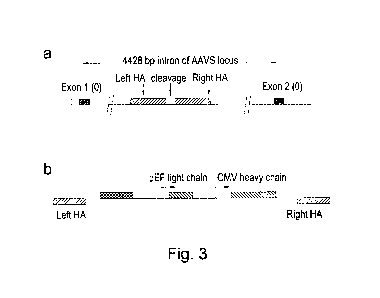
Examples herein we have demonstrated targeting to the AAVS locus but
alternative loci could
be targeted. For example the Type I collagen gene locus has been used for
efficient transgene
expression [44].
CA 02947513 2016-10-31
WO 2015/166272 PCT/GB2015/051287
19
Alternatively heterologous recognition sites for targeted nucleases, including
meganucleases, ZFNs and TALE nucleases could be introduced in advance for
subsequent
library targeting. In Examples herein, we describe the use of a TALE nuclease
recognising a
sequence within the AAVS locus to introduce by homologous recombination, an I-
Sce1
meganuclease recognition sequence and heterologous TALE nuclease recognition
sites within
the AAVS locus. Nuclease-directed targeting could be used to drive insertion
of target
sequences by homologous recombination or NHEJ using vector DNA or even double
stranded
oligonucleotides [45]. As an alternative, non-specific targeting methods could
be used to
introduce targeting sites through the use of transposon-directed integration
[46] to introduce
recognition sites for site-specific nucleases. Viral-based systems, such as
lentivirus, applied at
low titre could also be used to introduce targeting sites. Transfection of DNA
coupled with
screening for single copy insertion has also been used to identify unique
integration sites [17].
Such non-specific approaches would be particularly useful in the case of cells
which do not
have an obvious site to target or for genomes which have not been sequenced or
for genomes
for which no existing TALE nucleases, ZFNs or Cas9/CRISPR systems are
available. Once a
cell line has been established following random insertion of a nuclease
recognition site, the cell
line can be used subsequently to create libraries of binders where all clones
of the library
contain the transgene at the fixed locus using nuclease-directed integration.
In the Examples presented, three different plasmids are used encompassing
pairs of
TALE nucleases or ZFNs on individual plasmids with a separate plasmid for
donor DNA. In the
case of meganuclease the site-specific nuclease is encoded by a single gene
and this is
introduced on one plasmid with the donor DNA present on a second plasmid. Of
course,
combinations could be used incorporating two or more of these elements on the
same plasmid
and this could enhance the efficiency of targeting by reducing the number of
number of
plasmids to be introduced. In addition it may be possible to pre-integrate the
nuclease(s) which
could also be inducible to allow temporal control of nuclease activity as has
been demonstrated
for transposases [46]. Finally the nuclease could be introduced as recombinant
protein or
protein:RNA complex (for example in the case of an RNA directed nuclease such
as
CRISPR:Cas9).
Locus
A recognition sequence for the site-specific nuclease may be present in
genomic DNA,
or episomal DNA which is stably inherited in the cells. Donor DNA may
therefore be integrated
at a genomic or episomal locus in the cellular DNA.
CA 02947513 2016-10-31
WO 2015/166272 PCT/GB2015/051287
In its simplest form a single gene encoding a binder (binder gene) is targeted
to a single
site within the eukaryotic genome. Identification of a cell demonstrating a
particular binding
activity or cellular phenotype will allow direct isolation of the gene
encoding the desired property
(e.g., by PCR from mRNA or genomic DNA). This is facilitated by using a unique
recognition
sequence for the site-specific nuclease, occurring once in the cellular DNA.
Cells used for
creation of the library may thus contain a nuclease recognition sequence at a
single fixed locus,
i.e., one identical locus in all cells. Libraries produced from such cells
will contain donor DNA
integrated at the fixed locus, i.e., occurring at the same locus in cellular
DNA of all clones in the
library.
Optionally, recognition sequences may occur multiple times in cellular DNA, so
that the
cells have more than one potential integration site for donor DNA. This would
be a typical
situation for diploid or polyploid cells where the recognition sequence is
present at
corresponding positions in a pair of chromosomes, i.e., replicate loci.
Libraries produced from
such cells may contain donor DNA integrated at replicate fixed loci. For
example libraries
produced from diploid cells may have donor DNA integrated at duplicate fixed
loci and libraries
produced from triploid cells may have donor DNA integrated at triplicate fixed
loci. Many
suitable mammalian cells are diploid, and clones of mammalian cell libraries
according to the
invention may have donor DNA integrated at duplicate fixed loci.
The sequence recognised by the site-specific nuclease may occur at more than
one
independent locus in the cellular DNA. Donor DNA may therefore integrate at
multiple
independent loci. Libraries of diploid or polyploid cells may comprise donor
DNA integrated at
multiple independent fixed loci and/or at replicate fixed loci.
In cells containing recognition sequences at multiple loci (whether replicate
or
independent loci), each locus represents a potential integration site for a
molecule of donor
DNA. Introduction of donor DNA into the cells may result in integration at the
full number of
nuclease recognition sequences present in the cell, or the donor DNA may
integrate at some
but not all of these potential sites. For example, when producing a library
from diploid cells
containing recognition sequences at first and second fixed loci (e.g.,
duplicate fixed loci), the
resulting library may comprise clones in which donor DNA is integrated at the
first fixed locus,
clones in which donor DNA is integrated at the second fixed locus, and clones
in which donor
DNA is integrated at both the first and second fixed loci.
Methods of producing libraries may therefore involve site-specific nuclease
cleavage of
multiple fixed loci in a cell, and integration of donor DNA at the multiple
fixed loci. As noted
above, in cases where there are multiple copies of the same recognition
sequence (e.g., as
occurs when targeting endogenous loci in diploid or polyploid cells) it is
possible that two binder
genes will be integrated, particularly when an efficient targeting mechanisms
is used, with only
CA 02947513 2016-10-31
WO 2015/166272 PCT/GB2015/051287
21
one gene being specific to the target. This can be resolved during subsequent
screening once
binder genes have been isolated.
In some instances it may be desirable to introduce more than one binder per
cell. For
example bi-specific binders could be generated from two different antibodies
coming together
and these may have properties absent in the individual binders [47]. This
could be achieved by
introducing different antibody genes into both alleles at duplicate fixed loci
or by targeting
different antibody populations into independent fixed loci using the methods
described herein.
Furthermore a binder may itself be composed of multiple chains (e.g., antibody
VH and VL
domains presented within a Fab or IgG format). In this case it may be
desirable to integrate the
different sub-units into different loci. These could be integrated within the
same cycle of
nuclease-directed integration, they could be integrated sequentially using
nuclease-directed
integration for one or both integration steps.
Introduction of donor DNA
Numerous methods have been described for introducing donor DNA into eukaryotic
cells,
including transfection, infection or electroporation. Transfection of large
numbers of cells is
possible by standard methods including polyethyleneimine¨mediated transfection
as described
herein. In addition methods are available for highly efficient electroporation
of 1010 cells in 5
minutes, e.g., http://www.maxcyte.com.
Combinatorial libraries could be created wherein members of multimeric binding
pairs
(e.g., VH and VL genes of antibody genes) or even different parts of the same
binder molecule
are introduced on different plasmids. Introduction of separate donor DNA
molecules encoding
separate binders or binder subunits may be done simultaneously or
sequentially. For example
an antibody light chain could be introduced by transfection or infection, the
cells grown up and
selected if necessary. Other components could then be introduced in a
subsequent infection or
transfection step. One or both steps could involve nuclease-directed
integration to specific
genomic loci.
Integration of donor DNA
The donor DNA is integrated into the cellular DNA, forming recombinant DNA
having a
contiguous DNA sequence in which the donor DNA is inserted at the integration
site. In the
present invention, integration is mediated by the natural DNA repair
mechanisms that are
endogenous to the cell. Thus, integration can be allowed to occur simply by
introducing the
donor DNA into a cell, allowing the site-specific nuclease to create an
integration site, and
allowing the donor DNA to be integrated. Cells may be kept in culture for
sufficient time for the
CA 02947513 2016-10-31
WO 2015/166272 PCT/GB2015/051287
22
DNA to be integrated. This will usually result in a mixed population of cells,
including (i)
recombinant cells into which the donor DNA has integrated at the integration
site created by the
site-specific nuclease, and optionally (ii) cells in which donor DNA has
integrated at sites other
than the desired integration site and/or optionally (iii) cells that into
which donor DNA has not
integrated. The desired recombinant cells and the resulting clones of the
library may thus be
provided in a mixed population of other eukaryotic cells. Selection methods
described
elsewhere herein may be used to enrich for cells of the library.
Endogenous DNA repair mechanisms in eukaryotic cells include homologous
recombination, non-homologous end joining (NHEJ) and microhomology-directed
end joining.
The efficiency of DNA modification by such processes can be increased by the
introduction of
double stranded breaks (DSBs) in the DNA and efficiency gains of 40,000 fold
have been
reported using rare cutting endonucleases (meganucleases) such as I-Sce1 [48,
49, 50].
Unlike the site-specific recombination involved in systems such as the Flp-In
system [16],
the present invention does not require exogenous recombinases or engineered
recombinase
recognition sites. Therefore, optionally the present invention does not
include a step of
recombinase-mediated DNA integration in creating the library, and/or
optionally the eukaryotic
cells into which the donor DNA is introduced lack a recombination site for a
site-specific
recombinase. The mechanisms and practicalities of directed insertion of donor
DNA into cellular
DNA by recombinases and nucleases are very distinct. As discussed by Jasin
1996 [50]:
"... the reaction catalyzed by site-specific recombinases is quite distinct
from cellular
repair of DSBs. Site-specific recombinases, such as cre, synapse two
recognition sites and
create single-strand breaks within the sites, thus forming Holliday
intermediates. The
intermediates are resolved to produce deletions, inversions and insertions
(cointegrants), all of
which restore the two recognition sites. The reaction is absolutely precise
and, hence, reversible.
The breaks are never exposed to the cellular repair machinery."
In contrast site-specific nuclease act to create breaks or nicks within the
cellular DNA
(e.g., genomic or episomal), which are exposed to and repaired by endogenous
cellular repair
mechanisms such as homologous recombination or NHEJ. Recombinase-based
approaches
have an absolute requirement for pre-integration of their recognition sites,
so such methods
require engineering of the "hot spot" integration site into the cellular DNA
as a preliminary step.
With nuclease-directed integration it is possible to engineer nucleases or
direct via guide RNA
in the case of CRISPR:Cas9 to recognise endogenous loci, i.e., nucleic acid
sequences
occurring naturally in the cellular DNA. Finally, at a practical level
nuclease-directed approaches
are more efficient for direct integration of transgenes at the levels required
to make large
libraries of binders.
CA 02947513 2016-10-31
WO 2015/166272 PCT/GB2015/051287
23
The DNA repair mechanism by which the donor DNA is integrated in methods of
the
invention can be pre-determined or biased to some extent by design of the
donor DNA and/or
choice of site-specific nuclease.
Homologous recombination is a natural mechanism used by cells to repair double
stranded breaks using homologous sequence (e.g., from another allele) as a
template for repair.
Homologous recombination has been utilised in cellular engineering to
introduce insertions
(including transgenes), deletions and point mutations into the genome.
Homologous
recombination is promoted by providing homology arms on the donor DNA. The
original
approach to engineering higher eukaryotic cells typically used homology arms
of 5-10 kb within
a donor plasmid to increase efficiency of targeted integration into the site
of interest. Despite
this, homologous recombination driven purely by long homology arms, is less
efficient than Flp
and Ore directed recombination particularly in higher eukaryotes with large
genomes.
Homologous recombination is particularly suitable for eukaryotes such as
yeast, which has a
genome size of only 12.5 x 10⁶ bp, where it is more effective compared with
higher eukaryotes
with larger genomes e.g., mammalian cells with 3000 x 106 bp.
Homologous recombination can also be directed through [52] nicks in genomic
DNA and
this could also serve as a route for nuclease-directed integration into
genomic DNA. Two
distinct pathways have been shown to promote homologous recombination at
nicked DNA. One
is essentially similar to repair at double strand breaks, utilizing
Rad51/Brca2, while the other is
inhibited by Rad51/Brca2 and preferentially uses single¨stranded DNA or nicked
double
stranded donor DNA [51].
Non homologous end-joining (NHEJ) is an alternative mechanism to repair double
stranded breaks in the genome where the ends of DNA are directly re-ligated
without the need
for a homologous template. Nuclease-directed cleavage of genomic DNA can also
enhance
transgene integration via non-homology based mechanisms. This approach to DNA
repair is
less accurate and can lead to insertions or deletions. NHEJ nonetheless
provides a simple
means of integrating in-frame exons into intron or allows integration of
promoter:gene cassettes
into the genome. Use of non-homologous methods allows the use of donor vectors
which lack
homology arms thereby simplifying the construction of donor DNA.
It has been pointed out that short regions of terminal homology are used to re-
join DNA
ends and it was hypothesized that 4bp of microhomology might be utilized for
directing repairing
at double strand breaks, referred to as microhomology-directed end joining
[50].
CA 02947513 2016-10-31
WO 2015/166272 PCT/GB2015/051287
24
Donor DNA
The donor DNA will usually be circularised DNA, and may be provided as a
plasmid or
vector. Linear DNA is another possibility. Donor DNA molecules may comprise
regions that do
not integrate into the cellular DNA, in addition to one or more donor DNA
sequences that
integrate into the cellular DNA. The DNA is typically double-stranded,
although single-stranded
DNA may be used in some cases. The donor DNA contains one or more transgenes
encoding a
binder, for example it may comprise a promoter:gene cassette.
In the simplest format double-stranded, circular plasmid DNA can be used to
drive
homologous recombination. This requires regions of DNA flanking the transgenes
which are
homologous to DNA sequence flanking the cleavage site in genomic DNA.
Linearised double-
stranded plasmid DNA or PCR product or synthetic genes could be used to drive
both
homologous recombination and NHEJ repair pathways. As an alternative to double-
stranded
DNA it is possible to use single-stranded DNA to drive homologous
recombination [52]. A
common approach to generating single-stranded DNA is to include a single-
stranded origin of
replication from a filamentous bacteriophage into the plasmid.
Single-stranded DNA viruses such as adeno-associated virus (AAV) have been
used to
drive efficient homologous recombination where the efficiency has been shown
to be improved
by several orders of magnitude [53, 54]. Systems such as the AAV systems could
be used in
conjunction with nuclease-directed cleavage for the construction of large
libraries of binders.
The benefits of both systems could be applied to targeting of libraries of
binders. The packaging
limit of AAV vectors is 4.7 kb but the use of nuclease digestion of target
genomic DNA will
reduce this allowing larger transgene constructs to be incorporated.
A molecule of donor DNA may encode a single binder or multiple binders.
Optionally,
multiple subunits of a binder may be encoded per molecule of donor DNA. In
some
embodiments, donor DNA encodes a subunit of a multimeric binder.
Promoters and genetic elements for selection
Transcription of the binder from the encoding donor DNA will usually be
achieved by
placing the sequence encoding the binder under control of a promoter and
optionally one or
more enhancer elements for transcription. A promoter (and optionally other
genetic control
elements) may be included in the donor DNA molecule itself. Alternatively, the
sequence
encoding the binder may lack a promoter on the donor DNA, and instead may be
placed in
operable linkage with a promoter on the cellular DNA, e.g., an endogenous
promoter or a pre-
integrated exogenous promoter, as a result of its insertion at the integration
site created by the
site-specific nuclease.
CA 02947513 2016-10-31
WO 2015/166272 PCT/GB2015/051287
Donor DNA may further comprise one or more further coding sequences, such as
genetic elements enabling selection of cells containing or expressing the
donor DNA. As with
the sequence encoding the binder, discussed above, such elements may be
associated with a
promoter on the donor DNA or may be placed under control of a promoter as a
result of
integration of the donor DNA at a fixed locus. The latter arrangement provides
a convenient
means of selecting specifically for those cells which have integrated the
donor DNA at the
desired site, since these cells should express the genetic element for
selection. This may be, for
example, a gene conferring resistance to a negative selection agent such as
blasticidin or
puromycin. One or more selection steps may be applied to remove unwanted
cells, such as
cells that lack the donor DNA or which have not integrated the donor DNA at
the correct position.
Alternatively these cells may be permitted to remain mixed with clones of the
library.
The expression of a membrane anchored binder could itself be used as a form of
selectable marker. For example if a library of antibody genes, formatted as
IgG or scFv-Fc
fusions are introduced, then cells which express the antibody can be selected
using secondary
reagents which recognise the surface expressed Fc using methods described
herein. Upon
initial transfection with donor DNA encoding the transgene under the control
of an exogenous
promoter, transient expression (and cell surface expression) of the binder
will occur and it will
be necessary to wait for transient expression to abate (to achieve targeted
integration of e.g., 1-
2 antibody genes/cell).
As an alternative a construct encoding a membrane tethering element (e.g., the
Fc
domain of the present example fused to the PDGF receptor transmembrane domain)
could be
pre-integrated before the library of binders is introduced. If this membrane-
tethering element
lacks a promoter or is encoded within an exon which is out of frame with the
preceding exon
then surface expression will be compromised. Targeted integration of an
incoming donor
molecule can then correct this defect (e.g., by targeting a promoter or an "in-
frame" exon into
the intron which is upstream of the defective tethering element). If the frame
"correcting exon"
also encodes a binder then a fusion will be produced between the binder and
the membrane
tethering element resulting in surface expression of both. Thus correctly
targeted integration will
result in-frame expression of the membrane tethering element alone or as part
of a fusion with
the incoming binder. Furthermore if the incoming library of binders lack a
membrane tethering
element and these are incorrectly integrated they will not be selected. Thus
expression of the
binder itself on the cell surface can be used to select the population of
cells with correctly
targeted integration.
CA 02947513 2016-10-31
WO 2015/166272 PCT/GB2015/051287
26
Number of clones and library diversity
Yeast display libraries of 107-1010 have previously been constructed and
demonstrated
to yield binders in the absence of immunisation or pre-selection of the
population [9, 55, 56, 57].
Many of the previously published mammalian display libraries used antibody
genes derived
from immunised donors or even enriched antigen-specific B lymphocytes, given
the limitations
of library size and variability when using cells from higher eukaryotes.
Thanks to the efficiency
of gene targeting described in the present invention large, naïve libraries
can be constructed in
higher eukaryotes such as mammalian cells, which match those described for
simpler
eukaryotes such as yeast.
Following integration of donor DNA into the cellular DNA, the resulting
recombinant cells
are cultured to allow their replication, generating a clone of cells from each
initially-produced
recombinant cell. Each clone is thus derived from one original cell into which
donor DNA was
integrated at an integration site created by the site-specific nuclease.
Methods according to the
present invention are associated with a high efficiency and high fidelity of
donor DNA integration,
and a library according to the present invention may contain at least 100,
103, 104, 105, 106, 107,
108, 109 or 1010 clones.
Using nuclease-directed integration it is possible to target 10 % or more of
transfected
mammalian cells. It is also practical to grow and transform >1010 cells (e.g.
from 5 litres of cells
growing at 2 x10⁶ cells/m1). Transfection of such large numbers of cells could
be done using
standard methods including polyethyleneimine¨mediated transfection as
described herein. In
addition methods are available for highly efficient electroporation of 1010
cells in 5 minutes e.g.
http://www.maxcyte.com. Thus using the approach of the present invention it is
possible to
create libraries in excess of 109 clones.
When the population of donor DNA molecules that is used to create the library
contains
multiple copies of the same sequence, two or more clones may be obtained that
contain DNA
encoding the same binder. It can also be the case that a clone may contain
donor DNA
encoding more than one different binder, for example if there is more than one
recognition
sequence for the site-specific nuclease, as detailed elsewhere herein. Thus,
the diversity of the
library, in terms of the number of different binders encoded or expressed, may
be different from
the number of clones obtained.
Clones in the library preferably contain donor DNA encoding one or two members
of the
repertoire of binders and/or preferably express only one or two members of the
repertoire of
binders. A limited number of different binders per cell is an advantage when
it comes to
identifying the clone and/or DNA encoding a particular binder identified when
screening the
library against a given target. This is simplest when clones encode a single
member of the
CA 02947513 2016-10-31
WO 2015/166272 PCT/GB2015/051287
27
repertoire of binders. However it is also straightforward to identify the
relevant encoding DNA for
a desired binder if a clone selected from a library encodes a small number of
different binders,
for example a clone may encode two members of the repertoire of binders. As
discussed
elsewhere herein, clones encoding one or two binders are particularly
convenient to generate
by selecting a recognition sequence for the site-specific nuclease that occurs
once per
chromosomal copy in a diploid genome, as diploid cells contain duplicate fixed
loci, one on each
chromosomal copy, and the donor DNA may integrate at one or both fixed loci.
Thus, clones of
the library may each express only one or two members of the repertoire of
binders.
Binders displayed on the surface of cells of the library may be identical to
(having the
same amino acid sequence as) other binders displayed on the same cell. The
library may
consist of clones of cells which each display a single member of the
repertoire of binders, or of
clones displaying a plurality of members of the repertoire of binders per
cell. Alternatively a
library may comprise some clones that display a single member of the
repertoire of binders, and
some clones that display a plurality of members (e.g., two) of the repertoire
of binders.
Accordingly, a library according to the present invention may comprise clones
encoding
more than one member of the repertoire of binders, wherein the donor DNA is
integrated at
duplicate fixed loci or multiple independent fixed loci.
As noted above, it is easiest to identify the corresponding encoding DNA for a
binder if
the corresponding clone expresses only one binder. Typically, a molecule of
donor DNA will
encode a single binder. The binder may be multimeric so that a molecule of
donor DNA includes
multiple genes or open reading frames corresponding to the various subunits of
the multimeric
binder.
A library according to the present invention may encode at least 100, 103,
104, 106 or 106,
107, 108, 109 or 1019 different binders. Where the binders are multimeric,
diversity may be
provided by one or more subunits of the binder. Multimeric binders may combine
one or more
variable subunits with one or more constant subunits, where the constant
subunits are the same
(or of more limited diversity) across all clones of the library. In generating
libraries of multimeric
binders, combinatorial diversity is possible where a first repertoire of
binder subunits may pair
with any of a second repertoire of binder subunits.
Binders
A "binder" in accordance with the present invention is a binding molecule,
representing a
specific binding partner for another molecule. Typical examples of specific
binding partners are
antibody-antigen and receptor-ligand.
CA 02947513 2016-10-31
WO 2015/166272 PCT/GB2015/051287
28
The repertoire of binders encoded by a library will usually share a common
structure and
have one or more regions of diversity. The library therefore enables selection
of a member of a
desired structural class of molecules, such as a peptide or a scFv antibody
molecule. For
example, the binders may be polypeptides sharing a common structure and having
one or more
regions of amino acid sequence diversity.
This can be illustrated by considering a repertoire of antibody molecules.
These may be
antibody molecules of a common structural class, e.g., IgG, Fab, scFv-Fc or
scFv, differing in
one or more regions of their sequence. Antibody molecules typically have
sequence variability
in their complementarity determining regions (CDRs), which are the regions
primarily involved in
antigen recognition. A repertoire of binders in the present invention may be a
repertoire of
antibody molecules which differ in one or more CDRs, for example there may be
sequence
diversity in all six CDRs, or in one or more particular CDRs such as the heavy
chain CDR3
and/or light chain CDR3.
Antibody molecules and other binders are described in more detail elsewhere
herein.
The potential of the present invention however extends beyond antibody display
to include
display of libraries of peptides or engineered proteins, including receptors,
ligands, individual
protein domains and alternative protein scaffolds [58, 59]. Nuclease-directed
site-specific
integration can be used to make libraries of other types of binders previously
engineered using
other display systems. Many of these involve monomeric binding domains such as
DARPins
and lipocalins, affibodies and adhirons [58, 59, 152]. Display on eukaryotes,
particularly
mammalian cells, also opens up the possibility of isolating and engineering
binders or targets
involving more complex, multimeric targets. For example T cell receptors
(TCRs) are expressed
on T cells and have evolved to recognise peptide presented in complex with MHC
molecules on
antigen presenting cells. Libraries encoding and expressing a repertoire of
TCRs may be
generated, and may be screened to identify binding to MHC peptide complexes as
further
described elsewhere herein.
For multimeric binders, donor DNA encoding the binder may be provided as one
or more
DNA molecules. For example, where individual antibody VH and VL domains are to
be
separately expressed, these may be encoded on separate molecules of donor DNA.
The donor
DNA integrates into the cellular DNA at multiple integration sites, e.g., the
binder gene for the
VH at one locus and the binder gene for the VL at a second locus. Methods of
introducing donor
DNA encoding separate binder subunits are described in more detail elsewhere
herein.
Alternatively, both subunits or parts of a multimeric binder may be encoded on
the same
molecule of donor DNA which integrates at a fixed locus.
A binder may be an antibody molecule or a non-antibody protein that comprises
an
antigen-binding site. An antigen binding site may be provided by means of
arrangement of
CA 02947513 2016-10-31
WO 2015/166272 PCT/GB2015/051287
29
peptide loops on non-antibody protein scaffolds such as fibronectin or
cytochrome B etc., or by
randomising or mutating amino acid residues of a loop within a protein
scaffold to confer binding
to a desired target [60, 61, 62]. Protein scaffolds for antibody mimics are
disclosed in
WO/0034784 in which the inventors describe proteins (antibody mimics) that
include a
fibronectin type III domain having at least one randomised loop. A suitable
scaffold into which
to graft one or more peptide loops, e.g., a set of antibody VH CDR loops, may
be provided by
any domain member of the immunoglobulin gene superfamily. The scaffold may be
a human or
non-human protein.
Use of antigen binding sites in non-antibody protein scaffolds has been
reviewed
previously [63]. Typical are proteins having a stable backbone and one or more
variable loops,
in which the amino acid sequence of the loop or loops is specifically or
randomly mutated to
create an antigen-binding site having for binding the target antigen. Such
proteins include the
IgG-binding domains of protein A from S. aureus, transferrin, tetranectin,
fibronectin (e.g. 10th
fibronectin type III domain) and lipocalins. Other approaches include small
constrained peptide
e.g., based on" knottin" and cyclotides scaffolds [64]. Given their small size
and complexity
particularly in relation to correct formation of disulphide bond, there may be
advantages to the
use of eukaryotic cells for the selection of novel binders based on these
scaffolds. Given the
common functions of these peptides in nature, libraries of binders based on
these scaffolds may
be advantageous in generating small high affinity binders with particular
application in blocking
ion channels and proteases.
In addition to antibody sequences and/or an antigen-binding site, a binder may
comprise
other amino acids, e.g., forming a peptide or polypeptide, such as a folded
domain, or to impart
to the molecule another functional characteristic in addition to ability to
bind antigen. A binder
may carry a detectable label, or may be conjugated to a toxin or a targeting
moiety or enzyme
(e.g., via a peptidyl bond or linker). For example, a binder may comprise a
catalytic site (e.g., in
an enzyme domain) as well as an antigen binding site, wherein the antigen
binding site binds to
the antigen and thus targets the catalytic site to the antigen. The catalytic
site may inhibit
biological function of the antigen, e.g., by cleavage.
Antibody molecules
Antibody molecules are preferred binders. Antibody molecules may be whole
antibodies
or immunoglobulins (Ig), which have four polypeptide chains ¨ two identical
heavy chains and
two identical light chains. The heavy and light chains form pairs, each having
a VH-VL domain
pair that contains an antigen binding site. The heavy and light chains also
comprise constant
regions: light chain CL, and heavy chain CHI, CH2, CH3 and sometimes CH4 (the
fifth domain
CA 02947513 2016-10-31
WO 2015/166272 PCT/GB2015/051287
CH4 is present in human IgM and IgE). The two heavy chains are joined by
disulphide bridges
at a flexible hinge region. An antibody molecule may comprise a VH and/or a VL
domain.
The most common native format of an antibody molecule is an IgG which is a
heterotetramer consisting of two identical heavy chains and two identical
light chains. The
heavy and light chains are made up of modular domains with a conserved
secondary structure
consisting of a four-stranded antiparallel beta-sheet and a three-stranded
anti-parallel beta-
sheet, stabilised by a single disulphide bond. Antibody heavy chains each have
an N terminal
variable domain (VH) and 3 relatively conserved "constant" immunoglobulin
domains (CH1,
CH2, CH3) while the light chains have one N terminal variable domain (VL) and
one constant
domains (CL). Disulphide bonds stabilise individual domains and form covalent
linkages to join
the four chains in a stable complex. The VL and CL of the light chain
associates with VH and
CH1 of the heavy chain and these elements can be expressed alone to form a Fab
fragment.
The CH2 and CH3 domains (also called the "Fc domain") associate with another
CH2:CH3 pair
to give a tetrameric Y shaped molecule with the variable domains from the
heavy and light
chains at the tips of the "Y". The CH2 and CH3 domains are responsible for the
interactions with
effector cells and complement components within the immune system. Recombinant
antibodies
have previously been expressed in IgG format or as Fabs (consisting of a dimer
of VH:CH1 and
a light chain). In addition the artificial construct called a single chain Fv
(scFv) could be used
consisting of DNA encoding VH and VL fragments fused genetically with DNA
encoding a
flexible linker.
Binders may be human antibody molecules. Thus, where constant domains are
present
these are preferably human constant domains.
Binders may be antibody fragments or smaller antibody molecule formats, such
as single
chain antibody molecules. For example, the antibody molecules may be scFv
molecules,
consisting of a VH domain and a VL domain joined by a linker peptide. In the
scFv molecule, the
VH and VL domains form a VH-VL pair in which the complementarity determining
regions of the
VH and VL come together to form an antigen binding site.
Other antibody fragments that comprise an antibody antigen-binding site
include, but are
not limited to, (i) the Fab fragment consisting of VL, VH, CL and CH1 domains;
(ii) the Fd
fragment consisting of the VH and CH1 domains; (iii) the Fv fragment
consisting of the VL and
VH domains of a single antibody; (iv) the dAb fragment [65, 66, 67], which
consists of a VH or a
VL domain; (v) isolated CDR regions; (vi) F(ab')2 fragments, a bivalent
fragment comprising two
linked Fab fragments (vii) scFv, wherein a VH domain and a VL domain are
linked by a peptide
linker which allows the two domains to associate to form an antigen binding
site [68, 69]; (viii)
bispecific single chain Fv dimers (PCT/US92/09965) and (ix) "diabodies",
multivalent or
multispecific fragments constructed by gene fusion (W094/13804; [70]). Fv,
scFv or diabody
CA 02947513 2016-10-31
WO 2015/166272 PCT/GB2015/051287
31
molecules may be stabilised by the incorporation of disulphide bridges linking
the VH and VL
domains [71].
Various other antibody molecules including one or more antibody antigen-
binding sites
have been engineered, including for example Fab2, Fab3, diabodies, triabodies,
tetrabodies and
minibodies (small immune proteins). Antibody molecules and methods for their
construction
and use have been described [72].
Other examples of binding fragments are Fab', which differs from Fab fragments
by the
addition of a few residues at the carboxyl terminus of the heavy chain CH1
domain, including
one or more cysteines from the antibody hinge region, and Fab'-SH, which is a
Fab' fragment in
which the cysteine residue(s) of the constant domains bear a free thiol group.
A dAb (domain antibody) is a small monomeric antigen-binding fragment of an
antibody,
namely the variable region of an antibody heavy or light chain. VH dAbs occur
naturally in
camelids (e.g., camel, llama) and may be produced by immunizing a camelid with
a target
antigen, isolating antigen-specific B cells and directly cloning dAb genes
from individual B cells.
dAbs are also producible in cell culture. Their small size, good solubility
and temperature
stability makes them particularly physiologically useful and suitable for
selection and affinity
maturation. Camelid VH dAbs are being developed for therapeutic use under the
name
"nanobodies TM".
Synthetic antibody molecules may be created by expression from genes generated
by
means of oligonucleotides synthesized and assembled within suitable expression
vectors, for
example as described by Knappik et al. [73] or Krebs et al. [74].
Bispecific or bifunctional antibodies form a second generation of monoclonal
antibodies
in which two different variable regions are combined in the same molecule
[75]. Their use has
been demonstrated both in the diagnostic field and in the therapy field from
their capacity to
recruit new effector functions or to target several molecules on the surface
of tumour cells.
Where bispecific antibodies are to be used, these may be conventional
bispecific antibodies,
which can be manufactured in a variety of ways [76], e.g., prepared chemically
or from hybrid
hybridomas, or may be any of the bispecific antibody fragments mentioned
above. These
antibodies can be obtained by chemical methods [77, 78] or somatic methods
[79, 80] but
likewise and preferentially by genetic engineering techniques which allow the
heterodimerisation
to be forced and thus facilitate the process of purification of the antibody
sought [81]. Examples
of bispecific antibodies include those of the BiTET" technology in which the
binding domains of
two antibodies with different specificity can be used and directly linked via
short flexible
peptides. This combines two antibodies on a short single polypeptide chain.
Diabodies and scFv
CA 02947513 2016-10-31
WO 2015/166272 PCT/GB2015/051287
32
can be constructed without an Fc region, using only variable domains,
potentially reducing the
effects of anti-id iotypic reaction.
Bispecific antibodies can be constructed as entire IgG, as bispecific Fab2, as
Fab'PEG,
as diabodies or else as bispecific scFv. Further, two bispecific antibodies
can be linked using
routine methods known in the art to form tetravalent antibodies.
Bispecific diabodies, as opposed to bispecific whole antibodies, may also be
particularly
useful. Diabodies (and many other polypeptides, such as antibody fragments) of
appropriate
binding specificities can be readily selected. If one arm of the diabody is to
be kept constant, for
instance, with a specificity directed against an antigen of interest, then a
library can be made
where the other arm is varied and an antibody of appropriate specificity
selected. Bispecific
whole antibodies may be made by alternative engineering methods as described
in Ridgeway et
al., 1996 [82].
A library according to the invention may be used to select an antibody
molecule that
binds one or more antigens of interest. Selection from libraries is described
in detail below.
Following selection, the antibody molecule may then be engineered into a
different format
and/or to contain additional features. For example, the selected antibody
molecule may be
converted to a different format, such as one of the antibody formats described
above. The
selected antibody molecules, and antibody molecules comprising the VH and/or
VL CDRs of the
selected antibody molecules, are an aspect of the present invention. Antibody
molecules and
their encoding nucleic acid may be provided in isolated form.
Antibody fragments can be obtained starting from an antibody molecule by
methods
such as digestion by enzymes e.g. pepsin or papain and/or by cleavage of the
disulphide
bridges by chemical reduction. In another manner, the antibody fragments can
be obtained by
techniques of genetic recombination well known to the person skilled in the
art or else by
peptide synthesis by means of, for example, automatic peptide synthesisers, or
by nucleic acid
synthesis and expression.
It is possible to take monoclonal and other antibodies and use techniques of
recombinant DNA technology to produce other antibodies or chimaeric molecules
that bind the
target antigen. Such techniques may involve introducing DNA encoding the
immunoglobulin
variable region, or the CDRs, of an antibody to the constant regions, or
constant regions plus
framework regions, of a different immunoglobulin. See, for instance, EP-A-
184187, GB
2188638A or EP-A-239400, and a large body of subsequent literature.
Antibody molecules may be selected from a library and then modified, for
example the in
vivo half-life of an antibody molecule can be increased by chemical
modification, for example
PEGylation, or by incorporation in a liposome.
CA 02947513 2016-10-31
WO 2015/166272 PCT/GB2015/051287
33
Sources of binder genes
The traditional route for generation of monoclonal antibodies utilises the
immune system
of laboratory animals like mice and rabbits to generate a pool of high
affinity antibodies which
are then isolated by the use of hybridoma technology. The present invention
provides an
alternative route to identifying antibodies arising from immunisation. VH and
VL genes could be
amplified from the B cells of immunised animals and cloned into an appropriate
vector for
introduction into eukaryotic libraries followed by selection from these
libraries. Phage display
and ribosome display allows very large libraries (>109 clones) to be
constructed enabling
isolation of human antibodies without immunisation. The present invention
could also be used in
conjunction with such methods. Following rounds of phage display selection,
the selected
population of binders could be introduced into eukaryotic cells by nuclease-
directed integration
as described herein. This would allow the initial use of very large libraries
based in other
systems (e.g., phage display) to enrich a population of binders while allowing
their efficient
screening using eukaryotic cells as described above. Thus the invention can
combine the best
features of both phage display and eukaryotic display to give a high
throughput system with
quantitative screening and sorting.
Using phage display and yeast display it has previously been demonstrated that
it is also
possible to generate binders without resorting to immunisation, provided
display libraries of
sufficient size are used. For example multiple binders were generated from a
non-immune
antibody library of >107 clones [83]. This in turn allows generation of
binders to targets which
are difficult by traditional immunisation routes e.g., generation of
antibodies to "self-antigens" or
epitopes which are conserved between species. For example, human/mouse cross-
reactive
binders can be enriched by sequential selection on human and then mouse
versions of the
same target. Since it is not possible to specifically immunise humans to most
targets of interest,
this facility is particularly important in allowing the generation of human
antibodies which are
preferred for therapeutic approaches.
In examples of mammalian display to date, where library sizes and quality were
limited,
binders have only been generated using repertoires which were pre-enriched for
binders, e.g.,
from immunisation or from engineering of pre-existing binders. The ability to
make large libraries
in eukaryotic cells and particularly higher eukaryotes creates the possibility
of isolating binders
direct from these libraries starting with non-immune binders or binders which
have not
previously been selected within another system. With the present invention it
is possible to
generate binders from non-immune sources. This in turn opens up the
possibilities for using
binder genes from multiple sources. Binder genes could come from PCR of
natural sources
such as antibody genes. Binder genes could also be re-cloned from existing
libraries, such as
CA 02947513 2016-10-31
WO 2015/166272 PCT/GB2015/051287
34
antibody phage display libraries, and cloned into a suitable donor vector for
nuclease-directed
integration into target cells. Binders may be completely or partially
synthetic in origin.
Furthermore various types of binders are described elsewhere herein, for
example binder genes
could encode antibodies or could encode alternative scaffolds [58, 59],
peptides or engineered
proteins or protein domains.
Binder display
To provide a repertoire of binders for screening against a target of interest,
the library
may be cultured to express the binders in either soluble secreted form or in
transmembrane
form. For cell surface display it is necessary to retain the expressed binder
on the surface of the
cell which encodes it. Binders may comprise or be linked to a membrane anchor,
such as a
transmembrane domain, for extracellular display of the binder at the cell
surface. This may
involve direct fusion of the binder to a membrane localisation signal such as
a GPI recognition
sequence or to a transmembrane domain such as the transmembrane domain of the
PDGF
receptor [84]. Retention of binders at the cell surface can also be done
indirectly by association
with another cell surface retained molecule expressed within the same cell.
This associated
molecule could itself be part of a heterodimeric binder, such as tethered
antibody heavy chain in
association with a light chain partner that is not directly tethered.
Although cell surface immobilisation facilitates selection of the binder, in
many
applications it is necessary to prepare cell-free, secreted binder. It will be
possible to combine
membrane tethering and soluble secretion using a recapture method of attaching
the secreted
binders to cell surface receptors. One approach is to format the library of
binders as secreted
molecules which can associate with a membrane anchored molecule expressed
within the
same cell which can function to capture a secreted binder. For example, in the
case of
antibodies or binder molecules fused to antibody Fc domains, a membrane
tethered Fc can
"sample" secreted binder molecules being expressed in the same cell resulting
in display of a
monomeric fraction of the binder molecules being expressed while the remainder
is secreted in
a bivalent form (US 8,551,715). An alternative is to use a tethered IgG
binding domain such as
protein A.
Other methods for retaining secreted antibodies with the cells producing them
are
reviewed in Kumar at al. (2012) [85] and include encapsulation of cells within
microdrops, matrix
aided capture, affinity capture surface display (ACSD), secretion and capture
technology
(SECANT) and "cold capture" [85]. In examples given for ACSD and SECANT [85],
biotinylation
is used to facilitate immobilisation of streptavidin or a capture antibody on
the cell surface. The
captured molecule in turn captures secreted antibodies. In the example of
SECANT in vivo
CA 02947513 2016-10-31
WO 2015/166272 PCT/GB2015/051287
biotinylated of the secreted molecule occurs. Using the "cold capture"
technique secreted
antibody can be detected on producer cells using antibodies directed to the
secreted molecule.
It has been proposed that this due to association of the secreted antibody
with the glycocalyx of
the cell [86]. Alternatively it has been suggested that the secreted product
is trapped by staining
antibodies on the cell surface before being endocytosed [87]. The above
methods have been
used to identify high expressing clones within a population but could
potentially be adapted for
identification of binding specificity, provided the association has sufficient
longevity at the cell
surface.
Even when the binder is directly tethered to the cell surface it is possible
to generate a
soluble product. For example the gene encoding the selected binder can be
recovered and
cloned into an expression vector lacking the membrane anchored sequence.
Alternatively, and
as demonstrated in the Examples, an expression construction can be used in
which the
transmembrane domain is encoded within an exon flanked by recombination sites,
e.g., ROX
recognition sites for Dre recombinase [ 88 ]. In this example the exon
encoding the
transmembrane domain can be removed by transfection with a gene encoding Dre
recombinase
to switch expression to a secreted form. As demonstrated herein, secreted
antibody was
produced by this method without the need for recombinase action. This is
presumably as a
result of alternative splicing [89].
Any of the above methods or other suitable approaches can be used to ensure
that
binders expressed by clones of a library are displayed on the surface of their
expressing cells.
Screening to identify binders to a target of interest
As noted, the eukaryotic cell library may be used in a method of screening for
a binder
that recognises a target. Such a method may comprise:
providing a library as described herein,
culturing cells of the library to express the binders,
exposing the binders to the target, allowing recognition of the target by one
or more
cognate binders, if present, and
detecting whether the target is recognised by a cognate binder.
Selections could be carried using a range of target molecule classes, e.g.,
protein,
nucleic acid, carbohydrate, lipid, small molecules. The target may be provided
in soluble form.
The target may be labelled to facilitate detection, e.g., it may carry a
fluorescent label or it may
be biotinylated. Cells expressing a target-specific binder may be isolated
using a directly or
indirectly labelled target molecule, where the binder captures the labelled
molecule. For
example, cells that are bound, via the binder:target interaction, to a
fluorescently labelled target
CA 02947513 2016-10-31
WO 2015/166272 PCT/GB2015/051287
36
can be detected and sorted by flow cytometry or FACS to isolate the desired
cells. Selections
involving cytometry require target molecules which are directly fluorescently
labelled or are
labelled with molecules which can be detected with secondary reagents, e.g.,
biotinylated target
can be added to cells and binding to the cell surface can be detected with
fluorescently labelled
streptavidin such as streptavidin-phycoerythrin. A further possibility is to
immobilise the target
molecule or secondary reagents which bind to the target on a solid surface,
such as magnetic
beads or agarose beads, to allow enrichment of cells which bind the target.
For example cells
that bind, via the binder: target interaction, to a biotinylated target can be
isolated on a substrate
coated with streptavid in, e.g., streptavid in-coated beads.
In screening libraries it is preferable to over-sample, i.e., screen more
clones than the
number of independent clones present within the library to ensure effective
representation of the
library. Identifying binders from very large libraries provided by the present
invention could be
done by flow sorting but this would take several days, particularly if over-
sampling the library. As
an alternative initial selections could be based on the use of recoverable
antigen, e.g.,
biotinylated antigen recovered on streptavidin-coated magnetic beads. Thus
streptavidin-coated
magnetic beads could be used to capture cells which have bound to biotinylated
antigen.
Selection with magnetic beads could be used as the only selection method or
this could be
done in conjunction with flow cytometry where better resolution can be
achieved, e.g.,
differentiating between a clone with higher expression levels and one with a
higher affinity [56,
57].
The in vitro nature of display technology approaches makes it is possible to
control
selection in a way that is not possible by immunisation, e.g., selecting on a
particular
conformational state of a target [90, 91]. Targets could be tagged through
chemical modification
(fluorescein, biotin) or by genetic fusion (e.g. protein fused to an epitope
tag such as a FLAG
tag or another protein domain or a whole protein). The tag could be nucleic
acid (e.g., DNA,
RNA or non-biological nucleic acids) where the tag is part fused to target
nucleic acid or could
be chemically attached to another type of molecule such as a protein. This
could be through
chemical conjugation or through enzymatic attachment [92]. Nucleic acid could
be also fused to
a target through a translational process such as ribosome display. The "tag"
may be another
modification occurring within the cell (e.g., glycosylation, phosphorylation,
ubiqitinylation,
alkylation, PASylation, SUMO-lation and others described at the Post-
translational Database
(db-PTM) at http://dbptm.mbc.nctu.edu.twistatistics.php) which can be detected
via secondary
reagents. This would yield binders which bind an unknown target protein on the
basis of a
particular modification.
CA 02947513 2016-10-31
WO 2015/166272 PCT/GB2015/051287
37
Targets could be detected using existing binders which bind to that target
molecule, e.g.,
target specific antibodies. Use of existing binders for detection will have
the added advantage of
identifying binders within the library of binders which recognise an epitope
distinct from the
binder used for detection. In this way pairs of binders could be identified
for use in applications
such as sandwich ELISA. Where possible a purified target molecule would be
preferred.
Alternatively the target may be displayed on the surface of a population of
target cells and the
binders are displayed on the surface of the library cells, the method
comprising exposing the
binders to the target by bringing the library cells into contact with the
target cells. Recovery of
the cells expressing the target (e.g., using biotinylated cells expressing
target) will allow
enrichment of cells which express binders to them. This approach would be
useful where low
affinity interactions are involved since there is the potential for a strong
avidity effect.
The target molecule could also be unpurified recombinant or unpurified native
targets
provided a detection molecule is available to identify cell binding (as
described above). In
addition binding of target molecules to the cell expressing the binder could
be detected
indirectly through the association of target molecule to another molecule
which is being
detected, e.g., a cell lysate containing a tagged molecule could be incubated
with a library of
binders to identify binders not only to the tagged molecule but also binders
to its associated
partner proteins. This would result in a panel of antibodies to these partners
which could be
used to detect or identify the partner (e.g., using mass spectrometry).
Cellular fractionation
could be used to enrich targets from particular sub-cellular locations.
Alternatively differential
biotinylation of surface or cytoplasmic fractions could be used in conjunction
with streptavidin
detection reagents for eukaryotic display [93,94]. The use of detergent
solubilised target
preparations is a particularly useful approach for intact membrane proteins
such as GPCRs and
ion channels which are otherwise difficult to prepare. The presence of
detergents may have a
detrimental effect on the eukaryotic cells displaying the binders requiring
recovery of binder
genes without additional growth of the selected cells.
Following detection of target recognition by a cognate binder, cells of a
clone containing
DNA encoding the cognate binder may be recovered. DNA encoding the binder may
then be
isolated (e.g., identified or amplified) from the recovered clone, thereby
obtaining DNA encoding
a binder that recognises the target.
Exemplary binders and targets are detailed elsewhere herein. A classic example
is a
library of antibody molecules, which may be screened for binding to a target
antigen of interest.
Other examples include screening a library of TCRs against a target
MHC:peptide complex or
screening a library of MHC:peptide complexes against a target TCR.
CA 02947513 2016-10-31
WO 2015/166272 PCT/GB2015/051287
38
TCR:MHC and other receptor interactions
T cell receptors (TCRs) are expressed on T cells and have evolved to recognise
peptide
presented in complex with MHC molecules on antigen presenting cells. TCRs are
heterodimers
consisting in 95% of cases of alpha and beta heterodimers and in 5% of cases
of gamma and
delta heterodimers. Both monomer units have an N terminal immunoglobulin
domain which has
3 variable complementarity determining regions (CDRs) involved in driving
interaction with
target. The functional TCR is present within a complex of other sub-units and
signalling is
enhanced by co-stimulation with CD4 and CD8 molecules (specific for class I
and class II MHC
molecules respectively). On antigen presenting cells, proteins are processed,
and presented on
the cell surface in complex with MHC molecules which are themselves part of a
multimeric
protein complex. TCRs recognizing peptides originating from "self' are removed
during
development and the system is poised for recognition of foreign peptides
presented on antigen
presenting cells to effect an immune response. The outcome of recognition of a
peptide:MHC
complex depends on the identity of the T cell and the affinity of that
interaction.
It would be valuable to identify the genes encoding TCRs or MHC:peptide
complexes
which drive interactions involved in pathological conditions, e.g., as occurs
in autoimmune
disease. In the case of autoimmune disease, identification of interacting
partners, e.g., a TCR
driving a pathogenic condition could pave the way to either specifically
blocking such
interactions or removing offending cytotoxic cells. It would be desirable to
engineer TCRs for
altered binding e.g. higher affinity to targets of interest, e.g., in re-
targeting T cells in cancer or
enhancing the effect of existing T cells [95]. Alternatively the behaviour of
regulatory or
suppressive T cells might be altered as a therapeutic modality, e.g., for
directing or enhancing
immunotherapy of cancer by introducing specific TCRs into T cells or by using
expressed TCR
protein as therapeutic entities [96].
Display of libraries of TCRs on surface of yeast cells and mammalian cells has
previously been demonstrated. In the case of yeast cells it was necessary to
engineer the TCR
and present it in a single chain format. Since the affinity of interaction
between TCR and
peptide:MHC complex is low, the soluble component (e.g., peptide:MHC in this
case) is usually
presented in a multimeric format. TCR specificity has been engineered for
peptides in complex
with MHC class I [97] and MHC class II [98]. TCRs have also been expressed on
the surface of
a mutant mouse T cells (lacking TCR alpha and beta chains) and variant TCRs
with improved
binding properties have been isolated [99]. For example Chervin et al.
introduced TCRs by
retroviral infection and an effective library size of 104 clones was generated
[100]. Using
nuclease-directed integration of binders as proposed here, a similar approach
could be taken to
engineering T cells. As well as selecting TCRs with altered recognition
properties, display
CA 02947513 2016-10-31
WO 2015/166272 PCT/GB2015/051287
39
libraries could be used to screen libraries of peptide or of MHC variants for
recognition by TCRs.
For example peptide:MHC complexes have been displayed on insect cells and used
to epitope
map TCRs presented in a multimeric format [101].
As noted, screening methods may involve displaying the repertoire of binders
on the cell
surface and probing with a target presented as a soluble molecule, which may
be a multimeric
target. An alternative, which can be especially useful with multimeric
targets, is to screen
directly for cell:cell interactions, where binder and target are presented on
the surface of
different cells. For example if activation of a TCR of interest led to
expression of a reporter gene
this could be used to identify activating peptides or activating MHC molecules
presented within
a peptide:MHC library. In this particular example the reporter cell does not
encode the library
member but could be used to identify the cell which does encode it. The
approach could
potentially extend to a "library versus library" approach. For example
extending the example
described above, a TCR library could be screened against a peptide:MHC
library. More broadly
the example of screening a library of binders presented on one cell surface
using a binding
partner on another cell could be extended to other types of cell:cell
interactions e.g.,
identification of binders which inhibit or activate signalling within the
Notch or Wnt pathways.
Thus the present invention could be used in alternative cell based screening
system including
recognition systems based on cell:cell interactions.
As an example, chimeric antigen receptors ("CARS") represent a fusion between
an
antibody binding domain (usually formatted as a scFv) and a signalling domain.
These have
been introduced into T cells with the aim of re-directing the T cell in vivo
to attack tumour cells
through antibody recognition and binding to tumour-specific antigens. A number
of different
factors could affect the success of this strategy including the combination of
antibody specificity,
format, antibody affinity, linker length, fused signalling module, expression
level in T cells, T cell
sub-type and interaction of the CAR with other signalling molecules [102,
103]. The ability to
create large libraries of CARs in primary T cells incorporating individual or
combinations of the
above variables would allow a functional search for effective and optimal CAR
construction.
This functional "search" could be carried out in vitro or in vivo. For example
Alonso-Camino
(2009) have fused a scFv recognizing CEA to the chain of the TCR:CD3 complex
and
introduced this genetic construct into a human Jurkat cell line [104]. Upon
interaction with CEA
present on either HeLa cells or tumour cells they showed upregulation of the
early T cell
activation marker CD69. This approach could be used to identify CAR fusion
constructs with
appropriate activation or inhibitory properties using cultured or primary
cells.
Going one step further functionality of CAR constructs could be assessed in
vivo. For
example a library of CARs constructed in primary mouse T cells could be
introduced into tumour
CA 02947513 2016-10-31
WO 2015/166272 PCT/GB2015/051287
bearing mice to identify T cell clones stimulated to proliferate through
encounter with tumour. If
necessary this T cell library could be pre-selected based on antigen binding
specificity using the
methods described above. In either case the incoming library of binders could
be used to
replace an existing binder molecule (e.g., MHC or TCR or antibody variable
domain).
Phenotype screens
Described here are various methods for selection of binders which modify cell
signalling
and cellular behaviour.
A library may be screened for altered cellular phenotype as a result of the
action of the
binder on its target.
Antibodies which modify cell signalling by binding to ligands or receptors
have a proven
track record in drug development and the demand for such therapeutic
antibodies continues to
grow. Such antibodies and other classes of functional binders also have
potential in controlling
cell behaviour in vivo and in vitro. The ability to control and direct
cellular behaviour however
relies on the availability of natural ligands which control specific
signalling pathways.
Unfortunately many natural ligands such as those controlling stem cell
differentiation (e.g.,
members of FGF, TGF-beta, Wnt and Notch super-families) often exhibit
promiscuous
interactions and have limited availability due to their poor
expression/stability profiles. With their
exquisite specificity, antibodies have great potential in controlling cellular
behaviour.
The identification of functional antibodies that modify cell signalling has
historically been
relatively laborious involving picking clones, expressing antibody,
characterising according to
sequence and binding properties, conversion to mammalian expression systems
and addition to
functional cell based assays. The eukaryotic display approach described herein
will reduce this
effort but there is still a requirement for production of antibody and
addition to a separate
reporter cell culture. Therefore, a preferred alternative may be to directly
screen libraries of
binders expressed in eukaryotic cells for the effect of binding on cell
signalling or cell behaviour
by using the production cell itself as a reporter cell. Following introduction
of antibody genes,
clones within the resulting population of cells showing alteration in reporter
gene expression or
altered phenotypes can be identified.
A number of recent publications have described the construction of antibody
libraries by
cloning repertoires of antibody genes into reporter cells [47, 105, 106].
These systems combine
expression and reporting within one cell, and typically introduce a population
of antibodies
selected against a pre-defined target (e.g., using phage display).
A population of antibody genes may be introduced into reporter cells to
produce a library
by methods described herein, and clones within the population with an antibody-
directed
CA 02947513 2016-10-31
WO 2015/166272 PCT/GB2015/051287
41
alteration in phenotype (e.g., altered gene expression or survival) can be
identified. For this
phenotypic-directed selection to work there is a requirement to retain a
linkage between the
antibody gene present within the expressing cell (genotype) and the
consequence of antibody
expression (phenotype). This has been achieved previously either through
tethering the
antibody to the cell surface [47] as described for antibody display or through
the use of semi-
solid medium to retain secreted antibodies in the vicinity of producing cells
[105]. Alternatively
antibodies and other binders can be retained inside the cell [107]. Binders
retained on the cell
surface or in the surrounding medium can interact with an endogenous or
exogenous receptor
on the cell surface causing activation of the receptor. This in turn can cause
a change in
expression of a reporter gene or a change in the phenotype of the cell. As an
alternative the
antibody can block the receptor or ligand to reduce receptor activation. The
gene encoding the
binder which causes the modified cellular behaviour can then be recovered for
production or
further engineering.
An alternative to this "target-directed" approach, it is possible to introduce
a "naïve"
antibody population which has not been pre-selected to a particular target
[108]. The cellular
reporting system is used to identify members of the population with altered
behaviour. Since
there is no prior knowledge of the target, this non-targeted approach has a
particular
requirement for a large antibody repertoire, since pre-enrichment of the
antibody population to
the target is not possible. This approach will benefit from using nuclease-
directed transgene
integration as described in the present invention.
The "functional selection" approach could be used on other applications
involving
libraries in eukaryotic cells, particularly higher eukaryotes such as
mammalian cells. The
antibody could be fused to a signalling domain such that binding to target
causes activation of
the receptor. Kawahara et al. have constructed chimeric receptors where an
extra-cellular scFv
targeting fluorescein was fused to a spacer domain (the D2 domain of the Epo
receptor) and
various intracellular cytokine receptor domain including the thrombopoeitin
(Tpo) receptor,
erythropoietin (Epo) receptor, gp130, IL-2 receptor and the EGF receptor [109,
110, 111].
These were introduced into an IL-3 dependent proB cell line (BaF3) [27], where
chimaeric
receptors were shown to exhibit antigen-dependent activation of the chimaeric
receptor leading
to IL-3 independent growth. This same approach was used in model experiments
to
demonstrate antigen mediated chemoattraction of BaF3 cells [110]. The approach
was
extended beyond stable culture cells to primary cells exemplified by the
survival and growth of
Tpo-responsive haematopoeitic stem cells [112] or IL2 dependant primary T
cells where normal
stimulation by Tpo and IL-2 respectively was replaced by fluorescein directed
stimulation of
scFv chimaeric receptors. Thus a system based on chimaeric antibody-receptor
chimaeras can
CA 02947513 2016-10-31
WO 2015/166272 PCT/GB2015/051287
42
be used to drive target dependent gene expression or phenotypic changes in
primary or stable
reporter cells. This capacity could be used to identify fused binders which
drive a signalling
response or binders which inhibit the response.
In a modification of the above approach separate VH and VL domains from an
anti-
lysozyme antibody were fused to the Epo intracellular domain [113]. Cells grew
in response to
addition of lysozyme indicating an antigen induced dimerisation or
stabilisation of the separate
VH and VL fusion partners. Thus three interacting components come together for
an optimal
response in this system.
Although described here with reference to antibody molecules, the above
methods may
also be adapted and performed with libraries of other binders.
Protein fragment complementation represents an alternative system for studying
and for
selecting protein:protein interactions in mammalian cells [114, 115]. This
involves restoring
function of split reporter proteins through protein:protein interactions.
Reporter proteins which
have been used include ubiquitin, DNAE intein, beta-galactosidase,
dihydrofolate reductase,
GFP, firefly luciferase, beta-lactamase, TEV protease. For example a recent
example of this
approach is the mammalian membrane 2 hybrid (MaMTH) approach where association
of a bait
protein:split ubiquitin:transcription factor fusion with a partner
protein:split ubiquitin restores
ubiquitin recognition and liberates the transcription factor to effect
reporter gene expression
[116]. Again binders which interfere with or enhance this interaction could be
identified through
perturbed signalling.
Recovery and reformatting of binders and encoding DNA
Following selection of a binder or clone of interest from the library, a
common next step
will be to isolate (e.g., identify or amplify) the DNA encoding the binder.
Optionally, it may be
desired to modify the nucleic acid encoding the binder, for example to
restructure the binder
and/or to insert the encoding sequence into a different vector.
Where the binder is an antibody molecule, a method may comprise isolating DNA
encoding the antibody molecule from cells of a clone, amplifying DNA encoding
at least one
antibody variable region, preferably both the VH and VL domain, and inserting
DNA into a
vector to provide a vector encoding the antibody molecule. A multimeric
antibody molecule
bearing a constant domain may be converted to a single chain antibody molecule
for expression
in a soluble secreted form.
Antibodies may be presented in different formats but whatever format an
antibody is
selected in, once the antibody gene is isolated it is possible to reconfigure
it in a number of
different formats. Once VH or VL domains are isolated, they can be re-cloned
into expression
CA 02947513 2016-10-31
WO 2015/166272 PCT/GB2015/051287
43
vectors encompassing the required partner domains (see Example 1 showing a
dual promoter
IgG expression cassette).
A reformatting step may comprise reformatting of binders composed of a pair of
subunits
(e.g., scFv molecules), to a different molecular binder format (e.g., Ig or
Fab) in which the
original pairing of the subunits is maintained. Such methods are described in
more detail
elsewhere herein and can be used for monoclonal, oligoclonal or polyclonal
clone reformatting.
The method can be used to convert "en masse" an entire output population from
any of the
commonly used display technologies including phage, yeast or ribosome display.
Display of scFvs on the surface of mammalian cells fused to Fc domains.
Although many antibody phage display libraries are formatted to display scFvs,
eukaryotic display systems will allow presentation in Fab or IgG format. To
take full advantage
of the potential for IgG/Fab expression, particularly when using scFvs from
other display
systems will be necessary to take selected linked VH and VL domains within a
bacterial
expression system and express them within a eukaryotic system fused to
appropriate constant
domains. Described here is a method to convert scFv populations to
immunoglobulin (Ig) or
fragment, antigen binding (Fab) format in such a way that original VH and VL
chain pairings are
maintained. In the present invention, conversion is possible using individual
clones, oligoclonal
mixes or whole populations formatted as scFv while retaining the original
pairing of VH and VLs
chains. The method proceeds via the generation of an intermediate non-
replicative "mini-circle"
DNA which brings in a new "stuffer" DNA fragment. The circular DNA is
linearised (e.g., by
restriction digestion or PCR) which alters the relative position of the
original VH and VL
fragments and places the "stuffer" DNA between them. Following linearization
the product can
be cloned into a vector of choice, e.g., a mammalian expression vector. In
this way all of the
elements apart from the VH and VL can be replaced. Elements for bacterial
expression can be
replaced with elements for mammalian expression and fusion to alternative
partners. The
complete conversion process only requires a single transformation step of E.
coli bacteria to
generate a population of bacterial colonies each harbouring a plasmid encoding
a unique Ig or
Fab formatted recombinant antibody. Extending beyond conversion of scFv to
IgG/Fab, the
method can be employed to reformat any two joined DNA elements to clone into a
vector such
that after re-formatting each DNA element is surrounded by different DNA
control features whilst
maintaining the original pairing. A previous method has been described wherein
2 sequential
cloning steps are used [117] to replace these elements in contrast to the
present method which
proceeds via an intermediate non-replicative circular intermediate.
CA 02947513 2016-10-31
WO 2015/166272 PCT/GB2015/051287
44
A method of restructuring a binder, or population of binders, may comprise
converting
scFv to Ig or a fragment thereof, e.g., Fab. The method may comprise
converting nucleic acid
encoding scFv to DNA encoding an immunoglobulin (Ig) or fragment thereof such
as Fab format,
in such a way that the original variable VH and VL chain pairings are
maintained. Preferably the
conversion proceeds via circular DNA intermediate which may be a non-
replicative "mini-circle"
DNA. The method requires a single transformation of E. coli for the direct
generation of bacterial
transformants harbouring plasmids encoding Ig or Fab DNA.
The method may be used for monoclonal, oligoclonal or polyclonal clone
reformatting.
The method may be used to convert "en masse" an entire output population from
any of the
commonly used display technologies including phage, yeast or ribosome display.
More generally, this aspect of the invention relates to a method that allows
the
reformatting of any two joined DNA elements into a vector where the DNA
elements are cloned
under the control of separate promoters, or separated by alternative control
elements, but
maintaining the original DNA pairing.
Examples 7 and 14 describe such methods in further detail.
Isolation, and optional restructuring, of DNA encoding binders may be followed
by
introduction of that DNA into further cells to create a derivative library as
described elsewhere
herein, or DNA encoding one or more particular binders of interest may be
introduced into a
host cell for expression. The host cell may be of a different type compared
with the cells of the
library from which it was obtained. Generally the DNA will be provided in a
vector. DNA
introduced into the host cell may integrate into cellular DNA of the host
cell. Host cells
expressing the secreted soluble antibody molecule can then be selected.
Host cells encoding one or more binders may be provided in culture medium and
cultured to express the one or more binders.
Derivative libraries
Following production of a library by the method of the invention, one or more
library
clones may be selected and used to produce a further, second generation
library. When a
library has been generated by introducing DNA into eukaryotic cells as
described herein, the
library may be cultured to express the binders, and one or more clones
expressing binders of
interest may be recovered, for example by selecting binders against a target
as described
elsewhere herein. These clones may subsequently be used to generate a
derivative library
containing DNA encoding a second repertoire of binders.
To generate the derivative library, donor DNA of the one or more recovered
clones is
mutated to provide the second repertoire of binders. Mutations may be
addition, substitution or
CA 02947513 2016-10-31
WO 2015/166272 PCT/GB2015/051287
deletion of one or more nucleotides. Where the binder is a polypeptide,
mutation will be to
change the sequence of the encoded binder by addition, substitution or
deletion of one or more
amino acids. Mutation may be focussed on one or more regions, such as one or
more CDRs of
an antibody molecule, providing a repertoire of binders of a common structural
class which differ
in one or more regions of diversity, as described elsewhere herein.
Generating the derivative library may comprise isolating donor DNA from the
one or
more recovered clones, introducing mutation into the DNA to provide a
derivative population of
donor DNA molecules encoding a second repertoire of binders, and introducing
the derivative
population of donor DNA molecules into cells to create a derivative library of
cells containing
DNA encoding the second repertoire of binders.
Isolation of the donor DNA may involve obtaining and/or identifying the DNA
from the
clone. Such methods may encompass amplifying the DNA encoding a binder from a
recovered
clone, e.g., by PCR and introducing mutations. DNA may be sequenced and
mutated DNA
synthesised.
Mutation may alternatively be introduced into the donor DNA in the one or more
recovered clones by inducing mutation of the DNA within the clones. The
derivative library may
thus be created from one or more clones without requiring isolation of the
DNA, e.g., through
endogenous mutation in avian DT40 cells.
Antibody display lends itself especially well to the creation of derivative
libraries.
Once antibody genes are isolated, it is possible to use a variety of
mutagenesis approaches
(e.g., error prone PCR, oligonucleotide-directed mutagenesis, chain shuffling)
to create display
libraries of related clones from which improved variants can be selected. For
example, with
chain-shuffling the DNA encoding the population of selected VH clone,
oligoclonal mix or
population can be sub-cloned into a vector encoding a suitable antibody format
and encoding a
suitably formatted repertoire of VL chains [118]. Alternatively and again
using the example of
VHs, the VH clone, oligomix or population could be introduced into a
population of eukaryotic
cells which encode and express a population of appropriately formatted light
chain partners
(e.g., a VL-CL chain for association with an IgG or Fab formatted heavy
chain). The VH
population could arise from any of the sources discussed above including B
cells of immunised
animals or scFv genes from selected phage populations. In the latter example
cloning of
selected VHs into a repertoire of light chains could combine chain shuffling
and re-formatting
(e.g., into IgG format) in one step.
A particular advantage of display on eukaryotic cells is the ability to
control the
stringency of the selection/screening step. By reducing antigen concentration,
cells expressing
the highest affinity binders can be distinguished from lower affinity clones
within the population.
The visualisation and quantification of the affinity maturation process using
flow cytometry is a
CA 02947513 2016-10-31
WO 2015/166272 PCT/GB2015/051287
46
major benefit of eukaryotic display as it gives an early indication of
percentage positives in
naïve library and allows a direct comparison between the affinity of the
selected clones and the
parental population during sorting. Following sorting, the affinity of
individual clones can be
determined by pre-incubating with a range of antigen concentrations and
analysis in flow
cytometry or with a homogenous Time Resolved Fluorescence (TRF) assay or using
surface
plasmon resonance (SPR) (Biacore).
Characteristics and form of the library
The present invention enables construction of eukaryotic cell libraries having
many
advantageous characteristics. The invention provides libraries having any one
or more of the
following features:
Diversity. A library may encode and/or express at least 100, 108, 104, 105
106, 107, 108 or
109 different binders.
Uniform integration. A library may consist of clones containing donor DNA
integrated at a
fixed locus, or at a limited number of fixed loci in the cellular DNA. Each
clone in the library
therefore contains donor DNA at the fixed locus or at least one of the fixed
loci. Preferably
clones contain donor DNA integrated at one or two fixed loci in the cellular
DNA. As explained
elsewhere herein, the integration site is at a recognition sequence for a site-
specific nuclease.
Integration of donor DNA to produce recombinant DNA is described in detail
elsewhere herein
and can generate different results depending on the number of integration
sites. Where there is
a single potential integration site in cells used to generate the library, the
library will be a library
of clones containing donor DNA integrated at the single fixed locus. All
clones of the library
therefore contain the binder genes at the same position in the cellular DNA.
Alternatively where
there are multiple potential integration sites, the library may be a library
of clones containing
donor DNA integrated at multiple and/or different fixed loci. Preferably, each
clone of a library
contains donor DNA integrated at a first and/or a second fixed locus. For
example a library may
comprise clones in which donor DNA is integrated at a first fixed locus,
clones in which donor
DNA is integrated at a second fixed locus, and clones in which donor DNA is
integrated at both
the first and second fixed loci. In preferred embodiments there are only one
or two fixed loci in
the clones in a library, although it is possible to integrate donor DNA at
multiple loci if desired for
particular applications. Therefore in some libraries each clone may contain
donor DNA
integrated at any one or more of several fixed loci, e.g., three, four, five
or six fixed loci.
For libraries containing binder subunits integrated at separate sites, clones
of the library
may contain DNA encoding a first binder subunit integrated at a first fixed
locus and DNA
CA 02947513 2016-10-31
WO 2015/166272 PCT/GB2015/051287
47
encoding a second binder subunit integrated at a second fixed locus, wherein
the clones
express multimeric binders comprising the first and second subunits.
Uniform transcription. Relative levels of transcription of the binders between
different
clones of the library is kept within controlled limits due to donor DNA being
integrated at a
controlled number of loci, and at the same locus in the different clones
(fixed locus). Relatively
uniform transcription of binder genes leads to comparable levels of expression
of binders on or
from clones in a library. Binders displayed on the surface of cells of the
library may be identical
to (having the same amino acid sequence as) other binders displayed on the
same cell. The
library may consist of clones of cells which each display a single member of
the repertoire of
binders, or of clones displaying a plurality of members of the repertoire of
binders per cell.
Alternatively a library may comprise some clones that display a single member
of the repertoire
of binders, and some clones that display a plurality of members (e.g., two) of
the repertoire of
binders. Preferably clones of a library express one or two members of the
repertoire of binders.
For example, a library of eukaryotic cell clones according to the present
invention may
express a repertoire of at least 103, 104, 105 106, 107, 108 or 109 different
binders, e.g., IgG, Fab,
scFv or scFv-Fc antibody fragments, each cell containing donor DNA integrated
at a fixed locus
in the cellular DNA. The donor DNA encodes the binder and may further comprise
a genetic
element for selection of cells into which the donor DNA is integrated at the
fixed locus. Cells of
the library may contain DNA encoding an exogenous site-specific nuclease.
These and other features of libraries according to the present invention are
further
described elsewhere herein.
The present invention extends to the library either in pure form, as a
population of library
clones in the absence of other eukaryotic cells, or mixed with other
eukaryotic cells. Other cells
may be eukaryotic cells of the same type (e.g., the same cell line) or
different cells. Further
advantages may be obtained by combining two or more libraries according to the
present
invention, or combining a library according to the invention with a second
library or second
population of cells, either to facilitate or broaden screening or for other
uses as are described
herein or which will be apparent to the skilled person.
A library according to the invention, one or more clones obtained from the
library, or host
cells into which DNA encoding a binder from the library has been introduced,
may be provided
in a cell culture medium. The cells may be cultured and then concentrated to
form a cell pellet
for convenient transport or storage.
Libraries will usually be provided in vitro. The library may be in a container
such as a
cell culture flask containing cells of the library suspended in a culture
medium, or a container
comprising a pellet or concentrated suspension of eukaryotic cells comprising
the library. The
library may constitute at least 75 %, 80 %, 85 % or 90 % of the eukaryotic
cells in the container.
CA 02947513 2016-10-31
WO 2015/166272 PCT/GB2015/051287
48
Brief Description of the Drawings
Embodiments of the invention will now be described in more detail, with
reference to the
accompanying drawings, which are as follows:
Figure 1. Vector for expression of IgG formatted antibodies
a. pDUAL D1.3, a dual promoter expression vector for IgG secretion (in in
"pCMV/myc/ER"
vector backbone)
b. pINT3-D1.3, a dual promoter expression vector for IgG secretion (in "pSF-
CMV-fl-Pac1"
vector backbone)
c. pCMV/myc/ER vector backbone. ECoR1 site precedes CMV promoter. BstB1 and
BstZ171 sites flank the SV40 poly A sites.
d. pSF-CMV-fl-Pac1 vector backbone (Oxford Genetics)
e. synthetic gene with exon encoding PDGFR transmembrane region (TM) and exon
causing secretion (sec). Solid arrows represent Rox recombination sites.
Figure 2. Sequence of pD1 (SEQ ID NO: 1, 2, 3, 4 & 5): a dual promoter
antibody expression
cassette for surface expression.
Features:
pEF promoter 13-1180
BM40 leader 1193-1249
Humanised D1.3 VL 1250-1578
Human C kappa 1577-1891BGH poly A 1916-2130
CMV promoter 2146-2734
Mouse VH leader with intron 32832-3414
Humanised D1.3 VH 3419-3769
Optimised human IgG2 CH1-CH3
Figure 3. Construction of AAVS Donor plasmid (pD2)
a. Representation of the human AAVS locus. Exon 1 and Exon 2 of the AAVS
locus
(encoding protein phosphatase 1, regulatory subunit 12C, PPP1R12C) are
separated by an
intron of 4428 bp. Splicing is in frame "0" i.e. splicing occurs between 2
intact codons from each
exon. TALENs and CRISPR/Cas9 constructs are available to cleave within this
intron. Hatched
blocks below the line represent the regions to the left and right of this
cleavage site that are
CA 02947513 2016-10-31
WO 2015/166272 PCT/GB2015/051287
49
used within vector constructs to drive homologous recombination into this
locus (AAVS
homology arm left "Left HA" and AAVS homology arm right ("right HA").
b. Representation of the antibody encoding donor plasmid pD2. Left and
right homology
arms are shown at the ends of the construct representation. A synthetic
Blasticidin gene is
preceded by a splice acceptor which creates an "in-frame" fusion with AAVS
exon 1. Also
shown is the antibody expression cassette consisting of a D1.3 light chain and
a D1.3 IgG2
heavy chain driven by pEF and CMV promoters respectively.
c. Sequence of donor construct pD1-huD1.3 (SEQ ID NO: 6, 7 & 8). AAVS
homology arms
are shown underlined and emboldened. For brevity antibody cassette (previously
shown in
Figure 2) is not shown in detail. Restriction sites used in clone are shown
emboldened. The
sequence of the plasmid backbone is shown up to the ampicillin resistance
gene.
Figure 4. Expression of IgG on cell surface
a, c. Analysis was focused on viable cells using forward scatter and staining
in the FL3 channel
(a, c). Cells positive for staining in the FL3 channel (representing non-
viable cells which took up
7-AAD) were excluded. All cells were transfected with pD2-D1.3 in absence (a,
b) or presence
(c.d) of the AAVS TALENs and were stained with anti-Fc antibody.
Figure 5. Antibody binding of antigen on cell surface. Viable cells were
selected on the basis of
Forward Scatter and occlusion of 7AAD (a). Cells were incubated with
fluorescently labelled hen
egg lysozyme (b).
Figure 6. Effect of TALEN¨directed genomic cleavage on integration of
blasticidin resistance
gene. Figure shows number of colonies under the conditions described.
Figure 7. Analysis of integration of pD2-D1.3 donor plasmid into AAVS locus.
Following transfection cells were selected in Blasticidin and genomic DNA was
prepared.
Samples 1-9 benefitted from addition of AAVS-directed TALE nucleases, samples
10-11 were
from clones arising from cell transfected in the absence of TALE nuclease.
Genonnic DNA
analysis carried out by PCR as described in text. a. Verification from 5' end
of integration site. B.
Verification from 3' end of integration site.
Figure 8. Construction of the scFv-Fc expression vector pD6
a. Antibodies formatted as scFv are cloned into the Nco1/Not1 sites to create
an "in-frame"
fusion with the human Fc region of IgG2.
b. Sequence of pD6 from the Nco1 to Pme1 sites (SEQ ID NO: 9, 10 &11).
CA 02947513 2016-10-31
WO 2015/166272 PCT/GB2015/051287
Figure 9. Selection of binders from cell surface scFv-Fc library (from
selected phage
populations). Flow cytometry analysis is shown of cells with:
a-c. integrated anti-CD229 sFv-Fc population from 2 rounds of phage display
selection on
CD229
d. f integrated anti-p-galactosidase sFv-Fc population from 1 round of phage
display selection
on p-galactosidase (3-gaIR1 cells)
e. integrated anti-p-galactosidase sFv-Fc population from 2 rounds of phage
display selection
on p-galactosidase
Sample (a) shows unstained cells and the rest were stained with human anti-Fc-
phycoerythrin
(in FL2) and 100nM appropriate biotinylated antigen/streptavidin FITC (in
FL1). Cells were
analysed after 13 days (a,b, d, e). Examples c and f show cells stained after
20 days and the
marked region shows cells collected by flow cytometry
ft 3-gaIR1 cells selected by flow cytometry (Fig 6 f) were grown for 22 days
and re-analysed for
scFv-Fc expression and antigen binding (using 100nM antigen). g. show the
unstained
equivalent.
j. shows unsorted 3-gaIR1 cells from the original population (as in d) which
had been grown for
42 days after transfection (D. Unlabelled cells of each population are shown
for comparison (g, i)
Figure 10. Mammalian display and sorting of IgG formatted library.
A population of antibodies were selected on P-galactosidase using 1 or 2
rounds phage display,
reformatted as IgG and targeted via nuclease-directed integration into the
AAVS locus of
HEK293 cells. Panels a, b show cells derived from the round 1 phage population
either a,
unsorted (after 38 days growth) or b, sorted by flow cytometry and grown for
19 days. Panels c,
d show cells derived from round 2 phage population either c, unsorted (after
38 days growth or
d, sorted and grown for 19 days.
Figure 11. Construction of large naïve scFv-Fc library and selection of
binders
Cells from the naïve scFv-Fc library were stained with 500nM biotinylated
antigen and
streptavidin-FITC along with phycoerythrin¨labelled anti-Fc antibody as
before. Region shows
cells which were selected by flow sorting. Samples were labelled with
biotinylated:
a. CD28
b. p-galactosidase
c. Thyroglobulin
d. EphB4
CA 02947513 2016-10-31
WO 2015/166272 PCT/GB2015/051287
51
Figure 12. Targeting vector to introduce intron containing "multiple landing"
sites for comparison
of integration methods.
a. Intermediate GFP expression plasmid (pD3)
b. AAVS1_directed targeting vector (pD4). "Landing site" incorporates elements
for
directing integration which are FRT, lox 2272, I-Sce1 meganuclease and GFP
TALEN
Following integration of pD4 into the AAVS locus, multiple recombination or
nuclease cleavage
sites are present within the genome. The incoming pD5 plasmid (fig 15) has
left and right
homology arms equivalent to the sequence present on either side of the
"landing site" to drive
antibody insertion by homologous recombination.
Figure 13. Sequence of pD4 (SEQ ID NO: 12, 13, 14, 15, 16, 17 & 18.).
Sequence features include:
AAVS Left homology 19-822
FRT site 832-879, Lox 2272 site 884-917, I-Sce1meganuclease site 933-950
GFP left TALEN binding 954-968, GFP right TALEN binding 984-997
T2A 1041-1103
GFP 1104-1949
PGK promoter 2178-2691
Puronnycin delta thymidine kinase 2706-4307
loxP 4634-4667
AAVS right homology 4692-5528
Figure 14. Verification of integration of "multiple landing site" intron in
clone 6F.
Following transfection cells were selected in puromycin and genomic DNA was
prepared.
Samples 1 represents the whole selected population. Sample 2 represents clone
6F, sample 3
is a clone transfected in the absence of TALENs and sample 4 is wild-type
HEK293 cells.
Primers and conditions described in text were used to verify by PCR the
correct integration at
the 5' and 3' ends of the genomic insertion. The major (correct sized) band is
seen for the
selected clone (6F) as well as the selected population.
Figure 15. Sequence of donor plasmid for integration into Flp/GFP TALEN sites
(pD5) (SEQ ID
NO: 19, 20 & 21). Features include:
AAVS HA 13-233
FRT site 243-290
Lox2272 295-328
CA 02947513 2016-10-31
WO 2015/166272 PCT/GB2015/051287
52
I-Sce1 344-361
Blasticidin resistance 417-818
Poly A 832-1070
Figure 16. Sequence of I-Sce1 meganuclease construct (SEQ ID NO: 22 & 23)
Figure 17. Flow cytometry analysis comparing nuclease-directed integration
using I-Sce1
meganuclease with recombinases.
Clone 6F cells were co-transfected with pD5-D1.3 and plasmids encoding the
indicated
nuclease/recombinase. Cells were selected with blasticidin and analysed 3 days
after
transfection using biotinylated anti-human Fc antibodies and streptavidin
phycoerythrin.
Percentage positive cells are indicated (also summarized in Table 5)
a. Non-transfected, b Donor only c. I-Sce1, d, eGFP TALEN, e. Cre, f. Flp
recombinase
(encoded by p0G44 plasmid).
Figure 18. Nuclease-directed integration drives homologous recombination and
"non-
homologous end joining" (NHEJ).
a. Representation of structure of plasmid pD5 used to target the multiple
"landing site" within the
intron of "clone 6F cells showing position of primer J48. The "landing site"
in this plasmid
incorporates a FRT site, a 1ox2272 site and an I-Sce1 meganuclease site (but
no GFP TALEN
site).
b. Representation of integration site within clone 6F (derived from pD4)
showing position of
primer J44. "Landing site" incorporates elements for directing integration
which are FRT, lox
2272, I-Sce1 meganuclease and GFP TALEN.
c. Representation of clone 6F integration site after homologous recombination
of pD5, showing
position of primers J44 and J48.
d. Representation of integration site of clone 6F after NHEJ or Flp
recombination of pD5,
showing position of primers J44, J46 and reverse primer J44. The double headed
arrow
indicates the "extra" plasmid derived DNA incorporated by NHEJ or Flp-directed
integration.
Note in this example the incoming plasmid DNA (pD5) has homology arms (which
direct
homologous recombination but are not required for NHEJ). These sequences are
retained after
integration by NHEJ, causing a duplication of the sequence represented within
the homology
arms with one pair coming from the plasmid in this case and the other pair
representing the
endogenous genomic sequences. For simplicity the plasmid encoded homology arms
are not
shown, just their equivalent sequence within the genome.
CA 02947513 2016-10-31
WO 2015/166272 PCT/GB2015/051287
53
e. Primers 44 and 48 were used as PCR primers for samples i-iv where genomic
DNA from cells
transfected with the following nucleases/integrases were used:
i. Sce1, ii. TALEN (GFP), iii. Flp (p0G44), iv. Donor only. Molecular weight
markers were
"GeneRuler lkb ladder (New England Biolabs). Primers J44 and J48 reveal
homologous
recombination has occurred producing a band of 1928bp (indicated by arrow) in
nuclease
cleaved samples i and ii.
Primers 44 and 46 were used for samples v-viii where genomic DNA from the
following samples
was used. v. Sce1, vi. TALEN (GFP), vii. Flp (p0G44), viii. Donor only.
Primers J44 and J46 reveal that cleavage of donor and genomic DNA by I-Sce1
meganuclease
has resulted in NHEJ (sample v.) producing a band of 1800bp (indicated by
arrow). As expected
a similar sized band was achieved by Flp mediated integration (vii). NHEJ has
not occurred with
GFP TALEN since there was no cleavage site in the incoming plasmid.
Figure 19. Secretion of IgG antibodies into culture supernatant.
a. Coomassie stained gel of protein A purified IgG from culture supernatants.
i. IgG purified from supernatant of pD2-D1.3 cells without transfection of Dre
recombinase gene.
ii. IgG purified from supernatant of pD2-D1.3 cells transfected with Dre
recombinase gene.
Polyclonal ELISA of secreted antibodies. Sorted cells from the experiment
shown in figure 9H
(originally from antibody population cells selected by 1 round of phage
display) were grown for 7
days post sorting and the culture supernatant collected. ELISA plates were
coated with either
p-galactosidase (1Oug/m1) or BSA (1Oug/m1) overnight. Culture supernatants
were diluted down
to 66% after mixing with a 33% volume of 6% Marvel-PBS (this is described
above as the 'neat'
sample). Supernatant was also diluted 1/10 in PBS and mixed with 6 % MPBS in
the same
manner. Detection of bound scFv-Fc fusion was performed using anti-Human IgG-
Eu (Perkin
Elmer Cat 1244-330).
Figure 20. Preparation of DNA fragments for the conversion of selected
populations of scFv to
IgG format. (a) Generation of CL-pA-CMV-Sigp DNA insert (as depicted in Figure
21b). PCR
amplification from plasmid pD2 with primers 2595 and 2597 and gel purified.
Lane m, Generuler
1 kb ladder (Thermo, SM031D), lane 1 CL-pA-CMV-Sigp DNA insert. (b) Generation
of scFv
DNA insert was as described in Example 6. Lane m, Generuler 1 kb ladder
(Thermo, SM031D),
lane 1 blank, lane 2 purified scFv, lane 3 p-galactosidase round 1 output scFv
population, lane 4
P-galactosidase round 2 output scFv population, lane 5 CD229 round 2 output
scFv population.
(c) Purification of Nhel and Xhol digested "mini-circle" DNA. Ligations
between Ncol/Notl
digested DNA encoding scFv (Figure 21, insert a) and DNA encoding constant
light (CO chain,
poly A (pA), CMV promoter and signal peptide (Figure 21, insert b) to form
"mini-circle" DNA
CA 02947513 2016-10-31
WO 2015/166272 PCT/GB2015/051287
54
(Figure 21c) were spin column purified, digested with Nhel and Xhol and
purified by 1 %
agarose gel. Lanes are m, Generuler 1 kb ladder (Thermo, SM031D), lane 113-
galactosidase
round 1 output, lane 2 p-galactosidase round 2 output, lane 3 CD229 round 2
output.
Linearised product at 2.6 kb, indicated by arrow, was excised and purified.
Figure 21. Schematic representation of the conversion process from scFv to IgG
format. A
DNA insert (a) encoding the antibody VH and VL domains is ligated with DNA
fragment (b)
encoding a constant light (CL) chain, a polyadenylation sequence (pA) a
cytomegalovirus (CMV)
promoter and a signal peptide (SigP). The joining of DNA molecules a and b to
create a non-
replicative DNA "mini-circle" c is facilitated by a "sticky-end" ligation.
After ligation, the "mini-
circle" c is linearized with restriction enzymes Nhel and Xhol. Linearized
product d is then
purified and ligated with the digested vector e. The vector e includes a pEF
promoter and SigP
sequence upstream of the Nhel site and encodes the antibody constant heavy
(CH) domains 1
to 3 downstream of the Xhol site. The product of ligation of insert d with
vector e would result in
plasmid f, which can be used to transform bacteria and growth with a suitable
selectable marker
would allow the production and purification of plasmid DNA by standard
methods. Purified
plasmid f can be introduced into mammalian cells [134] for heterologous Ig
antibody expression.
Alternatively DNA encoding CH1-3 in vector e, could be replaced with DNA
encoding a single
CHI domain for Fab expression. VH and VL are antibody variable heavy and light
chain
respectively. DNA encoding an elongation factor promoter (pEF) an antibody
constant light
chain (CL) and constant heavy domains 1 to 3 (CH1-3), a polyadenylation
sequence (pA) a
cytomegalovirus (CMV) promoter and a signal peptide (SigP) are depicted.
Figure 22. Additional example of preparation of DNA fragments required for the
conversion of
scFv to IgG (a) scFv inserts generated as described in Example 14 were
separated on a 1%
agarose TBE gel. Lanes 1 and 14 is a 500 bp DNA ladder starting at 500 bp.
Lanes 2 to 13 are
scFv PCRs. (b) The purification of the linearised "mini-circle" d (Figure 21)
was performed by
separation on a 1% agarose TBE gel. From left to right, the first lane is a
DNA ladder (1 kb
ladder, Lifetech, 15615-024) and remaining lanes linearised "mini-circle" d.
(c) As (b) except the
CMV promoter is replaced by a P2A sequence and the DNA ladder employed was
Generuler 1
kb ladder (Thermo, SM031D).
Figure 23. Nuclease-directed integration of binder genes using flow
electroporation systems.
A 50:50 mix pD6 plasmids encoding either an anti-FGFR1 or an FGFR2 antibody
was
electroporated using a Flow electroporation system. After 13 days blasticidin
selection cells
were labelled with FGFR1-Fc labelled with Dyelight-633 (FGFR1-Dy633) or FGFR2-
Fc labelled
with Dyelight 488 (FGFR2-Dy488). Dot blots represent:
CA 02947513 2016-10-31
WO 2015/166272 PCT/GB2015/051287
a. Single staining FGFR2-488 (sample lb from Table 7)
b. single staining FGFR1-633 (sample lb from Table 7)
c. Dual staining FGFR1-633/ FGFR2-488 (sample lb from Table 7)
d. Dual staining FGFR1-633/ FGFR2-488 (sample 3 from Table 7)
Figure 24. Recovery of antibody genes after flow sorting.
A population of antibodies was selected by one round of phage display and a
mammalian
display library was created by Flow electroporation using the Maxcyte system.
Cells were sorted
using either 1nM or lOnM antigen and mRNA isolated directly. The antibody
genes were
recovered by PCR, cloned into a bacterial expression vector and the proportion
of ELISA
positives determined ("lnM output", "10nM output"). This was compared with the
original round
1 output ("R1 phage output"). Plot shows the profile of ELISA signals obtained
with each
population. ELISA signal for each
Figure 25. pINT20 vector for expression of T Cell Receptors.
a. Representation of the dual promoter plasmid pINT20 showing AAVS homology
arms,
puromycin selectable gene (with region around splice acceptor site shown
below). Alpha
chain (encompassing variable alpha, mouse alpha constant-CD3c) is flanked by
Nhel,
Notl and Acc65I restriction sites and is under the control of pEF promoter.
The beta
chain (encompassing variable beta, mouse beta constant-CD3) is flanked by
Ncol,
Xhol and hind3 sites and is under the control of the CMV promoter.
b. Sequence at the splice acceptor and beginning of puromycin gene (SEQ ID NO:
24 &
25)
c. Sequence of T cell receptor clone c12/c2 alpha chain construct showing
Nhel, Notl
and Acc65I restriction sites (SEQ ID NO: 26 & 27).
d. Sequence of T cell receptor clone c12/c2 beta chain construct showing Ncol.
Xhol and
Hind 3 restriction sites (SEQ ID NO: 28 & 29).
e. Sequence of T cell receptor clone 4JFH alpha chain construct showing
Nhel/Notl
restriction sites (SEQ ID NO: 30 & 31).
f. Sequence of T cell receptor clone 4JFH beta chain construct showing
Ncol/Xhol restriction sites (SEQ ID NO: 32 & 33).
g. Strategy and primer used to mutate CDR3 of c12/c2 TCR alpha chain (SEQ ID
NO: 34,
35, 36 & 37).
CA 02947513 2016-10-31
WO 2015/166272 PCT/GB2015/051287
56
h. Strategy and primer used to mutate CDR3 of c12/c2 TCR beta chain (SEQ ID
NO: 38,
39, 40 & 41).
(N= A, C, G, T; S= C OR G; W= A OR T)
Figure 26. Recognition of peptide;MHC complexes by T cell receptors introduced
into
mammalian cells by nuclease-directed integration.
TCR1 is TCR c12/c2 recognising peptide 1 (SLLMWITQV) in the form of complex
with
phycoerythrin-labelled HLA-A2 (peptide 1). TCR2 is TCR 4JFH recognizing
peptide 2
(ELAGIGILTV) in the form of complex with phycoerythrin-labelled HLA-A2
(peptide 2). Sample a
and c show cells expressing TCR1 exposed to peptide 1 (a) or peptide 2 (c).
Sample b and d
show cells expressing TCR2 exposed to peptide 1 (a) or peptide 2 (c).
Samples e and f show non-transfected HEK293 cells labelled with peptide 1 (e)
or peptide 2 (f).
g. Plasmid encoding TCR1 was mixed with 100 fold excess of TCR2 plasmid,
introduced by
nuclease-directed integration into HEK cells. 1.15% of cells and was labelled
with peptide 1.
1.15% of cells were positive.
h. Plasmid encoding TCR2 was mixed with 100 fold excess of TCR1 plasmid,
introduced by
nuclease-directed integration and was labelled with peptide 2. 0.62% of cells
were labelled.
Positive cells collected by flow sorting and mRNA recovered for analysis of
specific TCR
enrichment.
Samples i-I illustrate expression of a T cell library in HEK293 cells. TCR
library was introduced
by Maxcyte electroporation and selected for 11 days in puromycin
I. Shows cells labelled with an APC labeled anti-TCR antibody (y axis).
j. Shows cells labelled with phycoerythrin-labelled peptide 1:MHC (x axis)
k. Shows untransfected cells labelled with both anti-TCR antibody and peptide
1:MHC
I. Shows TCR1 library transfected cells labelled with both anti-TCR antibody
and peptide 1:MHC
Samples m-n illustrate expression of TCRs in Jurkat cells. TCR1 was delivered
by Amaxa
electroporation and selected for 25 days in puromycin. Plasmid was transfected
in presence (m)
or absence (n) of TALE nuclease and was incubated with an APC labelled anti-
TCR p chain
antibody. Samples o-r illustrates T cell receptor activation of the same TCR1-
transfected Jurkat
cells. All cells are labelled with anti-CD69 antibody (y axis). Sample o was
unstimulated and p
was stimulated for 24 hours with an anti-CD3 antibody. Samples q and r were
incubated for 24
hours with 2 ul and 6ulrespectively of PE labeled MHC:peptide 1. All cells
were also exposed to
CD28 antibody.
CA 02947513 2016-10-31
WO 2015/166272 PCT/GB2015/051287
57
Figure 27. pINT21 CAR1 and pINT21 CAR2 vectors for introduction of Chimeric
Antigen
Receptor (CAR) libraries into human cells.
Representation of the single promoter plasmid pINT21 showing AAVS homology
arms,
puromycin selectable gene, CMV promoter driving fusion of binder to CD3C
signalling domain.
Nco1 and Not 1 sites are sued for cloning the binder.
a. pINT21 CAR1 fuses the binder to the juxtamembrane, transmembrane and
signalling
domain of CD3C.
b. pINT21 CAR2 fuses the binder to CD8 hinge and transmembrane domain, 4-1BB
and
CD3C activation domains
c. Sequence of CD3C in pINT 21_CAR1 (SEQ ID NO: 42, 43 & 44)
d. Sequence of CD8, 4-1BB and CD3C in pINT 21_CAR2 (SEQ ID NO: 45 & 46)
e. Sequence of FMC63 H-L (anti CD19 antibody) (SEQ ID NO: 47 & 48)
Figure 28 Expression of scFv and alternative scaffold within chimeric antigen
receptor construct
introduced into human cells by nuclease-mediated integration.
HEK cells were transfected with anti-FGFR1 antibodies (b) or lox1 adhiron (c)
and labelled with
labelled FGFR1 and lox1 respectively. As control the same antigens were
incubated with non-
transfected HEK293 cells (a and c respectively).
Populations from phage display libraries selected on mesothelin and CD229 were
introduced
into HEK cells by nuclease-mediated integration (f and h respectively) and
were selected in
puromycin for 11 days. These cells or untransfected HEK293 cells were
incubated with labelled
mesothelin (e, f) or CD229 (g, h). e and h represent untransfected HEK293
cells.
Figure 29. Sequence of alternative binder scaffolds for mammalian display
Libraries of different binder formats can easily be introduced by nuclease-
directed integration
using vector described herein. By example Adhiron constructs were prepared
with flanking
Nco1 and Not 1 sites for introduction into CAR constructs or Fc fusion
constructs.
a. Sequence of lox1 binding Adhiron_lox1A (SEQ ID NO: 49 & 50)
b. Sequence of lox1 binding Adhiron_lox1B (SEQ ID NO: 51 & 52)
(Variable loops are shown emboldened and underlined on the protein sequence).
c. Potential mutagenic primers for construction of library of binders
within loop 1 (adhiron
mut1) (SEQ ID NO: 53, 55 & 56) or loop 2 (adhiron mut2) (SEQ ID NO: 54, 57 &
58),
Below is a representation of the region covered by the primers (lower strand)
showing
protein translation. n represents variable number of NNS codons giving rise to
different loop
CA 02947513 2016-10-31
WO 2015/166272 PCT/GB2015/051287
58
lengths.
d. Sequence of trypsin binding knottin MCoTI-II with flanking Nco1 and Not1
sites allowing
knottin expression within vectors described herein. Sequence of first loop is
underlined
SEQ ID NO: 59 & 60).
e. Strategy for creation of library of knottin mutants. In this example loop 1
is replaced by
randomised amino acids. In this example VNS codons are introduced (V=A, C or
G)
providing 24 codons encoding 17 amino acids. This sequence can be introduced
into a
clone encoding MC0TI-11 using standard methods (SEQ ID NO: 61, 62 & 63).
Figure 30. Example sequences for Nuclease mediated antibody gene insertion by
ligation or
microhomology-mediated end-joining (MMEJ).
a. Sequence of pD7-Sce1 (nucleotides 1-120) (SEQ ID NO: 64, 65). The sequence
is as pD6
(see Figure 3c, 8) except the AAVS left arm between EcoR1 and Nsi1 has been
replaced by the
I-Sce1 meganuclease recognition (bold). Also the AAVS right arm between Asc1
and Mlu1 has
been replaced by an insert encoded by primers 2723 and 2734 (not shown).
b. Sequence of pD7-0bLiGaRe (nucleotides 1-120) (SEQ ID NO: 66, 67). The
sequence is as
pD6 (see Figure 3c, 8) except the AAVS left arm between EcoR1 and Nsi1 has
been replaced
by the AAVS TALE right and left arm recognitions sites. Also the AAVS right
arm between Asc1
and Mlu1 has been replaced by an insert encoded by primers 2723 and 2734 (not
shown).
Figure 31. Nuclease directed integration of binders into the ROSA 26 locus.
a. Shows sequence of left homology arm up to the beginning of the puromycin
gene
showing primers and restriction sites mentioned in example 22 (SEQ ID NO: 68).
b. Sequence of right homology arm for nuclease directed integration into the
ROSA 26
locus showing primers and restriction sites mentioned in example 22 (SEQ ID
NO: 69).
CA 02947513 2016-10-31
WO 2015/166272 PCT/GB2015/051287
59
Examples
Example 1. Construction of vectors for expression of IqG formatted antibodies
To effect genetic selections of binders (e.g. antibody, protein or peptide) it
is necessary
to introduce a gene encoding this binder and to drive expression of this gene
from an
exogenous promoter, or by directing integration of the transgene downstream of
a promoter pre-
existing in the cellular DNA, e.g., an endogenous promoter. Antibodies
represent the most
commonly used class of binders and they can be formatted for expression in
different forms. In
examples below, we describe expression of a single gene format where a scFv is
fused to a Fc
domain (scFv-Fc). We also exemplify expression of antibodies formatted as
human IgG2
molecules. To express IgG or FAb formatted antibodies in producer cells such
as higher
eukaryotes, it is necessary to express the separate heavy and light chains.
This can be done by
introducing separate plasmids encoding each chain or by introducing them on a
single plasmid.
Within a single plasmid the 2 chains can be expressed from a multi-cistronic
single mRNA.
Expression of distinct proteins from a single message requires elements such
as an Internal
Ribosome Entry (IRE) sequences which enables translation to initiate at a
secondary
downstream location. Alternatively, sequence elements promoting stalling/re-
initation of
translation such as viral 2A sequences could be used [119].
Alternatively, multiple distinct proteins can be expressed from a single
plasmid using
multiple promoters. Figure la and lb show the organisation of 2 similar
expression cassettes
within different vector backbones (pDUAL and pINT3) which were developed for
expression of
secreted IgG formatted antibodies. These expression cassettes were created
using a
combination of gene synthesis and polymerase chain reaction amplification of
standard
elements such as promoters and poly A sequences. First separate plasmids were
created within
pCMV/myc/ER (Figure lc, Life Technologies) for expression of antibody heavy
chain
(pBIOCAM1-NewNot) and light chain (pBIOCAM2-pEF). The elements from pBIOCAM2-
pEF
(including pEF promoter, light chain gene and poly A site) were cloned into
pBIOCAM1-
NewNot) to create pDUAL. The examples shown include VH and VL domains from a
humanised
anti-lysozyme antibody called D1.3 [120] and are referred to as pDUAL-D1.3 and
pINT3-D1.3.
The elements of pDUAL D1.3 represented in Figure la are present between the
EcoR1 and
BGH polyA site of the plasmid backbone from pCMV/myc/ER (Life Technologies Cat
V82320
Figure 1c).
In a similar way separate light chain and heavy chain cassettes were
introduced into
pSF-pEF (Oxford Genetics 0G43) and pSF-CMV-Fl-Pacl (Oxford Genetics 0G111)
respectively to create pINT1 and pINT2. These were combined by cloning the
light chain
CA 02947513 2016-10-31
WO 2015/166272 PCT/GB2015/051287
cassette (including pEF promoter, light chain gene and poly A site) upstream
of the CMV
promoter in pINT2, to create pINT3. The elements of pINT3-D1.3 represented in
Figure lb are
cloned between the first Bg12 and the Sbfl represented within the plasmid pSF-
CMV-Fl-Pacl
(Figure id, Oxford Genetics 0G111).
The immediate early promoter of cytomegalovirus (CMV promoter) is a powerful
promoter and was used to drive expression of heavy chains. pDUAL D1.3 also
incorporates an
adenovirus 2 tripartite leader (TPL) and enhanced major late promoter (enh
MLP) immediately
downstream of the CMV promoter [121]. Elongation factor-1 alpha protein is
ubiquitously and
abundantly expressed in most eukaryotic cells and its promoter (pEF promoter)
is commonly
used for driving transgene expression [122]. In pDUAL-D1.3 and pINT3-D1.3 the
pEF promoter
is used to drive antibody light chain expression. The polyadenylation sites
originating in bovine
growth hormone (BGH polyA) is present at the end of each expression cassette.
Secretion of the separate heavy and light chains in the endoplasmic reticulum
(and
ultimately culture supernatant) is directed by 2 different leader sequences.
Light chain secretion
is directed by a BM40 leader sequence [123]. This is followed by Nhel and Notl
cloning sites
which allow in-frame cloning of VL genes which are in turn fused to a human C
kappa gene.
Secretion of the heavy chain is directed by a leader split by an intron
originating from a mouse
VH gene (as found in pCMV/myc/ER). The leader is followed by Ncol and Xhol
sites allowing
in frame cloning of antibody VH genes followed by a codon optimised IgG2 gene.
The VL and
VH genes of the humanised D1.3 antibody [120] were cloned into the Nhel/Notl
and
Ncol/Xhol sites respectively within pDUAL-D1.3 and pINT3-D1.3.
Membrane anchored versions of these plasmids were created for mammalian
display.
Plasmid pD1 was created by digesting pDUAL-D1.3 with Bsu36I (which cuts in CH3
domain of
the IgG2 heavy chain gene) and with BstZ171, which cuts in the backbone after
the SV40 poly
A region of the neomycin resistance cassette (Figure 1c). This therefore
removes most of the
CH3 domain and the entire neomycin expression cassette. The CH3 domain is
replaced by a
synthetic insert with compatible Bsu36I and BstZ171 ends (represented in
Figure le). The
synthetic insert was designed to replace the stop codon at the end of the
antibody CH3 domain
with a splice donor and intron which causes splicing of the CH3 terminus to an
exon encoding
the human PDGF receptor transmembrane domain [84] the first 5 intracellular
residues, a stop
codon and an additional splice donor. This is followed by an additional intron
and splice
acceptor followed by a codon for single amino acid then a stop codon (Figure
le). The 2
synthetic introns which flank the exon encoding the transmembrane domain were
designed with
ROX recognition sites located within them. ROX sites are recognized by Dre
recombinase
causing recombination between DNA containing these sites [88]. Inclusion of 2
ROX sites
CA 02947513 2016-10-31
WO 2015/166272 PCT/GB2015/051287
61
flanking the transmembrane domain-encoding exon creates the potential to
remove this exon by
the transfection of a gene encoding Rox recombinase. This would be anticipated
to create a
secreted antibody product.
Figure 2 shows the sequence of the resulting dual promoter antibody expression
plasmid
expressing a humanised D1.3 anti-lysozyme antibody (hereafter referred to as
pD1-D1.3 (SEQ
ID NO: 1). Anti-lysozyme binding specificity is incorporated through inclusion
of VH and VL
sequences from D1.3 [120] between Nco/Xho1 and Nhe1/Not1 restriction sites
respectively.
The sequence is shown from the ECoR1 site to the BstZ171. The sequences beyond
the
ECoR1 and BstZ171 sites are from the vector backbone as represented in Figure
1c.
Example 2. Construction of vector (pD2) for targeting an antibody cassette to
the AAVS locus
Cleavage within the genome using site-specific nucleases facilitates the
insertion of
heterologous DNA through homologous recombination or non-homologous end
joining (NHEJ).
Human HEK293 cells were cleaved with nucleases targeting the first intron of
the protein
phosphatase 1, regulatory subunit 12C (PPP1R12C) gene. This locus was
identified as a
common integration site of adeno-associated virus and is referred to as the
AAVS site (Figure
3a). The AAVS site is considered a "safe harbour" locus for insertion and
expression of
heterologous genes in human cells [124].
Following site-specific cleavage within the genome it is possible to promote
integration of
a protein expressing cassette using homologous recombination. To do this it is
necessary to
flank the expression cassette with regions homologous to the sequences found
on either side of
the genomic cleavage site. To direct integration into the AAVS locus, an 804
bp section of the
AAVS locus 5' to the intended cleavage site, was PCR amplified to create an
EcoR1 and an Mfe
1 site at the 5' and 3' end respectively. This product, representing the left
homology arm for
targeting the antibody cassette, was cloned into the EcoR1 site of pD1
recreating the EcoR1
site at the 5' end. For the right homology arm an 836bp section of the AAVS
locus, 3' of the
cleavage site, was PCR amplified to create Bstz171 sites at each terminus and
this was cloned
into the Bstz171 of pD1. The construct is represented in Figure 3b and the
sequence of the
resulting construct (pD2) is shown in Figure 3c.
During cloning of the AAVS left homology arm Nsi1 and Pad 1 restriction sites
were also
inserted at the 3' end. These sites were subsequently used to clone a
synthetic intron followed
by a blasticidin gene with an accompanying poly A site. The blasticidin gene
lacks a promoter
but is preceded by a splice acceptor site that creates an in-frame fusion with
the upstream exon
from the AAVS locus (Figure 3a, b). Integration into the AAVS locus causes
expression of the
CA 02947513 2016-10-31
WO 2015/166272 PCT/GB2015/051287
62
promoter-less blasticidin gene. The sequence of this final construct, called
pD2, is shown in
Figure 3c (.
The sequence of the antibody cassette, encompassing the pEF promoter, D1.3
light
chain, poly A region, CMV promoter, D1.3 heavy chain, alternative splice sites
and poly A site,
is shown in Figure 2. To avoid duplication this sequence is represented in
Figure 3c as a block
labelled "D1.3 ANTIBODY EXPRESSION CASSETTE".
Example 3. AAVS TALEN-directed integration of IgG construct for cell surface
antibody
expression and antigen binding
HEK293F cells (Life Technologies), grown in Freestyle medium were transfected
with
pD2-D1.3 DNA in the presence or absence of an AAVS directed TALEN vector pair.
An AAVS
TALEN pair ("AAVS original") was previously described [125] and recognises the
sequence:
LEFT TALEN: 5' (T)CCCCTCCACCCCACAGT (SEQ ID NO: 70)
Spacer 5' GGGGCCACTAGGGAC (SEQ ID NO: 71)
Right TALEN: complement of 5' AGGATTGGTGACAGAAAA (SEQ ID NO: 72)
(i.e. 5' TTTTCTGTCACCAATCCT (SEQ ID NO: 73)
An alternative, more efficient AAVS targeted TALEN pair was identified and
used in later
experiments (pZT-AAVS1 L1 TALE-N and pZT-AAVS1 R1 TALE, Cat No GE601A-1 System
Biosciences). This pair, which recognises the same site (but not the first "T"
residue shown in
brackets above), are referred to as the "AAVS-SBI" TALEN pair.
Cells were seeded at 0.5 x10⁶ cells/ml and transfected next day at 106
cells/ml using
DNA:polyethylene imine (PolyPlus) added at a ratio of 1:2 (w/w). Cells were
transfected with 0.6
pg/ml of pD2 and were co-transfected with either pcDNA3.0 as a control (0.6 pg
/m1) or the
combined left and right "original AAVS" TALEN plasmids (0.3 pg each/m1). pD3
which
expresses EGFP from the CMV promoter (see below) was included in the
experiment as a
transfection control and showed 35% transfection efficiency. Cells were
selected in suspension
culture using Freestyle medium (Life Technology) supplemented with
5ug/mlblasticidin.
To determine whether antibody expression had occurred on the cell surface,
cells were
stained with an anti-human Fc antibody according to the following protocol:
1. 16 days after transfection, 0.5-1 x 10⁶ cells from the populations
selected with
Blasticidin were centrifuged for 2 minutes (200-300xg) at 4 C.
2. Wash cells with 1m1 wash buffer (0.1%BSA in PBS Gibco #10010) and spin
cells for 2
minutes (200-300xg) at 4 C.
CA 02947513 2016-10-31
WO 2015/166272 PCT/GB2015/051287
63
3. Resuspend cells in 100p1 staining buffer (1%BSA in PBS) and add 5-10u1
of
fluorochrome - conjugated antibodies. Antibodies were phycoerythrin -labelled
anti-human IgG
Fc (clone HP6017, Cat. No. 409304, Biolegend) or phycoerythrin-labelled mouse
IgG2a, K
isotype control (Cat. No. 400214, Biolegend). Incubate for > 30 min at 4 C in
the dark.
4. Wash twice with 1m1 wash buffer and resuspend in 500ulwash buffer.
5. Add 5u1 of cell viability staining solution (#00-6993-50 eBioscience)
containing 5Oug/m1
7-amino-actinomycin D (7-AAD) to identify dead cells.
6. Cells were analysed on a (Beckton Dickinson FACS II) flow cytometer.
Figure 4 shows that there was a significantly higher population of antibody
expressing
cells when pD2-D1.3 is transfected in the presence of the AAVS targeted TALEN
with 86%
positive compared with pD2-D1.3 alone with 1.5% positive.
The functionality of the surface expressed anti-lysozyme antibody was
determined by
assessing binding to labelled antigen. Hen egg lysozyme (Sigma: L6876) was
labelled using
Lightning-Link Rapid conjugation system (Dylight 488, Innova Biosciences: 322-
0010) as
follows:
1. Add 10u1 LL-Rapid Modifier reagent to 100u1 lysozyme (200ug dissolved in
100u1 PBS)
and mix gently.
2. Add the mix to Lightning-Link Rapid mix and resuspend gently by
pipetting up and
down.
3. Incubate the mix for 15-30 minutes in the dark at room temperature.
4. Add 10u1 LL-Rapid Quencher reagent to the reaction and mix gently.
5. Store at 4 C. Final concentration of lysosyme-Dy488 is 1.6 pg/pl.
6. Use 6ullysosyme-Dy488 (-bug) per staining.
7. Staining, washing and flow cytometry was as described above.
Analysis shows that 86% of cells transfected with pD2-huD1.3 bound labelled
HEL (as
judged by the M1 gate) compared with 0.29% for un-transfected cells (Figure
5).
Example 4. Site-specific nucleases (AAVS directed TALENs) enhance donor DNA
integration
Transfected cells were also plated out and selected with blasticidin to
determine the
number of cells in which expression of the promoterless blasticidin gene was
activated. 24
hours after transfection cells were plated at 0.25 x 10⁶ cells/10cm petri dish
(tissue culture
treated) and were grown in 10% foetal bovine serum (10270-106, Gibco) and 1%
Minimal
CA 02947513 2016-10-31
WO 2015/166272 PCT/GB2015/051287
64
Essential medium non-essential amino acid (MEM_NEAA #11140-035 Life
Technologies).
5ug/m1 blasticidin was added after another 24 hours and medium was changed
every 2 days.
After 9 days cells which did not receive pD2 plasmid were all dead. After 12
days plates were
stained with 2% methylene blue (in 50% methanol). Colony density was too high
for accurate
quantitation but showed an increased number of blasticidin resistant colonies
in the presence of
the AAVS TALENs suggesting targeted integration into the AAVS locus. A reduced
amount of
DNA was introduced for more accurate quantitation.
Transfections were carried out as described earlier using either 50, 200 or
400ng pD2-
D1.3/106 cells in presence or absence of the AAVS TALENs (0.3ug/m1 of each
TALEN where
present, Table 1A). The total DNA input was adjusted to 1.2 ug DNA per 106
cells with control
plasmid pcDNA3Ø After 24hrs of transfection, 0.25x10⁶ cells were plated in a
10cm dish and
7.5ug/m1 blasticidin was added after 24hrs of plating. 10 days after
blasticidin selection the
colonies are stained with 2% methylene blue (in 50% methanol). Results are
shown in Figure 6
and summarised in Table 1A. This shows that co-transfection of DNA encoding
AAVS-directed
TALENs increases the number of blasticidin resistant colonies achieved by
approximately 10
fold.
A comparison was carried out between "AAVS original" and the "AAVS SBI" TALEN
pairs targeting the AAVS locus. Table 1B shows an increased number of
blasticidin resistant
colonies using the "AAVS SBI" TALEN pair.
Table 1. Quantitation of blasticidin-resistant colonies from transfection of
pD2-D1.3
A.
Enzyme plasmid pD2-D1.3 donor With AAVS TALEN Without AAVS TALEN
(ng/106 cells)
AAVS original 50 319 32
AAVS original 200 526 41
AAVS original 400 686 75
CA 02947513 2016-10-31
WO 2015/166272
PCT/GB2015/051287
B.
Enzyme plasmid pD2-D1.3
donor With AAVS TALEN Without AAVS TALEN
(ng/106 cells) (Control pcDNA3.0)
AAVS original 300 1420 111
AAVS original 1000 1080 127
AAVS original 3000 560 70
AAVS-S B I 300 2800 111
AAVS-SB I 1000 1630 127
AAVS-SB I 3000 870 70
Here we have compared the effect of TALEN nuclease addition using either cell
surface
antibody expression (Example 3) or activation of a promoter-less blasticidin
gene (Example 4).
The benefit of nuclease-directed integration is more obvious when measuring
antibody
expression compared to effect on blasticidin-resistant colonies. One likely
explanation is that the
levels of expression required to effect survival in the presence of
blasticidin may be significantly
less than the expression levels required to detect IgG2 expression on the
surface. Thus
misincorporation/splicing of the promoter-less blasticidin gene could lead to
a low level
expression of the blasticidin resistance gene causing a higher background of
blasticidin
resistant colonies in the absence of significant antibody expression.
Example 5. Determination of accuracy of integration using AAVS TALEN.
To investigate the accuracy of integration, colonies were picked from the
experiment in
Example 4/Table 1A (from duplicate, unstained plates), expanded and genomic
DNA from these
cells was used as template in PCR. For preparation of genomic DNA, cells were
harvested and
were re-suspended in 700 pL of lysis buffer (10 mM Tris.CI, pH=8.0, 50mM EDTA,
200 mM
NaCl, 0.5% SDS, supplemented with 0.5 mg/mL of Proteinase K (added just before
lysis). The
cell re-suspension in lysis buffer was then transferred to a microfuge tube
and kept at 60 C for
about 18 hours. Next day, 700 pL of isopropanol was added to the lysate in
order to precipitate
genomic DNA. The microfuge tube was spun at 13,000 rpm for 20 minutes. The
genomic DNA
pellet was then washed with 70% ethanol, and spun at 13,000 rpm for another 10
minutes. After
spinning, the supernatant was carefully separated taking care not to touch the
genomic DNA
pellet. The genomic DNA pellet was then re-suspended in 100 pL buffer
containing 10 mM Tris
(pH 8.0), and 1mM EDTA and kept at 60 C for 30 minutes keeping the lid open
in order to get
rid of traces of ethanol. To this 100 pL solution, RNAse A was added (final
concentration of 20
CA 02947513 2016-10-31
WO 2015/166272
PCT/GB2015/051287
66
pg/mL), and incubated at 60 C for about one hour. Genomic DNA concentration
was measured
using nanodrop spectrophotometer (Nanodrop).
To identify correct integration, PCR primers were designed which hybridise in
the AAVS
genomic locus beyond the left and right homology arms. These were paired up
with insert
specific primers. At the 5' end the primers were:
AAVS-Left-arm-junction-PCR-Forvv (9625) 5' CCGGAACTCTGCCCTCTAAC (SEQ ID NO:
74)
BSD_Junction PCR-rev (9626): 5'
TAGCCACAGAATAGTCTTCGGAG (SEQ ID NO: 75)
These give a product of 1.1 kb where correct integration occurs. 8/9 clones
arising from
AAVS directed integration gave a band of correct size (Figure 7a, b). 2
blasticidin resistant
clones derived without TALENs did not give a product (Figure 11a) indicative
of random
integration. At the 3' end primers were:
Donor_plasmid_seq_PDGFRTM-2 Forw 5' ACACGCAGGAGGCCATCGTGG (SEQ ID NO: 76)
AAVS1_right arm junction_PCR_rev 5' TCCTGGGATACCCCGAAGAG (SEQ ID NO: 77)
These give a product of 1.5kb with correct integration. 7/9 clones arising
from AAVS
directed integration gave a band of correct size. 2 blasticidin resistant
clones derived without
TALENs did not give a product (Figure 11b). Thus the majority of blasticidin
resistant cells arise
from correct integration into the AAVS locus whereas blasticidin resistant
colonies arising in the
absence of TALENs are not correctly integrated.
Example 6. Construction of an scFv display library from a selected population
from phaqe
display and selection via mammalian display
scFy formatted soluble antibodies have previously been expressed from the
vector
pBIOCAM5-3F where expression is driven by the CMV promoter and the vector
provides a C-
terminal fusion partner, consisting of human Fc, His6 and 3xFLAG, to the
antibody gene [105,
126]. This was modified to create the vector pBIOCAM5newNot where the Not1
site was
embedded within the Fc region of the antibody (as shown in Figure 8). This was
used as a
starting point to create the vector pD6 (Figure 8) for expression of scFv-Fc
fusions tethered to
the cell surface. Primers (2598 and 2619) were designed to allow amplification
of the CMV
promoter-scFv-Fc expression cassette from pBIOCAM5newNotPrimer 2598 hybridises
upstream of the CMV promoter and places a Pad site (underlined) at the end.
CA 02947513 2016-10-31
WO 2015/166272 PCT/GB2015/051287
67
2598: TTTTTTTTAATTAA GATTATTGACTAGTTATTAATAGTAATCAATTACGGGGTC
(SEQ ID NO: 78)
Primer 2619 hybridises near the end of the Fc domain and introduces a slice
donor site
and Pme1 site (underlined) at the beginning of the intron.
2619: TTTTTTGTTTAAACTTACCTTGGATCCCTTGCCGGGGCTCAGGCTCAGGGAC
(SEQ ID NO: 79)
The resulting FOR product is compatible with the Pad 1 and Pme1 sites of pD2
(Figure 3).
Digestion of pD2 with Pad 1 and Pme1 removes: pEF promoter-leader--light chain-
CMV
promoter-leader-heavy chain
Cloning of the Pac1/Pme1 cut PCR product insets: CMV promoter-leader-Nco1/Not1
sites-
human Fc.
Cloning in this way positions the scFv-Fc cassette appropriately for splicing
to the
downstream trans-membrane domain previously described for igG presentation on
the cell
surface in pD2. The final vector pD6 is shown in Figure 8, the sequence ofD6
from Nco1 to
Pme1 sites is shown.
Phage display selections were carried out using the McCafferty phage display
library [7]
using beta-galactosidase (Rockland, Cat B000-17) and CD229 (R and D Systems,
Cat 898-CD-
050) as antigens. Methods for selection and sub-cloning were essentially as
described
previously [6, 7, 118, 127]. scFv genes from populations arising from one or
two rounds of
selection on beta-galactosidase and two rounds of selection on CD229 were
recovered by PCR.
Primers M13Leadseq hybridises within the bacterial leader sequence preceding
the scFv gene
and Notmycseq hybridises in the myc tag following the scFv gene in the phage
display vector
[127].
M13Leadseq (SEQ ID NO: 80)
AAA TTA TTA TTC GCA ATT COT TTG GTT GTT OCT
Notnnycseq (SEQ ID NO: 81)
GGC CCC ATT CAG ATC CTC TIC TGA GAT GAG
CA 02947513 2016-10-31
WO 2015/166272 PCT/GB2015/051287
68
PCR product was digested with Nco1 and Not1, the digested insert was gel
purified. The
digested product was ligated into the Nco1 and Not 1 sites of the bacterial
expression plasmid
pSANG10-3F and antibody expressed and screened as described [127]. After 2
rounds of
selection on beta-galactosidase and CD229, 40/190 (21%) and 35/190 (18%)
clones were
found to be positive by ELISA.
550ng of Nco/Not cut insert was also ligated into the Nco1 and Not 1 sites of
pD6 (2.4
pg) to create a construct expressing a fusion between the scFv and the Fc
region of human
IgG2. Ligated DNA was transformed into electro-competent NEB5alpha cells (New
England
Biolabs, Cat C2989) which generated a library size of 2-3 x10 clones for each
population. DNA
was prepared and was co-transfected into 100mIs HEK293 cells grown in
Freestyle medium as
described above using 0.3 pg donor DNA (pD6-library) per 106 cells. Cells were
co-transfected
with 0.5 pg each of "AAVS-SBI" TALENs (pZT-AAVS1 L1 TALE-N and pZT-AAVS1 R1
TALE,
Cat No GE601A-1 System Biosciences).
24 hours after transfection the volume of the bulk culture was doubled and 24
hours later
blasticidin (10 pg/ml) was added. Medium was refreshed every 3-4 days and
after 6 days
blasticidin concentration was increased to 20 pg/ml.
In order to determine the library size, 20,000 cells were plated in a 10cm
petri dish
(tissue culture treated) 24 hours after transfection and were grown in 10%
foetal bovine serum
(10270-106, Gibco) and 1% Minimal Essential medium non-essential amino acid
(MEM_NEAA
#11140-035 Life Technologies). 10 pg/ml blasticidin was added after another 24
hours and
medium was changed every 2 days. After 8 days plates were stained with 2%
methylene blue
(in 50% methanol). Results are shown in Table 2. This shows that libraries of
around 3 x 10⁶
clones (representing 3% of transfected cells) were obtained for the 3
populations.
Table 2. Determination of scFv-Fc library size.
_______________________________________________________________________________
____ ¨
Sample No colonies/20,000 cells No colonies/106 cells
Library size
_______________________________________________________________________________
____ ¨
p-galactosidase Rd1 546 27,300 2.7 x 10⁶
p-galactosidase Rd2 654 32,700 3.2 x 10⁶
_______________________________________________________________________________
____ _
CD229 Rd2 556 27,800 2.8x 10⁶
The protocol for labelling and flow sorting 10-20 x 106 cells is shown below.
Initial
analysis was carried out 13 days post-transfection using only 106 cells/sample
and with reduced
incubation volumes (reagent volumes that are 1/i 0th of those shown).
Figure 9 shows that at 13 days post-transfection at least 43-46% of cells
express scFv-
Fc fusion on the cell surface and this can be detected using either FITC or
phycoerythrin-
CA 02947513 2016-10-31
WO 2015/166272 PCT/GB2015/051287
69
labelled anti-Fc antibodies. Binding of biotinylated beta-galactosidase is
also detected within
this population using either FITC or phycoerythrin-labelled streptavidin.
Using streptavidin-FITC
11.8% and 39% of the cell were positive for both antibody expression and
antigen binding using
libraries derived from output populations arising from 1 or 2 rounds of phage
display selection
respectively. For CD229 derived from 2 rounds of phage display, 66% of cells
were positive for
scFv-Fc and 24% of these were positive for CD229 binding (15% of the total
population).
At 20 days after transfection cells were labelled according to the protocol
below (using
biotinylated antigen/phycoerythrin-labelled streptavidin and FITC-labelled
anti human Fc).
1. Harvest, wash and adjust cells in 15-20x106 cells per sample. Spin down
cells at 250 g
for 4', RT, wash cells with 1 ml PBS+0.1%BSA (4 C), spin down cells at 250 g
for 4', RT,
resuspend in 1 ml PBS+1%BSA
2. Add biotinylated antigen to a final conc. 100 nM and incubate 30' at 4 C
3. Wash the cells 2 times 1m1 of 0.1% BSA by centrifugation at 1500 rpm for
5 minutes
4. Add either:
pl of FITC-labelled streptavidin ( 1 pg/ml, Sigma Cat S3762) and 20 pl of
phycoerythrin-labelled anti human Fc (200 pg/ml, BioLegend Cat. 409304),
or:
pl phycoerythrin-labelled streptavidin (200 pg/ml, Biolegend Cat 405203) and
20 pl of
FITC-labelled anti human Fc (200 pg/ml, Biolegend Cat 409310) PBS+1%BSA, for
15 at 4 C in
the dark
5. Wash the cells 2 times 1m1 of 0.1% BSA by centrifugation at 1500 rpm for
5 minutes
6. resuspend them in 500 pl ice cold PBS+1%BSA
7. Add 20u1 of 7AAD/vial for viability staining
For sorting cells were gated on the basis of cell size, granularity, pulse
width and viability
(via 7-AAD staining, forward scatter and side scatter. Results are shown in
Figure 9c and f. In
total 10 million cells were sorted and 3.1% and 7% of doubly positive cells
were collected for
libraries derived from output populations arising from 2 rounds of CD229
(CD229 R2) selection
and 1 round of p-galactosidase selection (p-gaIR1) respectively.
Selected cells from the 3-gaIR1-derived cells were grown for a further 20 days
and re-
analysed (Figure 9h). This shows that the majority of cells now express scFv-
Fc and bind 13 -
galactosidase. This figure also shows that the proportion of double positive
cells within the
unselected population has not diminished 42 days after transfection (Figure
9k).
CA 02947513 2016-10-31
WO 2015/166272 PCT/GB2015/051287
Genomic DNA was prepared from 150,000-106 sorted cells. Genomic DNA was
prepared using method described earlier or using a GenElute mammalian genomic
DNA
miniprep kit (Sigma G1N10).
scFv genes were FOR amplified from genomic DNA using the following primers:
2623 (SEQ ID NO: 82)
TAAAGTAGG CGGTCTTGAGACG
2624 (SEQ ID NO: 83)
GAAGGTGCTGTTGAACTGTTCC
PCR reactions were carried out using Phusion polymerase (NEB Cat M0532S) in
manufacturer's buffer containing 0.3uM of each primer and 3% DMSO. 100-1000ng
of genomic
DNA was used as template in a 50u1 reaction. 30 cycles were carried out at 98
C for 10 secs,
55 C for 25 secs, 72 C for 45secs. This gave a product of 1.4kB which was
digested with Nco1
and Not1. A band of approximately 750-800bp was generated and gel purified
before cloning
into pSANG10. Ligated DNA was transformed into BL21 cells (Edge Bio Ultra BL21
(DE3)
competent cells, Cat. 45363). In this way scFv fragments derived from the
sorted population can
be expressed in bacteria as described previously [7, 127].
As an alternative to isolating the antibody gene and expressing in an
alternative
vector/host combination, it is possible to derive secreted antibody directly
from the selected
cells either following single cell cloning or using a sorted population to
generate a polyclonal
antibody mix. To exemplify this culture supernatant was taken from sorted
cells (from 13gaIR1
cells) after 7 days in culture. This was shown to be positive in ELISA using
plates coated with
pgalactosidase (see Example 13 and Figure 19b).
Example 7. Construction and selection from an IqG display library from a
selected population
from phaqe display
DNA fragments encoding scFv, representing the round 1 and 2 antibody phage
display
outputs of selections against ii-galactosidase and CD229, were generated as
described in
Example 6. The scFv populations were converted to IgG format according to
Example 14 and
as detailed in the method below.
A DNA insert encoding the human kappa light chain constant domain (CO,
polyadenylation sequence (pA), CMV promoter and signal peptide from murine VH
chain
(represented between the Not1 and Nco1 sites of pD2 shown in Figure 21b) was
PCR amplified
from plasmid pD2 with primers 2595 (GAGGGCTCTGGCAGCTAGC) (SEQ ID NO: 84) and
CA 02947513 2016-10-31
WO 2015/166272 PCT/GB2015/051287
71
2597 (TCGAGACTGTGACGAGGCTG) (SEQ ID NO: 85). PCR reactions were carried out
using
KOD hot start polymerase (Novagen Cat 71086-4) in manufacturer's buffer
containing 0.25 pM
of each primer. 10 ng of pD2 plasmid DNA was used as template in a 50u1
reaction. 25 cycles
were carried out at 98 C for 10 secs, 55 C for 25 secs, 72 C for 40 secs. This
gave a product of
1.8 kB which was digested with Ncol and Not1 and gel purified (Figure 20a
represented as CL-
pA-CMV-SigP insert in Figure 21b).
DNA fragments encoding scFv, representing the round 1 and 2 antibody phage
display
outputs of selections against 13-galactosidase and CD229, were generated as
described in
Example 6. Figure 20b shows scFv populations selected against 13-galactosidase
and CD229
separated by 1 agarose gel electrophoresis.
Ligations between scFv insert and CL-pA-CMV-SigP insert were performed by
incubating
Ncol / Notl digested scFv insert (1 pg) with Ncol / Notl digested CL-pA-CMV-
SigP insert (1 pg)
with T4 DNA ligase (1.5 pl, Roche, 10-481-220-001) in manufacturer's buffer in
a total volume of
40 pl to form the "mini-circle" depicted in Figure 21c. Ligations were
incubated at 16 C for 16
hours, purified by spin column, digested with Nhel and Xhol and the 2.6 kb
product (depicted in
Figure 21d) purified by electrophoretic separation on 1% agarose gel (Figure
20c).
The DNA insert depicted in Figure 21d encoding VL-CL-pA-CMV-SigP-VH (0.5 pg)
was
ligated with Nhel / Xhol digested, gel purified vector pD2 (0.7 pg) (Figure
21e) with T4 DNA
ligase (1.5 pl, Roche, 10-481-220-001) in manufacturer's buffer in a total
volume of 40 pl to
produce the targeting vector depicted in Figure 21f. This encodes populations
of antibodies
formatted as IgGs, originating from first or second round antibody phage
display selections to 13-
galactosidase or CD229. Ligations were incubated at 16 C for 16 hours,
purified by spin
column and eluted with HPLC grade water.
Ligated DNA was transformed into electro-competent NEB5alpha cells (New
England
Biolabs, Cat C2989) which generated a library size of 1-4 x10⁶ clones for each
population. DNA
was prepared and was co-transfected into 100mIs HEK293 cells grown in
Freestyle medium as
described above using 0.3 pg donor DNA (pD6-library) per 106 cells. Cells were
co-transfected
with 0.5 pg each of "AAVS-SBI" TALENs (pZT-AAVS1 L1 TALE-N and pZT-AAVS1 R1
TALE,
Cat No GE601A-1 System Biosciences).
24 hours after transfection the volume of the bulk culture was doubled and 24
hours later
blasticidin (10 pg/ml) was added. Medium was refreshed every 3-4 days and
after 6 days
blasticidin concentration was increased to 20 pg/ml.
In order to determine the library size, 250,000 cells were plated in a 10cnn
petri dish
(tissue culture treated) 24 hours after transfection and were grown in 10%
foetal bovine serum
(10270-106, Gibco) and 1% Minimal Essential medium non-essential amino acid
(MEM_NEAA
CA 02947513 2016-10-31
WO 2015/166272 PCT/GB2015/051287
72
#11140-035 Life Technologies). 10 pg/ml blasticidin was added after another 24
hours and
medium was changed every 2 days. After 8 days plates were stained with 2%
methylene blue
(in 50% methanol). Results are shown in Table 3. This shows that libraries of
between 5 x 10⁵
and 9 x 10⁵ clones (representing 0.5% to 0.9% of transfected cells) were
obtained for the 3
populations.
Table 3. Determination of size of mammalian display libraries formatted as
IgG.
Sample No. colonies/0.25 x No. colonies/106 cells Library size
(x105)
106 cells
13-galactosidase Rd1 1337 5348 5.3
P-galactosidase Rd2 1972 7888 7.9
CD229 Rd2 2175 8700 8.7
The bulk of the population of cells transfected with the outputs from 1 or 2
rounds of
selection on 13-galactosidase were selected in blasticidin containing medium
as described earlier.
After 19 days 10-20 x 106 cells were labelled and flow sorting carried out as
described in
example 6. Sorted cells were grown for 17 days and re-analysed by flow
cytometry (Figure 10).
This showed that the majority of cells were now double positive for IgG
expression and binding
to f3-galactosidase.
Genomic DNA was prepared from the sorted cells and DNA encoding the IgG insert
was
isolated by PCR. The IgG-encoding insert was amplified using KOD polymerase
(Merck, cat.
no. 71086-3), with annealing temperature of 60 C and employing 30 cycles.
Manufacturer
provided buffer with 5% DMSO was used with 0.3 pM of primers 2597 (SEQ ID NO:
54) and
2598 (SEQ ID NO: 47). The product of desired size was gel purified. The gel
purified product
was then used for nested PCR using KOD polymerase (Merck, cat. no. 71086-3) in
manufacturer's buffer with 5% DMSO using 0.3 pM of primer 2625 (SEQ ID NO: 55)
in
combination with either primer 1999 (SEQ ID NO: 56) (for R1 sample), or 2595
(SEQ ID NO: 53)
(for 4R1 and 5R1), using annealing temperature of 60 C employing 30 cycles.
These nested
FOR products were gel purified and subjected to double digestion with Nhel-HF
(NEB, cat. no.
R3131S) and Xhol (NEB, cat. no. R0146S) in order to ligate them with similarly
double digested
pINT3 (figure 1) for expression of soluble IgG formatted binders. Primer
sequences are:
2597: AGGGGTTTTATGCGATGGAGTT (SEQ ID NO: 85)
2598: GTTACAGGTGTAGGTCTGGGTG (SEQ ID NO: 78)
CA 02947513 2016-10-31
WO 2015/166272 PCT/GB2015/051287
73
2625: CCTTGGTGCTGGCACTCGA (SEQ ID NO: 86)
1999: AAAAAGCAGGCTACCATGAGGGCCTGGATCTTCTTTCTCC (SEQ ID NO: 87)
2595: GAGGGCTCTGGCAGCTAGC (SEQ ID NO: 84)
Example 8. Construction and selection from a naïve scFv library
Schofield et al. [7] describe the construction of a phage display library
(McCafferty
library") wherein antibody genes from the B-lymphocytes of a number of human
donors were
first cloned into an "intermediate library" before re-cloning into the final
functional phage display
library. This same intermediated library and the same methodology was used to
generate a new
library (IONTAS library) of 4 x 10¹⁰ clones. Plasmid DNA was prepared from
this library taking
care to ensure sufficient representation of the library within the bacterial
inoculation. A number
of PCR reactions were set up using a total of 2ug of DNA template. The PCR
product was
digested with Nco1 and Not 1 gel purified and ligated as described in Example
6. 9.3ug of pD6
and 0.93ug of PCR insert were ligated overnight, the ligation reaction cleaned
up using phenol
chloroform extraction and the DNA electroporated into DH5alpha cells as
described previously
[7]. As a result a library of 2.4 x 10⁸ clones was created within the scFv-Fc
display vector. DNA
was prepared from this "naïve library" cloned in pD6 and transfected into 1
litre of HEK293F
cells (Life Technologies) grown in Freestyle medium (as described above).
0.3ug pD6-library
DNA, 0.5ug of each "AAVS-SBI" TALEN pair. 24 hours after transfection the
culture volume was
doubled and 48 hour after transfection Blasticidin selection was commenced as
described
above. The library size was determined by plating aliquots of the culture 24
hours after
transformation and selecting on blasticidin as described above. A library of
0.9 x 10⁷ clones was
created.
A number of antigens were biotinylated using EZ-Link Sulfo-NHS-LC-Biotin kit
(Pierce
Cat. No. 21327) according to manufacturer's instructions. Antigens were bovine
thyroglobulin
(Calbiochem Cat. 609310), human CD28-Fc chimera (R and D Systems, Cat 342-CD-
200) and
mouse EphB4-Fc chimera (R and D Systems, Cat. 446-B4-200). Biotinylated 13-
galactosidase
(Rockland Cat. B000-17) was also used.
Transfected cells were selected in blasticidin in liquid culture as described
above for 17
days. Cells were harvested, washed and adjusted to 15-20x106 cells per sample.
Cells were
prepared as described and biotinylated antigen added to a concentration of 500
nM. Labelling
and flow sorting a as described above. Using control cells incubated with only
the
phycoerythrin-labelled anti-Fc antibody a "gate" was created which included
0.05% of these
cells. Using the same gate for labelled cells between 0.28 - 0.51 % of cells
were included
CA 02947513 2016-10-31
WO 2015/166272 PCT/GB2015/051287
74
(Figure 11). These were collected and grown to allow additional rounds of
sorting and
amplification of scFv genes from the naïve library.
Example 9. Creation of a cell line with multiple "landing sites" to compare
nuclease and
recombinase-directed approaches to genomic integration
To allow comparison of integration methods based on either genomic cleavage or
recombinase-mediated integration an AAVS-directed targeting vector (pD4) was
constructed
which introduces an intron with multiple "landing sites" (Example 3). These
include an FRT site
recognized by Flp recombinase and a pair of a lox2272/IoxP sites recognized by
Ore
recombinase. To allow targeted cleavage, pD4 also includes a sequence from GFP
for which a
TALEN pair have been designed [128] and an I-Sce 1 meganuclease site to allow
endonuclease-directed integration. A compatible incoming donor plasmid was
constructed
(pD5) with appropriate recognition sites such that nuclease or recombinase
directed integration
causes activation of a promoter-less blasticidin gene and integration an
antibody expression
cassette.
The organisation of the plasmid and the sequence of pD4 is shown in Figure
12b. An
intermediate plasmid pD3 was first created which encompasses a GFP gene under
the control
of the CMV promoter followed by a puromycin/Thymidine kinase gene fusion under
the control
of the PGK promoter (Figure 12a). This was created by digesting pBIOCAM1-
newNot with Sadl
(at the end of the CMV promoter) and BstB1 (between the Neo gene and the poly
A site, Figure
la). This removed the neomycin expression cassette and allows replacement with
a synthetic
insert encompassing an enhanced Green Fluorescent Protein (EGFP) gene under
the control of
the CMV promoter. This GFP construct was fused at the C terminus to residues
422-461 of a
mutated mouse ornithine decarboxylase PEST sequence. This PEST sequence is
incorporated
in plasmid pZsGreen1 -DR (Clontech) and has been shown to reduce the half-life
of the fused
GFP to 1 hour. A cassette encoding a PGK promoter, Puromycin/Thymidine kinase
gene fusion
(Puro deltaTK) and polyA cassette was excised from the plasmid pFLEXIBLE [129]
using Xmnl
and Fsel and was cloned into Smal and Fsel sites present in the original
synthetic insert. The
resultant plasmid (called pD3) encodes a CMV-driven GFP gene and a puromycin
resistance
gene driven from a PGK promoter.
To create the final targeting vector pD4, the CMV promoter was removed and
AAVS
homology arms were inserted. An 850 bp section of the AAVS locus was PCR
amplified to
create an AAVS left homology arm flanked by an EcoR1 at the 5' end and an Mrel
at the 3' end.
This was cloned into the ECoRl/Mrel site of pD3 thereby removing the CMV
promoter. An Nsil
site was also incorporated at the 3' end of this AAVS left homology arm. The
neighbouring Mrel
CA 02947513 2016-10-31
WO 2015/166272 PCT/GB2015/051287
and Nsi1 sites were used to introduce a synthetic fragment fusing an intron to
the EGFP gene
as shown in Figure 13. The synthetic intron preceding the EGFP gene
incorporates:
a FRT recognition site for Flp recombinase
a lox 2272 recombination site
an I-Sce1 meganuclease site
GFP TALEN recognition site
a T2A ribosomal stalling sequence [130]
The AAVS right homology arm was generated by PCR to create a Hpa1 and BstZ171
sites at 5' and 3' ends. This fragment was cloned into the Hpa1 and BstZ171
sites of pD3. The
resulting plasmid pD4, encodes a puromycin resistance cassette ("Puro
deltaTK") and can be
used to introduce a "landing sites" into the AAVS locus incorporating various
nuclease and
recombinase sites for comparison. The sequence of pD4 is shown in Figure 13
(SEQ ID NO: 5,
SEQ ID NO: 6 and SEQ ID NO: 7). The AAVS left and right targeting arms are
shown in detail in
Figure 3 and are therefore abbreviated in Figure 13.
The intron introduced into pD4 contains a TALEN recognition sites originating
from GFP
[128]. The eGFP directed TALEN pair (eGFP-TALEN-18-Left and eGFP-TALEN-18-
right)
recognise the sequence shown below (all sequences and primers are presented in
a 5' to 3'
direction) where capitals represent the recognition site of left and right
TALENs and the lower
case shows the spacer sequence. The right hand TALEN recognises the complement
of the
sequence shown. The first base pair is equivalent of the minus16 position of
the sequence
relative to the initiating ATG sequence of GFP (shown underlined in the
spacer).
TCCACCGGTCGCCAccatcigtgagcaagggCGAGGAGCTGTTCA (SEQ ID NO: 88)
The plasmid pD4 also incorporates an I-Sce1 meganuclease site and an FRT site
recognised by Flp recombinase. Finally pD4 incorporates lox 2272 and loxP
(which are mutually
incompatible) flanking the GFP and puromycin expression cassettes.
Incorporation of these
same 2 loxP sites flanking the donor plasmid (pD5 below) affords an
opportunity to substitute
the integrated cassette (including the PGK puro delta TK cassette) replacing
it with an incoming
cassette driving expression of blasticidin and antibodies via recombinase
mediated cassette
exchange.
CA 02947513 2016-10-31
WO 2015/166272 PCT/GB2015/051287
76
Creation of a cell line by transfection of pD4.
HEK293F cells were resuspended at 106 cells/ml and DNA:polyethylene imine
(PolyPlus) added at a ratio of 1:2 (w/w). Cells were transfected with 0.6
pg/ml of pD4 and were
co-transfected with either the "original AAVS" TALEN pair or pcDNA3.0 as a
control (0.6 pg/ml).
pD3 which expresses EGFP from the CMV promoter was included in the experiment
as a
transfection control and showed 35% transfection efficiency. After 24 hours
transfected cells
were plated at 0.5 x 10⁶ cells/10cm petri dish (tissue culture treated) and
were grown in 10%
foetal bovine serum (10270-106, Gibco) and 1% Minimal Essential medium non-
essential
amino acid (MEM_NEAA #11140-035 Life Technologies). 5pg/m1 puromycin was added
after
another 24 hours and medium was changed every 2 days. After 5 days
untransfected cells or
cells transfected with pD3 only were dead. After 12 days there were
approximately 200 colonies
on cells transfected with pD4 only and approximately 400 colonies on cells
transfected with pD4
and the AAVS TALEN pair.
The puromycin resistant population arising from transfection with pD4 and the
AAVS
TALEN pair was analysed for correct integration. In addition a single colony
was picked from
this population (clone 6F) and compared with a colony from the puromycin
resistant population
arising from pD4 transfection in the absence of the AAVS TALEN pair. To
identify correct
integration, PCR primers were designed which hybridise in the AAVS genomic
locus beyond the
left and right homology arms. These were paired up with insert specific
primers. At the 5' end
the primers were:
AAVS1 _ HA-L _Nested Forw1 GTGCCCTTGCTGTGCCGCCGGAACTCTGCCCTC
(SEQ ID NO: 89)
EGFP_Synthetic_gene_Rev_Assembly TTCACGTCGCCGTCCAGCTCGAC (SEQ ID NO: 90)
Purotk_seq fow2 TCCATACCGACGATCTGCGAC (SEQ ID NO: 91)
AAVS1_Right_arm_Junction_PCR_Rev TCCTGGGATACCCCGAAGAG (SEQ ID NO: 77)
Figure 14 shows that clone 6F and the population are correct at both left and
right ends
but the clone picked from the non-AAVS directed population is negative. Thus
PCR analysis
indicates that the accuracy of integration of the donor cassette is greater
when directed by
AAVS TALEN cleavage of genomic DNA.
pD4 introduces a promoter-less, in-frame GFP gene driven from the AAVS
promoter.
Flow cytometry of the puromycin resistant population showed an absence of GFP
expression.
CA 02947513 2016-10-31
WO 2015/166272 PCT/GB2015/051287
77
This failure to express could be due to the combination of a short half-life
(from the murine
ornithine decarboxylase PEST sequence element) combined with reduced
expression arising
from the use of the T2A promoter. In fact it was found that addition of the
T2A element in front
of a promoterless blasticidin element (as described for pD2) reduced the
number of blasticidin
resistant colonies by 4 fold. Despite the absence of GFP expression, the
integration of multiple
landing sites still affords an opportunity for comparison of recombinase-
directed versus DNA
cleavage directed genomic integration.
Example 10. Construction of a vector for inserting an antibody cassette (pD5)
into the "multiple
landing" site
Following introduction of the "multiple landing site" intron into the AAVS
locus, it is
possible to introduce an antibody cassette via nuclease-directed or
recombinase-directed
means. To do this a donor plasmid pD5 was created where the expression
cassette is flanked
by left and right homology arms which are equivalent to the sequences flanking
the GFP TALEN
cleavage site introduced into pD4. pD5 does not itself incorporate an intact
GFP TALEN
recognition site and integration is driven by homologous recombination.
Homology-directed
integration of the donor plasmid will lead to introduction of a blasticidin
gene which lacks a
promoter but is preceded by a splice acceptor site that creates an in-frame
fusion with the
upstream exon from the AAVS locus as described earlier. Integration into the
AAVS locus will
cause expression of the promoter-less blasticidin gene. The inserted cassette
also encodes an
IgG formatted antibody heavy and light chains under the control of pEF and CMV
promoters
respectively as described above. pD5 incorporates an I-Sce1 meganuclease site
which can lead
to cleavage of the incoming donor affording an opportunity for NHEJ (see
Example 12). An FRT
site is also incorporated into the donor plasmid pD5 allowing recombinase-
directed
incorporation of the promoter-less blasticidin gene and antibody expression
cassettes at the
same locus. As discussed above Cre recombinase will act on the loxP sites in
donor and
genomic DNA to direct recombinase-mediated cassette exchange.
The sequence of pD5 is shown in Figure 15. The sequence at the 5' end of the
GFP
TALEN site is from the AAVS locus. A 267 bp section of the AAVS locus upstream
of the
TALEN cleavage site was generated by PCR. Primers were used which created an
EcoR1 and
an Mfe 1 site at the 5' and 3' end and the product was cloned into the ECoR1
site of pD1-D1.3.
The EcoR1 site is re-created at the 5' end. During cloning of the left
homology arm an Nsi1 and
Pad 1 was also inserted at the 3' end. A right homology arm, incorporating
approximately 700bp
equivalent to the sequence 3' of the GFP TALEN was created by PCR assembly.
PCR primers
CA 02947513 2016-10-31
WO 2015/166272 PCT/GB2015/051287
78
introduce BstZ171 sites at 5' and at the 3' end of the assembled fragment and
this was cloned
into the BstZ171 site of pD1-D1.3. The PCR primers also introduced a Hpa1 site
at the 5' end.
A PCR fragment encompassing the intron (which incorporates recognition sites
for GFP
TALEN, I-Sce1endonuclease, Flp recombinase and Ore recombinase), a splice
acceptor region,
a Blasticidin resistance gene and poly A site (described above) was created
with Nsi1 site at the
5' end and a Pad 1 site at the 3' end. This was cloned into the Nsi1 and Pad 1
site of the plasmid
described above to create pD5-D1.3 (sequence shown in Figure 15 and plasmid
structure
shown in Figure 18a).
Example 11 Comparison of nuclease-directed and Flp-directed integration of an
antibody
expression cassette
The Flp-In system which has previously been used for recombinase-mediated
integration of antibody expression cassettes [18] uses a mutant Flp
recombinase (in the plasmid
p0G44) which possesses only 10% of the activity at 37 C of the native Flp
recombinase [19]. A
variant of Flp recombinase (Flpe) with better thermostability and activity at
37 C than wild type
has been identified [19, 20]. This was further improved by codon optimization
to create Flpo
[131] encoded within plasmid cCAGGS-Flpo (Genebridges Cat. A203). The effect
of both
variants of Flp recombinase (encoded within p0G44 and cCAGGS- Flpo) was
compared.
Recombination directed by Ore recombinase was also examined by co-transfecting
cells with a
plasmid which encodes Ore recombinase [132] (pCAGGS-Cre, Genebridges Cat.
A204). In
each vector the recombinase is expressed under the control of the chicken-13-
actin promoter
and a CMV immediate early enhancer. An SV40 Large T nuclear localization
sequence is used
for nuclear localisation [20]. In the original vectors (cCAGGS-Flpo and pCAGGS-
Cre)
recombinase expression was linked to a puromycin resistance gene by an
internal ribosomal
entry site (IRES) which was removed using standard molecular biology
techniques.
An experiment was carried out to compare the efficiencies of genomic cleavage-
directed
versus recombinase directed integration of an antibody cassette. The outcome
was assessed in
2 ways:
1. Measuring the number of blasticidin-resistant colonies arising from
integration of a
promoter-less blasticidin gene
2. Assessing the extent of antibody expression achieved by the different
approaches.
As described in Example 9, the recognition sites for Ore recombinase (1ox2272
and loxP)
and Flp recombinase (FRT) were previously integrated into the AAVS locus
within clone 6F. In
addition recognition sites for a GFP TALEN pair and for the meganuclease I-
Sce1 are also
CA 02947513 2016-10-31
WO 2015/166272 PCT/GB2015/051287
79
present within the same intron. The donor plasmid pD5-D1.3 carries the same
recognition sites
(apart from GFP TALEN) within an intron upstream of a promoter-less
blasticidin gene. Correct
integration will lead to activation of the blasticidin gene. pD5-D1.3 also
encodes an IgG
formatted D1.3 antibody gene which will be expressed on the cell surface.
Co-transfection of pD5-D1.3 with p0G44 or pCAGGS- Flpo (encoding 2 variants of
Flp
recombinase) should result in integration of the entire pD5 plasmid into the
FRT site of clone 6F.
The donor plasmid pD5-D1.3 also has lox2272 site within the synthetic intron
upstream of the
blasticidin gene and a loxP site at the end of the antibody expression
cassette. Under the action
of Cre recombinase expressed from pCAGGS-Cre, recombinase-mediated cassette
exchange
should result in the integration of the blasticidin and antibody expression
cassettes into the
1ox2722 and loxP sites within clone 6F.
The efficiency of vector integration using recombinase-directed approaches
with
genomic cleavage-directed approaches was compared using a pair of TALENs (eGFP-
TALEN-
18-Left and eGFP-TALEN-18-right) directed towards a sequence from GFP (Reyon
etal., 2012).
In the case of GFP TALENs the element between the left and right homology arms
will be
integrated following genomic cleavage by TALENs.
To allow comparison with I-Sce1 meganuclease a codon optimised gene encoding I-
Sce1 was constructed (Figure 16). This gene has an N terminal HA epitope
tag/SV40 nuclear
localisation signal (NLS) at the N terminus and is flanked by Nco1 and Xba1
sites at the 5' and
3' termini. The gene was cloned into the vector pSF-CMV-F1-Pac1 (Oxford
Genetics 0G111)
where expression is driven from the CMV promoter.
Transfections were carried out using the clone 6F with a correctly integrated
"multiple
landing site". Cells were suspended at 106/m1 and transfected with 5Ong of pD5-
D1.3 donor
plasmid/106 cells along with enzyme encoding plasmids (Table 4A).
After 24 hours transfected cells were plated at 0.5 x 10⁶ cells/10cm petri
dish (tissue
culture treated) and were grown in 10% foetal bovine serum (10270-106, Gibco)
and 1%
Minimal Essential medium non-essential amino acid (MEM_NEAA #11140-035 Life
Technologies). 5ug/m1 blasticidin was added after another 24 hours and medium
was changed
every 2 days. After 12 days plates were stained with 2% methylene blue (in 50%
methanol) and
colony numbers counted (Table 2). In a direct comparison between Flp
recombinase, Cre
recombinase and TALEN, the greatest number of colonies were obtained through
use of the
GFP TALEN where there was a 9-fold increase compared with "donor only" (Table
4A). It also
appears that the use of the optimised Flpo gene actually resulted in a
reduction of the number
of blasticidin resistant colonies compared to "donor only" control, presumably
through toxicity of
the enhanced activity Flp recombinase. There was also an increase in colony
number using
pCAGGS-Cre compared to the donor only control.
CA 02947513 2016-10-31
WO 2015/166272 PCT/GB2015/051287
A second experiment was carried out comparing GFP TALEN with both enhanced Flp
(from cCAGGS- Flpo) as well as the low activity Flp enzyme encoded within
p0G44 from the
Flp-In system (as used by Zhou et al. [17, 18, US7,884,054]. These were
compared with GFP
TALEN and Cre recombinase (Table 4B). Cells were transfected with the amounts
DNA shown
per million cells. 0.25 x 10⁶ cells were plated out and the number of
blasticidin resistant colonies
determined as described above. Cells were also selected for blasticidin
resistance in liquid
culture for 30 days before determining the proportion of cells which expressed
surface IgG (as
described above). Table 4B shows that the TALEN was superior to the other
approaches in
terms of number of resistant colonies. Again the use of optimised Flp within
cCAGGS- Flpo
actually caused a reduction in number of blasticidin resistant colonies
compared to "donor only"
controls. Cre recombinase again led to an increase in blasticidin colony count
compared with
control whereas the Flp gene within p0G44 showed only a marginal increase
compared with
control.
Table 4. Comparison of TALE nuclease-directed and recombinase-directed
integration
approaches.
A.
Enzyme plasmid Amount pD5-D1.3 donor (ng/106 Blasticidin
resistant colonies
cells) (colonies/106 cells)
_______________________________________________________________________________
____ _
GFP TALEN pair 0.575 g 50 152 (304)
each
pCAGGS-Flpo 1.15 g 50 1(2)
pCAGGS-Cre 1.15 g 50 57(114)
control ( pCDNA3.0) 1.15 g 50 17(34)
B.
Sample¨
pD5-D1.3 Blasticidin Blasticidin colonies Percentage
positive ir 1
2pg each per 106 Donor colonies per 106 cells flow
cells pg/106 cells (percentage
blasticidin resistant)
_______________________________________________________________________________
____ ¨
GFP TALEN pair 0.6 270 1080(1.1%) 95.6
_______________________________________________________________________________
____ ¨
cCAGGS- Flpo 0.6 35 140 (0.14% Too few cells
_______________________________________________________________________________
____ _
p0G44 0.6 96 384 (0.38%) 6.4
pCAGGS-Cre 0.6 180 720 (0.72%) 4
_______________________________________________________________________________
____ _
Control (PCDNA3.0) 0.6 81 324 (0.32%) 37.3
_______________________________________________________________________________
____ _
CA 02947513 2016-10-31
WO 2015/166272 PCT/GB2015/051287
81
C.
Sample pD5-D1.3 Donor Blasticidin colonies¨
Percentage positive ir
2pg each per 106 pg/106 cells flow
cells
GFP TALEN 2 210 95.3
GFP TALEN 6 120 73.6
cCAGGS- Flpo 2 62 Too few cells
cCAGGS- Flpo 6 41 Too few cells
p0G44 2 178 35.6
p0G44 6 63 58.7
pCAGGS-Cre 2 84 40.9
pCAGGS-Cre 6 52 Too few cells
Control (PCDNA3.0) 2 340 65.6
Control (PCDNA3.0) 6 82 55.5
With the addition of more donor DNA (Table 4C) there was an increase in colony
numbers at intermediate levels (2 pg/million cells) and a decline at higher
levels across the
board (6 pg/million cells). None of the other samples achieved levels of
antibody display seen
with the GFP TALEN directed integration.
Cells were also selected for blasticidin resistance in liquid culture and the
cells stained
for antibody expression as described above. TALEN-directed integration gave a
significantly
higher proportion of antibody-positive cells compared with the other
approaches. Cells
transfected with cCAGGS- Flpo and with high concentrations of pCAGGS-Cre were
not healthy
and there were insufficient numbers to carry out flow cytometry.
The comparison was extended to included I-Sce endonuclease. A synthetic gene
encoding I-Sce1 was synthesised (Figure 16) and cloned into the Nco1/Xba 1
site of pSF-CMV-
fl -Pac1 (Oxford Genetics). Cells were suspended at 106/m1 and transfected for
each ml of cells
(106 cells/m1) with 300ng of pD5-D1.3 donor plasmid along with plasmids
encoding enzymes
(lug/b6 cells). Next day 0.05 ml of cells were plated and selected in
blasticidin and stained
after 14 days as described. Table 5 shows that the highest number of
blasticidin resistant
colonies came from I-Sce1 meganuclease followed by the eGFP TALEN pair. Both
Cre and Flp
recombinase (encoded within p0G44) gave numbers slightly higher than the
"donor only'
control. As before transfection with the Flpe encoding plasmids actually
reduced colony
numbers compared to "donor only".
CA 02947513 2016-10-31
WO 2015/166272 PCT/GB2015/051287
82
Table 5. Comparison of meganuclease-directed and recombinase-directed
integration
approaches
Sample pD5- Blasticidin Blasticidin colonies Percentage
Percentage
2 g each per D1.3 colonies per 106 cells positive
in positive in
106 cells Donor (percentage flow d7 flow
pg/106 blasticidin d13
cells resistant)
GFP TALEN 0.6 90 1800 27.6 55%
pair
1-Sce1 150 3000 29.9 47%
meganuclease
p0G44 0.6 60 1200 2.79 6.5%
pCAGGS-Cre 0.6 56 1120 2.95 6.6%
cCAGGS- Flpo 0.6 4 80 5.9 (low cell Too few
cells
nos)
Control 40 800 3.3 4.9%
(PCDNA3.0)
(SBI AAVS into 251 (x2) 10040 (1%) ND ND
WT HEKs
After transfection the bulk of cells were selected for blasticidin resistance
in liquid culture
and after 7 and 13 days were stained with an anti-Fc phycoerythrin labelled
antibody as
described above. Figure 17 (summarised in Table 5) shows significantly higher
antibody
expression was achieved for cells transfected with I-Sce1 endonuclease and
eGFP TALEN
(47% and 55% respectively) compared to "donor only" (4.9%). In contrast the
percentage of
antibody positive cells when cells were co-transfected with plasmids encoding
Flp recombinase
(p0G44) or Cre recombinase were 6.6 and 6.5% respectively. The proportion of
antibody
positive cells continues to increase with continued selection in blasticidin
and achieves 85-90%
antibody positive in the case of the I-Sce1 and EGFP TALEN transfected samples
when
assayed on day 19. Thus meganucleases provide an alternative approach to
effect nuclease-
directed integration of antibody-encoding transgenes.
CA 02947513 2016-10-31
WO 2015/166272 PCT/GB2015/051287
83
Example 12. Nuclease-directed integration of an antibody cassette can occur by
both
homologous recombination and NHEJ
The efficiency of integration of transgenes into cellular DNA can be enhanced
by the
introduction of double stranded breaks (DSBs). Endogenous DNA repair
mechanisms in
eukaryotic cells include homologous recombination non-homologous end joining
(NHEJ) and
variants of these. All provide a means for introducing genes encoding binders
within a library.
Homologous recombination provides a precise join between the regions of
homology and the
inserted transgene but require the provision of regions of homology in the
donor plasmid. DNA
for homologous recombination can be provided as linear or circular DNA. With
NHEJ the ends
of DNA are directly re-ligated without the need for a homologous template.
This approach to
DNA repair is less accurate and can lead to insertions or deletions. NHEJ
nonetheless provides
a simple means of integrating in-frame exons into intron or allows integration
of promoter:gene
cassettes into the genome. Use of non-homologous methods allows the use of
donor vectors
which lack homology arms thereby simplifying the construction of donor DNA.
Clone 6F has GFP TALEN and I-Sce1 nuclease recognition sites integrated into
the
genome and these will be cleaved when these nucleases are provided. The donor
vector pD5
does not have a GFP TALE nuclease recognition site but has homology arms
flanking the
cleavage site and so is expected to integrate by homologous recombination
only. Cleavage of
genomic DNA at the neighbouring I-Sce1 meganuclease will also lead to
integration of the pD5
elements by homologous recombination. pD5 however also has an I-Sce1
meganuclease site
which can be cleaved in vivo when I-Sce1 is provided. This will create a
linear DNA product
which can potentially be integrated by NHEJ. As described earlier there may
even be efficiency
advantages using in vivo cleavage of donor DNA when NHEJ is used.
Figure 18a represents the incoming pD5-D1.3 donor DNA and Figure 18b
represents the
genomic locus of clone 6F cells incorporating the "multiple landing" site.
Figure 18c represents
the consequence of homologous recombination between pD5-D1.3 (Figure 18a) and
the
multiple landing site of clone 6F (Figure 18b). Figure 18d in contrast
represents the
consequence of NHEJ. In this case extra DNA from the backbone of the incoming
plasmid is
incorporated (represented by a double arrow). Flp mediated recombination at
the "multiple
landing" site will lead to a similar product. In order to determine which
route is being used with
the samples described in Example 11 (shown in Figure 17) genomic DNA was
prepared from
the blasticidin selected population as described before. A reverse PCR primer
(J44) was
designed which hybridizes to the integrated PGK promoter. This was used in
conjunction with
either J48 which hybridises at the end of the IgG protein. Primers J44 and J48
were designed to
reveal homologous recombination producing a band of 1928bp when I-Sce1 is
responsible for
CA 02947513 2016-10-31
WO 2015/166272 PCT/GB2015/051287
84
integration (indicated by arrow in Figure 18e). (Potentially a band of 5131 bp
could be produced
by this primer pair when NHEJ has occurred but this longer product was not
visible in the
genomic PCRs of this experiment.)
Primer J46 was designed to hybridise within the 13-lactamase gene within the
vector
backbone. Primers J44 and J46 are anticipated to produce a band of 1800bp when
NHEJ has
occurred. A similar sized band is expected where Flp recombinase has led to
recombinase-
mediated integration.
J44: AAAAGCGCCTCCCCTACCCGGTAGAAT (SEQ ID NO: 92)
J46: GGCGACACGGAAATGTTGAATACTCAT (SEQ ID NO: 93)
J48: CACTACACCCAGAAGTCCCTGAGCCTG (SEQ ID NO: 94)
Figure 18e clearly reveals that homologous recombination occurs only with the
samples
treated with GFP TALEN and I-Sce1 meganuclease ((i and ii compared with iii
and iv). In
contrast NHEJ only occurs when cleavage is effected by I-Sce1 meganuclease
(Figure 18e v.)
but not GFP TALEN (Figure 18e vi). As expected a similar size band is found in
the sample
treated with Flp recombinase (Figure 18e vii). Thus this experiment reveals
nuclease-directed
integration of an antibody cassette can occur by both homologous recombination
and NHEJ.
Example 13. Generation of secreted and membrane bound antibody fragments from
the same
cell
As described above, mammalian display vectors pD2 and pD5 were constructed
with an
exon encoding a transmembrane domain flanked by two ROX recognition sites
recognised by
Dre recombinase [88]. In order to determine whether it was possible to convert
from a
membrane bound form to a secreted form, the blasticidin resistant population
arising from
transfection with pD2-D1.3/AAVS TALEN pair was re-transfected with the plasmid
encoding Dre
recombinase (pCAGGs-Dre). This was based on the plasmid pCAGGs-Dre-IRES puro
[88]
which drives the Dre recombinase gene from a CAGGs promoter (GeneBridges
A205). The
puromycin resistance gene was removed using standard molecular biology
techniques. After 22
days of blasticidin selection, cells were set up at 0.5 x 10⁶ cells/ml and
transfected as described
earlier with 0.5 pg pCAGGs-Dre per 106 cells. After 6 days supernatants were
collected,
antibody purified using protein A and samples run on an SDS-PAGE gel and
stained with
Coomassie blue. Figure 19a shows that secreted antibody was found in the
supernatant even
without transfection with the Dre recombinase gene. This may arise from
alternative splicing
where the exon encoding the transmembrane domain is skipped. Alternatively
antibody in the
CA 02947513 2016-10-31
WO 2015/166272 PCT/GB2015/051287
culture supernatant could arise from cleavage of the membrane bound antibody.
Transfection of
Dre recombinase increased the level of secreted antibody (Figure 19a).
Production of secreted scFv-Fc fusion was also demonstrated in the experiment
describe in example 7 (Figure 9h). Antibody scFv populations selected by 1
round of phage
display on [3-galactosidase were introduced into the pD6 vector and integrate
into the AAVS
locus of HEK293 cells using the AAVS TALEN. Antigen binding cells were sorted
by flow sorting
and selected cells were grown for 7 days post-sorting without a change of
medium to allow
antibody to accumulate. ELISA plates were coated with either13-galactosidase
(bug/m1) or BSA
(1Oug/m1) overnight. Culture supernatants from 7 day cultures were mixed with
a 50% volume
of 6% Marvel-PBS and the sample tested in triplicate. A 1/10 dilution was also
tested. Detection
of bound scFv-Fc fusion was performed using anti-Human IgG-Eu (Perkin Elmer
Cat 1244-330).
Figure 19b shows that antibody binding can be detected directly from culture
supernatants
either neat or with a 1/10 dilution. This illustrates that both surface
display and antibody
secretion can be achieved within the same cells without additional steps. It
will be possible to
derive secreted antibody directly from the selected cells either following
single cell cloning or
using a sorted population as shown here generate a polyclonal antibody mix.
Example 14. A simple method for conversion of scFvs to IgG or Fab format
A novel method was invented to effect the conversion of antibodies formatted
as scFv to
an IgG format as described in Example 7. This conversion is a necessary
process during
antibody drug discovery projects employing scFv antibody phage display
libraries where an IgG
or Fab formatted antibody is required as the final format. Current methods are
tedious and
involve individual cloning of the variable heavy (VH) and variable light (VL)
chains into suitable
expression vectors. Furthermore conversion of a population of scFvs "en masse"
is not
possible because the link between the VH and VL chains is lost. This is a
problem because both
the VH and VL chains contribute to antigen binding specificity. The current
inability to easily
convert populations of scFv to Ig or Fab format limits the ability to screen
large numbers of
antibodies in the final format they will be used in the clinic. The ability to
screen recombinant
antibodies in Ig or Fab format for target binding, cell reporter screens and
biophysical properties
and function including aggregation state is a necessary step to choose
candidate antibody
drugs as clinical candidates. The greater number of antibodies tested at this
stage, in IgG or
Fab format, the greater the chance of selecting the best antibody drug
candidate.
Described here is a method to convert single chain antibody (scFv) populations
to
immunoglobulin (Ig) or Fab format in such a way that the original variable
heavy (VH) and
variable light (VL) chain pairings are maintained. The method allows one to
convert monoclonal,
CA 02947513 2016-10-31
WO 2015/166272 PCT/GB2015/051287
86
oligoclonal or polyclonal scFvs simultaneously to Ig or Fab format.
Preferably, the method
proceeds via the generation of a non-replicative "mini-circle" DNA. Preferably
the complete
conversion process entails a single transformation of bacteria such as E. coli
to generate a
population of bacterial colonies each harbouring a plasmid encoding a unique
Ig or Fab
formatted recombinant antibody. This is distinct from alternative methods
requiring two separate
cloning and transformation steps [117].
More broadly this aspect of the invention relates to a method of converting a
genetic
construct with 3 linked genetic elements A, B and C (represented by the VH,
linker and VL
respectively in the case of an scFv) to a format where the order of the
flanking elements (A and
C) are reversed, in a single cloning step. The intermediate element could be
retained but most
usefully the method permits the replacement of this intermediate element by a
new element D
(to give C-D-A). In the example of conversion of a scFv to an IgG or Fab then
C is an antibody
VL domain and A is a VH domain. In this example element D encapsulate a light
chain constant
domain, poly A site, promoter and leader sequence fused to the VH (element A).
In the process
the product (C-D-A) is re-cloned allowing the flanking sequences to also be
changed. In the
scFv to IgG conversion example the VL element is preceded by a promoter and
leader
sequence and the VH is followed by a CH1-CH2-CH3 domain in the case of IgG
formatted
antibodies and by a CHi domain in the case of Fab formatted antibodies. The
method could be
applied more broadly where elements A and C (using above nomenclature) could
represent
other genetic elements, e.g., in construction of proteins with circularly
permutation where the
original N and C termini are fused and novel internal termini are engineered.
Figure 21 illustrates the conversion process schematically using scFv to IgG
conversion
as an example. A DNA insert (a) encoding the antibody VH and VL domains is
ligated with DNA
fragment (b) encoding a constant light (CL) chain, a polyadenylation sequence
(pA), a
cytomegalovirus (CMV) promoter and a signal peptide (SigP). DNA fragment (b)
could also
encode any promoter in place of the CMV promoter. Also the pA-CMV cassette
could be
replaced by an internal ribosomal entry sites (IRES) [119] or a 2A type "self-
cleaving" small
peptide [130, 133]. The joining of DNA molecules (a) and (b) to create a non-
replicative DNA
"mini-circle" (c) is facilitated by a "sticky-end" ligation. In Figure 21,
Ncol and Notl sites are
employed because these were used in the creation of the McCafferty phage
display library [7]
however any suitable restriction sites could be used to create the non-
replicative "mini-circle" c.
After ligation, the "mini-circle" c is linearized with restriction enzymes
Nhel and Xhol, the
recognition sites of which flank the linker between the VH and VL domains.
Nhel and Xhol were
chosen to illustrate this invention because they were used in the creation of
the McCafferty
phage display library [7], however any suitable restriction sites could be
used.
CA 02947513 2016-10-31
WO 2015/166272 PCT/GB2015/051287
87
Linearised product d is then purified and ligated with the digested vector
(e). The vector
(e) includes a CMV or pEF promoter and signal sequence upstream of the Nhel
site and
encodes the antibody constant heavy (CH) domains 1 to 3 downstream of the Xhol
site. The
vector would also encode a bacterial origin or replication and antibiotic
resistance marker (not
shown) to enable selection and replication of the resultant plasmid DNA in
bacteria. The product
of ligation of insert (d) with vector (e) would result in plasmid f, which can
be used to transform
bacteria and growth with a suitable selectable marker would allow the
production and
purification of plasmid DNA by standard methods. Purified plasmid f can be
introduced into
mammalian cells [134] for heterologous 1g antibody expression. Alternatively
DNA encoding
CH1_3 in vector (e), could be replaced with DNA encoding a single CHi domain
for Fab
expression.
In the detailed method description below used to illustrate this invention,
the insert b
contains either a CMV promoter or a P2A peptide which enables expression of
the separate
antibody light and heavy chains from a single messenger RNA (mRNA). The method
is non-
obvious and was refined after several experimental attempts. For example,
initially linearisation
of the DNA "mini-circle" (c) was attempted by PCR. However this resulted in
the amplification of
homo-dimer side-products, resulting in a low yield of the desired product (d).
In contrast, direct
digestion of the DNA "mini-circle" (c) provided sufficient material (d) to
allow the method to be
successfully implemented. Secondly, in an attempt to prevent undesired homo-
dimer product,
insert (a) was initially dephosphorylated. However, this required careful
control to prevent "end"
digestion resulting in product lacking the desired "sticky-ends" for ligation.
The optimal method
does not include dephosphorylation to maximise the proportion of ligation
competent product.
Lastly, careful control of the ratios used in the ligation of DNA inserts (a)
and (b) was required to
maximise the yield of the DNA "mini-circle" (c).
1. Preparation of PCR scFv inserts
From a bacterial glycerol stock, harbouring plasmid DNA encoding scFv scrape
into 50u1 of
water. Dilute this 1 in 10. Use 5u1 of this for PCR reaction containing
forward primer
pSANG1OpelB (CGCTGCCCAGCCGGCCATGG SEQ ID NO: 95) (2.5 pl, 5 pM), reverse
primer
2097 (GATGGTGATGATGATGTGCGGATGCG SEQ ID NO: 96), (2.5 pl, 5 pM), 10x KOD
buffer (KOD hot start kit from Merck, 71086-4), dNTPs (5 pl, 2 mM), MgSO4 (2
pl, 25 mM), KOD
hot start polymerase (2.5 units) in a total volume of 50 pl. Cycling
conditions were 94 C for 2
min then 25 cycles of 94 C 30sec, 54 C for 30sec then 72 C for 1min. PCR clean
up was
performed by spin column (Qiagen or Fermentas) and the PCR reactions eluted in
90 pl. Figure
22a shows 1 pl of PCR reaction loaded on a 1% agarose TBE gel. Purified scFv
DNA (80 pl, 8
pg) was digested by the addition of buffer 4 (New England Biolabs), BSA (0.1
mg / ml) and 40
CA 02947513 2016-10-31
WO 2015/166272 PCT/GB2015/051287
88
units of Ncol-HF and Notl-HF in a total volume of 100 pl and incubated for 2
hours at 37 C.
Inserts were purified with a Qiagen PCR cleanup kit, eluted in 30 pl and the
DNA concentration
measured by measuring the absorbance at 260 nM with a nanodrop
spectrophotometer
(Thermo).
2. Ligation of DNA inserts (Figure 21a and b)
A ligation reaction is performed to produce the DNA "mini-circle" (Figure
21c). The ligation
reaction contains insert b (125 ng), scFy insert a (Figure 21) (125ng), 10x
ligation buffer (Roche
T4 DNA ligase kit, 1.5u1), T4 DNA ligase (1 unit) in a total volume of 15 pl.
Incubate 1-2 hr 21 C.
Water (35 pl) was added to the ligation mix and purified with a Qiagen PCR
cleanup kit and
eluted in 30 pl.
3. Digestion of DNA "mini-circle" (Figure 21c) with Xho1 / Nhe1
Purified ligation reaction (28 pl) is digested by the addition of buffer 4
(New England Biolabs, 3.5
pl), BSA (0.1 mg I ml) and 10 units of Ncol-HF and Notl-HF in a total volume
of 35 pl and
incubated for 2 hours at 37 C. This is then purified by separation on a 1 A)
agarose TBE
(Figure 22b). Alternatively Figure 22c shows a linearised "mini-circle"
containing a P2A
sequence in place of a CMV promoter. DNA band at 2.6 kb (Figure 22b) is
excised and purified
with Qiagen gel extraction kit and eluted in 30 pl.
4. Ligation of linearised DNA "mini-circle" d with pINT3 (Xhol / Nhel cut)
vector and
transformation of E. coli DH5a.
A standard ligation was set-up with pINT3 cut vector (50 ng), linearised "mini-
circle" d (20 ng),
10x ligase buffer (Roche, 1.5p1) and 1 unit of 14 DNA ligase (NEB) in a final
volume of 15 pl.
Incubation was at 21 C for 2hrs. Transformation of E. coli DH5alpha chemically
competent cells,
subcloning efficiency, (lnvitrogen, cat. 18265017) was according to the
manufacturer's
instructions. 80 pl of chemically competent DH5a cells were added to 6 pl
ligation mix, placed
on ice for 1 hour and heat shocked at 42 C 1 min, ice for 2 min and then
transferred to a 14 ml
polypropylene tube containing 900 pl SOC media and incubated at 37 C for 1
hour and plated
on LB amp plates.
Example 15: Construction of large display libraries in mammalian cells by
nuclease-directed
integration using flow electroporation
Electroporation is an efficient way of introducing DNA, RNA and protein into
cells and
electroporation flow systems allow for efficient introduction of DNA into
large numbers of
CA 02947513 2016-10-31
WO 2015/166272 PCT/GB2015/051287
89
mammalian cells. For example the "MaxCyte STX Scalable Transfection System"
(Maxcyte)
permits the electroporation of 1010 cells within 30 mins, creating the
potential for transfecting up
to 1011 cells in a day. Cells and DNA are mixed and passed from a reservoir to
an
electroporation chamber, electroporated, pumped out and the process repeated
with a fresh
aliquot of cells and DNA. The same method can be applied for introduction of
DNA, RNA,
protein or mixtures thereof into cultured cells (e.g., human HEK293 cells or
Jurkat cells) or
primary cells e.g. human lymphocytes [135]. Flow electroporation has been used
to efficiently
introduce DNA, RNA and protein into a large number of primary and cultured
cells.
Here we exemplify the use of such a system to introduce donor DNA, encoding
antibody genes,
by co-transfecting with DNA encoding a pair of TALE nucleases targetted to the
human AAVS
locus of human HEK293 cells and Jurkat cells.
The distribution of the 2 different antibody specificities was determined by
flow cytometry using
fluorescently-labelled antigen. The generation of antibodies recognising the
FGF receptors
FGFR1 or FGFR2 has been described previously [105]. Clones a-FGFR1_A and a-
FGFR2_A
(described therein) were cloned into pD6 as described in example 6. In
addition a population of
scFv antibodies selected from the "McCafferty" phage display library [7] using
one round of
phage display on p-galactosidase (p-gal) were also cloned into this vector (as
described in
example 6).
HEK293 cells were centrifuged and re-suspended in a final volume of 108
cells/ml in the
manufacturer's electroporation buffer (Maxcyte Electroporation buffer, Thermo
Fisher Scientific
Cat. No. NC0856428)). An aliquot of 4 x 10⁷ cells (0.4m1) was added to the
electroporation
cuvette with 100 pg DNA (i.e., 2.5 pg/106 cells). The amounts of the different
components used
are shown below. Donor DNA encoding antibodies a-FGFR1_A and a-FGFR2_A was
provided
as an equimolar mix with the total amount per 106 cellsshown in Table 6 below.
DNA encoding
AAVS-SBI TALENs (pZT-AAVS1 L1 and pZT-AAVS R1 Systems Bioscience Cat. No.
GE601A-
1) was used as an equimolar mix with the total amount per 106 cells shown in
Table 6 below. In
samples without added TALENs, the input DNA was brought to 2.5 pg/106 cells
using control
plasmid pcDNA3Ø
The percentage transfection efficiency was calculated by counting the number
of blasticidin
colonies achieved for a given input of total cells. The fold difference
compared to negative
controls (i.e., no TALEN DNA added) is shown in brackets. Finally, the number
of transformed
colonies achievable by running a full cycle of the Maxcyte system, involving
electroporation of
CA 02947513 2016-10-31
WO 2015/166272 PCT/GB2015/051287
1010 cells is calculated in the last column. This represents a single cycle of
approximately 30
minutes, giving the potential to run multiple cycles in a day. Thus, the daily
output could be 5-
10 -fold higher. Large scale fermentation and culture systems such as Wavebag
system (GE
Healthcare) or the Celltainer system (Celltainer Biotech) can be used to
generate cells for
transfection and can be used to cultivate the resulting libraries.
Sample aFGFR1_A/aFGFR2_A ng TALEN % transfection No clones
per
ng donor DNA/106 cells DNA/106 cells 1010 cells
1 580 1920 5.1 (51x) 5.1 x 10⁸
lb 580 640 3.3 (33x) 3.3 x 10⁸
2 580 - 0.1 - 0.1 x 10⁸
3 194 1920 2.7 (89x) 2.7 x 10⁸
4 194 - 0.03 0.03x 10⁸
5 1185 1315 5.8(25x) 5.8x 10⁸
6 1185 - 0.23 0.23x 10⁸
7 1825 675 6.1 (11x) 6.1 x 10⁸
8 1825 - 0.57 0.57 x 10⁸
9 580 (FGFR1 alone) 1920 5.3 5.3 x 10⁸
10 580 (FGFR2 alone) 1920 5.3 5.3 x 10⁸
Sample 13-galactosidase ng TALEN % transfection No clones per
donor DNA/106 DNA/106 cells 1010 cells
cells
11 580 1920 4.5 4.5x 10⁸
12 580 - 0.21 0.21 x 10⁸
13 1185 1315 5.5 5.5 x 10⁸
14 1185 - 0.21 0.21 x 10⁸
Table 6. Electroporation of HEK293 cells
This example demonstrates that it is possible to make very large libraries of
cells with integrated
antibody cassettes. The transfection efficiency ranged from 2.7 to 6.1%. In
the case of the 13-
galactosidase selected population (sample 13) a library of 5.5 x 10⁸ clones
can be created in a
single flow electroporation session. With more than one session in a day, a
library of 2-5 x 10⁹
clones can be generated.
CA 02947513 2016-10-31
WO 2015/166272 PCT/GB2015/051287
91
After 13 days of blasticidin selection (10pg/m1), cells were labelled with
phycoerythrin labelled
anti-Fc antibody (Biolegend, Cat. No. 409304) as described earlier. Of the
antibody population
selected on P-galactosidase, 34-36% of cells were positive for Fc expression
and 11-13% were
positive for binding of Dyelight-633-labelled antigen at 10nM concentration.
Where FGFR binding clones were used, 98-99% of cells were positive for Fc
expression. Use of
a mixture of a-FGFR1 A and a-FGFR2_A antibodies affords an opportunity to
examine the
proportion of cells containing multiple integration events. For an individual
cell with a correctly-
integrated cassette (e.g., a-FGFR1_A) there is approximately a 50:50 chance
that a second
integration will be of the alternative specificity (i.e., a-FGFR2 A). If there
are frequent multiple
integrations, then the proportion of double-positive clones will be high,
however, the proportion
of double-positive clones was not found to be high, illustrating the fidelity
of the nuclease¨
directed library integration system in generating one antibody gene/per cell.
The ability of the
surface displayed anti-FGFR antibodies to specifically bind their appropriate
antigen was
confirmed. Expression of antigen was from a plasmid pTT3DestrCD4(d3+4)-His10
[134]
encoding mouse Fgfr1 ectodomain (ENSMUSP00000063808). This was used to
transfect
HEK293 suspension cells and secreted Fgfr1-rCd4-His10 purified by immobilised
metal affinity
chromatography as described previously [134]. Mouse Fgfr2 ectodomain was PCR
amplified
from IMAGE clone 9088089 using primers:
2423 (TTTTTTCCATGGGCCGGCCCTCCTTCAGTTTAGTTGAG) (SEQ ID NO: 97)
and
2437 (TTTTTTGCGGCCGCGGAAGCCGTGATCTCCTTCTCTCTC) (SEQ ID NO: 98),
digested with Ncol/Notl and cloned into expression plasmid pBIOCAM5 [126].
Fgfr2-Fc was
expressed by transient transfection of HEK293 cells as described previously
[134] and purified
by affinity chromatography.
Transfected populations were probed for dual binding using both of the
labelled antigens and
the proportion of double positives was low. In this experiment the optimal
balance of library size
(2.7 x 10⁸ clones/per Maxcyte session) with low percentage of double positives
(3.5%) was
found using 197ng donor DNA per 106 cells (figure 23).
This proportion of double positives could represent mis-incorporation of a
second antibody
cassette but given the efficiency of nuclease-directed integration of the
library it is also possible
that both alleles (the AAVS locus in this example) could be targeted with
incoming binders in a
proportion of cells. The presence of two different antibody genes within a
cell in itself does not
CA 02947513 2016-10-31
WO 2015/166272 PCT/GB2015/051287
92
prevent the isolation of binders or their encoding genes but this can be
circumvented by first
modifying the target cell at a single locus to introduce a single nuclease
targeting site, e.g., a
pre-integrated Sce1 meganuclease site as demonstrated in example 9.
Example 16. Recovery of genes encoding binders from sorted library
populations.
Phage display selections were carried on 13-galactosidase (as described in
example 6) and
antibody populations from 1-2 rounds of selection were cloned into vector pD6
and introduced
into the AAVS locus of HEK293 cells as described in example 6. 13-
galactosidase was labelled
using Lightning Link Dyelight-633 (Innova Bioscience Cat. No 325-0010)
according to
manufacturer's instructions. Transfected cell populations were selected for 25
days in blasticidin
(10pg/m1) and were labelled with 10nM Dyelight-633 labelled P-galactosidase
and
phycoerythrin-labelled anti-Fc (Biolegend Cat. No. 409304). Cells were
incubated with
antibodies for 30 minutes at 4 C, washed twice in PBS/0.1% BSA, re-suspended
in PBS/0.1%
BSA and double-positive cells sorted using a flow sorter.
Sorted cells were expanded and a second round of sorting was carried out using
10nM antigen.
Cells were grown and either genomic DNA or mRNA was isolated from the sorted,
selected
populations.
Where binders encompassing different chains (e.g., IgG formatted antibodies)
are present on
the same genomic sequence (e.g., by introduction on the same plasmid) but are
transcribed into
different mRNAs it may be optimal to recover the separate genes encoding the
multimeric
binder by amplification from genomic DNA. As an alternative, binders
encompassing multiple
protein chains can be encoded on the same mRNA through the use of "internal
ribosome entry
sequence" (IRES) elements or sequences such as viral P2A or T2A sequences that
promote
translational stalling/protein cleavage [133, 136]. In this case and in the
case of binders
encoded on a single protein chain it will also be possible to isolate the
encoded genes from
mRNA.
Genomic DNA was prepared using the "DNeasy blood and tissue kit" (QIAGEN Cat.
No. 69504).
mRNA was prepared using an "Isolate II RNA mini kit" (Bioline Bio-52072). For
amplification
from genomic DNA a PCR reaction was set up using Phusion polymerase with the
"2xPhusion
GC" mix according to manufacturer's instructions. Primers which flank the Nco1
and Not1
cloning sites, e.g., primers 2622 (GAACAGGAACACGGAAGGTC) (SEQ ID NO: 99) and
2623
(TAAAGTAGGCGGTCTTGAGACG) (SEQ ID NO: 82) were used to amplify the antibody
cassette (98 C 10 secs, 58 C 20 secs, 72 C 90 secs for 35 cycles.
CA 02947513 2016-10-31
WO 2015/166272 PCT/GB2015/051287
93
Genes encoding selected scFv genes were amplified from mRNA. Total RNA was
isolated from
the sorted cells using the "Isolate ll RNA mini kit" (Bioline Cat No Bio-
52072). cDNA was
synthesized from 2pg RNA using Superscript II reverse transcriptase (Life
Technologies, Cat no
180064-022). The selected scFv genes were then amplified from the cDNA by PCR
using KOD
Hot Start DNA polymerase (Merck Millipore Cat. No. 71086-3) using primers
which flank the
Nco1 and Not1 cloning sites. In this case, primers:
41679 ATGAGTTGGAGCTGTATCATCC (SEQ ID NO: 100) and
2621 GCATTCCACGGCGGCCGC (SEQ ID NO: 101)
were used to amplify the antibody cassette (95 C for 20 seconds, 60 C for 10
seconds, 70 C
for 15 seconds, for 25 cycles). PCR products were digested with Nco1 and Not 1
before cloning
into the Nco/Not1 site of bacterial antibody expression vector pSANG10.
Construction of
pSANG10, methods for bacterial expression and screening by ELISA are described
in Martin et
al 2006 [127].
ELISA screening of the population from 1 round of selection by phage display
revealed that 0/90
of the clones were positive. In contrast, when this same population was
further subjected to
mammalian display and the scFv gene population was recovered and screened,
27/90 clones
(30%) were positive in ELISA. This illustrates that it is possible to carry
out mammalian display
on a library and recover an enriched population of binders.
Example 15 describes the introduction of the population selected by 1 round of
phage display
on 13-galactosidase into HEK cells using flow electroporation. The mammalian
display cell
population was selected in blasticidin as before and was subjected to flow
sorting using 10nM of
labelled 13-galactosidase. After 9 days growth 75% of cells were found by flow
cytometry to be
positive for [3-galactosidase binding using 10nM 13 -galactosidase). These
were sorted and
expanded further. To illustrate the potential to drive stringency, labelling
was carried out using
either 1nM or 10nM antigen concentrations. 20.3% and 55.9% of cells
respectively were sorted
from each population. After sorting, mRNA was prepared immediately from the
sorted
population without additional cell culture. Following cloning, expression in
bacteria and ELISA
screening (as before) it was found that the success rate in ELISA increased
with increasing
stringency during flow sorting. The clones displaying highest signal level
came from this group
and the number of positives clones was also improved (Figure 24). This
illustrates the ability to
drive stringency of selection within display populations, reflected in the
better performance of
the resulting antibodies.
CA 02947513 2016-10-31
WO 2015/166272 PCT/GB2015/051287
94
Example 17. Incorporation of T cell receptors (TCRs) genes for mammalian
library production
by nuclease-directed integration
The methods described herein have application beyond display of antibodies. To
demonstrate
the potential for screening libraries of T-cell receptors using nuclease-
directed integration, a
vector construct (pINT20) allowing expression of T-cell receptors was
constructed.
pINT20 (figure 25a) is a dual promoter vector for targeting the human AAVS
locus. It has left
and right homology arms as presented in figure 3. The left AAVS homology arm
is flanked by
unique AsiSI and Nsi1 sites and is followed by a splice acceptor and a
puromycin gene. The
sequence between the end of the left homology arm and the splice acceptor is
the same as
previously described and the puromycin gene commences with an ATG in frame
with the
upstream exon ((figure 25B and as also shown for the blasticidin gene in
figure 3). Correct
nuclease-directed integration will lead to in-frame splicing of a puromycin
gene which is spliced
to an endogenous upstream exon which itself driven by the endogenous AAVS
promoter
allowing selection of clones with correct integration. The puromycin
resistance gene is followed
by an SV40 polyadenylation site. The right AAVS homology arm is flanked by
unique BstZ171
and Sbf1 sites.
pINT20 is configured with a pEF promoter (from pSF-pEF, Oxford Genetics Cat.
No. 0G43),
which allows genes of interest to be cloned into Nhe1/Kpn1 sites. The Nhe1
site is preceded by
a secretion leader sequence and the Kpn1 site is followed by the
polyadenylation signal of
bovine growth hormone (bGH poly A) as shown previously in figure 2. Downstream
is a CMV
promoter (from pSF-CMV-fl-Pact Oxford Genetics Cat No 0G111) allowing cloning
via Nco
and Not (as shown in figure 2) or Hind3. The Nco1 site is preceded by a
secretion leader
sequence and the cassette is followed by a bGH polyA site.
To exemplify display and enrichment of T cell receptors (TCRs) a TCR
recognising a cancer
marker described by Li et al. (2005) and later by Zhao et al. (2007)[137, 138]
was used. This
TCR called c12c2 recognises the peptide SLLMWITQV (SE ID NO: 102) presented on
HLA-A2
with an affinity of 450nM. This peptide represents residues 157-165 from NY-
ESO-1 (NY-ESO-1
157-165). This is an affinity-matured variant of a parental antibody called
1G4 with affinity of 32
pM.
A second TCR was used which recognises another cancer marker. The parental
MEL5 TCR
recognises the peptide MART-1 26-35 ("Melanoma antigen recognised by T cells-
1") presented
CA 02947513 2016-10-31
WO 2015/166272 PCT/GB2015/051287
on HLA-A2 with peptide sequence ELAGIGILTV (SEQ ID NO: 103). This TCR was
affinity
matured by phage display to give clone a24/1317 with 0.5nM affinity described
in Madura at al.
(2013) [139]. The structure of the complex between TCR and MHC:peptide complex
has been
solved (pdb code 4JFH) and the clone is hereafter named as "4JFH". The same
parental TCR
has also been engineered based on design by Pierce et al. (2014) [140] and the
structure of the
complex solved.
According to Debets and colleagues the attachment of the CD3 4 domain as shown
tends to
cause association of the heterologous gene even in the presence of native
human TCRs [141,
142]. The CD3 element used is composed of an extracellular domain, a
transmembrane
domain and a complete cytoplasmic domain. In addition substitution of human
constant
domains by mouse constant domains in the heterologous genes also tends to
drive their
association over association with endogenous human constant domains [143].
Finally the use of
mouse constant domains offers the option of detecting the heterologous TCR
chains against a
background of human TCRs. These elements were incorporated into the design of
the TCR
expression cassette.
Two synthetic genes were designed and synthesised giving rise to gene
constructs with the
following structure:
Human TCR Va-mouse a constant-human CD3 4
Human TCR V13-mouse 13 constant-human CD3 4
The sequence of the synthetic gene incorporating the a chain and the 13 chain
constructs
incorporating the variable domains of c12/c2 is shown in figure 25c and d.
These are cloned into
the Nhe1/Kpn1 sites and the Nco1/Hind3 sites of pINT20 respectively. The
construct encoding
this TCR is called pINT20-c12/c2. In the first instance the synthetic gene was
designed to
incorporate a Vu Ca domain (flanked by Nhe1 and Not 1 sites) and Vp domain
(flanked by
Nco1/Xho1 sites encoding the TCR c12/c2). These elements can then be replaced
by
alternative TCRs using these restriction sites.
Two additional synthetic genes were made encoding the Vu and V13 domains of
4JFH [139]
(figure 25e and f). The construct encoding this TCR is called pINT20-4JFH.
107 HEK293 cells were transfected using 3pg of pINT20-c12/c2 and pINT20-4JHF
as donor
DNA (300ng donor DNA per 106 cells) in the ratios shown in Table 7. pINT20-
c12/c2 is referred
CA 02947513 2016-10-31
WO 2015/166272 PCT/GB2015/051287
96
to as TCR1 and pINT20-4JHF is referred to as TCR2. 5pg each of pZT-AAVS1 L1
and pZT-
AAVS R1 TALENs were added to 10 cells (500ng each per 106 cells) with the
exception of
sample 6 where this was replaced by 10pg of control DNA (pcDNA3.0). DNA was
introduced by
polyethyleneimine transfection, as described above.
Sample TCR1/TCR2 ratio
1 1:100
2 100:1
3 50:50
4 100% TCR1
100% TCR2
6 50:50 (no TALE
nuclease)
Table 7
Following 12 days in selection (0.25pg/m1 puromycin) cells were labelled with
target antigen.
The peptide:MHC complexes recognised by the TCRs described above are presented
as a
phycoerythrin labelled pentamer (Proimmune). c12/c2 recognises peptide
SLLMWITQV
presented on HLA-A2 representing NY-ESO-1 157-165 (Proimmune product code
390). 4JHF
(also known as a24/[317) recognises peptide ELAGIGILTV presented on HLA-A2
representing
MelanA / MART 26-35 (Proimmune product code 082). In each case the MHC:peptide
complex
was labelled with phycoerythrin and used according to manufacturer's
instructions. Figure 26 (a-
d) shows that each TCR is specific for the expected MHC: peptide complex (a,
d) and fails to
bind to the non-cognate peptide in complex (figure 26 b, c). DNA encoding each
TCR was
mixed with a 100-fold excess of DNA encoding the other (samples 1-2, Table 7).
HEK293 cells
were transfected and selected in puromycin. Sorting of antigen positive cells
was performed
after 14 days of puromycin selection (figure 26 g, h).
In order to quantitate the level of enrichment within the flow-sorted output
populations, the TCR
genes were recovered by PCR amplification and the relative amounts of each TCR
species
determined following cloning. Total RNA was isolated from the sorted
population. cDNA
synthesis was performed as described in example 16. Primers to amplify the TCR
alpha and
beta chain were 1999 /2782 and 41679 / 2789 respectively (Table 8). PCR
amplification
employed KOD hot start polymerase using the manufacturer's recommended
protocol (EMD
CA 02947513 2016-10-31
WO 2015/166272 PCT/GB2015/051287
97
Millipore, 71086, EMD Millipore). PCR reaction conditions were 95 C (2 mins)
and 25 cycles of
95 C (20s), 60 C (10s), 70 C (15s) followed by 70 C (5 mins). The amplified
TCR alpha and
beta chains were digested with Nhel/Notl or Ncol/Xhol and sub-cloned into
vectors with
compatible sites (in this case pBIOCAM1-Tr-N Nhel/Notl or pBIOCAM2-Tr-N
(Ncol/Xhol) cut
vectors respectively). PCR of individual clones were amplified with a c2c12
(TCR1) specific
TCR alpha primer (2781) or a 4JFH (TCR2) specific TCR alpha primers (4JFH-Va-
F) and vector
specific primers to assay for TCR clone identity. After sorting of the sample
1 (Table 7) where a
ratio of 1:100 TCR1/TCR2 donor plasmid was employed (Table 7) with enrichment
for TCR1
specific clones using peptide SLLMWITQV presented on HLA-A2 (Proimmune,
product code
390), the proportion of TCR1 clones, as determined by colony PCR, increased to
11/15(73%).
Enrichment by sorting the sample 2 where a ratio of 100:1 TCR1TTCR2 donor
plasmid was
employed (Table 7), with peptide ELAGIGILTV presented on HLA-A2 (Proimmune
product code
082) resulted in an increase of the proportion of TCR2 clones to 4/15 (27%),
determined by
colony PCR.
To demonstrate library selection using nuclease-directed integration, a mutant
library based on
c12/c2 was created by cloning a repertoire of genes encoding mutant TCR alpha
chains along
with a repertoire of genes encoding mutant TCR beta chains. Such a library
could be created
using oligonucleotide-directed mutagenesis approaches. e.g., methods based on
Kunkel
mutagenesis [144]. As an alternative and by way of example, a PCR assembly
approach was
used to create a mutant TCR alpha chain (as a Nhe1/Kpn1 fragment) and a mutant
TCR beta
chain (as a Kpn/Hind 3 fragment, including the CMV promoter) which is cloned
into the
Nhe1/Hind 3 site of pINT20. This was done using primers shown in Table 8.
4JFH-Va-F
(SEQ ID NO: 104) ACACACGCTAGCCAGAAAGAGGTGGAACAG
1999
(SEQ ID NO: 87) AAAAAGCAGGCTACCATGAGGGCCTGGATCTTCTTTCTCC
2770 CAAAGAACAGCTCGCCGGTSNNCCCGASSNNGGAGCTGG
(SEQ ID NO: 105) CGCAAAAGTAC
2771 CTCGCCCGAAGGTGGGAATGTANGWTCCSNNSNNAAGTG
(SEQ ID NO: 106) GGCGCACGGCGCAC
2780
(SEQ ID NO: 107) CTGGCAGCTAGCAAGCAGGAAG
2781 TACATTCCCACCTTCGGGCGAG
CA 02947513 2016-10-31
WO 2015/166272 PCT/GB2015/051287
98
(SEQ ID NO: 108)
2782
(SEQ ID NO: 109) TTEITTGCGGCCGCGGACAGGTICTG
2783
(SEQ ID NO: 110) CGTAAGCTGGTACCTTATTATCTAGGG
2785
(SEQ ID NO: 111) CCCTAGATAATAAGGTACCAGCTTACG
2787
(SEQ ID NO: 112) ACCGGCGAGCTGTTCTTTG
2788
(SEQ ID NO: 113) AGTGACAAGCTTTTATTATCTGGGTG
2789
(SEQ ID NO: 114) CAGGTCCTCGAGCACTGTC
41679
(SEQ ID NO: 100) ATGAGTTGGAGCTGTATCATCC
(N=A, C, G, T. S= C OR G, W =A OR T)
Table 8. Primers used in library construction and clone recovery
A mutant oligonucleotide was designed (primer 2771) which randomised 2 amino
acid positions
within CDR3 of the c12/c2 alpha chain and provide the option of either serine
or threonine at
another position (primer 2771 is also represented by the lower strand of
figure 25 g). Primer
2771 was used in conjunction with primer 2780 to create mutant TCR alpha
repertoire going
from the Nhe1 cloning site incorporating the region of CDR3 mutagenesis with
an invariant
sequence at the end. Primer 2781 is complementary to the invariant 5' end of
primer 2771.
PCR with primers 2781 and 2783 provided the remainder of the TCR alpha-CD3
zeta cassette
up to the Kpn1 site. PCR assembly of the 2 PCR fragments is used to create the
TCR alpha-
CD3 zeta fragment which can be cloned into pINT20 following digestion with
Nhe1 and Kpn1.
A second mutant oligonucleotide (primer 2770) was designed which randomised 2
amino acid
positions within CDR3 of the c12/c2 beta chain and provide the option of
either valine or leucine
at another position (primer 2770 represented by the lower strand of figure
25h). Primer 2770
was used in conjunction with primer 2785 to create a mutant TCR beta
repertoire from the Kpn1
cloning site incorporating the region of CDR3 mutagenesis with an invariant
sequence at the
CA 02947513 2016-10-31
WO 2015/166272 PCT/GB2015/051287
99
end. Primer 2787 is complementary to the invariant 5' end of primer 2700. PCR
with primers
2787 and 2788 provided the remainder of the TCR beta-CD3 zeta cassette up to
the Hind 3 site.
PCR assembly of these 2 PCR fragments is used to create a TCR beta-CD3 zeta
fragment
which can be cloned into pINT20. A complete repertoire incorporating mutations
at both CDR 3
of alpha and beta chains was created by cloning of the Nhe1/Kpn fragment, the
Kpn1/Hind3
fragment into Nhe1/Hind3 digested pINT20. Following ligation the library was
cloned into
electrocompetent DH1OB cells. Plasmid DNA was prepared and the DNA transfected
into
HEK293 cells along with vectors encoding TALE nuclease as described earlier
(an equimolar
mix of pZT-AAVS1 L1 and pZT-AAVS R1 Systems Bioscience Cat. No. GE601A-1).
Following ligation of the mutant alpha and beta chains of the c12/c2 mutant
library into pINT20,
DNA was electroporated into DH1OB cell, plasmid DNA was prepared and the
library was co-
transfected with TALE nuclease targeting the AAVS locus. Transfection was
performed using
Maxcyte electroporation. Growth and selection were as described above.
Quantitation of the
library size, by titration of transfected cells and plate-based selection in
puromycin, indicated
that a library size 5 x 10⁸ was created. After puromycin selection for 11 days
cells were labelled
with an APC-labelled antibody specific to the f3 chain of the mouse TCR (Life
Technologies Cat
H57-957). Figure 26 i-j shows 38% of clones in the population express a T cell
receptor. Of this
TCR positive population, 13% also bound peptide1 (5% of the total population).
By this
approach clones with improved expression or peptide:MHC binding activity can
be isolated.
From figure 26 it can be seen that each T cell receptor recognised only its
cognate antigen.
Furthermore when a mixture of the two different specificities are used it is
possible to distinguish
each of them through the labelled antigen. This approach also allows TCR
clones with improved
affinity (or expression) to be distinguished within the mutant TCR library by
identifying a subset
of the library that was labelled to a greater extent than the parental clone
T cell receptor genes were also introduced into Jurkat cells by
electroporation. Jurkat cells were
centrifuged and re-suspended in a final volume of 108 cells/ml in the
manufacturer's
electroporation buffer (Maxcyte Electroporation buffer, Thermo Fisher
Scientific Cat. no
NC0856428)). An aliquot of 4 x 10 cells (0.4m1) was added to the 0C400
electroporation
cuvette with 40 pg DNA (i.e., 1 pg/106 cells). DNA consisted of a mixture of
donor plasmid DNA
(pINT20-c12/c2 or pINT20-4JHF or pINT20-c12/c2 TCR library, 9.2 pg) and an
equimolar mix of
DNA (30.8 pg total) of DNA encoding the AAVS-SBI TALENs (pZT-AAVS1 L1 and pZT-
AAVS
R1 Systems Bioscience Cat No GE601A-1). In samples without added TALENs the
input DNA
was brought to 1 pg/106 cells using control plasmid pcDNA3Ø An alternative
method of
CA 02947513 2016-10-31
WO 2015/166272 PCT/GB2015/051287
100
introducing T cell receptor genes into Jurkat cells used the 4D-Nucleofector
(Lonza). Here, the
transfection protocol followed the manufacturer's instructions according to
the SE cell-line kit
(Lonza, Cat. PBC1-02250). Briefly, 2 pg of DNA, consisting of a mixture of
donor plasmid DNA
(pINT20-c12/c2 or pINT20-4JHF or pINT20-c12/c2 TCR library, 0.46 pg) and an
equimolar mix
of DNA (1.54 pg total) of DNA encoding the AAVS-SBI TALENs (pZT-AAVS1 L1 and
pZT-AAVS
R1 Systems Bioscience Cat No GE601A-1) was transfected per 106 Jurkat cells.
The pulse
code setting was CL120 and cell type programme was specific for Jurkat E6.1
(ATCC) cells.
Figure 26 demonstrates that TCR expression was achieved and recognition of the
appropriate
peptide:MHC molecule was achieved. This was dependent on the use of TALE
nuclease
(compare figure 26 m and n). Signalling through the introduced TCR was also
achieved using
the relevant peptide:MHC molecule.
pINT20-c12/c2 transfected Jurkat cells or untransfected Jurkat cells were
plated in a 96-well
plate at a density of 1x10⁶/ml, 200p1 per well. Cells were stimulated with
either 2p1 or 6p1 per
well of PE labelled peptide 1-MHC pentamer (ProImmune) or 2pg/mlanti-human CD3
(BD
Pharmingen, Cat 555329) in the presence and absence of anti-human CD28 (BD
Pharmingen
Cat 555725) at 2pg/ml. After a 24 hour incubation at 37 C and 5% CO2, the
activation of Jurkat
cells was detected by investigating CD69 expression. Cells were stained with
50p1 PBS + 1%
BSA + 0.5plof anti-human CD69-APC (Invitrogen, Cat. MHCD6905) per well for 45
minutes at
4 C.
Figure 26 (sample o and p) demonstrates up-regulation of CD69 upon stimulation
with CD3.
The figure also shows the effect of adding 2p1 (q) or 6p1 (r) of peptide1:MHC.
A population of
double-positive cells which bind peptide:MHC and express CD69 is obvious. This
example
shows cells incubated in the presence of CD28 but the same effect was observed
in the
absence of CD28 (not shown).
This example demonstrates the potential of nuclease-directed integration of
libraries of an
alternative type of binder, i.e., T cell receptors. We demonstrate that it is
possible to express
and detect TCR expression on the cell surface using specific antibodies. We
also demonstrate
that these T cell receptors specifically recognise their respective targets.
We have also
constructed a mutant library allowing selection of improved binders. Finally
we have
demonstrated that library screening based on activation of TCR signalling in T
cells is possible.
Here we have used a cultured human T cell line. It is also possible to
introduce DNA into
primary T cells by Maxcyte electroporation. Methods for the isolation and
preparation of primary
CA 02947513 2016-10-31
WO 2015/166272 PCT/GB2015/051287
101
T lymphocytes are known to those skilled in the art (e.g., Cribbs etal., 2013,
Oelke et al. 2003
[145, 146]. Exposure of TCR transfected lymphocytes to multimeric peptide:MHC
can then be
used to achieve activation either through exposure to peptide:MHC multimers
[146] or to
antigen presenting cells loaded with the appropriate peptide [146, 147].
Activation can be
detected either through expression of reporter genes or through up-regulation
of endogenous
genes such as CD69 [104, 148].
Example 18. Display of libraries of chimeric antiqen receptors on mammalian
cells
Activation of T cells normally occurs through interaction of the T Cell
Receptor (TCR) with
specific peptide:MHC complexes. This in turn leads to signalling directed via
CD3 and other T
cell signalling molecules. As an alternative to target recognition directed by
the TCR it has been
shown that alternative binding molecules such as single chain Fvs can be
presented on T cells
as fusions to downstream signalling molecules in a way that re-directs T cell
activation to the
molecule recognised by the scFy (or alternative binder). In this way T cell
activation is no longer
limited to the molecular recognition directed towards peptide:MHC complexes by
TCRs but can
be directed to other cell surface molecules. This alternative format wherein a
non-TCR binding
entity is fused to a signalling component is referred to as a "chimeric
antigen receptor" (CAR). In
the case of T cells this has been shown to be an important and valuable means
of re-directing T
cell activation.
For any given target it is still not clear what the optimal epitope or
affinity features should be for
incorporation into a CAR [103]. Features of the CAR design such as linker
length, or choice of
transmembrane domain may in turn affect what constitutes an optimal epitope.
The combination
of antigen density on target and non-target cells together with the choice of
signalling domain
could affect the optimal affinity requirements. The ability to present
libraries of chimeric antigen
receptors on T cells affords an opportunity to identify optimal binding
specificity, binder format,
linker length/sequence, variants of fused signalling module, etc., either
alone or in combination.
Here we demonstrate the utility of nuclease-directed integration for
construction of libraries of
chimeric antigen receptors in mammalian cells. The vector pINT21 (figure 27a)
is a single CMV
promoter vector for convenient expression and secretion of binders such as
scFvs flanked by
Nco1/Not1 restriction sites to allow in frame expression with an upstream
leader sequence and
a downstream fusion partner (as shown earlier in figure 8). The CAR expression
cassette in
pINT21 is flanked by AAVS homology arms as described earlier in figure 3.
The vector pINT21-CAR1 (figure 27a, c) fuses binders such as single chain Fvs
to the
transmembrane domain and intracellular domain of CD3 (figure 27c and as
described for TCR
CA 02947513 2016-10-31
WO 2015/166272 PCT/GB2015/051287
102
expression in figure 25). This format is often referred to as a "first
generation" chimeric antigen
receptor. Signalling domains from other co-stimulatory molecules have also
been used to
provide additional signals and these have been shown to give improved
signalling. These have
been referred to as second and third generation chimeric antigen receptors.
For example
pINT21-CAR2 (figure 27 b, d)fuses the binder (conveniently cloned in this case
as an Nco1/Not
1 fragment to a previously described second generation domain (WO 2012/079000
Al)
consisting of:
The hinge and transmembrane domain from CD8
4-1BB signalling domain
CD3 signalling domain
By way of example a number of different binder groups were cloned into the
Nco/Not1 sites of
pINT20-CAR1 and pINT20-CAR2. CD19 has previously been used in a number of
different
studies to target B-cell malignancies references in Sadelain etal. (2013)
[103]. A previously
described anti-CD19 antibody (WO 2012/079000 A1) (called FMC63) was prepared
as a
synthetic scFv gene in either a VH-linker-VL configuration or a VL-linker-VH
configuration
(FMC63 H-L or FMC L-H respectively, Figure 27E shows the sequence of FMC63 H-
L. FMC63
L-H was configured with the variable domains in VL-linker-VH configuration
flanked by Nco1
and Not1 at 5' and 3' ends respectively.
As controls, scFvs with alternative binding specificities were also cloned
into pINT20. These
include anti-FGFR1_A [105] and an anti-desmin control antibody [7]. In
addition, Adhirons [152]
recognising lox1 (figure 29 a, b) were introduced as an example of an
alternative format of
binder configured as a CAR fusion (see example 19 for description).
To demonstrate the creation of libraries of binders presented in a CAR format,
populations of
scFv-formatted antibodies selected on mesothelin and CD229 were cloned.
Mesothelin is a cell
surface glycoprotein which is highly expressed in a number of cancers
including mesothelioma.
A number of antibody-based formats are under development and in clinical trial
including CARs
directed to mesothelin [149]. CD229 represents another potential tumour-
associated antigen
which could be targeted by immune therapy in cancers such as chronic
lymphocytic leukaemia
and multiple myeloma [150, 151].
A population of antibodies recognising either mesothelin or CD229 was created
by selection
using the "McCafferty" phage display library as described in example 6 and ref
7). Two rounds
of selection were carried out and the scFv-encoding genes recovered using
primers
CA 02947513 2016-10-31
WO 2015/166272 PCT/GB2015/051287
103
M131eadseq and Notmycseq (example 6). Products were cut with Nco1/Not 1, gel
purified and
cloned into pINT21-CAR2. These were directed into the AAVS locus of HEK293
cells by TALE
nuclease cleavage to generate a library of 4.8 x 10⁵ for CD229 and 6.4 x 10⁵
for mesothelin
(representing a 30x and 53x increase in library compared to samples
transfected in the absence
of TALE nuclease).
pINT20-CAR1 and pINT20-CAR2 were introduced into HEK293 cells by PEI
transfection. Here
donor plasmid DNA (pINT20-CAR1 or pINT20-CAR2, 6 pg was mixed with an
equimolar mix of
DNA (20 pg total) of DNA encoding the AAVS-SBI TALENs (pZT-AAVS1 L1 and pZT-
AAVS R1
Systems Bioscience Cat No GE601A-1) in Freestyle 293 media (Lifetech, Cat.
12338-026),
linear PEI (52 pl, 1 mg/ml, Polysciences Inc.) added and incubated at room
temperature for 10
mins. The mixture is then added to 20 ml HEK293 suspension cells (1 x 10⁶ cell
/ ml) in a 125
ml vented Erlenmeyer flask. pINT20-CAR1 and pINT20-CAR2 were also introduced
into Jurkat
cells by electroporation. Jurkat cells were centrifuged and re-suspended in a
final volume of 108
cells/ml in the manufacturer's electroporation buffer (Maxcyte Electroporation
buffer, Thermo
Fisher Scientific Cat. no NC0856428)). An aliquot of 4 x 10 cells (0.4m1) was
added to the
0C400 electroporation cuvette with 40 pg DNA (i.e., 1 pg/106 cells). DNA
consisted of a mixture
of donor plasmid DNA (pINT20-CAR1 and pINT20-CAR2, 9.2 pg) and an equimolar
mix of DNA
(30.8 pg total) of DNA encoding the AAVS-SBI TALENs (pZT-AAVS1 L1 and pZT-AAVS
R1
Systems Bioscience Cat No GE601A-1). In samples without added TALENs, the
input DNA was
brought to 1 pg/106 cells using control plasmid pcDNA3.0
Fluorescent labelling of the various antigens was performed using Lightning-
Link Rapid Dye-
Light 633 conjugation kit (Innova Biosciences, cat. 325-0010). Preparation of
FGFR1 and
FGFR2 is described in example 15. Lox1 and CD229 were from Rand D Systems
(Cat. Nos.
1798-LX-050, and 898-CD050 respectively), CD19-Fc and mesothelin were from
(AcroBiosystems Cat. No. CD9-H5259 and. MSN-H526x respectively).
Figure 28 b illustrates display of nuclease-directed anti-FGFR1 antibody [105]
within a second
generation CAR construct (pINT21-CAR2-FGFR1_A). Figure 28d also illustrates
display of an
alternative scaffold molecule (an Adhiron ref [152]) as a fusion with a second
generation
chimeric antigen receptor (pINT21-CAR2-lox1). Figure 28 f and g also
illustrate positive clones
within a library of scFvs selected on mesothelin or CD229.
In this example CARs were introduced into HEK cells and Jurkat cells but this
could equally be
done by introducing the constructs into primary cells such as human T
lymphocytes (e.g., as
CA 02947513 2016-10-31
WO 2015/166272 PCT/GB2015/051287
104
described by Sadelain etal. (2013) [103], for example using electroporation
[135]. Expression
constructs for CAR expression in lymphocytes may be further optimised, e.g. by
optimising
mRNA stability and translation through variation in 5' and 3'untranslated
regions, poly A length
etc., as has previously been described [135]. Signalling of CAR constructs
introduced into
primary T lymphocytes or T lymphocyte cell lines can be induced by exposure to
cells
expressing target antigen or using multimeric antigen, e.g. antigen
immobilised on a surface or
presented on beads [104, 148].
Example 19. Display of libraries of alternative scaffolds constructed in
mammalian cells via
nuclease-directed integration
The method described for constructing libraries of binders can be employed
beyond display of
antibodies and T cell receptors. A number of alternative scaffolds have been
described allowing
construction of libraries of variants from which novel binding specificities
have been isolated,
e.g., Tiede etal. (2014) [152] and references therein. In the example
described by Tiede etal.
(2014) a stable, versatile scaffold based on a consensus sequence from plant-
derived
phytocystatins was used. This scaffold is referred to as an Adhiron and Figure
29a shows a
synthetic gene encoding an Adhiron which was selected to bind to loxl (WO
2014125290 Al).
Figure 29 B shows an alternative loxl binder (loxl B). Both were synthesised
and cloned into
the Ncol/Notl site of pINT2O_CAR2 to create a fusion with the downstream
partner.
A library can be constructed by randomising loop residues (e.g., by Kunkel
mutagenesis or PCR
assembly as described above in example 17). By way of example, figure 29c
shows the design
of a mutant oligonucleotides useful to create a library following the same
approach as described
in example 17. In this case, randomisation is achieved by introducing a
variable number of NNS
residues, although alternative strategies known to those skilled in the art
could be used.
As another example figure 29 d and e demonstrates the means to create a
library of binders by
nuclease-directed integration based on a knottin scaffold [156]. Knottins are
peptides of
approximately 30 amino acids which are stabilised by three disulphide bonds,
with one threaded
through the other two to create a "knotted" structure. Figure 29 d shows the
trypsin binding
knottin MCoTI-11 with an Ncol site at the 5' end and a Not site at the 3' end
allowing in-frame
expression with the vectors described herein. As an example for library
construction the 6
amino acids of the first loop (underlined in figure 29d) can be mutated with
variable number of
amino acids. Figure 29e illustrates a mutagenic strategy replacing the 6 amino
acids of loop 1
with 10 randomised amino acids using the codons VNS (where V= A, C or G and S
= C or G).
CA 02947513 2016-10-31
WO 2015/166272 PCT/GB2015/051287
105
The VNS codon encompasses 24 codons encoding 17 amino acids which exclude
cysteines.
This strategy is for illustrative purposes and alternative mutagenic
strategies will be known to
those skilled in the art.
Example 20. Nuclease-directed introduction of antibody libraries using
CRISPR/Cas9
Nuclease-directed integration via CRISPR/Cas9 was demonstrated using the
"Geneart CRISPR
nuclease vector kit" (Lifetech A21175). In this system, a U6 RNA polymerase
III promoter
drives expression of a target complementary CRIPSR RNA (crRNA) which is linked
to a trans-
activating crRNA (tracrRNA). The crRNA and tracrRNA together make up a guide
RNA which
directs the cleavage specificity of a Cas9 protein encoded on the same
"GeneArt CRISPR
nuclease vector" (see manufacturer's instructions). The vector is provided as
a linearised
plasmid into which a short double-stranded oligonucleotide with appropriate 3'
overhangs is
cloned. Cleavage specificity is then determined by the sequence of the cloned
segment. Two
different targeting sequences were designed to direct cleavage to the human
AAVS locus
describe above.
The sequences were:
CRISPR1 double-stranded DNA insert:
5' GGGGCCACTAGGGACAGGATGTTTT (SEQ ID NO: 115)
3' GTGGCCCCCGGTGATCCCTGTCCTAC (SEQ ID NO: 116)
CRISPR2 double-stranded DNA insert:
5' GTCACCAATCCTGTCCCTAGGTTTT (SEQ ID NO: 117)
3' GTGGCCAGTGGTTAGGACAGGGATC (SEQ ID NO: 118)
The resulting guide RNAs target cleavage within the same region of the AAVS
locus as the
TALE nucleases described above (with CRISPR2 being in the reverse orientation
from
CRISPR1). Thus the AAVS homology arms used previously to direct integration of
the
expression cassette can be used for integration directed by these CRISPR guide
RNAs.
Linearised vector and double stranded oligonucleotides were ligated and
transformed into
electrocompetent DH1 OB cells. Cloning of the correct insert was confirmed by
sequencing and
plasmid DNA was prepared. The Cas9/CRISPR2 construct (encompassing the CRISPR2
oligonucleotide) was transfected together with donor DNA encoding a 8-
galactosidase library
selected by 1 round of phage display selection (example 15). These were
transfected into
HEK293 cells using Maxcyte electroporation system with the OC-400 assembly. 4
x 107 cells
CA 02947513 2016-10-31
WO 2015/166272 PCT/GB2015/051287
106
were transfected with 23.2 pg of donor DNA representing a population of scFv
genes selected
by one round of phage display on 13-galactosidase cloned into pD6. Cells were
co-transfected
with either 77 pg of Cas9-CRISPR2 plasmid, or 77 pg TALEN plasmid (38.5 pg
each of pZT-
AAVS1 L1 and pZT-AAVS) or 77 pg control plasmid
Sample no Nuclease used Transfection efficiency % Number of clones per 1010
cells
1 Cas9/CRISPR 2 10.53 (29x) 10 x 108
2 AAVS TALEN 5.1 (14x) 5.1 x10⁸
3 None (pCDNA3.0) 0.36 0.36 x 10⁸
Table 9
Titration of the number of transformants formed by Cas9/CRISPR2 transfection
(by measuring
blasticidin resistance colonies) revealed that 1053 blasticidin resistant
colonies were generated
from plating 10,000 cells, equating to a transfection efficiency of 10.5%
(Table 9). In the case of
TALE nuclease-directed a transfection efficiency of 5.1% was achieved. In
contrast, in the
absence of the Cas9/CRISPR2 construct only 0.36% transfection efficiency was
achieved.
As an alternative to transfection using plasmid DNA to introduce the Cas9
protein and guide
RNA into cells it is also possible to directly introduce a nucleoprotein
complex consisting of
Cas9 protein (Toolgen, Inc.) and a guide RNA. Guide RNA was prepared by
Toolgen, Inc.
using in vitro transcription from a T7 promoter as a single transcript which
included the TRACR
sequence (underlined) preceded by sequence complimentary to the target DNA (in
bold) as
shown below.
CRISPR 1 RNA (SEQ ID NO: 119:
5'
GGGGGGCCACUAGGGACAGGAUGUUUUAGAGCUAGAAAUAGCAAGUUAAAAUAAGGCU
AGUCCGUUAUCAACUUGAAAAAGUGGCACCGAGUCGGUGCUUUU
CRISPR 2 RNA (SEQ ID NO: 120):
5' GGGUCACCAAUCCUGUCCCUAGGUUUUAGAGCUAGAAAUAGCAAGUUAAAAUAAGGC
UAGUCCGUUAUCAACUUGAAAAAGUGGCACCGAGUCGGUGCUUUU
6.6 pg of Cs9 protein, 4.6pg of RNA and 10pg of donor DNA (encoding the anti-
FGFR1 _A
antibody in pD6) were introduced into 107 HEK293 cells by Maxcyte
electroporation as
CA 02947513 2016-10-31
WO 2015/166272 PCT/GB2015/051287
107
described above. Transfection efficiencies of 2.2% and 2.9% were achieved for
CRISPR 1 and
CRISPR2 RNA respectively with 0.7% and 0.8% in the absence of added Cas9:RNA
protein
complex.
These guide RNAs target the same sequences encoded by CRISPR1 and CRISPR2. As
an
alternative, crRNA and tracrRNA can be made by chemical synthesis (e.g., GE
Dharmacon).
Example 21: Nuclease mediated antibody gene insertion by ligation or
nnicrohomology-mediated
end-joining (MMEJ)
Although homologous recombination (HR) is useful for the precise insertion of
large DNA
fragments, this requires the construction of large targeting vectors
incorporating long homology
arms. This can make the construction of large libraries more difficult due to
the reduced
transformation efficiency of larger DNA constructs. Alternatively, simple
ligation reactions can
occur between the chromosomal DNA and targeting vector, if a nuclease
recognition sequence
is incorporated into the targeting vector. The ligation reactions can either
be "sticky-end"
employing, for example, zinc finger nucleases (Orlando at al., 2010) [45] or
TALENs (Cristea et
al., 2013) [22] which can make double-strand breaks (DSBs) that leave 5'
overhangs or "blunt-
end" employing CRISP/Cas9 ribonucleoprotein. An example of nuclease gene
integration by
ligation using I-Scel meganuclease was shown by the construction of vector pD7-
Sce1. pD7 is
derived from p06 (figure 8) but the left and right AAVS homology arms were
replaced with short
double stranded oligonucleotides. The left AAVS homology arm of the pD vector
series is
flanked by EcoR1 and Nsi1 restriction enzymes (see figure 3). To convert pD6
to pD7-Scel,
this was replaced by a double-stranded oligonucleotide insert formed by
primers 2778 and 2779
encoding an I-Scel meganuclease recognition sequence with "sticky ends"
compatible with the
"sticky ends" formed by EcoRI / Nsil. The right hand AAVS homology arm is
flanked by Asc1
and Mlu1 restriction sites (figure 3). The right homology arm was replaced by
a double-stranded
oligonucleotide insert with "sticky ends" compatible with the "sticky ends"
formed by Ascl / Mlul
digestion and is formed by primers 2723 and 2724.
2723 CGCGCCAGAAGTCTCACCAAGCCCA
(SEQ ID NO: 121)
2724 CGCGTGGGCTTGGTGAGACTTCTGG
(SEQ ID NO: 122)
2768 AATTCTCCCCTCCACCCCACAGTAGGGACAGTGGGGCCA
(SEQ ID NO: 123) GGATTGGTGACAGAAAATGCA
CA 02947513 2016-10-31
WO 2015/166272 PCT/GB2015/051287
108
2769 TTTTCTGTCACCAATCCTGGCCCCACTGTCCCTACTGTGG
(SEQ ID NO: 124) GGTGGAGGGGAG
2778 AATTCTAGGGATAACAGGGTAATATGCA
(SEQ ID NO: 125)
2779 TATTACCCTGTTATCCCTAG
(SEQ ID NO: 126)
2808 AATTCTTTTCTGTCACCAATCCTGGGGCCACTAGGGACAC
(SEQ ID NO: 127) TGTGGGGTGGAGGGGATGCA
2809 TCCCCTCCACCCCACAGTGTCCCTAGTGGCCCCAGGATTG
(SEQ ID NO: 128) GTGACAGAAAAGAATTG
Table 10. Primers for pD7 and pINT19 construction
Antibodies recognising Fgfr1 and Fgfr2 (example 15) were cloned into pD7 to
create pD7-Scel
anti-Fgfr1 and pD7-Scel anti-Fgfr2 respectively. These were co-transfected
with the I-Sce1
expression plasmid (example 11, figure 16) into the HEK293 clone 6F cell line
(see example 11)
which contains an integrated I-Sce1 recognition site.
Ligation of DSBs in the chromosome and targeting vector generated by zinc
finger nuclease or
TALE nucleases can also be achieved. By inverting the zinc finger nuclease or
TALEN
recognition sites on the targeting vector this can ensure that the product of
the insertion is no
longer a target for cleavage in a method termed "Obligate ligation-gated
recombination" or
ObLiGaRe (Maresca etal., 2013) [153]. pD7-0bLiGaRe vectors can be generated in
the same
way as described above for the creation of pD7-Scel. In this case, the left
hand homology arm
is replaced by an oligonucleotide consisting of primers 2808 and 2809 encoding
an inverted
TALEN recognition site (shown in bold) and spacer region. The right hand
homology arm is
replaced with primers 2723 and 2809 as described above.
An alternative to simple ligation reactions between DSBs in the chromosome and
targeting
vector, mediated by non-homologous end joining (NHEJ) is microhomology-
mediated end-
joining (MMEJ). MMEJ is a DSB repair mechanism that uses microhomologous
sequences
between 5 to 25 bp for error-prone end joining (McVey and Lee, 2008) [154]. A
strategy for
precise gene integration has been devised where the genomic sequence and the
targeting
vector contain the same TALE nuclease pair recognition sequence, but a
different vector spacer
sequence in which the anterior and posterior half are switched. The genomic
sequence and
CA 02947513 2016-10-31
WO 2015/166272 PCT/GB2015/051287
109
vector can be cut by the same TALEN pair and MMEJ takes place via the
microhomologous
DNA ends. The resultant integrated targeting vector is no longer a target for
TALE nuclease
because of the shortened spacer region which are not optimal for TALE nuclease
cleavage
(Nakade etal., 2014) [155].
The MMEJ AAVS targeting vector pD7-MMEJ can be generated in the same way as
described
above for the creation of pD7-Sce1. In this case the left hand homology arm is
replaced by an
oligonucleotide consisting of primers 2768 and 2769 encoding the TALEN
recognition site
(shown in bold) and switched spacer region (underlined). The right hand
homology arm is
replaced with primers 2723 and 2809 as described above.
Example 22: Design of primers for creation of single (CMV) promoter cassette
flanked by
ROSA26 arms (pINT19-ROSA)
This example is intended to demonstrate that antibody or alternative binding
molecule genes
can be integrated into the genome of mammalian cells by nuclease directed
methods and the
resultant clones screened for a desired function by either reporter or
phenotypic screening. An
example of this was previously demonstrated where antibody genes were
integrated into the
chromosome of mouse embryonic stem (ES) cells and individual ES colonies
screened for their
ability to maintain pluripotency when subjected to differentiation conditions
[105]. Antibody
genes recovered from ES colonies which maintained a pluripotent phenotype were
shown to
block the FGFR1 / FGF4 signaling pathway. A problem with this previously
reported method is
that homologous recombination can result in small library sizes, thus limiting
its ability to directly
screen for rare clones present in large binding molecule libraries. Nuclease-
mediated gene
integration methods for antibody and binding molecule gene integration are
more efficient
resulting in larger library size generation and thus more likely to generate
mammalian cell
libraries capable of identifying functional antibodies by phenotypic or
reporter cell screening.
The donor targeting vector pINT19 is designed to integrate antibody genes into
the mouse
ROSA26 locus by nuclease directed methods for direct functional screening.
pINT19 is a single
CMV promoter vector for scFv-Fc fusion expression. The expression cassette is
flanked by
mouse ROSA26 arms. Since the upstream exon is untranslated, the puromycin gene
is
preceded by a splice acceptor and further down has a KOZAK sequence leading
into the
puromycin gene.
CA 02947513 2016-10-31
WO 2015/166272 PCT/GB2015/051287
110
The AAVS left homology arm and puromycin resistance gene of pINT18 was
replaced by a
cassette encoding the ROSA26 left homology, splice acceptor, optimized kozak
consensus
sequence and puromycin resistance gene. The ROSA26 left homology arm was
initially
amplified from pGATOR (Melidoni etal., 2013 (105)) as two fragments which
knocked out an
internal Notl site. The two fragments, generated by primers J60 / 2716 and
2715 / 2706 were
combined in an assembly PCR with primers J60 and 2706 and digested with AsiSI
and Nsil.
The splice acceptor was amplified from pGATOR using primers 2709 and 2710 and
the
puromycin resistance cassette amplified with primers 2745 (which included a
region
homologous to the splice acceptor and optimized Kozak consensus) and J59. The
splice
acceptor region and puromycin resistance cassette were combined in assembly
PCR using
primers 2709 and J59 and digested with Nsi1 and Bg12. The ROSA26 left arm
homology and
splice acceptor-puromycin cassettes were ligated with pINT18 (AsiS1 / Bg12)
vector.
To complete the ROSA targeting vector the right hand ROSA26 homology arm,
downstream of
CMV¨scFv-Fc cassette, was introduced to replace the pINT18 AAVS right homology
arm. This
was performed by PCR of the ROSA right homology arm, present in pGATOR
(Melidoni et al.,
2013) using primers J61 and J62 to amplify a fragment with BstZ171 at one end
and Sbf1 at the
other. Primer 61 was positioned to exclude an endogenous Sbf1 site 65 bp up
from ROSA ZFN
cleavage site. Figure 31 shows the sequences of ROSA26 left and right homology
arms.
J60 ACACACGGTACCGCGATCGCGCTGATTGG AsiSI-rosa26-L-F
(SEQ ID NO: 129) CTTCTTTTCCTC
2706 TTTTTTATGCATTCTAGAAAGACTGGAGTT Nsil-rosa26-L-R
(SEQ ID NO: 130) GCAGA
2715 GAGCGTCCGCCCACCCTC ROSA-Left-
(SEQ ID NO: 131) Notl knockout F
2716 GAGGGTGGGCGGACGCTC ROSA-Left-
(SEQ ID NO: 132) Notl knockout R
2709 TTTTTTATGCATTAAGGGATCTGTAGGGCG Splice_acceptor-
(SEQ ID NO: 133) CAG F-Nsil
2710 GTGAATTCCTAGAGCGGCCTC Splice_acceptor-
(SEQ ID NO: 134)
2745 GAGGCCGCTCTAGGAATTCACGCCGCCAC Overlap-Puro-
CA 02947513 2016-10-31
WO 2015/166272 PCT/GB2015/051287
111
(SEQ ID NO: 135) CATGACCGAGTACAAGCCCAC F+kozak
J59 AAAAAAAGATCTGTGTGTTTCGAATCAGGC Bg12-Puro-R
(SEQ ID NO: 136) ACCGGGCTTGCGGGTCAT
J61 tttlitGTATACGGGAATTGAACAGGTGTAAAA ROSA-Right_F-
(SEQ ID NO: 137) TTG BstZ171
J62 TTTTTTCCTGCAGGAGGTTGGATTCTCAAT ROSA-Right_R-
(SEQ ID NO: 138) ACATCTATTGTTG Sbfl
2701 GCCGACGTCTCGTCGCTGATGTTTT
(SEQ ID NO: 139)
2702 ATCAGCGACGAGACGTCGGCCGGTG
(SEQ ID NO: 140)
2703 CGCCCATCTTCTAGAAAGACGTTTT
(SEQ ID NO: 141)
2704 GTCTTTCTAGAAGATGGGCGCGGTG
(SEQ ID NO: 142)
Table 11. Primers for nuclease-directed targeting of the mouse ROSA 26 locus
Integration of pINT19, encoding antibody or alternative binding molecules,
into the mouse
ROSA26 locus could be achieved by nuclease-directed introduction of antibody
libraries using
CRISPR/Cas9 as described in Example 20. Here, nuclease-directed integration
via
CRISPR/Cas9 could be demonstrated using the "Geneart CRISPR nuclease vector
kit" (Lifetech
A21175). In this system, a U6 RNA polymerase III promoter drives expression of
a target
complementary CRIPSR RNA (crRNA) which is linked to a trans-activating crRNA
(tracrRNA).
The crRNA and tracrRNA together make up a guide RNA which directs the cleavage
specificity
of a Cas9 protein encoded on the same "GeneArt CRISPR nuclease vector" (see
manufacturer's instructions). The vector is provided as a linearised plasmid
into which a short
double-stranded oligonucleotide with appropriate 3' overhangs is cloned.
Cleavage specificity is
then determined by the sequence of the cloned segment. 2 different targeting
sequences were
designed to direct cleavage to the mouse ROSA26 encoded by primers 2701/2702
and
2703/2704 (see Table 10).
CA 02947513 2016-10-31
WO 2015/166272 PCT/GB2015/051287
112
As an alternative, Zinc finger nucleases which cleave within the ROSA26 locus
have been
described [34]. These could be constructed in an appropriate expression vector
as described for
Sce-I meganuclease (figure 16, example 11).
Nuclease-mediated integration of donor plasmid pINT19 would give rise to
clones expressing
secreted antibody which could bind endogenous receptor or ligand resulting in
either
antagonism [105,107] or agonism [47,106,108] of receptor signalling pathways.
To enable the
linkage between cellular phenotype and the functional activity of the secreted
antibody, cells
can be plated at a low density in semi-solid media so that individual colonies
propagate and
antibody expression can be initiated via an inducible promoter [105].
Alternatively, a
constitutive promoter could be employed for antibody gene expression. The semi-
solid media
would maintain an elevated local concentration of the endogenously-expressed
antibody, so
that any phenotypic change specific to a colony arising from an individual
cell would be caused
by the unique antibody expressed from that particular clone. If a rapid
response reporter, such
as Rex or Nanog promoter fused to a reporter gene, was employed it would be
possible to plate
cells at a low density in semi-solid media, harvest and then screen by flow
cytometry.
Alternatively, the stop codon downstream of the antibody gene in pINT19 could
be replaced by
a transmembrane domain to enable tethering of the antibody to the cell
surface. The stop codon
downstream of the antibody gene in pINT19 could also be replaced by an
endoplasmic
reticulum (ER) retention signal sequence to enable retention of antibodies in
the ER and
potential down-regulation of an endogenously expressed target receptor or any
secreted protein
or peptide. pINT19 is designed specifically to target the mouse ROSA26 locus
and can be
employed for phenotypic screening of antibodies or alternative binding
molecules in mouse ES
cells. However, nuclease-directed antibody or binder molecule gene integration
methods could
also be applied to other functional screens such as those described using the
lentiviral
approach [47,106,107,108].
CA 02947513 2016-10-31
WO 2015/166272 PCT/GB2015/051287
113
References
All references listed below and others cited anywhere in this disclosure are
incorporated herein
by reference in their
entire
1 Russell, S. J., Hawkins, R. E., & Winter, G. (1993). Retroviral vectors
displaying functional
antibody fragments. Nucleic Acids Res, 21(5), 1081-1085.
2 Boublik, Y., Di Bonito, P., & Jones, I. M. (1995). Eukaryotic virus display:
Engineering the
major surface glycoprotein of the Autographa californica nuclear polyhedrosis
virus (AcNPV) for
the presentation of foreign proteins on the virus surface. Nature
Biotechnology, 13(10), 1079-
1084.
3 Mottershead, D. G., Alfthan, K., Ojala, K., Takkinen, K., & Oker-Blom, C.
(2000). Baculoviral
display of functional scFv and synthetic IgG-binding domains. Biochemical and
Biophysical
Research Communications, 275(1), 84-90.
4 Oker-Blom, C., Airenne, K. J., & Grabherr, R. (2003). Baculovirus display
strategies:
Emerging tools for eukaryotic libraries and gene delivery. Briefings in
Functional Genomics and
Proteomics, 2(3), 244-253.
Edwards, B. M., Barash, S. C., Main, S. H., Choi, G. H., Minter, R., Ullrich,
S., et al. (2003).
The Remarkable Flexibility of the Human Antibody Repertoire; Isolation of Over
One Thousand
Different Antibodies to a Single Protein, BLyS. Journal of Molecular Biology,
334(1), 103-118.
doi:10.1016/j.jmb.2003.09.054
6 Pershad, K., Pavlovic, J. D., Graslund, S., Nilsson, P., Colwill, K., Karatt-
Vellatt, A., et al.
(2010). Generating a panel of highly specific antibodies to 20 human SH2
domains by phage
display. Protein Engineering, Design and Selection, 23(4), 279-288.
doi:10.1093/protein/gzq003
7 Schofield, D. J., Pope, A. R., Clemente!, V., Buckell, J., Chapple, S.,
Clarke, K. F., et al.
(2007). Application of phage display to high throughput antibody generation
and
characterization. Genome Biol, 8(11), R254.
8 Salema, V., Marin, E., Martinez-Arteaga, R., Ruano-Gallego, D., Fraile, S.,
Margolies, Y., et al.
(2013). Selection of Single Domain Antibodies from Immune Libraries Displayed
on the Surface
of E. coli Cells with Two [3-Domains of Opposite Topologies. PLoS ONE, 8(9),
e75126.
doi:10.1371/journal.pone.0075126.s007
9 Chao, G., Lau, W. L., Hackel, B. J., Sazinsky, S. L., Lippow, S. M., &
Wittrup, K. D. (2006).
Isolating and engineering human antibodies using yeast surface display. Nat
Protoc, 1(2), 755-
768.
CA 02947513 2016-10-31
WO 2015/166272 PCT/GB2015/051287
114
Higuchi, K., Araki, T., Matsuzaki, 0., Sato, A., Kanno, K., Kitaguchi, N., &
Ito, H. (1997). Cell
display library for gene cloning of variable regions of human antibodies to
hepatitis B surface
antigen. J Immunol Methods, 202(2), 193-204.
11 Ho, M., Nagata, S., & Pastan, I. (2006). Isolation of anti-CD22 Fv with
high affinity by Fv
display on human cells. Proc Natl Acad Sci U S A, /03(25), 9637-9642.
doi:10.1073/pnas.0603653103
12 Ho, M., & Pastan, I. (2009). Display and selection of scFv antibodies on
HEK-293T cells.
Methods Mol Biol, 562, 99-113. doi:10.1007/978-1-60327-302-2_8
13 Akamatsu, Y., Pakabunto, K., Xu, Z., Zhang, Y., & Tsurushita, N. (2007).
Whole IgG surface
display on mammalian cells: Application to isolation of neutralizing chicken
monoclonal anti-IL-
12 antibodies. Journal of Immunological Methods, 327(1-2), 40-52.
doi:10.1016/j.jim.2007.07.007
14 Beerli, R. R., Bauer, M., Buser, R. B., Gwerder, M., Muntwiler, S., Maurer,
P., et al. Isolation
of human monoclonal antibodies by mammalian cell display. Proc Natl Acad Sci U
S A. 2008
Sep 23;105(38):14336-41. doi: 10.1073/pnas.0805942105
Breous-Nystrom, E., Schultze, K., Meier, M., Flueck, L., Holzer, C., Boll, M.,
et al. (2013).
Retrocyte DisplayA0 technology: Generation and screening of a high diversity
cellular antibody
library. Methods, 1-11. doi:10.1016/j.ymeth.2013.09.003
16 Grindley, Whiteson & Rice. Mechanisms of Site-Specific Recombination. Annu
Rev Biochem
2006 75:567-605
17 Zhou, C., Jacobsen, F. W., Cai, L., Chen, Q., & Shen, W. D. (2010).
Development of a novel
mammalian cell surface antibody display platform. mAbs, 2(5), 508-518
18 Li, C. Z., Liang, Z. K., Chen, Z. R., Lou, H. B., Zhou, Y., Zhang, Z. H.,
et al. (2012).
Identification of HBsAg-specific antibodies from a mammalian cell displayed
full-length human
antibody library of healthy immunized donor. Cellular and Molecular
Immunology, 9(2), 184-190.
19 Buchholz, F., Ringrose, L., Angrand, P. 0., Rossi, F., & Stewart, A. F.
(1996). Different
thermostabilities of FLP and Cre recornbinases: Implications for applied site-
specific
recombination. Nucleic Acids Res, 24(21), 4256-4262.
Schaft, J., Ashery-Padan, R., Van Hoeven, F. D., Gruss, P., & Francis Stewart,
A. (2001).
Efficient FLP recombination in mouse ES cells and oocytes. Genesis, 31(1), 6-
10.
21 Moehle, E. A., Moehle, E. A., Rock, J. M., Rock, J. M., Lee, Y.-L., Lee, Y.
L., et al. (2007).
Targeted gene addition into a specified location in the human genome using
designed zinc
finger nucleases. Proc Nat! Acad Sci U S A, 104(9), 3055-3060.
doi:10.1073/pnas.0611478104
CA 02947513 2016-10-31
WO 2015/166272 PCT/GB2015/051287
115
22 Cristea, S., Freyvert, Y., Santiago, Y., Holmes, M. C., Urnov, F. D.,
Gregory, P. D., & Cost,
G. J. (2013). In vivo cleavage of transgene donors promotes nuclease-mediated
targeted
integration. Biotechnology and Bioengineering, 110(3), 871-880.
doi:10.1002/bit.24733
23 Letourneur, F., & Malissen, B. (1989). Derivation of a T cell hybridoma
variant deprived of
functional T cell receptor alpha and beta chain transcripts reveals a
nonfunctional alpha-mRNA
of BW5147 origin. European Journal of Immunology, 19(12), 2269-2274.
doi:10.1002/eji.1830191214
24 Kanayama, N., Todo, K., Reth, M., & Ohmori, H. (2005). Reversible switching
of
immunoglobulin hypermutation machinery in a chicken B cell line. Biochem
Biophys Res
Commun, 327(1), 70-75. doi:10.1016/j.bbrc.2004.11.143
25 Lin, W., Kurosawa, K., Murayama, A., Kagaya, E., & Ohta, K. (2011). B-cell
display-based
one-step method to generate chimeric human IgG monoclonal antibodies. Nucleic
Acids
Research, 39(3), e14¨e14. doi:10.1093/nar/gkq1122
26 Adachi, N., So, S., Iiizumi, S., Nomura, Y., Murai, K., Yamakawa, C., et
al. (2006). The
human pre-B cell line Nalm-6 is highly proficient in gene targeting by
homologous recombination.
DNA and Cell Biology, 25(1), 19-24. doi:10.1089/dna.2006.25.19
27 Palacios, R., & Steinmetz, M. (1985). IL3-Dependent mouse clones that
express B-220
surface antigen, contain Ig genes in germ-line configuration, and generate B
lymphocytes in
vivo. Cell, 41(3), 727-734
28 Barretina, J., Caponigro, G., Stransky, N., Venkatesan, K., Margolin, A.
A., Kim, S., et al.
(2012). The Cancer Cell Line Encyclopedia enables predictive modelling of
anticancer drug
sensitivity. Nature, 483(7391), 603-607. doi:10.1038/nature11003
29 Forbes, S. A., Bindal, N., Bamford, S., Cole, C., Kok, C. Y., Beare, D., et
al. (2011).
COSMIC: mining complete cancer genomes in the Catalogue of Somatic Mutations
in Cancer.
Nucleic Acids Research, 39(Database issue), D945-50. doi:10.1093/nar/gkq929
30 Silva, G., Poirot, L., Galetto, R., Smith, J., Montoya, G., Duchateau, P.,
& Paques, F. (2011).
Meganucleases and other tools for targeted genome engineering: Perspectives
and challenges
for gene therapy. Current Gene Therapy, 11(1), 11-27
31 Epinat, J. C., Silva, G. H., Paques, F., Smith, J., & Duchateau, P. (2013).
Engineered
meganucleases for genome engineering purposes. Topics in Current Genetics
(Vol. 23, pp.
147-185).
32 Szczepek, M., Brondani, V., Bache!, J., Serrano, L., Segal, D. J., &
Cathomen, T. (2007).
Structure-based redesign of the dimerization interface reduces the toxicity of
zinc-finger
nucleases. Nature Biotechnology, 25(7), 786-793. doi:10.1038/nbt1317
CA 02947513 2016-10-31
WO 2015/166272 PCT/GB2015/051287
116
33 Doyon, Y., Vo, T. D., Mendel, M. C., Greenberg, S. G., Wang, J., Xia, D.
F., et al. (2011).
Enhancing zinc-finger-nuclease activity with improved obligate heterodimeric
architectures. Nat
Methods, 8(1), 74-79. doi:10.1038/nmeth.1539
34 Perez-Pinera, P., Ousterout, D. G., Brown, M. T., & Gersbach, C. A. (2012).
Gene targeting
to the ROSA26 locus directed by engineered zinc finger nucleases. Nucleic
Acids Research,
40(8), 3741-3752. doi:10.1093/nar/gkr1214
35 Urnov, F. D., Miller, J. C., Lee, Y.-L., Beausejour, C. M., Rock, J. M.,
Augustus, S., et al.
(2005). Highly efficient endogenous human gene correction using designed zinc-
finger
nucleases. Nature, 435(7042), 646-651. doi:10.1038/nature03556
36 Maresca, M., Lin, V. G., Guo, N., & Yang, Y. (2013). Obligate ligation-
gated recombination
(ObLiGaRe): custom-designed nuclease-mediated targeted integration through
nonhomologous
end joining. Genome Research, 23(3), 539-546. doi:10.1101/gr.145441.112
37 Bogdanove, A. J., & Voytas, D. F. (2011). TAL effectors: customizable
proteins for DNA
targeting. Science, 333(6051), 1843-1846. doi:10.1126/science.1204094
38 Reyon, D., Tsai, S. Q., Khgayter, C., Foden, J. A., Sander, J. D., & Joung,
J. K. (2012).
FLASH assembly of TALENs for high-throughput genome editing. Nature
Biotechnology, 30(5),
460-465. doi:10.1038/nbt.2170
39 Boissel, S., Jarjour, J., Astrakhan, A., Adey, A., Gouble, A., Duchateau,
P., et al. (2013).
megaTALs: a rare-cleaving nuclease architecture for therapeutic genome
engineering. Nucleic
Acids Research. doi:10.1093/nar/gkt1224
40 Beurdeley, M., Bietz, F., Li, J., Thomas, S., Stoddard, T., Juillerat, A.,
et al. (2013). Compact
designer TALENs for efficient genome engineering. Nature Communications, 4,
1762.
doi:10.1038/ncomms2782
41 Sampson, T. R., & Weiss, D. S. (2014). Exploiting CRISPR/Cas systems for
biotechnology.
BioEssays : News and Reviews in Molecular, Cellular and Developmental Biology,
36(1), 34-38.
doi:10.1002/bies.201300135
42 Shalem, 0., Sanjana, N. E., Hartenian, E., Shi, X., Scott, D. A.,
Mikkelsen, T. S., et al.
(2014). Genome-scale CRISPR-Cas9 knockout screening in human cells. Science,
343(6166),
84-87. doi:10.1126/science.1247005
43 Wang, T., Wei, J. J., Sabatini, D. M., & Lander, E. S. (2014). Genetic
screens in human cells
using the CRISPR-Cas9 system. Science, 343(6166), 80-84.
doi:10.1126/science.1246981
44 Beard, C., Hochedlinger, K., Plath, K., Wutz, A., & Jaenisch, R. (2006).
Efficient method to
generate single-copy transgenic mice by site-specific integration in embryonic
stem cells.
Genesis, 44(1), 23-28.
CA 02947513 2016-10-31
WO 2015/166272 PCT/GB2015/051287
117
45 Orlando, S. J., Santiago, Y., DeKelver, R. C., Freyvert, Y., Boydston, E.
A., Moehle, E. A., et
al. (2010). Zinc-finger nuclease-driven targeted integration into mammalian
genomes using
donors with limited chromosomal homology. Nucleic Acids Research, 38(15),
e152.
doi:10.1093/nar/gkq512
46 Cadinanos 2007
47 Zhang, H., Yea, K., Xie, J., Ruiz, D., Wilson, I. A., & Lerner, R. A.
(2013). Selecting agonists
from single cells infected with combinatorial antibody libraries. Chemistry &
Biology, 20(5), 734-
741. doi:10.1016/j.chembio1.2013.04.012
48 Porteus, M. H., & Baltimore, D. (2003). Chimeric nucleases stimulate gene
targeting in
human cells. Science, 300(5620), 763. doi:10.1126/science.1078395
49 Rouet, P., Smih, F., & Jasin, M. (1994). Introduction of double-strand
breaks into the
genome of mouse cells by expression of a rare-cutting endonuclease. Molecular
and Cellular
Biology, /4(12), 8096-8106.
50 Jasin, M. (1996). Genetic manipulation of genomes with rare-cutting
endonucleases. Trends
in Genetics, 12(6), 224-228
51 Davis, L., & MaizeIs, N. (2011). DNA nicks promote efficient and safe
targeted gene
correction. PLoS ONE, 6(9), e23981. doi:10.1371/journal.pone.0023981
52 Fujioka, K., Aratani, Y., Kusano, K., & Koyama, H. (1993). Targeted
recombination with
single-stranded DNA vectors in mammalian cells. Nucleic Acids Res, 2/(3), 407-
412
53 Khan, I. F., Hirata, R. K., & Russell, D. W. (2011). AAV-mediated gene
targeting methods for
human cells. Nat Protoc, 6(4), 482-501. doi:10.1038/nprot.2011.301
54 Deyle, D. R., & Russell, D. W. (2009). Adeno-associated virus vector
integration. Current
Opinion in Molecular Therapeutics, 1/(4), 442-447
55 Benatuil, L., Perez, J. M., Belk, J., & Hsieh, C. M. (2010). An improved
yeast transformation
method for the generation of very large human antibody libraries. Protein
Engineering, Design
and Selection, 23(4), 155-159.
56 Feldhaus, M. J., Siegel, R. W., Opresko, L. K., Coleman, J. R., Feldhaus,
J. M., Yeung, Y. A.,
et al. (2003). Flow-cytometric isolation of human antibodies from a nonimmune
Saccharomyces
cerevisiae surface display library. Nat Biotechnol.
57 Zhao, A., Nunez-Cruz, S., Li, C., Coukos, G., Siegel, D. L., & Scholler, N.
(2011).
Rapid isolation of high-affinity human antibodies against the tumor vascular
marker
Endosialin/TEM1, using a paired yeast-display/secretory scFv library platform.
Journal of
Immunological Methods, 363(2), 221-232. doi:10.1016/j.jim.2010.09.001
CA 02947513 2016-10-31
WO 2015/166272 PCT/GB2015/051287
118
58 Skerra, A. (2007). Alternative non-antibody scaffolds for molecular
recognition. Curr Opin
Biotechnol, 18(4), 295-304. doi:10.1016/j.copbio.2007.04.010
59 Gebauer, M., & Skerra, A. (2009). Engineered protein scaffolds as next-
generation antibody
therapeutics. Current Opinion in Chemical Biology, 13(3), 245-255.
doi:10.1016/j.cbpa.2009.04.627
60 Haan & Maggos (2004) BioCentury, 12(5): A1-A6
61 Koide et al. (1998) Journal of Molecular Biology, 284: 1141-1151.
62 Nygren et al. (1997) Current Opinion in Structural Biology, 7: 463-469
63 Wess, L. In: BioCentury, The Bernstein Report on BioBusiness, 12(42), A1-
A7, 2004
64 Chang, H.-J., Hsu, H.-J., Chang, C.-F., Peng, H.-P., Sun, Y.-K., Yu, H.-M.,
et al. (2009).
Molecular Evolution of Cystine-Stabilized Miniproteins as Stable Proteinaceous
Binders.
Structure, /7(4), 620-631. doi:10.1016/j.str.2009.01.011
65 Ward, E.S. et al., Nature 341, 544-546 (1989)
66 McCafferty et al Nature, 348, 552-554 (1990)
67 Holt et al Trends in Biotechnology 21, 484-490 (2003)
68 Bird et al, Science, 242, 423-426, (1988)
69 Huston et al, PNAS USA, 85, 5879-5883, (1988)
70 Holliger, P. et al, PNAS USA 90 6444-6448, (1993)
71 Reiter, Y. et al, Nature Biotech, 14, 1239-1245, (1996)
72 Holliger & Hudson, Nature Biotechnology 23(9):1126-1136 (2005)
73 Knappik et al. J. MoL Biol. 296, 57-86 (2000)
74 Krebs et al. Journal of Immunological Methods 254, 67-84 (2001)
75 Holliger and Bohlen Cancer and metastasis rev. 18: 411-419 (1999)
76 Holliger, P. and Winter G. Current Opinion Biotechnol 4, 446-449 (1993)
77 Glennie M J et al., J. ImmunoL 139, 2367-2375 (1987)
78 Repp R. et al., J. Hemat. 377-382 (1995)
79 Staerz U. D. and Bevan M. J. PNAS 83 (1986)
80 Suresh M. R. et al., Method Enzymol. 121: 210-228 (1986)
81 Merchand et al., Nature Biotech. 16:677-681 (1998)
82 Ridgeway, J. B. B. et al, Protein Eng., 9, 616-621, (1996)
83 Marks, J. D., Hoogenboom, H. R., Bonnert, T. P., McCafferty, J., Griffiths,
A. D., & Winter, G.
(1991). By-passing immunization. Human antibodies from V-gene libraries
displayed on phage.
J Mol Biol, 222(3), 581-597.
CA 02947513 2016-10-31
WO 2015/166272 PCT/GB2015/051287
119
84 Gronwald, R. G. K., Grant, F. J., Haldeman, B. A., Hart, C. E., O'Hara, P.
J., Hagen, F. S., et
al. (1988). Cloning and expression of a cDNA coding for the human platelet-
derived growth
factor receptor: Evidence for more than one receptor class (Vol. 85, pp. 3435-
3439). Presented
at the Proceedings of the National Academy of Sciences of the United States of
America.
85 Kumar, N., & Borth, N. (2012). Flow-cytometry and cell sorting: an
efficient approach to
investigate productivity and cell physiology in mammalian cell factories.
Methods, 56(3), 366-
374. doi:10.1016/j.ymeth.2012.03.004
86 Brezinsky, S. C. G., Chiang, G. G., Szilvasi, A., Mohan, S., Shapiro, R.
I., MacLean, A., et al.
(2003). A simple method for enriching populations of transfected CHO cells for
cells of higher
specific productivity. J Immunol Methods, 277(1-2), 141-155.
87 Pichler, J., Hesse, F., Wieser, M., Kunert, R., Galosy, S. S., Mott, J. E.,
& Borth, N. (2009). A
study on the temperature dependency and time course of the cold capture
antibody secretion
assay. Journal of Biotechnology, 141(1-2), 80-83.
doi:10.1016/j.jbiotec.2009.03.001
88 Anastassiadis, K., Fu, J., Patsch, C., Hu, S., Weidlich, S., Duerschke, K.,
et al. (2009). Dre
recombinase, like Cre, is a highly efficient site-specific recombinase in E.
coli, mammalian cells
and mice. Disease Models & Mechanisms, 2(9-10), 508-515.
doi:10.1242/dmm.003087
89 Horlick, R. A., Macomber, J. L., Bowers, P. M., Neben, T. Y., Tomlinson, G.
L., Krapf, I. P.,
et al. (2013). Simultaneous surface display and secretion of proteins from
mammalian cells
facilitate efficient in vitro selection and maturation of antibodies. J Biol
Chem, 288(27), 19861-
19869. doi:10.1074/jbc.M113.452482
90 Biffi, G., Tannahill, D., McCafferty, J., & Balasubramanian, S. (2013).
Quantitative
visualization of DNA G-quadruplex structures in human cells. Nature Chemistry,
5(3), 182-186.
doi:10.1038/nchem.1548
91 Gao, J., Sidhu, S. S., & Wells, J. A. (2009). Two-state selection of
conformation-specific
antibodies. Proc Natl Acad Sci US A, 106(9), 3071-3076.
doi:10.1073/pnas.0812952106
92 Gu, G. J., Friedman, M., Jost, C., Johnsson, K., Kamali-Moghaddam, M.,
Pluckthun, A., et al.
(2013). Protein tag-mediated conjugation of oligonucleotides to recombinant
affinity binders for
proximity ligation. N Biotechnol, 30(2), 144-152.
doi:10.1016/j.nbt.2012.05.005
93 Cho, Y. K., & Shusta, E. V. (2010). Antibody library screens using
detergent-solubilized
mammalian cell lysates as antigen sources. Protein Eng Des Se!, 23(7), 567-
577.
doi:10.1093/protein/gzq029
94 Tillotson, B. J., Cho, Y. K., & Shusta, E. V. (2013). Cells and cell
lysates: a direct approach
for engineering antibodies against membrane proteins using yeast surface
display. Methods,
60(1), 27-37. doi:10.1016/j.ymeth.2012.03.010
CA 02947513 2016-10-31
WO 2015/166272 PCT/GB2015/051287
120
95 Kunert, A., Straetemans, T., Govers, C., Lamers, C., Mathijssen, R.,
Sleijfer, S., & Debets, R.
(2013). TCR-Engineered T Cells Meet New Challenges to Treat Solid Tumors:
Choice of
Antigen, T Cell Fitness, and Sensitization of Tumor Milieu. Frontiers in
Immunology, 4, 363.
doi:10.3389/fimmu.2013.00363
96 Liddy, N., Bossi, G., Adams, K. J., Lissina, A., Mahon, T. M., Hassan, N.
J., et al. (2012).
Monoclonal TCR-redirected tumor cell killing. Nature Medicine, /8(6), 980-987.
doi:10.1038/nm.2764
97 Holler, P. D., Holman, P. 0., Shusta, E. V., O'Herrin, S., Wittrup, K. D.,
& Kranz, D. M.
(2000). In vitro evolution of a T cell receptor with high affinity for
peptide/MHC. Proc Nat! Acad
Sci US A, 97(10), 5387-5392. doi:10.1073/pnas.080078297
98 Weber, K. S., Donermeyer, D. L., Allen, P. M., & Kranz, D. M. (2005). Class
II-restricted T
cell receptor engineered in vitro for higher affinity retains peptide
specificity and function. Proc
Nat! Acad Sci U S A, /02(52), 19033-19038. doi:10.1073/pnas.0507554102
99 Kessels, H. W., van Den Boom, M. D., Spits, H., Hooijberg, E., &
Schumacher, T. N. (2000).
Changing T cell specificity by retroviral T cell receptor display. Proc Nat!
Acad Sci U S A, 97(26),
14578-14583. doi:10.1073/pnas.97.26.14578
100 Chervin, A. S., Aggen, D. H., Raseman, J. M., & Kranz, D. M. (2008).
Engineering higher
affinity T cell receptors using a T cell display system. Journal of
Immunological Methods, 339(2),
175-184.
101 Crawford, F., Jordan, K. R., Stadinski, B., Wang, Y., Huseby, E., Marrack,
P., et al. (2006).
Use of baculovirus MHC/peptide display libraries to characterize T-cell
receptor ligands.
Immunological Reviews, 210, 156-170. doi:10.1111/j.0105-2896.2006.00365.x
102 Hinrichs, C. S., & Restifo, N. P. (2013). Reassessing target antigens for
adoptive T-cell
therapy. Nat Biotechnol, 31(11), 999-1008. doi:10.1038/nbt.2725
103 Sadelain, M., Brentjens, R., & Riviere, I. (2013). The basic principles of
chimeric antigen
receptor design. Cancer Discovery, 3(4), 388-398. doi:10.1158/2159-8290.CD-12-
0548
104 Alonso-Camino, V., Sanchez-Martin, D., Compte, M., Sanz, L., & Alvarez-
Vallina, L. (2009).
Lymphocyte display: a novel antibody selection platform based on T cell
activation. PLoS ONE,
4(9), e7174. doi:10.1371/journal.pone.0007174
105 Melidoni, A. N., Dyson, M. R., Wormald, S., & McCafferty, J. (2013).
Selecting antagonistic
antibodies that control differentiation through inducible expression in
embryonic stem cells.
Proceedings of the National Academy of Sciences, 110(44), 17802-17807.
doi:10.1073/pnas.1312062110
CA 02947513 2016-10-31
WO 2015/166272 PCT/GB2015/051287
121
106 Zhang, H., Wilson, I. A., & Lerner, R. A. (2012). Selection of antibodies
that regulate
phenotype from intracellular combinatorial antibody libraries. Proceedings of
the National
Academy of Sciences, 109(39), 15728-15733. doi:10.1073/pnas.1214275109
107 Xie, J., Yea, K., Zhang, H., Moldt, B., He, L., Zhu, J., & Lerner, R. A.
(2014). Prevention of
cell death by antibodies selected from intracellular combinatorial libraries.
Chemistry & Biology,
21(2), 274-283.
108 Yea, K., Zhang, H., Xie, J., Jones, T. M., Yang, G., Song, B. D., &
Lerner, R. A. (2013).
Converting stem cells to dendritic cells by agonist antibodies from unbiased
morphogenic
selections (Vol. 110, pp. 14966-14971). Presented at the Proceedings of the
National Academy
of Sciences of the United States of America. doi:10.1073/pnas.1313671110
109 Kawahara, M., Kimura, H., Ueda, H., & Nagamune, T. (2004). Selection of
genetically
modified cell population using hapten-specific antibody/receptor chimera.
Biochem Biophys Res
Commun, 3/5(1), 132-138. doi:10.1016/j.bbrc.2004.01.030
110 Kawahara, M., Shimo, Y., Sogo, T., Hitomi, A., Ueda, H., & Nagamune, T.
(2008). Antigen-
mediated migration of murine pro-B Ba/F3 cells via an antibody/receptor
chimera. Journal of
Biotechnology, 133(1), 154-161. doi:10.1016/j.jbiotec.2007.09.009
111 Sogo, T., Kawahara, M., Ueda, H., Otsu, M., Onodera, M., Nakauchi, H., &
Nagamune, T.
(2009). T cell growth control using hapten-specific antibody/interleukin-2
receptor chimera.
Cytokine, 46(1), 127-136. doi:10.1016/j.cyto.2008.12.020
112 Kawahara, M., Chen, J., Sogo, T., Teng, J., Otsu, M., Onodera, M., et al.
(2011). Growth
promotion of genetically modified hematopoietic progenitors using an
antibody/c-Mpl chimera.
Cytokine, 55(3), 402-408. doi:10.1016/j.cyto.2011.05.024
113 Ueda, H., Kawahara, M., Aburatani, T., Tsumoto, K., Todokoro, K., Suzuki,
E., et al. (2000).
Cell-growth control by monomeric antigen: the cell surface expression of
lysozyme-specific Ig V-
domains fused to truncated Epo receptor. J lmmunol Methods, 241(1-2), 159-170.
114 Kerppola, T. K. (2009). Visualization of molecular interactions using
bimolecular
fluorescence complementation analysis: Characteristics of protein fragment
complementation.
Chemical Society Reviews, 38(10), 2876-2886.
115 Michnick, S. W., Ear, P. H., Manderson, E. N., Remy, I., & Stefan, E.
(2007). Universal
strategies in research and drug discovery based on protein-fragment
complementation assays.
Nature Reviews Drug Discovery, 6(7), 569-582. doi:10.1038/nrd231
116 Petschnigg, J., Groisman, B., Kotlyar, M., Taipale, M., Zheng, Y., Kurat,
C. F., at al. (2014).
The mammalian-membrane two-hybrid assay (MaMTH) for probing membrane-protein
interactions in human cells. Nat Methods. doi:10.1038/nmeth.2895
CA 02947513 2016-10-31
WO 2015/166272 PCT/GB2015/051287
122
117 Renaut, L., Monnet, C., Dubreuil, 0., Zaki, 0., Crozet, F., Bouayadi, K.,
et at. (2012).
Affinity maturation of antibodies: Optimized methods to generate high-quality
scfv libraries and
isolate igg candidates by high-throughput screening. Methods in Molecular
Biology (Vol. 907, pp.
451-461)
118 Dyson, M. R., Zheng, Y., Zhang, C., Colwill, K., Pershad, K., Kay, B. K.,
et al. (2011).
Mapping protein interactions by combining antibody affinity maturation and
mass spectrometry.
Anal Biochem, 417(1), 25-35. doi:10.1016/j.ab.2011.05.005
119 de Felipe P (2002) Polycistronic viral vectors. Curr Gene Ther 2: 355-378.
doi:
10.2174/1566523023347742.
120 Foote, J., & Winter, G. (1992). Antibody framework residues affecting the
conformation of
the hypervariable loops. J Mol Biol, 224(2), 487-499.
121 Massie, B., Dionne, J., Lamarche, N., Fleurent, J., & Langelier, Y.
(1995). Improved
adenovirus vector provides herpes simplex virus ribonucleotide reductase R1
and R2 subunits
very efficiently. Nature Biotechnology, 13(6), 602-608.
122 Kim, D. W., Uetsuki, T., Kaziro, Y., Yamaguchi, N., & Sugano, S. (1990).
Use of the human
elongation factor 1 alpha promoter as a versatile and efficient expression
system. Gene, 91(2),
217-223.
123 Holden, P., Keene, D. R., Lunstrum, G. P., Bachinger, H. P., & Horton, W.
A. (2005).
Secretion of cartilage oligomeric matrix protein is affected by the signal
peptide. J Biol Chem,
280(17), 17172-17179.
124 Sadelain, M., Papapetrou, E. P., & Bushman, F. D. (2012). Safe harbours
for the
integration of new DNA in the human genome. Nature Reviews Cancer, 12(1), 51-
58.
125 Sanjana, N. E., Cong, L., Zhou, Y., Cunniff, M. M., Feng, G., & Zhang, F.
(2012). A
transcription activator-like effector toolbox for genome engineering. Nat
Protoc, 7(1), 171-192.
doi:10.1038/nprot.2011.431
126 Falk, R., Falk, A., Dyson, M. R., Melidoni, A. N., Parthiban, K., Young,
J. L., et al. (2012).
Generation of anti-Notch antibodies and their application in blocking Notch
signalling in neural
stem cells. Methods, 58(1), 69-78. doi:10.1016/j.ymeth.2012.07.008
127 Martin, C. D., Rojas, G., Mitchell, J. N., Vincent, K. J., Wu, J.,
McCafferty, J., & Schofield,
D. J. (2006). A simple vector system to improve performance and utilisation of
recombinant
antibodies. BMC Biotechnology, 6, 46.
128 Reyon, D., Tsai, S. Q., Khgayter, C., Foden, J. A., Sander, J. D., &
Joung, J. K. (2012).
FLASH assembly of TALENs for high-throughput genome editing. Nature
Biotechnology, 30(5),
460-465. doi:10.1038/nbt.2170
CA 02947513 2016-10-31
WO 2015/166272 PCT/GB2015/051287
123
129 Van Der Weyden, L., Adams, D. J., Harris, L. W., Tannahill, D., Arends, M.
J., & Bradley, A.
(2005). Null and conditional Semaphorin 3B alleles using a flexible puroAtk
LoxP/FRT vector.
Genesis, 41(4), 171-178
130 de Felipe, P., & Ryan, M. D. (2004). Targeting of proteins derived from
self-processing
polyproteins containing multiple signal sequences. Traffic, 5(8), 616-626.
131 Raymond, C. S., & Soriano, P. (2007). High-efficiency FLP and PhiC31 site-
specific
recombination in mammalian cells. PLoS ONE, 2(1), e162.
doi:10.1371/journal.pone.0000162
132 Kranz, A., Fu, J., Duerschke, K., Weidlich, S., Naumann, R., Stewart, A.
F., &
Anastassiadis, K. (2010). An improved Flp deleter mouse in C57BI/6 based on
Flpo
recombinase. Genesis, 48(8), 512-520. doi:10.1002/dvg.20641
133 Szymczak AL, Vignali DA (2005) Development of 2A peptide-based strategies
in the design
of multicistronic vectors. Expert Opin Biol Ther 5: 627-638. doi:
10.1517/14712598.5.5.627
134 Chapple, S.D., Crofts, A.M., Shadbolt, S.P., McCafferty, J., and Dyson,
M.R. (2006).
Multiplexed expression and screening for recombinant protein production in
mammalian cells.
BMC biotechnology 6, 49.
135. Zhao, Y. at al. Multiple injections of electroporated autologous T cells
expressing a
chimeric antigen receptor mediate regression of human disseminated tumor.
Cancer Research
70, 9053-9061 (2010).
136 Szymczak, A. L. et al. Correction of multi-gene deficiency in vivo
using a single 'self-
cleaving' 2A peptide¨based retroviral vector. Nature biotechnology 22, 589-594
(2004).
137. Li, Y. et a/. Directed evolution of human T-cell receptors with picomolar
affinities by
phage display. Nat. BiotechnoL 23, 349-354 (2005).
138. Zhao, Y. et al. High-affinity TCRs generated by phage display provide
CD4+ T cells with
the ability to recognize and kill tumor cell lines. J. ImmunoL 179, 5845-5854
(2007).
139. Madura, F. at aL T-cell receptor specificity maintained by altered
thermodynamics. The
Journal of biological chemistry 288, 18766-18775 (2013).
140. Pierce, B. G. etal. Computational design of the affinity and specificity
of a therapeutic T
cell receptor. PLoS Comput Biol 10, e1003478 (2014).
141. Sebestyen, Z. et al. Human TCR that incorporate CD3zeta induce highly
preferred
pairing between TCRalpha and beta chains following gene transfer. J. ImmunoL
180, 7736-
7746 (2008).
142. Roszik, J. et al. T-cell synapse formation depends on antigen recognition
but not CD3
interaction: studies with TCR:, a candidate transgene for TCR gene therapy.
Eur. J. Immunol.
41, 1288-1297 (2011).
CA 02947513 2016-10-31
WO 2015/166272 PCT/GB2015/051287
124
143. Cohen, C. J., Zhao, Y., Zheng, Z., Rosenberg, S. A. & Morgan, R. A.
Enhanced
antitumor activity of murine-human hybrid T-cell receptor (TCR) in human
lymphocytes is
associated with improved pairing and TCR/CD3 stability. Cancer Research 66,
8878-8886
(2006).
144. Huovinen, T. et al. Primer extension mutagenesis powered by selective
rolling circle
amplification. PLoS ONE 7, e31817 (2012).
145. Cribbs, A. P., Kennedy, A., Gregory, B. & Brennan, F. M. Simplified
production and
concentration of lentiviral vectors to achieve high transduction in primary
human T cells. BMC
biotechnology 13, 98 (2013).
146. OeIke, M. et al. Ex vivo induction and expansion of antigen-specific
cytotoxic T cells by
HLA-Ig-coated artificial antigen-presenting cells. Nat Med 9, 619-624 (2003).
147. Wolf!, M. & Greenberg, P. D. Antigen-specific activation and cytokine-
facilitated
expansion of naive, human CD8+ T cells. Nature Protocols 9, 950-966 (2014).
148. Lipowska-Bhalla, G., Gilham, D. E., Hawkins, R. E. & Rothwell, D. G.
Isolation of tumor
antigen-specific single-chain variable fragments using a chimeric antigen
receptor bicistronic
retroviral vector in a Mammalian screening protocol. Hum Gene Ther Methods 24,
381-391
(2013).
149. Kelly, R. J., Sharon, E., Pastan, I. & Hassan, R. Mesothelin-targeted
agents in clinical
trials and in preclinical development. MoL Cancer Ther. 11, 517-525 (2012).
150. Atanackovic, D. et al. Surface molecule CD229 as a novel target for the
diagnosis and
treatment of multiple nnyeloma. Haematologica 96, 1512-1520 (2011).
151. Bund, D., Mayr, C., Kofler, D. M., Hallek, M. & Wendtner, C.-M. Human Ly9
(CD229) as
novel tumor-associated antigen (TAA) in chronic lymphocytic leukemia (B-CLL)
recognized by
autologous CD8+ T cells. Exp. HematoL 34, 860-869 (2006).
152. Tiede, C. et al. Adhiron: a stable and versatile peptide display scaffold
for molecular
recognition applications. Protein Eng. Des. Se!. 27, 145-155 (2014).
153. Maresca, M., Lin, V. G., Guo, N. & Yang, Y. Obligate ligation-gated
recombination
(ObLiGaRe): custom-designed nuclease-mediated targeted integration through
nonhomologous
end joining. Genome Res. 23, 539-546 (2013).
154. McVey, M. & Lee, S. E. MMEJ repair of double-strand breaks (director's
cut): deleted
sequences and alternative endings. Trends Genet. 24, 529-538 (2008).
155. Nakade, S. et al. Microhomology-mediated end-joining-dependent
integration of donor
DNA in cells and animals using TALENs and CRISPR/Cas9. Nat Commun 5, 5560
(2014).
156. Chiche, L. et al. Squash inhibitors: From structural motifs to
macrocyclic knottins. Current
Protein & Peptide Science 5, 341-349.